genome biology - san diego hospital, healthcare · gastroenteritis worldwide. results: using...
TRANSCRIPT

This Provisional PDF corresponds to the article as it appeared upon acceptance. Copyedited andfully formatted PDF and full text (HTML) versions will be made available soon.
A proteome-wide protein interaction map for Campylobacter jejuni
Genome Biology 2007, 8:R130 doi:10.1186/gb-2007-8-7-r130
Jodi R Parrish ([email protected])Jingkai Yu ([email protected])
Guozhen Liu ([email protected])Julie A. Hines ([email protected])
Jason E. Chan ([email protected])Bernie A. Mangiola ([email protected])
Huamei Zhang ([email protected])Svetlana Pacifico ([email protected])Farshad Fotouhi ([email protected])
Victor J. DiRita ([email protected])Trey Ideker ([email protected])
Phillip Andrews ([email protected])Russell L. Finley Jr. ([email protected])
ISSN 1465-6906
Article type Research
Submission date 28 December 2006
Acceptance date 5 July 2007
Publication date 5 July 2007
Article URL http://genomebiology.com/2007/8/7/R130
This peer-reviewed article was published immediately upon acceptance. It can be downloaded,printed and distributed freely for any purposes (see copyright notice below).
Articles in Genome Biology are listed in PubMed and archived at PubMed Central.
For information about publishing your research in Genome Biology go to
http://genomebiology.com/info/instructions/
Genome Biology
© 2007 Parrish et al., licensee BioMed Central Ltd.This is an open access article distributed under the terms of the Creative Commons Attribution License (http://creativecommons.org/licenses/by/2.0),
which permits unrestricted use, distribution, and reproduction in any medium, provided the original work is properly cited.

A proteome-wide protein interaction map for Campylobacter jejuni
Jodi R. Parrish*, Jingkai Yu
*, Guozhen Liu
*, Julie A. Hines
*, Jason E. Chan
§, Bernie A.
Mangiola*, Huamei Zhang
*, Svetlana Pacifico
*, Farshad Fotouhi
†, Victor J. DiRita
¶, Trey
Ideker§, Phillip Andrews
¥, and Russell L. Finley Jr.
*, ‡
*Center for Molecular Medicine and Genetics and
‡Department of Biochemistry and
Molecular Biology, Wayne State University School of Medicine; †Department of
Computer Science, Wayne State University, Detroit, MI USA; §Department of
Bioengineering and Program in Bioinformatics, University of California at San Diego, San
Diego, CA USA; ¶Department of Microbiology and Immunology, University of Michigan
Medical School; ¥Department of Biological Chemistry, University of Michigan Medical
School, Ann Arbor, MI USA.
Correspondence: Russell L. Finley Jr., Email: [email protected]
Jodi R. Parrish: [email protected]
Jingkai Yu: [email protected]
Guozhen Liu: [email protected]
Julie A. Hines: [email protected]
Jason E. Chan: [email protected]
Bernie A. Mangiola: [email protected]
Huamei Zhang: [email protected]
Svetlana Pacifico: [email protected]
Farshad Fotouhi: [email protected]
Victor J. DiRita: [email protected]
Trey Ideker: [email protected]
Phillip Andrews: [email protected]

2
Abstract
Background: Data from large-scale protein interaction screens for humans and model
eukaryotes has been invaluable for developing systems-level models of biological
processes. Despite this value, only a limited amount of interaction data is available for
prokaryotes. Here we report the systematic identification of protein interactions for the
bacterium Campylobacter jejuni, a food-borne pathogen and a major cause of
gastroenteritis worldwide.
Results: Using high-throughput yeast two-hybrid screens we detected and reproduced
11,687 interactions. The resulting interaction map includes 80% of the predicted C. jejuni
NCTC11168 proteins and places a large number of poorly characterized proteins into
networks that provide initial clues about their functions. We used the map to identify a
number of conserved subnetworks by comparison to protein networks from Escherichia
coli and Saccharomyces cerevisiae. We also demonstrate the value of the interactome data
for mapping biological pathways by identifying the C. jejuni chemotaxis pathway. Finally,
the interaction map also includes a large subnetwork of putative essential genes that may
be used to identify potential new antimicrobial drug targets for C. jejuni and related
organisms.
Conclusions: The C. jejuni protein interaction map is one of the most comprehensive yet
determined for a free-living organism and nearly doubles the binary interactions available
for the prokaryotic kingdom. This high level of coverage facilitates pathway mapping and
function prediction for a large number of C. jejuni proteins as well as orthologous proteins
from other organisms. The broad coverage also facilitates cross-species comparisons for
the identification of evolutionarily conserved subnetworks of protein interactions.

3
Background
A catalog of all the protein interactions that occur in an organism could provide a useful
starting point for understanding the functions of proteins and entire biological systems.
Several research groups have performed large-scale screens with the goal of identifying all
of the protein interactions, or the interactome, for a given organism. One productive
approach has been to co-affinity purify (co-AP) members of protein complexes using
affinity-tagged bait proteins and then to identify the complex members using mass
spectrometry (MS). This approach has been particularly useful for single-cell model
organisms like Escherichia coli and Saccharomyces cerevisiae, in which large sets of
affinity-tagged proteins can be expressed readily and co-AP/MS can be performed on large
quantities of cells [1-6]. A complementary approach that detects binary protein
interactions rather than protein complexes is the yeast two-hybrid system [7]. In contrast
to the co-AP/MS studies, large-scale yeast two-hybrid screens measure interactions in an
artificial setting, the yeast nucleus, with the goal of mapping all of the possible specific
binary interactions that may occur in vivo. Large-scale yeast two-hybrid screens have been
used to probe the interactomes of a wide range of organisms from viruses to humans (see
refs. [8-10] for reviews). The yeast two-hybrid screens and the co-AP/MS studies provide
at least a static picture of protein interactions that may occur under one or a defined set of
in vivo conditions. The resulting interaction maps can provide a framework for
understanding pathways and molecular machines, particularly when combined with other
types of functional genomics data including gene phenotypes and dynamic information
such as gene expression, protein expression, and protein localization data.
Very few bacterial species have been analyzed at the proteome level for protein
interactions. For example, large-scale systematic determination of binary protein
interactions has only been described for one bacterium to date, Helicobacter pylori [11].
That study resulted in interactions covering 46 % of the H. pylori proteome (Additional
data file 1). Meanwhile, E. coli is the only bacterium for which protein complex
purifications have been applied at the proteome scale [1, 6]. Binary protein interactions
predicted from these studies include 80% of the E. coli proteome. With the immense
number and diversity of different bacterial species that exist, a huge reservoir of

4
prokaryotic protein interactions have yet to be sampled.
Campylobacter jejuni is a Gram-negative food-borne pathogen that is a major cause of
gastroenteritis in humans [12]. Infection with C. jejuni has also been associated with the
autoimmune peripheral neuropathy known as Guillain Barré syndrome and
immunoproliferative small intestinal disease [13-15]. Despite the importance of C. jejuni
as a pathogen, much remains to be learned about its biology and mechanisms for causing
disease. The functions of over 50% of the 1654 proteins predicted to be encoded by the C.
jejuni NCTC11168 genome are either unknown or poorly characterized as implied from
their unnamed gene status [16]. Clues about the functions of these proteins could come
from protein interaction data, yet very little such data exists. Most of the protein
interaction data for C. jejuni comes from small-scale experiments with individual proteins
or from the somewhat less reliable method of predicting interactions based on
measurements with orthologous proteins in other organisms. Despite the proven utility of
protein interaction data, most of the C. jejuni proteins are not yet known or predicted to be
involved in an interaction. Thus, interactome data could significantly aid C. jejuni
research. Because co-AP/MS studies would be difficult for this organism we set out to
map interactions using the two-hybrid system.
Here we report the results of a proteome-scale systematic screen of C. jejuni protein
interactions. Using a comprehensive yeast two-hybrid approach we tested over 89% of the
predicted C. jejuni NCTC11168 proteins for interactions and identified thousands of novel
protein interactions covering 80% of the proteome. For each interaction we generated a
confidence score that reflects its probability of being biologically relevant, resulting in
2,884 interactions with high confidence scores. We demonstrate how this data can be used
to map pathways, generate hypotheses about protein function and network evolution, and
to identify potential new drug targets. We have assembled all of the interactions from this
study into a single comprehensive C. jejuni protein interaction database [17] that also
contains computational predictions [18] and interolog [19] predictions based on E. coli
and H. pylori protein interactions. The interaction data can be readily accessed and
downloaded using the web-based application tool called IM Browser [20].

5
Results
Systematic identification of protein interactions for C. jejuni NCTC11168
We used a yeast two-hybrid pooled matrix approach [21, 22] to screen for binary
interactions among the predicted C. jejuni NCTC11168 proteins. We generated two arrays
of yeast strains that expressed full length C. jejuni ORFs fused to the LexA DNA-binding
domain (BD) or a transcription activation domain (AD), respectively (Materials and
Methods). Over 89% of the predicted C. jejuni ORFs are represented in the arrays (Table
1). To sample all possible binary interactions, each member of the BD array was mated
with pools containing approximately 96 AD strains and the resulting diploids were assayed
for reporter activity. Each BD strain was then tested with every AD strain comprising the
pools with which it was positive to identify the specific interacting protein pair. The
activities of the two yeast two-hybrid reporters were independently quantified based on
growth on selective media and color on X-Gal plates, as previously described [21]. Our
screen initially detected a total of 16,022 putative interactions with above-threshold
reporter scores (after subtraction of background activity for BD fusions capable of
activating the reporters on their own). An additional 82 unique interactions were identified
using a library screen (Materials and Methods). We retested the combined 16,104 initial
positives in individual one-on-one mating assays of BD strains and AD strains, and
reproduced 11,687 of them. The majority of non-repeating interactions initially had shown
low levels of reporter activity. The 11,687 repeated interactions were included in our final
data set (CampyYTH v3.1) (Figure 1a).
The interaction map includes all of the major protein types and is not significantly enriched
for any particular gene classification (Additional data file 2). As expected, however,
integral membrane proteins are slightly depleted (Additional data file 3), which was likely
due to failure to reach the nucleus or improper folding in the nuclear environment. The
high coverage (80% of the predicted proteome) can be attributed in part to the number of
proteins tested, to the systematic pooled matrix approach, and to the use of regulated
promoters to detect interactions with toxic proteins or proteins that activated the reporters

6
on their own. For example, proteins toxic or inhibitory to yeast were successfully assayed
by expressing the fusion proteins with an inducible rather than constitutive promoter [23].
Constitutive expression of inhibitory proteins can result in down regulation of the fusion
proteins and loss of the ability to detect interactions [21]. In this study we found that 114
(7%) of the proteins in our array were either toxic or inhibitory to yeast (Additional data
file 4). Nevertheless we were able to detect over 700 interactions that involved these
proteins, including the well-known GroES-GroEL interaction.
Data quality and confidence scores.
To help distinguish true positives from false positives we applied a statistical method to
generate confidence scores for each interaction [24, 25]. We used logistic regression to
assign weights to a set of experimental interaction attributes based on how well they
correlated with biological significance. Sets of putative true positives and false positives
were used to train the scoring system on biological significance (Materials and Methods).
One interaction attribute that strongly correlated with putative true positives, for example,
was the level of reporter activity, an attribute not determined in most previous large-scale
two-hybrid screens. An attribute that correlated with false positives was the number of
interaction per protein. The weighted attributes were combined in a model that assigned
probability scores between 0 and 1 to each interaction. Choosing 0.5 as the threshold
between low and high confidence interactions resulted in 2884 (25%) of the reproduced
interactions falling into the higher confidence set (Figure 2a), which covered 67% of the C.
jejuni proteins. As an independent test of the confidence-scoring system, we demonstrated
that interactions with higher confidence scores were significantly more likely to involve
pairs of proteins known to function in the same biological process, as would be expected
for true positives, than do an equal number of randomly selected low scoring interactions
(p < 3*10-57
) (Figure 2b). For this analysis we used the biological role classifications that
had been assigned previously [26], but which played no part in generating the confidence
scores. Similarly, we found that the higher confidence interactions generally included
more pairs of proteins that share more detailed Gene Ontology [27] functional annotations
(Figure 2c). Combined, these analyses indicate that the confidence scores are a useful
measure of biological significance to guide future studies.

7
To further assess the quality of the C. jejuni interaction data we compared it to E. coli and
H. pylori data sets presumed to be enriched for true positives. First, we considered a set of
high-confidence E. coli protein interactions from literature-cited low-throughput
experiments compiled within the Database of Interacting Proteins (DIP) [28]. Reciprocal
best-match C. jejuni orthologs of the E. coli proteins were used to predict 147 conserved C.
jejuni interactions or interologs. The overlap between the two-hybrid data and the
predictions from the E. coli reference set was 28 of 147, significantly (p-value = 2x10-11
)
more than the overlap between the reference set and random maps with the same size and
topology as the two-hybrid map (Figure 3a). The overlaps between our data and interologs
predicted from H. pylori yeast two-hybrid data [11] or E. coli protein complexes [1, 6]
were also significantly greater than expected by chance (Figure 3b-d). Moreover, the
fraction of C. jejuni data that overlaps with the reference set is similar to that for the E. coli
and H. pylori high throughput data sets (Table 2). This analysis suggests that the C. jejuni
yeast two-hybrid map has rates of true positives, false positives, and false negatives similar
to the previous maps for E. coli and H. pylori.
The C. jejuni protein interaction network
The entire data set of C. jejuni interactions and the subset of higher confidence interactions
each assemble primarily into single large network components containing 99% and 95% of
their interactions, respectively (Figure 1). Both networks have characteristics similar to
those observed for other large-scale protein interaction datasets (Additional data file 5).
Global analysis of the connectivity (k) of each protein, also known as a protein’s degree,
revealed a network in which most proteins have few connections, some (hubs) have many
connections, and the distribution of interactions per protein is nonrandom (Figure 4a, b). A
rank-degree plot of the CampyYTH v3.1 data is best modeled by an exponential curve
rather than the power law expected for a scale-free network [29] (Figure 4c). In many
studies, the process of selecting the higher confidence interactions has involved removal of
the most highly connected proteins, or in our case, trimming interactions preferentially
from those proteins. While this enriches for biologically relevant true positives, it may
also change the topology of the network. Consistent with this, the higher confidence C.

8
jejuni network appears to be scale-free (Figure 4d).
Several studies have shown that highly interconnected regions of experimentally derived
protein interaction maps correspond to biologically relevant protein modules, such as
complexes or pathways. Proteins with related functions, for example, tend to be clustered
into highly interconnected subnetworks [25, 30, 31]. Moreover, interactions within more
highly interconnected regions of protein networks tend to be enriched for true positives
[32, 33]. This suggests that clustering is a biological feature of a protein interaction map.
The C. jejuni protein network has many groups of highly interconnected proteins, as
indicated by its average clustering coefficient (0.10), which is high compared to other
large-scale interaction maps (Additional data file 5). The C. jejuni higher confidence set,
for example, is more highly clustered than the Drosophila interaction map (average
clustering coefficient of 0.05 vs. 0.02, respectively) even though the average number of
interactions per protein in the two maps is similar. This could be explained by the fact that
the C. jejuni map covers much more of the proteome than the Drosophila map. Indeed,
among all the maps there is a general trend of increased clustering as the coverage
increases (Additional data file 5).
Cross-species protein interaction network conservation
We compared the C. jejuni protein interaction network to protein networks from E. coli, H.
pylori, and S. cerevisiae using the NetworkBlast algorithm, which can identify
subnetworks that are conserved among species (Materials and Methods) [34]. The
algorithm identified 48 conserved subnetworks between C. jejuni and E. coli, and 19
between C. jejuni and S. cerevisiae. Representative conserved subnetworks are shown in
Figure 5a. The subnetworks were found to be statistically significant compared to a
random distribution generated by the NetworkBlast algorithm (Additional data file 6).
Most of the conserved subnetworks were enriched for proteins with specific gene ontology
functions (Additional data file 6), suggesting that they represent important functional
pathways or protein complexes. Surprisingly, comparison of C. jejuni and H. pylori, two
organisms from the same order, resulted in no significant conserved subnetworks. This is
possibly a result of low interactome coverage in the H. pylori protein-protein interaction
9
network relative to the others (0.93 interactions per protein in H. pylori versus 1.47, 2.44,
or 9.52 interactions per protein in the entire proteome of E. coli, S. cerevisiae, or C. jejuni,
respectively). Furthermore, the fraction of the genome covered by the interaction networks
differs markedly between species. Because the NetworkBlast algorithm identifies densely
conserved regions of protein networks, sparse regions conserved between H. pylori and C.
jejuni would not have been detected. Further analysis of the conserved subnetworks in this
study allowed the prediction of a total of 379 new C. jejuni protein interactions (Additional
data file 7). These interactions were not present in the experimental yeast two-hybrid
analysis, but were derived from the significant conserved subnetworks based on the
presence of the orthologous interactions in E. coli or S. cerevisiae (Materials and
Methods). Such predictions have become a powerful way to construct more complete
interaction maps using incomplete experimental data [34, 35].
To explore the potential relationships among conserved subnetworks, we used hierarchical
clustering to group proteins by their subnetwork memberships (Figure 5b). These clusters
support the idea, previously argued by Gavin et al. [3], that the network is composed of a
set of functional “cores” that interact with interchangeable “modules” to constitute distinct
cellular functions. Both cores and modules appear as groups of proteins with similar
profiles of subnetwork membership; however, while core proteins appear in many
subnetworks, modules appear in relatively few. Moreover, cores may appear in the
presence or absence of multiple modules, whereas modules are generally found only in the
presence of a particular core. These data suggest a higher level of organization amongst
protein interactions within organism-wide interaction networks. Additionally, hierarchical
clustering also reveals that the conserved portion of the C. jejuni protein-protein interaction
network generated from the comparison of C. jejuni and E. coli is distinct from that
generated by the comparison of C. jejuni and S. cerevisiae. This may reflect key
differences in divergence between the prokaryotes C. jejuni and E. coli versus the
eukaryote S. cerevisiae.
A framework for protein function predictions and pathway mapping
Examination of proteins in the C. jejuni map that have been assigned a function (for

10
example, based on sequence similarity to characterized proteins) reveals that proteins
involved in the same process tend to interact with each other more frequently than
expected by chance (Additional data file 10). This is consistent with the idea that
interacting proteins in the map often function in the same pathway or protein complex.
The C. jejuni interaction map, therefore, can be used to predict the biological role of
uncharacterized proteins based on the functions of interacting proteins, as demonstrated for
eukaryotic protein networks [30]. An analysis of proteins involved in flagellum
biosynthesis provides a useful example. The C. jejuni interaction map includes an
interaction between FliS, a putative flagellum assembly export chaperone, and FlaA and
FlaB, the flagellin subunits comprising the flagellum. This is consistent with orthologous
protein interactions detected in Salmonella typhimurium [36], and in the solved Aquifex
aeolicus co-crystal structure of FliS complexed with a FliC (flagellin) fragment [37].
Unique to our C. jejuni dataset, however, is the additional interaction detected between
FliS and the secreted protein FlaC. Despite homology to FlaA and FlaB at the N and C
termini, FlaC is not a component of the flagellum, but rather may have a role in cell
invasion [38]. Experimental data indicates that the flagellar apparatus is required for
secretion of FlaC [38]. Our interaction data suggests that FliS may help mediate FlaC
export. The map likewise connects 663 other poorly characterized proteins into networks
that provide initial clues about their functions (Figure 1).
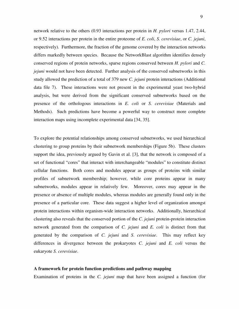
The C. jejuni protein interaction dataset can also serve as a framework for mapping
functional pathways, such as the chemotaxis signaling pathway (Figure 6a, b). Although
not well characterized in C. jejuni, orthologs have been identified for the prototypical
chemotaxis proteins CheW, CheA, CheY, and FliM [26, 39]. In the canonical pathway,
chemoattractants bind chemoreceptors known as methyl-accepting chemotaxis proteins
(MCPs), which then activate the histidine kinase CheA in a complex stabilized by CheW.
CheA phosphorylates CheY, which then interacts with the FliM protein at the base of the
flagellar motor resulting in changes in the direction of flagellar rotation. A search of the C.
jejuni map for interactions involving motility and chemotaxis-related proteins reveals a
large connected subnetwork of proteins (Figure 6a). The subnetwork includes the expected
interactions between a putative MCP (Cj0262c) and CheW, CheW and CheA, and CheA

11
and CheY (Figure 6b). The interaction between CheY and FliM, however, was missed,
most likely because it depends upon CheY phosphorylation on a specific aspartate residue
[40, 41], a modification unlikely to be provided by yeast. We also identified interactions
between the poorly characterized CheV protein, and three putative MCP proteins, Cj0262c,
Cj0448c, and Cj1110c, supporting previous suggestions that CheV may function early in
the signal transduction pathway, similar to CheW [39, 42]. Lastly, we detected an
interaction between CheA and Cj0643 (Figure 6a). This interaction was predicted
previously [43] because Cj0643 contains the conserved CheY-like receiver domain.
Cj0643 also contains a diguanylate-cyclase domain indicating the potential for 3’,5’-cyclic
diguanylic acid (cdiGMP) biosynthetic activity [44]. CdiGMP is a signaling molecule in
some bacteria [44]. Perhaps in C. jejuni the interaction between CheA and Cj0643 links
Cdi-GMP generation to conditions outside of the cell.
A network of putative essential genes
Several groups have shown that in yeast, essential genes, which are genes required for
growth or viability, are more likely to encode hubs in the protein network than nonessential
genes [45-47]. To explore the relationship between essential genes and protein interactions
in the C. jejuni network we generated a list of putative essential C. jejuni genes based on
orthology to genes proposed to be essential in E. coli and Bacillus subtilis based on
experimental evidence in those organisms (Materials and Methods). We found higher
percentages of putative essential genes amongst proteins with larger numbers of
interactions (Figure 7, see also Materials and Methods). It follows from this finding that,
like in yeast, hub proteins are more likely to be essential than non-hub proteins. Thus,
network topology may provide one way to estimate the potential importance of particular
genes and may be useful in searches for new candidate drug targets.
Essential proteins often function together in pathways or processes that are important for
cell growth or viability. Consistent with this, we found that the C. jejuni map contains
interactions between putative essential proteins significantly more frequently than expected
by chance (Additional data file 11). Similar results have been described for yeast protein
interaction maps [46]. One consequence of this enrichment for essential-essential

12
interactions is that groups of essential proteins can form interconnected subnetworks
within the interaction map. Additionally, the C. jejuni map may be used to predict that
some of the previously uncharacterized proteins may be important for growth or viability
based on their interactions with known essential proteins. To create a network enriched for
important proteins we identified a subnetwork of interconnected proteins predicted to be
essential in C. jejuni based on orthology to essential proteins in E. coli and B. subtilis
(Figure 8, triangular, diamond, and rectangular nodes). To identify additional putative
essential or important proteins, we added proteins that connect to two or more of the
essential nodes through high confidence interactions (circular nodes). The resulting map
(Figure 8) contains 264 proteins, many of which are of unknown function (yellow), and
identifies potential connections amongst many proteins involved in processes known to be
essential for viability, including ribosome function and DNA synthesis and repair. For
example, in Figure 8, BoxA highlights the interaction between RecJ and SSB. SSB is a
single-stranded DNA (ssDNA) binding protein that resolves secondary structure in ssDNA
(reviewed in [48]), while RecJ is a conserved exonuclease that degrades ssDNA [49]. Both
proteins have roles in homologous recombination and mismatch repair [48, 50, 51]. A
recent report has demonstrated that binding of ssDNA by SSB enhances RecJ binding and
exonuclease activity [52] suggesting a functional relationship between the two proteins.
This is further supported by the binary protein-protein interaction that we have detected in
C. jejuni (this study) and the purification of an E. coli protein complex containing RecJ
using affinity-tagged SSB [1].
The many uncharacterized proteins in the essential protein network are potentially
biologically important and may include potential novel drug targets. For example Figure
8, Box B highlights a protein of unknown function, Cj0189c, which has interaction
partners with 5 ribosomal proteins. Based on this and the fact that proteins with related
functions tend to interact, it is reasonable to hypothesize that Cj0189c may also be
involved in ribosome assembly or function. This is potentially significant given that the
ribosome and protein synthesis are frequent targets of antibiotics [53]. Box C highlights
the uncharacterized protein Cj0980, which is homologous to the dipeptidase, peptidase D.
In E. coli Peptidase D is one of the enzymes that generates cysteine by cleaving

13
cysteinylglycine [54]. In our map Cj0980 interacts with nine proteins predicted to be
essential. One of these proteins, Cj0240c, is a homolog of IscS, a cysteine desulfurase
required for the synthesis of all tRNA thiolated nucleosides in E. coli [55]. Interestingly,
four additional interactors of Cj0980 are tRNA synthetases. Whether or not their product
tRNAs are modified in C. jejuni has not been determined, but this series of interactions
suggests a possible pathway or protein complex that mediates the transfer of a thiol group
originating from cysteinylglycine to specific tRNAs.
Discussion
The large-scale interaction studies performed to date have fallen short of complete
interactome coverage. The most complete large-scale yeast two-hybrid screens have
covered only around 54% of the proteome in Drosophila [22, 25, 56], 46% in Helicobacter
pylori [11] and 55% in yeast [57-59], while Co-AP/MS studies have reached 80% and 67%
of the E. coli and yeast proteomes respectively [1-6] (Additional data file 1). Complete
interactome coverage should include most of the proteome, since most proteins are
believed to function at least in part through interactions with other proteins. A major
factor contributing to incomplete coverage is the incomplete nature of the high-throughput
screens, as indicated by the minimal rate of overlap observed between independent large-
scale screens (Additional data file 1) [22, 59]. Thus despite the usefulness of the data from
various interaction mapping efforts, the low interactome coverage is likely to limit efforts
to predict protein functions, map pathways, and characterize protein networks. Low
coverage also limits the opportunity for cross validation, which is particularly important
for high throughout data sets because they tend to have high rates of false positives [24,
60].
We have made substantial progress towards defining the C. jejuni interactome. Based on
the number of ORFs included in the interaction dataset we have covered 80% of the
proteome, and our higher confidence dataset covered 67%. An expected consequence of
performing high-throughput screens, which tend to be subsaturating, is that some
interactions that are detectable by two-hybrid are missed [10]. We set out to minimize
these false negatives by using a highly sensitive two-hybrid system, inducible promoters to

14
detect interactions with toxic proteins and transcriptional activators, and a pooled-matrix
mating scheme to maximize the number of interactions sampled. Despite these efforts,
some interactions will be missed, especially those that are refractory to standard two-
hybrid assays. Detection of these will require other technologies, such as isolation and
identification of protein complexes, and assays that target specific classes of proteins such
as membrane proteins [61, 62]. Interaction networks may also be made more complete by
using computational approaches to predict missed interactions [34, 35]. In this study we
applied a comparative algorithm to align protein networks from C. jejuni to the
interactomes of other species to generate further predictions of protein interactions. Like
the high throughput experimental data, these predictions provide a guide for directed
validation studies.
An unfortunate side effect of large-scale protein interaction datasets is the presence of
significant numbers of false positive interactions. We addressed this problem in two ways.
First, we retested every interaction in a second independent two-hybrid assay. Second, we
calculated probability scores that correlate with the likelihood that an interaction is
biologically relevant. One advantage to this confidence scoring system is that it scores
interactions rather than proteins and therefore does not specifically delete any proteins.
Several studies including ours have found an inverse correlation between the biological
significance of an interaction and the total number of interactions for the two proteins
involved; the more interactions that a protein has, the less likely they are to be biological
true positives. One approach to increasing the overall confidence of a data set, therefore, is
to delete these “sticky” proteins. In contrast, it is possible to identify biologically relevant
interactions involving these proteins by using a statistical scoring system that weighs
multiple attributes according to their correlation with biological significance. With such a
scoring system an interaction may be penalized because it involves a sticky protein, but
redeemed due to some other attribute. This is the case, for example, in our data with the
interactions FliS-FlaC, GroEL-GroES, Ilvl-IlvH, PyrB-PyrC2, and TrxA-TrxB, all of
which involve proteins with more than 60 interactions, yet have confidence scores above
0.8, and are likely to be biologically significant.

15
Another advantage to this scoring system is that it allows user-defined confidence intervals
to be chosen based on particular analysis needs. Global analyses, for example, may benefit
from using the highest confidence data set. More focused analyses involving one or few
proteins, on the other hand, may tolerate lower confidence interactions because validation
experiments can be performed. This reduces the chances of missed interactions.
Importantly, some low confidence interactions may be found to be biologically significant
by experimental validation or by considering additional information not used in the scoring
system. For example, by considering pairs of proteins with known functions, one can find
a number of likely true positives with confidence scores below 0.2, including DnaX-DnaN,
ExbD1-ExbD3, and FabF-FabG.
Finally, the confidence that we have in any particular interaction can change as new data
becomes available about the two proteins or about the interaction itself. We have shown
that the scores we assigned to the C. jejuni two-hybrid data correlate with biological
significance such that more of the interactions with higher scores will be biologically
significant than those with lower scores, and vice versa. Nevertheless, a fraction of the low
confidence interactions are true positives and some of the high confidence interactions are
false positives. It is expected that these will be sorted out using new, increasingly accurate
confidence scoring systems that are based, for example, on new information as it becomes
available. Thus, we have defined the scoring of the C. jejuni two-hybrid data presented
here as version 1.0.
Conclusions
Interactome maps such as the one generated in our study begin to provide a tally of the
binary protein interactions that can occur within an organism. Although incomplete, the
data can provide a framework for understanding dynamic biological processes, such as the
C. jejuni chemotaxis response. The map also can be mined for subnetworks of biological
interest such as essential gene networks that suggest candidate drug targets. Comparative
analyses of protein interaction maps generated for humans and model eukaryotes have
provided insights into the function and evolution of proteins and their regulatory networks.
The protein interactions detected for each species also have enabled the prediction of

16
interactions in other species, which is particularly important given the difficulty of
obtaining complete coverage in high throughput screens, and the lack of suitable screening
systems for many species. The C. jejuni interaction map generated here substantially
increases the protein interactions detected thus far for the prokaryotic domain of life. The
map should provide a useful starting point for predicting the functions of uncharacterized
proteins and for mapping functional pathways in C. jejuni and other prokaryotes.
Materials and Methods
Strains and Plasmids.
The two-hybrid system used here is based on the version originally described by Brent and
colleagues [63]. C. jejuni ORFs were cloned into the yeast two-hybrid vector pJZ4-NRT
for expression of activation domain (AD) fusions driven by the yeast GAL1 promoter [22],
and pHZ5-NRT for expression of LexA DNA binding domain (BD) fusions driven by the
yeast MAL62 promoter [23]. Both vectors contain recombination tags for direct cloning of
tagged inserts (see below). Yeast strain RFY231 (MATα trp1∆::hisG his3 ura3-1
leu2::3LexAop-LEU2) contained the AD plasmids, while Y309 (MATa trp1∆::hisG
his3∆200 leu2-3 lys2∆201 ura3-52 mal- pSH18-34(URA3, lacZ)) contained the BD
plasmids. The reporter genes include LEU2 facilitating growth on medium lacking
leucine, and lacZ, expression of which turns yeast colonies blue when the substrate X-Gal
is present.
Generation of yeast two-hybrid arrays for C. jejuni.
PCR amplification of over 87% of the predicted ORFs from C. jejuni NCTC11168
genomic DNA was previously described [64]. The amplification products included the 21
bp recombination tags 5RT1 and 3RT1 at their 5’ and 3’ ends, respectively, which match
identical sites flanking the insertion site in the yeast two-hybrid vectors. PCR products
were cloned into the vectors via homologous recombination in yeast as described
previously [22]. To validate the identity of the insert in each vector, the 5’ ends of the
inserted PCR products were sequenced. 1398 BD strains and 1442 AD strains containing
the two-hybrid vectors with inserts were generated of which 90% have been sequence

17
verified. Most of the ORFs missing from the arrays failed PCR amplification prior to
cloning.
High-throughput yeast two-hybrid analysis.
We mated BD and AD strains using a two-phase pooling (pooled matrix) strategy as
described previously [21, 22]. Briefly, 15 pools of approximately 96 AD strains each were
generated, along with one additional pool of 32 strains. Each pool was mated with
individual BD strains arrayed on 96-well plates, and the resulting diploids were assayed for
reporter activities. Positive BD strains were then mated with each member of the positive
AD pool arrayed on 96-well plates to identify the interacting pairs. Reporter activities
were scored using a custom program for image analysis [65] and at least one manual
scoring. LacZ scores ranged from 0 (white) to 5 (dark blue) and Leu scores ranged from 0
(no growth) to 3 (heavy growth); combined scores ranged from 0 to 8. Many BDs have
some level of background activity due to activation independent of the AD fusion or non-
specific interactions. To correct for these we calculated the average interaction score for
each BD based on at least 96 interaction assays and subtracted this background from the
reporter scores for each of its interactions. Of these corrected scores, only those >= 1 were
considered initial positives and were retested (see below). A small subset of BD strains
(94 total) was also assayed using a library approach as described [21, 22]. Briefly, BD
strains were individually mated with a single pool containing almost all of the AD strains
(except Cj1718c (leuB) and Cj1546, which activate reporters without a BD). Up to 30
diploids with reporter activity were picked for each BD. Their AD inserts were PCR
amplified and restriction digested to identify strains carrying the same clones. Single
representatives from each restriction fragment class (RFC) were then sequenced to identify
the inserts. Of the 134 interactions detected, 52 (39%) were also identified in the two-
phase matrix screen. Combined, 16,104 unique interactions were retested in one-on-one
binary mating assays between individual AD and BD strains on 96-well plates. A total of
11,687 interactions proved repeatable (background-corrected combined activity score >=
1) including 73% of those from the two-phase matrix screen, 75% of those from the library
screen, and 100% of those detected in both screens. The majority of interactions that failed
to repeat had been low-scoring (less than 2) in the initial screen. The 11,687 interactions
18
that repeated were combined with 325 non-repeated interactions that had high confidence
scores (see below) to create a data set containing 12,012 interactions, which we named
CampyYTH v3.1. This version of the dataset was subsequently used for bioinformatics
analysis as indicated. The interaction data can be visualized and downloaded at [17]. The
CampyYTH v3.1 data is also listed in Additional data file 13.
Assignment of confidence scores
Confidence scores were determined for each interaction based on methods described by
Bader et al. [24, 25]. We fit a generalized linear model [66] using experimental and
topological attributes of yeast two-hybrid interactions, including the number of interactions
for each protein in a pair and the Leu and lacZ reporter activities Fitting the model
required both positive and negative training sets. Because a reference set of known
interactions is not available for C. jejuni, we derived a set of positive training data (85
interactions total) by assuming that the conserved interactions (reciprocal best match
interologs) in common with either the E. coli low-throughput interaction set [28], the H.
pylori yeast two-hybrid set [11], or the E. coli protein complex set [1] are likely to be true
positives. We derived a set of likely true negatives (111 total) for the negative training
data by considering interactions between proteins whose orthologs in E. coli or H. pylori
were separated in the respective interaction maps by greater than the average distance of all
pairs (>= 4). Positive and negative training cases were weighted inversely to the number
of interactions in each set. When training sets are weighted this way, a confidence score
greater than 0.5 means that available data and features support that a specific interaction
has a better than random chance to be a true interaction; this allows 0.5 to be used as the
threshold between high and low confidence interactions. Validation using protein features
not used in the scoring system support the choice of 0.5 as a threshold for higher
confidence interactions (discussed further in Additional data file 14; see also Figure 2c). Of
the attributes tested, the numbers of interactions per protein were found to be negative
predictors of biologically relevant interactions, while reporter activities were positive
predictors. To evaluate the scoring model, we performed a stratified five-fold cross
validation. Cross validation reported a precision of 91.4% and a recall of 78.9%, which
gave us confidence that it is a reasonably well-fitted model. We then used the full sets of

19
positives and negatives in training and obtained our final logistic model. The final model
was used to compute confidence scores for 16,104 initial positive interactions prior to
retesting. Of these, 3,209 scored higher than 0.5, which we define as the High Confidence
Set (HCS). Of the interactions with high confidence scores (>0.5), 90% corresponded to
interactions that repeated when retested, while only 68% of the low confidence interactions
repeated. Further discussion and details of the confidence scoring system are available in
Additional data file 14.
Evaluating the confidence score model.
Main role annotations ‘mainrole’ were downloaded from [67]. Excluding self-interactions,
out of the 3209 high confidence interactions, 2599 have ‘mainrole’ annotations, and 454
share at least one ‘mainrole’ annotation. We generated 5000 groups of 2599 randomly
selected interactions that have ‘mainrole’ annotations and have a confidence score lower
than 0.5. The number of pairs in each set that share ‘mainrole’ annotations was counted.
The distribution was plotted in a histogram and compared with the high confidence set
(Figure 2b). To examine whether high confidence interactions tend to share more detailed
GO [27] annotations, we grouped interactions into confidence bins so that each bin
contains only interactions with scores falling into a specific range. For each interaction, we
determined the deepest level of GO biological process annotations shared by the pair of
genes, and calculated the average depth of shared biological process for each group. Since
GO ontology for C. jejuni NCTC11168 was not available, we used annotations for best
match orthologs of C. jejuni RM1221 genes [68]. Figure 2c shows that there is a general
pattern of increased depth of shared GO terms for interactions with confidence score
higher than 0.5. This fact also suggests that our choice of 0.5 as a high confidence
threshold is meaningful.
Assessment of functional enrichments
The frequency of each Gene Ontology (GO) description from the iProClass database [69],
amongst all of the proteins comprising the proteome was determined and compared to their
frequency within the CampyYTH v3.1 dataset or the high confidence subset (Additional
data file 3). A similar analysis was performed using the functional classifications assigned

20
by the Sanger Institute [26] (Additional data file 2). We also looked for pairs of GO
annotations that were enriched in the interaction data (Additional data file 10). To do this
we counted the number of interactions having a specific pair of GO terms. We mapped the
annotations to level 5; that is, for a protein with GO annotation A that is at a deeper level
than 5, we mapped A to level 5 using ‘parent’ and ‘part of’ relationships in the ontologies,
and we discarded A if it was above level 5. Self-interactions were excluded from the
analysis. We did the same for all GO terms annotated to a protein. To compute the
significance of finding specific GO pairs, we generated 2000 random networks by
randomly switching pairs of links while maintaining the degree distribution of the original
map, and counted the number of times we found each GO pair in each randomized
network. For each GO pair, a p-value was computed based on the distribution of the 2000
counts (assuming normal distribution) and the count in the original yeast two hybrid map.
The p-value represents the probability of seeing such a pair in a random network. We listed
only pairs with a p-value less than 5%.
Comparative network analysis
Additional details are in Additional file 14. Protein-protein interactions from C. jejuni
were compared with those from E. coli [1]; H. pylori [11]; and S. cerevisiae from the
Database of Interacting Proteins [28]. Corresponding protein sequences were obtained
from the following sources: C. jejuni NCTC11168 [26]; E. coli [70]; H. pylori [71]; and S.
cerevisiae [72]. We used NetworkBlast to identify significant conserved protein-protein
interaction subnetworks [34]. A stand-alone Java version of the program is available at
[73]. Briefly, the algorithm takes as input a pair of protein-protein interaction networks,
one for each of two species, along with a set of homology relationships between the
proteins of the two networks. We constructed the homology relationships from an all-
versus-all BLAST of the complete set of protein sequences for each of the two species,
taking the top 10 hits with E-value <= 10-10
. Next, a network alignment graph was created
where each node represents a homologous pair of proteins from species 1 and 2 (e.g., a1
and a2) and each edge represents a conserved interaction (a1/a2 connects to b1/b2 if the a-
b interaction is found in both species; interactions may be either direct (distance 1) or
indirect (distance 2), in which a-b is connected through a common neighbor, i.e., a-c-b). A

21
greedy search is initiated from each node to identify conserved protein subnetworks,
defined as dense subgraphs within the network alignment graph (of maximum size 15
proteins per species). When multiple subnetworks contain protein homologs that overlap
by >=50%, only the complex with the highest density was included in the final result. GO
annotations [27] of proteins in each conserved complex were analyzed to identify
significant functional enrichments (Additional data file 6). We calculated a hypergeometric
p-value of enrichment for each GO annotation in the three divisions of the GO hierarchy
and constrained the annotations by requiring that at least half of the proteins in a complex
ascribe to the enrichment. The most specific annotations with hypergeometric p-value <
0.05 in each of the three divisions were then assigned to each complex. A complete list of
conserved complexes between C. jejuni and E. coli or S. cerevisiae is available for
download at [73]. The significant conserved subnetworks provided predictions of 379 new
C. jejuni protein-protein interactions not found in the two-hybrid screens (Additional data
file 7). A protein pair (a,b) was predicted to interact directly if: 1., both a and b were
present in the same significant conserved complex; 2., this pair was observed to interact
indirectly in C. jejuni; and 3., this pair corresponded to a direct interaction in the
comparison species’ network.
Clustering of conserved subnetworks
Since proteins can belong to more than one complex, we clustered the significant
conserved subnetworks by protein membership, in effect ‘superclustering’ the interactions
(Figure 5b). An n×m matrix was constructed, where n is the number of significant
subnetworks and m is the number of unique proteins involved in any of the significant
subnetworks. Using the open source tool ClustArray [74], we clustered the proteins
hierarchically using the unweighted pair group method with arithmetic mean (UPGMA)
and clustered the subnetworks with a combination k-means algorithm followed by
UPGMA hierarchical clustering. The number of clusters k=3 was chosen as the parameter
that approximately minimized within-cluster variability and maximized between-cluster
variability (data not shown). Identities of complexes and proteins are shown in the high
resolution image of the hierarchical clustering in Additional data file 8. Lists of the
proteins comprising complexes are available for download at [73].

22
Essential gene analysis and network assembly
We generated lists of putative C. jejuni NCTC11168 essential proteins by identifying
reciprocal best match orthologs of likely essential proteins from B. subtilis [75] and E. coli
[76]. We removed genes from our putative essential list if viable null mutants have been
reported (Dr. B. Wren, personal communication). To examine the relationship between
essentiality and centrality in the interaction map, we computed the numbers of essential
and non-essential proteins in groups having the same number of interactions (degree) in the
higher confidence data set (interactions with confidence scores >0.5). The result is shown
in Figure 7, where r values in the graphs represent Pearson correlation coefficient between
the fractions and the degrees. Figure 7 shows that there is a correlation between degree of
proteins and the likelihood of being essential. A similar result was obtained with the entire
dataset CampyYTH v3.1 (not shown). Lastly, we computed the fraction of essential and
non-essential neighbors of each essential protein and compared this to the fraction for
random groups of proteins (of the same size as the set of essential proteins). The results
shown in Additional data file 11 indicate that essential genes tend to have more neighbors
that are also essential. p-values indicate the probability of seeing the real fraction (the red
dot) by chance.
Additional Data Files
The following additional data are available with the online version of this paper.
Additional data file 1 is a table summarizing proteome coverage from large-scale
interaction screens. Additional data file 2 is a table listing the representation of functional
categories amongst the proteins in the CampyYTH v3.1 dataset. Additional data file 3 is a
table listing the Gene Ontology (GO) category representation amongst the proteins in
CampyYTH v3.1. Additional data file 4 lists C. jejuni genes that were toxic or inhibitory to
yeast growth. Additional data file 5 is a table comparing network features across
organisms. Additional data file 6 lists conserved subnetworks between C. jejuni and E.
coli or C. jejuni and yeast. Additional data file 7 lists predicted C. jejuni protein
interactions. Additional data file 8 is a higher resolution version of Figure 5, showing
hierarchical clustering of conserved subnetworks. Additional data file 9 is a table listing

23
enriched functions within the cores and modules of Figure 5. Additional data file 10 is a
table showing gene ontology enrichment amongst the C. jejuni protein interactions.
Additional data file 11 is a figure showing that essential proteins interact with each other
more often than expected by chance. Additional data file 12 is a table of C. jejuni
interologs predicted from large-scale protein interaction analyses performed for E. coli or
H. pylori. Additional data file 13 is an annotated list of all C. jejuni protein interactions in
the CampyYTH v3.1 dataset. Additional data file 14 includes supplementary materials and
methods.
Acknowledgements
We thank Thawornchai Limjindaporn, Dima El-Khechen, Keith Gulyas, Meghan Hurt, and
Rohinton Tarapore for technical assistance, and Michigan Proteome Consortium members
and Janine Maddock for helpful discussions. This work was supported in part by Grant
RR18327 from The National Center for Research Resources, a component of The National
Institute of Health, and by grant HG001536 from the National Human Genome Research
Institute.
References
1. Butland G, Peregrin-Alvarez JM, Li J, Yang W, Yang X, Canadien V, Starostine A,
Richards D, Beattie B, Krogan N et al: Interaction network containing
conserved and essential protein complexes in Escherichia coli. Nature 2005,
433(7025):531-537.
2. Gavin AC, Bosche M, Krause R, Grandi P, Marzioch M, Bauer A, Schultz J, Rick
JM, Michon AM, Cruciat CM et al: Functional organization of the yeast
proteome by systematic analysis of protein complexes. Nature 2002,
415(6868):141-147.
3. Gavin AC, Aloy P, Grandi P, Krause R, Boesche M, Marzioch M, Rau C, Jensen
LJ, Bastuck S, Dumpelfeld B et al: Proteome survey reveals modularity of the
yeast cell machinery. Nature 2006, 440(7084):631-636.
4. Ho Y, Gruhler A, Heilbut A, Bader GD, Moore L, Adams SL, Millar A, Taylor P,
Bennett K, Boutilier K et al: Systematic identification of protein complexes in
Saccharomyces cerevisiae by mass spectrometry. Nature 2002, 415(6868):180-
183.
5. Krogan NJ, Cagney G, Yu H, Zhong G, Guo X, Ignatchenko A, Li J, Pu S, Datta N,
Tikuisis AP et al: Global landscape of protein complexes in the yeast
Saccharomyces cerevisiae. Nature 2006, 440:637-643.

24
6. Arifuzzaman M, Maeda M, Itoh A, Nishikata K, Takita C, Saito R, Ara T,
Nakahigashi K, Huang HC, Hirai A et al: Large-scale identification of protein-
protein interaction of Escherichia coli K-12. Genome Res 2006, 16(5):686-691.
7. Fields S, Song O: A novel genetic system to detect protein-protein interactions.
Nature 1989, 340(6230):245-246.
8. Fields S: High-throughput two-hybrid analysis. The promise and the peril.
Febs J 2005, 272(21):5391-5399.
9. Cusick ME, Klitgord N, Vidal M, Hill DE: Interactome: gateway into systems
biology. Hum Mol Genet 2005, 14 Spec No. 2:R171-181.
10. Parrish JR, Gulyas KD, Finley RL, Jr.: Yeast two-hybrid contributions to
interactome mapping. Curr Opin Biotechnol 2006, 17:387-393.
11. Rain JC, Selig L, De Reuse H, Battaglia V, Reverdy C, Simon S, Lenzen G, Petel
F, Wojcik J, Schachter V et al: The protein-protein interaction map of
Helicobacter pylori. Nature 2001, 409(6817):211-215.
12. Blaser MJ: Epidemiologic and clinical features of Campylobacter jejuni
infections. J Infect Dis 1997, 176 Suppl 2:S103-105.
13. Godschalk PC, Heikema AP, Gilbert M, Komagamine T, Ang CW, Glerum J,
Brochu D, Li J, Yuki N, Jacobs BC et al: The crucial role of Campylobacter
jejuni genes in anti-ganglioside antibody induction in Guillain-Barre syndrome. J Clin Invest 2004, 114(11):1659-1665.
14. Nachamkin I, Allos BM, Ho TW: Campylobacter jejuni infection and the
association with Guillain-Barré Syndrome. In: Campylobacter. Edited by
Nachamkin I, Blaser MJ, 2nd edn. Washington, D.C.: ASM Press; 2000: 155-175.
15. Lecuit M, Abachin E, Martin A, Poyart C, Pochart P, Suarez F, Bengoufa D,
Feuillard J, Lavergne A, Gordon JI et al: Immunoproliferative small intestinal
disease associated with Campylobacter jejuni. N Engl J Med 2004, 350(3):239-
248.
16. Campylobacter Resource Facility
[http://www.lshtm.ac.uk/pmbu/crf/updated_embl.htm]
17. Finley Lab [http://proteome.wayne.edu]
18. Bowers PM, Pellegrini M, Thompson MJ, Fierro J, Yeates TO, Eisenberg D:
Prolinks: a database of protein functional linkages derived from coevolution.
Genome Biol 2004, 5(5):R35.
19. Walhout AJ, Boulton SJ, Vidal M: Yeast two-hybrid systems and protein
interaction mapping projects for yeast and worm. Yeast 2000, 17(2):88-94.
20. Pacifico S, Liu G, Guest S, Parrish JR, Fotouhi F, Finley RL, Jr.: A database and
tool, IM Browser, for exploring and integrating emerging gene and protein interaction data for Drosophila. BMC Bioinformatics 2006, 7(1):195.
21. Zhong J, Zhang H, Stanyon CA, Tromp G, Finley RL, Jr.: A strategy for
constructing large protein interaction maps using the yeast two-hybrid system: regulated expression arrays and two-phase mating. Genome Res 2003,
13(12):2691-2699.
22. Stanyon CA, Liu G, Mangiola BA, Patel N, Giot L, Kuang B, Zhang H, Zhong J,
Finley RL, Jr.: A Drosophila protein-interaction map centered on cell-cycle
regulators. Genome Biol 2004, 5(12):R96.
25
23. Finley RL, Jr., Zhang H, Zhong J, Stanyon CA: Regulated expression of proteins
in yeast using the MAL61-62 promoter and a mating scheme to increase dynamic range. Gene 2002, 285(1-2):49-57.
24. Bader JS, Chaudhuri A, Rothberg JM, Chant J: Gaining confidence in high-
throughput protein interaction networks. Nat Biotechnol 2004, 22(1):78-85.
25. Giot L, Bader JS, Brouwer C, Chaudhuri A, Kuang B, Li Y, Hao YL, Ooi CE,
Godwin B, Vitols E et al: A protein interaction map of Drosophila
melanogaster. Science 2003, 302(5651):1727-1736.
26. Parkhill J, Wren BW, Mungall K, Ketley JM, Churcher C, Basham D,
Chillingworth T, Davies RM, Feltwell T, Holroyd S et al: The genome sequence
of the food-borne pathogen Campylobacter jejuni reveals hypervariable sequences. Nature 2000, 403(6770):665-668.
27. Ashburner M, Ball CA, Blake JA, Botstein D, Butler H, Cherry JM, Davis AP,
Dolinski K, Dwight SS, Eppig JT et al: Gene ontology: tool for the unification of
biology. The Gene Ontology Consortium. Nat Genet 2000, 25(1):25-29.
28. Xenarios I, Rice DW, Salwinski L, Baron MK, Marcotte EM, Eisenberg D: DIP:
the database of interacting proteins. Nucleic Acids Res 2000, 28(1):289-291.
29. Tanaka R, Yi TM, Doyle J: Some protein interaction data do not exhibit power
law statistics. FEBS Lett 2005, 579(23):5140-5144.
30. Schwikowski B, Uetz P, Fields S: A network of protein-protein interactions in
yeast. Nat Biotechnol 2000, 18(12):1257-1261.
31. Li S, Armstrong CM, Bertin N, Ge H, Milstein S, Boxem M, Vidalain PO, Han JD,
Chesneau A, Hao T et al: A map of the interactome network of the metazoan C.
elegans. Science 2004, 303(5657):540-543.
32. Saito R, Suzuki H, Hayashizaki Y: Interaction generality, a measurement to
assess the reliability of a protein-protein interaction. Nucleic Acids Res 2002,
30(5):1163-1168.
33. Goldberg DS, Roth FP: Assessing experimentally derived interactions in a small
world. Proc Natl Acad Sci U S A 2003, 100(8):4372-4376.
34. Sharan R, Suthram S, Kelley RM, Kuhn T, McCuine S, Uetz P, Sittler T, Karp RM,
Ideker T: Conserved patterns of protein interaction in multiple species. Proc
Natl Acad Sci U S A 2005, 102(6):1974-1979.
35. Jansen R, Yu H, Greenbaum D, Kluger Y, Krogan NJ, Chung S, Emili A, Snyder
M, Greenblatt JF, Gerstein M: A Bayesian networks approach for predicting
protein-protein interactions from genomic data. Science 2003, 302(5644):449-
453.
36. Ozin AJ, Claret L, Auvray F, Hughes C: The FliS chaperone selectively binds the
disordered flagellin C-terminal D0 domain central to polymerisation. FEMS
Microbiol Lett 2003, 219(2):219-224.
37. Evdokimov AG, Phan J, Tropea JE, Routzahn KM, Peters HK, Pokross M, Waugh
DS: Similar modes of polypeptide recognition by export chaperones in
flagellar biosynthesis and type III secretion. Nat Struct Biol 2003, 10(10):789-
793.
38. Song YC, Jin S, Louie H, Ng D, Lau R, Zhang Y, Weerasekera R, Al Rashid S,
Ward LA, Der SD et al: FlaC, a protein of Campylobacter jejuni TGH9011

26
(ATCC43431) secreted through the flagellar apparatus, binds epithelial cells and influences cell invasion. Mol Microbiol 2004, 53(2):541-553.
39. Marchant J, Wren B, Ketley J: Exploiting genome sequence: predictions for
mechanisms of Campylobacter chemotaxis. Trends Microbiol 2002, 10(4):155-
159.
40. Welch M, Oosawa K, Aizawa S, Eisenbach M: Phosphorylation-dependent
binding of a signal molecule to the flagellar switch of bacteria. Proc Natl Acad
Sci U S A 1993, 90(19):8787-8791.
41. Sanders DA, Gillece-Castro BL, Stock AM, Burlingame AL, Koshland DE, Jr.:
Identification of the site of phosphorylation of the chemotaxis response regulator protein, CheY. J Biol Chem 1989, 264(36):21770-21778.
42. Hendrixson DR, Akerley BJ, DiRita VJ: Transposon mutagenesis of
Campylobacter jejuni identifies a bipartite energy taxis system required for motility. Mol Microbiol 2001, 40(1):214-224.
43. Wojcik J, Boneca IG, Legrain P: Prediction, assessment and validation of
protein interaction maps in bacteria. J Mol Biol 2002, 323(4):763-770.
44. Camilli A, Bassler BL: Bacterial small-molecule signaling pathways. Science
2006, 311(5764):1113-1116.
45. Gandhi TK, Zhong J, Mathivanan S, Karthick L, Chandrika KN, Mohan SS,
Sharma S, Pinkert S, Nagaraju S, Periaswamy B et al: Analysis of the human
protein interactome and comparison with yeast, worm and fly interaction datasets. Nat Genet 2006, 38(3):285-293.
46. Reguly T, Breitkreutz A, Boucher L, Breitkreutz BJ, Hon GC, Myers CL, Parsons
A, Friesen H, Oughtred R, Tong A et al: Comprehensive curation and analysis of
global interaction networks in Saccharomyces cerevisiae. J Biol 2006, 5(4):11.
47. Jeong H, Mason SP, Barabasi AL, Oltvai ZN: Lethality and centrality in protein
networks. Nature 2001, 411(6833):41-42.
48. Meyer RR, Laine PS: The single-stranded DNA-binding protein of Escherichia
coli. Microbiol Rev 1990, 54(4):342-380.
49. Lovett ST, Kolodner RD: Identification and purification of a single-stranded-
DNA-specific exonuclease encoded by the recJ gene of Escherichia coli. Proc
Natl Acad Sci U S A 1989, 86(8):2627-2631.
50. Lovett ST, Clark AJ: Genetic analysis of the recJ gene of Escherichia coli K-12.
J Bacteriol 1984, 157(1):190-196.
51. Burdett V, Baitinger C, Viswanathan M, Lovett ST, Modrich P: In vivo
requirement for RecJ, ExoVII, ExoI, and ExoX in methyl-directed mismatch repair. Proc Natl Acad Sci U S A 2001, 98(12):6765-6770.
52. Han ES, Cooper DL, Persky NS, Sutera VA, Jr., Whitaker RD, Montello ML,
Lovett ST: RecJ exonuclease: substrates, products and interaction with SSB.
Nucleic Acids Res 2006, 34(4):1084-1091.
53. Poehlsgaard J, Douthwaite S: The bacterial ribosome as a target for antibiotics.
Nat Rev Microbiol 2005, 3(11):870-881.
54. Suzuki H, Kamatani S, Kim ES, Kumagai H: Aminopeptidases A, B, and N and
dipeptidase D are the four cysteinylglycinases of Escherichia coli K-12. J
Bacteriol 2001, 183(4):1489-1490.

27
55. Lauhon CT: Requirement for IscS in biosynthesis of all thionucleosides in
Escherichia coli. J Bacteriol 2002, 184(24):6820-6829.
56. Formstecher E, Aresta S, Collura V, Hamburger A, Meil A, Trehin A, Reverdy C,
Betin V, Maire S, Brun C et al: Protein interaction mapping: a Drosophila case
study. Genome Res 2005, 15(3):376-384.
57. Uetz P, Giot L, Cagney G, Mansfield TA, Judson RS, Knight JR, Lockshon D,
Narayan V, Srinivasan M, Pochart P et al: A comprehensive analysis of protein-
protein interactions in Saccharomyces cerevisiae. Nature 2000, 403(6770):623-
627.
58. Ito T, Tashiro K, Muta S, Ozawa R, Chiba T, Nishizawa M, Yamamoto K, Kuhara
S, Sakaki Y: Toward a protein-protein interaction map of the budding yeast: A
comprehensive system to examine two-hybrid interactions in all possible combinations between the yeast proteins. Proc Natl Acad Sci U S A 2000,
97(3):1143-1147.
59. Ito T, Chiba T, Ozawa R, Yoshida M, Hattori M, Sakaki Y: A comprehensive two-
hybrid analysis to explore the yeast protein interactome. Proc Natl Acad Sci U
S A 2001, 98(8):4569-4574.
60. von Mering C, Krause R, Snel B, Cornell M, Oliver SG, Fields S, Bork P:
Comparative assessment of large-scale data sets of protein-protein interactions. Nature 2002, 417(6887):399-403.
61. Karimova G, Pidoux J, Ullmann A, Ladant D: A bacterial two-hybrid system
based on a reconstituted signal transduction pathway. Proc Natl Acad Sci U S A
1998, 95(10):5752-5756.
62. Johnsson N, Varshavsky A: Split ubiquitin as a sensor of protein interactions in
vivo. Proc Natl Acad Sci U S A 1994, 91(22):10340-10344.
63. Gyuris J, Golemis E, Chertkov H, Brent R: Cdi1, a human G1 and S phase
protein phosphatase that associates with Cdk2. Cell 1993, 75(4):791-803.
64. Parrish JR, Limjindaporn T, Hines JA, Liu J, Liu G, Finley RL, Jr.: High-
throughput cloning of Campylobacter jejuni ORfs by in vivo recombination in Escherichia coli. J Proteome Res 2004, 3(3):582-586.
65. Jafari-Khouzani K, Soltanian-Zadeh H, Fotouhi F, Parrish JR, Finley RL, Jr.:
Automated segmentation and classfication of high throughput yeast assay spots. Transactions on Medical Imaging 2007, In Press.
66. McCullagh P, Nelder JA: Generalized linear models, 2nd edn. Boca Raton, Fla.:
Chapman & Hall/CRC; 1998.
67. Welcome Trust Sanger Institute [http://www.sanger.ac.uk/Projects/C_jejuni/]
68. Fouts DE, Mongodin EF, Mandrell RE, Miller WG, Rasko DA, Ravel J, Brinkac
LM, DeBoy RT, Parker CT, Daugherty SC et al: Major structural differences
and novel potential virulence mechanisms from the genomes of multiple campylobacter species. PLoS Biol 2005, 3(1):e15.
69. Wu CH, Huang H, Nikolskaya A, Hu Z, Barker WC: The iProClass integrated
database for protein functional analysis. Comput Biol Chem 2004, 28(1):87-96.
70. Blattner FR, Plunkett G, 3rd, Bloch CA, Perna NT, Burland V, Riley M, Collado-
Vides J, Glasner JD, Rode CK, Mayhew GF et al: The complete genome
sequence of Escherichia coli K-12. Science 1997, 277(5331):1453-1474.

28
71. Wu CH, Huang H, Arminski L, Castro-Alvear J, Chen Y, Hu ZZ, Ledley RS,
Lewis KC, Mewes HW, Orcutt BC et al: The Protein Information Resource: an
integrated public resource of functional annotation of proteins. Nucleic Acids
Res 2002, 30(1):35-37.
72. Christie KR, Weng S, Balakrishnan R, Costanzo MC, Dolinski K, Dwight SS,
Engel SR, Feierbach B, Fisk DG, Hirschman JE et al: Saccharomyces Genome
Database (SGD) provides tools to identify and analyze sequences from Saccharomyces cerevisiae and related sequences from other organisms.
Nucleic Acids Res 2004, 32(Database issue):D311-314.
73. Ideker Lab [http://chianti.ucsd.edu/]
74. Knudsen S, Workman C, Sicheritz-Ponten T, Friis C: GenePublisher: Automated
analysis of DNA microarray data. Nucleic Acids Res 2003, 31(13):3471-3476.
75. Kobayashi K, Ehrlich SD, Albertini A, Amati G, Andersen KK, Arnaud M, Asai K,
Ashikaga S, Aymerich S, Bessieres P et al: Essential Bacillus subtilis genes. Proc
Natl Acad Sci U S A 2003, 100(8):4678-4683.
76. Baba T, Ara T, Hasegawa M, Takai Y, Okumura Y, Baba M, Datsenko KA, Tomita
M, Wanner BL, Mori H: Construction of Escherichia coli K-12 in-frame, single-
gene knockout mutants: the Keio collection. Mol Syst Biol 2006, 2:2006 0008.

29
Figure Legends
Figure 1 C. jejuni protein interaction networks. (a) The C. jejuni interaction dataset (CampyYTH
v3.1), and (b) the higher confidence subset. In each case most of the proteins (square
nodes) are connected into a single large network; the unconnected interactions are in the
upper right of each panel. The networks in (a) and (b) connect over 79% (663 total) and
65% (548 total) of the unnamed and presumed poorly characterized proteins (yellow
nodes), respectively.
Figure 2 Confidence scores assigned to the C. jejuni protein interactions. (a) The distribution of
confidence scores generated for the CampyYTH v3.1 protein interactions are shown in red.
The distributions of scores for the training sets containing likely true positives (green) or
true negatives (black) are also shown. (b) Protein interaction pairs with high confidence
scores (HCS; confidence scores >0.5) share the same functions significantly more
frequently (p-value < 3*10-57
) than protein pairs comprising interactions with low
confidence scores (LCS; confidence scores <=0.5). Protein “self” interactions were
excluded from the analysis. (c) The average depth of shared Gene Ontology biological
process annotation was determined for the interactions comprising each confidence score
bin. Higher confidence interactions generally involve proteins with the same functional
annotation at greater depths of precision. The two dotted line segments are linearly fitted
lines between average GO depth and bin number in two regions, from 0.5 to 0.9 and 0.9 to
1.0. Protein “self” interactions were excluded from the analysis.
Figure 3 Comparison of the C. jejuni interaction map with other datasets. The interactions found in
common, or overlap (red dots) between the C. jejuni two-hybrid map and interologs
predicted from other organisms was determined. This was compared to the overlap
between the interolog datasets and 2,000 random maps generated by randomly switching
pairs of links in the original yeast two-hybrid map; which preserves network degree
distribution. (a) The two-hybrid map shared 28 interactions with a reference set containing
147 interologs of E. coli low-throughput literature-cited protein interactions, significantly
greater than the overlap with the random maps. (b) 50 C. jejuni interactions were shared
with 1165 interologs predicted from the H. pylori protein interaction dataset [11]. (c) 124
interactions were shared with a set of 3743 interologs predicted from a large-scale E. coli
protein complex study [1]. (d) 76 interactions were shared with a set of 4056 interologs
predicted from a second E. coli protein complex pull-down study [6]. A complete list of
the predicted interologs used for these analyses can be found in Additional data file 12.
Figure 4 Characteristics of the C. jejuni protein network. (a) Degree frequency distribution for the
entire two-hybrid data set (CampyYTH v3.1). k = degree, the number of connections to a
protein. P(k) = the probability that a node has k connections. A power law fit yields: y =

30
0.4153 x-1.29
; R2 = 0.88. (b) Degree frequency distribution for the high confidence dataset
(confidence scores > 0.5). A power law fit yields: y = 482.2 x-1.53
, R2 = 0.89. (c) Rank-
degree distribution for the entire two-hybrid data set. The semi-log plot more closely fits
an exponential curve (red line, R2 = 0.97) than a power law curve (black line, R
2 = 0.81.
(d) Rank-degree distribution for the high confidence data. The semi-log plot more closely
fits a power law curve (black line, R2 = 0.91) consistent with a scale-free network. (e) (f)
The distribution of the average clustering coefficient (C) for degree k for the entire two-
hybrid data set (e) and the high confidence set (f). C is equal to the number of interactions
among a protein’s interactors as a fraction of all possible interactions. (g) Frequency of
pathlength (the shortest distance in interactions between two nodes) for the entire data set.
(h) Frequency of pathlength for the high confidence data.
Figure 5 Identification of conserved core subnetworks. (a) Representative examples of subnetworks
conserved between two organisms. C. jejuni subnetworks are on the left. The top and
middle subnetworks (#142 and #307 in Additional data file 6) are conserved with E. coli.
The bottom subnetwork (# 56) is conserved with yeast S. cerevisiae. Bold lines represent
direct interactions, whereas thin lines represent indirect interactions that are direct in the
comparison organism (i.e., these are predicted interactions). Gene names can be read by
zooming in. A complete list of conserved subnetworks between E. coli and S. cerevisiae is
available for download at [73]. (b) Hierarchical clustering of the conserved subnetworks.
In the clustergram, rows represent proteins and columns represent C. jejuni subnetworks
that are conserved with either yeast (left) or E. coli (right). Cores (boxed in red) and
modules (boxed in blue) are defined as groups of proteins with similar profiles of
subnetwork membership. The cores and modules are enriched for specific functions. For
example, Core 1: Serine family amino acid metabolism; Module 1-1: serine family amino
acid biosynthesis; Module 1-2: generation of precursor metabolites and energy; Module 1-
3: oxygen and reactive oxygen species metabolism. A larger version of this figure
including complex and protein names (Additional data file 8) and a list of function
enrichments (Additional data file 9) are available in Additional data files.
Figure 6 Identification of the C. jejuni motility protein network. (a) The subset of high confidence
interactions involving all proteins annotated [26] as having roles in motility. Only six
small networks fall outside of the single large network. Protein colors are as follows: blue,
putative chemotaxis proteins; red, putative methyl-accepting chemotaxis proteins (MCP);
green, putative flagellar/motility proteins; and yellow, proteins not annotated as motility-
related. The box highlights CheA and its interactors including Cj0643 (see text). Gene
names can be read by zooming in. (b) A subsection of the motility network highlighting
the proteins in the canonical chemotaxis signal transduction pathway (MCP, CheW, CheA,
CheY, and FliM) and their interactors. Proteins are colored as in (a), above. To improve
visibility of interactions comprising the chemotaxis backbone, nodes not previously
identified as related to chemotaxis or motility (yellow) were removed if they connected to
only one red, blue, or green node.

31
Figure 7.
Fraction of putative C. jejuni essential genes among genes of the same degree. C. jejuni
genes in the higher confidence interaction map (confidence score >0.5) were collected into
different groups according to their degrees (number of interacting partners). For each
degree group the fraction of putative essential genes was computed and plotted as shown.
Solid lines in the graphs were fitted using the available data points. The r values represent
Pearson correlation coefficients between fractions of putative essential genes and their
degrees. (a) Putative C. jejuni essential genes are orthologs of E. coli genes identified as
essential by Baba et al., [76]. (b) Putative C. jejuni essential genes are orthologs of B.
subtilits genes identified as essential by Kobayashi et al., [75]. (c) Putative C. jejuni
essential genes are the intersection of genes predicted to be essential from the E. coli and
B. subtilis sets. (d) Putative C. jejuni essential genes are the union of the E. coli and B.
subtilis sets.
Figure 8 A C. jejuni network enriched for putative essential proteins. The network contains C.
jejuni orthologs of genes proposed to be essential in E. coli (triangles), B. subtilis
(diamond), or in both organisms (rectangles). Additional proteins (circles) were included
only if they interacted with more than one of the putative essential proteins. All of the
protein interactions shown have confidence scores >0.5. The map contains 264 proteins
and 480 interactions. Proteins are colored based on their functional classification [26]; red,
ribosomal protein synthesis and modification; blue, DNA replication,
restriction/modification, recombination and repair; green, cell envelope; turquoise,
aminoacyl tRNA synthetase and modification; orange, biosynthesis of amino acids and
fatty acids; purple, energy and central intermediary metabolism; lavender, cofactor,
prosthetic group and carrier biosynthesis; gray, purines, pyrimidines, nucleosides and
nucleotide biosynthesis; brown, transcription and translation; yellow, hypothetical; pink,
misc. Gene names can be read by zooming in.

32
Table 1. Summary of array generation and interaction testing.
ORFs total 1654
ORFs cloned 1477
BD fusions 1398
AD fusions 1442
Assays performed ~336,000
Interactions1 11,687
BD proteins 637
AD proteins 1248
Unique ORFs 1321
Higher confidence
interactions1,2
2884
BD proteins 589
AD proteins 923
Unique ORFs 1067 1Interactions that repeated upon retesting. 2An additional 325 interactions received high confidence scores (> 0.5),
but were not repeated in a second two-hybrid assay.
Table 2. Comparison of C. jejuni, H. pylori, and E. coli protein interaction sets to an E.
coli reference set containing 599 low-throughput literature-cited interactions1.
Ref. set
interologs2
Overlap with
ref. set
interologs
Overlap (%) with total
interactions detected
for each study
Fraction of proteins in
each study with
orthologs in the ref. set
C. jejuni 147 28 (19%) 0.24 7.9% C. jejuni (HC) 147 27 (18%) 0.95 8.5% H. pylori
3 84 10 (12%) 0.70 7.9%
E. coli4 599 81 (14%) 1.32 11.2%
E. coli5 599 49 (8%) 0.44 11.2%
1Reference set was derived from DIP [28]
2Interolog lists were generated separately for each organism based on BLASTP reciprocal best
match determination of orthologs. 3H. pylori interactions were detected by Rain et al. [11] 4E. coli interactions were predicted from Butland et al. [1] 5E. coli interactions were predicted from Arrifuzzaman et al. [6]

(a) (b)
Figure 1

(a)
Pairs with Shared Main Role
(b) Ran dom sets of L CS interactions
0
200
400
600
200 300 400 500
HCSHC S
Confidence
(c)
2
3
4
5
6
Confidence
Figure 2

E. coli reference set
overlap
Fre
quency
0 10 20 30 40
0300
600
p!value == 2.32 !! 10""11
(a)
Interologs predicted from H. pylori
overlap
Fre
quency
0 10 20 30 40 50 60
0150
300
p!value == 2.16 !! 10""55
(b)
Interologs predicted from E. coli complexes
overlap
Fre
quency
20 40 60 80 100 120 140
060
120
p!value == 3.12 !! 10""47
(c)
Interologs predicted from E. coli complexes
overlap
Fre
quency
30 40 50 60 70 80 90 100
0100
250
p!value == 5.96 !! 10""4
(d)
Figure 3
Figure 3

0
0.02
0.04
0.06
0.08
0.1
0 50 100 150 200 250
Degree (k)
0.0001
0.001
0.01
0.1
1
1 10 100 1000
Log [k]
R2 = 0.9674
R2 = 0.8111
0.1
1
10
100
1000
10000
0 50 100 150 200 250
Degree (k)
0.0010
0.0100
0.1000
1.0000
1 10 100 1000
Log (k)
0
200000
400000
600000
800000
1000000
1200000
1 2 3 4 5 6
Pathlength
0
0.05
0.1
0.15
0.2
0.25
0 50 100 150
Degree (k)
0.0001
0.001
0.01
0.1
1
1 10 100 1000
Log [k]
R2 = 0.7874
R2
= 0.9112
0.1
1
10
100
1000
10000
0 20 40 60 80 100 120
Degree (k)
(d)
0.0001
0.001
0.01
0.1
1
1 10 100 1000
Log [k]
0
100000
200000
300000
400000
500000
600000
1 2 3 4 5 6 7 8 9
Pathlength
(c)
(a) (b)
(e) (f)
(g) (h)
Figure 4

(a) (b)
Figure 5
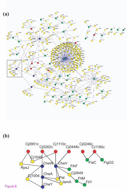
Cj0951cCj0262c
Cj1110cCj0448c
Cj0246c
CheVCheW
CheA
CheY
Pal
IamA
RpsJ
Cj1648
Cj1004
FlhF
Cj0649
FliMFliY
FlaC FlgG2
Cj1190c
(b)
(a)
Figure 6

0 5 10 15 20 25 30
0.1
0.3
0.5
E. coli
degree
essential gene f
raction
r=0.741
(a)
0 5 10 15 20 25 30
0.1
0.3
0.5
B. subtilis
degree
essential gene f
raction
r=0.701
(b)
0 5 10 15 20 25 30
0.1
0.3
0.5
E. coli AND B. subtilis
degree
essential gene fra
ction
r=0.765
(c)
0 5 10 15 20 25 30
0.1
0.3
0.5
E. coli OR B. subtilis
degree
essential gene fra
ction
r=0.634
(d)
Figure 7
Figure 7

A
B
C
R ib osom al p r ote in s y n th es is an d mod i fica ti o n
DN A r e p lica ti o n, r es tric ti o n / mod i fica ti o n,
r e c om b ina ti o n an d r e p air
C e ll e n v e lo p e
A m in o ac y l tRN A s y n the ta se and mod i fica ti o n
B ios y n th es is o f a m in o aci d s an d f a tt y aci ds
E n e r g y an d c e n tral in te r med iar y met a b o li sm
C of ac to r , p r o s th et i c g r o u p , and carri e r
b i os y n th es i s
P urin es, p y ri m id in es, nucl eos i de s , and
nucl eot i de b i os y n th es i s
T ran s cri pt i o n an d t ran s la ti o n
H yp ot h et ical
M i s c e llan eo u s
Figure 8

Additional files provided with this submission:
Additional file 1: additional data file1.doc, 109Khttp://genomebiology.com/imedia/1044524182143940/supp1.docAdditional file 2: additional data file2.doc, 93Khttp://genomebiology.com/imedia/1758688196143940/supp2.docAdditional file 3: additional data file3.doc, 489Khttp://genomebiology.com/imedia/9690706631439409/supp3.docAdditional file 4: additional data file4.doc, 259Khttp://genomebiology.com/imedia/9949176981439409/supp4.docAdditional file 5: additional data file5.doc, 91Khttp://genomebiology.com/imedia/2108952569143942/supp5.docAdditional file 6: additional data file6.doc, 202Khttp://genomebiology.com/imedia/1857446829143942/supp6.docAdditional file 7: additional data file7.xls, 70Khttp://genomebiology.com/imedia/5664509541481513/supp7.xlsAdditional file 8: additional data file8.pdf, 226Khttp://genomebiology.com/imedia/1624429514143942/supp8.pdfAdditional file 9: additional data file9.doc, 58Khttp://genomebiology.com/imedia/2876435611439422/supp9.docAdditional file 10: additional data file10.xls, 354Khttp://genomebiology.com/imedia/2043143998143942/supp10.xlsAdditional file 11: additional data file11.doc, 81Khttp://genomebiology.com/imedia/1687877911143942/supp11.docAdditional file 12: additional data file12.xls, 739Khttp://genomebiology.com/imedia/4263355981439422/supp12.xlsAdditional file 13: additional data file13.xls, 4881Khttp://genomebiology.com/imedia/3915758471439422/supp13.xlsAdditional file 14: additional data file14.doc, 95Khttp://genomebiology.com/imedia/1767656161143942/supp14.doc