novel orthoreovirus detected in a child hospitalized with acute gastroenteritis
TRANSCRIPT
High Similarity of Novel Orthoreovirus Detected in a ChildHospitalized with Acute Gastroenteritis to MammalianOrthoreoviruses Found in Bats in Europe
Andrej Steyer,a Ion Gutiérrez-Aguire,b,e Marko Kolenc,a Simon Koren,c Denis Kutnjak,b Marko Pokorn,d Mateja Poljšak-Prijatelj,a
Nejc Racki,b Maja Ravnikar,b,e Martin Sagadin,a Adela Fratnik Steyer,a Nataša Toplakc
Institute of Microbiology and Immunology, Faculty of Medicine, University of Ljubljana, Ljubljana, Sloveniaa; National Institute of Biology, Ljubljana, Sloveniab; Omegad.o.o., Ljubljana, Sloveniac; Department of Infectious Diseases, University Medical Centre Ljubljana, Ljubljana, Sloveniad; The Centre of Excellence for Biosensors,Instrumentation and Process Control—COBIK, Solkan, Sloveniae
Mammalian orthoreoviruses (MRVs) are known to cause mild enteric and respiratory infections in humans. They are widespread and infect abroad spectrum of mammals. We report here the first case of an MRV detected in a child with acute gastroenteritis, which showed the highestsimilarity to an MRV reported recently in European bats. An examination of a stool sample from the child was negative for most commonviral and bacterial pathogens. Reovirus particles were identified by electron microscopic examination of both the stool suspension and cellculture supernatant. The whole-genome sequence was obtained with the Ion Torrent next-generation sequencing platform. Prior to sequenc-ing, the stool sample suspension and cell culture supernatant were pretreated with nucleases and/or the convective interaction medium (CIM)monolithic chromatographic method to purify and concentrate the target viral nucleic acid. Whole-genome sequence analysis revealed thatthe Slovenian SI-MRV01 isolate was most similar to an MRV found in a bat in Germany. High similarity was shared in all genome segments,with nucleotide and amino acid identities between 93.8 to 99.0% and 98.4 to 99.7%, respectively. It was shown that CIM monolithic chroma-tography alone is an efficient method for enriching the sample in viral particles before nucleic acid isolation and next-generation sequencingapplication.
Reoviridae is a highly diverse virus family, including viruses ca-pable of infecting various host species (mammals, reptiles,
fish, birds, protozoa, fungi, plants, and insects). These viruses con-sist of an icosahedric capsid and a segmented genome of 10 to 12double-stranded RNA (dsRNA) segments (1). They are unenvel-oped and relatively stable in the environment (2). Two subfamilieshave been described within the Reoviridae family: Spinareovirinaeand Sedoreovirinae, including 9 and 6 genera, respectively (3). Inhuman medicine, the most recognized are Rotaviruses within theSedoreovirinae subfamily and Orthoreoviruses within Spinoreoviri-nae. Orthoreoviruses, with mammalian orthoreovirus (MRV) asthe type species, were already recognized in the 1950s as respira-tory and enteric orphan viruses (4). They were found in hosts withor without clinical manifestations (1). MRVs have been reportedto date in various mammalian hosts, including human and animalspecies. In the last few years, they have often been described as thesole pathogen in various hosts presenting severe clinical manifes-tations, such as hemorrhagic enteritis, acute respiratory infec-tions, central nerve system implications, and others (5–10). Thereis consequently increasing concern about the widespread natureand pathogenesis of these viruses. A German group of researchersrecently reported infections of bat species with MRVs, which re-sulted in evident pathological signs in the organs of infected bats(11). This was the first report of reoviruses in bats that were notclustered into the species Pteropine orthoreovirus but in MRVs.Almost at the same time, an Italian group published data on reo-virus detection in various bat species, reporting nucleotide se-quences of partial L1 and complete S1 segments, which showedthe highest similarity to the German bat isolate (12). Both researchgroups speculated on bat-to-human interspecies transmission,but there was no evidence to support this hypothesis. A closelyrelated MRV found in a dog with hemorrhagic enteritis had been
previously described by another Italian group. The authors pro-posed that these viruses might be important zoonotic pathogens(13).
The zoonotic potential of reoviruses has already been de-scribed and discussed elsewhere (6, 7, 11). The transmission ofreoviruses from one host to another is not limited to close contactsbut extends to indirect transmission. Infection through contami-nated food, water, or other factors in the environment is highlypossible, since infective reovirus particles have been found in en-vironmental samples (14–17). Viral persistence outside the host isone of the advantageous features that enables them to spread effi-ciently.
Researchers throughout the world have recently focused veryintensively on zoonotic or potentially zoonotic viruses. Bats are ofspecial interest because of their diversity, the wide spectrum ofvirus populations found in different bat samples, and a specialvirus-host interaction (18, 19). Screening of animals for poten-tially zoonotic viruses has resulted in the discovery of novel vi-ruses. In the last few years, the pathogen discovery field has madea major step forward. Application of new technologies, such asnext-generation sequencing (NGS), enables researchers to take a
Received 13 June 2013 Returned for modification 22 July 2013Accepted 5 September 2013
Published ahead of print 11 September 2013
Address correspondence to Andrej Steyer, [email protected].
Supplemental material for this article may be found at http://dx.doi.org/10.1128/JCM.01531-13.
Copyright © 2013, American Society for Microbiology. All Rights Reserved.
doi:10.1128/JCM.01531-13
3818 jcm.asm.org Journal of Clinical Microbiology p. 3818–3825 November 2013 Volume 51 Number 11
Dow
nloa
ded
from
http
s://j
ourn
als.
asm
.org
/jour
nal/j
cm o
n 08
Feb
ruar
y 20
22 b
y 20
01:6
48:2
e80:
6601
:cd8
5:8a
c8:6
d12:
d896
.
different approach to pathogen discovery, gathering a hugeamount of genomic information from a sample of interest in ashort period of time (20). In the last decade, the price of sequenc-ing technology has decreased drastically, while the capacity ofNGS data obtained has increased sharply (20). This research toolalso has major potential as part of microbiological diagnostics incombination with classical or standard microbiological method-ologies (21). In the future, NGS could help resolve clinical cases ofinfections with undetermined etiology. However, when usingNGS in pathogen discovery or diagnostics, the preparation ofhigh-quality and high-quantity nucleic acid is of major concern(22). Improved and optimized sample preparation, purification,and concentration of the target are essential for achieving the bestresults in NGS. Isolation of as pure as possible viral nucleic acidenables easier sequencing of the target of interest and faster bioin-formatics workflow. Convective interaction medium (CIM) chro-matography is the method of choice for virus concentration andpurification from different samples (23, 24).
In this work, we discovered an MRV strain with high similarityto orthoreoviruses detected in European bats. The MRV wasfound in a child with acute gastroenteritis requiring hospitaliza-tion. This is an indication of zoonotic transmission. The wholevirus genome was sequenced using the Ion Torrent platform. It ishighly possible that this group of MRVs detected in bats in Europeis widespread and could easily cross species barriers. However, thedetailed pathogenesis has yet to be determined. To the best of ourknowledge, this is the first report of a bat MRV detected in ahuman with a clinical manifestation. In addition, a new approachfor sample preparation prior to NGS is presented. A chromato-graphic method using CIM monoliths was used, which proved tobe effective in enriching and purifying the sample of MRV prior toRNA extraction and NGS application.
CASE REPORT
A 17-month-old boy presented at the Department of InfectiousDiseases, University Medical Centre Ljubljana, Ljubljana, Slove-nia, with a 5-day history of nonbloody diarrhea. Stools were fre-quent; the child refused to drink and after 4 days developed fever.His previous medical history was unremarkable, with only a fever-related thrombocytopenia at the age of 3 months. He had beenvaccinated against rotavirus. He attended a day care center atwhich no gastroenteritis cases were observed.
On admission, the child was mildly dehydrated and afebrile,and the abdomen was not sensitive to palpation. The C-reactiveprotein (CRP) concentration was 20 mg/liter, the white blood cell(WBC) count was 8.4 � 109/liter (with normal differential), thered blood cell (RBC) count was 4.94 � 1012/liter, the hemoglobin(Hb) level was 124 g/liter, the hematocrit (Hct) was 0.384, and theblood sugar, blood urea nitrogen, creatinine, and electrolytes werenormal. The child was given 500 ml of intravenous fluids anddischarged home. The next day he returned to the departmentbecause of ongoing fever, frequent nonbloody stools accompa-nied by colicky pains, and red and swollen gums with oral ulcers.Aphthous stomatitis was diagnosed, in addition to gastroenteritis,and the child was admitted for parenteral rehydration and dis-charged the next day. He made an uneventful recovery. The totalduration of diarrhea was 8 days.
A stool sample was analyzed according to the standard diag-nostic protocol, including classical bacterial examination withculturing techniques, checking for the most common bacterial
pathogens (Campylobacter spp., Salmonella sp., pathogenic Esch-erichia coli, Yersinia sp., and Shigella sp.). A virological examina-tion was carried out at the same time: negative-staining electronmicroscopy (EM) and enzyme-linked immunosorbent assays(ELISAs) for group A rotaviruses and adenoviruses 40 and 41(Meridian Bioscience, Inc., Cincinnati, OH) and in-house real-time reverse transcription (RT)-PCR for noroviruses of geno-groups I and II and astroviruses, as described previously (25, 26).Reoviruses were observed by electron microscopy in the stoolsample taken at the first admission.
MATERIALS AND METHODSVirus identification and cell culture propagation. Cell line LLC-MK2(rhesus monkey kidney epithelial cells) grown in modified Eagle’s me-dium (MEM) supplemented with 10% fetal calf serum was used for viruspropagation. A 10% stool suspension was prepared and clarified by cen-trifugation at 1,600 � g. Clarified supernatant of a 10% stool suspensionwas filtered through a 0.22-�m-pore filter (Millipore, Billerica, MA) andinoculated on a 48-h cell culture monolayer in 25-cm2 flasks (TPP, Trasa-dingen, Switzerland). After incubation for 1 h at 37°C, the inoculum wasremoved, and cells were supplemented with the original growth mediumand incubated further at 37°C in an atmosphere with 5% CO2 until theappearance of a cytopathic effect (CPE). A 10-fold serial dilution of reo-virus isolate was prepared to determine the 50% tissue culture infectivedose (TCID50) value (27). A known concentration of reovirus isolatehelped us to estimate the reovirus concentration in the child’s stool sam-ple using real-time RT-PCR, as described previously (11).
In addition, infected cells were prepared for thin sectioning. Briefly,the specimen was fixed with 2% (vol/vol) glutaraldehyde in 0.1 M phos-phate-buffered saline (PBS; pH 7.4) for 2 h, rinsed with 3 changes of PBS,and postfixed with 1% (vol/vol) OsO4 in PBS for 2 h. After being washed,the specimen was dehydrated in a graded series of ethanol and embeddedin epoxy (agar low-viscosity) resin, following the standard protocol. Ul-trathin sections were collected on carbon-coated 200 mesh copper gridsand stained with 1% uranyl acetate and 1% lead citrate. In addition, clar-ified stool suspension was used for direct EM examination of the sampleafter negative staining with 2% phosphotungstic acid (pH 4.5). EM gridswere screened at 80 kV in a JEM 1200 EXII transmission electron micro-scope (JEOL, Tokyo, Japan).
Sample preparation for NGS. The RNA for the sequencing procedurewas obtained from two sources: clarified stool suspension and cell culturesupernatant after virus propagation. Two samples (6 ml of stool and 6 mlof cell culture supernatant) were incubated with 1,000 U/ml of Benzonase(Novagen, San Diego, CA) for 1 h at room temperature (samples 1 and 2)to digest nonencapsidated nucleic acid. Samples were then diluted 6� inchromatography running buffer (50 mM HEPES; pH 7) and left overnightat 4°C. An additional 6-ml sample of cell culture supernatant was dilutedin the same way but without Benzonase treatment (sample 3). Ten milli-liters of all three samples was loaded into a 0.34-ml-volume CIM-QA disk(BIA Separations, Ajdovšcina, Slovenia), using an AKTA purifier chro-matographic system (GE Healthcare, Uppsala, Sweden). After loading,nonbound material was washed with running buffer, and the elution ofbound viruses proceeded in two different ways. The Benzonase-treatedstool suspension and cell culture samples (samples 1 and 2) were eluted ina single step, by including 1 M NaCl in the running buffer. The Benzo-nase-untreated cell culture sample (sample 3) was eluted by using a gra-dient of 0 to 500 mM NaCl in �60 column volumes in order to purify theviruses as much as possible. In all three cases, the elution was collected in1-ml fractions. The presence and approximate number of putative reovi-ruses in the elution fractions were estimated by the virus particle countingmethod under an electron microscope using a negative-staining tech-nique (28). To have an indication of the presence of eukaryotic nucleicacid, RNA was extracted from the fractions using a QIAamp virus RNA kit(QIAgen, Valencia, CA) and applied to a eukaryotic 18S assay (Life Tech-nologies, Applied Biosystems Division, Foster City, CA), in a one-step
Bat MRV in Hospitalized Child with Diarrhea
November 2013 Volume 51 Number 11 jcm.asm.org 3819
Dow
nloa
ded
from
http
s://j
ourn
als.
asm
.org
/jour
nal/j
cm o
n 08
Feb
ruar
y 20
22 b
y 20
01:6
48:2
e80:
6601
:cd8
5:8a
c8:6
d12:
d896
.

real-time RT-PCR format, using an Ag-Path master mix (Life Technolo-gies) and an ABI 7900 HT real-time PCR system. To overcome the lowersensitivity of the electron microscopy technique and in order to confirmthe presence of reovirus, the RNA from fractions 6 to 10 obtained in thestool sample run, in which no virus particles were observed, was applied toRT-PCR using a set of reovirus generic primers (5). Fractions that wereselected for the sequencing procedure (fractions 6 to 10, 3 to 6, and 15 to18 for runs involving samples 1, 2, and 3, respectively) were pooled, andRNA was extracted from the whole pooled volume using a TRIzol LS PlusRNA purification kit (Life Technologies, Invitrogen Division, Carlsbad,CA). The final purified RNA solution obtained from each of the threesamples was further evaporated to a final volume of �100 �l in a GeneVacpersonal evaporator (Genevac, Ltd., Ipswitch, United Kingdom).
Ion Torrent library preparation and sequencing. The RNA librarywas prepared using an Ion Total RNA-Seq kit v2 (Life Technologies, In-vitrogen Division, Darmstadt, Germany) according to the manufacturer’sprotocol (4476286, revision D) for a low input of starting material (be-tween 10 and 100 ng). The amount and size distribution of library DNAfragments were determined with a Labchip GX instrument (Caliper, LifeSciences, MA). Emulsion PCR and enrichment steps were carried outusing an Ion OneTouch200 template kit v2 DL as described in the proto-col (MAN0006957, revision 5.0). Assessment of the Ion Sphere particlequality was undertaken between the emulsion PCR and enrichment stepswith an Ion Sphere quality control kit using a Qubit 2.0 fluorometer (LifeTechnologies). Each library was sequenced on a separate Ion 314 Chip(Life Technologies). Signal processing and base calling were performedwith Torrent Suite software version 3.4.2. Adapter sequences weretrimmed using the same software.
Complete genome sequence generation. Trimmed reads were usedfor de novo assembly using CLC Genomic Workbench 6.0 (CLC Bio, Aar-hus, Denmark), with default program parameters (see Table S2 in thesupplemental material). The generated contigs were compared for simi-larity against all virus sequences deposited in the NCBI GenBank databaseusing the Basic Local Alignment Search Tool (BLASTn and BLASTx). Inall three samples, the majority of the contigs from all 10 viral genomesegments showed the highest identity values with the MRV strain 342/08genome, deposited in GenBank under accession no. JQ412755 toJQ412764 (11). This strain was therefore selected as the reference genome.Reads obtained from each sample were initially mapped separately to thereference sequence using default parameters (see Table S2). The generatedconsensus sequences were additionally compared, and no difference wasobserved between the consensus sequences of the three different samples.The reads of all three samples were subsequently mapped together to thereference. After removal of duplicate reads, all of the single nucleotidepolymorphisms (SNPs) and indels (in comparison to the reference ge-nome sequence) were explored visually. They were considered to be reli-able if they were covered with at least 30 reads and were derived from boththe positive and negative strands of RNA.
In order to compare the efficiencies of host (LLC-MK2 cell culture)nucleic acid removal between samples 2 and 3, reads were mapped to theMacaca mulatta genome (rheMac3; Beijing Genomics Institute, Shen-zhen, China) (GenBank assembly ID GCA_000230795.1). To prevent un-specific mapping, reads smaller than 25 bp were removed, and highlystringent parameters were used for these mappings (given in Table S2 inthe supplemental material).
Sequence analysis. Consensus genome segments were further ana-lyzed for open reading frames (ORFs), which were determined using Ge-neious 6.0.5 version (Biomatters, Ltd., Auckland, New Zealand), and de-duced amino acid sequences were obtained.
For phylogenetic analysis, whole-genome MRV sequences avail-able in GenBank were downloaded, and multiple sequence alignments(ClustalW) were carried out. A neighbor-joining phylogenetic tree(Kimura 2-parameter system) was made to show the phylogenetic rela-tionships, using the MEGA 4 software system (29). Branch support wasassessed by bootstrap analysis of 1,000 replicates. For reassortment or
recombination events, the concatenated whole-genome sequence of theSlovenian MRV was compared to other whole-genome MRVs fromGenBank. A similarity plot was constructed using SimPlot software, ver-sion 3.5.1 (30).
Nucleotide sequence accession numbers. The nucleotide sequencesobtained in this study have been deposited in GenBank under accessionno. KF154724 to KF154733.
RESULTS
Bacterial laboratory examination of the patient’s stool sample wasnegative at the first and second admissions. Virological examina-tion was performed only on the sample at first admission and wasnegative for specific antigen detection tests for group A rotavirusesand adenoviruses 40 and 41, and molecular tests were negative fornoroviruses in genogroups I and II and astroviruses, which are themost prevalent pathogens detected in the age group of the case.However, clear 75-nm reovirus particles were observed upon EMexamination of the stool suspension (Fig. 1A). After inoculation ofthe filtered stool suspension on an LLC-MK2 cell culture, CPEappeared 48 h postinoculation, comprising cell rounding with anotable membrane and final cell lysis. Again, reovirus particleswere observed under an electron microscope, on examination ofthe cell culture supernatant without a prior concentration step(Fig. 1B) and in a thin section of infected cell culture (Fig. 1C). Inultrathin sections, several cytoplasmic inclusions with denselypacked virus particles were found. An electron-dense center and aclear outer rim were observed in all particles.
The cell culture supernatant was cleared, and the reovirusconcentration was determined to be 2.43 � 108 TCID50/ml. The re-gression equation was obtained from the real-time RT-PCR anal-ysis of serial 10-fold dilutions of reovirus isolate (y � 40.73 �3.49x; R2 � 0.999). The theoretical TCID50/ml was calculated forreoviruses in child’s stool sample, using the quantification cycle(Cq) value of the reovirus real-time RT-PCR analysis. The concen-tration was estimated to 2.26 � 107 TCID50/ml, considering the10% stool sample dilution.
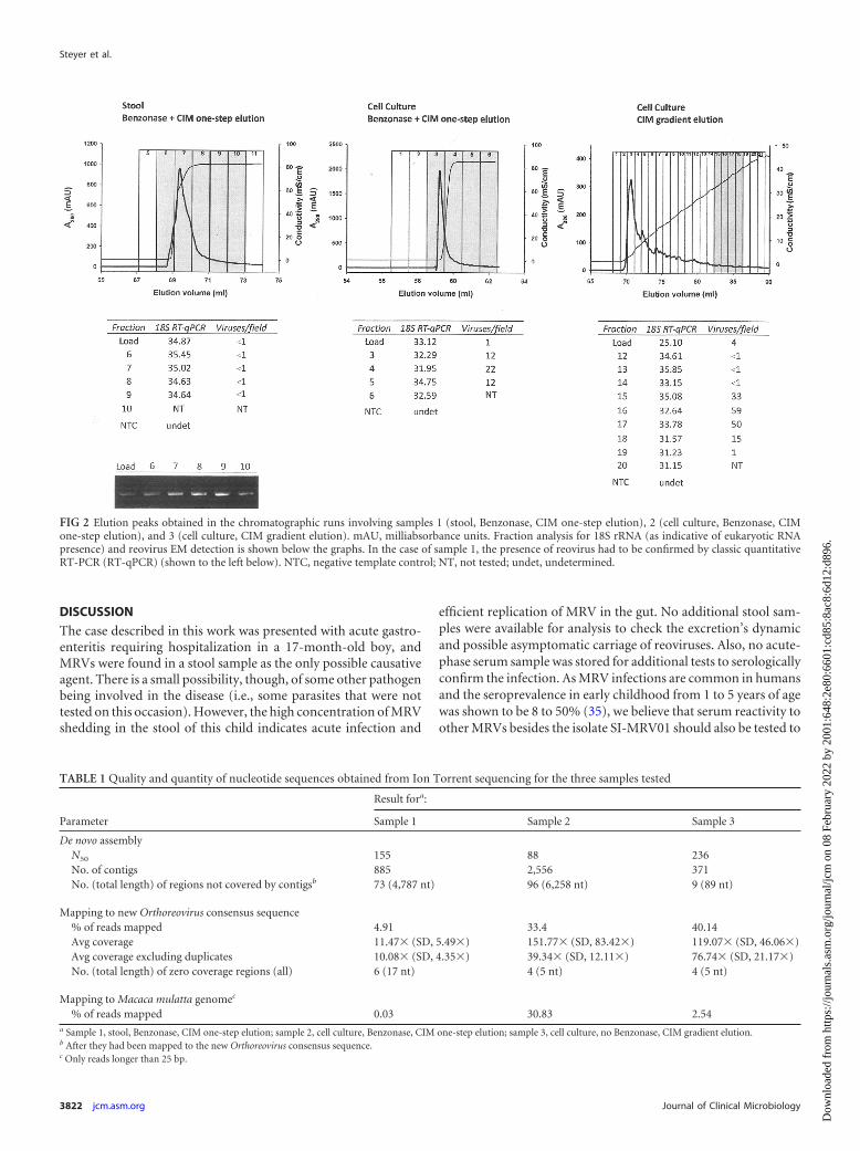
Sample preparation and sequencing on the Ion Torrent PGMplatform. The high 18S real-time RT-PCR Cq values (�34) ob-served in the load of the first two samples treated with nucleases(in comparison with nontreated sample 3; Cq � 25) indicates thatBenzonase degraded the nucleic acids present in the sample (Fig.2). Comparing the 18S Cq values of the load and elution fractionsin these two samples, it can be concluded that one-step elution didnot further clean the sample from eukaryotic NA, although it al-lowed a single order of magnitude concentration of the viruses insample 2 (1 virus per field of 400 mesh EM grid in the load com-pared to 12 to 22 in the eluted fractions). In sample 1 (stool sus-pension), the amount of virus was below the limit of detection(LOD) of the electron microscopy already in the load, and thepresence of viruses was only detected by conventional RT-PCR(Fig. 2), in which a certain concentration was again observed whencomparing the intensities of the agarose gel bands from load andelution fractions. Sample 3 showed the largest amount of virus inthe eluted fractions (up to 59 viruses per field of 400 mesh EMgrid), and it also allowed concentration of the viruses from 4 vi-ruses per field to up to 59. Moreover, the gradient allowed moreefficient separation of the virus-rich fraction, as seen from thechromatogram (Fig. 2) and from the 18S assay Cq values, whichincreased from 25 up to 35 in fraction 15. In this case, nonviral NAwas removed physically from the sample, so a lower risk for the
Steyer et al.
3820 jcm.asm.org Journal of Clinical Microbiology
Dow
nloa
ded
from
http
s://j
ourn
als.
asm
.org
/jour
nal/j
cm o
n 08
Feb
ruar
y 20
22 b
y 20
01:6
48:2
e80:
6601
:cd8
5:8a
c8:6
d12:
d896
.

presence of small interfering fragments is expected in comparisonto the Benzonase-treated ones.
The joint data set from all three sequencing runs consisted of861,215 reads. The average depth of coverage across all segmentsof the virus genome was 257.41�, with a standard deviation (SD)of 117.29�. After removal of duplicate reads, the average depth ofcoverage amounted to 101.88� (SD, 25.13�). The comparison ofthe sequencing outputs of the three samples pretreated differentlyis summarized in Table 1. In the case of sample 1, only 4.91% ofreads aligned to the final consensus sequence used as a referencefor comparisons. Much higher enrichment for viral sequences wasseen in the case of samples isolated from tissue culture: 33.4% ofreads aligned to the reference in case of sample 2. In the case of
sample 3 with gradient elution, an even higher proportion of se-quences aligned to the reference (40.14% of reads). Of all threesamples, sample 3 had the highest N50 value for the de novo assem-bly of reads, and the contigs produced covered almost the entirevirus genome. In addition, the percentage of high-quality readsthat map to the M. mulatta genome (source species for cell culturecells) is much lower in sample 3 (2.5%) than in sample 2 (30.8%)(Table 1), indicating that the elution gradient was more efficient atremoving background NA than the Benzonase treatment com-bined with single-step elution. Since the total number of reads wasa lot higher for sample 2 than for sample 3, more data and betterdepth of coverage were obtained from the former. However, in-creased depth in the case of sample 2 was shown to be a conse-quence of a high number of duplicate reads (Table 1). Due to thesmall size of the viral genome, a very good average coverage depthwas achieved even on the smallest PGM sequencing chip.
Sequence analysis. Nucleotide and deduced amino acid se-quences were obtained and further analyzed. Following BLAST ofthe nucleotide sequence, the newly sequenced Slovenian reovirusisolate SI-MRV01 showed the highest nucleotide sequence iden-tity with reovirus strain 342/08, isolated from an insectivorous batin Germany, in all genome segments except S2, in which a slightlylower nucleotide identity was observed (see Table S1 in the sup-plemental material). The nucleotide identity with the 342/08strain varied from 93.8% for the S2 segment to 99.0% for the L2segment. The amino acid sequence identity consequently alsoshowed the highest identity with the 342/08 strain in all genomesegments (98.4 to 99.7%) (see Table S1). In addition, phylogeneticanalysis confirmed the highest relationship of Slovenian humanreovirus isolate SI-MRV01 with German bat reovirus isolate 342/08. Both isolates clustered together in a separate branch in allgenome segments except S1 and S2 (Fig. 3; see Fig. S1 to S7 in thesupplemental material). For the S1 segment, the Slovenian SI-MRV01 strain was found in a specific bat cluster formed withinthe mammalian reovirus serotype 3 group. For this segment, thehighest identity was shown with the bat reovirus characterizedrecently in Italy (12). In the S2 segment, SI-MRV01 did not showrelatedness to any specific strain but was clustered in the serotype3 group, together with porcine strain SC-A, MRV-HLJ/2007, civetstrain MPC/04, and bat strain 342/08. According to the results ofthe SimPlot analysis, there are some indications of possible ge-nome reassortments. Most indications of a reassortment eventwere found for the L2 segment, comparing the SI-MRV01 strainwith the porcine GD-1, 729, and civet MPC/04 strains (Fig. 4).
The SI-MRV01 strain’s deduced amino acid sequence was an-alyzed for indicative amino acid positions related to pathogenesis.In previous studies, two amino acid positions, 350D and 419E, inthe �1 protein (S1 gene) were shown to be indicative of neurotro-pism (31, 32). However, in the SI-MRV01 strain, those sites were350I and 419E. The significance of such an amino acid composi-tion is not known and needs to be examined. It was also shownthat 249I is required for protease resistance of the �1 protein,which enables efficient viral spread and replication (33) and wasalso found in the SI-MRV01 strain. The S1 segment is also impor-tant in fusogenic reoviruses, since it encodes the fusion proteinFAST (fusion-associated transmembrane protein) (34). However,the Slovenian SI-MRV01 strain does not have the FAST codingregion. In the cell culture, no syncytia were observed, which is inconcordance with molecular findings of the absent FAST codingregion.
FIG 1 Electron micrographs of reoviruses in stool suspension (A) (magnifi-cation, 100,000�) cell culture supernatant (B) (magnification, 100,000�),and ultrathin section of LLC-MK2 cells (C) (magnification, 10,000�) infectedwith the SI-MRV01 orthoreovirus strain. Arrows in Fig. 1C indicate reovirusparticles.
Bat MRV in Hospitalized Child with Diarrhea
November 2013 Volume 51 Number 11 jcm.asm.org 3821
Dow
nloa
ded
from
http
s://j
ourn
als.
asm
.org
/jour
nal/j
cm o
n 08
Feb
ruar
y 20
22 b
y 20
01:6
48:2
e80:
6601
:cd8
5:8a
c8:6
d12:
d896
.
DISCUSSION
The case described in this work was presented with acute gastro-enteritis requiring hospitalization in a 17-month-old boy, andMRVs were found in a stool sample as the only possible causativeagent. There is a small possibility, though, of some other pathogenbeing involved in the disease (i.e., some parasites that were nottested on this occasion). However, the high concentration of MRVshedding in the stool of this child indicates acute infection and
efficient replication of MRV in the gut. No additional stool sam-ples were available for analysis to check the excretion’s dynamicand possible asymptomatic carriage of reoviruses. Also, no acute-phase serum sample was stored for additional tests to serologicallyconfirm the infection. As MRV infections are common in humansand the seroprevalence in early childhood from 1 to 5 years of agewas shown to be 8 to 50% (35), we believe that serum reactivity toother MRVs besides the isolate SI-MRV01 should also be tested to
FIG 2 Elution peaks obtained in the chromatographic runs involving samples 1 (stool, Benzonase, CIM one-step elution), 2 (cell culture, Benzonase, CIMone-step elution), and 3 (cell culture, CIM gradient elution). mAU, milliabsorbance units. Fraction analysis for 18S rRNA (as indicative of eukaryotic RNApresence) and reovirus EM detection is shown below the graphs. In the case of sample 1, the presence of reovirus had to be confirmed by classic quantitativeRT-PCR (RT-qPCR) (shown to the left below). NTC, negative template control; NT, not tested; undet, undetermined.
TABLE 1 Quality and quantity of nucleotide sequences obtained from Ion Torrent sequencing for the three samples tested
Parameter
Result fora:
Sample 1 Sample 2 Sample 3
De novo assemblyN50 155 88 236No. of contigs 885 2,556 371No. (total length) of regions not covered by contigsb 73 (4,787 nt) 96 (6,258 nt) 9 (89 nt)
Mapping to new Orthoreovirus consensus sequence% of reads mapped 4.91 33.4 40.14Avg coverage 11.47� (SD, 5.49�) 151.77� (SD, 83.42�) 119.07� (SD, 46.06�)Avg coverage excluding duplicates 10.08� (SD, 4.35�) 39.34� (SD, 12.11�) 76.74� (SD, 21.17�)No. (total length) of zero coverage regions (all) 6 (17 nt) 4 (5 nt) 4 (5 nt)
Mapping to Macaca mulatta genomec
% of reads mapped 0.03 30.83 2.54a Sample 1, stool, Benzonase, CIM one-step elution; sample 2, cell culture, Benzonase, CIM one-step elution; sample 3, cell culture, no Benzonase, CIM gradient elution.b After they had been mapped to the new Orthoreovirus consensus sequence.c Only reads longer than 25 bp.
Steyer et al.
3822 jcm.asm.org Journal of Clinical Microbiology
Dow
nloa
ded
from
http
s://j
ourn
als.
asm
.org
/jour
nal/j
cm o
n 08
Feb
ruar
y 20
22 b
y 20
01:6
48:2
e80:
6601
:cd8
5:8a
c8:6
d12:
d896
.

exclude possible cross-reactivity. Although there was no serolog-ical proof of MRV infection causing gastroenteritis, we have pre-sented data that demonstrate MRV as the possible causative agentof the disease.
It is well known that the disadvantage of electron microscopy isits low sensitivity. For successful detection of viruses with electronmicroscopy, the concentration of viral particles should be higherthan 106 per milliliter of suspension (36). Bearing in mind thedilution factor of 10 (when preparing a 10% stool suspension),this means that the MRV concentration in the patient’s stool sam-ple was at least of 107 viruses per milliliter or even higher. More-over, the viruses found in the stool sample were infective, sincethey were successfully propagated in a cell culture. The high con-centration of reoviruses in stool sample was confirmed also by thetheoretically calculated TCID50/ml, which was as high as 2.26 �107. All these facts suggest high replication activity of MRV in thepatient’s gut.
After the MRV isolate was cultured and genotyped, the parentswere contacted with regard to the child’s potential animal expo-sure. The family lived in a renovated house in a village close to thecity, where no bats had been observed. The only animal that hadclose contact with the child was a dog at the grandparents’ house,
but the animal was predominantly kept indoors. At the time of thechild’s illness, none of the family members experienced similarsymptoms. The parents stated that, at the time of the disease, thechild was known to ingest nonfood items (e.g., mild form of pica).According to the reovirus inactivation studies, showing their per-sistent stability in environment, indirect infection through con-taminated surfaces could also be possible (2, 17, 37). The source ofinfection therefore remains unknown. Unfortunately, no bat stoolsamples from the child’s residence were available for analysis toinvestigate the possible source of infection. It would be interestingin the future to screen bats in Slovenia for reoviruses, in order tohave a clear picture of reovirus molecular epidemiology and tolink possible zoonotic transmissions, such as the one described inour case.
MRVs were traditionally believed to be causative agents of mildrespiratory and enteric infections, without significant clinical im-pact (1). However, in the last decade or two, increasing reports onsevere human infections with reoviruses have been published, in-cluding central nervous system involvement (10, 38, 39). Thepathogenesis of reoviruses is not yet elucidated in detail, but someparameters influencing tissue tropism, efficient spread within thehost, and severe outcomes of infection are already known (31–33).
FIG 3 Phylogenetic analysis of the L1, M1, S1, and S2 genome segments for the Slovenian strain and most related whole-genome strains from GenBank. Theneighbor-joining algorithm was used for the construction of the phylogenetic tree with bootstrap values of 1,000 replicates shown at the branches. The scale barrepresents the p-distance. �, Slovenian MRV isolate SI-MRV01; o, Italian MRV strain identified in a bat; Œ, German MRV isolate 342/08 identified in a bat.
Bat MRV in Hospitalized Child with Diarrhea
November 2013 Volume 51 Number 11 jcm.asm.org 3823
Dow
nloa
ded
from
http
s://j
ourn
als.
asm
.org
/jour
nal/j
cm o
n 08
Feb
ruar
y 20
22 b
y 20
01:6
48:2
e80:
6601
:cd8
5:8a
c8:6
d12:
d896
.

It is thus important for diagnostic personnel to be aware of apossible reovirus etiology and to have tools to prove this infection.However, any reovirus-positive result should be interpreted care-fully, with a broad spectrum of cotested pathogens and clinicalpicture of the patient. Whether or not zoonotic transmission ofreoviruses is one of the possible factors associated with a severeclinical outcome is not yet clear. Nevertheless, there are reports inthe literature of bat reoviruses in humans with severe clinical man-ifestations. Those reovirus isolates, though, all clustered to Ptero-pine reovirus species and not to MRV species (6–8). This study isthe first description of a bat MRV found in humans. ProbablyMRVs are widely distributed among bat species, since they havebeen found in insectivorous bats in Germany and in Italy (11, 12).
Whole-genome sequence comparison of our strain to MRVsavailable in GenBank clearly shows that it is most closely related toMRVs found in bats in Germany. This was supported by highnucleotide and amino acid identities, phylogenetic analysis of sep-arate genome segments, and whole-genome sequences, includingSimPlot analysis. Genome reassortment is indicated by a similar-ity plot of reovirus genome sequences most closely related to theSlovenian SI-MRV01 strain (Fig. 4). A sharp decrease in nucleo-tide sequence identities for some of the genome segments wasobserved, like the L2 and S1 segments, comparing SI-MRV01 tothe GD-1 and MPC/04 strains, and the S2 segment, comparingSI-MRV01 to the 342/8 strain. Unfortunately, no other strainswith higher nucleotide identity were found in GenBank to explainthis diversity or possible reassortment events.
Finding an MRV in the stool sample of a hospitalized childwith diarrhea was surprising for us, since no such case had previ-ously been described in our laboratory. In the cell culture unit, noprevious work with reoviruses was performed, there was no reo-virus nucleic acid isolated in the molecular laboratory, nor had areovirus genome amplification project been performed. Thus, thepossibility of sample contamination is negligible. In addition toantigen detection tests and real-time RT-PCR assays for the detec-
tion of the most common viral causes of gastroenteritis, electronmicroscopy is still an important diagnostic tool routinely used inour laboratory. It is possible with the electron microscope to de-tect various viruses associated with rare cases, not included inspecific antigen and/or molecular testing procedures. It is a“catch-all” method, providing a broad-spectrum analysis of ex-amined samples (40). For rapid and accurate diagnostics, combi-nation of classical and new techniques in diagnostic virology isoften needed. This study demonstrates a good case of such aninterplay. Moreover, an improvement in sample preparation forsuccessful downstream application was introduced with virus en-richment using CIM monolithic support. It was shown that gra-dient elution, used in sample 3 without Benzonase treatment, inprinciple allows more detailed separation between the eluted mol-ecules, including viruses and nucleic acids. In addition, virus par-ticles were concentrated in specific fractions after chromato-graphic separation. This was confirmed also by sequencing results.In sample 3, the total number of reads was the lowest, but thepercentage of orthoreovirus reads was the highest.
Summarizing the results of our study, there are two major out-puts of this work. First, the detection of an MRV with high identityto MRV isolates found in European bats is an important indicatorof bat-to-human interspecies transmission, as was already specu-lated by German and Italian groups (11, 12). The real epidemio-logical situation regarding MRVs in bats and their transmission toother animal species should be further investigated in order tounderstand the full zoonotic potential and pathogenesis of theseviruses. The second output is a tool for improving the samplepretreatment in the search for enrichment in viral particles andnucleic acid. The pretreatment step is essential in NGS applica-tion, in which the target nucleic acid is expected to be present withhigh background. The described CIM chromatographic approachdemonstrates an excellent method combining concentration andpurification of the target, providing a new potential shortcut forNGS application in diagnostic clinical virology.
FIG 4 Similarity plot analysis of the whole-genome nucleotide sequence comparing the Slovenian orthoreovirus strain with some of the most related strains fromGenBank.
Steyer et al.
3824 jcm.asm.org Journal of Clinical Microbiology
Dow
nloa
ded
from
http
s://j
ourn
als.
asm
.org
/jour
nal/j
cm o
n 08
Feb
ruar
y 20
22 b
y 20
01:6
48:2
e80:
6601
:cd8
5:8a
c8:6
d12:
d896
.

ACKNOWLEDGMENTS
We thank Irena Šest for technical support with electron microscopy, di-agnostics, and sample preparation and Snežana Kramar for assistancewith cell culture propagation.
This work was financially supported by the Slovenian Research Agency(contracts no. L2-4314 and J3-4252).
REFERENCES1. Schiff LA, Nibert ML, Tyler KL. 2007. Orthoreoviruses and their repli-
cation, p 1854 –1915. In Knipe DM, Howley PM, Griffin DE, Lamb RA,Martin MA, Roizman B, Straus SE (ed), Fields virology, 5th ed. LippincottWilliams & Wilkins, Philadelphia, PA.
2. Katz BD, Margolin AB. 2007. Inactivation of hepatitis A HM-175/18f,reovirus T1 Lang and MS2 during alkaline stabilization of human biosol-ids. J. Appl. Microbiol. 103:2225–2233.
3. Day JM. 2009. The diversity of the orthoreoviruses: molecular taxonomyand phylogenetic divides. Infection, genetics and evolution. J. Mol. Epi-demiol. Evol. Genet. Infect. Dis. 9:390 – 400.
4. Sabin AB. 1959. Reoviruses. Science 130:1387–1389.5. Ouattara LA, Barin F, Barthez MA, Bonnaud B, Roingeard P, Goudeau
A, Castelnau P, Vernet G, Paranhos-Baccala G, Komurian-Pradel F.2011. Novel human reovirus isolated from children with acute necrotizingencephalopathy. Emerg. Infect. Dis. 17:1436 –1444.
6. Chua KB, Voon K, Crameri G, Tan HS, Rosli J, McEachern JA, SulurajuS, Yu M, Wang LF. 2008. Identification and characterization of a neworthoreovirus from patients with acute respiratory infections. PLoS One3:e3803. doi:10.1371/journal.pone.0003803.
7. Chua KB, Voon K, Yu M, Keniscope C, Abdul Rasid K, Wang LF. 2011.Investigation of a potential zoonotic transmission of orthoreovirus asso-ciated with acute influenza-like illness in an adult patient. PLoS One6:e25434. doi:10.1371/journal.pone.0025434.
8. Cheng P, Lau CS, Lai A, Ho E, Leung P, Chan F, Wong A, Lim W. 2009.A novel reovirus isolated from a patient with acute respiratory disease. J.Clin. Virol. 45:79 – 80.
9. Hermann L, Embree J, Hazelton P, Wells B, Coombs RT. 2004. Reovi-rus type 2 isolated from cerebrospinal fluid. Pediatr. Infect. Dis. J. 23:373–375.
10. Tyler KL, Barton ES, Ibach ML, Robinson C, Campbell JA, O’DonnellSM, Valyi-Nagy T, Clarke P, Wetzel JD, Dermody TS. 2004. Isolationand molecular characterization of a novel type 3 reovirus from a child withmeningitis. J. Infect. Dis. 189:1664 –1675.
11. Kohl C, Lesnik R, Brinkmann A, Ebinger A, Radonic A, Nitsche A,Muhldorfer K, Wibbelt G, Kurth A. 2012. Isolation and characterizationof three mammalian orthoreoviruses from European bats. PLoS One7:e43106. doi:10.1371/journal.pone.0043106.
12. Lelli D, Moreno A, Lavazza A, Bresaola M, Canelli E, Boniotti MB,Cordioli P. 2013. Identification of mammalian orthoreovirus type 3 inItalian bats. Zoonoses Public Health 60:84 –92.
13. Decaro N, Campolo M, Desario C, Ricci D, Camero M, Lorusso E, EliaG, Lavazza A, Martella V, Buonavoglia C. 2005. Virological and molec-ular characterization of a mammalian orthoreovirus type 3 strain isolatedfrom a dog in Italy. Vet. Microbiol. 109:19 –27.
14. Lodder WJ, de Roda Husman AM. 2005. Presence of noroviruses andother enteric viruses in sewage and surface waters in The Netherlands.Appl. Environ. Microbiol. 71:1453–1461.
15. Lodder WJ, van den Berg HH, Rutjes SA, de Roda Husman AM. 2010.Presence of enteric viruses in source waters for drinking water productionin The Netherlands. Appl. Environ. Microbiol. 76:5965–5971.
16. Spinner ML, Di Giovanni GD. 2001. Detection and identification ofmammalian reoviruses in surface water by combined cell culture and re-verse transcription-PCR. Appl. Environ. Microbiol. 67:3016 –3020.
17. Irving LG, Smith FA. 1981. One-year survey of enteroviruses, adenovi-ruses, and reoviruses isolated from effluent at an activated-sludge purifi-cation plant. Appl. Environ. Microbiol. 41:51–59.
18. Wong S, Lau S, Woo P, Yuen KY. 2007. Bats as a continuing source ofemerging infections in humans. Rev. Med. Virol. 17:67–91.
19. Wang LF, Walker PJ, Poon LL. 2011. Mass extinctions, biodiversity and
mitochondrial function: are bats ‘special’ as reservoirs for emerging vi-ruses? Curr. Opin. Virol. 1:649 – 657.
20. Lipkin WI. 2013. The changing face of pathogen discovery and surveil-lance. Nat. Rev. Microbiol. 11:133–141.
21. Capobianchi MR, Giombini E, Rozera G. 2013. Next-generationsequencing technology in clinical virology. Clin. Microbiol. Infect. 19:15–22.
22. Beerenwinkel N, Gunthard HF, Roth V, Metzner KJ. 2012. Challengesand opportunities in estimating viral genetic diversity from next-generation sequencing data. Front. Microbiol. 3:329. doi:10.3389/fmicb.2012.00329.
23. Gutierrez-Aguirre I, Steyer A, Banjac M, Kramberger P, Poljsak-Prijatelj M, Ravnikar M. 2011. On-site reverse transcription-quantitativepolymerase chain reaction detection of rotaviruses concentrated from en-vironmental water samples using methacrylate monolithic supports. J.Chromatogr. A 1218:2368 –2373.
24. Kovac K, Gutierrez-Aguirre I, Banjac M, Peterka M, Poljsak-Prijatelj M,Ravnikar M, Mijovski JZ, Schultz AC, Raspor P. 2009. A novel methodfor concentrating hepatitis A virus and caliciviruses from bottled water. J.Virol. Methods 162:272–275.
25. Kageyama T, Kojima S, Shinohara M, Uchida K, Fukushi S, HoshinoFB, Takeda N, Katayama K. 2003. Broadly reactive and highly sensitiveassay for Norwalk-like viruses based on real-time quantitative reversetranscription-PCR. J. Clin. Microbiol. 41:1548 –1557.
26. Svraka S, van der Veer B, Duizer E, Dekkers J, Koopmans M, VennemaH. 2009. Novel approach for detection of enteric viruses to enable syn-drome surveillance of acute viral gastroenteritis. J. Clin. Microbiol. 47:1674 –1679.
27. Reed LJ, Muench H. 1938. A simple method of estimating fifty per centendpoints. Am. J. Hyg. (Lond.) 27:493– 497.
28. Zheng YZ, Webb R, Greenfield PF, Reid S. 1996. Improved method forcounting virus and virus like particles. J. Virol. Methods 62:153–159.
29. Tamura K, Dudley J, Nei M, Kumar S. 2007. MEGA4: Molecular Evo-lutionary Genetics Analysis (MEGA) software version 4.0. Mol. Biol. Evol.24:1596 –1599.
30. Lole KS, Bollinger RC, Paranjape RS, Gadkari D, Kulkarni SS, NovakNG, Ingersoll R, Sheppard HW, Ray SC. 1999. Full-length humanimmunodeficiency virus type 1 genomes from subtype C-infected sero-converters in India, with evidence of intersubtype recombination. J. Virol.73:152–160.
31. Bassel-Duby R, Spriggs DR, Tyler KL, Fields BN. 1986. Identification ofattenuating mutations on the reovirus type 3 S1 double-stranded RNAsegment with a rapid sequencing technique. J. Virol. 60:64 – 67.
32. Kaye KM, Spriggs DR, Bassel-Duby R, Fields BN, Tyler KL. 1986.Genetic basis for altered pathogenesis of an immune-selected antigenicvariant of reovirus type 3 (Dearing). J. Virol. 59:90 –97.
33. Chappell JD, Barton ES, Smith TH, Baer GS, Duong DT, Nibert ML,Dermody TS. 1998. Cleavage susceptibility of reovirus attachment pro-tein sigma1 during proteolytic disassembly of virions is determined by asequence polymorphism in the sigma1 neck. J. Virol. 72:8205– 8213.
34. Shmulevitz M, Duncan R. 2000. A new class of fusion-associated smalltransmembrane (FAST) proteins encoded by the non-enveloped fuso-genic reoviruses. EMBO J. 19:902–912.
35. Tai JH, Williams JV, Edwards KM, Wright PF, Crowe JE, Jr, DermodyTS. 2005. Prevalence of reovirus-specific antibodies in young children inNashville, Tennessee. J. Infect. Dis. 191:1221–1224.
36. Hazelton PR, Gelderblom HR. 2003. Electron microscopy for rapid di-agnosis of infectious agents in emergent situations. Emerg. Infect. Dis.9:294 –303.
37. Ward RL, Ashley CS. 1978. Heat inactivation of enteric viruses in dewa-tered wastewater sludge. Appl. Environ. Microbiol. 36:898 –905.
38. Tyler KL. 1998. Pathogenesis of reovirus infections of the central nervoussystem. Curr. Top. Microbiol. Immunol. 233:93–124.
39. Johansson PJ, Sveger T, Ahlfors K, Ekstrand J, Svensson L. 1996.Reovirus type 1 associated with meningitis. Scand. J. Infect. Dis. 28:117–120.
40. Biel SS, Gelderblom HR. 1999. Diagnostic electron microscopy is still atimely and rewarding method. J. Clin. Virol. 13:105–119.
Bat MRV in Hospitalized Child with Diarrhea
November 2013 Volume 51 Number 11 jcm.asm.org 3825
Dow
nloa
ded
from
http
s://j
ourn
als.
asm
.org
/jour
nal/j
cm o
n 08
Feb
ruar
y 20
22 b
y 20
01:6
48:2
e80:
6601
:cd8
5:8a
c8:6
d12:
d896
.