uva-dare (digital academic repository) wound healing in ... · -- ulcus phagedenicum --...
TRANSCRIPT

UvA-DARE is a service provided by the library of the University of Amsterdam (http://dare.uva.nl)
UvA-DARE (Digital Academic Repository)
Wound healing in diabetic ulcers
Loots, M.A.M.
Link to publication
Citation for published version (APA):Loots, M. A. M. (2002). Wound healing in diabetic ulcers.
General rightsIt is not permitted to download or to forward/distribute the text or part of it without the consent of the author(s) and/or copyright holder(s),other than for strictly personal, individual use, unless the work is under an open content license (like Creative Commons).
Disclaimer/Complaints regulationsIf you believe that digital publication of certain material infringes any of your rights or (privacy) interests, please let the Library know, statingyour reasons. In case of a legitimate complaint, the Library will make the material inaccessible and/or remove it from the website. Please Askthe Library: https://uba.uva.nl/en/contact, or a letter to: Library of the University of Amsterdam, Secretariat, Singel 425, 1012 WP Amsterdam,The Netherlands. You will be contacted as soon as possible.
Download date: 26 Aug 2019

Chapterr 2
Differentiall diagnosis off leg ulcers
Mekkess JR, Loots MAM, vann der Wal AC, Bos JD.
SubmittedSubmitted for publication
ChapterChapter 2
INTRODUCTIO N N
Chronicc ulceration of the lower leg including the foot is a frequent condition, causingg pain, social discomfort, and generating considerable costs. Prevalence numberss range from 0.15-0.30% in the overall population, 1% in the adult population,, to 3-5% in the over-65 population.!": In western countries, the incidencee of ulceration is rising as a result of the ageing population and increasedd risk factors for atherosclerotic occlusion like smoking, obesity, and diabetes.1 1
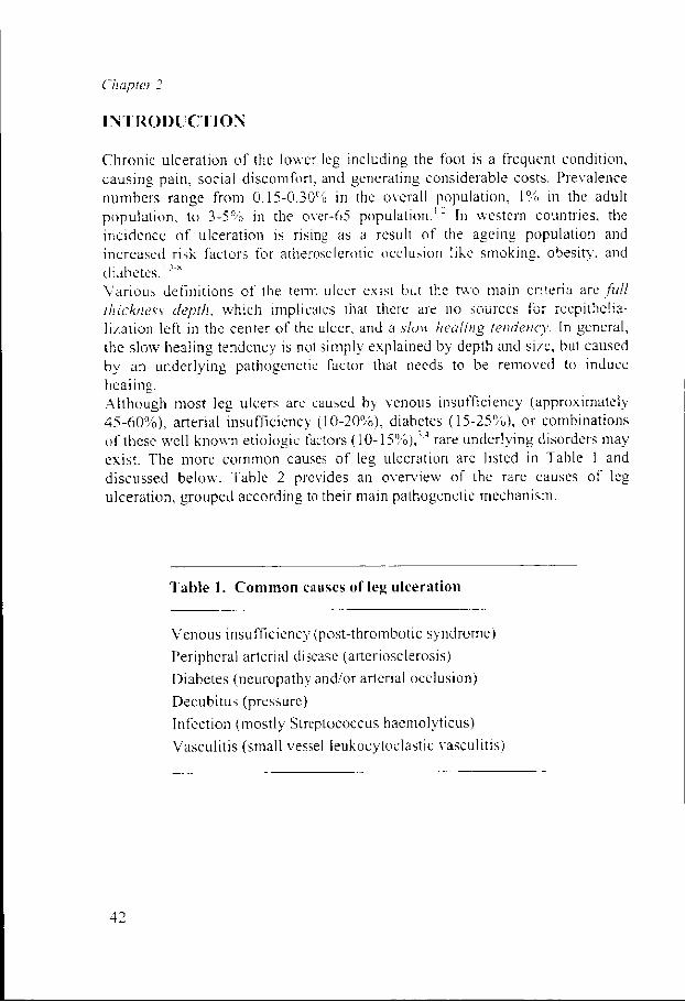
Variouss definitions of the term ulcer exist but the two main criteria arc full thicknessthickness depth, which implicates that there are no sources for reepithelia-lizationn left in the center of the ulcer, and a slow healing tendency. In general, thee slow healing tendency is not simply explained by depth and size, but caused byy an underlying pathogenetic factor that needs to be removed to induce healing. . Althoughh most leg ulcers arc caused by venous insufficiency (approximately 45-60%),, arterial insufficiency (10-20%), diabetes (15-25%), or combinations off these well known etiologic factors (10-15%),M rare underlying disorders may exist.. The more common causes of leg ulceration are listed in Table 1 and discussedd below. Table 2 provides an overview of the rare causes of leg ulceration,, grouped according to their main pathogenetic mechanism.
TableTable 1. Common causes of leg ulceration
Venouss insufficiency (post-thrombotic syndrome) Peripherall arterial disease (arteriosclerosis) Diabetess (neuropathy and/or arterial occlusion) Decubituss (pressure) Infectionn (mostly Streptococcus hacmolyticus) Vasculitiss (small vessel leukocytoclastic vasculitis)
42 2

DifferentialDifferential diagnosis oj leg ulcers
Tablee 2. Rare causes of leg ulceration
Venouss insufficiency and dependency -- venae communicantes insufficiency -- congenital hypoplasia / aplasia of valves -- weakness of the venous wall (collagen
disorders) ) -- arteriovenous anastomosis, angiodysplasia -- compression or obstruction of veins
(tumors,, enlarged lymph nodes, pelvic veinn thrombosis)
-- ulcerating thrombophlebitis, ruptured varices s
-- dependency syndrome (immobility, arthrosis,, rheumatoid arthritis, paresis, paralysis,, orthopedic malformations)
Arteria ll diseases -- arterial thrombosis macro-thrombocmbo-
lismm and micro-thromboembolism (fibrin, platelets) )
-- fat embolism (hypercholesterolemia, hyperlipidemia) )
-- detachment of cholesterol containing plaquess from aorta, aneurysm or atrium (atriall fibrillation)
-- thromboangiitis obliterans (m. Buerger) -- arteriovenous anastomosis (congenital /
traumatic) ) -- trauma, rupture, infection, vascular
procedures s -- fibromuscular dysplasia
Microcirculator yy disorders -- hypertension: ulcus hypertensivum
(Martorcll'ss ulcer) -- Raynaud's phenomenon, scleroderma -- increased blood viscosity (increased
fibrinogenfibrinogen level, paraneoplastic, paraproteinemia,, leukemia)
-- blood transfusion reactions
Physicall or chemical injur y -- trauma, burn wounds, freezing, electricity -- röntgen damage, intra-articular injection of
Yttrium-90 0 -- chemical (corrosive agents), sclerotherapy -- artificial (automutilation)
Vasculitis s -- large vessel: giant cell arteritis
(polymyalgiaa rheumatica, Takayasu's arteritis) )
-- medium size: polyarteritis nodosa, Kawasaki'ss disease
-- small vessel: Wegener's granulomatosis, allergicc granulomatosis (Churg-Strauss), microscopicc polyangiitis, Henoch Schonleinn purpura, essential cryoglobulinemicc vasculitis, erythema induratumm Bazin, livedo reticularis, livedo-vasculitiss and Sneddon's syndrome
Infectiouss diseases -- ulcerating pyoderma (S. aureus), ecthyma
(Streptococcuss haemolyticus), gas gangrenee (Clostridium), ecthyma gangrenosumm (Pseudomonas), septic embolismm (Meningococcus and others), anthraxx (Bacillus anthracis), diphteria (Corynebacteriumm diphteriae)
-- osteomyelitis (several microorganisms) -- complications by secondary wound
infections s -- herpes, lues, lues II, lues maligna (lues III ,
gummata),, granuloma inguinale, chancroid d
-- lepra, framboesia (yaws), ulcerating cutaneouss tuberculosis, lupus vulgaris, atypicall mycobacteria, Buruli ulcus (Mycobacteriumm ulcerans), papulonecrotic tuberculide e
** tularemia (Franeiscella tularensis), Chiclcroo ulcer (Leishmaniasis), bos-yaws (Leishmaniasiss mucocutanea), tropica! ulcerr (Baeteroidcs, Borrelia vincenti and otherr bacteria)
-- Madura foot, Maduramycosis (eumyectomaa / mycetoma), chromoblastomycosis,, coccidiomycosis, sporotrichosis,, granuloma trichopfiyticum
-- amoebiasis, Kala azar -- histoplasmosis ** bacillary angiomatosis
43 3

ChapterChapter 2
Tablee 2 (continued). Rare causes of leg ulceration
Hematologicc disorders anemia,, essential thrombocytemia, thromboticc thrombocytopenic purpura, sicklee cell anemia, granulocytopenia. GöPÜ-deficieney.. Thalassemia, hereditary' spherocytosis,, polycythemia, leukemia. monoclonall dysprotcmemia (Waldenstrom'ss disease, myeloma), polyclonall dysprotcmemia (cryofibrinogenemia,, purpura hyperglobulinemia,, cold agglutinins)
Neuropathicc diseases lepra,, tabes dorsalis. syringomyelia, alcoholl neuropathy, spina bifida, multiple sclerosis,, poliomyelitis
Clottin gg disorders Factorr V- Leiden, lupus anticoaguians, anticardiolipinn (antiphospholipid syndrome),, disturbed fibrinolysis, factor XII II deficiency (may be associated with colitiss ulcerosa), antithrombin III deficiency,, protein C or S deficiency. Marcoumarr necrosis, large haematoma. ulceratingg thrombophlebitis, purpura fulminans,, diffuse intravasal coagulation
Metabolicc diseases diabetess meliitus, necrobiosis lipoidica, porphyriaa cutanea tarda, gout, calciphylaxis,, calcinosis cutis, homocysteinuria,, prolidase deficiency, hyperoxaluria a
Ulceratingg tumors basall cell carcinoma, squamous cell carcinoma,, malignant melanoma, metastasis,, lymphoma, leukemia, lymphosarcoma,, cutaneous T-cell
lymphoma.. Hodgkin's disease, Kaposi's sarcoma,, pseudoepitheliomatous hyperplasia,, epithelioma (Marjolin's ulcer),, rhabdomyosarcoma, hemangiosarcoma.. lymphangiosarcoma
Ulceratingg skin diseases pyodermaa gangrenosum, erythema exudativumm multiforme, pemphigoid and otherr bullous diseases, malignant atrophic papulosiss (Dcgos), erythema induratum (Bazin),, sarcoidosis, erythema elevatum diutinum,, Behcet's disease, cutaneous discoidd and systemic lupus erythematosus, lichenn planus, contact dermatitis, panniculitis,, fat necrosis / pancreatic fat necrosis,, trench foot, insect bites, toe web infection n
Drugg reactions steroidd ulcus (intralesional injection), vaccinationn ulcer (BCG), halogens, ergotamin,, methotrexate, hydroxyureum. paravasall injection of cytostatic drugs. GM-CSF F
Miscellaneous s -- pemiosis (chilblains) -- erythermalgia'erythromelalgia -- corpus alienum, orthopedic fixation
materials s -- hemangioma, Stewart Bluefarb syndrome -- Klinefelter̂ syndrome -- rheumatoid arthritis, Felly's syndrome -- ulcus phagedenicum -- acro-osteopathia ulceromuülans (Bureau-
Barrière) ) -- complement C3 deficiency -- Langcrhans cell histiocytosis
44 4

DifferentialDifferential diagnosis oj leg ulcers
Venouss insufficiency
Venouss ulceration is caused by increased pressure in the venous system. The mainn cause of venous ulceration is insufficiency of the valves in the deep venouss system and the lower perforating veins. These veins and a good function off their valves are necessary for the return of venous blood to the heart at each contractionn of the calf muscles ('the muscle pump'). Intact valves but absent musclee contraction (immobility, paresis) may also cause edema and ulceration, aa condition known as dependency syndrome. Valve insufficiency may be acquiredd as in the post-thrombotic syndrome or caused by congenital weakness off valves or vessels. The exact pathogenetic cascade leading from valve insufficiencyy to ulceration is still not fully elucidated.4 The clinical symptoms of venouss insufficiency are edema, lipodermatosclerosis, hyperpigmentation, hyperkeratosis,, and atrophie blanche preceding ulceration.5 On a microvascular level,, the observations are microlymphangiopathy, dilatation of larger lymph vessels,vessels,99 dilatation and elongation of capillaries,10 occlusion of capillaries by microthrombi,111 or white cells,12 reduction of the number of functional capillaries,9100 increased capillary passage, leakage of plasma proteins and even erythrocytes,, leading to iron accumulation in the interstitium, partly in siderophages,, fibrin deposition, and ingrowth of fibroblasts along the fibrin fibrils."" The functional alterations are reduction, reversion, and stagnation of bloodd flow in the capillaries of prc-nccrotic skin, increased pressure in the capillaries,, increased blood flow in the deeper stratum reticulare capillary network,, increased blood flow and arteriovenous shunting nearby ulcers, and decreasedd skin oxygen pressure in areas at risk.1013 The laboratory alterations cann be anaemia, elevated erythrocyte sedimentation rate, iron deficiency, zinc deficiency,, decreased fibrinolytic activity, increased plasma and full blood viscosity,144 or clotting disorders predisposing to thrombosis.15,16 Some of these observations,, like shunting of blood near ulcers, the fibrin cuff, iron accumulation,, white cell accumulation, decreased fibrinolytic activity, and variouss inflammatory responses to the vascular damage have been isolated from theirr context and promoted to be 'the final cause of venous ulceration', while theyy represent rather epiphenomena than causative factors. Most authors believe thatt the changes on the microvascular level are sufficient to explain venous ulceration.10,11,14'177 The capillary changes lead to reduction of blood flow, disturbedd rheological conditions,14 sludging and aggregation of cells, and finally too microthrombi formation and occlusion of capillaries.in,l! In addition, increasedd pressure in the venous system increases transendothelial and interendotheliall capillary passage, resulting in a protein-rich edema. Edema in itselff may contribute to tissue hypoxia because it simply increases the diffusion distancee for oxygen around the nourishing capillaries.13 In the end this results in
45 5

ChapterChapter 2
aa fibrotic and edematous skin area where a considerable amount of capillaries is missing,, while the remaining are malformed and dysfunctional. The slightest traumaa or infection in these areas disturbs the balance between oxygen supply andd demand and a chronic non-healing ulcer develops. Thee relative frequency of venous leg ulcers is diminishing as a result of improvedd community care, improved prevention, diagnosis and treatment of thrombosis,, and an increase in arterial ulcers.4':" Still, the costs associated with venouss leg ulcers are considerable, approximately 200 million pounds yearly in thee UK,|V and 1 billion dollar in the US,lh'2'J were the yearly costs for hard-to-heall ulcers may be up to 27.500 dollar per patient/1
Thee mainstay of treatment (and prevention of new ulcers) is control of edema byy adequate compression therapy. Provided that the patients are bandaged by-experiencedd personnel, there arc no differences between non-clastic, short stretch,, two-layer or multi-layer compression bandages.4"^':"! Because many patientss have ulcers of combined etiology, e.g. venous and diabetes, or venous andd arterial, it is recommended to rule out arterial insufficiency before applying aa compression bandage, especially when elastic bandages are used. This can be donee by measuring the anklc-brachial index (ABI), which should be over 0.8."'244 Surgical restoration or replacement of destroyed deep venous valves is stilll not a routine option.2?:(1 Superficial incompetent veins can be ligated and/or removed,, or embolized by sclerocompression therapy, but their role in the etiologyy of venous leg ulcers is limited. Insufficient perforating veins, especiallyy the lower Cockett-veins are of haemodynamic importance because theyy transmit the high pressure to the overlying skin, and they can be ligated by (multiple)) incisions or new subfascial endoscopic techniques,2 but the value of thiss procedure in the presence of deep venous insufficiency is doubtful.25''5 With thee possible exception of pentoxifylline,"25 there is no sufficient evidence that systemicc drugs are beneficial.:Cv Patients with resistant or large ulcers may requiree hospitalization, additional wound bed preparation and skin grafting. An old-fashionedd but efficient method is full-thickness autologuous skin grafting, usingg punch biopsy grafts.:i<) Commercially cultured allogeneic skin grafts like Apligraf** (Novartis) have also been reported to accelerate healing,31 '3: especially inn a subgroup of ulcers of long duration." but more data are needed on cost-effectiveness,, long term results, and recurrence rates.33 The transplanted allogeneicc cells do not survive, eventually they are replaced by the patients own fibroblastss and keratinocytcs, but the cell-seeded skin equivalents induce a healingg tendency, probably through cytokine production.12 Since a decade it is possiblee to culture an autologuous dermal-epidermal skin equivalent from a smalll tissue specimen of the patients upper leg, within 14-21 days.30 These graftss will not be rejected, but the procedure is logistically complicated and expensive. .
46 6

DifferentialDifferential diagnosis of leg ulcers
Lowerr extremity arterial disease
Thee incidence of critical leg ischemia is increasing.3,1*'34 Risk factors for arterioscleroticc occlusion are diabetes (4 to 5-fold increase of incidence of peripherall vascular disease), smoking, hyperlipidemia, hypertension, obesity, andd age. Some influencing of risk factors is possible by education of the public, orr by drug therapy (antihypertensive drugs, antilipemic agents, transdermal nicotine,, low-dose aspirin)/'"'"71 Arteriosclerotic occlusion usually affects the entiree femoro-popliteal traject including important distal branches (arteria peronea,, tibialis anterior and tibialis posterior), and may lead to extensive distal damage.. It may also affect only small-sized branches, leading to limited infarctionn of skin and subcutaneous tissue with a relatively good prognosis. The latterr variant is not detected by a routine vascular work-up. Large occlusions requiree surgical interventions, which may be revascularization by means of bypassess using the patients own veins or artificial vein grafts, or by means of intravascularintravascular procedures such as balloon dilatation (PTA, percutaneous transluminall angioplasty), sometimes combined with thrombolysis and the placementt of stents.3S The average costs of a surgical intervention for critical leg ischemia,, including all necessary measures to secure graft maintenance during a 5-yearr follow-up period are $35.000-47.000.36'37 Femoro-popliteal vein grafts havee higher patency rates than femoro-crural procedures.34 Ideally, necrotic tissuee can be excised in the same operation session, and in some cases, vascularizedd flaps can be used to close the defects. The efficacy of pharmacologicall treatment of existing disease with vasoactive or anticoagulativcc drugs is disappointing; antilipemic agents are under investigationn and may be useful. A future development, although still in the experimentall phase, may be intravascular gene therapy with VEGF, vascular endotheliall growth factor, which may induce collateral neovascularisation in inoperablee ischemic legs.3S
Diabetes s
Amongg diabetic patients, 2-3% will develop a foot ulcer each year, 15% will developp a foot ulcer during lifetime.*'"19 The average costs per case from ulcer presentationn to complete resolution may be 4.730-10.930 pounds, and up to 20.800-31.8000 pounds when amputation is required.40'41
Inn the classic diabetic foot, distal sensorimotor and autonomic neuropathy is the majorr cause, often combined with arterial insufficiency caused by atheroscleroticc occlusion of the tibioperoneal arteries, with sparing of the pedal arteries.7'422 Approximately 60-70% has neuropathy only, 15-20% has peripheral vascularvascular disease only, and 15-20% has a mixture of both.43 The contribution of
47 7

ChapterChapter 2
occlusivee microvascular disease in the etiology of diabetic foot ulcers has not beenn confirmed by histology, vascular casting, or vascular resistance studies andd therefore seems to be a misconception.44 The frequency and severity of woundd infection is increased in diabetes, which may be related to high glucose levelss or impairment of granulocytic function and chemotaxis.45 In addition, theree seems to be prolonged inflammation, impaired neovascularization, decreasedd synthesis of collagen, an abnormal pattern of synthesis of extracellularr matrix proteins, and decreased fibroblast proliferation.46
Thee main principles of treatment are relief of any pressure at the wound site, aggressivee surgical debridement, adequate control of infection (beware of osteomyelitis),, arterial reconstruction if necessary, and strict control of glucose levels.. Pressure relief may be accomplished by total contact casting (TCC), wrhichh is the most extensively studied technique,47 orthopedic shoes or bed rest. Debridementt of devitalized tissue at frequent intervals has been shown to heal neuropathicc ulcers more rapidly.48 There are littl e data to support the use of enzymaticc or other non-surgical debridement strategies. Iff the standard measures fail, some benefit may be derived from new therapeuticall options such as recombinant human growth factors, bio-engineeredd skin substitutes, dressings made of extracellular matrix molecules likee collagen or hyaluronic acid, and a variety of synthetic dressings.49 Although theree is no evidence that any specific dressing type accelerates the healing process,4'' the beneficial effect of a moist wound environment has been well established.. Randomized controlled clinical trials with growth factors in diabeticc ulcers have shown efficacy of topically applied PDGF-BB (platelet-derivedd growth factor),50 and G-CSF (granulocyte-colony stimulating factor).51
Thee main effect of biologic skin substitutes is to promote wound healing by stimulatingg the host to produce various cytokines. Dermagraft'*, a bioabsorbable polyglactinn mesh seeded with cultured neonatal dermal fibroblasts, induced healingg in 50% of diabetic ulcers after 8 weeks of treatment, versus 7.7% in the controll group.52 However, no significant differences in ulcer recurrence rates weree noted.52 Apligraf*', an allogeneic bilayered cultured skin equivalent, appliedd for 4 weeks, achieved complete wound healing at 12 weeks in 56% of patientss with diabetic foot ulcers, versus 38% in the control group.53
Recentt research indicates that the incidence of both vascular and neurological complicationss of diabetes can be significantly reduced when intensified insulin therapyy maintains blood glucose concentrations at near-normal levels.49,54
Significantt steps are undertaken towards fully automatic control of glucose levelss by an implantable artificial pancreas.55 Management of dyslipidemia also deservess attention. And finally, patient education increases their awareness of potentiall hazards (pressure, minor skin trauma) and reduces infection and ulcer recurrence. .
48 8

DifferentialDifferential diagnosis of leg ulcers
Decubitus s
Pressuree ulcers develop when soft tissue is compressed between a bony prominencee and an external surface for a prolonged period of time. It usually occurss in hospitalized patients that are temporarily or permanently unable to changee their position due to circumstances like general anaesthesia, sedation, coma,, paresis/spinal injury, or fractures. Additional risk factors are incontinence,, bad nutritional state, increased body temperature, diabetes, peripherall arterial diseases and age.5fl'57 Decubitus can be divided into four stages,, depending on the extend of tissue damage; stage I: non blanchable erythema;; stage II: partial thickness loss of skin layers (blister, abrasion); stage III :: full thickness loss exposing subcutaneous fat (superficial ulcer); stage IV: exposedd muscle or bone (deep ulcer or necrosis).18
Thee prevalence of pressure ulcers ranges from 6.8-14.6% in home care settings, 5.1-15.6%% in general hospitals, to 25-41% in geriatric nursing homes.5fi The costss generated by decubitus ulcers are enormous. Rough estimations indicate thatt the annual costs of pressure sore treatment in the UK are around 150.000.0000 pounds.18 Obviously, maximum attention should be given to preventivee measures. It is generally recommended to have a decubitus protocol availablee to all staff which contains a validated scale for risk assessment. Dependingg on the risk assessment, preventive measures can be taken varying fromm frequent inspection, general measures to diminish pressure (spreading the bodyy weight over an area as large as possible), frequent changes of position, andd the use of special foam or airchamber matresses, low-air-loss systems or air-fluidizedd mattresses.57 Ulcer treatment consists of surgical removal of necroticc tissue, followed by the repeated application of dressings (saline soaked gauzes,, hydrogels, hydrocolloids, and many others) that further remove debris andd induce granulation tissue formation.
Hypertensionn and ulcus hypertensivum Martorel l
Hypertensionn is a known risk factor for atherosclerotic occlusion. In addition, anti-hypertensivee drugs (beta-blockers) may interfere with wound healing due too peripheral vasoconstriction." A rare condition exists called Martorell's ulcer, seenn in patients with prolonged, severe or sub-optimally controlled hypertension.599 The ulceration is secondary to tissue ischemia caused by increasedd vascular resistance. The ulcers are usually located at the lower limb, abovee the ankle region, contain black necrosis and are extremely painful. By definition,, the distal arterial pulsations are normal, and the diagnosis is made by histologicalhistological examination, which shows concentric intima thickening and marked hypertrophyy of the media of small size and medium size arteries, and by
49 9
ChapterChapter 2
exclusionn oï other conditions that may cause ulceration in tins area. The differentiall diagnosis consists of arteriosclerotic occlusion of small size arteries, diabeticc angiopathy, vasculitis, thromboembolic occlusion (e.g. in atrial fibrillation),, and pyoderma gangrenosum. Treatment consists of reducing hypertension,, avoiding beta-blockers, adequate control of pain, and local wound care. .
Vasculitis s
Vasculitiss denotes a heterogenous group of diseases characterized by inflammatoryy vessel damage. Several subdivisions can be made, based on vessell size (large vessel, medium size, small vessel), infiltrate type (polymorphonuclear,, mononuclear, granulomatous), or clinical presentation/'0
Cutaneouss vasculitis may present as purpura, erythema, urticaria, noduli, bullae, orr skin infarction leading to ulceration. Cutaneous ulceration is usually caused byy medium size to small vessel - leukocytoclastic vasculitis.61 Persistent or progressivee ulceration due to histologically confirmed vasculitis is an indication forr immunosuppressive therapy. Ulceratingg vasculitis may be caused by anti-ncutrophil cytoplasmic antibodies (ANCA),, autoantibodies against antigens in neutrophils, such as myeloperoxidasee (MPO) and proteinase 3 (PR3). Using indirect immuno-fluorescencee techniques, ANCA can be delected in a perinuclear pattern (pANCA,, often anti-MPO) or a cytoplasmatic pattern (cANCA. often anti-PR3).. They were first identified in Wegener's granulomatosis, later also in other typess of small vessel vasculitis, now classified as AN'CA-associated vasculitides (Wegener'ss disease, microscopic polyarteritis, idiopathic glomerulonephritis, andd Churg-Strauss syndrome)/'"
Felty'ss syndrome
Felty'ss syndrome, defined by the triad of rheumatoid arthritis, splenomegaly andd neutropenia, is associated with skin ulcers, probably caused by vasculitis. In general,, the incidence of leg ulcers in rheumatoid arthritis is slightly increased.tM,,,~~ In a minority of patients the ulceration is caused by vasculitis, otherr explanations are venous insufficiency and dependency (impairment of the venouss pump caused by immobility and ankle joint dysfunction), deformities, trauma,, ill-fittin g shoes (pressure), neuropathy, co-existing arterial insufficiency,, or pyoderma gangrenosum/0 If vasculitis can be confirmed histologically,, immuno-suppressiva are indicated.
:>u u
DifferentialDifferential diagnosis of leg ulcers
Clottin gg disorders
Hypercoagulablee disorders may cause ulceration, either indirectly as a consequencee of venous thrombosis, or directly by thrombus formation in small arteries,, arterioles, capillaries or venules.66'6 A growing number of hereditary or acquiredd conditions predisposing to thrombosis have been identified (Table 2), suchh as the antiphospholipid syndrome, deficiency of antithrombin 111, protein CC or protein S,6H or abnormal clotting factors (Factor V Leiden, Factor II mutant).1516,677 Not the laboratory abnormalities, but the specific clinical picture determiness whether a patient should be treated with anticoagulant drugs.
Antiphospholipidd syndrome
Thiss rare syndrome is characterized by the presence of circulating autoantibodiess against phospholipid compounds. It is associated with an increasedd risk for venous or arterial thrombosis, thrombocytopenia, and habitual abortus.. The cutaneous symptoms (ulceration, livedo reticularis, acrocyanosis, Raynaud'ss phenomenon, capillaritis and thrombophlebitis) can all be explained byy vascular thrombosis. The two most frequently found antibodies are lupus anticoagulanss and anticardiolipine. Presence of lupus anticoagulans is often accompaniedd by a prolonged prothrombin time and activated partial tromboplastinn time, hence the confusing term anti-coagulans, but it is associated withh an increased risk for thrombosis. Antiphospholipids have been found in a growingg number of diseases, especially auto-immune diseases (systemic lupus erythematosus,, auto-immune thrombocyte purpura and hacmolytic anemia, rheumatoidd arthritis, Sjogren's syndrome, giant cell arteritis, dermatomyositis, Behcet'ss disease, polyarteritis nodosa), malignancies, hematologic disorders (myelofibrosis,, von Willebrand's disease, paraproteinemia), infections (lues, lepra,, tuberculosis, mycoplasma, borreliosis, HIV, endocarditis, hepatitis) and neurologicall disorders (Sneddon's syndrome, myasthenia gravis, multiple sclerosis).69 9
Combinationss of vasculitis and clotting disorders
Thiss combination (Fig. 1) may be more frequent than the current literature suggests.. After the introduction in our department of routine screening for clottingg disorders (Table 3) in patients with ulceration caused by histologically confirmedd vasculitis, hypercoagulable states were found in 5 out of 1 1 subsequentlyy admitted patients (4 x Factor V-Leiden, 1 x lupus anti-coagulans). Thee two rare conditions together predispose for necrosis. Vasculitis damages
51 1

ChapterChapter 2
thee vascular wall, but not in all patients this leads to ulceration. An additional hypercoagulablee state may lead to extensive microvascular thrombi formation. Forr Factor V-Leiden such a sequence of events is likely. The vascular damage initiatess the coagulation cascade, prothrombin is converted to thrombin, thrombinn activates Factor V en VII . Coagulation is normally controlled by circulatingg antitrombin III , and locally by thrombomodulin, which is present on endotheliall cells and binds thrombin. The thrombin-thrombomodulin complex activatess protein C. Activated protein C (and protein S) inactivates Factor Va en Vila.. But the mutant Factor V-Leiden (506R -*506Q) is resistant to inactivation byy protein C. As a consequence, the local protection mechanism against thrombosiss is not working adequately.
Figuree 1. A 40-year old male patient with livedo reticularis and multiple ulcers caused by leukocytoclasticc vasculitis in combination with Factor V-Leiden. Histology: microvascular occlusionn by platelet-rich thrombi (anti-CD61 immuno-staining for thrombocytes).
52 2
DifferentialDifferential diagnosis of leg ulcers
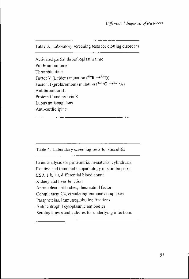
Tablee 3. Laboratory screening tests for clotting disorders
Activatedd partial thromboplastin time
Prothrombinn time Thrombinn time Factorr V (Leiden) mutation (mR ->506Q) Factorr II (prothrombin) mutation (:o210G ->202IOA)
Antithrombinn III Proteinn C and protein S Lupuss anticoagutans Anti-cardiolipine e
Tablee 4. Laboratory screening tests for vasculitis
Urinee analysis for proteinuria, hematuria, cylindruria
Routinee and immunohistopathology of skin biopsies ESR,, Hb, Ht, differential blood count Kidneyy and liver function Antinuclearr antibodies, rheumatoid factor Complementt C4, circulating immune complexes Paraproteins,, Immunoglobuline fractions Antineutrophill cytoplasmic antibodies
Serologicc tests and cultures for underlying infections

ChapterChapter 2
Infectiouss diseases
Sonicc micro-organisms can cause tissue necrosis, like the notorious beta-haemolyticall Streptococcus pyogenes. This bacteria causes a range of severe clinicall symptoms varying from erysipelas, punched-out ulcers (ecthyma), deep cellulitis,, to fasciitis necroticans, sepsis and multi-organ failure. Immediate high-dosee antibiotic treatment is necessary', with attention for combined infectionss with Staphylococcus aureus and anaerobic species. Al ll chronic wounds are secondarily contaminated with bacteria, but in most cases,, with the exception of the micro-organisms listed in Table 2, they are not off pathogenetic importance. Wound Cultures are often routinely performed, but givee only information about the bacterial flora in the superficial layers. The decisionn to prescribe systemic antibiotics should be based on the combination of culturee results and clinical criteria, like signs of infection (fever, erythema, calor).. In osteomyelitis, a common complication of neuropathic ulcers, efforts shouldd be made to obtain representative cultures from the bone or deepest tissue layers,, prior to antibiotic treatment, which should be given in high doses, preferablyy parenterally. and for at least 6 weeks." The diagnosis has become easierr after the introduction of labeled leukocyte scanning and especially magneticc resonance imaging (MR1).
Acquiredd immune deficiency due to HIV-infection reintroduced ulcerative conditionss that were thought to be eradicated, like tertiary lues and ulcerating tuberculosis,, and may be associated with atypical, large ulcers caused by herpes simplexx or cytomegalovirus. Finally, increased traveling around the world has broughtt tropical ulcerating infections to western countries, especially Leishmaniasis. .
Ulceratingg skin diseases
Severall skin disorders present with ulceration as the first symptom (Table 2). Thee most impressive ulcerating dermatosis is pyoderma gangrenosum, often not recognized.. The etiology is unknown, it is associated with colitis ulcerosa, morbuss Crohn, arthritis, paraproteinemia, myeloma, leukemia, and many other internall diseases. Pyoderma gangrenosum causes deep necrotic ulcers, usually withh an elevated violaceous border, and the ulceration is progressive if left untreated.. It may be provoked by wounding the skin, hence its occurrence aroundd scars, anus praeter. and donor sites used for grafting the original lesions. Onlyy treatment with sulphasalazinc. prednisone, ciclosporin or other immuno-modulatoryy drugs will stop the process.

DifferentialDifferential diagnosis of leg ulcers
Diagnosticc approach in patients with leg ulcers
Thee localization may give the first clue, venous leg ulcers predominantly occur inn the gaiter area, above the malleoli, arterial ulcers at the toes, on the shin and overr pressure points, diabetic ulcers over pressure points, especially the distal metatarsall joints.71 Venous insufficiency can often be diagnosed without additionall vascular investigations, on the presence of typical skin signs like edema,, hemosiderin pigmentation, hyperkeratosis, and atrophie blanche.
Suspicionn of arterial disease requires a routine vascular work up, starting with clinicall examination, palpation of arteries, assessment of skin color and temperature,, and calculation of the ankle-brachial index (ABI) for the arteria dorsaliss pedis and tibialis posterior. In healthy subjects, the ABI is around or abovee 1.0, but an ABI above 0.8 is still considered normal, and a safe threshold too apply compression therapy in venous leg ulcer patients.5,:2,:M An ABI below 0.55 indicates arterial insufficiency.s If high systolic pressures are measured, one shouldd consider the possibility that the arteries are difficult to compress due to calciumm deposits. This can make the ABI unreliable."4 Toe pressure and transcutaneouss oxygen pressure measurement, Duplex scanning and finally diagnosticc angiography complete the vascular workup. A new diagnostic methodd is MRA (magnetic resonance angiography).
Diabetess is readily detected by routine laboratory investigations, which in case off ulcer patients should include serum glucose (and, if elevated, HbAk), cholesteroll and triglycerides, iron, hemoglobin, erythrocyte sedimentation rate, andd differential leukocyte counts. In case of diabetes, neuropathy may be assessedd by measuring the thresholds for perception of vibration (using a biothesiometer)) and light touch (using Semmcs-Weinstein monofilaments). And althoughh not as sensitive as MRI, plain radiography of bones suspected for osteomyelitiss is useful.
Ann irregular border, black necrosis, erythema or bluish or purple discoloration off adjacent skin arc suggestive for vasculitis. Histologic examination of a skin specimen,, taken from vital skin adjacent to the ulcer can confirm the diagnosis. Numerouss specialized staining techniques are available to detect vascular pathology,, micro-organisms, malignancies, dermatological disorders, or (metabolic)) storage diseases. Therefore the pathologist should receive detailed informationn about the clinical problem and the differential options that are still open.. If vasculitis is suspected, additional laboratory investigations (Table 4) shouldd be performed to identify underlying disorders associated with vasculitis.
55 5

ChapterChapter 2
Clinicall signs of a hypercoagulable stale, like repeated thrombophlebitis or unexplainedd thrombosis at young age, are an indication for screening for clottingg disorders. Whether all patients with skin necrosis caused by vasculitis shouldd be screened routinely for hypercoagulability needs to be further documentedd in larger patient series. It seems wise to consider the possibility of itss existence, and this also pertains to the other relatively rare conditions listed inn Table 2/:" w
Inn interpreting Table 2, one should realize that the majority (90-95%) of ulcers iss venous, arterial, diabetic, or of mixed etiology, and that the other conditions arcc rare. They should be taken into consideration only if an ulcer can not be categorizedd under one of the trivial causes, or fails to respond to adequate treatment,, or in case of additional suggestive clinical signs or laboratory abnormalities. .
Withh good knowledge of the large differential diagnosis of leg ulceration, and withh the efforts and the specialized skills of all specialties involved, the expandingg diagnostic and technical possibilities, and the enormous arsenal of woundd care products available to us, including the new biotechnology-based productss like cultured skin and growth factors, it should be possible to overcomee or at least control the burden of leg ulceration in our ageing population. .
56 6

DifferentialDifferential diagnosis of leg ulcers
REFERENCES S
1.. Ryan TJ. The epidemiology of leg ulcers. In: Westerhof W. (ed.)- Leg Ulcers: Diagnosiss and Treatment. Elsevier Science Publishers BV, Amsterdam, 1993, chapterr 3.
2.. Baker SR, Stacey MC, Jopp-McKay AG, Hoskin SE, Thompson PJ. Epidemiology off chronic venous ulcers. Br J Surg 1991 ;78:864-867.
3.. Liedberg E, Persson BM. Increased incidence of lower limb amputation for arterial occlusivee disease, ActaOrth Scand 1983;54:230-234.
4.. Bello YM, Phillips TJ. Management of venous ulcers. J Cutan Med Surg 1998;3: 6-12. .
5.. Valencia IC, Falabella A, Kirsner RS, Eaglstein WH. Chronic venous insufficiency andd venous leg ulceration. J Amer Acad Dermatol 2001 ;44:40 3 -421.
6.. Leng GC, Davis M, Baker D. Bypass surgery for lower leg ischemia (Cochrane Reviewr).. In: The Cochrane Library, Issue 1. Oxford, Update Software, 2001.
7.. Shaw JE, Boulton AJ. The pathogenesis of diabetic foot problems: an overview. Diabetess 1997;46(suppl. 2):58-61.
8.. Frykberg RG. Epidemiology of the diabetic foot: ulcerations and amputations. Adv Woundd Care 1999;12:139-141.
9.. Partsch H. Investigations on the pathogenesis of venous leg ulcers. Acta Chir Scandd 1988;554(suppi. l):25-29.
10.. Junger M, Steins A, Hahn M, Hafner HM, Microcirculatory dysfunction in chronic venouss insufficiency (CV1). Microcirculation 2000;7(suppl.):3-12.
11.. Leu HJ. Morphology of chronic venous insufficiency; light and electron microscopicc examinations. Vasa 1991;20:330-342.
12.. Thomas PRS, Nash GB, Dormandy JA. White cell accumulation in dependent legs off patients with venous hypertension: a possible mechanism for trophic changes in thee skin. Br Med J 1988;296:1693-1695.
13.. Mani R, White JE, Barrett DF, Weaver PW. Tissue oxygenation, venous ulcers and fibrinn cuffs. J R Soc Med 1989;82:345-346.
14.. Ernst E, Matrai A, Vinnemeier E, Marshall M. Blood rheology in post-thrombotic syndrome-- a pilot study. Phlebology 1986;3:181-183.
15.. Peus D, Heit JA, Pittelkow MR. Activated protein C resistance caused by factor V genee mutation; common coagulation defect in chronic venous leg ulcers'? J Am Acadd Dermatol 1997;36:616-620.
16.. Maessen-Visch MB, Hamulyak K, Tazelaar DJ, Crombag NHCMN, Neuman HAM.. The prevalence of Factor V Leiden mutation in patients with leg ulcers and venouss insufficiency. Arch Dermatol 1999;135:41-44.
17.. Vanscheidt W, Laaff H, Weiss J. Immunohistochemical investigation of dermal capillariess in chronic venous insufficiency. Acta Derm Venereol 1991 ;71:17-19.
18.. Phillips TJ. Chronic cutaneous ulcers: etiology and epidemiology. J Invest Dermatoll 1994;102(suppl.):38-41.
19.. Renton EJ. Pharmacological treatments of venous leg ulcers. Journal of Wound Caree 1999;8:195-197.
20.. Miller III OF, Phillips TJ. Leg ulcers. J Am Acad Dermatol 2000;43:91-95.
57 7
ChapterChapter 2
21.. Schonfeid WH, Vill a K.F, Fastenau JM, Mazonson PD, Falanga V. An economic assessmentt of Apligraf (Graftskin) for the treatment of hard-to-heal venous leg ulcers.. Wound Repair Regeneration 2000;8:251-257.
22.. Vowden K., Vowden P. Managing leg ulcers: a review of the clinical guidelines. Nursingg Times 2000;96:19-20.
23.. Cullum N, Nelson EA, Fletcher AW, Sheldon TA. Compression for venous leg ulcerss (Cochrane Review). In: The Cochrane Library, Issue 1. Oxford, Update Software,, 2001.
24.. Wutschert R. Bounameaux 11. Predicting healing of arterial leg ulcers by means of segmentall systolic pressure measurements. Vasa 1998;27:224-228.
25.. Abidia A, Hardy SC. Surgery for deep venous incompetence (Cochrane Review). In:: The Cochrane Library. Issue 1. Oxford, Update Software. 2001.
26.. Gloor B, Largiader J. Surgical reconstruction of incompetent deep valves. Eur J Vasee Surg 1997;13:98-100.
27.. Gloviczki P. Subfascial endoscopic perforator vein surgery: indications and results. Vasee Med 1999;4:173-180.
28.. Jull AB. Waters J, Aroll B. Oral pentoxifylline for treating venous leg ulcers (Cochranee Review). In: The Cochrane Library, Issue 1. Oxford. Update Software, 2001. .
29.. Clement DL. Venous ulcers reappraisal: insights from an international task force. J Vasee Res 1999;36(suppl. l):42-47.
30.. Mol MAE, Westerhof W. Nanninga PB, van Eendenburg JP, Mekkes JR, van Ginkell CJW. Grafting of venous leg ulcers: an intra-individual comparison betweenn cultured skin equivalents and full thickness punches, J Am Acad Dermatol 1991;24:77-82. .
31.. Sabolinski ML. Alvarez O, Auletta M, Mulder G, Parenteau NL, Cultured skin as 'smartt material1 for healing wounds: experience in venous leg ulcers. Biomaterials 1997;17:311-320. .
32.. Falanga V, Margolis D, Alvarez O, Auletta M, Maggiacomo F, Altman M, Jensen J,, Sabolinski M, Hardin-Young J. Rapid healing of venous ulcers and lack of clinicall rejection with an allogeneic cultured human skin equivalent. Arch Dermatoll 1998;134:293-300.
33.. Jones JE, Nelson EA. Skin grafting for venous leg ulcers (Cochrane Review). In: Thee Cochrane Library, Issue 1. Oxford, Update Software, 2001.
34.. Dormandy JA, Stock G (editors). Critical leg ischemia - its pathophysiology and management.. Springer-Verlag, Berlin Heidelberg, 1990. Chapter 2 and 6.
35.. Cleveland TJ, Gaines P. Stenting in peripheral vascular disease. Hosp Med 1999; 60:630-632. .
36.. Wixon CL, Mill s JL, Wcstcrband A, Hughes JD, Ihnat DM. An economic appraisal off lower extremity bypass graft maintenance. J Vase Surg 2000;32:1-12.
37.. Eneroth M, Apelqvist J, Troeng T, Pcrsson BM. Operations, total hospital stay and costss of critical leg ischemia. A population-based longitudinal outcome study of 3211 patients. Acta Orthop Scand 1996;67:459-465.
38.. Turunen MP, Hiltunen MO, Yla-Herttuala S. Gene therapy for angiogenesis, restenosis,, and related diseases. Exp Gerontology 1999;34:567-574.
58 8

DifferentialDifferential diagnosis of leg ulcers
39.. Reiber GE, Lipsky BA, Gibbons GW. The burden of diabetic foot ulcers. Am J Surgg I998;176(suppl. 2A):5-iO.
40.. Apelqvist J, Term vail GR, Persson U, Larsson J. Diabetic foot ulcers in a multidisciplinaryy setting: an economic analysis of primary healing and healing withoutt amputation. J Int Med 1994;235:463-471.
41.. Tennvall GR, Apelqvist J, Eneroth M. Costs of deep foot infections in patients with diabetess mellitus. Pharmacoeconomics 2000;18:225-238,
42.. Sumpio BE. Foot ulcers. N Engl J Med 2000;343:787-793. 43.. Pecaro RE, Reiber GE, Burgess EM. Pathways to diabetic limb amputation: basis
forr prevention. Diabetes Care 1990;13:513-521. 44.. Logerfo FW, Coffman JD. Vascular and microvascular disease of the foot in
diabetes.. N Engl J Med 1984;311:1615-1619. 45.. Singer AJ, Clark, RAF. Cutaneous wound healing. N Engl J Med 1999;341:738-
746. . 46.. Loots MAM, Lamme EN, Zeegelaar JE, Mekkes JR, Bos JD, Middelkoop E.
Differencess in cellular infiltrate and extracellular matrix of chronic diabetic and venouss ulcers versus acute wounds. J Invest Dermatol 1998; 111:850-857.
47.. American Diabetes Association. Consensus development conference on diabetic foott wound care. Diabetes Care 1999;22:1354-1360.
48.. Steed DL, Donohoe D, Webster MW, Lindslcy L. Effect of extensive debridement andd treatment on the healing of diabetic foot ulcers. Diabetic Ulcer Study Group. J Amm Coll Surg 1996;183:61-64.
49.. Caputo GM, Cavanagh PR, Ulbrecht JS, Gibbons GW, Karchmer AW. Assessment andd management of foot disease in patients with diabetes. N Engl J Med I994;331: 854-860. .
50.. Steed DL. Clinical evaluation of recombinant human platelet-derived growth factor forr the treatment of lower extremity diabetic ulcers. Diabetic Ulcer Study Group. J Vasee Surg 1995;21:71-78.
51.. Cough A, Clapperton M, Rolando N, Foster AVM , Philpott-Howard J, Edmonds ME.. Randomised placebo-controlled trial of granulocyte-colony stimulating factor inn diabetic foot infection. Lancet 1997;350:855-859.
52.. Gentzkow GD, Iwasaki SD, Hershon KS, Mengel M, Prendergast JJ, Ricotta JJ, Steedd DP, Lipkin S. Use of Dermagraft, a cultured human dermis, to treat diabetic foott ulcers. Diabetes Care 1996;19:350-354.
53.. Veves A, Falanga V, Armstrong DG, Sabolinski ML. Graftskin, a human skin equivalent,, is effective in the management of noninfected neuropathic diabetic foot ulcers.. A prospective randomized multicenter clinical trial. Diabetes Care 200I;24: 290-295. .
54.. The Diabetes Control and Complications Trial Research Group. The effect of intensivee treatment of diabetes on the development and progression of long-term complicationss in insulin-dependent diabetes mellitus. N Engl J Med 1993;329: 977-986. .
55.. Jaremko J, Rorstad O. Advances towards the implantable artificial pancreas for treatmentt of diabetes. Diabetes Care 1998;21:444-450.
59 9

ChapterChapter 2
56.. Bergquist S, Frantz R. Pressure ulcers in community-based older adults receiving homee health care. Prevalence, incidence, and associated risk factors. Adv Wound Caree 1999;12:339-351.
57.. EPL'AP Guidelines, on the prevention and treatment of pressure ulcers, European Pressuree Ulcer Advisory Panel, Oxford, 1998.
58.. Diehm C. Effects of beta-adrenergic blocking drugs on arterial blood flow. VASA 1984;13:201-206. .
59.. Graves JW, Morris JC. MartorelFs hypertensive leg ulcer: case report and concise revieww of the literature. J Hum Hypertens 2001 ;15:279-283.
60.. Jenette JC, Falk R, Andrassy K. et al. Nomenclature of systemic vasculitides. Proposall of an international consensus conference. Arthritis Rheum 1994;37;187-192. .
6!.. Lotti T, Ghersetich I, Comacchi C, Jorizzo JL. Cutaneous small-vessel vasculitis. J Amm Acad Dermatol 1998;39:667-687,
62.. Irvine AD, Bruce IN. Microscopic polyangiitis. Delineation of a cutaneous-limited variantt associated with antimyeloperoxidase autoantibody. Arch Dermatol 1997; 133:474-477. .
63.. Niles JL. Antineutrophil cytoplasmic antibodies in the classification of vasculitis. Annn Rev Med 1996;47:303-313.
64.. McRorie ER, Jobanputra P. Ruckley CV, Nuki G. Leg ulceration in rheumatoid artritis.. Br J Rheumatol 1994:33:1078-1084.
65.. Pun YLW, Barraclough DRE, Muirden KD. Leg ulcers in rheumatoid arthritis. Medd J Australia 1990:153;585-587.
66.. Marechal V, De Maistre E, Barbaud A, Albuisson E, Lecompte T, Gobert B, Bene M,, Faure G. Schmutz J. Activated protein C resistance and cardiolipin antibodies inn leg ulcers. Ann Dermatol Venereol 2000;127:585-589.
67.. Hackenjos K, Bek M, Schopf E, Vanscheidt W. Recurrent ulcerations on both legs sincee early childhood due to a factor V gene mutation. Dermatology 1997; 194: 297-298. .
68.. Kulthanan K, Krudum T, Pintadit P, Khokkaseam R. Kullavanijaya P. Chronic leg ulcerss associated with hereditary protein S deficiency. Int J Dermatol 1997:39: 198-212. .
69.. Nahass GT. Antiphospholipid antibodies and the antiphospholipid antibody syndrome.. J Am Acad Dermatol 1997;36:149-168.
70.. Lipsky BA. Osteomyelitis of the foot in diabetic patients. Clin Infect Dis 1997;25: 1318-1366. .
71.. London NJ. Donnely R. ABC of arterial and venous disease. Ulcerated lower limb. BMJJ 2000;320:1589-1591.
72.. Olin JW. Thromboangiitis obliterans (Buerger's disease). N Eng J Med 2000;343: 864-869. .
73.. Peters W, Lee P. Radiation necrosis overlying the ankle joint after injection with yttrium-90.. Annals of Plastic Surgery 1994;32:542-543.
74.. Skaria AM, Rufieux P, Piletta P, Chavaz P, Saurat JH. Borradori L. Takayasu arteritiss and cutaneous necrotizing vasculitis. Dermatology 2000;200:129-143.
60 0

DifferentialDifferential diagnosis of leg ulcers
75.. Biancari F, Kantonen I, Peltomaa R, Lepantalo M Iloprost in the management of legg ulcer in polyarteritis nodosa. A case report. Int Angiol 1999;18:335-336.
76.. Carrascosa JM, Ribera M, Bielsa I, Raventós A, Vaquero M. Bacillary angiomatosiss presenting as a malleolar ulcer. Arch Dermatol 1995;131:963-964,
77.. Robinson-Bostom L, DiGiovanni JJ. Cutaneous manifestations of end-stage renal disease.. J Am Acad Dermatol 2000;43:975-986.
78.. Shih HA, Kao DM, Elenitsas R, Leyden JJ. Livedo reticularis, ulcers, and peripherall gangrene: cutaneous manifestations of primary hyperoxaluria. Arch Dermatoll 2000;136:1272-1274.
79.. Hafner J, Trueb RM. Management of vasculitic leg ulcers and pyoderma gangrenosum.. Curr Probl Dermatol 1999;27:277-285.
80.. Guven FO, Bozdag KE, Ermete M, Karaman A. Degos' disease. Int J Dermatol 2000;39:361-362. .
81.. Ollert MW, Thomas P, Korting HC, Schram W, Braun-Falco O. Erythema induratumm of Bazin. Evidence of T-lymphocyte hyperresponsiveness to purified proteinn derivative of tuberculin: report of two cases and treatment. Arch Dermatol 1993;129:469-473. .
82.. McDonagh AJ, Colver GB. Ulcerated nodules on the elbows, fingers, and knees. Erythemaa elevatum diutinum (EED). Arch Dermatol 1993;129:1043-1047.
83.. Takeuchi A, Hashimoto T. Oral prostaglandin El as a therapeutic modality for leg ulcerss in Behcet's disease. Int J Clin Pharmacol Res 1987;7:283-289.
84.. Best PJ, Daoud MS, Pittelkow MR, Petitt RM. Hydroxyurea-induced leg ulceration inn 14 patients. Ann Intern Med 1998;128:29-32.
85.. Cohen JS. Erythromelalgia: New theories and new therapies. J Amer Acad Dermatoll 2000;43:841-847.
86.. Utermann S, Kahle B, Petzoldt D. Successful long-term therapy of Stewart-Bluefarbb syndrome. Hautarzt 2000;51:335-339.
87.. ZoIIner TM, Veraart JC, Wolter M, Hesse S, Villemur B, Wenke A, Werner RJ, Boehnckee WH, Fost SS, Scharrer I, Kaufmann R. Leg ulcers in Klinefelter's syndrome-furtherr evidence for an involvement of plasminogen activator inhibitor-1.. Br J Dermatol 1997;136:341-344.
88.. Robinson DC, Adriaans B, Hay RJ, Yesudian P. The clinical and epidemiologic featuress of tropical ulcer (tropical phagedenic ulcer). Int J Dermatol 1988;27:49-53. .
89.. Moins-Teisserenc HT, Gadola SD, Cella M, Dunbar PR, Exley A, Blake N, Bayca! C,, Lambert J, Bigliardi P, Willemsen M, Jones M, Buechner S, Colonna M, Gross WL,, Cerundolo V. Association of a syndrome resembling Wegener's granulomatosiss with low surface expression of HLA class-I molecules. Lancet 1999;354:1598-603. .
90.. Falanga V (editor). Text atlas of wound management. Martin Dunitz Ltd, London, 2000. .
61 1
