sede administrativa biologia bÁsica e aplicada … · the other hand thalassemia major, a disease...
TRANSCRIPT
INST
ITU
TO
DE C
IÊNC
IAS B
IOM
ÉDIC
AS A
BEL SA
LAZ
AR
FAC
ULD
AD
E DE C
IÊNC
IAS
FAC
ULD
AD
E DE M
EDIC
INA
Joana Matos das N
eves. Resistance of ferroportin to hepcidin
binding causes pulmonary iron overload and restrictive lung
disease
Resistance of ferroportin to hepcidin binding
causes pulmonary iron overload and restrictive
lung disease
Joana Matos das N
eves
Resistance of ferroportin to hepcidin binding causes pulmonary iron overload and restrictive lung disease
Joana Matos das Neves
D 2017
D.IC
BAS 2017
SEDE AD
MIN
ISTRATIVA
DOUTORAMENTO
BIOLOGIA BÁSICA E APLICADA

i
Joana Matos das Neves
Resistance of ferroportin to hepcidin binding causes pulmonary iron
overload and restrictive lung disease
Tese de Candidatura ao grau de Doutor em
Biologia Básica e Aplicada submetida ao Instituto
de Ciências Biomédicas Abel Salazar da
Universidade do Porto.
Orientador – Doutora Martina U. Muckenthaler
Categoria – Professor
Afiliação – University of Heidelberg, Germany
Co-orientador – Doutora Maria da Graça Porto
Categoria – Professora Catedrática
Afiliação – Instituto de Ciências Biomédicas Abel
Salazar, Universidade do Porto, Portugal

ii
This work was supported by Fundação para a Ciência e a Tecnologia by means of a
PhD fellowship (SFRH / BD / 51702 / 2011) awarded to Joana Matos das Neves through
the Graduate Program in Areas of Basic and Applied Biology (GABBA), Universidade do
Porto, Portugal. The work was also supported with funding from Deutsches Zentrum für
Lungenforschung (DZL).

iii
Aos meus pais,

iv
Acknowledgements
First of all, I want to thank my supervisor Prof. Dr. Martina Muckenthaler. I am very grateful for
the opportunity to join your laboratory, where I had a wonderful environment to develop my PhD
project. Thank you for all our scientific discussions and for believing in me. Thank you for your
guidance and for simultaneously giving me the freedom I needed to follow my own ideas, which
allowed me to grow as a scientist.
I thank Prof. Dr. Graça Porto for accepting me as her student. Thank you for your interest in my
work, for your support and for being always available to help me.
I would like to thank the current and former members of the Muckenthaler group. Thank you for
the friendly work environment and for the lovely time we spent together inside and outside the
lab. A very special acknowledgement to Dr. Sandro Altamura. Thank you for all the guidance
and mentoring during this period of my life. Thank you for everything you taught me and for all
the support - my PhD will always be connected to you. A big “thank you” goes to my friend and
colleague Ana Rita da Silva. We have gone through our PhDs in parallel, always being there for
each other, both inside and outside the lab. Thank you for the unforgettable moments we spent
together in Heidelberg. I also thank Milene Costa da Silva, my GABBA and lab colleague, with
whom I shared a desk every single day of my PhD.
I would like to express my gratitude to my collaborators, Prof. Dr. Christian Mühlfeld, Dr.
Christina Brandenberger, Dr. Simone Kraut and Prof. Dr. Norbert Weissmann for their
contribution to this work.
I am extremely grateful to the GABBA program for this amazing opportunity. I thank all the
professors and the current and former directors: Prof. Dr. António Amorim and Prof. Dr.
Alexandre do Carmo. A special “thank you” to my colleagues and friends of the 15th edition of
the GABBA program, with whom I had the privilege to share the beginning of this journey.
I would like to thank all my friends outside the lab. A special “obrigada” to my Portuguese friends
here in Heidelberg that made this new city feel like home. A big thanks to my Portuguese friends
back in Portugal – thank you for not letting the distance affect our friendship. It warms my heart
to know that we can still count on each other after all these years.
I want to thank my family, especially my parents who were always there for me. Despite living in
different countries, you made sure I would never feel alone. Thank you for all the hours we spent
on skype and for all the packages/letters/postcards we exchanged throughout the years – those
small gifts were crucial to fight my home sickness. I know that I can always count on you and I
know I wouldn’t have made it without your love and support. Obrigada!
Finally, I thank Roman Teimer. Thank you for all the support and advice. Thank you for cheering
me up when things went wrong and for celebrating with me when things worked out. Thank you
for your patience, for your silliness, and for your love. Thank you for accepting me exactly as I
am and for making me believe in myself. Obrigada!

v
Abstract
Emerging evidence suggests that pulmonary iron accumulation is implicated in a spectrum of
chronic and acute lung diseases, such as cystic fibrosis and pulmonary alveolar proteinosis. In
patients with chronic obstructive pulmonary disease, iron deposits in alveolar macrophages and
the percentage of iron loaded macrophages is associated with increased disease severity. On
the other hand thalassemia major, a disease characterized by transfusional iron overload, has
been associated with impaired lung function. However, the mechanism(s) involved in pulmonary
iron deposition and its role in the in vivo pathogenesis of lung diseases remained unknown.
The supply of iron to the lung depends on its systemic plasma availability, which is controlled by
the hepcidin/ferroportin regulatory axis. The work presented here aims to understand whether a
disruption in the hepcidin/ferroportin regulatory system and the subsequent increase in systemic
iron levels affect lung iron homeostasis and function. To achieve this goal, we took advantage of
a murine disease model of hereditary hemochromatosis type 4 (Slc40a1C326S), hallmarked by a
C326S amino acid substitution in ferroportin that impairs hepcidin binding.
We show that resistance of ferroportin to hepcidin binding causes pulmonary iron accumulation
in defined lung cell types. The increase in pulmonary iron levels correlates with an increase in
lipid peroxidation, suggesting that iron-mediated oxidative stress could contribute to the
pathogenesis of lung diseases. Measurements of lung function in aged Slc40a1C326S/C326S mice
revealed classical signs of restrictive lung disease, such as a decrease in total lung capacity and
lung compliance. Furthermore, Slc40a1C326S/C326S mice show a severe decrease in blood oxygen
saturation when compared to wild-type animals. Taken together, these findings implicate iron
overload in lung pathology, which is so far not considered a classical iron-related disorder.

vi
Resumo
A acumulação de ferro nos pulmões está associada a um largo espectro de doenças
respiratórias, tais como a fibrose quística e a proteinose alveolar pulmonar. Em pacientes com
doença pulmonar crónica obstrutiva, a deposição de ferro foi detectada em macrófagos
alveolares e a percentagem de macrófagos com acumulação de ferro foi associada ao grau de
severidade da doença. Por outro lado, pacientes com formas severas de talassemia (uma
doença caracterizada por elevados níveis sistémicos de ferro) apresentam frequentemente
alterações da função pulmonar. No entanto, o(s) mecanismo(s) envolvido(s) na acumulação de
ferro nos pulmões e o papel do ferro na patologia de doenças respiratórias ainda não é
conhecido.
O fornecimento de ferro às células do pulmão depende da sua disponibilidade sistémica na
corrente sanguínea, a qual é controlada pela sistema regulatório hepcidina/ferroportina. O
trabalho apresentado nesta tese teve como objectivo perceber como é que uma disrupção no
sistema hepcidina/ferroportina e o consequente aumento nos níveis sistémicos de ferro afectam
a homeostase de ferro no pulmão e a função pulmonar. Para atingir este objectivo, analisámos
um ratinho modelo da doença hemocromatose hereditária tipo 4 (Slc40a1C326S), que é
caracterizado pela substituição de uma cisteína por uma serina na posição 326 da ferroportina.
Esta mutação na ferroportina impede a ligação da hepcidina.
Nesta tese mostramos que uma disrupção no sistema regulatório hepcidina/ferroportina causa
acumulação de ferro no pulmão, a qual é restricta a tipos celulares específicos. O aumento dos
níveis de ferro neste orgão está associado a um aumento na peroxidação de lípidos, o que
sugere que o stress oxidativo mediado por ferro pode contribuir para a patogénese de doenças
pulmonares. Testes de função pulmonar em ratinhos Slc40a1C326S/C326S com 9 meses revelaram
sinais característicos de doença pulmonar restritiva, tal como uma diminuição da capacidade
pulmonar total e da compliance pulmonar. Além disso, ratinhos Slc40a1C326S/C326S apresentam
uma redução severa na saturação de oxigénio no sangue. Em conclusão, estes resultados
revelam uma contribuição do aumento dos níveis de ferro na etiopatogenia de doenças
pulmonares, as quais até agora não têm sido consideradas doenças tipicamente associadas a
alterações na homeostase do ferro.

vii
List of abbreviations
AM – Alveolar macrophage
AT1 – Alveolar epithelial type 1 cell
AT2 – Alveolar epithelial type 2 cell
Bach1 – Btb And Cnc Homology 1
BAL – Bronchoalveolar lavage
BMDM – Bone marrow-derived macrophages
BMP – Bone morphogenic proteins
Ccl – C-C motif chemokine ligand
CCSP – Club cell secretory protein
CD – Cluster of differentiation
cDNA – Complementary DNA
CO2 – Carbon dioxide
COPD – Chronic obstructive pulmonary disease
Cxcl – C-X-C motif chemokine ligand
DAB – 3,3-diaminobenzidinetetrahydrochloride
DcytB – Duodenal cytochrome B
DMT1 – Divalent metal transporter 1
DNA – Deoxyribonucleic acid
dNTP – Deoxynucleotide triphosphate
ECM – Extracellular matrix
EDTA – Ethylenediaminetetraacetic acid
ELISA – Enzyme-linked immunosorbent assay
EPO – Erythropoietin
ERFE – Erythropherrone
FeNTA – Iron-nitrilotriacetate
FPN – Ferroportin
FtH – Ferritin heavy chain
FtL – Ferritin light chain

viii
g – Gram
GDF-15 – Growth differentiation factor 15
GM-CSF – Granulocyte-macrophage colony-stimulating factor
GM-R – Granulocyte-macrophage colony-stimulating factor receptor
Hamp-/- – Hepcidin knock out
HBSS – Hank’s balanced salt solution
HFE – Human hemochromatosis protein
HH – Hereditary hemochromatosis
HIF – Hypoxia inducible factor
HJV – Hemojuvelin
HO – Heme oxygenase
HRG-1 – Mammalian homologue heme responsive gene 1
IL – Interleukin
IRE – Iron-responsive element
IRIDA – Iron-refractory iron deficiency anemia
IRP – Iron regulatory protein
JAK – Janus kinase
LIP – Labile Iron Pool
LPS – Lipopolysaccharide
MAPK – Mitogen-activated protein kinases
M-CSF – Macrophage colony-stimulating factor
mRNA – Messenger RNA
Nrf2 – Nuclear Factor Erythroid 2-like
NTBI – Non-transferrin-bound iron
O2 – Oxygen
PBS – Phosphate-buffered saline
PCR – Polymerase chain reaction
PHD – Prolyl hydroxylases
PI3K – Phosphatidylinositol 3-Kinase
PNEC – Pulmonary neuroendocrine cell

ix
Ppia – Peptidylprolyl isomerase A
ppm – parts per million
proSP-C – Prosurfactant protein C
pVHL – von Hippel-Lindau tumor suppressor
qRT-PCR – Quantitative reverse transcription-polymerase chain reaction
RNA – Ribonucleic acid
ROS – Reactive oxygen species
RPL19 – Ribosomal protein L19
SEM – Standard error of the mean
SiglecF – Sialic acid-binding immunoglobulin-like lectin F
SMA – Smooth muscle actin
SMAD – Mothers against decapentaplegic homolog
sMAF – Small Maf proteins
SMC – Smooth muscle cells
SP – Surfactant protein
STAT – Signal transduction and activator of transcription
STEAP3 – Six Transmembrane Epithelial Antigen of the Prostate 3
TBARS – Thiobarbituric acid reactive substances
Tf-Fe2 – Diferic transferrin
TfR1 – Transferrin receptor 1
TfR2 – Transferrin receptor 2
TGF- – Transforming growth factor
TLR – Toll-like receptor
TMPRSS6 – Transmembrane protease, serine 6
TNF – Tumor necrosis factor
TWSG1 – Twisted gastrulation homolog 1
UTR – Untranslated regions
WT – wild-type
ZIP – Zrt/IRT-like protein
x
Table of Contents
Acknowledgements ................................................................................................... iv
Abstract ...................................................................................................................... v
Resumo ...................................................................................................................... vi
List of abbreviations ................................................................................................ vii
Table of Contents ...................................................................................................... x
Chapter I General Introduction ................................................................................. 1
1. Mammalian Iron Homeostasis ....................................................................... 2
1.1 Biological role of iron ................................................................................... 2
1.2 Iron distribution ............................................................................................ 2
1.3 Iron absorption, utilization, storage and recycling ....................................... 2
1.4 Cellular Iron Homeostasis ........................................................................... 6
1.5 Systemic Iron Homeostasis ......................................................................... 7
1.5.1 Hepcidin/Ferroportin Axis ..................................................................... 8
1.5.2 Regulation of hepcidin expression ........................................................ 8
1.5.3 Regulation of ferroportin expression ................................................... 11
1.6 Iron related disorders ................................................................................ 13
1.6.1 Iron overload diseases ........................................................................ 13
1.6.2 Iron deficiency diseases ..................................................................... 14
2. Mammalian Respiratory System .................................................................. 15
2.1 Pulmonary vasculature .............................................................................. 16
2.2 Trachea, bronchi and bronchioles ............................................................. 17
2.3 Alveoli structure ......................................................................................... 18
2.4 Alveolar Macrophages............................................................................... 20
xi
3. Lung iron homeostasis ................................................................................ 23
3.1 Iron metabolism in the lung ....................................................................... 23
3.2 Lung diseases associated with iron overload ............................................ 25
4. Research Aims .............................................................................................. 27
Chapter II Materials and Methods .......................................................................... 28
Chapter III Results ................................................................................................... 38
Increased pulmonary iron content in Slc40a1C326S mice ..................................... 39
Iron accumulation in the lung of Slc40a1C326S mice is restricted to specific
cell types ............................................................................................................ 43
Iron loaded alveolar macrophages in Slc40a1C326S mice ..................................... 48
Slc40a1C326S/C326S mice present classical signs of restrictive lung disease ......... 58
Slc40a1C326S/C326S mice show decreased blood oxygen saturation ..................... 62
Chapter IV Discussion............................................................................................. 63
Chapter VI References ............................................................................................ 75
Peer-reviewed article associated with this thesis ................................................ 88

1
Chapter I
General Introduction

General Introduction
2
1. Mammalian Iron Homeostasis
1.1 Biological role of iron
Iron is one of the most abundant elements in the crust of the Earth and it plays a key role in the
biology of almost all living organisms. It has the remarkable ability to exist in multiple oxidation
states, ranging from -2 to +6; the divalent ferrous (Fe2+) and the trivalent ferric (Fe3+) being the
most common species [1]. By readily accepting and donating electrons, iron acts as an important
co-factor for enzymes and proteins involved in fundamental metabolic processes, such as DNA
synthesis (e.g. ribonucleotide reductase), oxygen transport (e.g. hemoglobin), and mitochondrial
respiration (e.g. cytochromes) [1]. However, this ability to fluctuate between different oxidation
states also explains why free iron is very reactive and potentially toxic. Iron catalyzes the
formation of reactive oxygen species (ROS) through “Fenton-type” reactions [2]. These highly
reactive radicals can damage lipids, proteins, and nucleic acids, leading to cellular damage and
tissue injury [3].
The binding of iron to proteins and prostethic groups counteracts its chemical reactivity and
potentially deleterious effects. Nevertheless, iron levels must be strictly controlled in order to
meet the cellular and systemic metabolic needs while preventing detrimental iron overload.
1.2 Iron distribution
The average adult human body contains 3-4 grams (g) of iron. Even though all cells contain
small amounts of iron in iron-containing proteins, most of the body’s iron is present in the
hemoglobin of erythrocytes (2-3 g). Iron is delivered to tissues and cells through the
bloodstream, which contains 2-4 mg of iron bound to the iron-transport protein transferrin
(reviewed in [4]).
1.3 Iron absorption, utilization, storage, and recycling
Iron absorption
Dietary iron is mainly found in the form of ferric iron (Fe3+) or associated with heme groups and
is absorbed by duodenal enterocytes. Since there is no regulated pathway to excrete iron from
the organism, the control of intestinal iron absorption is crucial to maintain adequate body iron
levels (reviewed in [5]). Inorganic Fe3+ needs to be reduced to Fe2+ by the membrane-associated
ferrireductase duodenal cytochrome B (DcytB) before being transported to the cytosol via the
divalent metal transporter 1 (DMT1) expressed in the brush-border membrane of enterocytes
General Introduction
3
(Figure 1.1) [6, 7]. Heme iron is absorbed by an independent mechanism that is not clearly
known yet. Subsequently, iron is released intracellularly from heme by heme oxygenase (HO) 1
[8]. Cytosolic iron can be used for metabolic processes or exported into the bloodstream via the
only known cellular iron exporter ferroportin expressed in the basolateral membrane of
enterocytes (Figure 1.1) [9, 10]. The export of iron via ferroportin is coupled with the reoxidation
of Fe2+ to Fe3+, a process that is catalyzed by hephaestin, a trans-membrane ferroxidase [11].
Ferric iron is then loaded onto the glycoprotein transferrin, which has two high-affinity sites for
Fe3+ [12]. By maintaining iron in a chemically inert form, iron binding to transferrin prevents the
generation of reactive radicals and allows its delivery to every cell of the organism.
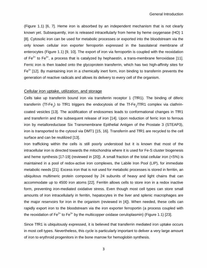
Cellular iron uptake, utilization, and storage
Cells take up transferrin bound iron via transferrin receptor 1 (TfR1). The binding of diferic
transferrin (Tf-Fe2) to TfR1 triggers the endocytosis of the Tf-Fe
2/TfR1 complex via clathrin-
coated vesicles [13]. The acidification of endosomes leads to conformational changes in TfR1
and transferrin and the subsequent release of iron [14]. Upon reduction of ferric iron to ferrous
iron by metalloreductase Six Transmembrane Epithelial Antigen of the Prostate 3 (STEAP3),
iron is transported to the cytosol via DMT1 [15, 16]. Transferrin and TfR1 are recycled to the cell
surface and can be reutilized [13].
Iron trafficking within the cells is still poorly understood but it is known that most of the
intracellular iron is directed towards the mitochondria where it is used for Fe-S cluster biogenesis
and heme synthesis [17-19] (reviewed in [20]). A small fraction of the total cellular iron (<5%) is
maintained in a pool of redox-active iron complexes, the Labile Iron Pool (LIP), for immediate
metabolic needs [21]. Excess iron that is not used for metabolic processes is stored in ferritin, an
ubiquitous multimeric protein composed by 24 subunits of heavy and light chains that can
accommodate up to 4500 iron atoms [22]. Ferritin allows cells to store iron in a redox inactive
form, preventing iron-mediated oxidative stress. Even though most cell types can store small
amounts of iron intracellularly in ferritin, hepatocytes in the liver and splenic macrophages are
the major reservoirs for iron in the organism (reviewed in [4]). When needed, these cells can
rapidly export iron to the bloodstream via the iron exporter ferroportin (a process coupled with
the reoxidation of Fe2+ to Fe3+ by the multicopper oxidase ceruloplasmin) (Figure 1.1) [23].
Since TfR1 is ubiquitously expressed, it is believed that transferrin mediated iron uptake occurs
in most cell types. Nevertheless, this cycle is particularly important to deliver a very large amount
of iron to erythroid progenitors in the bone marrow for hemoglobin synthesis.
General Introduction
4
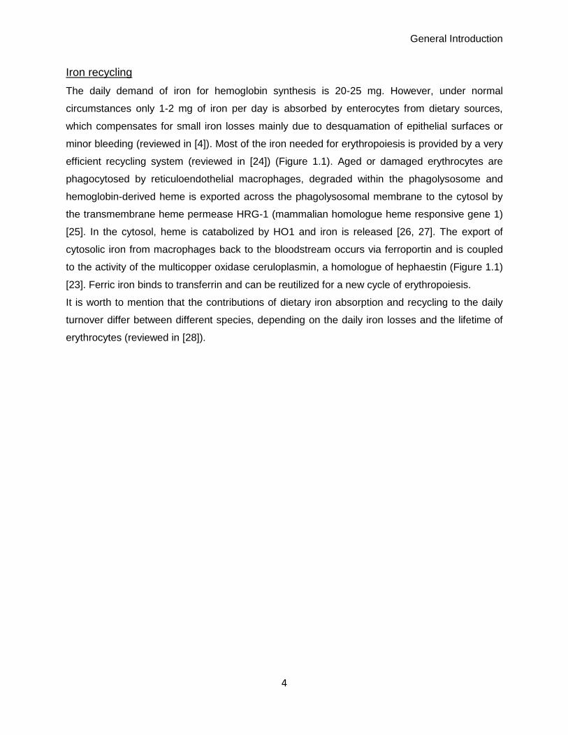
Iron recycling
The daily demand of iron for hemoglobin synthesis is 20-25 mg. However, under normal
circumstances only 1-2 mg of iron per day is absorbed by enterocytes from dietary sources,
which compensates for small iron losses mainly due to desquamation of epithelial surfaces or
minor bleeding (reviewed in [4]). Most of the iron needed for erythropoiesis is provided by a very
efficient recycling system (reviewed in [24]) (Figure 1.1). Aged or damaged erythrocytes are
phagocytosed by reticuloendothelial macrophages, degraded within the phagolysosome and
hemoglobin-derived heme is exported across the phagolysosomal membrane to the cytosol by
the transmembrane heme permease HRG-1 (mammalian homologue heme responsive gene 1)
[25]. In the cytosol, heme is catabolized by HO1 and iron is released [26, 27]. The export of
cytosolic iron from macrophages back to the bloodstream occurs via ferroportin and is coupled
to the activity of the multicopper oxidase ceruloplasmin, a homologue of hephaestin (Figure 1.1)
[23]. Ferric iron binds to transferrin and can be reutilized for a new cycle of erythropoiesis.
It is worth to mention that the contributions of dietary iron absorption and recycling to the daily
turnover differ between different species, depending on the daily iron losses and the lifetime of
erythrocytes (reviewed in [28]).

General Introduction
5
Figure 1.1 – Systemic iron homeostasis. Left panel: Dietary iron absorption occurs at the brush-border membrane
of duodenal enterocytes. Ferric iron is reduced to ferrous iron by Duodenal cytochrome B (DcytB) and is transported
across the membrane via Divalent metal transporter 1 (DMT1). Heme iron is taken up by an unknown mechanism and
iron is released from heme intracellularly by heme oxygenase 1 (HO1). The export of iron across the basolateral
membrane of enterocytes occurs via ferroportin (FPN) and is coupled with the reoxidation of ferrous to ferric iron – a
process that is catalyzed by hephaestin. In the blood, ferric iron circulates bound to transferrin (Tf-Fe2). Center lower
panel: Transferrin-bound iron can be taken up via Transferrin receptor 1 (TfR1) by every cell type in the organism,
including hepatocytes which store high amounts of iron in ferritin. Hepatocytes can also take up high amounts of non-
transferrin-bound iron (NTBI) via ZIP14. When required, iron can be exported from hepatocytes via FPN back to
circulation (a process coupled with the reoxidation of Fe2+
to Fe3+
by ceruloplasmin). Right panel: Senescent
erythrocytes are engulfed by reticuloendothelial macrophages. Iron is released from heme by HO1 and it can be either
stored in ferritin or released to circulation, depending on the body iron needs. Iron export from macrophages via FPN
is also coupled with the activity of ceruloplasmin. Center upper panel: Hepcidin produced by hepatocytes has the
ability to decrease cellular iron export by binding to FPN and promoting its internalization and degradation. Hepcidin
expression is controlled by several factors: body iron stores, inflammation, erythropoietic signals and hypoxia. Image
created based on image from Darshan et al., 2010 [29].

General Introduction
6
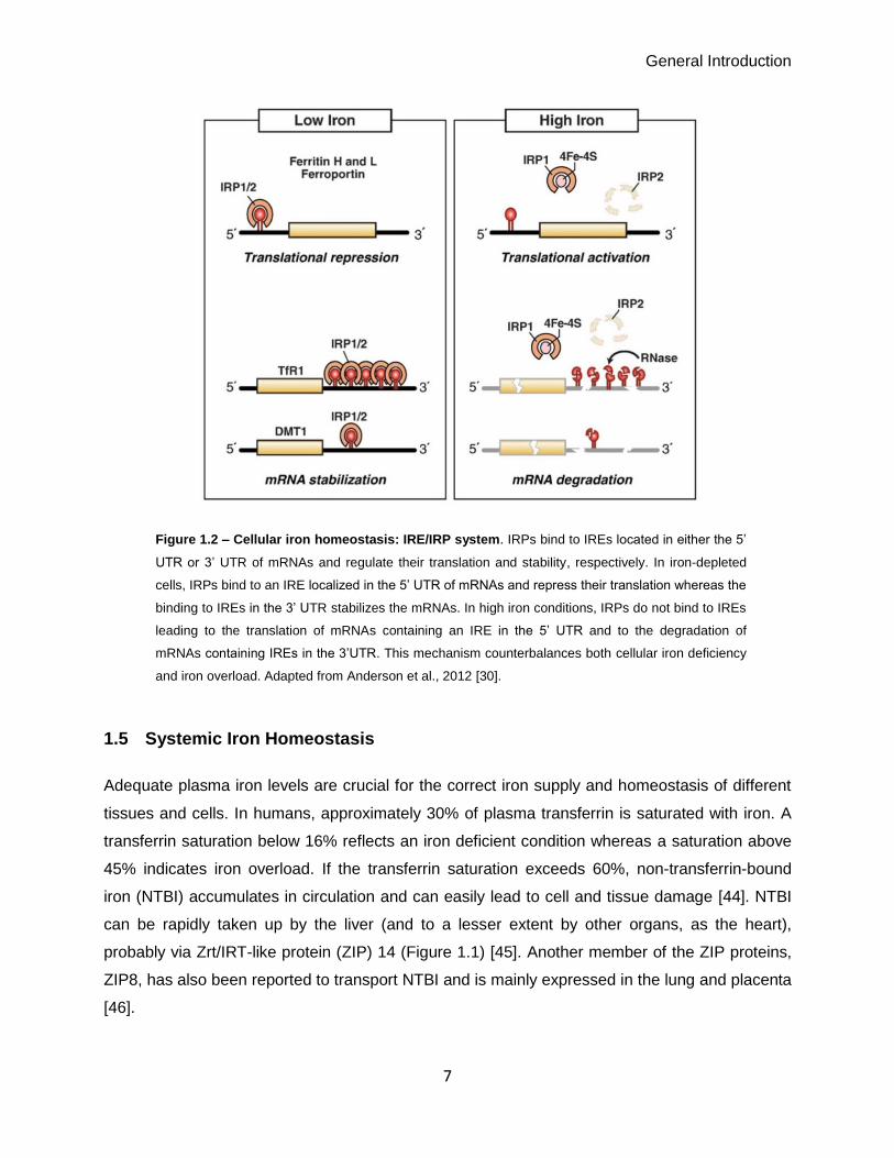
1.4 Cellular Iron Homeostasis
The coordination between cellular iron uptake, utilization, storage, and export is crucial to
maintain cellular iron homeostasis: cells must have sufficient iron to meet their metabolic needs
while avoiding toxic iron overload. The post-transcriptional regulatory system IRE/IRP plays a
critical role in this process by controlling the expression of iron-related proteins according to
intracellular levels of iron (Figure 1.2) (reviewed in [30] and [31]). It relies on the cytosolic iron
regulatory protein 1 and 2 (IRP1 and IRP2) and their interaction with conserved hairpin
structures known as iron-responsive elements (IREs) present in the 5’ or 3’ untranslated regions
(UTRs) of mRNAs of several iron-regulated genes. Transcripts containing IREs in the 3’ UTR are
stabilized after the binding of IRPs whereas the translation of transcripts containing an IRE in the
5’ UTR is blocked upon IRP binding.
In iron-depleted cells, IRPs bind to the IRE in the 5’ UTR of ferritin light chain (FtL), ferritin heavy
chain (FtH), and Fpn mRNA, inhibiting their translation [9, 10, 32, 33]. IRPs bind also to IREs in
the 3’ UTR of TfR1 mRNA and to a single IRE in the 3’ UTR of Dmt1 mRNA, blocking their
degradation [34-36]. This leads to increased iron uptake and decreased iron storage and export,
resulting in higher intracellular iron availability. In iron-replete cells, IRP1 is converted from its
RNA-binding form to the cytoplasmatic aconitase form containing a [4Fe–4S] cluster and IRP2 is
targeted to proteasomal degradation [37-39]. Consequently, IRPs cannot bind to IREs. This
leads to increased iron storage and export by increasing ferritin and Fpn mRNA translation and
decreased iron uptake due to increased degradation of TfR1 and Dmt1 mRNAs.
The vital role of the IRE/IRP regulatory system is highlighted by the observation that genetic
ablation of IRP1 and IRP2 in the mouse results in early embryonic lethality [40]. Interestingly,
animals lacking only one of the IRPs are viable but present different phenotypes [41, 42],
indicating that the two IRPs have only partially redundant functions (reviewed in [43]).
General Introduction
7
Figure 1.2 – Cellular iron homeostasis: IRE/IRP system. IRPs bind to IREs located in either the 5’
UTR or 3’ UTR of mRNAs and regulate their translation and stability, respectively. In iron-depleted
cells, IRPs bind to an IRE localized in the 5’ UTR of mRNAs and repress their translation whereas the
binding to IREs in the 3’ UTR stabilizes the mRNAs. In high iron conditions, IRPs do not bind to IREs
leading to the translation of mRNAs containing an IRE in the 5’ UTR and to the degradation of
mRNAs containing IREs in the 3’UTR. This mechanism counterbalances both cellular iron deficiency
and iron overload. Adapted from Anderson et al., 2012 [30].
1.5 Systemic Iron Homeostasis
Adequate plasma iron levels are crucial for the correct iron supply and homeostasis of different
tissues and cells. In humans, approximately 30% of plasma transferrin is saturated with iron. A
transferrin saturation below 16% reflects an iron deficient condition whereas a saturation above
45% indicates iron overload. If the transferrin saturation exceeds 60%, non-transferrin-bound
iron (NTBI) accumulates in circulation and can easily lead to cell and tissue damage [44]. NTBI
can be rapidly taken up by the liver (and to a lesser extent by other organs, as the heart),
probably via Zrt/IRT-like protein (ZIP) 14 (Figure 1.1) [45]. Another member of the ZIP proteins,
ZIP8, has also been reported to transport NTBI and is mainly expressed in the lung and placenta
[46].
General Introduction
8
The coordination of iron fluxes from tissues to circulation is essential to maintain transferrin
saturation at physiological levels and is mainly controlled by the hepcidin/ferroportin regulatory
system (Figure 1.1) (reviewed in [5]).
1.5.1 Hepcidin/Ferroportin Axis
Hepcidin is a 25 amino acid peptide hormone synthesized and secreted to the plasma by
hepatocytes [47]. It emerged as a key molecule in systemic iron homeostasis due to its ability to
regulate cellular iron efflux: hepcidin acts by post-translationally controlling the plasma
membrane concentration of ferroportin (reviewed in [5] and [28]). Ferroportin (FPN), a protein
containing 12-transmembrane domains, is the only known cellular iron exporter [9, 10, 48]. It is
highly conserved and has been identified in several vertebrates as well as in species as distant
as Caenorhabditis elegans (reviewed in [28]). Ferroportin expression is abundant in cells that
handle major iron fluxes such as duodenal enterocytes, kupffer cells and splenic macrophages,
and to a lesser extent in hepatocytes.
Hepcidin binding to ferroportin induces its ubiquitination, internalization, and subsequent
lysosomal degradation [49, 50]. This leads to a decrease in iron export from ferroportin-
expressing cells and therefore lowers systemic iron levels.
1.5.2 Regulation of hepcidin expression
The expression of hepcidin in hepatocytes rapidly responds to different stimuli, such as systemic
iron levels, inflammation, erythropoietic activity, and hypoxia (Figure 1.1). A dysregulation of
hepcidin production leads to iron-related disorders.
The production of hepcidin by other cell types has been described in the literature but at a much
lower level [51-54] and its role in systemic iron homeostasis is not clear yet.
Hepcidin regulation by systemic iron availability
Hepcidin plays a central role in the regulation of systemic iron levels. Conversely, its expression
is regulated by plasma iron levels and iron stores. Mice maintained on a high iron diet increase
hepatic hepcidin expression in order to decrease the levels of circulating iron whereas a low iron
diet induces the opposite response [55].
The bone morphogenic protein (BMP)/mothers against decapentaplegic homologue (SMAD)
pathway has been described as the key signaling system for the regulation of hepcidin. Through
a yet to be identified mechanism, BMP6 levels are increased in conditions of iron overload [56].
General Introduction
9
BMP6 binds and activates the BMP serine/threonine kinase receptors expressed at the cell
membrane of hepatocytes, which leads to the phosphorylation of the SMAD1/5/8 complex [56-
58]. This complex recruits SMAD4 and translocates to the nucleus, inducing hepcidin
transcription (reviewed in [59]). Consistently, BMP6 knock out mice show hepcidin deficiency
and iron overload [60, 61]. A recent study showed that BMP6 is mainly produced by liver
endothelial cells and that loss of BMP6 specifically in these cells recapitulates the
hemochromatosis phenotype observed in the global BMP6 knock out mice [62]. BMP2 and
BMP4 were shown to also up-regulate hepcidin expression in vitro [63]. The in vivo role of BMP2
in controlling hepcidin expression has recently been confirmed by Koch and colleagues [64].
The activation of the BMP signaling is enhanced by the BMP co-receptor hemojuvelin (HJV)
[65]. Under iron deficient conditions, the transmembrane protease serine 6 (TMPRSS6, also
known as matriptase 2) and the protease furin cleave HJV, abrogating its function as a BMP co-
receptor and negatively regulating hepcidin expression [66, 67]. Attenuation of hepcidin
activation can also be achieved through a negative feedback regulatory system mediate by
SMAD proteins, such as SMAD7 [68].
The sensing of extracellular iron occurs by the interaction of transferrin-bound iron
(holotransferrin) with a complex composed of TfR1, TfR2 (homologous to TfR1 but with lower
affinity to holotransferrin), and human hemochromatosis protein (HFE), which is also expressed
at the cell surface of hepatocytes and interacts with both TfR1 and TfR2 (reviewed in [5]). The
interaction of HFE with TfR1 and TfR2 depends on the holotransferrin concentration. Under
conditions of high iron levels, holotransferrin binds to TfR1 and HFE is released, since the
binding site of holotransferrin overlaps the HFE binding site [69, 70]. As a consequence, HFE
binds to TfR2 and this complex enhances hepcidin expression [69, 70]. The signaling pathway
downstream of the HFE/TfR2 complex is still not completely understood but it might involve a
cross-talk between BMP and mitogen-activated protein kinases (MAPK) signaling pathways [71].
Hepcidin regulation by inflammation
Iron is an essential element for pathogens. As a host defense strategy, hepcidin expression
increases during inflammation in an attempt to decrease plasma iron availability for extracellular
pathogens (reviewed in [72]). The inflammatory cytokine interleukin (IL) 6 seems to be a key
factor that activates hepcidin expression in hepatocytes by triggering the activation of a signaling
pathway mediated by Janus kinase (JAK) and signal transduction and activator of transcription
(STAT) 3 [73-75]. Upon the binding of IL6 to its receptor, JAK kinase is activated and
phosphorylates STAT3, which is then translocated to the nucleus and directly activates hepcidin
expression by binding to its promoter. Interestingly, the integrity of the BMP signaling pathway is

General Introduction
10
necessary to increase hepcidin expression during inflammation since liver specific SMAD4
knock out mice fail to increase hepcidin levels upon IL-6 injection [76]. Moreover, a mutation in
the BMP-responsive element in the hepcidin promoter impairs hepcidin expression in response
to IL-6 [77].
Hepcidin regulation by erythropoietic signals
Erythropoiesis depends on iron availability since iron is needed for heme and hemoglobin
synthesis: 20-25 mg of iron are recycled every day for the synthesis of new red blood cells. In
conditions of high erythropoietic activity, hepcidin expression is downregulated in order to
increase iron availability. In 2014, erythroferrone (ERFE), a hormone produced by erythroblasts,
was identified as the erythroid factor responsible for hepcidin repression [78]. ERFE-deficient
mice do not suppress hepcidin expression after hemorrhage. However, the ERFE receptor and
ERFE signaling pathway are still not known [78]. Two other factors, growth differentiation factor
15 (GDF-15) and twisted gastrulation homolog 1 (TWSG1), might also be involved in reducing
hepcidin expression in conditions of high erythropoietic demand [79, 80].
Hepcidin regulation by hypoxia
Hepcidin expression is inhibited by hypoxia and different mechanisms have been proposed to
explain this observation.
The kidney senses systemic hypoxia and responds by producing an erythroid growth factor
known as erythropoietin (EPO) [81]. The secretion of EPO stimulates erythropoiesis in the bone
marrow in order to restore a proper delivery of oxygen to every cell type in the organism. EPO is
likely to suppress hepcidin expression indirectly by stimulating erythropoiesis and therefore
increasing ERFE [82] (section: “Hepcidin regulation by erythropoietic signals”).
The heterodimeric hypoxia-inducible factors (HIF-1/HIF- and HIF-2/HIF-) are known to
regulate the transcription of several iron-related genes, such as Fpn and Dmt1 in enterocytes
[83, 84], and it was reported that HIFs might directly repress hepcidin promoter activity under
hypoxic conditions [85]. Under normoxia, the oxygen- and iron-dependent prolyl hydroxylases
(PHDs) hydroxylate the HIF-1α and HIF-2α subunits, which then associate with the von Hippel-
Lindau tumor suppressor (pVHL). This complex is polyubiquitinated by an E3 ligase and HIFs
subunits are rapidly degraded. Under hypoxic conditions, the activity of PHDs is reduced due to
the lack of oxygen and HIF-1α and HIF-2α subunits are no longer hydroxylated and targeted for
proteasomal degradation (reviewed by [86]). As a consequence, HIF-1α and HIF-2α are stable
and recruit HIF-β (which is constitutively expressed), forming heterodimers that can directly
regulate gene expression.

General Introduction
11
However, later studies suggested that hepcidin regulation during hypoxia does not occur via
direct effects of the pVHL/PHD/HIF axis on the hepcidin promoter but probably through EPO-
mediated erythropoiesis [87, 88].
Nevertheless, HIF-1α may indirectly regulate hepcidin expression. A functional hypoxia-
responsive element was identified in the TMPRSS6 promoter region and mutations in this
element impair HIF1α-dependent induction of TMPRSS6 expression [89]. Similarly, hypoxia also
increases the expression of furin via HIF-1α [66, 90]. These observations suggest that increased
expression of TMPRSS6 and furin under hypoxic conditions may be an additional mechanism to
repress hepcidin expression.
1.5.3 Regulation of ferroportin expression
As previously mentioned, ferroportin (also known as SLC40A1, Ireg1 and MTP1) is the only
known cellular iron exporter [9, 10]. The systemic inactivation of the Fpn gene in mice results in
embryonic lethality [48]. Interestingly, mice in which the Fpn gene was inactivated in all tissues
except placenta and the extraembryonic visceral endoderm were viable, likely reflecting a
correct iron transfer from the mother to the embryo [48]. Nevertheless, these animals
accumulate iron in enterocytes, splenic and liver macrophages, and hepatocytes shortly after
birth – highlighting the major role of FPN in iron export in these cell types [48].
Contrary to hepcidin, FPN expression is regulated at several levels and not only at the
transcription level.
Transcriptional regulation of ferroportin
Heme, a product of hemoglobin degradation, increases Fpn transcription in macrophages. The
presence of heme induces the degradation of the transcriptional repressor Btb And Cnc
Homology 1 (Bach1) and allows the transcriptional activator Nuclear Factor Erythroid 2-like
(Nrf2) to bind to Small Maf proteins (sMAFs) and enhance Fpn transcription [91]. Additionally,
Fpn transcription is also increased by iron in macrophages [92] but the molecular mechanism
behind this observation is not known yet.
It has been reported that Fpn transcription is decreased under inflammatory conditions. Upon
lipopolysaccharide (LPS) stimulation, Fpn mRNA levels decrease in macrophages from the
spleen and the liver, probably in an attempt to reduce iron availability for extracellular pathogens
[93-95]. The molecular mechanism behind Fpn transcriptional repression during inflammation is
still not completely understood. The stimulation of toll-like receptors (TLR) 2/6 dependent
signaling pathways by the Mycoplasma–derived molecule FSL1 was shown to repress Fpn

General Introduction
12
transcription [96]. Upon Pseudomonas aeruginosa infection or LPS stimulation, Fpn mRNA
levels were shown to be reduced in a TLR 4 dependent manner [93, 97]. The direct effect of
increased inflammatory cytokines such as TNF still remains controversial [93, 98].
Hypoxia and iron deficiency were shown to induce Fpn transcription in duodenal enterocytes. In
these conditions, HIF2-is stabilized, forms a heterodimer with HIF-β, and binds directly to HIF
response elements present in the promoter of Fpn [83, 84]. Upon binding, the HIF2 heterodimer
stimulates Fpn expression and therefore increases the export of iron from enterocytes to the
bloodstream; this ensures an increase in dietary iron absorption in order to counteract the iron
deficiency or the hypoxia (by increasing the levels of iron available for erythropoiesis).
Post-transcriptional regulation of ferroportin
At the post-transcriptional level, FPN is regulated by the IRE/IRP system. As described in
section 1.4, when intracellular levels of iron are low, IRP1 or 2 bind to the IRE present in the 5’
UTR of Fpn mRNA, blocking its translation and therefore decreasing cellular iron export [9, 10].
When intracellular iron levels are high, Fpn mRNA is translated and iron export increases. This
mechanism prevents both toxic intracellular iron overload as well as detrimental intracellular iron
depletion (reviewed in [31]).
Interestingly, duodenal enterocytes and erythroid precursors additionally use an alternative
upstream promoter to express a Fpn transcript with an identical open reading frame but that
lacks the IRE in the 5’ UTR [99]. These cells can evade translational repression of FPN in
conditions of low iron levels. As a result, enterocytes can export iron into the plasma under iron
deficient conditions in order to restore systemic iron levels. This alternative transcript accounts
for most of the FPN expression in erythroid precursors and allows these cells to export iron in
conditions of iron deficiency, in order to increase its availability to other tissues in need.
Post-translational regulation of ferroportin
As described in section 1.5.1, FPN is regulated post-translationally by hepcidin [49]. The binding
of hepcidin to FPN, and the subsequent internalization and degradation of the latter, is
dependent on the fourth extracellular loop of FPN, which contains the amino acid cysteine (C) at
position 326. A point mutation in this amino acid (Cysteine Serine) was identified in a family
presenting an autosomal dominant form of hereditary hemochromatosis, with elevated
transferrin saturation and iron deposition in hepatocytes, but not in kupffer cells [100]. Additional
studies, both in vitro and in vivo, showed that this point mutation (C326S) prevents the binding of

General Introduction
13
hepcidin to FPN [101, 102], revealing the critical role of the thiol form of C326 for hepcidin
binding.
The exact mechanism underling FPN internalization and degradation has been investigated
thoroughly in the past years and it is believed that ubiquitination is the key signal for hepcidin-
induced ferroportin endocytosis [50].
1.6 Iron related disorders
The study of the molecular mechanisms underlying iron-related disorders in humans has been
very useful in the understanding of mechanisms regulating systemic and cellular iron
homeostasis. Most of the iron-related diseases, both from iron overload and from iron deficiency,
result from disruptions in the hepcidin/FPN regulatory system.
1.6.1 Iron overload diseases
Hereditary hemochromatosis (HH) is an iron overload disease characterized by excessive
dietary iron absorption that results in elevated plasma iron levels, high transferrin saturation, and
formation of NTBI. The excess of iron accumulates in several organs, e.g. the liver, leading to
increased oxidative stress, cell damage, and tissue injury. Different forms of HH exist that
present with different disease severities and result from different mutations. HH type 1, 2A and 3
are characterized by mutations in the iron-sensing machinery (HFE, HJV or TfR2, respectively)
that result in inappropriate low or absent levels of hepcidin [103-106]. HH type 2B results from a
mutation in the hepcidin gene itself [107]. Deficient hepcidin production observed in these
patients leads to high levels of FPN at the cell membrane, causing an increase in dietary iron
absorption by duodenal enterocytes and increased iron release from recycling macrophages.
These four subtypes of HH are recessive forms of the disease. HH type 4 is an autosomal
dominant form of hemochromatosis that results from “gain of function” mutations in FPN that
confer resistance to hepcidin binding [100, 108]. As a consequence, ferroportin remains stable
at the cell membrane independently of hepcidin levels, leading to elevated iron export into the
circulation. Hepatic cells become iron overloaded whereas splenic macrophages and kupffer
cells are iron depleted. This rare disorder is also known as non-classical ferroportin disease and
is the only HH subtype that is associated with high hepcidin levels, since the iron sensing
machinery still responds to increased systemic iron levels.
Loss of function mutations in FPN that lead to incorrect targeting of the protein to the plasma
membrane or the production of an inactive iron exporter result in a disorder known as classical

General Introduction
14
ferroportin disease (reviewed in [109]). Due to the inability to export iron, specialized iron-
exporting cells such as Kupffer cells and splenic macrophages are iron overloaded. As a
consequence of iron restriction in these cells, patients present normal to low plasma transferrin
saturation.
Other iron overload disorders, known as iron-loading anemias, can develop as a consequence
of an impaired erythropoiesis system. For example, in -thalassemia, the defective production of
the -globin chain in erythroid precursors leads to the precipitation of excess -chains and
results in the apoptosis of these cells. The lack of mature red blood cells and diminished oxygen
transport capacity stimulate EPO production, which in turn stimulates erythropoiesis but fails to
correct anemia because the precursors undergo apoptosis. High erythropoietic activity
suppresses hepcidin expression, leading to hyperabsorption of dietary iron and, consequently, to
systemic iron overload [110, 111].
1.6.2 Iron deficiency diseases
Insufficient dietary iron intake or blood losses can cause iron deficiency that commonly
manifests as anemia; since erythropoiesis requires large amounts of iron, a depletion of body
iron levels impairs this process and leads to low hemoglobin levels.
However, several other iron deficiency disorders result from elevated hepcidin expression. By
limiting iron availability for erythropoiesis, these diseases are also often associated with anemia.
Mutations in TMPRSS6 that inactivate its function as a negative regulator of hepcidin production
result in an autosomal recessive disorder known as familial iron-refractory iron deficiency
anemia (IRIDA) [112]. Although patients present with severe iron deficiency that should
suppress hepcidin expression, serum hepcidin levels are abnormally increased, causing
uncontrolled FPN degradation and preventing iron from being absorbed from diet and released
from stores.
Very high levels of hepcidin are also responsible for another iron-deficiency disorder named
anemia of inflammation or anemia of chronic diseases [113, 114]. The underlying cause of
elevated hepcidin expression in these diseases is attributed to an excess of inflammatory
cytokines, such as IL6 (reviewed in [115]). Despite the hypoferremia observed in these patients,
overproduction of hepcidin inhibits intestinal iron absorption and macrophage iron release.

General Introduction
15
2. Mammalian Respiratory System
The oxidative metabolism of cells requires high amounts of oxygen (O2). Oxygen is transported
in the bloodstream bound to hemoglobin in erythrocytes and is delivered to every cell type in the
body. During aerobic cellular respiration, oxygen is consumed and carbon dioxide (CO2) is
produced and released into the circulation.
In order to fulfill the cellular oxygen requirements and to excrete the waste CO2, the respiratory
system has evolved to allow an efficient gas exchange between the atmosphere and the
bloodstream. Inhaled air travels through a highly branched conducting tract (nose, pharynx,
larynx, trachea, bronchi, bronchioles) until it reaches the distal lung where most of the gas
exchange occurs (reviewed in [116] and [117]). In this region, the surface for gas exchange is
maximized by the septation into innumerous microscopic thin-walled air sacs called alveoli [118]
(reviewed in [119]). These structures are in close contact to a dense capillary network.
The alveolar epithelium is composed by two cell types – alveolar epithelial cells type 1 and type
2 (also known as pneumocytes type 1 and 2) – whereas the conducting airways are lined by an
epithelium composed of a variety of multi-ciliated and non-ciliated epithelial cells (Figure 1.3)
(reviewed in [120]). In healthy conditions, alveolar macrophages are the most common cell type
present within the alveolar space.
Millions of liters of air inhaled every day do not only provide the necessary oxygen to support life
but also expose the respiratory system to pathogens and toxic particles. For this reason, multiple
physical and chemical innate host-defense mechanisms exist within the respiratory tract. Tight
adhesions between epithelial cells and the presence of mucus and antimicrobial molecules that
enhance mucociliary clearance are among the most important features of the pulmonary innate
immunity (reviewed in [121] and [122]). In addition, pulmonary epithelial cells and alveolar
macrophages can recognize the presence of pathogens and trigger an inflammatory response, if
necessary. These cells produce cytokines and chemokines and recruit and activate other cells of
the immune system (reviewed in [121] and [123]).

General Introduction
16
Figure 1.3 – Structure of the mouse airway epithelium. The larger airways (trachea and bronchi) are lining by a
pseudostratified columnar epithelium mainly composed of ciliated airway epithelial cells, basal cells, few goblet cells
and Club cells. As the conducting airway branches into smaller airways (bronchioles and bronchio-alveolar duct), the
epithelium becomes a simple epithelium mainly composed of Club cells, with interspersed ciliated cells.
Neuroendocrine cells are present in low numbers in the airway epithelium and are commonly found in small clusters
called neuroepithelial bodies. Alveoli are lined by an uninterrupted epithelium consisting of alveolar epithelial type 1
and type 2 cells, surrounded by a dense capillary network. Alveolar macrophages are the primary immune cells
present in the airway lumen of a healthy lung. Image created based on image from Wong et al., 2009 [124].
2.1 Pulmonary vasculature
After oxygenation of cells throughout the body, deoxygenated blood is pumped from the right
ventricle of the heart to the lungs via the main pulmonary artery. This artery divides into the right
and the left main branches, which transport blood to the four right lobes and the left lobe of the
mouse lung (reviewed in [125]). Within each lobe, the arteries rapidly subdivide into branches
that follow the bronchial tree: arteries break into small arteries, followed by arterioles and then
innumerous capillaries that completely envelop the alveoli (reviewed in [125]).
Large pulmonary arteries are classified as elastic since their wall is mainly composed by elastic
fibers and some smooth muscle cells. As they divide and their vascular diameter decreases, the
composition of their wall changes leading to an increase in the smooth muscle cell layer and a

General Introduction
17
decrease in the elastic fibers – these arteries are classified as muscular arteries [126]. The wall
thickness of the vessels gradually decreases when they further subdivide: small arterioles are
surrounded by a non-uniform layer of smooth muscle cells and are followed by smaller non-
muscular arterioles located proximal to the terminal bronchi [127]. At the alveoli region, these
arterioles give rise to capillaries whose walls consist of a single layer of endothelial cells [128].
The thin pulmonary endothelium is ideal for gas exchange to occur: carbon dioxide is released
and oxygen is taken up. Oxygenated blood travels through the capillaries into venules, which are
in gross structure similar to arterioles (reviewed in [129]). Venules are distributed irregularly
within the lung and do not follow the bronchial tree. These vessels come together to form the
pulmonary veins and transport oxygenated blood to the left atrium of the heart. Oxygen can then
be delivered to cells throughout the body through the systemic circulation.
2.2 Trachea, bronchi and bronchioles
The trachea, bronchi and bronchioles form a tree-like system of branched tubes that carries air
to and from the alveoli. In the mouse lung, the trachea and main bronchi are lined by a
pseudostratified columnar epithelium whereas the smaller airways are lined by a simple
columnar epithelium (Figure 1.3) (reviewed in [120]). The epithelium is covered by the airway
lining fluid, which protects the underlying cells against desiccation and damage, and plays an
important role in the delivery of almost completely sterile gases to the alveoli.
Studies throughout the years have revealed a big diversity of cells that form the airway
epithelium. These cells can be broadly divided into the following groups:
Ciliated airway epithelial cells: Ciliated epithelial cells are considered terminally differentiated
cells [130]. They are present in large numbers in the mouse upper airways and each cell
possesses up to 300 cilia on its apical surface. The coordinated movement of cilia is essential to
move the mucus along the respiratory tract (see Goblet cells section). Cilia dysfunction impairs
this process and leads to recurrent respiratory infections [131].
Goblet cells: Goblet cells are non-ciliated secretory cells that produce and secret mucin, i.e.
highly glycosylated proteins that, together with water, form the mucus lining the airway
epithelium [132] (reviewed in [133]). Inhaled materials/pathogens get trapped in this mucus and
are subsequently removed from the respiratory system through the highly synchronized beat of
cilia from ciliated airway epithelial cells, in a process called mucociliary clearance (reviewed in

General Introduction
18
[134]). The importance of the mucus in the healthy lung is highlighted by the observation that
mice lacking the constitutively produced mucin MUC5b present chronic inflammation [135].
Basal cells: In the mouse lung, basal cells are mainly localized between ciliated and secretory
cells in the larger airways. The abundant cytoskeleton and the presence of desmosomes and
hemidesmosomes highlight the role of basal cells as major structural components of the
epithelium, attaching it to the underlying basement membrane [136, 137]. Moreover, basal cells
have also an important role in the homeostasis of the epithelium since they have the ability to
self-renew and give rise to secretory and ciliated airway epithelial cells [138, 139].
Club cells: Club cells are the main cell type present in mouse small airways. These non-ciliated
secretory cells produce and secrete several components of the airway lining fluid, such as the
club cell secretory protein (CCSP) (reviewed in [140]). The study of CCSP-/- mice revealed the
important role of club cells and their secretions in lung homeostasis: CCSP deficiency is
associated with atypical airway lining fluid composition, increased susceptibility to lung injury and
oxidative stress, as well as altered lung inflammatory responses [141-144] (reviewed in [124]).
Despite being differentiated cells, club cells have the potential to reenter the cell cycle and
contribute to the renewal of the bronchiolar airway epithelium [145, 146].
Pulmonary neuroendocrine cells: Pulmonary neuroendocrine cells (PNEC) are localized within
the epithelium lining the trachea, bronchi, and bronchioles, and they can be grouped in small
clusters called neuroepithelial bodies (reviewed in [147]). Even though these are the first
specialized cells to differentiate in the airway epithelium during development, they are present in
low numbers in the adult lung. During lung development, PNECs act as modulators of lung
growth and differentiation (reviewed in [147]); their function in the adult lung is not well known
yet but they might be involved in airway oxygen sensing [148]. Also, a recent study shows that
PNECs control lung immune responses by producing neuropeptides [149].
2.3 Alveoli structure
Alveoli are lined by an uninterrupted epithelium consisting of alveolar epithelial type 1 and type 2
cells (AT1 and AT2), which are in close contact with underlying capillaries and fibroblasts (Figure
1.3) [118] (reviewed in [150]). The alveolar surface is covered by the alveolar lining fluid, which
protects the underlying epithelial cells against desiccation and pathogens, and facilitates the
diffusion of gases.

General Introduction
19
AT1 cells are flat and cover >90% of the surface of alveoli [151]. Their high membrane to
cytoplasm ratio and close apposition to alveolar capillaries create a very thin air-blood barrier,
essential for a proper gas exchange. These cells are generally regarded as non-proliferative,
fully differentiated cells [152].
AT2 cells are cuboidal-shaped and are often localized in the “corners” of alveoli. Contrary to
AT1, AT2 cells have the ability to self-renew and differentiate into AT1 cells [153]. AT2 cells
synthetize, secrete, and recycle the pulmonary surfactant – a mix of extracellular proteins and
lipids present within the alveolar lining fluid that decreases alveolar surface tension and
contributes to host defense [154-156]. These cells contain large and abundant secretory
vesicles called lamellar bodies, which reflect their important secretory function [157].
The lung volume does not remain constant during respiration: it increases during inspiration and
decreases during expiration. By reducing the surface tension at the gaseous-aqueous
interphase of the alveoli, the surfactant plays an essential role in preventing the collapse of
alveoli at the end of expiration and reducing the effort needed to expand the lungs during
inspiration (reviewed in [158]). This way, the surfactant increases lung compliance, i.e. the
change in lung volume per unit of pressure change, meaning the ease with which the lungs can
be extended. Inversely, the surfactant decreases lung elastance which is a measure of lung
stiffness and is defined as the change in the pressure per unit of volume change.
Even though the exact composition of the surfactant is not completely known yet, it is believed to
be composed of four surfactant proteins (SP) that account for 10% of the surfactant and by
several different lipids that comprise 90% of the surfactant layer (reviewed in [158]). The
hydrophobic SP-B and SP-C have a major role in surfactant structure and function while the
other two surfactant proteins, SP-A and SP-D, are hydrophilic proteins and contribute to lung
immunity [159] (reviewed in [156]). They interact with various microorganisms and pathogen-
derived components and act as opsonins by binding and agglutinating pathogens, thus
enhancing phagocytosis. Moreover, these proteins also directly modulate the activity of alveolar
macrophages and other immune cells [160, 161] (reviewed in [156]). Consistently, mice deficient
in SP-D or SP-A are more susceptible to lung inflammation and infection than wild-type mice
[162-164].
AT2 cells play an important role in host defense not only by producing surfactant proteins but
also by producing cytokines and expressing ligands that regulate the activity of immune cells
[165-167] (reviewed in [168]).
Finally, both AT1 and AT2 cells express ion channels and might be involved in alveolar fluid
homeostasis [169, 170].

General Introduction
20
The thin air-blood barrier is exposed to two main physical stresses: the pressure within the
capillaries and the increased longitudinal tension of the alveolar wall when the lung is inflated to
high volumes (reviewed in [171]). Interstitial fibroblasts play an essential role in supporting the
alveolar structure by secreting extracellular matrix (ECM). In a healthy lung, fibroblasts
synthetize small amounts of ECM, enough to preserve the integrity of the alveolar barrier while
maintaining it as thin as possible. Different pathological conditions that lead to the recruitment
and activation of fibroblasts, differentiation into myofibroblasts, and increased production of ECM
contribute for increased lung stiffness and pathology of fibrotic lung diseases [172-174].
2.4 Alveolar Macrophages
Alveolar macrophages (AMs) are the primary immune cells present in the airway lumen of a
healthy lung. Due to the techniques used to investigate the biology of these cells, most studies
do not distinguish between macrophages present within the alveolar space and those present
within larger airspaces. For this reason, in this thesis, the term “alveolar macrophage” is used to
describe a mixed population of macrophages that reside in the lumen of the airways.
Being the first immune cells to contact with inhaled pathogens, AMs are extremely important in
lung immunological responses. However, they also play an equally important role in clearing
apoptotic cells, excessive surfactant and cellular debris in health and disease situations
(reviewed in [123]).
The unique environment in which AMs exist has a strong influence on their phenotype and
function, which is illustrated by the observation that peritoneal macrophages acquire expression
markers of AMs when transferred into the lung [175]. In mice, AMs are phenotypically very
different from other macrophage populations (reviewed in [123]). They express high levels of
cluster of differentiation (CD)-11c (also known as integrin x), a molecule that is not typically
expressed in macrophages but is a typical marker of dendritic cells [175-177]. Similarly, AMs
express CD205 (also known as DEC-205), which is also expressed by dendritic cells and has
not been reported to be expressed by macrophages [175, 178]. In contrast, AMs express low
levels of CD11b, which is typically expressed by other macrophage populations [175, 177].
Moreover, mouse AMs express intermediate/low levels of CD14 [176] and high levels of sialic
acid-binding immunoglobulin-like lectin F (SiglecF), which is mainly expressed by eosinophils
[176, 179].

General Introduction
21
Origin and differentiation of alveolar macrophages
The initial colonization of the airways with AMs is dependent on embryonically derived fetal
monocytes, which fully differentiate into AMs in the first days after birth [180] (reviewed in [181]).
Throughout life, AMs have the ability to self-renew and maintain the population with minor
contribution from circulating monocytes [182]. Even though conflicting studies exist, it is believed
that circulating monocytes only contribute to AM repopulation when these cells have a limited
capacity for proliferation, as in the case of radiation-induced depletion of AMs [182].
Both differentiation and survival of AMs is highly dependent on the granulocyte-macrophage
colony-stimulating factor (GM-CSF, also known as CSF2) and possibly also on surfactant
proteins [175, 180, 182]. The essential role of GM-CSF is highlighted by the observation that
GM-CSF-/- mice fail to generate mature and fully functional AMs [180].
GM-CSF has long been described as a key factor in the proliferation and maturation of myeloid
lineages (reviewed in [183]) but it has also been reported to act in other biological contexts such
as proliferation of endothelial progenitor cells and during embryo implantation and development
[184] (reviewed in [185]). The effects of GM-CSF are mediated through a heterodimeric receptor
composed of a ligand-specific -subunit (GM-R) and a common -subunit that is also a
component of IL-3 and IL-5 receptors [186-188]. The GM-Rsubunit binds to GM-CSF with low
affinity as a monomer but with high affinity when associated with the subunit [187]. Even
though the GM-R subunit is incapable of binding to GM-CSF as a monomer, it plays an
important role in GM-CSF-induced signal transduction. The high-affinity complex (GM-R/GM-
R) transduces signals through JAK-STAT-, MAPK-, and phosphatidylinositol 3-Kinase (PI3K)-
dependent pathways to alter the expression of target genes [184, 189-191].
Regulatory effects of the lung microenvironment
Due to their unique position in the airspace, AMs are exposed to air pollutants, pathogens, and
dust. AMs have to accurately discriminate between situations that require an inflammatory
response (pathogens) and those that require a tolerogenic response (lung microbiome [192] and
innocuous stimuli). For this reason, and in order to prevent unwanted inflammatory responses,
AM activation is negatively regulated via cell-cell interactions with airway epithelial cells
(reviewed in [123]). For example, the airway epithelium expresses CD200 which interacts with its
receptor expressed in AMs and inhibits their activation [166]. Additionally, the non-inflammatory
status of AMs is also maintained by the presence of secreted factors such as surfactant proteins
(SP-A and SP-D), transforming growth factor TGF- and IL-10 produced by the lung
epithelium [161, 193, 194] (reviewed in [123]). Hence, a combination of events that overcome

General Introduction
22
the inhibitory stimuli must occur for an inflammatory response to initiate. The presence of
pathogens in the airways activates TLR-dependent signaling pathways in AMs, releasing them
from the suppression mediated by epithelial cells and triggering their activation [193].
Furthermore, the destruction of the airway epithelium during inflammation leads to the loss of
negative regulators, such as CD200, enhancing the inflammatory status.
Once activated, AMs have a high phagocytic capacity and produce high levels of inflammatory
cytokines and chemotactic factors, which leads to the recruitment of other immune cells, such as
neutrophils, to the bronchoalveolar space [195-197] (reviewed in [198]).

General Introduction
23
3. Lung iron homeostasis
Like every other cell type in the body, lung cells take up iron from the bloodstream and use it to
fulfill their metabolic needs. However, they must also prevent toxic iron overload. In the particular
case of the lung, the risk of oxidative stress is very high since this organ is exposed to the
atmosphere and therefore to high levels of oxygen. Moreover, lung cells are not only exposed to
iron circulating in the blood but also to iron present in inhaled particles - the atmosphere is a
vehicle for the movement and redistribution of metals, such as iron (reviewed in [199]). Human
activities have contributed to increased levels of atmospheric iron via the production of air
pollution particles [200]. Iron is also present in cigarette smoke (a factor with a strong causative
link to pulmonary pathology) and accumulates in the lung: pulmonary iron levels are higher in
cigarette smokers when compared with nonsmokers [201].
Even though high levels of antioxidants, such as glutathione and ascorbic acid, are present in
the respiratory tract lining fluid, an imbalance in iron levels in the lung can lead to oxidative
stress [202] (reviewed in [203]).
3.1 Iron metabolism in the lung
Contrary to duodenal enterocytes, the lung did not evolve to absorb iron to meet the body
nutritional requirements. Instead, by being exposed to iron from blood and from inhaled
particles, the lung has an essential role in iron detoxification (review in [204]). In a healthy lung,
iron homeostasis is tightly controlled and the availability of free iron is limited. It has been
suggested that lung cells sequester iron intracellularly bound to ferritin and therefore in a less
reactive form (review in [204]). In fact, pulmonary ferritin levels increase upon iron exposure
[204, 205].
Several studies showed that lung cells express the same iron-related proteins that are
expressed in enterocytes and hepatocytes (reviewed in [204]) (Figure 1.4). However, how iron
homeostasis is maintained in the lung is not completely understood and conflicting data exist
between different studies.
Iron uptake by lung epithelial cells: Similar to duodenal enterocytes, lung epithelial cells express
DMT1 in their apical membrane and take up non-transferrin bound iron from the airway lumen
(Figure 1.4) [206, 207]. However, it is still not clear how the expression of DMT1 responds to
increased iron concentrations. Wang et al. observed that the mRNA and protein levels of the
DMT1 isoform without IRE increase in the airway epithelium in vitro and in vivo upon exposure to

General Introduction
24
iron whereas Giorgi et al. observed that the protein levels of DMT1 in the lung are unchanged in
mice injected intraperitoneally with iron-saccharate [206, 208]. The uptake of iron via DMT1
needs to be coupled with the reduction of ferric iron to ferrous iron (described in section 1.3.).
Turi et al. reported that DcytB is expressed in airway epithelial cells (similar to enterocytes) and
probably reduces iron before its uptake [209].
Giorgi et al. showed that ZIP14 is expressed in airway epithelial cells and that its expression
increases in conditions of iron overload [208]. Wang et al. reported that Zip14 levels in the lung
are very low but demonstrated that Zip8 is highly expressed in the lung [46]. For this reason,
ZIP8 is another candidate for the uptake of non-transferrin bound iron by lung cells.
Iron export by lung epithelial cells: In the model which proposes that iron is detoxified in the lung,
airway epithelial cells not only take up and store iron, but are also able to export it in a less
reactive form via ferroportin or bound to ferritin or transferrin (reviewed in [204]). The authors
suggested that iron could then be transported out of the lung via the mucociliary pathway or via
blood to the reticuloendothelial system for long term storage. However, conflicting data exist
regarding this model. Yang et al. reported that FPN is expressed at the apical membrane of
airway epithelial cells while Giorgi et al. showed that FPN is expressed in the cytoplasm of these
cells [208, 210]. On the other hand, the idea that FPN expression increases in lung cells upon
exposure to iron seems to be consistent in different studies [208, 211]. Taken together, more
studies are needed to fully understand if lung epithelial cells are able to export iron or not.
Alveolar macrophages: AMs play a key role in lung iron homeostasis. These cells have the
capacity to sequester large quantities of iron intracellularly bound to ferritin and therefore in a
less reactive form. AMs from smokers show very high levels of iron and it was suggested that
this storage prevents oxidative stress by decreasing the generation of extracellular hydroxyl
radicals [212]. Moreover, AMs from mice injected intraperitoneally with iron-saccharate (which
present increased pulmonary iron content) show increased iron levels, suggesting that AMs take
up the excess of iron in conditions of iron overload [208].
It was reported that AMs may take up both transferrin and non-transferrin bound iron as they
express TfR1 and DMT1 [207, 213, 214]. Via phagocytosis, alveolar macrophages can also
retain iron derived from senescent cells and from inorganic iron-rich dust (reviewed in [215]).
Whether AMs are able to export iron is currently not clear. It has been suggested that they might
export it via ferritin or FPN [204], but there is a lack of strong evidence to support this idea.
Further studies are necessary to confirm or exclude this hypothesis.

General Introduction
25
Hepcidin expression in the lung: Giorgi et al. showed that unlike hepatic hepcidin expression,
total lung hepcidin levels do not increase in response to iron [208]. Moreover, they suggested
that hepcidin expression in the lung does not alter iron homeostasis in this organ [208]. Frazier
et al. studied the expression of hepcidin in human airway epithelial cells in vitro and have come
to the same conclusion [216]. Contrasting these findings, Chen et al. showed that the silencing
of hepcidin expression in airway epithelial cells leads to a reduced degradation of FPN in the
lung in a mouse model of sepsis-induced acute lung injury [217]. They also observed a decrease
in intracellular iron content of AMs, probably as a consequence of increase FPN protein levels
[217]. Nguyen et al. reported that AMs also produce hepcidin and that its expression increases
upon treatment with LPS [214]. However, its role in lung iron homeostasis was not elucidated in
this study.
3.2 Lung diseases associated with iron overload
A dysregulation of lung iron homeostasis caused by either endogenous or exogenous factors is
likely to increase the availability of free iron. Increased iron levels in the lung caused by the
injection of intravenous iron result in increased oxidative stress [202], which can lead to tissue
injury and contribute to the pathogenesis of several diseases. In fact, increasing evidence shows
that several chronic and acute lung diseases are associated with a dysregulation of pulmonary
iron homeostasis (reviewed in [204]). Patients with idiopathic pulmonary alveolar proteinosis,
acute respiratory distress syndrome, cystic fibrosis, and chronic obstructive pulmonary disease
(COPD) show higher levels of iron and iron-related proteins (e.g. ferritin and TfR1) in the lung
when compared to healthy individuals [218-221]. In most of these studies, iron was detected
both extracellularly in the bronchoalveolar space and intracellularly. AMs were described as the
predominant iron loaded cell type and the percentage of iron loaded AMs in patients with COPD
was correlated with increased disease severity [218]. However, these studies mostly remain on
the observational level. Whether increased pulmonary iron levels contribute to the development
of these lung diseases is still poorly understood.
General Introduction
26
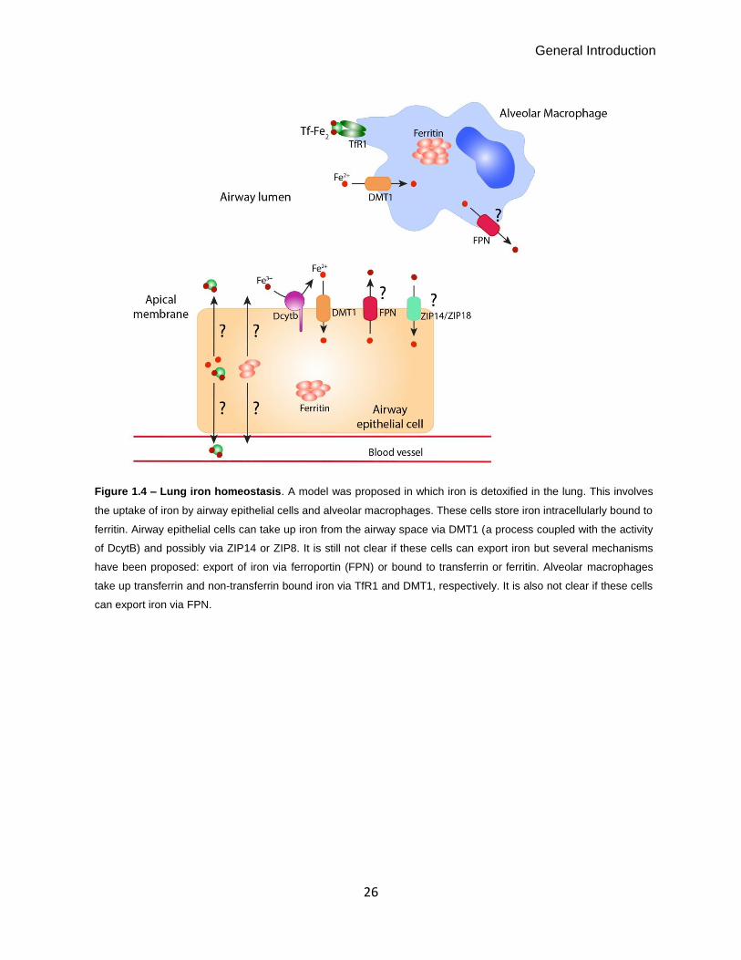
Figure 1.4 – Lung iron homeostasis. A model was proposed in which iron is detoxified in the lung. This involves
the uptake of iron by airway epithelial cells and alveolar macrophages. These cells store iron intracellularly bound to
ferritin. Airway epithelial cells can take up iron from the airway space via DMT1 (a process coupled with the activity
of DcytB) and possibly via ZIP14 or ZIP8. It is still not clear if these cells can export iron but several mechanisms
have been proposed: export of iron via ferroportin (FPN) or bound to transferrin or ferritin. Alveolar macrophages
take up transferrin and non-transferrin bound iron via TfR1 and DMT1, respectively. It is also not clear if these cells
can export iron via FPN.

General Introduction
27
4. Research Aims
The pathogenesis of lung diseases associated with iron overload is poorly understood. It is not
clear whether the observed increase in lung iron levels contributes to disease onset and
progression or if it represents a secondary effect resulting from lung injury.
Some studies have addressed the impact of increased lung iron content by the inhalation of
particles containing iron [222-224]. However, limited data exist regarding the impact of increased
circulating iron levels on pulmonary iron content and function.
The major aim of this work was to understand whether a disruption of the hepcidin/FPN
regulatory system and the subsequent increase in systemic iron levels affect lung iron
homeostasis and function. We analyzed a mouse model of hereditary hemochromatosis type 4
(Slc40a1C326S) which is characterized by a disrupted hepcidin/FPN regulatory circuitry [102]. The
disruption of the hepcidin/FPN interaction is caused by a missense mutation (C326S) introduced
into the murine ferroportin gene. This mutation was previously identified in patients [100] and
was shown to impair the binding of hepcidin to FPN in cellular assays [101]. As a consequence
of FPN stabilization and uncontrolled iron export, these mice show elevated plasma iron levels,
hepatic iron overload, and iron depletion in duodenal enterocytes and reticuloendothelial
macrophages [102].
The specific goals of this study were to:
- understand how the lung iron content and distribution is affected by a disruption of the
hepcidin/FPN regulatory system and increased systemic iron levels;
- characterize the expression of iron-related genes in order to dissect the molecular
mechanisms controlling lung iron homeostasis;
- identify the (patho)physiological consequences of increased pulmonary iron content in
terms of lung inflammation and lung function.

28
Chapter II
Materials and Methods

Materials and Methods
29
Mice
Slc40a1C326S mice analyzed were maintained on a pure C57BL/6N genetic background (>99.9%
congenic). As controls, age- and gender-matched wild-type C57BL/6N mice born and
maintained in the same breeding facility were used. Mice were housed in the Heidelberg
University animal facility under a constant light-dark cycle and maintained on a standard mouse
diet (LASQCdiet Rod18-A - LASvendi) containing 200 ppm iron with ad libitum access to food
and water.
All mouse breeding and animal experiments were approved by the Regierungspräsidium
Karlsruhe (Projects Nr T-81/14, T-66/13, G-41/16, G-39/16).
Since Hamp-/- mice have a different genetic background (C57BL/6J), wild-type and Slc40a1C326S
mice with the same genetic background were used for the comparison of lung non-heme iron
content between these three mouse models. These mice were housed in the EMBL animal
facility. Mouse breeding and the experiment were approved by and conducted in compliance
with the guidelines of the EMBL Institutional Animal Care and Use Committee.
Bronchoalveolar lavage and differential cell count
Mice were anesthetized via intraperitoneal injection of a combination of ketamine (Pfizer) and
xylazine (Bayer) (120 and 16 mg/kg respectively) and sacrificed by exsanguination. A median
sternotomy was performed, the trachea cannulated and the left mainstem bronchus ligated and
the right lung was lavaged with Phosphate Buffered Saline (PBS, Sigma). Bronchoalveolar
lavage (BAL) samples were centrifuged (5 minutes, 600g, 4°C) and BAL fluid supernatant was
harvested and stored at -80ºC. Total cell counts were determined using a haemocytometer and
differential cell counts were determined in cytospin preparations stained with May–Grünwald–
Giemsa (Merck). Alveolar macrophage size was determined in cytospin preparations by
measuring their surface area using the Analysis B imagine analysis software (Olympus).
Isolation and differentiation of bone marrow-derived macrophages (BMDM)
Bone marrow cells were flushed from tibia and femur using ice-cold Hank’s balanced salt
solution (HBSS, Sigma) and filtered through a 70 µm cell strainer (BD biosciences). Cells were
seeded at a density of 350000 cells/cm2 in RPMI1640-Glutamax medium (Life Technologies)
supplemented with 10% of heat-inactivated fetal bovine serum (Thermo scientific), 1%
Penicillin/streptomycin (Gibco) and 10 ng/mL M-CSF (Petrotech) and/or GM-CSF (Petrotech).
Materials and Methods
30
After 4 days, non-adherent cells were removed by HBSS washing and the medium was replaced
daily until cell harvesting (6 days after seeding).
To test the presence of ROS upon acute iron treatment, BMDM differentiated in the presence of
M-CSF were treated overnight with 75 µM of iron-nitrilotriacetate (FeNTA) before harvesting.
Isolation of splenic macrophages
Mouse splenic macrophages were magnetically separated from splenic cell suspensions in PBS-
EDTA 2mM using anti-F4/80 biotin antibody and anti-biotin MicroBeads (MACS, Miltenyi Biotec),
according to manufacturer’s instructions. Briefly, spleens were smashed using a 100 µm cell
strainer and splenic cell suspensions were subject to ACK lysing buffer (Gibco) to lyse red blood
cells. The suspensions were then incubated for 30 minutes with anti-F4/80 biotin antibody
(MACS, Miltenyi Biotec) at 4ºC, washed and incubated for 30 minutes with anti-biotin
MicroBeads (MACS, Miltenyi Biotec) at 4ºC. After washing, splenic macrophages were isolated
by magnetic cell sorting using MACS separation columns (Miltenyi Biotec).
RNA Extraction, Reverse transcription and qRT-PCR
RNA from tissues was isolated using Trizol (Life technologies). RNA from BMDM or splenic
macrophages was isolated using the RNeasy Plus Mini Kit (Qiagen). Alveolar macrophages
isolated through bronchoalveolar lavage from 6 mice were pooled and RNA extracted using the
Arcturus PicoPure RNA Isolation kit (Life technologies). The concentration and purity of the RNA
was determined by Nanodrop2000 (Thermo Scientific).
The isolated RNA was reverse transcribed in a 25 µl reaction mixture containing 1 µg (or 0,5 µg)
of RNA, 0,4 mM dNTPs (Carl Roth GMBH), 100 units of RevertAid H Minus Reverse
Transcriptase (Thermo Scientific), reaction buffer for RT (Thermo Scientific) and 8 ng/µl random
oligomers as primers (invitrogen). After denaturating the RNA and primers for 10 minutes at
70°C, the mix was incubated at 42°C for 90 minutes and then at 70°C for 10 minutes to stop the
reaction. The cDNA was diluted for subsequent quantitative reverse transcription-PCR (qRT-
PCR) analysis by adding 475 μl of H20 to cDNA obtained from tissues or 175 µl of H20 to cDNA
from cells.
SYBR green qRT-PCR was performed using the StepONE Plus real-time PCR system (Applied
Biosystems), under the following amplification conditions: 95°C 10 minutes, (95°C 15 seconds,
60°C 1 minute) × 40 cycles. The primers used are listed in Table 1.1. Relative mRNA expression
of the gene of interest was normalized to Rpl19 (Ribosomal Protein L19) or Ppia (Peptidylprolyl
isomerase A) mRNA expression. Results were calculated using the ΔΔCt method [225].

Materials and Methods
31
Table 1.1 - SYBR green qRT-PCR primers
Primer Forward Primer Reverse
TfR1 CCCATGACGTTGAATTGAACCT GTAGTCTCCACGAGCGGAATA
Dmt1 IRE AGCTAGGGCATGTGGCACTCT ATGTTGCCACCGCTGGTATC
Fpn TGTCAGCCTGCTGTTTGCAGGA TCTTGCAGCAACTGTGTCACCG
HO-1 AGGCTAAGACCGCCTTCCT TGTGTTCCTCTGTCAGCATCA
Rpl19 AGGCATATGGGCATAGGGAAGAG TTGACCTTCAGGTACAGGCTGTG
Gm-csf GCATGTAGAGGCCATCAAAGA CGGGTCTGCACACATGTTA
M-csf GGTGGACTGCCAGTATAGAAAG TCCCATATGTCTCCTTCCATAAA
Tnf TGCCTATGTCTCAGCCTCTTC GAGGCCATTTGGGAACTTCT
IL6 GCTACCAAACTGGATATAATCAGGA CCAGGTAGCTATGGTACTCCAGAA
IL1β GCAACTGTTCCTGAACTCAACT ATCTTTTGGGGTCCGTCAACT
Cd11b TGTCCCTGGCTGTTTCTACTG ATTCTCCTTGCAGTTTTGGTG
Cd11c TCGTATTTGGCTTCCCAGA CCATCATTAGACACCGTCACAT
SiglecF GAACTTACCTGGCACTGGTGTT GCTCCACTCTGCAGGACTTT
Spi-C TCCGCAACCCAAGACTCTTCAA GGGTTCTCTGTGGGTGACATTCC
Hepcidin ATACCAATGCAGAAGAGAAGG AACAGATACCACACTGGGAA
Zip8 GCAACAATTTTGCTCCCAAT TCCCTATGGAGATGTTTCTGTG
Zip14 TGGAACCCTCTACTCCAACG CTGAGGGTTGAAGCCAAAAG
Ppia ACGGCCGATGACGAGCCCTT TCTCCAGTGCTCAGAGCTCGAAA
Cd205 CTGAAGGCTGGCACACTTT CGATTCTTTCTATATGGAACACCTT
Cd14 AAAGAAACTGAAGCCTTTCTCG AGCAACAAGCCAAGCACAC
Cxcl1 AGACCATGGCTGGGATTCAC CGCGACCATTCTTGAGTGTG
Cxcl2 CCTGGTTCAGAAAATCATCCA CTTCCGTTGAGGGACAGC
Ccl2 CATCCACGTGTTGGCTCA GATCATCTTGCTGGTGAATGAGT
Ccl3 AGATTCCACGCCAATTCATC GCCGGTTTCTCTTAGTCAGGA
Ccl4 CTCAGCCCTGATGCTTCTCAC AGAGGGGCAGGAAATCTGAAC
Ccl20 AACTGGGTGAAAAGGGCTGT GTCCAATTCCATCCCAAAAA
Materials and Methods
32
Tissue iron quantification
Tissue non-heme iron content was measured using the bathophenanthroline method and
calculated against dry weight tissue [226]. Briefly, lung tissue was dried for 3 days at 45°C. Dried
tissue was weighted and a solution of 10% TCA (Carl Roth), 10% HCl (Merck) was added in the
proportion of 2μl of solution per 100μg of dried tissue, followed by incubation for 2 days at 65°C
with mild shaking. After centrifugation at 2500g for 5 minutes to remove cell debris, the
supernatant was used for iron quantification. A chromogenic solution of 3 M Na-acetate, 0,01%
bathophenanthrolone-disulfonic acid, 0,1% thioglycolic acid was used and a non-chromogenic
solution of 3 M Na-acetate, 0,1% thioglycolic acid was used as blank. Absorbance was
measured in a spectrophotometer at 535 nm. Non-heme iron levels are given as μg of iron per
gram of dried tissue.
Western blot
Protein lysates were obtained by homogenizing snap-frozen tissues or bone marrow-derived
macrophages in RIPA buffer (10 mM Tris-HCl pH8, 150 mM NaCl, 1 mM EDTA, 1% NP-40,
0,1% SDS) supplemented with protease inhibitors (Roche Diagnostics), as previously described
[227]. Lysates were incubated for 30 minutes on ice and debris was removed by centrifugation
at 14500g at 4°C for 10 minutes. Protein concentration was determined using the DC protein
assay following manufacturer’s instructions (BioRad). 50 μg of protein were subjected to
western-blot analysis with the antibodies listed in Table 1.2. Briefly, samples were mixed with 4x
Laemmli buffer (250 mM Tris-HCl pH 6,8, 8% SDS, 40% glycerol, 10% -mercaptoethanol,
0,08% bromophenol blue) and denaturated by heating at 95°C for 5 min. Samples were not
denatured for FPN protein analysis. After running the samples on a 12% SDS-PAGE, the
proteins were transferred to a PVDF membrane using wet transfer method. The membrane was
blocked with 5% nonfat dry milk in TBS-T buffer (20 mM Tris-HCl pH 7,6, 150mM NaCl, 0,1%
Tween-20) for 1 hour at room temperature. Primary antibodies were incubated for 1 hour at
room temperature or overnight at 4°C. After washing the membrane with TBS-T, the membranes
were incubated with secondary antibodies for 1 hour at room temperature (anti-rabbit IgG, anti-
goat IgG or anti-mouse IgG from Sigma-Aldrich). After washing the membrane with TBS-T, ECL-
Plus substrate (Amersham Biosciences) was used to visualize the immune complexes formed
on the blot. Western blot images were quantitatively acquired with the Vilber Lourmat Fusion-FX
Chemiluminescence system (Eberhardzell). β-actin was used as loading control.

Materials and Methods
33
Table 1.2 - Antibodies used for Western Blot (WB), immunohistochemistry (IHC) and immunocytochemistry (ICC)
Antigen Host WB
dilution
IHC
dilution
ICC
dilution
Reference/Supplier
FPN rabbit 1:500 / 1:200 MTP11-A / Alphadiagnostics
DMT1 rabbit / 1:100 / Gently provided by Dr. Bruno
Galy [228]
FtL goat 1:500 / / sc-14420 / SCBT
TfR1 mouse 1:1000 / / 136800 / Invitrogen
-actin mouse 1:5000 / / A1978 / Sigma-Aldrich
-tubulin IV mouse / 1:500 / MU178-UC / Biogenex
proSP-C rabbit / 1:5000 / ab28744-50 / Abcam
α-SMA rabbit / 1:300 / ab5694 / Abcam
ZIP8 rabbit 2 µg/ml / / Gently provided by Dr. Mitchell
Knutson
Desmin rabbit / 1:100 / Abcam
Lipid peroxidation - TBARS measurements
Thiobarbituric acid reactive substances (TBARS) levels were measured in samples of mouse
total lung using the QuantiChrom TBARS Assay Kit (BioAssay Systems) following
manufacturer’s instructions.
Iron quantification in the bronchoalveolar lavage fluid supernatant
BAL fluid supernatant was concentrated through Speed-Vacuum. Iron measurements were
performed using the SFBC kit (Biolabo) following manufacturer’s instructions.
Measurement of cytokine protein levels in bronchoalveolar lavage fluid
supernatant
Cytokine protein levels were determined in BAL fluid supernatant (50 μl) by applying Multiplex
bead-array based technology. Measurements were performed on a BioPlex200 System using
the Bio-Plex Pro Cytokine Reagent Kit and Bio-Plex Pro Mouse Cytokine sets (Bio-Rad)
according to manufacturer’s instructions. Cytokine protein levels are given as picograms (pg) in
total BAL fluid supernantant.
Materials and Methods
34
Flow cytometry analysis
Cells obtained from BAL were stained with CD45.2, CD11c and SiglecF antibodies for 30
minutes on ice (antibody dilutions and catalog numbers are reported in Table 1.3). To measure
oxidative stress, BAL cells and BMDM were incubated for 30 minutes at room temperature with
5 µM CellROX Green reagent (ThermoFisher). After washing, cells were analyzed by flow
cytometry using the BD LSRFortessa Cell Analyzer.
Table 1.3 - Antibodies used for Flow cytometry
Antigen Dilution Reference/Supplier
CD45.2 1/200 109820 / Biolegend
CD11c 1/200 550261 / BD Pharmingen
SiglecF 1/200 552126 / BD Pharmingen
Lung Function
Mice were anesthetized via intraperitoneal injection of Na+-pentobarbital (80 mg/kg),
tracheostomized and placed on the FlexiVent system (SCIREQ, Montreal, QC, Canada) for
measurements of pulmonary function. Mice were then paralyzed with pancuronium bromide
injected intraperitoneally (0,8 mg/kg) to avoid breathing artefacts during the measurement. Mice
were mechanically ventilated at a frequency of 150 breaths/minute, with a tidal volume of 11
mL/kg and a positive end expiratory pressure of 3 cm H2O to prevent alveolar collapse. A
maximum pressure of 30 cm H2O and a maximum volume of 1-2 mL were used to prevent
hyperinflation of the lung. Pressure-volume curves, total lung capacity, pulmonary compliance
and elastance were measured as previously described [172, 229]. All perturbations were
performed until at least three acceptable measurements were reached. Mice were then
sacrificed by exsanguination.
Blood Oxygen Saturation
Blood oxygen saturation was determined using a noninvasive pulse oximeter for laboratory
animals (MouseOx® Plus, Starr life science) following manufacturer’s instructions. Arterial blood
oxygen saturation was analyzed in conscious mice exposed to room air with a thigh clip sensor.
The oxygen saturation was measured when pulse waves were stable and regular in order to
obtain valid values.

Materials and Methods
35
Immunohistochemistry
Lungs were inflated with 4% neutral buffered formalin (Fischar) to 25 cm of fixative pressure,
fixed overnight by immersion in the same solution, dehydrated and paraffin embedded. Lungs
were sectioned at 1,5 or 5 μm and mounted on Superfrost Plus slides (Thermo scientific). Lung
sections were rehydrated and treated for 10 minutes with 3% H2O2 (Sigma Aldrich) to block
endogenous peroxidases. After washing in distilled water, tissue slides were subjected to
microwave-mediated antigen retrieval using the Citraplus reagent (Biogenex). After 30 minutes
of cooling, slides were washed 3 times in PBS and subjected to immunorecognition using the
Vectastain ABC mouse and rabbit kits (Vector Lab, Burlingame, CA, USA) following
manufacturer’s instructions. Antibody dilutions and catalog numbers are reported in Table 1.2.
Isotype antibodies were used at the same concentration as the primary antibodies to control for
antibody specificity. Tissue slides were developed using the Vector AEC substrate (Vector lab),
rinsed with distilled water, counterstained with Hematoxylin, washed in PBS and mounted using
the VectaMount AQ mounting medium (Vector lab). Images were digitally acquired with a Nikon
Ni-E microscope, using the Nikon NIS-Elements software.
Immunocytochemistry
BAL cells in cytospin preparations were fixed in cold acetone (-20ºC) for 10 minutes, washed in
PBS and treated for 10 minute with 3% H2O2 (Sigma Aldrich) to block endogenous peroxidases.
After washing in PBS, slides were subjected to immunorecognition using the Vectastain ABC
rabbit kit (Vector Lab, Burlingame, CA, USA) following manufacturer’s instructions. FPN antibody
dilution and catalog number are reported in Table 1.2. An isotype antibody was used at the
same concentration as the primary antibody to control for antibody specificity. Cytospin
preparations were developed using the Vector AEC substrate (Vector lab), rinsed with distilled
water, counterstained with Hematoxylin, washed in PBS and mounted using the VectaMount AQ
mounting medium (Vector lab). Images were digitally acquired with a Nikon Ni-E microscope,
using the Nikon NIS-Elements software. Splenic cells in cytospin preparations were subjected to
the same protocol and used as a positive control for FPN immunocytochemistry.
DAB-enhanced Perls’ stain
Lungs were inflated with 4% neutral buffered formalin (Fischar) to 25 cm of fixative pressure,
fixed overnight by immersion in the same solution, dehydrated and paraffin embedded. Lungs
were sectioned at 1,5 or 5 μm and mounted on Superfrost Plus slides (Thermo scientific). Lung
sections were rehydrated and stained for 10 minutes with potassium ferrocyanide/HCl solution
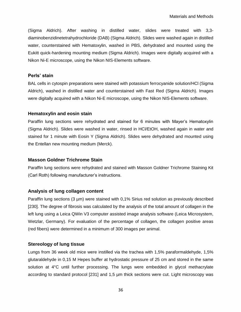
Materials and Methods
36
(Sigma Aldrich). After washing in distilled water, slides were treated with 3,3-
diaminobenzidinetetrahydrochloride (DAB) (Sigma Aldrich). Slides were washed again in distilled
water, counterstained with Hematoxylin, washed in PBS, dehydrated and mounted using the
Eukitt quick-hardening mounting medium (Sigma Aldrich). Images were digitally acquired with a
Nikon Ni-E microscope, using the Nikon NIS-Elements software.
Perls’ stain
BAL cells in cytospin preparations were stained with potassium ferrocyanide solution/HCl (Sigma
Aldrich), washed in distilled water and counterstained with Fast Red (Sigma Aldrich). Images
were digitally acquired with a Nikon Ni-E microscope, using the Nikon NIS-Elements software.
Hematoxylin and eosin stain
Paraffin lung sections were rehydrated and stained for 6 minutes with Mayer’s Hematoxylin
(Sigma Aldrich). Slides were washed in water, rinsed in HCl/EtOH, washed again in water and
stained for 1 minute with Eosin Y (Sigma Aldrich). Slides were dehydrated and mounted using
the Entellan new mounting medium (Merck).
Masson Goldner Trichrome Stain
Paraffin lung sections were rehydrated and stained with Masson Goldner Trichrome Staining Kit
(Carl Roth) following manufacturer’s instructions.
Analysis of lung collagen content
Paraffin lung sections (3 µm) were stained with 0,1% Sirius red solution as previously described
[230]. The degree of fibrosis was calculated by the analysis of the total amount of collagen in the
left lung using a Leica QWin V3 computer assisted image analysis software (Leica Microsystem,
Wetzlar, Germany). For evaluation of the percentage of collagen, the collagen positive areas
(red fibers) were determined in a minimum of 300 images per animal.
Stereology of lung tissue
Lungs from 36 week old mice were instilled via the trachea with 1,5% paraformaldehyde, 1,5%
glutaraldehyde in 0,15 M Hepes buffer at hydrostatic pressure of 25 cm and stored in the same
solution at 4°C until further processing. The lungs were embedded in glycol methacrylate
according to standard protocol [231] and 1,5 µm thick sections were cut. Light microscopy was

Materials and Methods
37
performed using a Leica DM6000B microscope (Leica, Wetzlar, Germany) attached to a
computer with the newCAST stereology software (Visiopharm, Horsholm, Denmark). Design-
based stereology was used to quantify the volume of the lung parenchyma and non-parenchyma
as previously described [231].
Erythropoietin (EPO) ELISA
Blood was centrifuged at 2000g for 10 minutes and plasma EPO levels were determined using a
mouse EPO ELISA kit (R&D systems) following manufacturer’s instructions.
CCL2 ELISA
Blood was centrifuged at 2000g for 10 minutes and plasma CCL2 levels were determined using
a mouse CCL2 ELISA kit (R&D systems) following manufacturer’s instructions.
Statistical analyses
Data are shown as mean ± standard error of the mean (SEM) and the number of mice (n) is
indicated. Statistical analyses were performed using Prism v6 (GraphPad Software, La Jolla,
CA). For comparisons between 2 groups, the two-tailed, Student’s t-test was used. For
comparisons between 3 or 4 groups, a one-way or two-way ANOVA with the Bonferroni post-test
was used. p-values <0.05 (*), <0.01 (**), <0.001 (***) and <0.0001 (****) are indicated.

38
Chapter III
Results

Results
39
Increased pulmonary iron content in Slc40a1C326S mice
The Slc40a1C326S mouse model exhibits a phenotype that resembles human hereditary
hemochromatosis type 4, presenting elevated plasma iron levels, high transferrin saturation,
parenchymal iron overload, and iron depletion in iron-exporting tissues such as duodenal
enterocytes and reticuloendothelial macrophages [102].
To assess whether a disruption in the hepcidin/FPN regulatory circuitry affects pulmonary
iron content, lungs from wild-type (Slc40a1wt/wt), heterozygous (Slc40a1wt/C326S) and homozygous
(Slc40a1C326S/C326S) mutant male and female mice were analyzed at different ages.
Measurements of non-heme iron levels revealed a genotype-specific and age-dependent
increase in pulmonary iron content in Slc40a1C326S mice, regardless of the gender (Fig. 2.1A).
To confirm that the increase in pulmonary iron content observed in Slc40a1C326S/C326S is
caused by the disruption of the hepcidin/FPN regulatory circuitry and is not a direct
consequence of the C326S point mutation per se, we compared the lung non-heme iron content
of Slc40a1C326S/C326S mice and hepcidin knock out (Hamp-/-) mice – both mouse models present a
disrupted hepcidin/FPN system but result from different genetic mutations. As observed in Fig.
2.1B, pulmonary iron levels are similar between Slc40a1C326S/C326S and Hamp-/- mice and
increased when compared to wild-type mice, indicating that the increased lung iron content of
Slc40a1C326S/C326S mice results from a non-functional hepcidin/FPN system.
Figure 2.1 – Lung iron content of Slc40a1C326S
mice
(A) Total lung non-heme iron levels measured in 10-, 24- and 36-week old male and female mice.
(B) Total lung non-heme iron levels measured in 50-week old male mice.
ns – non-significant

Results
40
A molecular analysis of iron-related genes in the lung was performed to evaluate whether
their expression responds to increased pulmonary iron content. At mRNA level, the expression
of TfR1 and Dmt1 isoform containing an IRE are decreased in Slc40a1wt/C326S and
Slc40a1C326S/C326S mice (Fig. 2.2A, B). TfR1 is decreased also at the protein level and correlates
with an increase in FtL protein levels (Fig. 2.2C). FPN shows a genotype-specific upregulation
both at mRNA and protein levels (Fig. 2.2C, D).
Figure 2.2 – Expression of iron-related genes in the lung of Slc40a1C326S
mice
(A) qRT-PCR analysis of TfR1 mRNA expression in total lung of 36-week old female mice.
(B) qRT-PCR analysis of Dmt1 IRE mRNA expression in total lung of 36-week old female mice.
(C) Western blot analysis of TfR1 (~100 kDa), FtL (~20 kDa), and FPN (~65 kDa) protein expression in total lung
of 36-week old female mice. -actin (42 kDa) was used as loading control.
(D) qRT-PCR analysis of Fpn mRNA expression in total lung of 36-week old female mice.
We have also analyzed the expression of two recently identified iron transporters: ZIP8
and ZIP14, which were reported to mediate the uptake of NTBI [232]. We observed that Zip8 is
highly expressed in the lung compared to the liver (Fig. 2.3A), an observation that supports a
study by Wang and colleagues [46]. The same group has shown that ZIP8 protein levels
increase in response to iron loading due to a post-transcriptional mechanism, without alterations
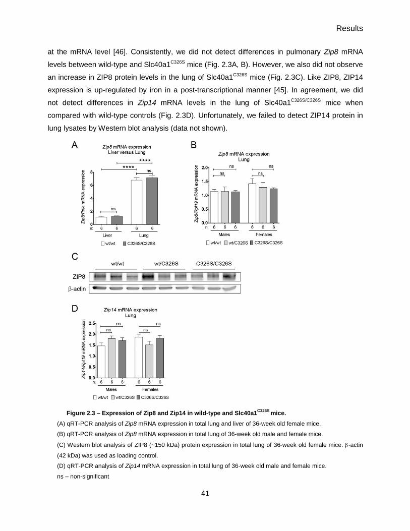
Results
41
at the mRNA level [46]. Consistently, we did not detect differences in pulmonary Zip8 mRNA
levels between wild-type and Slc40a1C326S mice (Fig. 2.3A, B). However, we also did not observe
an increase in ZIP8 protein levels in the lung of Slc40a1C326S mice (Fig. 2.3C). Like ZIP8, ZIP14
expression is up-regulated by iron in a post-transcriptional manner [45]. In agreement, we did
not detect differences in Zip14 mRNA levels in the lung of Slc40a1C326S/C326S mice when
compared with wild-type controls (Fig. 2.3D). Unfortunately, we failed to detect ZIP14 protein in
lung lysates by Western blot analysis (data not shown).
Figure 2.3 – Expression of Zip8 and Zip14 in wild-type and Slc40a1C326S
mice.
(A) qRT-PCR analysis of Zip8 mRNA expression in total lung and liver of 36-week old female mice.
(B) qRT-PCR analysis of Zip8 mRNA expression in total lung of 36-week old male and female mice.
(C) Western blot analysis of ZIP8 (~150 kDa) protein expression in total lung of 36-week old female mice. -actin
(42 kDa) was used as loading control.
(D) qRT-PCR analysis of Zip14 mRNA expression in total lung of 36-week old male and female mice.
ns – non-significant
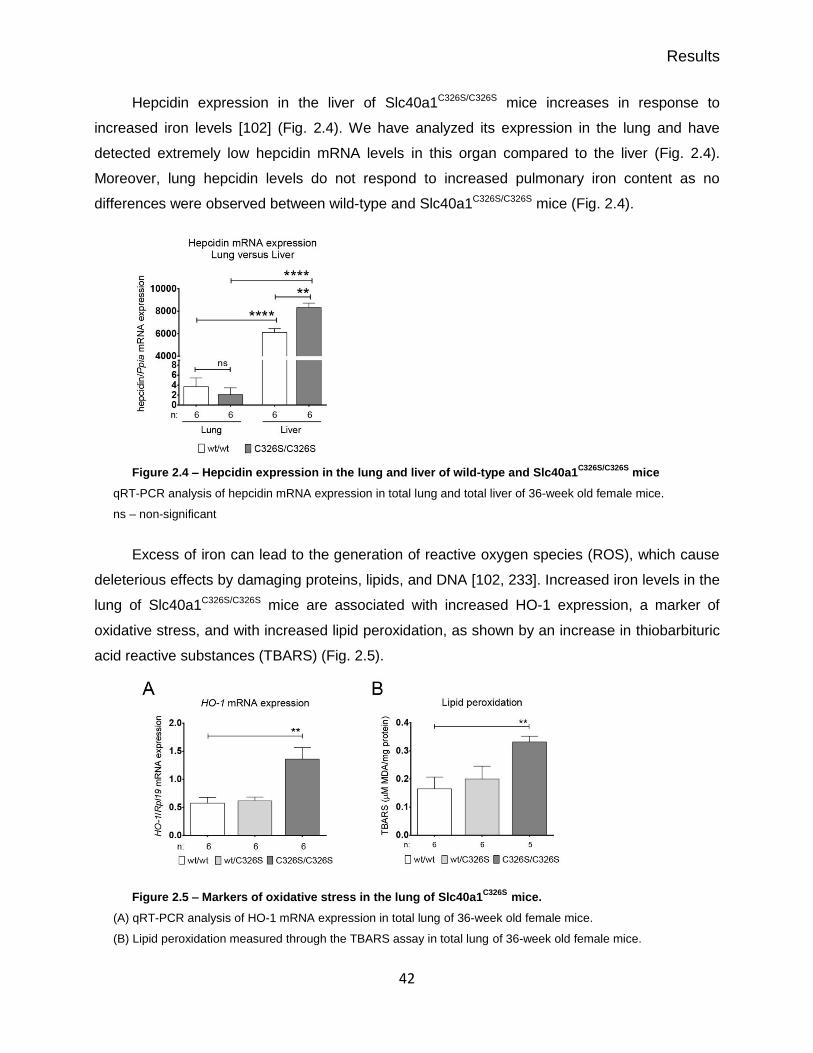
Results
42
Hepcidin expression in the liver of Slc40a1C326S/C326S mice increases in response to
increased iron levels [102] (Fig. 2.4). We have analyzed its expression in the lung and have
detected extremely low hepcidin mRNA levels in this organ compared to the liver (Fig. 2.4).
Moreover, lung hepcidin levels do not respond to increased pulmonary iron content as no
differences were observed between wild-type and Slc40a1C326S/C326S mice (Fig. 2.4).
Figure 2.4 – Hepcidin expression in the lung and liver of wild-type and Slc40a1C326S/C326S
mice
qRT-PCR analysis of hepcidin mRNA expression in total lung and total liver of 36-week old female mice.
ns – non-significant
Excess of iron can lead to the generation of reactive oxygen species (ROS), which cause
deleterious effects by damaging proteins, lipids, and DNA [102, 233]. Increased iron levels in the
lung of Slc40a1C326S/C326S mice are associated with increased HO-1 expression, a marker of
oxidative stress, and with increased lipid peroxidation, as shown by an increase in thiobarbituric
acid reactive substances (TBARS) (Fig. 2.5).
Figure 2.5 – Markers of oxidative stress in the lung of Slc40a1C326S mice.
(A) qRT-PCR analysis of HO-1 mRNA expression in total lung of 36-week old female mice.
(B) Lipid peroxidation measured through the TBARS assay in total lung of 36-week old female mice.

Results
43
Iron accumulation in the lung of Slc40a1C326S mice is restricted to specific
cell types
Histological analysis of the lung of Slc40a1C326S mice confirmed the iron overload
phenotype (Fig. 2.6). Furthermore, it revealed that iron does not accumulate uniformly in all lung
cell types but it is instead restricted to specific sub-sets of the lung cell population (Fig. 2.6).
Figure 2.6 – Iron accumulation in the lung of Slc40a1C326S
mice is restricted to specific cell types
DAB-enhanced Perls’ stain of lung sections of 36-week old female mice. The arrow shows an alveolar
macrophage with strong iron accumulation.
It has been previously proposed that airway epithelial cells can take up iron from the
airway lumen via DMT1 [204]. Using -tubulin IV as a marker of cilia, we detected DMT1 mainly
at the apical membrane of ciliated airway epithelial cells in wild-type mice (Fig. 2.7A). In line with
this observation, iron was frequently detected in ciliated epithelial cells lining the conducting
airways of Slc40a1C326S/C326S mice (Fig. 2.7B, C).
Iron deposition was occasionally detected in another epithelial cell type – alveolar type 2
cells (Fig. 2.8). These secretory cells are present in the alveolar epithelium and can be identified
by the expression of the prosurfactant protein C.
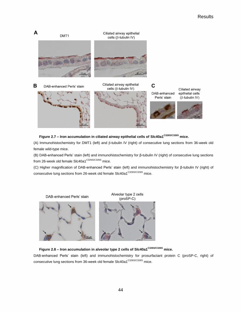
Results
44
Figure 2.7 – Iron accumulation in ciliated airway epithelial cells of Slc40a1C326S/C326S
mice.
(A) Immunohistochemistry for DMT1 (left) and β-tubulin IV (right) of consecutive lung sections from 36-week old
female wild-type mice.
(B) DAB-enhanced Perls’ stain (left) and immunohistochemistry for β-tubulin IV (right) of consecutive lung sections
from 26-week old female Slc40a1C326S/C326S
mice.
(C) Higher magnification of DAB-enhanced Perls’ stain (left) and immunohistochemistry for β-tubulin IV (right) of
consecutive lung sections from 26-week old female Slc40a1C326S/C326S
mice.
Figure 2.8 – Iron accumulation in alveolar type 2 cells of Slc40a1C326S/C326S
mice.
DAB-enhanced Perls’ stain (left) and immunohistochemistry for prosurfactant protein C (proSP-C, right) of
consecutive lung sections from 36-week old female Slc40a1C326S/C326S
mice.

Results
45
In addition, we detected iron accumulation in vascular smooth muscle cells (SMC) in the
lung of Slc40a1C326S/C326S mice, which were identified by the expression of alpha smooth muscle
actin (αSMA) (Fig. 2.9A). However, we have also detected iron-spared vascular SMCs (Fig.
2.9B), suggesting that the lung contains different populations of vascular SMCs in regard to iron
trafficking. Interestingly, iron accumulates in vascular SMCs that express low levels of desmin
whereas vascular SMCs expressing high levels of desmin are iron-spared (Fig. 2.10A). The
disparity in desmin expression between different vascular SMCs is not a consequence of the
C326S point mutation and/or alterations in iron levels since a differential expression of desmin
between different vascular SMCs was also observed in wild-type mice (Fig. 2.10B).
Figure 2.9 – Iron accumulation in lung vascular smooth muscle cells of Slc40a1C326S/C326S
mice.
(A) DAB-enhanced Perls’ stain (left and center) and immunohistochemistry for alpha smooth muscle actin (αSMA)
(right) of consecutive lung sections from 36-week old female Slc40a1C326S/C326S
mice.
(B) DAB-enhanced Perls’ stain (left) and immunohistochemistry for alpha smooth muscle actin (αSMA; right) of
consecutive lung sections from 36-week old female Slc40a1C326S/C326S
mice.

Results
46
Figure 2.10 – Iron accumulates differently in functionally distinct vascular smooth muscle cells.
(A) DAB-enhanced Perls’ stain (left) and immunohistochemistry for desmin (right) of consecutive lung sections
from 36-week old female Slc40a1C326S/C326S
mice. Arrows indicate vascular SMCs.
(B) Immunohistochemistry for desmin of lung sections from 36-week old female wild-type mice.
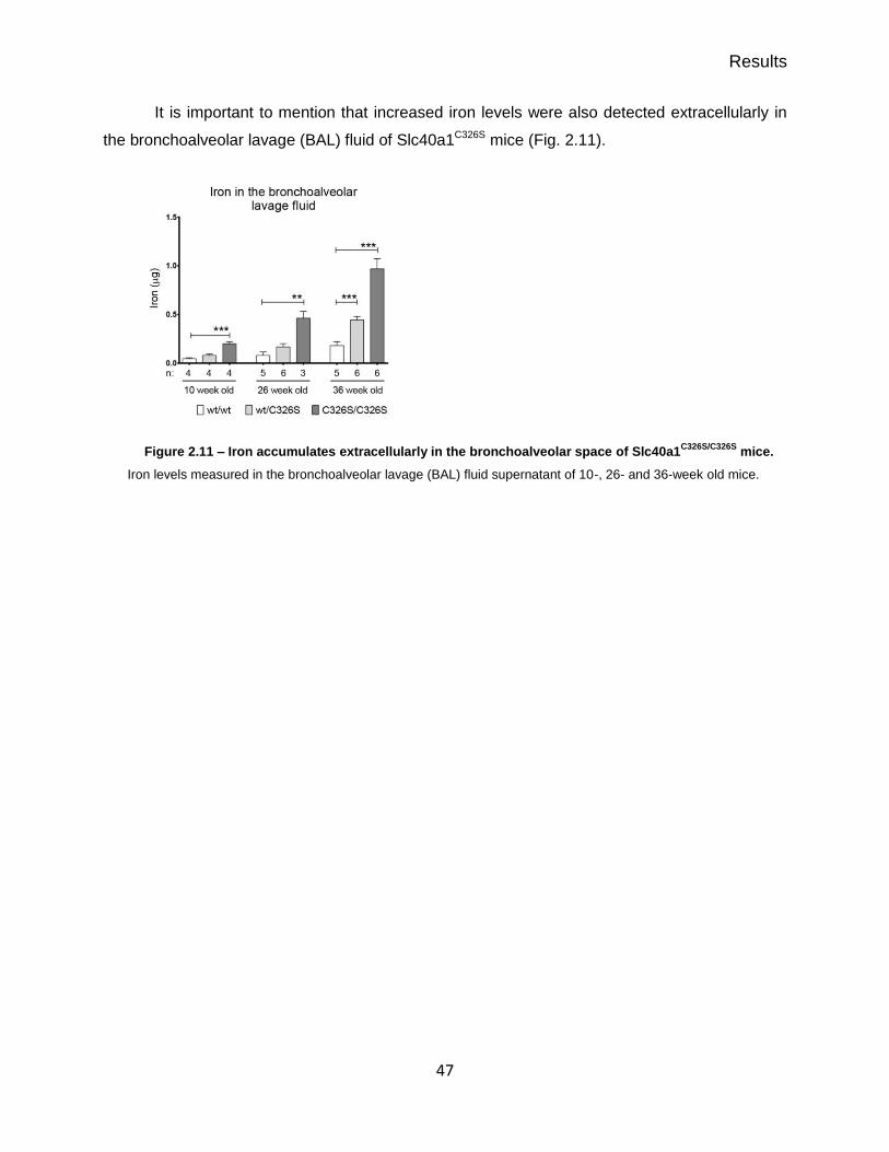
Results
47
It is important to mention that increased iron levels were also detected extracellularly in
the bronchoalveolar lavage (BAL) fluid of Slc40a1C326S mice (Fig. 2.11).
Figure 2.11 – Iron accumulates extracellularly in the bronchoalveolar space of Slc40a1C326S/C326S
mice.
Iron levels measured in the bronchoalveolar lavage (BAL) fluid supernatant of 10-, 26- and 36-week old mice.

Results
48
Iron loaded alveolar macrophages in Slc40a1C326S mice
Iron deposition in alveolar macrophages (AM) has been observed in different lung
diseases [218, 219]. Interestingly, histological analysis of lung sections from Slc40a1C326S/C326S
mice revealed the presence of strongly iron loaded AMs (Fig. 2.6 arrow). To further confirm this
observation, we analyzed the cellular fraction of the BAL. Differential May-Grünwald-Giemsa
stain of cytospin preparations showed that AMs were the predominant cell type in the BAL fluid
of wild-type and Slc40a1C326S/C326S mice (Fig. 2.12A, B). This analysis further revealed that the
total number of cells in the BAL fluid of Slc40a1C326S/C326S mice was increased when compared to
wild-type animals, mainly due to an increase in the number of AMs (Fig. 2.12B). The identity of
AMs was additionally confirmed by flow cytometry (gating strategy Fig. 2.12C) – more than 90%
of the immune cells in the BAL fluid of both wild-type and Slc40a1C326S/C326S mice corresponded
to AMs (CD45.2+ CD11c+ SiglecF+) (Fig. 2.12D) [176].
The increase in the number of AMs can result either from proliferation of a progenitor
population within the lung or from recruitment of circulating monocytes (reviewed in [123]).
Interestingly, the plasma levels of CCL2 in Slc40a1C326S/C326S mice have a strong tendency to be
increased (p-value = 0,06), which may directly contribute to the recruitment of monocytes and,
as a consequence, to an increase in the number of macrophages (Fig. 2.13A). We further
analyzed the mRNA levels of different chemokines in the lung of Slc40a1C326S/C326S mice. The
mRNA levels of C-C motif chemokine ligand (Ccl) 2, Ccl3, Ccl4, Ccl20 and C-X-C motif
chemokine ligand (Cxcl) 2 were not significantly up-regulated in the lung of Slc40a1C326S/C326S
mice but we observed a small increase in the expression levels of Cxcl1 (Fig. 2.13B-G). We
have also analyzed the expression levels of two cytokines involved in macrophage
differentiation: macrophage colony-stimulating factor (M-csf), which was decreased in the lung of
Slc40a1C326S/C326S mice, and granulocyte macrophage colony-stimulating factor (Gm-csf), which
showed a robust increase (Fig. 2.13H, I). The increase in Gm-csf mRNA levels is of particular
interest as this cytokine is a key player in the differentiation of AMs. Additional studies will be
necessary to understand the underlying mechanism causing the increase in the number of AMs
in Slc40a1C326S/C326S mice.

Results
49
Figure 2.12 – Alveolar macrophages in the bronchoalveolar space of Slc40a1C326S/C326S
mice
(A) May-Grünwald-Giemsa stain of cytospin preparations obtained from the bronchoalveolar lavage of 26-week old
male mice.
(B) Total cell count and differential cell count of the cell fractions of the bronchoalveolar lavage of 26-week old
male mice.
(C) Gating strategy used to identify the cells present in the bronchoalveolar lavage fluid of wild-type and
Slc40a1C326S/C326S
mice.
(D) Percentage of AM (CD11c+ Siglec
+) present in the leukocyte fraction (CD45.2
+) in the bronchoalveolar lavage
fluid of 24-week old male mice (n=3).
ns – non-significant
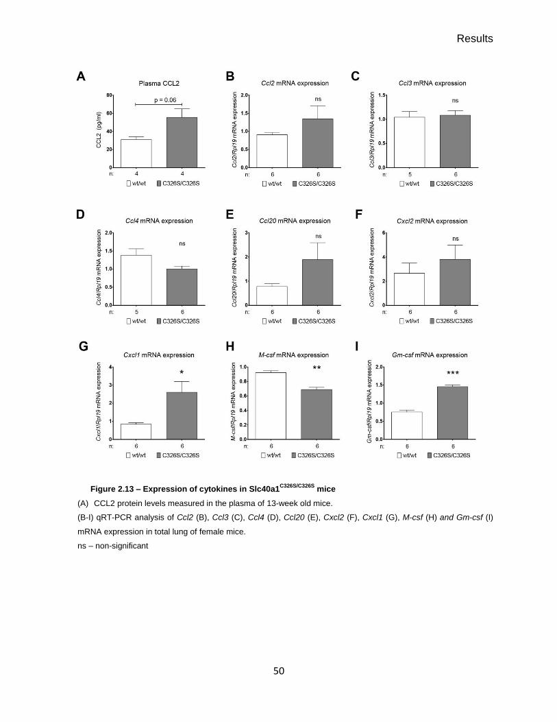
Results
50
Figure 2.13 – Expression of cytokines in Slc40a1C326S/C326S
mice
(A) CCL2 protein levels measured in the plasma of 13-week old mice.
(B-I) qRT-PCR analysis of Ccl2 (B), Ccl3 (C), Ccl4 (D), Ccl20 (E), Cxcl2 (F), Cxcl1 (G), M-csf (H) and Gm-csf (I)
mRNA expression in total lung of female mice.
ns – non-significant

Results
51
The presence of iron-loaded AMs in Slc40a1C326S/C326S mice was confirmed by Perls’ stain
on cytospin preparations of BAL cells (Fig. 2.14A). Intracellular iron accumulation in AMs
distinguishes them from hepatic Kupffer cells and splenic macrophages in Slc40a1C326S/C326S
mice: these cells are iron depleted due to uncontrolled iron export caused by the resistance of
FPN to hepcidin binding [102]. Curiously, we also detected iron spared AMs in BAL cytospin
preparations from Slc40a1C326S/C326S mice (Fig. 2.14A arrow). This finding indicates that the
population of AMs in Slc40a1C326S/C326S mice is heterogeneous regarding iron handling. This
heterogeneity in iron accumulation correlates with a heterogeneous expression of FPN in AMs
from Slc40a1C326S/C326S mice, probably explaining the phenotype (Fig. 2.14B). Interestingly, we
could not detect FPN in AMs from wild-type mice (Fig. 2.14B).
Figure 2.14 – Iron loaded alveolar macrophages in Slc40a1C326S/C326S
mice
(A) Perls’ stain of cytospin preparations of cells isolated from the bronchoalveolar lavage fluid of 24-week old male
mice. Arrow shows an iron spared alveolar macrophage.
(B) Immunocytochemistry for FPN in cytospin preparations of cells isolated from the bronchoalveolar lavage fluid
of 24-week old male mice.

Results
52
To further confirm the absence of FPN expression in AMs isolated from wild-type mice,
we analyzed the mRNA levels of Fpn in these cells. To this end, we compared AMs, which were
isolated from the bronchoalveolar lavage and identified by the expression of high levels of the
AM markers Cd11c and SiglecF, to splenic macrophages, which were isolated through magnetic
cell sorting and identified by the expression of high levels of the markers Cd11b and Spi-C (Fig.
2.15A-D). We observed that AMs express very low levels of Fpn mRNA in comparison to splenic
macrophages (Fig. 2.15E).
Figure 2.15 – Comparison between alveolar macrophages and splenic macrophages isolated from wild-
type mice
qRT-PCR analysis of Cd11c (A), SiglecF (B), Cd11b (C), Spi-C (D), and Fpn (E) mRNA expression in
alveolar macrophages and splenic macrophages isolated from 12-week old wild-type mice.

Results
53
Ferroportin transcription is regulated by different mechanisms, which mostly depend on
the cell type. For example, ferroportin transcription in duodenal enterocytes is mainly regulated
by hypoxia [84] while reticuloendothelial macrophages control ferroportin expression via a heme-
dependent pathway [91]. How ferroportin expression is regulated in AMs is still unknown.
GM-CSF is a key regulator of the differentiation of AMs: GM-CSF-/- mice fail to generate
mature and functional AMs [180]. Interestingly, GM-CSF was reported to repress FPN
expression in monocyte-derived macrophages after stressed erythrocyte challenge [234]. Since
GM-CSF is highly expressed in the lung compared to the spleen (Fig. 2.16A), where
macrophages express FPN, it is possible that GM-CSF represses ferroportin expression in AMs.
To test this hypothesis, we differentiated bone marrow hematopoietic progenitors in vitro
in the presence of GM-CSF and compared them to bone marrow hematopoietic progenitors
differentiated in the presence of M-CSF – cytokine that is commonly used to differentiate bone
marrow-derived macrophages (BMDM) in vitro. We observed that BMDM differentiated in the
presence of GM-CSF upregulate the expression of AMs markers SiglecF, Cd11c, and Cd205
(Fig. 2.16B-D) [123, 175, 176]. Moreover, the presence of GM-CSF leads to a downregulation of
Cd14, which is also expressed at intermediate/low levels in AMs (Fig. 2.16E) [123, 176]. These
observations indicate that BMDM differentiated in the presence of GM-CSF present an AM-like
phenotype, supporting the major role of GM-CSF in the differentiation of AMs in the lung.
However, AMs express low levels of Cd11b [176] (Fig. 2.15C), but its expression is not reduced
in BMDM differentiated in the presence of GM-CSF (Fig. 2.16F), suggesting that other factors
present within the alveolar microenvironment might be necessary to fully differentiate AMs.
We observed that in the presence of GM-CSF, the expression of FPN is downregulated
both at mRNA and protein level when compared to BMDMs differentiated in the presence of M-
CSF (Fig. 2.16G, H). The effect of GM-CSF is dominant, since it is able to downregulate Fpn
expression also in the presence of M-CSF (Fig. 2.16I). These observations support our
hypothesis that GM-CSF may mediate the suppression of ferroportin expression in AMs.
However, we observed AMs expressing FPN in Slc40a1C326S/C326S mice. These FPN+ AMs
might correspond to recruited macrophages that do not respond to the pulmonary GM-CSF or
AMs that are still undergoing the differentiation process.
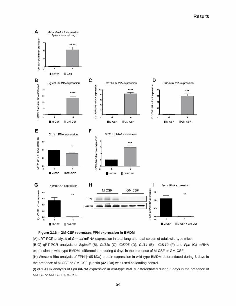
Results
54
Figure 2.16 – GM-CSF represses FPN expression in BMDM
(A) qRT-PCR analysis of Gm-csf mRNA expression in total lung and total spleen of adult wild-type mice.
(B-G) qRT-PCR analysis of SiglecF (B), Cd11c (C), Cd205 (D), Cd14 (E) , Cd11b (F) and Fpn (G) mRNA
expression in wild-type BMDMs differentiated during 6 days in the presence of M-CSF or GM-CSF.
(H) Western Blot analysis of FPN (~65 kDa) protein expression in wild-type BMDM differentiated during 6 days in
the presence of M-CSF or GM-CSF. -actin (42 kDa) was used as loading control.
(I) qRT-PCR analysis of Fpn mRNA expression in wild-type BMDM differentiated during 6 days in the presence of
M-CSF or M-CSF + GM-CSF.

Results
55
Both phenotype and function of macrophages are shaped by the environment to which
these cells are exposed (reviewed in [235]). Several studies have described that iron overload in
macrophages results in oxidative stress, which triggers a switch towards a pro-inflammatory
state [236-238]. Since we detected iron overloaded AMs in Slc40a1C326S/C326S mice (Fig. 2.14A),
we analyzed the degree of oxidative stress in these cells as well as the pulmonary inflammatory
status of Slc40a1C326S/C326S mice. Lung inflammation is hallmarked by an increased expression of
pro-inflammatory cytokines and a recruitment of immune cells into the bronchoalveolar
compartment.
Using the CellRox green as ROS reporter (a fluorogenic probe, which exhibits a strong
fluorescence upon oxidation by ROS), we did not detect differences in oxidative stress in AMs
between wild-type and Slc40a1C326S/C326S mice (Fig. 2.17A). In contrast, acute iron treatment of
BMDM causes the formation of ROS (Fig. 2.17B).
Despite the increase in the number of AMs (Fig. 2.12B), but consistent with the absence
of oxidative stress in these cells (Fig. 2.17A), AMs in Slc40a1C326S/C326S mice do not seem to be
morphologically activated as no significant difference in cell size was observed when compared
with wild-type AMs (Fig. 2.17C). In agreement, we did not detect an increase in mRNA and
protein levels of pro-inflammatory cytokines IL1β, IL6, and TNFα in total lung or in the BAL fluid
supernatant, respectively (Fig. 2.17D, E). Since inflammatory responses are regulated by
complex networks involving different cell types and the production of different cytokines, we
further analyzed the expression levels of the anti-inflammatory cytokines IL10 and IL13. We
failed to detect differences in the protein levels of these cytokines between wild-type and
Slc40a1C326S/C326S mice (Fig. 2.18).

Results
56
Figure 2.17 – Slc40a1C326S/C326S
mice do not show signs of pulmonary inflammation
(A) FACS analysis of the CellROX oxidative stress tracer of AMs isolated from the BAL of 24-week old male mice.
(B) FACS analysis of the CellROX oxidative stress tracer of iron-treated BMDM.
(C) Area of alveolar macrophages isolated from the bronchoalveolar lavage of 36-week old female mice.
(D) qRT-PCR analysis of IL1β, IL6 and Tnfα mRNA expression in total lung from 36-week old female mice.
(E) Luminex analysis of IL1β, IL6 and TNFα protein levels in the bronchoalveolar lavage fluid supernatant of 36-
week old female mice.
ns – non-significant , pg – picograms

Results
57
Figure 2.18 – Expression of anti-inflammatory cytokines in the lung of Slc40a1C326S/C326S
mice
Luminex analysis of IL10 and IL13 protein levels in the bronchoalveolar lavage fluid supernatant of 36-week old
female mice.
ns – non-significant , pg – picograms

Results
58
Slc40a1C326S/C326S mice present classical signs of restrictive lung disease
Increased iron levels and consequent increase in oxidative stress are known to cause
cellular damage and tissue injury [2, 102, 233]. We next investigated whether aged
Slc40a1C326S/C326S mice present alterations in lung function.
Measurements of lung function were performed in tracheostomised anaesthetized mice
using a lung function measurement device for small animals (FlexiVent of Scireq, Canada). This
analysis revealed classical signs of restrictive lung disease in Slc40a1C326S/C326S mice, such as a
decrease in total lung capacity and a decrease in pulmonary compliance, which reflects a
decrease in lung elasticity (Fig. 2.19A, B). Consistently, Slc40a1C326S/C326S mice showed an
increase in pulmonary elastance, a measurement of lung rigidity (Fig. 2.19C). This finding was
further confirmed by exposing the mice to fixed air pressures. As shown in the pressure-volume
curve, an increase in the applied pressure correlated with an increase in lung volume but to a
lesser extent in Slc40a1C326S/C326S mice when compared to wild-type mice (Fig. 2.19D).
We excluded the possibility that the observed differences between wild-type and
Slc40a1C326S/C326S mice were due to differences in body size. Despite the fact that aged
Slc40a1C326S/C326S mice present a lower body weight, most likely due to exocrine pancreatic
failure [102], the tibia length (a marker of body size) was comparable to age-matched wild-type
animals, indicating that both groups of mice have a similar size (Fig. 2.19E, F).

Results
59
Figure 2.19 – Lung function measurements and stereological analysis of the lung of Slc40a1C326S/C326S
mice
(A-D) Lung function measurements performed in 36-week old female mice. A) Total Lung Capacity; (B) Pulmonary
Dynamic Compliance; C) Pulmonary Elastance; D) Pressure-Volume (PV) curve with controlled pressure applied.
(E) Body weight of 10-, 26- and 34-week old female mice.
(F) Tibia size of 10-, 26- and 34-week old female mice.
(G) Volume of lung parenchyma measured in 36-week old female mice.
(H) Volume of lung non-parenchyma measured in 36-week old female mice.
ns – non-significant

Results
60
Together with Prof. Christian Mühlfeld and Dr. Christina Brandenberger (Hannover
Medical School), we performed a stereological analysis of the lung of Slc40a1C326S/C326S mice, as
previously described [231]. We applied a grid on images of lung sections obtained by light
microscopy and counted the number of points overlapping with the alveolar septa (P(alvsept)),
alveolar airspace (P(alvair)) and alveolar duct airspace (P(ductair)), which together correspond
to points overlapping the lung parenchyma. We have also counted the points overlapping with
the lung non-parenchyma (P(non parenchyma)), e.g. conducting airways and blood vessels. We
estimated the volume fraction (Vv) of the lung parenchyma and lung non-parenchyma as
following:
𝑉𝑣 (𝑝𝑎𝑟𝑒𝑛𝑐ℎ𝑦𝑚𝑎
𝑙𝑢𝑛𝑔) =
[𝑃(𝑑𝑢𝑐𝑡𝑎𝑖𝑟) + 𝑃(𝑎𝑙𝑣𝑠𝑒𝑝𝑡) + 𝑃(𝑎𝑙𝑣𝑎𝑖𝑟)]
𝑃(𝑙𝑢𝑛𝑔)
𝑉𝑣 (𝑛𝑜𝑛 𝑝𝑎𝑟𝑒𝑛𝑐ℎ𝑦𝑚𝑎
𝑙𝑢𝑛𝑔) =
𝑃(𝑛𝑜𝑛 𝑝𝑎𝑟𝑒𝑛𝑐ℎ𝑦𝑚𝑎)
𝑃(𝑙𝑢𝑛𝑔)
𝑃(𝑙𝑢𝑛𝑔) − sum of points overlapping the lung parenchyma and the lung non-parenchyma.
By multiplying the volume fraction of these compartments by the total volume of the lung
(assessed by the fluid displacement method), we calculated the total volume of the lung
parenchyma and the total volume of the lung non-parenchyma [231]. This analysis revealed a
reduction in the volume of the lung parenchyma in Slc40a1C326S/C326S mice, without alterations in
the volume of the lung non-parenchyma (Fig. 2.19G, H). These observations support the
restrictive pattern observed by measurements of lung function and indicate that the decrease in
total lung capacity results from a reduction in the volume of the lung parenchyma.
A pulmonary restrictive pattern is commonly associated with conditions that increase lung
stiffness, such as an increase in collagen deposition [172]. However, a marked replacement of
normal lung parenchyma by fibrotic tissue was not observed (Fig. 2.20).

Results
61
Figure 2.20 – The lung structure in Slc40a1C326S/C326S
mice is unaltered compared to wild-type mice.
(A) Hematoxylin & Eosin stain of lung sections from 36-week old female mice.
(B) Goldner Masson Trichrome stain of lung sections from 36-week old mice.
(C) Sirius red stain of lung sections from 36-week old mice.
(D) Collagen content calculated on Sirius Red stained lung sections from 34-week old mice.
Quantification of lung collagen deposition was performed by our collaborators Dr. Simone Kraut and Prof. Dr.
Norbert Weissmann.
ns – non-significant
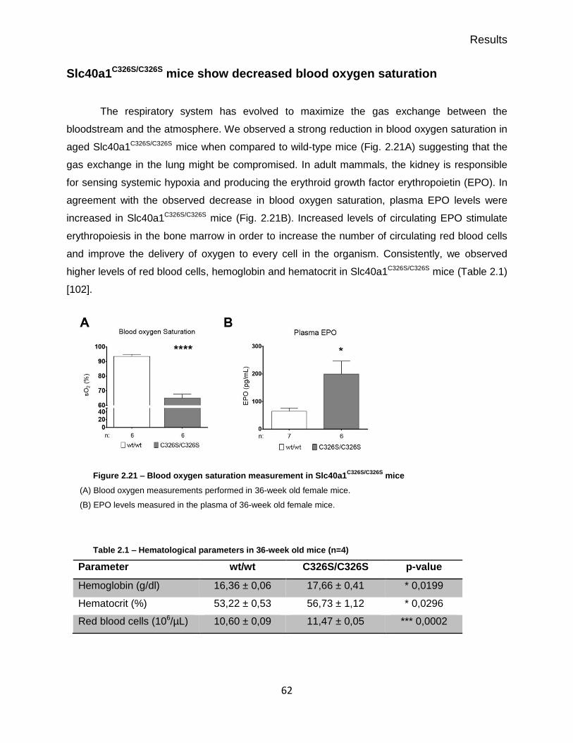
Results
62
Slc40a1C326S/C326S mice show decreased blood oxygen saturation
The respiratory system has evolved to maximize the gas exchange between the
bloodstream and the atmosphere. We observed a strong reduction in blood oxygen saturation in
aged Slc40a1C326S/C326S mice when compared to wild-type mice (Fig. 2.21A) suggesting that the
gas exchange in the lung might be compromised. In adult mammals, the kidney is responsible
for sensing systemic hypoxia and producing the erythroid growth factor erythropoietin (EPO). In
agreement with the observed decrease in blood oxygen saturation, plasma EPO levels were
increased in Slc40a1C326S/C326S mice (Fig. 2.21B). Increased levels of circulating EPO stimulate
erythropoiesis in the bone marrow in order to increase the number of circulating red blood cells
and improve the delivery of oxygen to every cell in the organism. Consistently, we observed
higher levels of red blood cells, hemoglobin and hematocrit in Slc40a1C326S/C326S mice (Table 2.1)
[102].
Figure 2.21 – Blood oxygen saturation measurement in Slc40a1C326S/C326S
mice
(A) Blood oxygen measurements performed in 36-week old female mice.
(B) EPO levels measured in the plasma of 36-week old female mice.
Table 2.1 – Hematological parameters in 36-week old mice (n=4)
Parameter wt/wt C326S/C326S p-value
Hemoglobin (g/dl) 16,36 ± 0,06 17,66 ± 0,41 * 0,0199
Hematocrit (%) 53,22 ± 0,53 56,73 ± 1,12 * 0,0296
Red blood cells (106/µL) 10,60 ± 0,09 11,47 ± 0,05 *** 0,0002

63
Chapter IV
Discussion

Discussion
64
Imbalances in iron homeostasis have been reported in a spectrum of lung diseases [204, 219].
However, the role of iron in lung pathology is poorly understood. In this work, we aim at
understanding whether a disruption in the hepcidin/ferroportin regulatory circuitry and the
subsequent increase in systemic iron levels affect lung iron homeostasis and function.
Here we show that resistance of ferroportin to hepcidin binding causes an age-dependent
pulmonary iron deposition in defined lung cell types (Fig. 3.1). Increased iron levels in this organ
correlated with increased lipid peroxidation, suggesting that iron-mediated oxidative stress may
contribute to the pathogenesis of lung diseases. Homozygous Slc40a1C326S/C326S mice show
classical signs of restrictive lung disease and a significant decrease in blood oxygen saturation.
These findings implicate iron overload in lung pathology, which has to date not been considered
a classical iron-related disorder.
Figure 3.1 – Lung iron accumulation and pathophysiological consequences of pulmonary iron overload
in Slc40a1C326S/C326S
mice
Iron accumulates in specific cell types of the lung of Slc40a1C326S/C326S
mice: ciliated airway epithelial cells,
vascular smooth muscle cells, alveolar type 2 cells and alveolar macrophages. Iron was also detected extracellularly
in the bronchoalveolar space. Although there is an increase in the number of alveolar macrophages,
Slc40a1C326S/C326S
mice do not show strong signs of pulmonary inflammation. Slc40a1C326S/C326S
mice show a
pulmonary restrictive pattern and decreased blood oxygen saturation.
FPN – Ferroportin; Fe – iron; sO2 – blood oxygen saturation.
Discussion
65
Slc40a1C326S mice show increased iron levels in the lung
There are limited reports regarding how lung iron homeostasis is affected by imbalanced
systemic iron levels. Here, we report that a disruption in the hepcidin/FPN regulatory circuitry
and the subsequent increase in systemic iron levels cause pulmonary iron accumulation in a
mouse model of HH type 4 (Slc40a1C326S). The observed increase in lung iron levels is not a
result of the point mutation in FPN per se, since hepcidin knock out mice showed similar levels
of pulmonary iron accumulation when compared to Slc40a1C326S/C326S mice. Increased systemic
and pulmonary iron levels in Slc40a1C326S mice result from an increase in dietary iron uptake
caused by the resistance of FPN to hepcidin binding.
To date, no data is available regarding the pulmonary iron content of patients with HH type 4.
However, the idea that increased pulmonary iron levels in humans can be caused by increased
dietary iron uptake is supported by the report of a patient that presented signs of intracellular
and extracellular lung iron accumulation after receiving therapeutic oral iron supplementation
[239]. Moreover, patients with thalassemia major, a disease characterized by transfusional iron
overload, were reported to accumulate iron in alveolar macrophages, further supporting the idea
that increased systemic iron levels in humans can affect pulmonary iron content [240, 241].
The IRE/IRP system is inactivated by increased intracellular iron levels (see introduction, section
1.4). This causes the degradation of the IRE-containing transcripts encoding for DMT1 and TfR1
and allows the translation of FtL and FPN. Consistent with the increase in pulmonary iron
content in Slc40a1C326S mice, we observed a decrease in mRNA and protein levels of TfR1 and
mRNA levels of the IRE-containing transcript of Dmt1, as well as an increase in the protein
levels of FtL. These results show that the expected molecular signature is preserved in the lung
of Slc40a1C326S mice. Furthermore, FPN protein levels showed a genotype-specific increase,
which may result from a combined effect of increased expression at transcriptional level (Fig.
2.2D), derepression of FPN translation by the inhibition of the IRE/IRP system [30] and the
hepcidin-resistant phenotype that post-translationally stabilizes the protein [102].
Giorgi et al. reported that intraperitoneal injection of iron-saccharate in mice leads to increased
systemic and pulmonary iron levels [208], supporting our finding that altered systemic iron
homeostasis affects lung iron content. However, in their study the expression of TfR1 and DMT1
in the lung did not change in response to increased pulmonary iron content [208]. The
differences in our observations might be explained by different sources of iron (dietary iron
versus iron-saccharate) and/or may reflect the fact that lung cells in Slc40a1C326S mice are
chronically exposed to high levels of iron in contrast to the acute iron treatment in iron-

Discussion
66
saccharate injected mice. Regarding FPN expression, they observed an increase in lung FPN
protein levels, consistent with our findings in Slc40a1C326S mice [208]. In their work, Giorgi and
colleagues further observed an increase in ZIP14 protein levels. Unfortunately, we were unable
to detect ZIP14 protein by western blot or immunohistochemistry in the lung of wild-type and
Slc40a1C326S mice.
The hormone hepcidin is mainly produced and secreted by hepatocytes and plays a major role
in systemic iron regulation (reviewed in [28]). Hepcidin expression in cells other than
hepatocytes has been reported, but at a much lower level [51-54]. We observed that in the lung,
hepcidin mRNA levels are very low compared to those in the liver. Furthermore, we observed
that contrary to hepatic hepcidin, pulmonary hepcidin expression does not respond to increased
iron levels, as no differences were observed between wild-type and Slc40a1C326S/C326S mice.
Giorgi and colleagues have also analyzed the expression of hepcidin in the lung of mice injected
intraperitoneally with iron-saccharate. Consistent with our data, they observed that pulmonary
hepcidin expression does not respond to an increased lung iron content [208].
Liver-specific hepcidin knock out mice completely recapitulate the hemochromatosis phenotype
observed in the global knock out mice, highlighting the predominant role of hepatic hepcidin in
the regulation of body iron homeostasis [242]. The role of extra-hepatic hepcidin in maintaining
cellular or/and systemic iron homeostasis is not completely known yet. A recently published
study has revealed a role for cardiac hepcidin in cardiac iron homeostasis through the analysis
of mice with a cardiomyocyte-specific deletion of hepcidin [243]. Airway epithelial cells in the
lung were shown to express hepcidin but its role in lung iron homeostasis is not clear yet [216,
217]. Frazier and colleagues reported that an increase in hepcidin expression in human airway
epithelial cells in vitro does not cause a decrease in FPN protein levels in these cells [216]. In
contrast, Chen and colleagues observed that, in a mouse model of sepsis-induced acute lung
injury, there is an increase in lung FPN protein levels upon the knock down of hepcidin in airway
epithelial cells [217]. The generation of mice with lung cell-type specific deletion of hepcidin
would help to better understand a possible role of lung hepcidin in the context of pulmonary iron
homeostasis.
The risk for oxidative stress in the respiratory tract is exceptionally high since it is continuously
exposed to the atmosphere and consequently to high levels of oxygen and pollutants. Under
healthy conditions, oxidative stress is prevented by the presence of antioxidants in the
respiratory lining fluid and the presence of alveolar macrophages, which are capable of
sequestering large amounts of iron intracellularly [203, 212]. Endogenous or exogenous

Discussion
67
conditions that cause an increase in the availability of free iron in the lung can result in oxidative
stress. For example, the pulmonary inhalation of iron oxide nanoparticles in rats leads to an
increase of ROS in the lung [223]. We observed an increase in oxidative stress in the lung of
Slc40a1C326S/C326S mice, which correlates with increased pulmonary iron content and the
presence of extracellular iron in the BAL fluid, probably reflecting a saturated ability of AMs and
other lung cells to store iron in a chemically inert form.
Iron accumulation in the lung of Slc40a1C326S mice is restricted to specific
cell types
We observed that iron does not accumulate uniformly in the lung of Slc40a1C326S mice but in
specific cells types: ciliated epithelial cells lining the conducting airways, alveolar type 2 cells,
vascular smooth muscle cells and AMs. Giorgi et al. reported that in the lung of mice injected
intraperitoneally with iron-saccharate, iron accumulates mainly in AMs [208]. Our observation
that iron accumulates in cell types other than AMs might be explained by an acute versus
chronic iron overload and/or differences in iron fluxes in wild-type mice intraperitoneally-injected
with iron compared to a genetic model with a disrupted hepcidin/FPN regulatory circuitry.
Previous work suggested that airway epithelial cells take up free catalytically active iron from the
airway lumen [204]. We observed that in wild-type mice DMT1 is mainly expressed at the apical
membrane of ciliated airway epithelial cells. Consistently, we frequently detected iron loaded
ciliated cells in the conducting airways of Slc40a1C326S/C326S mice. These observations support
the idea that these cells might be able to take up NTBI from the airway lumen.
However, contrary to what was suggested by Ghio and colleagues [211], we believe that ciliated
airway epithelial cells do not export iron through FPN since we detected iron deposition in these
cells in Slc40a1C326S/C326S mice – the resistance of FPN to hepcidin binding should otherwise
cause uncontrolled iron export. Interestingly, Giorgi and colleagues have detected FPN in the
cytoplasm and not in the plasma membrane of airway epithelial cells, a finding that would argue
against an active iron export via FPN [208].
Occasionally, we detected iron in another epithelial cell type of the lung of Slc40a1C326S mice –
alveolar type 2 cells. How iron is imported into these cells still remains to be elucidated.
Finally, we observed iron accumulation in vascular SMCs of the lung of Slc40a1C326S/C326S mice.
Notably, we could also detect iron-spared vascular SMCs in Slc40a1C326S/C326S mice indicating

Discussion
68
that vascular SMCs are heterogeneous in regard to iron loading. Phenotypic diversity has been
described in vascular SMCs and correlates with different functions, ranging from a contractile to
a synthetic function [244]. The expression levels of different SMC markers are commonly used
to define different phenotypes; for example, higher levels of desmin expression were associated
with a contractile phenotype [244]. In Slc40a1C326S/C326S mice, iron accumulates in vascular
SMCs that appear to be functionally different: iron-spared vascular SMCs express high levels of
desmin whereas iron-loaded vascular SMCs show lower levels of desmin. Since vascular SMCs
have a critical role in the normal regulation of the blood flow and pressure, we have an ongoing
project in the laboratory to address how increased systemic iron levels and/or iron accumulation
in a sub-population of vascular SMCs in Slc40a1C326S/C326S mice affects their role in maintaining
appropriate blood pressure under physiological and/or pathophysiological conditions. It is worth
to mention that altered iron homeostasis was reported in patients with idiopathic pulmonary
arterial hypertension [245]. Iron deficiency in rats was shown to cause pulmonary vascular
remodeling and was associated with increased pulmonary arterial hypertension, which can be
reverted by iron treatment [246].
Iron loaded alveolar macrophages and inflammatory status of Slc40a1C326S
mice
Iron sequestration by AMs has been described in different lung diseases and has been
recognized as a powerful protective mechanism against cytotoxic iron catalyzed oxidants [204,
212, 218]. We analyzed AMs from Slc40a1C326S/C326S mice and showed that their number is
increased when compared to wild-type animals. Whether this increase is due to a recruitment of
circulating monocytes or results from the proliferation of a progenitor population within the lung is
still not known. Even though monocyte recruitment has been mainly connected with increased
tissues levels of CCL2, we did not detect an increase in mRNA levels of Ccl2 in the lung of
Slc40a1C326S/C326S mice. Nevertheless, we observed a strong tendency for an increase in the
levels of CCL2 in the plasma of Slc40a1C326S/C326S mice, which may contribute for the recruitment
of monocytes into tissues. It is worth to mention that Slc40a1C326S/C326S mice also show an
increase in the number of peritoneal macrophages (unpublished data from our laboratory), which
might also be a consequence of increased systemic levels of CCL2. To further understand if
there is a recruitment of circulating monocytes to the lung of Slc40a1C326S/C326S mice, it would be
interesting to perform fate-mapping studies to trace monocyte progeny in Slc40a1C326S/C326S mice
[182]. We have also detected a mild increase in the levels of Cxcl1 in the lung of

Discussion
69
Slc40a1C326S/C326S mice, which is commonly associated with neutrophil recruitment. However, the
increase in the number of neutrophils in the bronchoalveolar space was not statistically
significant (Fig. 2.12B).
We detected strong iron deposition in AMs from Slc40a1C326S/C326S mice. This was an
unexpected result since, in the same mouse model, other resident macrophages such as
Kupffer cells and splenic macrophages are iron depleted due to the presence of a hepcidin-
resistant FPN on the cell surface of these cells [102]. Interestingly, we also observed iron spared
AMs, indicating that the population of AMs in Slc40a1C326S/C326S mice is heterogeneous in regard
to iron handling. This heterogeneity in iron accumulation most likely results from a
heterogeneous expression of FPN in AMs isolated from Slc40a1C326S/C326S mice. Notably, we
could not detect AMs expressing FPN in wild-type mice. This result contrasts with a previous
study, in which FPN was detected in intracellular vesicles of murine AMs [210].
How FPN expression is regulated in AMs is still not known. GM-CSF, which is known for its key
role in AM differentiation and which is highly expressed in the lung, has been described to
repress FPN expression in monocyte-derived macrophages upon stressed erythrocyte challenge
[234]. In line with this idea, we demonstrated the ability of GM-CSF to downregulate FPN
expression in BMDM in vitro. Nevertheless, we observed AMs expressing FPN in
Slc40a1C326S/C326S mice. The presence of GM-CSF in the lung would be expected to suppress
FPN expression. We speculate that FPN-positive AMs correspond to recruited macrophages
that do not respond to the presence of GM-CSF in the lung or to macrophages still undergoing
the differentiation process.
Our laboratory has previously described that iron overload in reticuloendothelial macrophages
results in oxidative stress and triggers a pro-inflammatory status [238]. Since we detected
increased iron levels in AMs, we analyzed the degree of oxidative stress in these cells and the
pulmonary inflammatory status of Slc40a1C326S/C326S mice. We did not detect differences in
oxidative stress between AMs of wild-type and Slc40a1C326S/C326S mice. By contrast, BMDM show
a strong increase in oxidative stress after an acute iron treatment. The absence of oxidative
stress in AMs probably results from an adaptation of these cells to chronic iron accumulation.
Consistent with the absence of oxidative stress, AMs from Slc40a1C326S/C326S mice do not appear
to be morphologically activated since we did not detect an increase in cell size when compared
to wild-type AMs.
Lung inflammation is hallmarked by an increased expression of pro-inflammatory cytokines and
a strong recruitment of immune cells into the bronchoalveolar compartment [247]. Despite the

Discussion
70
increase in the number of AMs, but in agreement with the lack of oxidative stress in these cells,
we did not observe a strong and robust increase in the expression of inflammatory cytokines in
the lung of Slc40a1C326S/C326S mice (Fig. 2.13; Fig. 2.17). Interestingly, the inhalation of iron-rich
particles leads to increased oxidative stress and lung inflammation [222-224]. This difference in
terms of pulmonary inflammation in response to increased iron levels is probably a consequence
of the different sources of iron (dietary iron versus inhalation of iron-rich particles) and a
difference in the exposure to iron (chronic versus acute). Despite the absence of pulmonary
inflammation in Slc40a1C326S/C326S mice, increased iron levels in the lung, and in particular in the
AMs, might affect the inflammatory response to pathogens. For example, low intracellular iron in
macrophages impairs signaling via the TLR4 pathway leading to reduced production of LPS-
induced cytokines [248]. An interesting follow up would be to perform LPS instillation in
Slc40a1C326S/C326S mice and analyze the production of inflammatory cytokines and the recruitment
of neutrophils to the bronchoalveolar space. Previous work from our group has described the
acute pulmonary inflammatory response of Hfe knock out mice upon intratracheal instillation of
LPS [247]. The study showed that the production of inflammatory cytokines was affected and
that there was an impairment in the recruitment of neutrophils to the lung of Hfe knock out mice.
However, in this study it was not possible to understand whether the observed effects were due
to the increase in systemic iron levels or due to the absence of HFE itself. Furthermore, Hfe
knock out mice do not present increased pulmonary iron content when compared to wild-type
mice [247].
In healthy lungs, the availability of free iron must be limited not only to reduce the risk of
oxidative stress but also to prevent pulmonary infections [249]. For example, increased iron
levels in the airways were related to the persistence of Pseudomonas aeruginosa infection in
cystic fibrosis patients [250]. Moreover, during Pseudomonas aeruginosa infection, the murine
host up-regulates the expression of proteins involved in iron-sequestration, highlighting the
concept that the competition for iron between the host and the pathogens is crucial to determine
the outcome of an infection [251]. Due to the increase in pulmonary iron content in
Slc40a1C326S/C326S mice, and particularly the presence of extracellular iron in the bronchoalveolar
space, it would be interesting to infect these animals with Pseudomonas aeruginosa and
determine whether they are more susceptible to infection.

Discussion
71
Slc40a1C326S/C326S mice show classical signs of restrictive lung disease and
decreased blood oxygen saturation
An increase in oxidative stress, resulting from increased iron levels, is known to cause cellular
damage and tissue injury [2, 102, 233]. Increased pulmonary iron levels in Slc40a1C326S/C326S
correlate with an increase in lipid peroxidation, suggesting that iron-mediated oxidative stress
could contribute to the pathogenesis of lung diseases. To determine whether increased iron
content affects pulmonary function, we performed measurements of lung function on aged mice.
This analysis revealed that Slc40a1C326S/C326S mice present classical signs of restrictive lung
disease, such as decreased total lung capacity, decreased pulmonary compliance and
increased elastance. Furthermore, stereological analysis of the lung revealed that the decrease
in total lung capacity in Slc40a1C326S/C326S mice results from a reduction in the volume of the lung
parenchyma.
To date, there are no reports of impaired lung function in human patients with HH type 4.
However, this may reflect the fact that patients identified with the FPN C326S mutation were
heterozygous [100] and clinical iron overload has been controlled by phlebotomy.
Interestingly, restrictive lung disease is the major pulmonary dysfunction observed in patients
with thalassemia major [252-257]. These patients are hallmarked by increased systemic iron
levels and parenchymal iron overload. Furthermore, iron accumulation has been observed in
AMs isolated from the BAL [240, 241]. Some studies reported that the restrictive pattern
correlates with serum ferritin levels and/or with age (which may reflect a chronic effect of iron
overload) [254, 256]. Our results strengthen the idea that the pulmonary restrictive pattern in
thalassemia patients may be a consequence of chronic iron overload.
A restrictive pattern is commonly associated with pulmonary fibrosis. However, histological
analysis of the lung of Slc40a1C326S/C326S mice did not reveal significant alterations in lung
collagen deposition. Even though the mechanism(s) leading to restrictive lung disease in
Slc40a1C326S/C326S mice is still not known, we can speculate about two possible causes:
pulmonary edema or altered function of the surfactant.
Increased iron levels and iron-mediated oxidative stress have been reported to disrupt the
blood-brain barrier permeability [258]. Consistently, unpublished data from our laboratory also
indicate that blood vessel permeability is affected by increased iron levels and/or oxidative
stress. We hypothesize that vessel permeability in the lung of Slc40a1C326S/C326S mice might be
compromised due to increased systemic and pulmonary iron levels and the subsequent increase
in oxidative stress. This would lead to fluid accumulation within the lung and contribute to the

Discussion
72
decrease in total lung capacity measured by pulmonary function tests. In addition, the increase
in blood vessel permeability in the lung would also offer a plausible explanation for the increase
in iron levels in the bronchoalveolar space. We have tried to test this hypothesis by injecting
Evans blue dye into the tail vein of Slc40a1C326S/C326S mice. Due to its high affinity to serum
albumin, this dye should not accumulate in the tissues when vessel permeability is intact.
Unfortunately, for reasons that are still not clear, vessels in the Slc40a1C326S/C326S mice are very
fragile, making tail vein injection hard to perform in a consistent and reproducible way.
The pulmonary surfactant decreases surface tension at the gaseous-aqueous interphase of the
lung, preventing it from collapsing and facilitating inflation [158]. We can speculate that the
oxidation of surfactant proteins and lipids by the presence of extracellular iron in the
bronchoalveolar space of Slc40a1C326S/C326S mice might alter the structure and function of the
surfactant, increasing the surface tension and making it harder to inflate. This would explain the
decrease in pulmonary compliance and increase in elastance observed in Slc40a1C326S/C326S
mice. Supporting this hypothesis, Rodriguez-Capote and colleagues showed that surfactant
oxidation caused by ROS interferes with its function through the oxidation of surfactant
phospholipids and proteins [259].
The main function of the lung is to allow for an efficient gas exchange between the atmosphere
and the bloodstream, in order to fulfill the oxygen requirements of all cells of the organism and to
excrete the waste CO2 produced during cellular respiration. Since lung function is impaired in
Slc40a1C326S/C326S mice, we analyzed blood oxygen saturation. We observed a strong decrease
in oxygen saturation in Slc40a1C326S/C326S compared to wild-type mice. The restrictive pattern
observed in Slc40a1C326S/C326S mice probably contributes to the observed hypoxemia but most
likely cannot fully explain such a strong phenotype. We can hypothesize that the reduction in
blood oxygen saturation can be caused by 1) a substantial ventilation/perfusion mismatch, 2) a
shunt flow of the blood, or 3) by a low cardiac output. Regarding the first hypothesis, it is
possible that an impairment of surfactant production and/or activity might lead to a partial
collapse of gas exchanging units within the lung resulting in ventilation/perfusion mismatch and
hypoxemia. Second, alterations in the vasculature may cause a shunting of unoxygenated blood
across the heart or the lungs. Finally, low cardiac output could also contribute to such a
phenotype by causing low venous oxygenation and leading to low arterial O2 saturation. Future
studies will be necessary to address these possibilities.
In adult mammals, systemic hypoxia is mainly sensed by the kidney, in particular by peritubular
fibroblasts of the renal cortex [81]. These cells produce and secrete EPO in response to
Discussion
73
hypoxia: low oxygen levels lead to the stabilization of the transcription factor subunit HIF-2α
which, together with HIF-β, activates Epo transcription [86]. Consistently, we detected increased
levels of EPO in the plasma of Slc40a1C326S/C326S mice. Erythropoiesis in the bone marrow is
stimulated by increased levels of circulating EPO, in an attempt to restore the proper delivery of
oxygen to cells. In agreement with these findings, high levels of red blood cells, hemoglobin and
hematocrit were detected in Slc40a1C326S/C326S mice. It is worth to mention that elevated
intracellular iron levels in Slc40a1C326S/C326S mice can also directly activate EPO production via
the IRP/IRE system – IRP1 is converted from its RNA-binding form to the cytoplasmatic
aconitase form containing a [4Fe–4S] cluster and consequently cannot bind the IRE present in
the 5’ UTR of the HIF-2α mRNA, leading to an increase in its translation [260].
Hepatic hepcidin transcriptional expression is regulated by different stimuli (see introduction,
section 1.5.2). Interestingly, Slc40a1C326S/C326S mice show increased systemic iron levels but
decreased blood oxygen saturation. These two stimuli are expected to have opposite effects on
hepcidin transcription: high levels of iron stimulate hepcidin expression whereas hypoxia
represses hepcidin expression. As previously reported by us, liver hepcidin expression is
increased in Slc40a1C326S/C326S mice [102], indicating that increased iron levels are dominant over
hypoxic signals in controlling hepcidin expression. Nevertheless, since hypoxia represses
hepcidin expression indirectly by inducing erythropoiesis and therefore increasing ERFE, it
would be interesting to analyze ERFE levels in the plasma of Slc40a1C326S/C326S mice.
Additionally, hypoxia can also indirectly repress hepcidin expression by the upregulation of
TMPRSS6 and furin expression caused by HIF-1α stabilization [66, 89].
Slc40a1C326S/C326S mice present increased plasma iron levels, hepatic iron overload, body weight
loss, and exocrine pancreatic failure [102]. For this reason, we cannot exclude that
pathophysiological consequences of iron overload in Slc40a1C326S/C326S mice other than the lung
may contribute to our observations. The restrictive pattern and decreased blood oxygen
saturation may result from a combination of high iron levels in the lung and other impairments
caused by increased systemic iron levels and tissue iron deposition. Finally, we can speculate
that an impaired lung function may contribute to the premature death observed in
Slc40a1C326S/C326S mice [102].
As a conclusion, our findings implicate iron overload in lung pathology, which is so far not
considered a classical iron-related disorder. With the exception of some studies in thalassemia

Discussion
74
patients, there is limited information regarding the pulmonary consequences of systemic and
lung iron overload in humans. Future analysis of lung iron content and lung function in patients
with iron-related diseases such as hereditary hemochromatosis will be essential to further
understand the physiological consequences of iron overload in human lung disorders.

75
Chapter VI
References
References
76
1. Ilbert, M. and V. Bonnefoy, 2013, Insight into the evolution of the iron oxidation pathways, Biochim Biophys Acta, 1827, 161-75
2. Stohs, S.J. and D. Bagchi, 1995, Oxidative mechanisms in the toxicity of metal ions, Free Radic Biol Med, 18, 321-36
3. Papanikolaou, G. and K. Pantopoulos, 2005, Iron metabolism and toxicity, Toxicol Appl Pharmacol, 202, 199-211
4. Ganz, T., 2013, Systemic iron homeostasis, Physiol Rev, 93, 1721-41 5. Hentze, M.W., et al., 2010, Two to tango: regulation of Mammalian iron metabolism, Cell,
142, 24-38 6. McKie, A.T., et al., 2001, An iron-regulated ferric reductase associated with the
absorption of dietary iron, Science, 291, 1755-9 7. Gunshin, H., et al., 1997, Cloning and characterization of a mammalian proton-coupled
metal-ion transporter, Nature, 388, 482-8 8. Ferris, C.D., et al., 1999, Haem oxygenase-1 prevents cell death by regulating cellular
iron, Nat Cell Biol, 1, 152-7 9. McKie, A.T., et al., 2000, A novel duodenal iron-regulated transporter, IREG1, implicated
in the basolateral transfer of iron to the circulation, Mol Cell, 5, 299-309 10. Donovan, A., et al., 2000, Positional cloning of zebrafish ferroportin1 identifies a
conserved vertebrate iron exporter, Nature, 403, 776-81 11. Vulpe, C.D., et al., 1999, Hephaestin, a ceruloplasmin homologue implicated in intestinal
iron transport, is defective in the sla mouse, Nat Genet, 21, 195-9 12. Schade, A.L. and L. Caroline, 1946, An Iron-binding Component in Human Blood
Plasma, Science, 104, 340-1 13. Harding, C., J. Heuser, and P. Stahl, 1983, Receptor-mediated endocytosis of transferrin
and recycling of the transferrin receptor in rat reticulocytes, J Cell Biol, 97, 329-39 14. Dautry-Varsat, A., A. Ciechanover, and H.F. Lodish, 1983, pH and the recycling of
transferrin during receptor-mediated endocytosis, Proc Natl Acad Sci U S A, 80, 2258-62 15. Ohgami, R.S., et al., 2005, Identification of a ferrireductase required for efficient
transferrin-dependent iron uptake in erythroid cells, Nat Genet, 37, 1264-9 16. Fleming, M.D., et al., 1998, Nramp2 is mutated in the anemic Belgrade (b) rat: evidence
of a role for Nramp2 in endosomal iron transport, Proc Natl Acad Sci U S A, 95, 1148-53 17. Taketani, S., et al., 2003, Involvement of ABC7 in the biosynthesis of heme in erythroid
cells: interaction of ABC7 with ferrochelatase, Blood, 101, 3274-80 18. Shaw, G.C., et al., 2006, Mitoferrin is essential for erythroid iron assimilation, Nature,
440, 96-100 19. Paradkar, P.N., et al., 2009, Regulation of mitochondrial iron import through differential
turnover of mitoferrin 1 and mitoferrin 2, Mol Cell Biol, 29, 1007-16 20. Hentze, M.W., M.U. Muckenthaler, and N.C. Andrews, 2004, Balancing acts: molecular
control of mammalian iron metabolism, Cell, 117, 285-97 21. Kakhlon, O. and Z.I. Cabantchik, 2002, The labile iron pool: characterization,
measurement, and participation in cellular processes(1), Free Radic Biol Med, 33, 1037-46
22. Arosio, P. and S. Levi, 2010, Cytosolic and mitochondrial ferritins in the regulation of cellular iron homeostasis and oxidative damage, Biochim Biophys Acta, 1800, 783-92
23. Harris, Z.L., et al., 1999, Targeted gene disruption reveals an essential role for ceruloplasmin in cellular iron efflux, Proc Natl Acad Sci U S A, 96, 10812-7
24. Ganz, T., 2012, Macrophages and systemic iron homeostasis, J Innate Immun, 4, 446-53 25. White, C., et al., 2013, HRG1 is essential for heme transport from the phagolysosome of
macrophages during erythrophagocytosis, Cell Metab, 17, 261-70 26. Delaby, C., et al., 2012, Subcellular localization of iron and heme metabolism related
proteins at early stages of erythrophagocytosis, PLoS One, 7, e42199
References
77
27. Poss, K.D. and S. Tonegawa, 1997, Heme oxygenase 1 is required for mammalian iron reutilization, Proc Natl Acad Sci U S A, 94, 10919-24
28. Ganz, T. and E. Nemeth, 2012, Hepcidin and iron homeostasis, Biochim Biophys Acta, 1823, 1434-43
29. Darshan, D., D.M. Frazer, and G.J. Anderson, 2010, Molecular basis of iron-loading disorders, Expert Rev Mol Med, 12, e36
30. Anderson, C.P., et al., 2012, Mammalian iron metabolism and its control by iron regulatory proteins, Biochim Biophys Acta, 1823, 1468-83
31. Muckenthaler, M.U., B. Galy, and M.W. Hentze, 2008, Systemic iron homeostasis and the iron-responsive element/iron-regulatory protein (IRE/IRP) regulatory network, Annu Rev Nutr, 28, 197-213
32. Hentze, M.W., et al., 1987, Identification of the iron-responsive element for the translational regulation of human ferritin mRNA, Science, 238, 1570-3
33. Muckenthaler, M., N.K. Gray, and M.W. Hentze, 1998, IRP-1 binding to ferritin mRNA prevents the recruitment of the small ribosomal subunit by the cap-binding complex eIF4F, Mol Cell, 2, 383-8
34. Gunshin, H., et al., 2001, Iron-dependent regulation of the divalent metal ion transporter, FEBS Lett, 509, 309-16
35. Mullner, E.W. and L.C. Kuhn, 1988, A stem-loop in the 3' untranslated region mediates iron-dependent regulation of transferrin receptor mRNA stability in the cytoplasm, Cell, 53, 815-25
36. Casey, J.L., et al., 1988, Iron-responsive elements: regulatory RNA sequences that control mRNA levels and translation, Science, 240, 924-8
37. Haile, D.J., et al., 1992, Cellular regulation of the iron-responsive element binding protein: disassembly of the cubane iron-sulfur cluster results in high-affinity RNA binding, Proc Natl Acad Sci U S A, 89, 11735-9
38. Haile, D.J., et al., 1992, Reciprocal control of RNA-binding and aconitase activity in the regulation of the iron-responsive element binding protein: role of the iron-sulfur cluster, Proc Natl Acad Sci U S A, 89, 7536-40
39. Iwai, K., R.D. Klausner, and T.A. Rouault, 1995, Requirements for iron-regulated degradation of the RNA binding protein, iron regulatory protein 2, EMBO J, 14, 5350-7
40. Smith, S.R., et al., 2006, Complete loss of iron regulatory proteins 1 and 2 prevents viability of murine zygotes beyond the blastocyst stage of embryonic development, Blood Cells Mol Dis, 36, 283-7
41. Ghosh, M.C., et al., 2013, Deletion of iron regulatory protein 1 causes polycythemia and pulmonary hypertension in mice through translational derepression of HIF2alpha, Cell Metab, 17, 271-81
42. Galy, B., et al., 2005, Altered body iron distribution and microcytosis in mice deficient in iron regulatory protein 2 (IRP2), Blood, 106, 2580-9
43. Wilkinson, N. and K. Pantopoulos, 2014, The IRP/IRE system in vivo: insights from mouse models, Front Pharmacol, 5, 176
44. Brissot, P., et al., 2012, Non-transferrin bound iron: a key role in iron overload and iron toxicity, Biochim Biophys Acta, 1820, 403-10
45. Nam, H., et al., 2013, ZIP14 and DMT1 in the liver, pancreas, and heart are differentially regulated by iron deficiency and overload: implications for tissue iron uptake in iron-related disorders, Haematologica, 98, 1049-57
46. Wang, C.Y., et al., 2012, ZIP8 is an iron and zinc transporter whose cell-surface expression is up-regulated by cellular iron loading, J Biol Chem, 287, 34032-43
47. Park, C.H., et al., 2001, Hepcidin, a urinary antimicrobial peptide synthesized in the liver, J Biol Chem, 276, 7806-10

References
78
48. Donovan, A., et al., 2005, The iron exporter ferroportin/Slc40a1 is essential for iron homeostasis, Cell Metab, 1, 191-200
49. Nemeth, E., et al., 2004, Hepcidin regulates cellular iron efflux by binding to ferroportin and inducing its internalization, Science, 306, 2090-3
50. Qiao, B., et al., 2012, Hepcidin-induced endocytosis of ferroportin is dependent on ferroportin ubiquitination, Cell Metab, 15, 918-24
51. Pinto, J.P., et al., 2010, Hepcidin messenger RNA expression in human lymphocytes, Immunology, 130, 217-30
52. Sow, F.B., et al., 2007, Expression and localization of hepcidin in macrophages: a role in host defense against tuberculosis, J Leukoc Biol, 82, 934-45
53. Kulaksiz, H., et al., 2005, The iron-regulatory peptide hormone hepcidin: expression and cellular localization in the mammalian kidney, J Endocrinol, 184, 361-70
54. Merle, U., et al., 2007, The iron regulatory peptide hepcidin is expressed in the heart and regulated by hypoxia and inflammation, Endocrinology, 148, 2663-8
55. Pigeon, C., et al., 2001, A new mouse liver-specific gene, encoding a protein homologous to human antimicrobial peptide hepcidin, is overexpressed during iron overload, J Biol Chem, 276, 7811-9
56. Kautz, L., et al., 2008, Iron regulates phosphorylation of Smad1/5/8 and gene expression of Bmp6, Smad7, Id1, and Atoh8 in the mouse liver, Blood, 112, 1503-9
57. Steinbicker, A.U., et al., 2011, Perturbation of hepcidin expression by BMP type I receptor deletion induces iron overload in mice, Blood, 118, 4224-30
58. Mayeur, C., et al., 2014, BMP type II receptors have redundant roles in the regulation of hepatic hepcidin gene expression and iron metabolism, Blood, 124, 2116-23
59. Parrow, N.L. and R.E. Fleming, 2014, Bone morphogenetic proteins as regulators of iron metabolism, Annu Rev Nutr, 34, 77-94
60. Andriopoulos, B., Jr., et al., 2009, BMP6 is a key endogenous regulator of hepcidin expression and iron metabolism, Nat Genet, 41, 482-7
61. Meynard, D., et al., 2009, Lack of the bone morphogenetic protein BMP6 induces massive iron overload, Nat Genet, 41, 478-81
62. Canali, S., et al., 2017, Endothelial cells produce bone morphogenetic protein 6 required for iron homeostasis in mice, Blood, 129, 405-414
63. Xia, Y., et al., 2008, Hemojuvelin regulates hepcidin expression via a selective subset of BMP ligands and receptors independently of neogenin, Blood, 111, 5195-204
64. Koch, P.S., et al., 2017, Angiocrine Bmp2 signaling in murine liver controls normal iron homeostasis, Blood, 129, 415-419
65. Babitt, J.L., et al., 2006, Bone morphogenetic protein signaling by hemojuvelin regulates hepcidin expression, Nat Genet, 38, 531-9
66. Silvestri, L., A. Pagani, and C. Camaschella, 2008, Furin-mediated release of soluble hemojuvelin: a new link between hypoxia and iron homeostasis, Blood, 111, 924-31
67. Silvestri, L., et al., 2008, The serine protease matriptase-2 (TMPRSS6) inhibits hepcidin activation by cleaving membrane hemojuvelin, Cell Metab, 8, 502-11
68. Mleczko-Sanecka, K., et al., 2010, SMAD7 controls iron metabolism as a potent inhibitor of hepcidin expression, Blood, 115, 2657-65
69. Goswami, T. and N.C. Andrews, 2006, Hereditary hemochromatosis protein, HFE, interaction with transferrin receptor 2 suggests a molecular mechanism for mammalian iron sensing, J Biol Chem, 281, 28494-8
70. Schmidt, P.J., et al., 2008, The transferrin receptor modulates Hfe-dependent regulation of hepcidin expression, Cell Metab, 7, 205-14
71. Ramey, G., J.C. Deschemin, and S. Vaulont, 2009, Cross-talk between the mitogen activated protein kinase and bone morphogenetic protein/hemojuvelin pathways is
References
79
required for the induction of hepcidin by holotransferrin in primary mouse hepatocytes, Haematologica, 94, 765-72
72. Ganz, T., 2009, Iron in innate immunity: starve the invaders, Curr Opin Immunol, 21, 63-7 73. Rodriguez, R., et al., 2014, Hepcidin induction by pathogens and pathogen-derived
molecules is strongly dependent on interleukin-6, Infect Immun, 82, 745-52 74. Nemeth, E., et al., 2004, IL-6 mediates hypoferremia of inflammation by inducing the
synthesis of the iron regulatory hormone hepcidin, J Clin Invest, 113, 1271-6 75. Wrighting, D.M. and N.C. Andrews, 2006, Interleukin-6 induces hepcidin expression
through STAT3, Blood, 108, 3204-9 76. Wang, R.H., et al., 2005, A role of SMAD4 in iron metabolism through the positive
regulation of hepcidin expression, Cell Metab, 2, 399-409 77. Verga Falzacappa, M.V., et al., 2008, A bone morphogenetic protein (BMP)-responsive
element in the hepcidin promoter controls HFE2-mediated hepatic hepcidin expression and its response to IL-6 in cultured cells, J Mol Med (Berl), 86, 531-40
78. Kautz, L., et al., 2014, Identification of erythroferrone as an erythroid regulator of iron metabolism, Nat Genet, 46, 678-84
79. Tanno, T., et al., 2007, High levels of GDF15 in thalassemia suppress expression of the iron regulatory protein hepcidin, Nat Med, 13, 1096-101
80. Tanno, T., et al., 2009, Identification of TWSG1 as a second novel erythroid regulator of hepcidin expression in murine and human cells, Blood, 114, 181-6
81. Suzuki, N., 2015, Erythropoietin gene expression: developmental-stage specificity, cell-type specificity, and hypoxia inducibility, Tohoku J Exp Med, 235, 233-40
82. Pak, M., et al., 2006, Suppression of hepcidin during anemia requires erythropoietic activity, Blood, 108, 3730-5
83. Taylor, M., et al., 2011, Hypoxia-inducible factor-2alpha mediates the adaptive increase of intestinal ferroportin during iron deficiency in mice, Gastroenterology, 140, 2044-55
84. Mastrogiannaki, M., et al., 2009, HIF-2alpha, but not HIF-1alpha, promotes iron absorption in mice, J Clin Invest, 119, 1159-66
85. Peyssonnaux, C., et al., 2007, Regulation of iron homeostasis by the hypoxia-inducible transcription factors (HIFs), J Clin Invest, 117, 1926-32
86. Haase, V.H., 2010, Hypoxic regulation of erythropoiesis and iron metabolism, Am J Physiol Renal Physiol, 299, F1-13
87. Liu, Q., et al., 2012, Hypoxia-inducible factor regulates hepcidin via erythropoietin-induced erythropoiesis, J Clin Invest, 122, 4635-44
88. Mastrogiannaki, M., et al., 2012, Hepatic hypoxia-inducible factor-2 down-regulates hepcidin expression in mice through an erythropoietin-mediated increase in erythropoiesis, Haematologica, 97, 827-34
89. Maurer, E., M. Gutschow, and M. Stirnberg, 2012, Matriptase-2 (TMPRSS6) is directly up-regulated by hypoxia inducible factor-1: identification of a hypoxia-responsive element in the TMPRSS6 promoter region, Biol Chem, 393, 535-40
90. McMahon, S., et al., 2005, Hypoxia-enhanced expression of the proprotein convertase furin is mediated by hypoxia-inducible factor-1: impact on the bioactivation of proproteins, J Biol Chem, 280, 6561-9
91. Marro, S., et al., 2010, Heme controls ferroportin1 (FPN1) transcription involving Bach1, Nrf2 and a MARE/ARE sequence motif at position -7007 of the FPN1 promoter, Haematologica, 95, 1261-8
92. Aydemir, F., et al., 2009, Iron loading increases ferroportin heterogeneous nuclear RNA and mRNA levels in murine J774 macrophages, J Nutr, 139, 434-8
93. Liu, X.B., et al., 2005, Regulation of hepcidin and ferroportin expression by lipopolysaccharide in splenic macrophages, Blood Cells Mol Dis, 35, 47-56

References
80
94. Yang, F., et al., 2002, Regulation of reticuloendothelial iron transporter MTP1 (Slc11a3) by inflammation, J Biol Chem, 277, 39786-91
95. Deschemin, J.C. and S. Vaulont, 2013, Role of hepcidin in the setting of hypoferremia during acute inflammation, PLoS One, 8, e61050
96. Guida, C., et al., 2015, A novel inflammatory pathway mediating rapid hepcidin-independent hypoferremia, Blood, 125, 2265-75
97. Peyssonnaux, C., et al., 2006, TLR4-dependent hepcidin expression by myeloid cells in response to bacterial pathogens, Blood, 107, 3727-32
98. Schubert, T.E., et al., 2012, Hypoferraemia during the early inflammatory response is dependent on tumour necrosis factor activity in a murine model of protracted peritonitis, Mol Med Rep, 6, 838-42
99. Zhang, D.L., et al., 2009, A ferroportin transcript that lacks an iron-responsive element enables duodenal and erythroid precursor cells to evade translational repression, Cell Metab, 9, 461-73
100. Sham, R.L., et al., 2005, Autosomal dominant hereditary hemochromatosis associated with a novel ferroportin mutation and unique clinical features, Blood Cells Mol Dis, 34, 157-61
101. Fernandes, A., et al., 2009, The molecular basis of hepcidin-resistant hereditary hemochromatosis, Blood, 114, 437-43
102. Altamura, S., et al., 2014, Resistance of ferroportin to hepcidin binding causes exocrine pancreatic failure and fatal iron overload, Cell Metab, 20, 359-67
103. Bridle, K.R., et al., 2003, Disrupted hepcidin regulation in HFE-associated haemochromatosis and the liver as a regulator of body iron homoeostasis, Lancet, 361, 669-73
104. Feder, J.N., et al., 1996, A novel MHC class I-like gene is mutated in patients with hereditary haemochromatosis, Nat Genet, 13, 399-408
105. Papanikolaou, G., et al., 2004, Mutations in HFE2 cause iron overload in chromosome 1q-linked juvenile hemochromatosis, Nat Genet, 36, 77-82
106. Nemeth, E., et al., 2005, Hepcidin is decreased in TFR2 hemochromatosis, Blood, 105, 1803-6
107. Roetto, A., et al., 2003, Mutant antimicrobial peptide hepcidin is associated with severe juvenile hemochromatosis, Nat Genet, 33, 21-2
108. Sham, R.L., et al., 2009, Hereditary hemochromatosis due to resistance to hepcidin: high hepcidin concentrations in a family with C326S ferroportin mutation, Blood, 114, 493-4
109. Pietrangelo, A., 2004, The ferroportin disease, Blood Cells Mol Dis, 32, 131-8 110. Origa, R., et al., 2007, Liver iron concentrations and urinary hepcidin in beta-thalassemia,
Haematologica, 92, 583-8 111. Kattamis, A., et al., 2006, The effects of erythropoetic activity and iron burden on
hepcidin expression in patients with thalassemia major, Haematologica, 91, 809-12 112. Finberg, K.E., et al., 2008, Mutations in TMPRSS6 cause iron-refractory iron deficiency
anemia (IRIDA), Nat Genet, 40, 569-71 113. Kim, A., et al., 2014, A mouse model of anemia of inflammation: complex pathogenesis
with partial dependence on hepcidin, Blood, 123, 1129-36 114. Zaritsky, J., et al., 2009, Hepcidin--a potential novel biomarker for iron status in chronic
kidney disease, Clin J Am Soc Nephrol, 4, 1051-6 115. Andrews, N.C., 2004, Anemia of inflammation: the cytokine-hepcidin link, J Clin Invest,
113, 1251-3 116. Herriges, M. and E.E. Morrisey, 2014, Lung development: orchestrating the generation
and regeneration of a complex organ, Development, 141, 502-13 117. Kling, M.A., 2011, A review of respiratory system anatomy, physiology, and disease in
the mouse, rat, hamster, and gerbil, Vet Clin North Am Exot Anim Pract, 14, 287-337, vi

References
81
118. Karrer, H.E., 1956, The ultrastructure of mouse lung; some remarks regarding the fine structure of the alveolar basement membrane, J Biophys Biochem Cytol, 2, 287-92
119. Burri, P.H., 2006, Structural aspects of postnatal lung development - alveolar formation and growth, Biol Neonate, 89, 313-22
120. Rock, J.R. and B.L. Hogan, 2011, Epithelial progenitor cells in lung development, maintenance, repair, and disease, Annu Rev Cell Dev Biol, 27, 493-512
121. Whitsett, J.A. and T. Alenghat, 2015, Respiratory epithelial cells orchestrate pulmonary innate immunity, Nat Immunol, 16, 27-35
122. Ganesan, S., A.T. Comstock, and U.S. Sajjan, 2013, Barrier function of airway tract epithelium, Tissue Barriers, 1, e24997
123. Hussell, T. and T.J. Bell, 2014, Alveolar macrophages: plasticity in a tissue-specific context, Nat Rev Immunol, 14, 81-93
124. Wong, A.P., A. Keating, and T.K. Waddell, 2009, Airway regeneration: the role of the Clara cell secretory protein and the cells that express it, Cytotherapy, 11, 676-87
125. Suresh, K. and L.A. Shimoda, 2016, Lung Circulation, Compr Physiol, 6, 897-943 126. Reid, L., 1965, The angiogram and pulmonary artery structure and branching (in the
normal and with reference to disease), Proc R Soc Med, 58, 681-4 127. Hislop, A. and L. Reid, 1978, Normal structure and dimensions of the pulmonary arteries
in the rat, J Anat, 125, 71-83 128. Low, F.N., 1952, Electron microscopy of the rat lung, Anat Rec, 113, 437-49 129. Townsley, M.I., 2012, Structure and composition of pulmonary arteries, capillaries, and
veins, Compr Physiol, 2, 675-709 130. Rawlins, E.L. and B.L. Hogan, 2008, Ciliated epithelial cell lifespan in the mouse trachea
and lung, Am J Physiol Lung Cell Mol Physiol, 295, L231-4 131. Popatia, R., K. Haver, and A. Casey, 2014, Primary Ciliary Dyskinesia: An Update on
New Diagnostic Modalities and Review of the Literature, Pediatr Allergy Immunol Pulmonol, 27, 51-59
132. Hovenberg, H.W., J.R. Davies, and I. Carlstedt, 1996, Different mucins are produced by the surface epithelium and the submucosa in human trachea: identification of MUC5AC as a major mucin from the goblet cells, Biochem J, 318 ( Pt 1), 319-24
133. Thornton, D.J., K. Rousseau, and M.A. McGuckin, 2008, Structure and function of the polymeric mucins in airways mucus, Annu Rev Physiol, 70, 459-86
134. Knowles, M.R. and R.C. Boucher, 2002, Mucus clearance as a primary innate defense mechanism for mammalian airways, J Clin Invest, 109, 571-7
135. Roy, M.G., et al., 2014, Muc5b is required for airway defence, Nature, 505, 412-6 136. Evans, M.J. and C.G. Plopper, 1988, The role of basal cells in adhesion of columnar
epithelium to airway basement membrane, Am Rev Respir Dis, 138, 481-3 137. Evans, M.J., et al., 1989, The role of basal cells in attachment of columnar cells to the
basal lamina of the trachea, Am J Respir Cell Mol Biol, 1, 463-9 138. Rock, J.R., et al., 2009, Basal cells as stem cells of the mouse trachea and human
airway epithelium, Proc Natl Acad Sci U S A, 106, 12771-5 139. Hong, K.U., et al., 2004, Basal cells are a multipotent progenitor capable of renewing the
bronchial epithelium, Am J Pathol, 164, 577-88 140. Singh, G. and S.L. Katyal, 1997, Clara cells and Clara cell 10 kD protein (CC10), Am J
Respir Cell Mol Biol, 17, 141-3 141. Stripp, B.R., et al., 2002, Clara cell secretory protein deficiency alters clara cell secretory
apparatus and the protein composition of airway lining fluid, Am J Respir Cell Mol Biol, 27, 170-8
142. Mango, G.W., et al., 1998, Clara cell secretory protein deficiency increases oxidant stress response in conducting airways, Am J Physiol, 275, L348-56

References
82
143. Chen, L.C., et al., 2001, Cutting edge: altered pulmonary eosinophilic inflammation in mice deficient for Clara cell secretory 10-kDa protein, J Immunol, 167, 3025-8
144. Harrod, K.S., et al., 1998, Clara cell secretory protein decreases lung inflammation after acute virus infection, Am J Physiol, 275, L924-30
145. Reynolds, S.D. and A.M. Malkinson, 2010, Clara cell: progenitor for the bronchiolar epithelium, Int J Biochem Cell Biol, 42, 1-4
146. Rawlins, E.L., et al., 2009, The role of Scgb1a1+ Clara cells in the long-term maintenance and repair of lung airway, but not alveolar, epithelium, Cell Stem Cell, 4, 525-34
147. Linnoila, R.I., 2006, Functional facets of the pulmonary neuroendocrine system, Lab Invest, 86, 425-44
148. Youngson, C., et al., 1993, Oxygen sensing in airway chemoreceptors, Nature, 365, 153-5
149. Branchfield, K., et al., 2016, Pulmonary neuroendocrine cells function as airway sensors to control lung immune response, Science, 351, 707-10
150. Herzog, E.L., et al., 2008, Knowns and unknowns of the alveolus, Proc Am Thorac Soc, 5, 778-82
151. Crapo, J.D., et al., 1982, Cell number and cell characteristics of the normal human lung, Am Rev Respir Dis, 126, 332-7
152. Desai, T.J., D.G. Brownfield, and M.A. Krasnow, 2014, Alveolar progenitor and stem cells in lung development, renewal and cancer, Nature, 507, 190-4
153. Barkauskas, C.E., et al., 2013, Type 2 alveolar cells are stem cells in adult lung, J Clin Invest, 123, 3025-36
154. Goerke, J., 1998, Pulmonary surfactant: functions and molecular composition, Biochim Biophys Acta, 1408, 79-89
155. Perez-Gil, J. and T.E. Weaver, 2010, Pulmonary surfactant pathophysiology: current models and open questions, Physiology (Bethesda), 25, 132-41
156. Wright, J.R., 2004, Host defense functions of pulmonary surfactant, Biol Neonate, 85, 326-32
157. Cerrada, A., et al., 2015, Pneumocytes Assemble Lung Surfactant as Highly Packed/Dehydrated States with Optimal Surface Activity, Biophys J, 109, 2295-306
158. Akella, A. and S.B. Deshpande, 2013, Pulmonary surfactants and their role in pathophysiology of lung disorders, Indian J Exp Biol, 51, 5-22
159. Williams, G.D., et al., 1999, Surfactant protein B deficiency: clinical, histological and molecular evaluation, J Paediatr Child Health, 35, 214-20
160. Sano, H., et al., 1999, Pulmonary surfactant protein A modulates the cellular response to smooth and rough lipopolysaccharides by interaction with CD14, J Immunol, 163, 387-95
161. Gardai, S.J., et al., 2003, By binding SIRPalpha or calreticulin/CD91, lung collectins act as dual function surveillance molecules to suppress or enhance inflammation, Cell, 115, 13-23
162. LeVine, A.M., et al., 1997, Surfactant protein A-deficient mice are susceptible to group B streptococcal infection, J Immunol, 158, 4336-40
163. LeVine, A.M., et al., 1998, Surfactant protein-A-deficient mice are susceptible to Pseudomonas aeruginosa infection, Am J Respir Cell Mol Biol, 19, 700-8
164. Fakih, D., et al., 2015, Protective effects of surfactant protein D treatment in 1,3-beta-glucan-modulated allergic inflammation, Am J Physiol Lung Cell Mol Physiol, 309, L1333-43
165. Zissel, G., et al., 2000, Human alveolar epithelial cells type II are capable of regulating T-cell activity, J Investig Med, 48, 66-75
166. Snelgrove, R.J., et al., 2008, A critical function for CD200 in lung immune homeostasis and the severity of influenza infection, Nat Immunol, 9, 1074-83

References
83
167. Lo, B., et al., 2008, Alveolar epithelial type II cells induce T cell tolerance to specific antigen, J Immunol, 180, 881-8
168. Fehrenbach, H., 2001, Alveolar epithelial type II cell: defender of the alveolus revisited, Respir Res, 2, 33-46
169. Johnson, M.D., et al., 2002, Alveolar epithelial type I cells contain transport proteins and transport sodium, supporting an active role for type I cells in regulation of lung liquid homeostasis, Proc Natl Acad Sci U S A, 99, 1966-71
170. Bove, P.F., et al., 2010, Human alveolar type II cells secrete and absorb liquid in response to local nucleotide signaling, J Biol Chem, 285, 34939-49
171. West, J.B., 2009, Comparative physiology of the pulmonary blood-gas barrier: the unique avian solution, Am J Physiol Regul Integr Comp Physiol, 297, R1625-34
172. Vanoirbeek, J.A., et al., 2010, Noninvasive and invasive pulmonary function in mouse models of obstructive and restrictive respiratory diseases, Am J Respir Cell Mol Biol, 42, 96-104
173. King, T.E., Jr., A. Pardo, and M. Selman, 2011, Idiopathic pulmonary fibrosis, Lancet, 378, 1949-61
174. Organ, L., et al., 2015, Structural and functional correlations in a large animal model of bleomycin-induced pulmonary fibrosis, BMC Pulm Med, 15, 81
175. Guth, A.M., et al., 2009, Lung environment determines unique phenotype of alveolar macrophages, Am J Physiol Lung Cell Mol Physiol, 296, L936-46
176. Misharin, A.V., et al., 2013, Flow cytometric analysis of macrophages and dendritic cell subsets in the mouse lung, Am J Respir Cell Mol Biol, 49, 503-10
177. Zaynagetdinov, R., et al., 2013, Identification of myeloid cell subsets in murine lungs using flow cytometry, Am J Respir Cell Mol Biol, 49, 180-9
178. Naessens, T., et al., 2012, Innate imprinting of murine resident alveolar macrophages by allergic bronchial inflammation causes a switch from hypoinflammatory to hyperinflammatory reactivity, Am J Pathol, 181, 174-84
179. Zhang, M., et al., 2007, Defining the in vivo function of Siglec-F, a CD33-related Siglec expressed on mouse eosinophils, Blood, 109, 4280-7
180. Guilliams, M., et al., 2013, Alveolar macrophages develop from fetal monocytes that differentiate into long-lived cells in the first week of life via GM-CSF, J Exp Med, 210, 1977-92
181. Epelman, S., K.J. Lavine, and G.J. Randolph, 2014, Origin and functions of tissue macrophages, Immunity, 41, 21-35
182. Hashimoto, D., et al., 2013, Tissue-resident macrophages self-maintain locally throughout adult life with minimal contribution from circulating monocytes, Immunity, 38, 792-804
183. Gasson, J.C., 1991, Molecular physiology of granulocyte-macrophage colony-stimulating factor, Blood, 77, 1131-45
184. Qiu, C., et al., 2014, GM-CSF induces cyclin D1 expression and proliferation of endothelial progenitor cells via PI3K and MAPK signaling, Cell Physiol Biochem, 33, 784-95
185. Robertson, S.A., 2007, GM-CSF regulation of embryo development and pregnancy, Cytokine Growth Factor Rev, 18, 287-98
186. Gearing, D.P., et al., 1989, Expression cloning of a receptor for human granulocyte-macrophage colony-stimulating factor, EMBO J, 8, 3667-76
187. Hayashida, K., et al., 1990, Molecular cloning of a second subunit of the receptor for human granulocyte-macrophage colony-stimulating factor (GM-CSF): reconstitution of a high-affinity GM-CSF receptor, Proc Natl Acad Sci U S A, 87, 9655-9

References
84
188. Martinez-Moczygemba, M. and D.P. Huston, 2003, Biology of common beta receptor-signaling cytokines: IL-3, IL-5, and GM-CSF, J Allergy Clin Immunol, 112, 653-65; quiz 666
189. Jeong, W., et al., 2014, Proliferation-stimulating effect of colony stimulating factor 2 on porcine trophectoderm cells is mediated by activation of phosphatidylinositol 3-kinase and extracellular signal-regulated kinase 1/2 mitogen-activated protein kinase, PLoS One, 9, e88731
190. Doyle, S.E. and J.C. Gasson, 1998, Characterization of the role of the human granulocyte-macrophage colony-stimulating factor receptor alpha subunit in the activation of JAK2 and STAT5, Blood, 92, 867-76
191. Watanabe, S., T. Itoh, and K. Arai, 1996, JAK2 is essential for activation of c-fos and c-myc promoters and cell proliferation through the human granulocyte-macrophage colony-stimulating factor receptor in BA/F3 cells, J Biol Chem, 271, 12681-6
192. Beck, J.M., V.B. Young, and G.B. Huffnagle, 2012, The microbiome of the lung, Transl Res, 160, 258-66
193. Fernandez, S., et al., 2004, Inhibition of IL-10 receptor function in alveolar macrophages by Toll-like receptor agonists, J Immunol, 172, 2613-20
194. Morris, D.G., et al., 2003, Loss of integrin alpha(v)beta6-mediated TGF-beta activation causes Mmp12-dependent emphysema, Nature, 422, 169-73
195. Reutershan, J., et al., 2005, Sequential recruitment of neutrophils into lung and bronchoalveolar lavage fluid in LPS-induced acute lung injury, Am J Physiol Lung Cell Mol Physiol, 289, L807-15
196. Xing, Z., et al., 1993, Polymorphonuclear leukocytes as a significant source of tumor necrosis factor-alpha in endotoxin-challenged lung tissue, Am J Pathol, 143, 1009-15
197. Steinmuller, C., et al., 2000, Local activation of nonspecific defense against a respiratory model infection by application of interferon-gamma: comparison between rat alveolar and interstitial lung macrophages, Am J Respir Cell Mol Biol, 22, 481-90
198. Tam, A., et al., 2011, The airway epithelium: more than just a structural barrier, Ther Adv Respir Dis, 5, 255-73
199. Mahowald, N.M., et al., 2009, Atmospheric iron deposition: global distribution, variability, and human perturbations, Ann Rev Mar Sci, 1, 245-78
200. Ghio, A.J., et al., 1999, Sulfate content correlates with iron concentrations in ambient air pollution particles, Inhal Toxicol, 11, 293-307
201. Ghio, A.J., et al., 2008, Particulate matter in cigarette smoke alters iron homeostasis to produce a biological effect, Am J Respir Crit Care Med, 178, 1130-8
202. Bailie, G.R., et al., 2013, Oxidative effect of several intravenous iron complexes in the rat, Biometals, 26, 473-8
203. Cross, C.E., et al., 1994, Oxidants, antioxidants, and respiratory tract lining fluids, Environ Health Perspect, 102 Suppl 10, 185-91
204. Ghio, A.J., 2009, Disruption of iron homeostasis and lung disease, Biochim Biophys Acta, 1790, 731-9
205. Ghio, A.J., et al., 1998, Disruption of normal iron homeostasis after bronchial instillation of an iron-containing particle, Am J Physiol, 274, L396-403
206. Wang, X., et al., 2002, Iron uptake and Nramp2/DMT1/DCT1 in human bronchial epithelial cells, Am J Physiol Lung Cell Mol Physiol, 282, L987-95
207. Ghio, A.J., et al., 2003, DMT1 expression is increased in the lungs of hypotransferrinemic mice, Am J Physiol Lung Cell Mol Physiol, 284, L938-44
208. Giorgi, G., M.C. Danna, and M.E. Roque, 2015, Iron homeostasis and its disruption in mouse lung in iron deficiency and overload, Exp Physiol,
209. Turi, J.L., et al., 2006, Duodenal cytochrome b: a novel ferrireductase in airway epithelial cells, Am J Physiol Lung Cell Mol Physiol, 291, L272-80

References
85
210. Yang, F., et al., 2005, Apical location of ferroportin 1 in airway epithelia and its role in iron detoxification in the lung, Am J Physiol Lung Cell Mol Physiol, 289, L14-23
211. Yang, F., et al., 2002, Iron increases expression of iron-export protein MTP1 in lung cells, Am J Physiol Lung Cell Mol Physiol, 283, L932-9
212. Olakanmi, O., et al., 1993, Iron sequestration by macrophages decreases the potential for extracellular hydroxyl radical formation, J Clin Invest, 91, 889-99
213. Hirata, T., et al., 1986, Expression of the transferrin receptor gene during the process of mononuclear phagocyte maturation, J Immunol, 136, 1339-45
214. Nguyen, N.B., et al., 2006, Hepcidin expression and iron transport in alveolar macrophages, Am J Physiol Lung Cell Mol Physiol, 291, L417-25
215. Mateos, F., J.H. Brock, and J.L. Perez-Arellano, 1998, Iron metabolism in the lower respiratory tract, Thorax, 53, 594-600
216. Frazier, M.D., et al., 2011, Hepcidin expression in human airway epithelial cells is regulated by interferon-gamma, Respir Res, 12, 100
217. Chen, Q.X., et al., 2014, Silencing airway epithelial cell-derived hepcidin exacerbates sepsis induced acute lung injury, Crit Care, 18, 470
218. Philippot, Q., et al., 2014, Increased iron sequestration in alveolar macrophages in chronic obstructive pulmonary disease, PLoS One, 9, e96285
219. Ghio, A.J., et al., 2012, Iron accumulates in the lavage and explanted lungs of cystic fibrosis patients, J Cyst Fibros, 12, 390-8
220. Ghio, A.J., et al., 2003, Iron and iron-related proteins in the lower respiratory tract of patients with acute respiratory distress syndrome, Crit Care Med, 31, 395-400
221. Ghio, A.J., et al., 2008, Iron homeostasis and oxidative stress in idiopathic pulmonary alveolar proteinosis: a case-control study, Respir Res, 9, 10
222. Lay, J.C., et al., 1999, Cellular and biochemical response of the human lung after intrapulmonary instillation of ferric oxide particles, Am J Respir Cell Mol Biol, 20, 631-42
223. Sadeghi, L., V. Yousefi Babadi, and H.R. Espanani, 2015, Toxic effects of the Fe2O3 nanoparticles on the liver and lung tissue, Bratisl Lek Listy, 116, 373-8
224. Park, E.J., et al., 2010, Inflammatory responses may be induced by a single intratracheal instillation of iron nanoparticles in mice, Toxicology, 275, 65-71
225. Pfaffl, M.W., 2001, A new mathematical model for relative quantification in real-time RT-PCR, Nucleic Acids Res, 29, e45
226. Torrance, J.D. and T.H. Bothwell, 1968, A simple technique for measuring storage iron concentrations in formalinised liver samples, S Afr J Med Sci, 33, 9-11
227. Galy, B., et al., 2004, Targeted mutagenesis of the murine IRP1 and IRP2 genes reveals context-dependent RNA processing differences in vivo, RNA, 10, 1019-25
228. Galy, B., et al., 2008, Iron regulatory proteins are essential for intestinal function and control key iron absorption molecules in the duodenum, Cell Metab, 7, 79-85
229. Mall, M.A., et al., 2008, Development of chronic bronchitis and emphysema in beta-epithelial Na+ channel-overexpressing mice, Am J Respir Crit Care Med, 177, 730-42
230. Kosanovic, D., et al., 2011, Therapeutic efficacy of TBC3711 in monocrotaline-induced pulmonary hypertension, Respir Res, 12, 87
231. Muhlfeld, C., L. Knudsen, and M. Ochs, 2013, Stereology and morphometry of lung tissue, Methods Mol Biol, 931, 367-90
232. Jenkitkasemwong, S., et al., 2012, Physiologic implications of metal-ion transport by ZIP14 and ZIP8, Biometals, 25, 643-55
233. Altamura, S. and M.U. Muckenthaler, 2009, Iron toxicity in diseases of aging: Alzheimer's disease, Parkinson's disease and atherosclerosis, J Alzheimers Dis, 16, 879-95
234. Theurl, I., et al., 2016, On-demand erythrocyte disposal and iron recycling requires transient macrophages in the liver, Nat Med,

References
86
235. Sica, A. and A. Mantovani, 2012, Macrophage plasticity and polarization: in vivo veritas, J Clin Invest, 122, 787-95
236. Kroner, A., et al., 2014, TNF and increased intracellular iron alter macrophage polarization to a detrimental M1 phenotype in the injured spinal cord, Neuron, 83, 1098-116
237. Recalcati, S., et al., 2012, Iron levels in polarized macrophages: regulation of immunity and autoimmunity, Autoimmun Rev, 11, 883-9
238. Vinchi, F., et al., 2016, Hemopexin therapy reverts heme-induced proinflammatory phenotypic switching of macrophages in a mouse model of sickle cell disease, Blood, 127, 473-86
239. Cimino-Mathews, A. and P.B. Illei, 2013, Cytologic and histologic findings of iron pill-induced injury of the lower respiratory tract, Diagn Cytopathol, 41, 901-3
240. Filosa, A., et al., 2000, Evidence of lymphocyte alveolitis by bronchoalveolar lavage in thalassemic patients with pulmonary dysfunction, Acta Haematol, 103, 90-5
241. Parakh, A., et al., 2007, Study of pulmonary function tests in thalassemic children, J Pediatr Hematol Oncol, 29, 151-5
242. Zumerle, S., et al., 2014, Targeted disruption of hepcidin in the liver recapitulates the hemochromatotic phenotype, Blood, 123, 3646-50
243. Lakhal-Littleton, S., et al., 2016, An essential cell-autonomous role for hepcidin in cardiac iron homeostasis, Elife, 5,
244. Rensen, S.S., P.A. Doevendans, and G.J. van Eys, 2007, Regulation and characteristics of vascular smooth muscle cell phenotypic diversity, Neth Heart J, 15, 100-8
245. Mathew, R., et al., 2016, Hematological disorders and pulmonary hypertension, World J Cardiol, 8, 703-718
246. Cotroneo, E., et al., 2015, Iron homeostasis and pulmonary hypertension: iron deficiency leads to pulmonary vascular remodeling in the rat, Circ Res, 116, 1680-90
247. Benesova, K., et al., 2012, Hfe deficiency impairs pulmonary neutrophil recruitment in response to inflammation, PLoS One, 7, e39363
248. Wang, L., et al., 2009, Selective modulation of TLR4-activated inflammatory responses by altered iron homeostasis in mice, J Clin Invest, 119, 3322-8
249. Khiroya, H. and A.M. Turner, 2015, The role of iron in pulmonary pathology, Multidiscip Respir Med, 10, 34
250. Reid, D.W., et al., 2007, Increased airway iron as a potential factor in the persistence of Pseudomonas aeruginosa infection in cystic fibrosis, Eur Respir J, 30, 286-92
251. Damron, F.H., et al., 2016, Dual-seq transcriptomics reveals the battle for iron during Pseudomonas aeruginosa acute murine pneumonia, Sci Rep, 6, 39172
252. Carnelli, V., et al., 2003, Pulmonary dysfunction in transfusion-dependent patients with thalassemia major, Am J Respir Crit Care Med, 168, 180-4
253. Guidotti, F., et al., 2016, Pulmonary dysfunction in thalassaemia major: is there any relationship with body iron stores?, Br J Haematol,
254. Kanj, N., et al., 2000, Relation of ferritin levels to pulmonary function in patients with thalassemia major and the acute effects of transfusion, Eur J Haematol, 64, 396-400
255. Bourli, E., et al., 2012, Restrictive pulmonary dysfunction and its predictors in young patients with beta-thalassaemia major, Pediatr Pulmonol, 47, 801-7
256. Factor, J.M., et al., 1994, Pulmonary function abnormalities in thalassemia major and the role of iron overload, Am J Respir Crit Care Med, 149, 1570-4
257. Piatti, G., et al., 2006, Lung function in beta-thalassemia patients: a longitudinal study, Acta Haematol, 116, 25-9
258. Won, S.M., et al., 2011, Iron mediates endothelial cell damage and blood-brain barrier opening in the hippocampus after transient forebrain ischemia in rats, Exp Mol Med, 43, 121-8

References
87
259. Rodriguez-Capote, K., et al., 2006, Reactive oxygen species inactivation of surfactant involves structural and functional alterations to surfactant proteins SP-B and SP-C, Biophys J, 90, 2808-21
260. Sanchez, M., et al., 2007, Iron-regulatory proteins limit hypoxia-inducible factor-2alpha expression in iron deficiency, Nat Struct Mol Biol, 14, 420-6

88
Peer-reviewed article associated
with this thesis

EBioMedicine 20 (2017) 230–239
Contents lists available at ScienceDirect
EBioMedicine
j ourna l homepage: www.eb iomed ic ine.com
Research Paper
Disruption of the Hepcidin/Ferroportin Regulatory System CausesPulmonary Iron Overload and Restrictive Lung Disease
Joana Neves a,b,c,d, Dominik Leitz d,e, Simone Kraut f, Christina Brandenberger g, Raman Agrawal d,e,Norbert Weissmann f, Christian Mühlfeld g, Marcus A. Mall b,d,e,1,Sandro Altamura a,b,1, Martina U. Muckenthaler a,b,d,⁎,1a Department of Pediatric Hematology, Oncology and Immunology - University of Heidelberg, Im Neuenheimer Feld 350, D-69120 Heidelberg, Germanyb Molecular Medicine Partnership Unit, D-69120 Heidelberg, Germanyc Graduate Program in Areas of Basic and Applied Biology, Abel Salazar Biomedical Sciences Institute, University of Porto, 4050-343 Porto, Portugald Translational Lung Research Center Heidelberg (TLRC), German Center for Lung Research (DZL), University of Heidelberg, D-69120 Heidelberg, Germanye Department of Translational Pulmonology, University of Heidelberg, D-69120 Heidelberg, Germanyf Justus-Liebig University of Giessen (JLUG), Excellence Cluster Cardiopulmonary System (ECCPS), Universities of Giessen and Marburg Lung Center (UGMLC), German Center for Lung Research(DZL), Germanyg Institute of Functional and Applied Anatomy, Biomedical Research in Endstage and Obstructive Lung Disease Hannover (BREATH), German Center for Lung Research (DZL), Hannover MedicalSchool, D-30625 Hannover, Germany
⁎ Corresponding author.E-mail address: [email protected]
1 Equal contribution.
http://dx.doi.org/10.1016/j.ebiom.2017.04.0362352-3964/© 2017 The Authors. Published by Elsevier B.V
a b s t r a c t
a r t i c l e i n f oArticle history:Received 19 January 2017Received in revised form 27 April 2017Accepted 28 April 2017Available online 29 April 2017
Emerging evidence suggests that pulmonary iron accumulation is implicated in a spectrum of chronic lung dis-eases. However, themechanism(s) involved in pulmonary iron deposition and its role in the in vivo pathogenesisof lung diseases remains unknown. Here we show that a point mutation in the murine ferroportin gene, whichcauses hereditary hemochromatosis type 4 (Slc40a1C326S), increases iron levels in alveolar macrophages, epithe-lial cells lining the conducting airways and lung parenchyma, and in vascular smooth muscle cells. Pulmonaryiron overload is associated with oxidative stress, restrictive lung disease with decreased total lung capacity andreduced blood oxygen saturation in homozygous Slc40a1C326S/C326S mice compared to wild-type controls.These findings implicate iron in lung pathology, which is so far not considered a classical iron-related disorder.
© 2017 The Authors. Published by Elsevier B.V. This is an open access article under the CC BY-NC-ND license(http://creativecommons.org/licenses/by-nc-nd/4.0/).
Keywords:Restrictive lung diseaseFerroportinHereditary hemochromatosisIron OverloadHepcidin resistance
1. Introduction
Imbalances of iron homeostasis are implicated in a spectrum ofacute and chronic lung diseases (Ghio, 2009; Ghio et al., 2013). Inpatients with chronic obstructive pulmonary disease (COPD), iron de-posits in alveolar macrophages (AM) and the percentage of iron loadedmacrophages is associated with increased disease severity (Philippot etal., 2014). On the other hand thalassemiamajor, a disease characterizedby transfusional iron overload, has been associated with impaired lungfunction (Carnelli et al., 2003; Guidotti et al., 2016; Kanj et al., 2000).However, whether pulmonary iron accumulation contributes to diseaseonset and progression is poorly understood.
Tissue iron levels must be maintained in such a way that both irondeficiency and iron overload are prevented. Excess free iron generatesoxidative stress causing cell damage and tissue injury (Muckenthaler
elberg.de (M.U. Muckenthaler).
. This is an open access article under
et al., 2017). In the lung, the risk for oxidative stress is exacerbated byits continuous exposure to an atmosphere with high oxygen levels.The mechanisms maintaining lung iron homeostasis are incompletelyunderstood. Like most other cells, lung cells acquire transferrin-boundiron from the plasma. In addition, lung airway epithelial cells take upiron via the divalent metal transporter 1 (DMT1) and sequester it inthe iron storage protein ferritin (Ghio, 2009; Wang et al., 2002). Uponiron exposure, pulmonary ferritin levels increase, possibly protectingfrom oxidative stress (Ghio, 2009; Ghio et al., 1998; Giorgi et al., 2015).
The supply of iron to the lung depends on its systemic plasmaavailability, which is controlled by the hepcidin/ferroportin regula-tory axis (Muckenthaler et al., 2017). Hepatic hepcidin produced inresponse to increased systemic iron levels binds to the iron exporterferroportin (FPN) to control its internalization, ubiquitination andsubsequent lysosomal degradation (Nemeth et al., 2004; Qiao et al.,2012). In turn, this decreases iron efflux from exporting cells, such asduodenal enterocytes and reticuloendothelial macrophages. Mutationsin the iron exporter FPN that confer resistance to hepcidin bindingcause hereditary hemochromatosis (HH) type 4, a genetically inherited
the CC BY-NC-ND license (http://creativecommons.org/licenses/by-nc-nd/4.0/).

231J. Neves et al. / EBioMedicine 20 (2017) 230–239
primary iron overload disorder hallmarked by increased systemic ironlevels, iron depletion in iron-exporting organs and severe iron deposi-tion in parenchymal tissues (Altamura et al., 2014; Sham et al., 2009;Sham et al., 2005).
Here, we aim to understand whether a disruption in the FPN/hepcidin regulatory system and the subsequent increase in systemiciron levels affect lung iron homeostasis and function. To achieve thisgoal, we took advantage of a murine disease model of HH type 4,hallmarked by a C326S amino acid substitution in FPN (Slc40a1C326S)(Altamura et al., 2014). We show that resistance of ferroportin tohepcidin binding causes pulmonary iron accumulation in definedlung cell types. Homozygous Slc40a1C326S/C326S mice show increasedoxidative stress in the lung, restrictive lung disease and decreasedblood oxygen saturation, revealing a role of iron in lung pathology.
2. Material and Methods
2.1. Mice
All mice analyzed were maintained on a pure C57BL/6N geneticbackground (N99.9% congenic). As controls, age- and gender-matchedwild-type C57BL/6Nmice born andmaintained in the same breeding fa-cility were used. Mice were housed in the Heidelberg University animalfacility under a constant light-dark cycle and maintained on a standardmouse diet (LASQCdiet Rod18-A - LASvendi) containing 200 ppm ironwith ad libitum access to food and water. All mouse breeding and ani-mal experiments were approved by the RegierungspräsidiumKarlsruhe(Projects Nr T-81/14, T-66/13, G-41/16, G-39/16).
2.2. Tissue Iron Quantification
Tissue non-heme iron content was measured using thebathophenanthroline method and calculated against dry weight tissue(Torrance and Bothwell, 1968).
2.3. RNA Extraction, Reverse Transcription and qRT-PCR
Total lung RNAwas isolated using Trizol (Life technologies) andwasreverse transcribed and used in SYBR-green qPCR, as described in(Altamura et al., 2010). mRNA expression was calculated relative toRPL19 and data were analyzed using the ΔΔCt method (Livak andSchmittgen, 2001). The primers used are listed in SupplementaryInformation.
2.4. Western Blotting
Protein lysates were obtained by homogenizing snap-frozen tissuesin RIPA buffer supplemented with protease inhibitors (Roche Diagnos-tics), as described in (Galy et al., 2004). Protein concentration was de-termined using the DC protein assay (BioRad). 50 μg of protein weresubjected to western-blot analysis with the antibodies listed in Supple-mentary Information. Western blot images were quantitatively ac-quired with the Vilber Lourmat Fusion-FX Chemiluminescence system(Eberhardzell). β-actin was used as loading control.
2.5. Lipid Peroxidation - TBARS Measurements
Thiobarbituric acid reactive substances (TBARS) levels weremeasured in samples of total lung from 36-week old mice using theQuantiChrom TBARS Assay Kit (BioAssay Systems) followingmanufacturer's instructions.
2.6. Bronchoalveolar Lavage and Differential Cell Count
Mice were anesthetized via intraperitoneal injection of a combina-tion of ketamine and xylazine (120 and 16 mg/kg respectively) and
sacrificed by exsanguination. A median sternotomy was performed,the trachea cannulated and the left mainstem bronchus ligated, andthe right lung was lavaged with Phosphate Buffered Saline (PBS). Bron-choalveolar lavage (BAL) sampleswere centrifuged and BAL fluid super-natant was harvested and stored at −80 °C. Total cell counts weredetermined using a haemocytometer and differential cell counts weredetermined in cytospin preparations stained with May–Grünwald–Giemsa (Merck).
2.7. Iron Quantification in the Bronchoalveolar Lavage Fluid Supernatant
BAL fluid supernatant was concentrated through Speed-Vacuum.Iron measurements were performed using the SFBC kit (Biolabor) fol-lowing manufacturer's instructions.
2.8. Measurement of Cytokine Protein Levels in Bronchoalveolar Lavage
Cytokine protein levels (IL6, IL1β and TNFα) were determined inBAL fluid supernatants applying Multiplex bead-array based technolo-gy. Measurements were performed on a BioPlex200 System using theBio-Plex Pro Cytokine Reagent Kit and Bio-Plex Pro Mouse Cytokinesets (Bio-Rad) according to manufacturer's instructions. Cytokine pro-tein levels are given as picograms in total BAL fluid supernatant.
2.9. Lung Function Measurements
Mice were anesthetized via intraperitoneal injection of Na+-pen-tobarbital (80 mg/kg), tracheostomized and placed on the FlexiVentsystem (SCIREQ, Montreal, QC, Canada) for measurements of pulmo-nary function. Mice were then paralyzed with pancuronium bromideinjected intraperitoneally (0.8 mg/kg) to avoid breathing artefactsduring the measurement. Mice were ventilated at a frequency of150 breaths/min, with a tidal volume of 11 mL/kg and a positiveend expiratory pressure of 3 cm H2O to prevent alveolar collapse.Pressure-volume curves, total lung capacity, pulmonary complianceand elastance were measured as previously described in (Mall et al.,2008) and (Vanoirbeek et al., 2010). All perturbations were performeduntil at least three acceptable measurements were reached.
2.10. Blood Oxygen Saturation
Oxygen saturation was determined using a noninvasive pulse oxim-eter for laboratory animals (MouseOx® Plus, Starr life science) follow-ing manufacturer's instructions. Arterial blood oxygen saturation wasanalyzed in consciousmice exposed to room air with a thigh clip sensor.The oxygen saturation was measured when pulse waves were stableand regular in order to obtain valid values.
2.11. Stereology of Lung Tissue
Lungs from 36 week old mice were instilled via the trachea with1.5% paraformaldehyde, 1.5% glutaraldehyde in 0.15 M Hepes bufferat hydrostatic pressure of 25 cm and stored in the same solution at 4°C until further processing. The lungs were embedded in glycolmethacrylate according to standard protocol (Muhlfeld et al., 2013)and 1.5 μm thick sections were cut. Light microscopy was performedusing a Leica DM6000B microscope (Leica, Wetzlar, Germany)attached to a computer with the newCAST stereology software(Visiopharm, Horsholm, Denmark). Design-based stereology wasused to quantify the volume of the lung parenchyma and non-paren-chyma as described in (Muhlfeld et al., 2013).
2.12. Erythropoietin (Epo) Measurements
Plasma Epo levels were determined using a mouse Epo ELISA kit(R&D systems) following manufacturer's instructions.
232 J. Neves et al. / EBioMedicine 20 (2017) 230–239
2.13. Isolation and Differentiation of Bone Marrow Derived Macrophages(BMDM)
Bone marrow cells were flushed from tibia and femur using ice-cold Hank's balanced salt solution (HBSS) and filtered through a 70μm cell strainer (BD biosciences). Cells were seeded at a density of350,000 cells/cm2 in RPMI1640-Glutamax medium (Life Technologies)supplemented with 10% of heat-inactivated fetal bovine serum(Hyclone, Thermo scientific), 1% Penicillin/streptomycin (Gibco) and10 ng/mLM-CSF (Peprotech). After 4 days, non-adherent cells were re-moved by HBSS washing and the medium was replaced daily until cellharvesting (6 days after seeding). Before harvesting, BMDMwere treat-ed overnight with 75 μM of iron-nitrilotriacetate (FeNTA).
2.14. Immunohistochemistry and Immunocytochemistry
Lungs were inflated with 4% neutral buffered formalin (Fischar) to25 cmoffixative pressure,fixed overnight by immersion in the same so-lution, dehydrated and paraffin embedded. Lungs were sectioned at 1.5or 5 μm and mounted on Superfrost Plus slides (ThermoFisher scientif-ic). Lung sections were rehydrated and treated for 10minwith 3%H2O2
(Sigma Aldrich) to block endogenous peroxidases. After washing indistilled water, tissue slides were subjected to microwave-mediatedantigen retrieval using the Citraplus reagent (Biogenex). After30 min of cooling, slides were washed 3 times in PBS and subjectedto immunorecognition using Vectastain ABC mouse and rabbit kits(Vector Lab, Burlingame, CA, USA) following manufacturer's instruc-tions. BAL cells in cytopsin preparations were fixed in cold acetone(−20 °C) for 10 min, washed in PBS and treated for 10 min with 3%H2O2 (Sigma Aldrich) to block endogenous peroxidases. After wash-ing in PBS, cytospin slides were subjected to immunorecognitionusing the Vectastain ABC rabbit kit (Vector Lab, Burlingame, CA,USA) following manufacturer's instructions. Antibody dilutions andcatalog numbers are reported in Supplementary information. Isotypeantibodies were used at the same concentration as the primary anti-bodies to control for antibody specificity. Slides were developedusing the Vector AEC substrate (Vector lab), rinsed with distilledwater, counterstained with Hematoxylin, washed in PBS andmounted using the VectaMount AQ mounting medium (Vector lab).Images were digitally acquired with a Nikon Ni-E microscope(Nikon Center of the University of Heidelberg), using the NikonNIS-Elements software. Splenic cells in cytospin preparations weresubjected to the same protocol as BAL cells in cytospin preparationsand used as a positive control for FPN immunocytochemistry.
2.15. DAB-enhanced Perls' Stain
Lungs were inflated with 4% neutral buffered formalin (Fischar)to 25 cm of fixative pressure, fixed overnight by immersion in thesame solution, dehydrated and paraffin embedded. Lungs were sec-tioned at 1.5 or 5 μm and mounted on Superfrost Plus slides(ThermoFisher scientific). Lung sections were rehydrated andstained for 10 min with a potassium ferrocyanide/HCl solution(Sigma Aldrich). After washing in distilled water, slides were treatedwith 3,3-diaminobenzidinetetrahydrochloride (DAB) (Sigma Al-drich). Slides were washed again in distilled water, counterstainedwith Hematoxylin, washed in PBS, dehydrated and mounted usingthe Eukitt quick-hardening mounting medium (Sigma Aldrich).
2.16. Perls' Stain
BAL cells in cytopsin preparations were stained with potassium fer-rocyanide/HCl solution (Sigma Aldrich), washed in distilled water andcounterstained with Fast Red (Sigma Aldrich). Images were digitally ac-quired with a Nikon Ni-E microscope, using the Nikon NIS-ElementsViewer software.
2.17. Hematoxylin and Eosin Stain
Paraffin lung sections were rehydrated and stained for 6 min withMayer's Hematoxylin (Sigma Aldrich). Slides were washed in water,rinsed in HCl/EtOH, washed again in water and stained for 1 min withEosin Y (Sigma Aldrich). Slides were dehydrated and mounted usingthe Entellan new mounting medium (Merck).
2.18. Masson Goldner Trichrome Stain
Paraffin lung sections were rehydrated and stained with MassonGoldner Trichrome Staining Kit (Carl Roth) following manufacturer'sinstructions.
2.19. Analysis of Lung Collagen Content
Paraffin lung sections (3 μm)were stainedwith 0.1% Sirius red so-lution as previously described (Kosanovic et al., 2011). The degree offibrosis was calculated by the analysis of the total amount of collagenin the left lung (Leica Microsystem, Wetzlar, Germany). For evalua-tion of the percentage of collagen, the collagen positive areas (red fi-bers) were determined in a minimum of 300 images per animal.
2.20. Flow Cytometry Analysis
Cells obtained from BAL were stained with CD45.2, CD11c andSiglecF antibodies for 30 min on ice (antibody dilutions and catalognumbers are reported in Supplementary information). To measure oxi-dative stress, BAL cells and BMDMwere incubated for 30min at RTwith5 μM CellROX Green reagent (ThermoFisher). After washing, cells wereanalyzed by flow cytometry using the BD LSRFortessa Cell Analyzer.
2.21. Statistical Analyses
Data are shown asmean± SEM. Statistical analyseswere performedusing Prism v6 (GraphPad Software, La Jolla, CA). Two-tailed, Student'st-test was used and p-values b 0.05 (*), b0.01 (**), b0.001 (***) andb0.0001 (****) are indicated.
3. Results
3.1. Iron Levels are Increased in the Lung of Slc40a1C326S Mice
Slc40a1C326Smice, a diseasemodel of HH type 4, is hallmarked by in-creased systemic iron levels, iron deposition in parenchymal tissues andiron depletion in iron exporting organs (Altamura et al., 2014). We nowanalyzed lung tissue of male and female heterozygous Slc40a1wt/C326S
and homozygous Slc40a1C326S/C326S mice and demonstrate that iron ac-cumulates in an age- and genotype-dependent manner (Fig. 1A).
Under physiological conditions, elevated cellular iron levels inacti-vate the iron-responsive element/iron-regulatory protein (IRE/IRP) sys-tem. This causes degradation of the transcripts encoding for the ironimporters divalentmetal transporter 1 (DMT1) and transferrin receptor1 (TfR1) and allows for the translation of the iron storage protein ferritinlight chain (FtL) and ferritin heavy chain (FtH) as well as for the iron ex-porter FPN (Anderson et al., 2012; Muckenthaler et al., 2008). Accord-ingly, mRNA and protein expression of TfR1 and mRNA expression ofthe IRE-containing transcript of DMT1 are decreased in iron loadedlungs of Slc40a1wt/C326S and Slc40a1C326S/C326S mice (Fig. 1B, C, D),while FtL protein expression is increased (Fig. 1D). Furthermore, FPNexpression shows a genotype-specific increase both at mRNA and pro-tein levels (Fig. 1D, E).
Excess of iron generates reactive oxygen species (ROS) that willdamage proteins, lipids and DNA (Altamura and Muckenthaler, 2009;Papanikolaou and Pantopoulos, 2005). Consistently, increased pulmo-nary iron levels in Slc40a1C326S/C326S mice correlate with increased

Fig. 1. Slc40a1C326S mice show increased pulmonary iron content. A) Total lung non-heme iron levelsmeasured inmale and female 10-, 24- and 36-week oldmice. B) qRT-PCR analysis ofTfR1mRNA expression in total lung of female 36-week oldmice. C) qRT-PCR analysis of DMT1-IREmRNA expression in total lung of female 36-week oldmice. D)Western Blot analysis ofTfR1, FtL and FPN protein expression in total lung of female 36-week oldmice. β-actinwas used as loading control. E) qRT-PCR analysis of FPNmRNA expression in total lung of female 36-week oldmice. F) qRT-PCR analysis of HO1mRNA expression in total lung of female 36-week oldmice. G) Lipid peroxidationmeasured by the TBARS assay in total lung of female 36-weekold mice.
233J. Neves et al. / EBioMedicine 20 (2017) 230–239

Fig. 2. Iron accumulation in the lung of Slc40a1C326S/C326Smice is restricted to specific cell types. A) DAB-enhanced Perls' staining of lung sections of female 36-week oldmice. Arrow showsan alveolarmacrophagewith strong iron accumulation. B) Immunohistochemistry for DMT1 (left) andβ-tubulin IV (right) of consecutive lung sections fromwild-type female 36-weekoldmice. C) DAB-enhanced Perls' staining (left) and immunohistochemistry for β-tubulin IV (right) of consecutive lung sections from Slc40a1C326S/C326S female 26-week oldmice. D) Highermagnification of DAB-enhanced Perls' staining (left) and immunohistochemistry for β-tubulin IV (right) of consecutive lung sections from Slc40a1C326S/C326S female 26-week oldmice. E)DAB-enhanced Perls' staining (right) and immunohistochemistry against prosurfactant protein C (proSP-C) (left) of consecutive lung sections from Slc40a1C326S/C326S female 36-week oldmice. Arrows indicate alveolar type II cells. F)DAB-enhancedPerls' staining (left and center) and immunohistochemistry for alpha smoothmuscle actin (αSMA) (right) of consecutive lungsections from Slc40a1C326S/C326S female 36-week old mice. G) Iron levels measured in the bronchoalveolar lavage (BAL) fluid supernatant of 10-, 26- and 36-week old mice.
234 J. Neves et al. / EBioMedicine 20 (2017) 230–239

235J. Neves et al. / EBioMedicine 20 (2017) 230–239
heme oxygenase 1 (HO-1) mRNA expression, a marker for oxidativestress (Fig. 1F), and with elevated lipid peroxidation (Fig. 1G).
Collectively, our results show that the disruption of the hepcidin/FPN regulatory axis causes iron overload and oxidative stress in thelung.
3.2. Iron Accumulates in Specific Pulmonary Cell Types in Slc40a1C326S Mice
Histological analysis of the lung of Slc40a1C326S mice confirmed theiron overload phenotype and revealed that iron deposition is restrictedto specific cell types (Fig. 2A).
Previous reports suggested that airway epithelial cells acquire ironfrom the airway lumen via DMT1 (Ghio, 2009). In wild-type mice, wedetected DMT1 mainly at the apical membrane of ciliated airway epi-thelial cells (Fig. 2B). In line with this observation, iron was commonlydetected in ciliated epithelial cells lining the conducting airways ofSlc40a1C326S/C326S mice (Fig. 2C and D). In addition, iron was detectedin a subset of another pulmonary epithelial cell type - alveolar type IIcells, which line the gas exchanging distal airspaces and produce surfac-tant proteins such as prosurfactant protein C (Fig. 2E).
Furthermore, vascular smooth muscle cells (SMC) characterizedby expression of alpha smooth muscle actin stain for iron (Fig. 2F).Interestingly, vascular SMCs are heterogeneous in regard to ironloading, whereby some show iron overload while others are iron-spared (Fig. S1A). This observation implies that the lung contains atleast two populations of vascular SMCs that are hallmarked by differ-ential iron trafficking. Future studies will have to determine whetherdifferent populations of vascular SMCs in terms of iron handling mayreflect upon their role in maintaining appropriate blood pressureunder physiological and/or pathophysiological conditions.
Importantly, increased iron levels were also detected in the bron-choalveolar lavage (BAL) fluid of Slc40a1C326S mice (Fig. 2G).
3.3. Alveolar Macrophages Accumulate Iron in Slc40a1C326S/C326S Mice
Iron accumulation in alveolar macrophages (AM) is a hallmark ofcertain lung diseases (Ghio et al., 2013; Philippot et al., 2014). Analysisof histological sections of Slc40a1C326S/C326S mice revealed severe ironaccumulation in AM (Fig. 2A arrow).We next analyzed the cellular frac-tion of the BAL by differential May-Grünwald-Giemsa stain and showedthat AM were the predominant cell type in the BAL fluid of wild-typeand Slc40a1C326S/C326S mice (Fig. 3A, Fig. 3B). Interestingly, the totalnumber of cells in theBALfluid of Slc40a1C326S/C326Smicewas increased,which is explained by an increase in AM (Fig. 3B), a finding confirmedby flow cytometry (Fig. 3C) (Misharin et al., 2013). In both wild-typeand Slc40a1C326S/C326S mice, N90% of the immune cells in the BAL fluidcorrespond to AM (CD45.2+ CD11c+ SiglecF+) (Fig. 3D). Perls' stainingof cytospin preparations revealed severe iron overload in AM ofSlc40a1C326S/C326S mice (Fig. 3E). This iron retention phenotype distin-guishes AM from hepatic Kupffer cells or splenic reticuloendothelialmacrophages in Slc40a1C326S/C326S mice, which are iron depleted(Altamura et al., 2014). It is of note that in the same cell preparationsof Slc40a1C326S/C326S mice we also detected iron spared AM (Fig. 3E,arrow), indicating that the population of AM in Slc40a1C326S/C326S miceis heterogeneous regarding iron handling. A heterogeneity in iron accu-mulation in AM of Slc40a1C326S/C326S mice is possibly explained by a dif-ferential expression of FPN (Fig. 3F).
We previously showed that macrophage iron overload results in ox-idative stress, which triggers a pro-inflammatory status (Vinchi et al.,2016). We next investigated the degree of oxidative stress in AM aswell as the pulmonary inflammatory status in Slc40a1C326S/C326S mice.By applying the CellROX green as ROS reporter, we failed to detectdifferences in oxidative stress in AM between wild-type andSlc40a1C326S/C326S mice (Fig. S2A). A lack of oxidative stress in iron-loaded AM may be explained by an adaptation to chronic iron accu-mulation. By contrast, acute iron treatment of bone marrow derived
macrophages (BMDM) triggers the formation of oxidative radicals(Fig. S2A). Despite increased numbers of AM in Slc40a1C326S/C326S
mice (Fig. 3B), and consistent with a lack of oxidative stress in AM,we failed to detect differences in mRNA and protein levels of inflam-matory cytokines (IL1β, IL6 and TNFα) in total lung or in the BAL su-pernatant, respectively (Fig. S2B, S2C). These observations indicatethat increased pulmonary iron levels did not cause pulmonary in-flammation in Slc40a1C326S/C326S mice.
3.4. Slc40a1C326S/C326S Mice Show Restrictive Lung Disease and DecreasedBlood Oxygen Saturation
Although no differences in lung structure were observed by his-tological analysis (Fig. S3A-D), we next investigated whetherSlc40a1C326S/C326S mice show alterations in lung function. Func-tional measurements in 36-week old Slc40a1C326S/C326S micerevealed classical signs of restrictive lung disease, such as de-creased total lung capacity and decreased lung compliance (Fig.4A, B). Consistently, Slc40a1C326S/C326S mice show increased pul-monary elastance, thus revealing elevated lung rigidity (Fig. 4C).This observation was confirmed by exposing the mice to fixed airpressures (Flexivent) as shown in the pressure-volume curve(Fig. 4D). We excluded the possibility that the observed differencesbetween wild-type and Slc40a1C326S/C326S mice were due to differencesin body size. Despite the fact that aged Slc40a1C326S/C326S mice presentwith lower body weight most likely due to exocrine pancreatic failure(Altamura et al., 2014), the tibia length, a marker for body size, is com-parable to age-matchedwild-typemice (Fig. S4A). Furthermore, we de-tected a decrease in total lung capacity already in 26-week oldSlc40a1C326S/C326S mice, which at this age still don't show differencesin body weight as well as tibia length (Figs. S4, S5). Stereological analy-sis of the lung shows that the volume of the lung parenchyma is reducedin Slc40a1C326S/C326S mice, without alterations in the volume of the lungnon-parenchyma (Fig. 4E, F). These observations indicate that the de-crease in total lung capacity of Slc40a1C326S/C326S mice results from a re-duction in the volume of the lung parenchyma.
A major function of the lung is to provide optimal diffusion condi-tions for efficient oxygen exchange between the atmosphere and theblood stream. Aged Slc40a1C326S/C326S mice show a marked reductionin blood oxygen saturation when compared to wild-type mice (Fig.4G). Systemic hypoxia is mainly sensed by the kidney which respondsby producing the erythroid growth factor, erythropoietin (Epo). Consis-tent with decreased oxygen saturation, plasma Epo levels are increasedin Slc40a1C326S/C326S mice (Fig. 4 H). Increased levels of circulating Epostimulate erythropoiesis in the bone marrow to restore sufficient oxy-gen delivery to every cell in the organism. In line with these findings,we observed higher levels of red blood cells, hemoglobin and hemato-crit in Slc40a1C326S/C326S mice (Table S1) (Altamura et al., 2014).
4. Discussion
Respiratory diseases are frequently associated with alterations inpulmonary iron homeostasis (Ghio, 2009). Patients with acute respira-tory distress syndrome, pulmonary alveolar proteinosis, cystic fibrosisand COPD show higher levels of iron in the lung when compared tohealthy individuals (Ghio et al., 2003; Ghio et al., 2012; Ghio et al.,2008; Philippot et al., 2014). So far, themechanism(s) leading to pulmo-nary iron accumulation and the role of iron in the development of lungdiseases remain unexplored.
Here, we show that the genetic disruption of the regulatory systemmaintaining systemic iron homeostasis causes age-dependent and ge-notype-specific pulmonary iron accumulation, increased lipid peroxida-tion, and gene responses consistent with pulmonary iron overload andoxidative stress; TfR1 and DMT1-IRE expression is decreased and FtLand FPN levels are increased. Elevated FPN protein levels likely resultfrom a combination of transcriptional responses, derepression of FPN

Fig. 3.Alveolarmacrophages in Slc40a1C326S/C326S mice show iron deposition. A)May-Grünwald-Giemsa of cytospin preparations of AMobtained from the BAL of male 26-week oldmice.B) Total cell count and AMcell count of the BAL fluid ofmale 26-week oldmice. C)Gating strategy used to identify the cells present in the BAL fluid ofwild-type and Slc40a1C326S/C326Smiceby FACS analysis. D) Percentage of AM (CD11c+ SiglecF+) present in the leukocyte fraction (CD45.2+) in the BAL fluid ofmale 24-week oldmice. E) Perls' stain of cytospin preparations ofcells isolated from the BAL fluid of male 24-week oldmice. Arrow points to an iron spared AM. F) Immunocytochemistry for ferroportin in cytospin preparations of cells isolated from theBAL fluid of male 24-week old mice.
236 J. Neves et al. / EBioMedicine 20 (2017) 230–239
translation via the IRE/IRP system and the hepcidin-resistant phenotypethat post-translationally stabilizes the FPN protein (Altamura et al.,2014; Anderson et al., 2012).
Our finding that increased systemic iron levels result in iron accu-mulation in the lung is supported by a previous study inwhich intraper-itoneal injection of iron-saccharate increased iron levels in the lung

Fig. 4. Impaired lung function and reduced blood oxygen saturation hallmark Slc40a1C326S/C326Smice. (A–D) Lung functionmeasurements performed in female 36-week oldmice. A) TotalLung Capacity; B) Dynamic Compliance; C) Elastance; D) Pressure-Volume (PV) curve with controlled pressure applied. E) Volume of lung parenchymameasured in female 36-week oldmice. F) Volume of lung non-parenchymameasured in female 36-week oldmice. G) Blood oxygen saturationmeasured in female 36-week oldmice. H) Epo levelsmeasured in the plasmaof female 36-week old mice.
237J. Neves et al. / EBioMedicine 20 (2017) 230–239
(Giorgi et al., 2015). While in this study, iron accumulated predomi-nantly in AM, we demonstrate that in addition ciliated airway epi-thelial cells, alveolar type II cells and vascular SMCs can load iron inSlc40a1C326S/C326S mice. The differences observed in these twomouse models of iron overload may be explained by an acute versuschronic iron overload, by different iron sources or possible local con-sequences of the disrupted FPN/hepcidin system.
We further show that iron accumulation in Slc40a1C326S/C326S micedoes not trigger pulmonary inflammation. However, the availability offree iron in the lung may determine the outcome of pulmonary infec-tions (Khiroya and Turner, 2015). For example, increased pulmonaryiron levels are related to the persistence of Pseudomonas aeruginosa in-fection in cystic fibrosis patients (Reid et al., 2007). Moreover, duringPseudomonas aeruginosa infection, themurine host up-regulates the ex-pression of proteins involved in iron-sequestration (Damron et al.,2016), highlighting the known competition for iron between the host
and the pathogen. Since there is a general increase in pulmonary ironlevels in Slc40a1C326S/C326S mice, and in particular in extracellular ironin the bronchoalveolar space, we hypothesize that Slc40a1C326S/C326S
mice may have an increased susceptibility to bacterial lung infections.However, this concept needs to be tested experimentally in futurestudies.
Slc40a1C326S/C326S mice present classical signs of restrictive lung dis-ease, with a decrease in total lung capacity and compliance and an in-crease in pulmonary elastance. A restrictive pattern is commonlyassociated with pulmonary fibrosis but collagen deposition in the lungof Slc40a1C326S/C326S mice is not significantly increased and thereforecannot explain the strong phenotype. Rodriguez-Capote and colleaguesshowed that the presence of ROS and the oxidation of surfactant phos-pholipids and proteins can alter surfactant function (Rodriguez-Capoteet al., 2006). It is thus possible that the presence of extracellular ironin the bronchoalveolar space of Slc40a1C326S/C326S mice may affect

238 J. Neves et al. / EBioMedicine 20 (2017) 230–239
surfactant activity, surface tension and lung compliance. However, themechanism(s) causing the restrictive pattern in Slc40a1C326S/C326S
mice still remain to be elucidated.It is of note that restrictive lung disease is the predominant pulmo-
nary dysfunction in patients with thalassemia major (Carnelli et al.,2003; Guidotti et al., 2016; Kanj et al., 2000). Due to ineffective erythro-poiesis and blood transfusion requirements, these patients present withincreased systemic iron levels and parenchymal iron overload. Wetherefore speculate that the pulmonary restrictive pattern in thalasse-mia major patients may be a consequence of increased systemic ironlevels and/or lung iron deposition (Carnelli et al., 2003; Kanj et al.,2000; Parakh et al., 2007).
Slc40a1C326S/C326S mice show a strong decrease in blood oxygen sat-uration, despite the absence of observable structural changes in thelung. Thisfinding can be causedby 1) a substantial ventilation/perfusion(V/Q) mismatch, 2) a shunt flow of the blood in Slc40a1C326S/C326S miceor 3) by a low cardiac output. With regard to the first hypothesis, theiron deposition observed in a subset of alveolar type II cells may leadto regional impairment of surfactant production, which may in turncause a partial collapse of gas exchanging units resulting in ventila-tion/perfusion mismatch and hypoxemia. Second, a shunting of unoxy-genated blood across the heart or the lungs might be caused byabnormal iron deposition in vascular smooth muscle cells leading tothe respective vascular dysfunction/abnormalities. Third, low cardiacoutput could also contribute to such a phenotype by causing low venousO2 andfinally low arterial O2 saturation. Future studieswill be necessaryto address these possibilities.
The decrease in blood oxygen saturation correlates with increasedplasma EPO levels in Slc40a1C326S/C326S mice. Hypoxia is the mainphysiological inducer of EPO production in the kidney via the hypox-ia inducible factor HIF2α/HIF1β heterodimer (Haase, 2010). InSlc40a1C326S/C326S mice iron may also directly increase HIF-2α mRNAtranslation by inhibiting the binding of IRPs to its 5′ UTR (Sanchez etal., 2007). Increased plasma levels of EPO stimulate erythropoiesis inthe bone marrow. Consistently, we detected high levels of red bloodcells, hemoglobin and hematocrit in Slc40a1C326S/C326S mice.
We cannot exclude that pathophysiological consequences of ironoverload in Slc40a1C326S/C326S mice other than in the lung may con-tribute to our observations. The restrictive pattern and decreasedblood oxygen saturation may be caused by a combination of highiron levels in the lung as well as other impairments triggered by anincrease in systemic iron levels and organ iron deposition(Altamura et al., 2014). To date, there is no evidence of lung pathol-ogy in human patients with HH type 4. However, this may reflectupon the fact that patients identified with the FPN C326S point mu-tation were heterozygous (Sham et al., 2005) and clinical iron over-load has been controlled by phlebotomy.
Taken together, our results show that a disruption in the hepcidin/FPN regulatory system leads to an age-dependent pulmonary iron depo-sition in specific lung cell types. Homozygous Slc40a1C326S/C326S miceshow restrictive lung disease and decreased blood oxygen saturation.These observations implicate iron in lung pathology, which so far isnot considered a classical iron-related disorder. Moreover, we speculatethat an impaired lung function may contribute to the premature deathobserved in Slc40a1C326S/C326S mice (Altamura et al., 2014).
Funding Sources
This work was supported by research funding from the DietmarHopp Stiftung (23011208) (M.U.M.), the German Federal Ministryof Education and Research 82DZL00401 and 82DZL004A1 (M.U.M.and M.A.M), the DFG via the Cluster of Excellence REBIRTH (C.M.),BREATH (C.M.), by a fellowship from Fundação para a Ciência e aTecnologia, Portugal (SFRH/BD/51702/2011) (J.N.) and by theDeutsche Forschungsgemeinschaft SFB1118 (S.A. and M.U.M.).
Conflict of Interest
The authors declare no conflict of interest.
Author Contributions
J.N. designed the project, performed experiments and wrote themanuscript; D.L. performed the blood oxygen saturationmeasurementsand provided expertise inmeasurements of lung function; S.K. andN.W.provided expertise in lung physiology and performed analysis of lungcollagen content; C.B. and C.M. performed the stereological analysis ofthe lung; R.A. provided expertise in measurements of lung functionand bronchoalveolar lavage analysis; M.A.M. designed and supervisedthe project; S.A. designed and supervised the project, performed exper-iments and wrote the manuscript; M.U.M. designed and supervised theproject and wrote the manuscript.
Acknowledgements
We thank the EMBL Flow Cytometry facility, in particular MaltePaulsen and Diana Ordonez, for technical assistance. We are gratefulto Prof. Dr. Ursula Klingmüller and Dr. Katharina Robichon for supportin the Luminex experiment.We thank the personal of the animal facilityof the Heidelberg University for the mouse housing and care and theNikon center at the University of Heidelberg for granting us access totheir facility. We thank Katja Müdder for technical assistance.
Appendix A. Supplementary data
Supplementary data to this article can be found online at http://dx.doi.org/10.1016/j.ebiom.2017.04.036.
References
Altamura, S., Muckenthaler, M.U., 2009. Iron toxicity in diseases of aging: Alzheimer's dis-ease, Parkinson's disease and atherosclerosis. J. Alzheimers Dis. 16:879–895. http://dx.doi.org/10.3233/JAD-2009-1010.
Altamura, S., D'Alessio, F., Selle, B., Muckenthaler, M.U., 2010. A novel TMPRSS6 mutationthat prevents protease auto-activation causes IRIDA. Biochem. J. 431:363–371. http://dx.doi.org/10.1042/BJ20100668.
Altamura, S., Kessler, R., Grone, H.J., Gretz, N., Hentze, M.W., Galy, B., Muckenthaler, M.U.,2014. Resistance of ferroportin to hepcidin binding causes exocrine pancreatic failureand fatal iron overload. Cell Metab. 20:359–367. http://dx.doi.org/10.1016/j.cmet.2014.07.007.
Anderson, C.P., Shen, M., Eisenstein, R.S., Leibold, E.A., 2012. Mammalian iron metabolismand its control by iron regulatory proteins. Biochim. Biophys. Acta 1823:1468–1483.http://dx.doi.org/10.1016/j.bbamcr.2012.05.010.
Carnelli, V., D'Angelo, E., Pecchiari, M., Ligorio, M., D'Angelo, E., 2003. Pulmonary dysfunc-tion in transfusion-dependent patients with thalassemia major. Am. J. Respir. Crit.Care Med. 168:180–184. http://dx.doi.org/10.1164/rccm.200211-1292OC.
Damron, F.H., Oglesby-Sherrouse, A.G., Wilks, A., Barbier, M., 2016. Dual-seq transcripto-mics reveals the battle for iron during Pseudomonas aeruginosa acute murine pneu-monia. Sci. Rep. 6:39172. http://dx.doi.org/10.1038/srep39172.
Galy, B., Ferring, D., Benesova, M., Benes, V., Hentze, M.W., 2004. Targeted mutagenesis ofthe murine IRP1 and IRP2 genes reveals context-dependent RNA processing differ-ences in vivo. RNA 10:1019–1025. http://dx.doi.org/10.1261/rna.7220704.
Ghio, A.J., 2009. Disruption of iron homeostasis and lung disease. Biochim. Biophys. Acta1790:731–739. http://dx.doi.org/10.1016/j.bbagen.2008.11.004.
Ghio, A.J., Carter, J.D., Richards, J.H., Brighton, L.E., Lay, J.C., Devlin, R.B., 1998. Disruption ofnormal iron homeostasis after bronchial instillation of an iron-containing particle.Am. J. Physiol. 274, L396–L403.
Ghio, A.J., Carter, J.D., Richards, J.H., Richer, L.D., Grissom, C.K., Elstad, M.R., 2003. Iron andiron-related proteins in the lower respiratory tract of patients with acute respiratorydistress syndrome. Crit. Care Med. 31:395–400. http://dx.doi.org/10.1097/01.CCM.0000050284.35609.97.
Ghio, A.J., Stonehuerner, J.G., Richards, J.H., Crissman, K.M., Roggli, V.L., Piantadosi, C.A.,Carraway, M.S., 2008. Iron homeostasis and oxidative stress in idiopathic pulmonaryalveolar proteinosis: a case-control study. Respir. Res. 9:10. http://dx.doi.org/10.1186/1465-9921-9-10.
Ghio, A.J., Roggli, V.L., Soukup, J.M., Richards, J.H., Randell, S.H., Muhlebach, M.S., 2012.Iron accumulates in the lavage and explanted lungs of cystic fibrosis patients.J. Cyst. Fibros. 12:390–398. http://dx.doi.org/10.1016/j.jcf.2012.10.010.
Ghio, A.J., Roggli, V.L., Soukup, J.M., Richards, J.H., Randell, S.H., Muhlebach, M.S., 2013.Iron accumulates in the lavage and explanted lungs of cystic fibrosis patients.J. Cyst. Fibros. 12:390–398. http://dx.doi.org/10.1016/j.jcf.2012.10.010.
239J. Neves et al. / EBioMedicine 20 (2017) 230–239
Giorgi, G., Danna, M.C., Roque, M.E., 2015. Iron homeostasis and its disruption in mouselung in iron deficiency and overload. Exp. Physiol. http://dx.doi.org/10.1113/EP085166.
Guidotti, F., Piatti, G., Marcon, A., Cassinerio, E., Giuditta, M., Roghi, A., Fasano, V.,Consonni, D., Cappellini, M.D., 2016. Pulmonary dysfunction in thalassaemia major:is there any relationship with body iron stores? Br. J. Haematol. http://dx.doi.org/10.1111/bjh.14396.
Haase, V.H., 2010. Hypoxic regulation of erythropoiesis and iron metabolism. Am.J. Physiol. Ren. Physiol. 299:F1–13. http://dx.doi.org/10.1152/ajprenal.00174.2010.
Kanj, N., Shamseddine, A., Gharzeddine,W., Kanj, M., Nasr, T.A., Koussa, S., Jibrail, J., Taher,A., 2000. Relation of ferritin levels to pulmonary function in patients with thalassemiamajor and the acute effects of transfusion. Eur. J. Haematol. 64:396–400. http://dx.doi.org/10.1034/j.1600-0609.2000.90106.x.
Khiroya, H., Turner, A.M., 2015. The role of iron in pulmonary pathology. Multidiscip.Respir. Med. 10:34. http://dx.doi.org/10.1186/s40248-015-0031-2.
Kosanovic, D., Kojonazarov, B., Luitel, H., Dahal, B.K., Sydykov, A., Cornitescu, T., Janssen,W., Brandes, R.P., Davie, N., Ghofrani, H.A., et al., 2011. Therapeutic efficacy ofTBC3711 in monocrotaline-induced pulmonary hypertension. Respir. Res. 12:87.http://dx.doi.org/10.1186/1465-9921-12-87.
Livak, K.J., Schmittgen, T.D., 2001. Analysis of relative gene expression data using real-time quantitative PCR and the 2(-Delta Delta C(T)) method. Methods 25:402–408.http://dx.doi.org/10.1006/meth.2001.1262.
Mall, M.A., Harkema, J.R., Trojanek, J.B., Treis, D., Livraghi, A., Schubert, S., Zhou, Z., Kreda,S.M., Tilley, S.L., Hudson, E.J., et al., 2008. Development of chronic bronchitis and em-physema in beta-epithelial Na+ channel-overexpressing mice. Am. J. Respir. Crit.Care Med. 177:730–742. http://dx.doi.org/10.1164/rccm.200708-1233OC.
Misharin, A.V., Morales-Nebreda, L., Mutlu, G.M., Budinger, G.R., Perlman, H., 2013. Flowcytometric analysis of macrophages and dendritic cell subsets in the mouse lung.Am. J. Respir. Cell Mol. Biol. 49:503–510. http://dx.doi.org/10.1165/rcmb.2013-0086MA.
Muckenthaler, M.U., Galy, B., Hentze, M.W., 2008. Systemic iron homeostasis and theiron-responsive element/iron-regulatory protein (IRE/IRP) regulatory network.Annu. Rev. Nutr. 28:197–213. http://dx.doi.org/10.1146/annurev.nutr.28.061807.155521.
Muckenthaler, M.U., Rivella, S., Hentze, M.W., Galy, B., 2017. A red carpet for iron metab-olism. Cell 168:344–361. http://dx.doi.org/10.1016/j.cell.2016.12.034.
Muhlfeld, C., Knudsen, L., Ochs, M., 2013. Stereology and morphometry of lung tissue.Methods Mol. Biol. 931:367–390. http://dx.doi.org/10.1007/978-1-62703-056-4_18.
Nemeth, E., Tuttle, M.S., Powelson, J., Vaughn, M.B., Donovan, A., Ward, D.M., Ganz, T.,Kaplan, J., 2004. Hepcidin regulates cellular iron efflux by binding to ferroportinand inducing its internalization. Science 306:2090–2093. http://dx.doi.org/10.1126/science.1104742.
Papanikolaou, G., Pantopoulos, K., 2005. Iron metabolism and toxicity. Toxicol. Appl.Pharmacol. 202:199–211. http://dx.doi.org/10.1016/j.taap.2004.06.021.
Parakh, A., Dubey, A.P., Chowdhury, V., Sethi, G.R., Jain, S., Hira, H.S., 2007. Study of pulmo-nary function tests in thalassemic children. J. Pediatr. Hematol. Oncol. 29:151–155.http://dx.doi.org/10.1097/MPH.0b013e318033a73d.
Philippot, Q., Deslee, G., Adair-Kirk, T.L., Woods, J.C., Byers, D., Conradi, S., Dury, S., Perotin,J.M., Lebargy, F., Cassan, C., et al., 2014. Increased iron sequestration in alveolar mac-rophages in chronic obstructive pulmonary disease. PLoS One 9, e96285. http://dx.doi.org/10.1371/journal.pone.0096285.
Qiao, B., Sugianto, P., Fung, E., Del-Castillo-Rueda, A., Moran-Jimenez, M.J., Ganz, T.,Nemeth, E., 2012. Hepcidin-induced endocytosis of ferroportin is dependent onferroportin ubiquitination. Cell Metab. 15:918–924. http://dx.doi.org/10.1016/j.cmet.2012.03.018.
Reid, D.W., Carroll, V., O'May, C., Champion, A., Kirov, S.M., 2007. Increased airway iron asa potential factor in the persistence of Pseudomonas aeruginosa infection in cystic fi-brosis. Eur. Respir. J. 30:286–292. http://dx.doi.org/10.1183/09031936.00154006.
Rodriguez-Capote, K., Manzanares, D., Haines, T., Possmayer, F., 2006. Reactive oxygenspecies inactivation of surfactant involves structural and functional alterations to sur-factant proteins SP-B and SP-C. Biophys. J. 90:2808–2821. http://dx.doi.org/10.1529/biophysj.105.073106.
Sanchez, M., Galy, B., Muckenthaler, M.U., Hentze, M.W., 2007. Iron-regulatory proteinslimit hypoxia-inducible factor-2alpha expression in iron deficiency. Nat. Struct. Mol.Biol. 14:420–426. http://dx.doi.org/10.1038/nsmb1222.
Sham, R.L., Phatak, P.D., West, C., Lee, P., Andrews, C., Beutler, E., 2005. Autosomal domi-nant hereditary hemochromatosis associated with a novel ferroportin mutation andunique clinical features. Blood Cells Mol. Dis. 34:157–161. http://dx.doi.org/10.1016/j.bcmd.2004.12.002.
Sham, R.L., Phatak, P.D., Nemeth, E., Ganz, T., 2009. Hereditary hemochromatosis due toresistance to hepcidin: high hepcidin concentrations in a family with C326Sferroportin mutation. Blood 114:493–494. http://dx.doi.org/10.1182/blood-2009-04-216226.
Torrance, J.D., Bothwell, T.H., 1968. A simple technique for measuring storage iron con-centrations in formalinised liver samples. South Afr. J. Med. Sci. 33, 9–11.
Vanoirbeek, J.A., Rinaldi, M., De Vooght, V., Haenen, S., Bobic, S., Gayan-Ramirez, G., Hoet,P.H., Verbeken, E., Decramer, M., Nemery, B., et al., 2010. Noninvasive and invasivepulmonary function in mouse models of obstructive and restrictive respiratory dis-eases. Am. J. Respir. Cell Mol. Biol. 42:96–104. http://dx.doi.org/10.1165/rcmb.2008-0487OC.
Vinchi, F., Costa da Silva, M., Ingoglia, G., Petrillo, S., Brinkman, N., Zuercher, A., Cerwenka,A., Tolosano, E., Muckenthaler, M.U., 2016. Hemopexin therapy reverts heme-inducedproinflammatory phenotypic switching of macrophages in a mouse model of sicklecell disease. Blood 127:473–486. http://dx.doi.org/10.1182/blood-2015-08-663245.
Wang, X., Ghio, A.J., Yang, F., Dolan, K.G., Garrick, M.D., Piantadosi, C.A., 2002. Iron uptakeand Nramp2/DMT1/DCT1 in human bronchial epithelial cells. Am. J. Phys. Lung Cell.Mol. Phys. 282:L987–L995. http://dx.doi.org/10.1152/ajplung.00253.2001.