iron homeostasis in mycobacterium tuberculosis ...jb.asm.org/content/198/18/2399.full.pdfiron...
TRANSCRIPT

Iron Homeostasis in Mycobacterium tuberculosis: Mechanistic Insightsinto Siderophore-Mediated Iron Uptake
Manjula Sritharan
Department of Animal Biology, University of Hyderabad, Hyderabad, India
Mycobacterium tuberculosis requires iron for normal growth but faces a limitation of the metal ion due to its low solubility atbiological pH and the withholding of iron by the mammalian host. The pathogen expresses the Fe3�-specific siderophores myco-bactin and carboxymycobactin to chelate the metal ion from insoluble iron and the host proteins transferrin, lactoferrin, andferritin. Siderophore-mediated iron uptake is essential for the survival of M. tuberculosis, as knockout mutants, which were de-fective in siderophore synthesis or uptake, failed to survive in low-iron medium and inside macrophages. But as excess iron istoxic due to its catalytic role in the generation of free radicals, regulation of iron uptake is necessary to maintain optimal levels ofintracellular iron. The focus of this review is to present a comprehensive overview of iron homeostasis in M. tuberculosis that isdiscussed in the context of mycobactin biosynthesis, transport of iron across the mycobacterial cell envelope, and storage of ex-cess iron. The clinical significance of the serum iron status and the expression of the iron-regulated protein HupB in tuberculosis(TB) patients is presented here, highlighting the potential of HupB as a marker, notably in extrapulmonary TB cases.
Iron is an essential micronutrient for all aerobic bacteria, exceptlactobacilli and Borrelia burgdorferi (1). It plays an important
role in vital biologic processes, including electron transport,where it participates in oxidation-reduction reactions by virtueof its transition between Fe3�and Fe2� states. Iron, however, isharmful at high concentrations, as it mediates the formation offree radicals that damage macromolecules, like DNA and pro-teins. Fe2�, via the Fenton reaction, catalyzes the formation ofhydroxyl radical (HO·), and the oxidized Fe3� reacts with an-other molecule of hydrogen peroxide to form the hydroperoxylradical (HOO·):
Fe2� � H2O2 → Fe3� � HO· � OH�
Fe3� � H2O2 → Fe2� � HOO· � H�
Despite the abundancy of iron, free iron is scarce at physiologicalpH, as it exists as insoluble iron oxides in the aerobic environ-ment. Iron, in the form of the insoluble Fe(OH)2
� (solubility,1.4 � 10�9 M at pH 7) is unavailable for bacteria that require 10�7
M iron for optimal growth (2). Pathogenic bacteria, includingMycobacterium tuberculosis, face an additional limitation of iron,as the mammalian host limits the amount of free iron by a processcalled nutritional immunity (3). Transferrin in the circulatingplasma and lactoferrin present in extracellular fluids and poly-morphonuclear leukocytes play important roles in reducing theavailability of iron to the pathogen by virtue of their high affinityfor Fe3� (4, 5). Mycobacterium tuberculosis, like other mycobacte-ria, produces Fe3�-specific high-affinity low-molecular-mass(�1,000 Da) compounds called siderophores for chelating themetal ion from insoluble and protein-bound iron. There are sev-eral reviews on mycobacterial siderophores and siderophore-me-diated iron uptake mechanisms (5–9). Here, the focus is to presenta comprehensive overview of iron homeostasis in M. tuberculosisand highlight its impact on the virulence of the pathogen. Recentadvances on transcriptional regulation of siderophore biosynthe-sis, namely, the role of the iron-regulated histone-like proteinHupB, revised models of transport of desferri- and ferrisidero-phores, and the importance of storage of excess iron by the ironstorage protein BfrB are presented here. Of clinical significance is
the low serum iron status and the expression of the iron-regulatedprotein HupB in tuberculosis (TB) patients, reflecting the iron-limiting conditions faced by M. tuberculosis.
MYCOBACTERIAL SIDEROPHORESNeed for two types of siderophores. Mycobacteria produce twotypes of siderophores, the hydrophobic mycobactins and the wa-ter soluble carboxymycobactins that scavenge iron from the im-mediate environment; saprophytic mycobacteria produce exo-chelins as the predominant extracellular siderophore. Mycobactinis restricted to the cell envelope, which contains complex lipids,including the highly hydrophobic mycobacterium-specific my-colic acids. This lipid-rich organization renders the outer mem-brane of mycobacteria much more rigid than Gram-negative bac-teria (10) and necessitates the presence of two siderophores for theuptake of iron. Unlike the TonB-dependent receptor-mediatedinternalization of the ferrisiderophore seen in Gram-negative or-ganisms (11), it is highly likely that transfer of iron occurs fromferricarboxymycobactin from the outside to mycobactin localizedclose to the cytoplasmic membrane (8, 12). Transfer of iron fromferricarboxymycobactin to mycobactin has been demonstrated(13), and, as discussed later in this review, it is proposed thatHupB, a 28-kDa iron-regulated cell wall-associated protein inM. tuberculosis (14), mediates this transfer.
Mycobactins. Mycobactins are essential for the in vivo growthand survival of M. tuberculosis (15). Almost all mycobacteria pro-duce mycobactin under iron-limiting conditions, with the excep-
Accepted manuscript posted online 11 July 2016
Citation Sritharan M. 2016. Iron homeostasis in Mycobacterium tuberculosis:mechanistic insights into siderophore-mediated iron uptake. J Bacteriol198:2399 –2409. doi:10.1128/JB.00359-16.
Editor: W. Margolin, University of Texas Medical School at Houston
Address correspondence to [email protected].
Supplemental material for this article may be found at http://dx.doi.org/10.1128/JB.00359-16.
Copyright © 2016, American Society for Microbiology. All Rights Reserved.
MINIREVIEW
crossmark
September 2016 Volume 198 Number 18 jb.asm.org 2399Journal of Bacteriology
on June 8, 2018 by guesthttp://jb.asm
.org/D
ownloaded from

tion of M. paratuberculosis. In fact, Snow and White first identifiedmycobactin as a growth factor for the in vitro growth of this my-cobacterial species (16). Snow and White, in their extensive struc-tural elucidation of mycobactins produced by different species ofthis genus (17), identified the core mycobactin molecule with spe-cies-specific structural variation at residues R1 to R5 (Fig. 1a). Thecore nucleus consists of 2-hydroxyphenyloxazoline moiety linkedby an amide bond to an acylated ε-N-hydroxylysine residue that isesterified at the �-carboxyl group with a �-hydroxy acid. The�-hydroxy acid is attached via an amide bond to the seven-mem-bered lactam ring, formed by the cyclization of a second ε-N-hydroxylysine. The hydroxamic acid groups (N-OH) of the ε-N-hydroxylysines, the phenolate oxygen atom, and the nitrogenatom of the oxazoline moiety (circled in Fig. 1) chelate Fe3� veryeffectively, explaining the high affinity of the molecule (�1030) forthe oxidized form of the metal ion (8), with poor binding seenwith Fe2�.
Figure 1 shows the substitutions at R1 to R5 contributing to thevariations among the mycobactins produced by different myco-bacterial species. R5, usually a long alkyl chain that contributes tothe hydrophobicity of the molecule, differs among the variousmycobactins in its length and unsaturation between �- and �-car-bons. While this alkyl chain is predominantly seen at this positionin most of the mycobactins, it is present at R3 in mycobactin fromMycobacterium marinum. At positions R1 and R2, a methyl groupmay or may not be present at the 6th position of the phenolic ringand the 5= position of the oxazoline moiety. At R3 and R4, thevariation can be seen in the alkyl substituents of the hydroxy acids.These differences contribute to the specificity of the mycobactin,making these molecules useful as taxonomical markers for theclassification of mycobacteria. As pointed out by Snow (18), this
variation possibly ensured that only the species that produced themolecule could effectively utilize it. The basis for this species spec-ificity needs to be explored further, and it remains to be seen if thisspecificity is associated with the iron transport system.
Carboxymycobactins and exochelins. Carboxymycobactinsare expressed as the sole extracellular siderophore by pathogenicmycobacteria, including M. tuberculosis (19) and M. bovis strains(20). These molecules have the same chemical structure of themycobactins produced by the respective strain but carry a shorteracyl chain at R5; in fact, the carboxymycobactins upon high-per-formance liquid chromatography (HPLC) purification are notseen as a single species but are a heterogenous group of moleculesthat differ in the length of this acyl chain (20). They, like myco-bactin, have high affinity for Fe3� and can remove insoluble andprotein-bound iron. They are the sole extracellular siderophoresin pathogenic mycobacteria and, as will be discussed later, exper-imental evidence shows that the disruption of their biosynthesisaffects the growth and viability of these organisms. It is unclearwhy the saprophytic Mycobacterium smegmatis, utilizing the pep-tidic exochelins as the major extracellular siderophore, producesthese compounds, albeit in low concentrations (21). The two ex-tracellular siderophores were first differentiated by virtue of theextractability of their ferric forms into chloroform (22); the car-boxymycobactins partitioned into the chloroform layer, and theexochelins were restricted to the aqueous layer.
Exochelins, as known to date, are produced by free-living bac-teria only. They are peptides, and structural elucidation of theexochelins from M. smegmatis (23) and exochelin MN from My-cobacterium neoaurum (24) show them to be pentapeptides andhexapeptides, respectively, that strongly chelate Fe3� via the hy-droxamic acid residues generated as a result of modifications of
FIG 1 Structural variations in mycobactin carboxymycobactin. The figure shows mycobactin T from M. tuberculosis, with the iron-chelating residues shown inred circles. R1 to R5 represent the residues that differ among mycobactins T, S, Av, M, H, and P from M. tuberculosis, M. smegmatis, M. avium, M. marinum, M.fortuitum, and M. phlei, respectively.
Minireview
2400 jb.asm.org September 2016 Volume 198 Number 18Journal of Bacteriology
on June 8, 2018 by guesthttp://jb.asm
.org/D
ownloaded from

the amino acids ornithine and histidine. The genome of M. tuber-culosis does not show the presence of any of the genes associatedwith the biosynthesis and transport of exochelin (25). Iron acqui-sition by the human pathogen Mycobacterium leprae is a mystery,as it lacks both the mbt biosynthetic machinery and the exochelin-linked genes (26) Interestingly, the pathogen acquired iron fromferriexochelin MN could not take up iron from ferriexochelin MSand other mycobacterial carboxymycobactins (27).
SIDEROPHORE BIOSYNTHESIS IN M. TUBERCULOSIS
In M. tuberculosis, the genes encoding the proteins for the assem-bly of the mycobactin carboxymycobactin are organized in twoclusters, namely, the mbt-1 (28) and mbt-2 (29) loci. The 24-kbmbt-1 locus consists of 10 genes, mbtA to mbtJ, and contains theinformation for synthesizing the core structure of the mycobactinmolecule (Fig. 2a). The mbt-2 cluster, composed of four genes,mbtK to mbtN (Fig. 2c), incorporates the hydrophobic aliphaticside chain onto the mycobactin backbone (29). A recent studyestablished the functionality of eight of the genes in the mbt-1cluster by using a systematic mutational approach in M. smegmatis
(30). Figure 2b gives a schematic representation of the nonribo-somal biosynthetic pathway for the assembly of the core mycobac-tin molecule. This involves the synthesis of salicylate by MbtI,hydroxylation of lysine by MbtG giving N6-hydroxy Lys, and as-sembly of the mycobactin backbone by the megasynthase com-plex, consisting of three nonribosomal peptide synthetases(NRPS; MbtB, MbtE, and MbtF) and two polyketide synthases(PKS; MbtC and MbtD). The roles of MbtJ and MbtH are yet to beidentified. The core mycobactin is then acylated by a long-chainfatty acyl group to form mycobactin, with the reactions mediatedby the products of the genes in the mbt-2 locus, namely, MbtL(FadD14, strain Rv1344), MbtM (FadD33, strain Rv1345), MbtN(FadE14, strain Rv1346), and MbtK (lysine N-acetyltransferase,strain Rv1347c). After the formation of the acyl chain by MbtL,MbtM, and MbtN (Fig. 2c), it is transferred (29) to the core my-cobactin by the MbtK-fatty acyl complex (Fig. 2d), forming thefunctional mycobactin. The mbt-2 cluster has been characterizedbased on in vitro studies, and the roles of these genes in sidero-phore biosynthesis are yet to be supported by genetic evidence.
FIG 2 Biosynthesis of mycobactin. (a) Organization of the mbt genes in the mbt-1 locus and the locations of the two IdeR boxes. (b) Sequential steps involvedin the nonribosomal synthesis of the core mycobactin molecule. The figure shows the different Mbt enzymes and ATP required to drive the specific reaction(s).ε-RHN-lysine represents an active intermediate of lysine that interacts with MbtE to form ε-RHN-lysine-MbtE. (c and d) Genes in the mbt-2 locus and thereactions leading to the formation of the MbtK-acyl complex (c), which transfers the acyl moiety to the core mycobactin molecule (d).
Minireview
September 2016 Volume 198 Number 18 jb.asm.org 2401Journal of Bacteriology
on June 8, 2018 by guesthttp://jb.asm
.org/D
ownloaded from
Mycobactins from different mycobacterial species differ in thelength of this acyl chain, and structural analysis of the proteinsencoded by the mbt-2 cluster in different mycobacterial speciesmay explain the heterogeneity of the aliphatic side chain added bythis locus. For example, MbtK mediating the acetylation of lysineresidues was found to show amino acid variations in the corre-sponding enzyme from Nocardia, a genus closely linked to myco-bacteria (29) that produces mycobactin-like siderophores callednocobactins under low-iron conditions. Whether these changes inMbtK are associated with the variations in the acetylation of lysineresidues in nocobactin needs to be experimentally proved.
TRANSCRIPTIONAL REGULATION OF MYCOBACTINBIOSYNTHESIS: ROLE OF IdeR AND HupBIdeR. Iron-dependent regulator (IdeR), first described by Schmittand coworkers (31), represses mycobactin biosynthesis in thepresence of iron. Structural elucidation of this 230-amino-acid-long protein coupled with functional characterization (32–35) ex-plains its role as a transcriptional regulator. It has three domains,with domain 1 (amino acids 1 to 73) binding DNA by virtue of itshelix-turn-helix motif, domain 2 bearing the two metal-bindingsites constituting the dimerization domain (residues 74 to 140),and domain 3, called the SH3-like domain (amino acids [aa] 151to 230). In the absence of iron, apo-IdeR is loosely held as a di-meric species that cannot bind DNA, and effective dimerizationoccurs only upon the addition of a divalent metal ion (35). In theabsence of metal ions, the SH3 domain prevents the formation ofthe stable dimer by binding to residues 125 to 139, called the tetherregion. When the metal binding sites are occupied, the SH3 do-main moves away from the tether region, and the stable IdeR-Fe2�complex, with its four iron atoms, can bind DNA strongly.Although iron is the preferred metal ion, IdeR can bind otherdivalent metal ions, such as Mn2�, Zn2�, Co2�, Ni2�, and Mg2�,but at higher concentrations.
IdeR, under iron-sufficient conditions, functions predomi-nantly as a negative regulator, switching off the synthesis of genesassociated with iron acquisition, but acts as a positive regulator ofthe iron storage genes bfrA and bfrB (36). The earlier report on theessentiality of IdeR in M. tuberculosis (37) was substantiated by arecent study in which the role of IdeR on iron homeostasis andvirulence was established using a conditional ideR mutant of M.tuberculosis that failed to survive in macrophages and experimen-tal mice (38). The mutant strain showed high levels of iron due touncontrolled mycobactin synthesis and low levels of the storageproteins BfrA and BfrB.
HupB (Rv2986c). HupB is annotated as a 22-kDa DNA-bind-ing histone-like protein (Hlp) in the genome of M. tuberculosis.The protein, containing 214 amino acids, has an N-terminal re-gion of 90 amino acids homologous to the Escherichia coli histone-like DNA-binding HU class of nucleoid proteins and a highly basicC-terminal region rich in lysine and arginine that is unique tomycobacteria. The presence of these basic amino acids gives theprotein a high pI value of 12.5 and accounts for its altered electro-phoretic mobility as a 28-kDa protein against its calculated mo-lecular mass of 22 kDa. Mycobacterial HupB shows considerablesequence variations in the C-terminal region. Phylogenetic anal-ysis of the protein from different mycobacterial species groupedthe pathogenic and nonpathogenic members as well-separatedclusters (see Fig. S1 in the supplemental material). The heteroge-neity of HupB was also seen among the members of the highly
conserved M. tuberculosis complex, the significance of which re-mains to be understood. Notable was the deletion of a 27-bpstretch, encoding the 9 amino acids at positions 137 to 145 in M.bovis BCG Pasteur. Also, M. marinum, a member of the M. tuber-culosis complex, forms a separate cluster with Mycobacterium ul-cerans (Fig. S1).
HupB, which has also been named HLPMt (histone-like pro-tein in M. tuberculosis), mycobacterial DNA-binding protein 1(MDP1), and laminin-binding protein (LBP) have been impli-cated in several biological functions, including immunoprolifera-tion (39), adhesion (40), assembly of the cell wall (41), and recom-bination (42). The association of HupB with iron metabolism inM. tuberculosis was first demonstrated by Yeruva and coworkers,who identified the protein as a 28-kDa cell wall-associated proteinin organisms (14) grown in iron-limiting medium (0.02 �g Feml�1; 0.36 �M Fe). Under high-iron conditions, hupB transcrip-tion is repressed by the IdeR-Fe2�complex, as seen from its inter-action with the two IdeR boxes located at positions �5 and �127upstream of hupB (43).
The functional characterization of HupB in iron homeostasiswas made possible by the generation of a hupB knockout (KO)mutant strain of M. tuberculosis (44). In iron-limiting medium,the KO strain, unlike the wild-type (WT) organisms, expressedmarkedly low levels of mycobactin and carboxymycobactin thatwere restored upon hupB complementation, indicating the role ofHupB in promoting mycobactin biosynthesis. It is therefore notsurprising that the hupB KO mutant strain, like the mbtB KOmutant strain of M. tuberculosis (15), could not multiply insidemacrophages, as both of them were unable to produce myco-bactin.
How do the levels of iron, IdeR, and HupB regulate the mbtbiosynthetic machinery? IdeR in the presence of iron negativelyregulates the mycobacterial mbt biosynthetic machinery by theclassical repression mechanism. The IdeR-Fe2� complex (36)binds specifically to a 19-bp consensus sequence called the ironbox or IdeR box (5=-TTAGGTTAGGCTAACCTAA-3=) in thepromoter DNA of the mbt genes. This blocks the transcription ofthe mbt genes by RNA polymerase (Fig. 3a) when the intracellulariron is high. Under iron-limiting conditions, as a stable IdeR-Fe2�
complex is not formed, the IdeR box remains unoccupied andenables RNA polymerase to transcribe the mbt genes (36). How-ever, the mere absence of the IdeR-Fe2� complex at the IdeR boxis not sufficient to initiate transcription and requires the bindingof HupB to a 10-bp sequence (5=-CACTAAAATT-3=) called theHupB box, located immediately upstream of the IdeR box (44).This explains the restoration of mycobactin biosynthesis uponcomplementation of the hup KO mutant strain with hupB. Thepresence of a functional HupB box in the hupB promoter DNAshowed that HupB potentiated not only mycobactin productionbut also its own synthesis.
Occupancy of the IdeR box or the HupB box by the respectiveproteins will determine if the mbt machinery will be repressed oractivated. This outcome is dictated by the iron concentration.While the intracellular iron concentration controlling mycobactinbiosynthesis in vivo is not known, organisms grown in axenic me-dium can be grown in defined low- and high-iron medium con-taining a calculated amount of iron. Thus, maximal mycobactinand carboxymycobactin were produced by M. tuberculosis in low-iron medium containing 0.36 �M Fe (14), with negligible levels ofboth the siderophores in high-iron medium (144 �M Fe). It is
Minireview
2402 jb.asm.org September 2016 Volume 198 Number 18Journal of Bacteriology
on June 8, 2018 by guesthttp://jb.asm
.org/D
ownloaded from

possible to maintain such defined iron-limiting conditions in vitrothat allow the cells to continue synthesizing the siderophores. Infact, mycobactin levels in M. smegmatis can reach values as high as10% of the cell dry weight (8).
The activation of the mbt machinery is proposed to occur whenHupB is positioned in the HupB box in the promoter DNA of thembt genes. In the mobility shift assays, a surprising and unex-pected finding was the requirement of iron for the binding ofHupB to the HupB box in the mbtB promoter DNA. This maysound paradoxical considering (i) there is a negative correlation ofHupB expression with iron levels and (ii) if iron is present, theIdeR-Fe2� complex will be formed, and it will occupy the IdeRbox and prevent HupB from occupying the adjacent HupB box.This was addressed by determining the concentration of ironneeded for IdeR and HupB to bind their respective binding re-gions in the mbtB promoter DNA. Mobility shift studies revealedIdeR required severalfold-higher concentrations of iron (�200�M Fe) to bind the mbtB promoter, while HupB needed at �25�M Fe. Thus, at 200 �M Fe, IdeR functions as a repressor anddownregulates both mycobactin and HupB expression. NeitherHupB nor the two siderophores were detected in wild-type M.tuberculosis grown in medium with �200 �M Fe (44). When ironlevels were lowered, HupB was induced much earlier than thesiderophores and was detected even at 144 �M Fe in the growthmedium. It will promote the transcription of mbtB by occupying
the HupB box in the mbtB promoter DNA, a process that canoccur when the IdeR box is empty. This situation is possible onlywhen the iron concentration drops to a value that does not allowthe formation of the IdeR-Fe2� complex. This explains the lowmycobactin production by the hupB-complemented KO straingrown in high-iron medium; despite the constitutive expressionof HupB, there was no mycobactin production in the presence ofiron, as the IdeR-Fe2� complex possibly occupied the IdeR boxand blocked the binding of HupB to the HupB box (44). Whenlow-iron conditions prevail, HupB will not only maintain its ownlevels but will ensure sufficient mycobactin production to scav-enge iron from the immediate environment.
While such defined conditions are possible in vitro, it is notappropriate to extrapolate them to in vivo situations, where fluc-tuations in iron levels will occur without reaching the so-calledhigh- and low-iron conditions established in vitro. This is becausesubtle changes will be perceived by the pathogen that will switchthe mbt machinery on or off. Thus, when the intracellular ironlevel goes below the concentration needed for formation of theIdeR-Fe2� complex, the mbt machinery will be activated, andwhen sufficient iron is taken up, there will be repression of myco-bactin synthesis. The mbt biosynthetic machinery is likely to beswitched on or off over a narrow range of iron concentrationswithout reaching the so-called high- and low-iron conditionsachieved in vitro.
FIG 3 Transcriptional regulation of mycobactin biosynthesis: a schematic model. (a) Repression of the mbt genes and hupB by IdeR-Fe2� complex. In thepresence of iron, IdeR forms a stable dimeric IdeR-Fe2� complex, with two iron atoms in each of the monomeric units. This complex binds to the IdeR box ironbox in the promoter DNA of the mbt genes and blocks their transcription. IdeR-Fe2� complex also binds to the two IdeR boxes in the hupB promoter region(positions �5 and �127), repressing the transcription of hupB. (b) Sequence of events under iron-limiting conditions leading to expression of mycobactin.When the IdeR-Fe2� complex cannot be formed under iron-limiting conditions, HupB binds to the HupB box, located upstream of the IdeR box, and promotesthe transcription of the mbt genes. Further, it positively regulates its own synthesis by binding to the HupB box in its promoter DNA.
Minireview
September 2016 Volume 198 Number 18 jb.asm.org 2403Journal of Bacteriology
on June 8, 2018 by guesthttp://jb.asm
.org/D
ownloaded from

Other proteins influencing mycobactin biosynthesis includethe MmpS4 and MmpS5 proteins (45), although the exact mech-anism of action remains to be understood.
TRANSPORT OF IRON ACROSS THE MYCOBACTERIALMEMBRANE
Early studies by Ratledge and Dover (8, 46) and recent reports (45,47–50) have addressed the transport of iron across the mycobac-terial envelope and export of the desferrisiderophore from thecytoplasm in M. smegmatis and M. tuberculosis. Unlike the wealthof information on the ferrisiderophore receptors in E. coli (11),identification and characterization of iron-regulated envelopeproteins (IREPs) as iron transporters have been slow to come inmycobacteria, mainly due to the difficulties faced in the geneticmanipulation of mycobacteria. To date, the ferriexochelin recep-tor in M. smegmatis and the IrtAB cytoplasmic transporter in M.tuberculosis have been characterized as transporters of iron, but acomplete understanding of the transport mechanism remains elu-sive.
A 29-kDa ferriexochelin receptor in M. smegmatis. Amongseveral IREPs expressed by M. smegmatis, a 29-kDa protein wascharacterized as a receptor for ferriexochelin (51, 52), both bydemonstrating direct interaction of the purified protein with ferri-exochelin MS and by inhibiting ferriexochelin-mediated iron up-take by preincubating live organisms with specific antibodiesagainst the 29-kDa IREP. Several mycobacteria express this pro-tein (53), and it was also identified in an in vivo-derived Mycobac-terium avium strain isolated from the liver of experimentally in-fected C57 black mice (53). Another IREP is the 21-kDa protein inM. neoaurum that was coexpressed with mycobactin and exoche-lin (54). Interestingly, an identical 21-kDa band was seen in thecell envelope fractions of M. leprae isolated from armadillo liver(53). It is possible that this IREP mediates iron uptake, as thishuman pathogen can take up iron only from exochelin MN fromM. neoaurum (27) and not from other mycobacterial sidero-phores.
IrtAB ABC transporter in M. tuberculosis. In M. tuberculosis,several researchers (55–57) reported iron-regulated proteinswhose specific roles in iron acquisition are suggestive and notestablished. The first protein shown to play a definitive role in thispathogen is IrtAB, first documented in 2006 (58). IrtAB is formedby the association of the membrane proteins IrtA and IrtB, en-coded by irtA and irtB (strains Rv1348 and Rv1349), respectively.There is 34% identity among these two proteins in the transmem-brane and carboxy-terminal domains, and they differ in the N-ter-minal region of a stretch of 272 amino acids present only in IrtA.Using an irtAB KO mutant strain of M. tuberculosis, the proteinwas functionally characterized as an iron transporter that medi-ated the internalization of iron using ferricarboxymycobactin asthe source of iron. In addition, the N-terminal domain of IrtA,referred to as IrtA-NTD, was shown to bind flavin adenine dinu-cleotide (FAD) and hypothesized to function as an FAD-depen-dent reductase (47), thereby implicating the IrtAB system in notonly transporting the metal ion but also catalyzing its reduction toFe2�. This irtAB KO mutant strain failed to survive inside humanmacrophages and in experimental mice, highlighting the in vivosignificance of this transporter.
MmpL4, MmpS4, MmpL5, and MmpS5 proteins. MmpL4,MmpL5, and the associated MmpS4 and MmpS5 proteins werefirst shown to play crucial roles in iron acquisition and virulence
of M. tuberculosis by Wells and his group (45). They developedspecific knockout mutants and showed that MmpS4 and MmpS5,with their respective MmpL4 and MmpL5 transport proteins,formed a novel siderophore export system for mycobactin andcarboxymycobactin. Interestingly, in a subsequent study (59), itwas demonstrated that the addition of exogenous siderophore tothe mmpS4 mmpS5 double KO mutant inhibited its growthdue to the toxicity of the accumulated desferrisiderophore. Whilethe mutant strain was able to take up the ferric forms of carboxy-mycobactin and mycobactin and utilize the iron, it was unable toexport the desferric form that clearly implicated that recycling ofthe siderophore was taking place in M. tuberculosis. However, thepicture is far from complete, as the outer membrane exporter hasyet to be identified.
In this context, it is appropriate to mention that the transcrip-tome data (GEO accession no. GSE53254) of the hupB knockoutmutant (44) showed a 3-fold lower transcript level of both mmpS5and mmpL5, with an �1.5-fold decrease in the mmpS4 andmmpL4 transcripts. Experimental evidence is necessary to estab-lish if HupB, by regulating the expression of the MmpL4, MmpL5,MmpL4, and MmpS4 proteins, plays an indirect role in sidero-phore export (44).
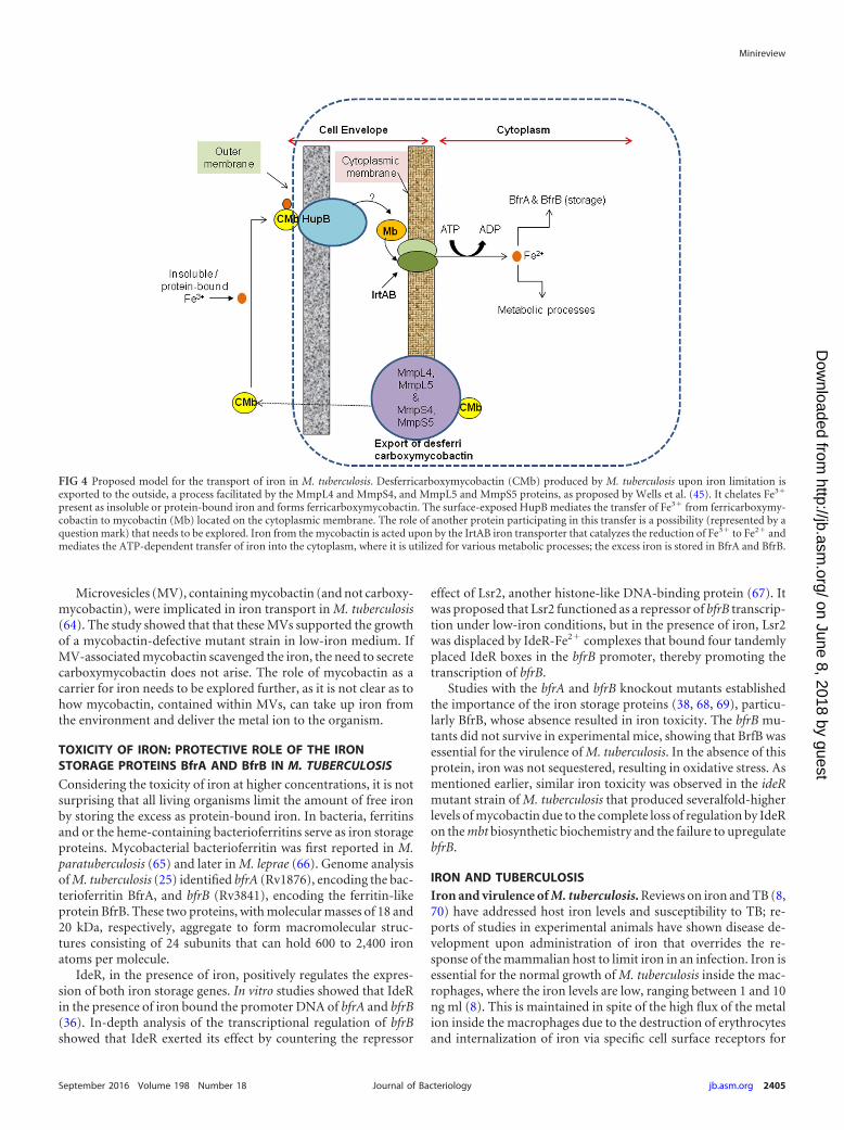
Proposed model for the transport of iron in M. tuberculosis.Figure 4 is a diagrammatic representation of the proposed modelof iron uptake in M. tuberculosis. When faced with low levels offree iron, the pathogen releases desferricarboxymycobactin intothe immediate environment. The MmpS4 and MmpS5 and theassociated MmpL4 and MmpL5 transport proteins are associatedwith this export process (45), and, once released the desferricar-boxymycobactin, chelates Fe3� from insoluble or protein-boundiron and forms ferricarboxymycobactin. Here, it is proposed thatiron from ferricarboxymycobactin is transferred to mycobactin inthe cell envelope of M. tuberculosis, with HupB functioning as theiron transporter. HupB is proposed as the iron transporterbased on its surface localization (60, 61), its property to bindFe3�, and interaction with ferricarboxymycobactin and ferri-mycobactin (62). HupB is not only seen in the 50S ribosomalsubunit (63) but in the cell wall (14), specifically on the cell surface(60). The purified protein bound radiolabeled ferricarboxymyco-bactin and ferrimycobactin in a dose-dependent manner, thespecificity of which was demonstrated by displacement of thebound label upon the addition of cold ferrisiderophore (61). AsHupB binds Fe3� (62), it is hypothesized that it mediates thetransfer of the metal from ferricarboxymycobactin to mycobactin,also reported in another study (13). The IrtAB transporter, local-ized on the cytoplasmic membrane, mediates the internalizationof the iron (47) after reduction of Fe3� to Fe2� by the FAD-de-pendent reductase activity of the IrtA protein. The metal ion, asFe2�, is transported into the cytoplasm by an energy-dependentprocess, and once inside, it is utilized for various metabolic pro-cesses, and the excess iron is stored in BfrA and BfrB.
The ESX-3 secretion pathway has been implicated in irontransport (48–50), and experimental evidence shows the essenti-ality of this pathway for the survival of M. tuberculosis inside mac-rophages. The transcription of esx-3 is controlled both by iron andzinc levels, with the participation of the respective iron and zincregulators IdeR and Zur. In M. smegmatis, only iron influencedthe expression of the components of esx-3. While knockout mu-tants show altered iron uptake, the exact role of the ESX-3 on irontransport is unclear.
Minireview
2404 jb.asm.org September 2016 Volume 198 Number 18Journal of Bacteriology
on June 8, 2018 by guesthttp://jb.asm
.org/D
ownloaded from
Microvesicles (MV), containing mycobactin (and not carboxy-mycobactin), were implicated in iron transport in M. tuberculosis(64). The study showed that that these MVs supported the growthof a mycobactin-defective mutant strain in low-iron medium. IfMV-associated mycobactin scavenged the iron, the need to secretecarboxymycobactin does not arise. The role of mycobactin as acarrier for iron needs to be explored further, as it is not clear as tohow mycobactin, contained within MVs, can take up iron fromthe environment and deliver the metal ion to the organism.
TOXICITY OF IRON: PROTECTIVE ROLE OF THE IRONSTORAGE PROTEINS BfrA AND BfrB IN M. TUBERCULOSIS
Considering the toxicity of iron at higher concentrations, it is notsurprising that all living organisms limit the amount of free ironby storing the excess as protein-bound iron. In bacteria, ferritinsand or the heme-containing bacterioferritins serve as iron storageproteins. Mycobacterial bacterioferritin was first reported in M.paratuberculosis (65) and later in M. leprae (66). Genome analysisof M. tuberculosis (25) identified bfrA (Rv1876), encoding the bac-terioferritin BfrA, and bfrB (Rv3841), encoding the ferritin-likeprotein BfrB. These two proteins, with molecular masses of 18 and20 kDa, respectively, aggregate to form macromolecular struc-tures consisting of 24 subunits that can hold 600 to 2,400 ironatoms per molecule.
IdeR, in the presence of iron, positively regulates the expres-sion of both iron storage genes. In vitro studies showed that IdeRin the presence of iron bound the promoter DNA of bfrA and bfrB(36). In-depth analysis of the transcriptional regulation of bfrBshowed that IdeR exerted its effect by countering the repressor
effect of Lsr2, another histone-like DNA-binding protein (67). Itwas proposed that Lsr2 functioned as a repressor of bfrB transcrip-tion under low-iron conditions, but in the presence of iron, Lsr2was displaced by IdeR-Fe2� complexes that bound four tandemlyplaced IdeR boxes in the bfrB promoter, thereby promoting thetranscription of bfrB.
Studies with the bfrA and bfrB knockout mutants establishedthe importance of the iron storage proteins (38, 68, 69), particu-larly BfrB, whose absence resulted in iron toxicity. The bfrB mu-tants did not survive in experimental mice, showing that BrfB wasessential for the virulence of M. tuberculosis. In the absence of thisprotein, iron was not sequestered, resulting in oxidative stress. Asmentioned earlier, similar iron toxicity was observed in the ideRmutant strain of M. tuberculosis that produced severalfold-higherlevels of mycobactin due to the complete loss of regulation by IdeRon the mbt biosynthetic biochemistry and the failure to upregulatebfrB.
IRON AND TUBERCULOSISIron and virulence of M. tuberculosis. Reviews on iron and TB (8,70) have addressed host iron levels and susceptibility to TB; re-ports of studies in experimental animals have shown disease de-velopment upon administration of iron that overrides the re-sponse of the mammalian host to limit iron in an infection. Iron isessential for the normal growth of M. tuberculosis inside the mac-rophages, where the iron levels are low, ranging between 1 and 10ng ml (8). This is maintained in spite of the high flux of the metalion inside the macrophages due to the destruction of erythrocytesand internalization of iron via specific cell surface receptors for
FIG 4 Proposed model for the transport of iron in M. tuberculosis. Desferricarboxymycobactin (CMb) produced by M. tuberculosis upon iron limitation isexported to the outside, a process facilitated by the MmpL4 and MmpS4, and MmpL5 and MmpS5 proteins, as proposed by Wells et al. (45). It chelates Fe3�
present as insoluble or protein-bound iron and forms ferricarboxymycobactin. The surface-exposed HupB mediates the transfer of Fe3� from ferricarboxymy-cobactin to mycobactin (Mb) located on the cytoplasmic membrane. The role of another protein participating in this transfer is a possibility (represented by aquestion mark) that needs to be explored. Iron from the mycobactin is acted upon by the IrtAB iron transporter that catalyzes the reduction of Fe3� to Fe2� andmediates the ATP-dependent transfer of iron into the cytoplasm, where it is utilized for various metabolic processes; the excess iron is stored in BfrA and BfrB.
Minireview
September 2016 Volume 198 Number 18 jb.asm.org 2405Journal of Bacteriology
on June 8, 2018 by guesthttp://jb.asm
.org/D
ownloaded from

transferrin, lactoferrin, and hemoglobin-haptoglobin. However,most of the iron is transferred to the bone marrow, and any freeiron is bound by transferrin and lactoferrin. Lactoferrin, by virtueof its ability to hold the metal ion even at acidic pH, plays animportant role in withholding iron to M. tuberculosis residingwithin the alveolar macrophages in patients with pulmonary TB.Thus, the elaboration of the siderophore machinery is necessary,as shown by the sequestration of the metal ion by M. tuberculosisfrom holotransferrin (71, 72) and from hololactoferrin (73, 74).
If iron was made available to the pathogen, as done experimen-tally in macrophage cultures or in experimental animals infectedwith M. tuberculosis, there was enhanced multiplication of thepathogen (3, 5, 75–77). When the iron was given along with ironchelator deferoxamine or apo-transferrin, there was inhibition ofgrowth of the pathogen, clearly establishing the role of iron in TB.These findings were later substantiated by using KO mutants withspecific defects in iron acquisition. As discussed earlier, mycobac-tin biosynthesis and transport via the siderophore system are es-sential for the in vivo survival of the pathogen, reaffirming theassociation of iron acquisition with virulence of the pathogen.
In vivo expression of HupB: can HupB serve as a biomarkerin TB patients? That the pathogen faces iron deprivation withinmacrophages is evident from the upregulation of the mbt genes(78, 79). It is thus likely that one or more components of the ironacquisition machinery can serve as marker(s) to reflect the ironstatus of the pathogen. Sivakolundu et al. (80) and Sritharan et al.(81) identified HupB as a putative marker in two independentstudies conducted on TB patients in India (80, 81). In both of thestudies performed on pulmonary and extrapulmonary TB pa-tients, two important observations were made. First, there wasnegative correlation of the titer of anti-HupB antibodies with se-rum iron levels, and second, anti-HupB antibody titers in ex-trapulmonary TB patients were notably high, with levels exceed-ing those seen in pulmonary TB patients. When the full-lengthHupB was used as antigen (80), anti-HupB antibodies in the se-rum level of extrapulmonary TB patients (optical density at 450nm [OD450], 1.230 0.341, P � 0.05, compared to 0.230 0.042in healthy endemic controls) were higher than those seen insmear-positive pulmonary TB patients (OD450, 0.678 0.205;P � 0.05). Interestingly, the titers were low in the household con-tacts of these patients (mean standard deviation [SD], 0.313 0.128), an observation of relevance due to the endemicity of thedisease. As mentioned, the antibody titer showed a statisticallysignificant (P � 0.01) negative correlation with serum iron andtotal iron binding capacity (TIBC); for example, the circulatingiron level in pulmonary TB patients was only 47.70 39.48 �gdl�1, compared to 107.74 45.74 �g dl�1 in the endemic healthycontrols (P � 0.05). The serum ferritin levels in TB patients werehigh, with some pulmonary patients showing values as high as2,500 ng ml�1 compared to the mean SD of 89.07 141.50 ngml�1 in the healthy controls. When three different antigenic frag-ments of HupB were used as antigens (81), HupB-F2 antigen bear-ing amino acids 63 to 161 was highly promising as an antigen,detecting remarkably high levels of anti-HupB antibodies in theserum of extrapulmonary TB patients. Also, the high antibodytiters in pulmonary TB patients with relapse of the disease sug-gested prolonged exposure of the tubercle bacilli, possibly as dor-mant bacilli to iron-limiting conditions inside the human host.
The potential of HupB as a marker for TB stems from thefinding that it can identify TB patients in a region that is endemic
for the disease and where BCG is used as a vaccine. This will haveconsiderable impact on the diagnosis of extrapulmonary TB,which is often difficult to diagnose with conventional tests. In fact,using hupB-specific primers, PCR identified M. tuberculosis as thecausative organism for the chronic inflammatory disease Takaya-su’s arteritis (82), comparable to the reference control amplifiedusing IS6110-specific primers.
CONCLUDING REMARKS
It is evident that iron is essential for the growth and survival of M.tuberculosis. The pathogen expresses the siderophores mycobactinand carboxymycobactin under iron-limiting conditions. It doesnot possess the exochelin machinery elaborated by the nonpatho-genic M. smegmatis and is thus dependent on the carboxymyco-bactin and mycobactin uptake system for acquiring this essentialmicronutrient. Thus, any disruption of the mbt biosynthetic ma-chinery affects its growth and survival, as demonstrated experi-mentally with the mbtB and hupB KO mutant strains. HupB playsan important role in sensing iron levels and functions as a positiveregulator of mycobactin biosynthesis. The clinical significance ofthe protein can be inferred from the high titers of anti-HupB an-tibodies in the serum of TB patients, the majority of whom pre-sented with low serum iron levels.
Remarkable progress has been made in understanding of thetranscriptional regulation of the mycobactin biosynthetic ma-chinery and expression of the iron storage proteins BfrA and BfrB.Iron homeostasis is a tightly controlled process, balancing ironuptake, utilization, and storage. Considering the essentiality ofiron and the various regulatory controls used by the pathogen tomaintain optimal iron for its growth and survival, it will be worthexploring these pathways to identify potential drug, vaccine, anddiagnostic targets. The diagnostic potential of HupB, particularlyfor extrapulmonary TB, is worth exploring. There is a need forbetter control measures for this dreaded disease, and advances indiagnosis, development of novel drugs, and vaccines are the needof the hour, with the iron acquisition machinery offering amplescope for this unmet need.
ACKNOWLEDGMENTS
I thank the funding bodies CSIR, DBT, and DST (Government of India)for funding some of the work presented in this review. I also thank theCommonwealth Commission for supporting the faculty fellowship and Split-Site student fellowships for my doctoral students to work in the TuberculosisResearch Group at the Veterinary Lab Agency, United Kingdom.
REFERENCES1. Posey JE, Gherardini FC. 2000. Lack of a role for iron in the Lyme disease
pathogen. Science 288:1651–1653. http://dx.doi.org/10.1126/science.288.5471.1651.
2. Chipperfield JR, Ratledge C. 2000. Salicylic acid is not a bacterial sidero-phore: a theoretical study. Biometals 13:165–168. http://dx.doi.org/10.1023/A:1009227206890.
3. Kochan I. 1973. The role of iron in bacterial infections, with special con-sideration of host-tubercle bacillus interaction. Curr Top Microbiol Im-munol 60:1–30.
4. Bullen JJ, Griffiths E. 1999. Iron-binding proteins and host defence, 2nded. John Wiley & Sons, Chichester, United Kingdom.
5. Lounis N, Truffot-Pernot C, Grosset J, Gordeuk VR, Boelaert JR. 2001.Iron and Mycobacterium tuberculosis infection. J Clin Virol 20:123–126.http://dx.doi.org/10.1016/S1386-6532(00)00136-0.
6. De Voss JJ, Rutter K, Schroeder BG, Barry CE, III. 1999. Iron acquisi-tion and metabolism by mycobacteria. J Bacteriol 181:4443– 4451.
7. Fang Z, Sampson SL, Warren RM, Gey van Pittius NC, Newton-Foot
Minireview
2406 jb.asm.org September 2016 Volume 198 Number 18Journal of Bacteriology
on June 8, 2018 by guesthttp://jb.asm
.org/D
ownloaded from

M. 2015. Iron acquisition strategies in mycobacteria. Tuberculosis (Ed-inb) 95:123–130. http://dx.doi.org/10.1016/j.tube.2015.01.004.
8. Ratledge C. 2004. Iron, mycobacteria and tuberculosis. Tuberculosis (Ed-inb) 84:110 –130. http://dx.doi.org/10.1016/j.tube.2003.08.012.
9. Rodriguez GM. 2006. Control of iron metabolism in Mycobacterium tu-berculosis. Trends Microbiol 14:320 –327. http://dx.doi.org/10.1016/j.tim.2006.05.006.
10. Liu J, Barry CE, III, Besra GS, Nikaido H. 1996. Mycolic acid structuredetermines the fluidity of the mycobacterial cell wall. J Biol Chem 271:29545–29551. http://dx.doi.org/10.1074/jbc.271.47.29545.
11. Braun V, Hantke K, Koster W. 1998. Bacterial iron transport: mecha-nisms, genetics and regulation. Met Ions Biol Syst 35:67–145.
12. Ratledge C, Patel PV, Mundy J. 1982. Iron transport in Mycobacteriumsmegmatis: the location of mycobactin by electron microscopy. J Gen Mi-crobiol 128:1559 –1565.
13. Gobin J, Horwitz MA. 1996. Exochelins of Mycobacterium tuberculosisremove iron from human iron-binding proteins and donate iron to my-cobactins in the M. tuberculosis cell wall. J Exp Med 183:1527–1532. http://dx.doi.org/10.1084/jem.183.4.1527.
14. Yeruva VC, Duggirala S, Lakshmi V, Kolarich D, Altmann F, SritharanM. 2006. Identification and characterization of a major cell wall-associated iron-regulated envelope protein (Irep-28) in Mycobacteriumtuberculosis. Clin Vaccine Immunol 13:1137–1142. http://dx.doi.org/10.1128/CVI.00125-06.
15. De Voss JJ, Rutter K, Schroeder BG, Su H, Zhu Y, Barry CE, III. 2000.The salicylate-derived mycobactin siderophores of Mycobacterium tuber-culosis are essential for growth in macrophages. Proc Natl Acad Sci U S A97:1252–1257. http://dx.doi.org/10.1073/pnas.97.3.1252.
16. Snow GA. 1965. The structure of mycobactin P, a growth factor for My-cobacterium johnei, and the significance of its iron complex. Biochem J94:160 –165. http://dx.doi.org/10.1042/bj0940160.
17. Snow GA, White AJ. 1969. Chemical and biological properties of myco-bactins isolated from various mycobacteria. Biochem J 115:1031–1050.http://dx.doi.org/10.1042/bj1151031.
18. Snow GA. 1970. Mycobactins: iron-chelating growth factors from myco-bacteria. Bacteriol Rev 34:99 –125.
19. Gobin J, Moore CH, Reeve JR, Jr, Wong DK, Gibson BW, HorwitzMA. 1995. Iron acquisition by Mycobacterium tuberculosis: isolation andcharacterization of a family of iron-binding exochelins. Proc Natl AcadSci U S A 92:5189 –5193. http://dx.doi.org/10.1073/pnas.92.11.5189.
20. Gobin J, Wong DK, Gibson BW, Horwitz MA. 1999. Characterization ofexochelins of the Mycobacterium bovis type strain and BCG substrains.Infect Immun 67:2035–2039.
21. Ratledge C, Ewing M. 1996. The occurrence of carboxymycobactin, thesiderophore of pathogenic mycobacteria, as a second extracellular sidero-phore in Mycobacterium smegmatis. Microbiology 142:2207–2212.
22. Macham LP, Ratledge C, Nocton JC. 1975. Extracellular iron acquisitionby mycobacteria: role of the exochelins and evidence against the partici-pation of mycobactin. Infect Immun 12:1242–1251.
23. Sharman GJ, Williams DH, Ewing DF, Ratledge C. 1995. Isolation,purification and structure of exochelin MS, the extracellular siderophorefrom Mycobacterium smegmatis. Biochem J 305:187–196.
24. Sharman GJ, Williams DH, Ewing DF, Ratledge C. 1995. Determinationof the structure of exochelin MN, the extracellular siderophore from My-cobacterium neoaurum. Chem Biol 2:553–561. http://dx.doi.org/10.1016/1074-5521(95)90189-2.
25. Cole ST, Brosch R, Parkhill J, Garnier T, Churcher C, Harris D, GordonSV, Eiglmeier K, Gas S, Barry CE, III, Tekaia F, Badcock K, Basham D,Brown D, Chillingworth T, Connor R, Davies R, Devlin K, Feltwell T,Gentles S, Hamlin N, Holroyd S, Hornsby T, Jagels K, Krogh A,McLean J, Moule S, Murphy L, Oliver K, Osborne J, Quail MA,Rajandream MA, Rogers J, Rutter S, Seeger K, Skelton J, Squares R,Squares S, Sulston JE, Taylor K, Whitehead S, Barrell BG. 1998. Deci-phering the biology of Mycobacterium tuberculosis from the complete ge-nome sequence. Nature 393:537–544. http://dx.doi.org/10.1038/31159.
26. Eiglmeier K, Parkhill J, Honore N, Garnier T, Tekaia F, Telenti A,Klatser P, James KD, Thomson NR, Wheeler PR, Churcher C, Harris D,Mungall K, Barrell BG, Cole ST. 2001. The decaying genome of Myco-bacterium leprae. Lepr Rev 72:387–398.
27. Hall RM, Wheeler PR, Ratledge C. 1983. Exochelin-mediated ironuptake into Mycobacterium leprae. Int J Lepr Other Mycobact Dis 51:490 – 494.
28. Quadri LE, Sello J, Keating TA, Weinreb PH, Walsh CT. 1998.
Identification of a Mycobacterium tuberculosis gene cluster encoding thebiosynthetic enzymes for assembly of the virulence-conferring sidero-phore mycobactin. Chem Biol 5:631– 645. http://dx.doi.org/10.1016/S1074-5521(98)90291-5.
29. Krithika R, Marathe U, Saxena P, Ansari MZ, Mohanty D, Gokhale RS.2006. A genetic locus required for iron acquisition in Mycobacterium tu-berculosis. Proc Natl Acad Sci U S A 103:2069 –2074. http://dx.doi.org/10.1073/pnas.0507924103.
30. Chavadi SS, Stirrett KL, Edupuganti UR, Vergnolle O, Sadhanandan G,Marchiano E, Martin C, Qiu WG, Soll CE, Quadri LE. 2011. Mutationaland phylogenetic analyses of the mycobacterial mbt gene cluster. J Bacte-riol 193:5905–5913. http://dx.doi.org/10.1128/JB.05811-11.
31. Schmitt MP, Predich M, Doukhan L, Smith I, Holmes RK. 1995.Characterization of an iron-dependent regulatory protein (IdeR) of My-cobacterium tuberculosis as a functional homolog of the diphtheria toxinrepressor (DtxR) from Corynebacterium diphtheriae. Infect Immun 63:4284 – 4289.
32. Chou CJ, Wisedchaisri G, Monfeli RR, Oram DM, Holmes RK, HolWG, Beeson C. 2004. Functional studies of the Mycobacterium tubercu-losis iron-dependent regulator. J Biol Chem 279:53554 –53561. http://dx.doi.org/10.1074/jbc.M407385200.
33. Pohl E, Holmes RK, Hol WG. 1999. Crystal structure of the iron-dependent regulator (IdeR) from Mycobacterium tuberculosis shows bothmetal binding sites fully occupied. J Mol Biol 285:1145–1156. http://dx.doi.org/10.1006/jmbi.1998.2339.
34. Semavina M, Beckett D, Logan TM. 2006. Metal-linked dimerization inthe iron-dependent regulator from Mycobacterium tuberculosis. Biochem-istry 45:12480 –12490. http://dx.doi.org/10.1021/bi060797s.
35. Wisedchaisri G, Chou CJ, Wu M, Roach C, Rice AE, Holmes RK,Beeson C, Hol WG. 2007. Crystal structures, metal activation, and DNA-binding properties of two-domain IdeR from Mycobacterium tuberculosis.Biochemistry 46:436 – 447. http://dx.doi.org/10.1021/bi0609826.
36. Gold B, Rodriguez GM, Marras SA, Pentecost M, Smith I. 2001. TheMycobacterium tuberculosis IdeR is a dual functional regulator that con-trols transcription of genes involved in iron acquisition, iron storage andsurvival in macrophages. Mol Microbiol 42:851– 865.
37. Rodriguez GM, Voskuil MI, Gold B, Schoolnik GK, Smith I. 2002. ideR,an essential gene in Mycobacterium tuberculosis: role of IdeR in iron-dependent gene expression, iron metabolism, and oxidative stress re-sponse. Infect Immun 70:3371–3381. http://dx.doi.org/10.1128/IAI.70.7.3371-3381.2002.
38. Pandey R, Rodriguez GM. 2013. IdeR is required for iron homeostasisand virulence in Mycobacterium tuberculosis. Mol Microbiol 91:98 –109.
39. Prabhakar S, Annapurna PS, Jain NK, Dey AB, Tyagi JS, Prasad HK.1998. Identification of an immunogenic histone-like protein (HLPMt) ofMycobacterium tuberculosis. Tuber Lung Dis 79:43–53. http://dx.doi.org/10.1054/tuld.1998.0004.
40. Soares de Lima C, Zulianello L, Marques MA, Kim H, Portugal MI,Antunes SL, Menozzi FD, Ottenhoff TH, Brennan PJ, Pessolani MC.2005. Mapping the laminin-binding and adhesive domain of the cell sur-face-associated HlpLBP protein from Mycobacterium leprae. Microbes In-fect 7:1097–1109. http://dx.doi.org/10.1016/j.micinf.2005.02.013.
41. Katsube T, Matsumoto S, Takatsuka M, Okuyama M, Ozeki Y, NaitoM, Nishiuchi Y, Fujiwara N, Yoshimura M, Tsuboi T, Torii M, OshitaniN, Arakawa T, Kobayashi K. 2007. Control of cell wall assembly by ahistone-like protein in mycobacteria. J Bacteriol 189:8241– 8249. http://dx.doi.org/10.1128/JB.00550-07.
42. Sharadamma N, Harshavardhana Y, Ravishankar A, Anand P, ChandraN, Muniyappa K. Molecular dissection of Mycobacterium tuberculosisintegration host factor reveals novel insights into the mode of DNA bind-ing and nucleoid compaction. Biochemistry 54:4142– 4160.
43. Pandey SD, Choudhury M, Sritharan M. 2014. Transcriptional regula-tion of Mycobacterium tuberculosis hupB gene expression. Microbiology160:1637–1647. http://dx.doi.org/10.1099/mic.0.079640-0.
44. Pandey SD, Choudhury M, Yousuf S, Wheeler PR, Gordon SV, RanjanA, Sritharan M. 2014. Iron-regulated protein HupB of Mycobacteriumtuberculosis positively regulates siderophore biosynthesis and is essentialfor growth in macrophages. J Bacteriol 196:1853–1865. http://dx.doi.org/10.1128/JB.01483-13.
45. Wells RM, Jones CM, Xi Z, Speer A, Danilchanka O, Doornbos KS, SunP, Wu F, Tian C, Niederweis M. 2013. Discovery of a siderophore exportsystem essential for virulence of Mycobacterium tuberculosis. PLoS Pathog9:e1003120. http://dx.doi.org/10.1371/journal.ppat.1003120.
Minireview
September 2016 Volume 198 Number 18 jb.asm.org 2407Journal of Bacteriology
on June 8, 2018 by guesthttp://jb.asm
.org/D
ownloaded from

46. Ratledge C, Dover LG. 2000. Iron metabolism in pathogenic bacteria.Annu Rev Microbiol 54:881–941. http://dx.doi.org/10.1146/annurev.micro.54.1.881.
47. Ryndak MB, Wang S, Smith I, Rodriguez GM. 2010. The Mycobac-terium tuberculosis high-affinity iron importer, IrtA, contains an FAD-binding domain. J Bacteriol 192:861– 869. http://dx.doi.org/10.1128/JB.00223-09.
48. Serafini A, Boldrin F, Palu G, Manganelli R. 2009. Characterization of aMycobacterium tuberculosis ESX-3 conditional mutant: essentiality andrescue by iron and zinc. J Bacteriol 191:6340 – 6344. http://dx.doi.org/10.1128/JB.00756-09.
49. Serafini A, Pisu D, Palu G, Rodriguez GM, Manganelli R. 2013. TheESX-3 secretion system is necessary for iron and zinc homeostasis in My-cobacterium tuberculosis. PLoS One 8:e78351. http://dx.doi.org/10.1371/journal.pone.0078351.
50. Siegrist MS, Unnikrishnan M, McConnell MJ, Borowsky M, ChengTY, Siddiqi N, Fortune SM, Moody DB, Rubin EJ. 2009. Mycobac-terial Esx-3 is required for mycobactin-mediated iron acquisition. ProcNatl Acad Sci U S A 106:18792–18797. http://dx.doi.org/10.1073/pnas.0900589106.
51. Dover LG, Ratledge C. 1996. Identification of a 29 kDa protein in theenvelope of Mycobacterium smegmatis as a putative ferri-exochelin recep-tor. Microbiology 142:1521–1530.
52. Hall RM, Sritharan M, Messenger AJ, Ratledge C. 1987. Iron transportin Mycobacterium smegmatis: occurrence of iron-regulated envelope pro-teins as potential receptors for iron uptake. J Gen Microbiol 133:2107–2114.
53. Sritharan M, Ratledge C. 1990. Iron-regulated envelope proteins of my-cobacteria grown in vitro and their occurrence in Mycobacterium aviumand Mycobacterium leprae grown in vivo. Biol Met 2:203–208. http://dx.doi.org/10.1007/BF01141360.
54. Sritharan M, Ratledge C. 1989. Co-ordinated expression of the compo-nents of iron transport (mycobactin, exochelin and envelope proteins) inMycobacterium neoaurum. FEMS Microbiol Lett 51:183–185.
55. Calder KM, Horwitz MA. 1998. Identification of iron-regulated proteinsof Mycobacterium tuberculosis and cloning of tandem genes encoding a lowiron-induced protein and a metal transporting ATPase with similarities totwo-component metal transport systems. Microb Pathog 24:133–143.http://dx.doi.org/10.1006/mpat.1997.9999.
56. Raghu B, Sarma GR. 1993. Isolation and characterization of sidero-phores and envelope proteins from mycobacteria. Biochem Mol BiolInt 31:333–339.
57. Wong DK, Lee BY, Horwitz MA, Gibson BW. 1999. Identification of fur,aconitase, and other proteins expressed by Mycobacterium tuberculosis un-der conditions of low and high concentrations of iron by combined two-dimensional gel electrophoresis and mass spectrometry. Infect Immun67:327–336.
58. Rodriguez GM, Smith I. 2006. Identification of an ABC transporterrequired for iron acquisition and virulence in Mycobacterium tuberculosis.J Bacteriol 188:424 – 430. http://dx.doi.org/10.1128/JB.188.2.424-430.2006.
59. Jones CM, Wells RM, Madduri AV, Renfrow MB, Ratledge C, MoodyDB, Niederweis M. 2014. Self-poisoning of Mycobacterium tuberculosis byinterrupting siderophore recycling. Proc Natl Acad Sci U S A 111:1945–1950. http://dx.doi.org/10.1073/pnas.1311402111.
60. Pethe K, Bifani P, Drobecq H, Sergheraert C, Debrie AS, Locht C,Menozzi FD. 2002. Mycobacterial heparin-binding hemagglutinin andlaminin-binding protein share antigenic methyllysines that confer resis-tance to proteolysis. Proc Natl Acad Sci U S A 99:10759 –10764. http://dx.doi.org/10.1073/pnas.162246899.
61. Yeruva VC. 2010. Iron limitation in Mycobacterium tuberculosis: studieson iron-regulated envelope proteins and catalase-peroxidases Universityof Hyderabad, Hyderabad, India.
62. Takatsuka M, Osada-Oka M, Satoh EF, Kitadokoro K, Nishiuchi Y,Niki M, Inoue M, Iwai K, Arakawa T, Shimoji Y, Ogura H, KobayashiK, Rambukkana A, Matsumoto S. 2011. A histone-like protein of myco-bacteria possesses ferritin superfamily protein-like activity and protectsagainst DNA damage by Fenton reaction. PLoS One 6:e20985. http://dx.doi.org/10.1371/journal.pone.0020985.
63. Lewin A, Baus D, Kamal E, Bon F, Kunisch R, Maurischat S,Adonopoulou M, Eich K. 2008. The mycobacterial DNA-binding
protein 1 (MDP1) from Mycobacterium bovis BCG influences variousgrowth characteristics. BMC Microbiol 8:91. http://dx.doi.org/10.1186/1471-2180-8-91.
64. Prados-Rosales R, Weinrick BC, Pique DG, Jacobs WR, Jr, CasadevallA, Rodriguez GM. 2014. Role for Mycobacterium tuberculosis membranevesicles in iron acquisition. J Bacteriol 196:1250 –1256. http://dx.doi.org/10.1128/JB.01090-13.
65. Brooks BW, Young NM, Watson DC, Robertson RH, Sugden EA,Nielsen KH, Becker SA. 1991. Mycobacterium paratuberculosis antigen D:characterization and evidence that it is a bacterioferritin. J Clin Microbiol29:1652–1658.
66. Pessolani MC, Smith DR, Rivoire B, McCormick J, Hefta SA, Cole ST,Brennan PJ. 1994. Purification, characterization, gene sequence, and sig-nificance of a bacterioferritin from Mycobacterium leprae. J Exp Med 180:319 –327. http://dx.doi.org/10.1084/jem.180.1.319.
67. Kurthkoti K, Tare P, Paitchowdhury R, Gowthami VN, Garcia MJ,Colangeli R, Chatterji D, Nagaraja V, Rodriguez GM. 2015. The myco-bacterial iron-dependent regulator IdeR induces ferritin (bfrB) by allevi-ating Lsr2 repression. Mol Microbiol 98:864 – 877. http://dx.doi.org/10.1111/mmi.13166.
68. Pandey R, Rodriguez GM. 2012. A ferritin mutant of Mycobacteriumtuberculosis is highly susceptible to killing by antibiotics and is unable toestablish a chronic infection in mice. Infect Immun 80:3650 –3659. http://dx.doi.org/10.1128/IAI.00229-12.
69. Reddy PV, Puri RV, Khera A, Tyagi AK. 2012. Iron storage proteins areessential for the survival and pathogenesis of Mycobacterium tuberculosisin THP-1 macrophages and the guinea pig model of infection. J Bacteriol194:567–575. http://dx.doi.org/10.1128/JB.05553-11.
70. McDermid JM, Prentice AM. 2006. Iron and infection: effects of hostiron status and the iron-regulatory genes haptoglobin and NRAMP1(SLC11A1) on host-pathogen interactions in tuberculosis and HIV.Clin Sci (Lond) 110:503–524. http://dx.doi.org/10.1042/CS20050273.
71. Clemens DL, Horwitz MA. 1996. The Mycobacterium tuberculosis phago-some interacts with early endosomes and is accessible to exogenously ad-ministered transferrin. J Exp Med 184:1349 –1355. http://dx.doi.org/10.1084/jem.184.4.1349.
72. Olakanmi O, Schlesinger LS, Ahmed A, Britigan BE. 2002. Intraphago-somal Mycobacterium tuberculosis acquires iron from both extracellulartransferrin and intracellular iron pools. Impact of interferon-gamma andhemochromatosis. J Biol Chem 277:49727– 49734.
73. Olakanmi O, Schlesinger LS, Ahmed A, Britigan BE. 2004. The nature ofextracellular iron influences iron acquisition by Mycobacterium tubercu-losis residing within human macrophages. Infect Immun 72:2022–2028.http://dx.doi.org/10.1128/IAI.72.4.2022-2028.2004.
74. Olakanmi O, Schlesinger LS, Britigan BE. 2007. Hereditary hemo-chromatosis results in decreased iron acquisition and growth by My-cobacterium tuberculosis within human macrophages. J Leukoc Biol 81:195–204.
75. Cronje L, Bornman L. 2005. Iron overload and tuberculosis: a case foriron chelation therapy. Int J Tuberc Lung Dis 9:2–9.
76. Cronje L, Edmondson N, Eisenach KD, Bornman L. 2005. Iron andiron chelating agents modulate Mycobacterium tuberculosis growth andmonocyte-macrophage viability and effector functions. FEMS Immu-nol Med Microbiol 45:103–112. http://dx.doi.org/10.1016/j.femsim.2005.02.007.
77. Schaible UE, Collins HL, Priem F, Kaufmann SH. 2002. Correction ofthe iron overload defect in beta-2-microglobulin knockout mice by lacto-ferrin abolishes their increased susceptibility to tuberculosis. J Exp Med196:1507–1513. http://dx.doi.org/10.1084/jem.20020897.
78. Schnappinger D, Ehrt S, Voskuil MI, Liu Y, Mangan JA, Monahan IM,Dolganov G, Efron B, Butcher PD, Nathan C, Schoolnik GK. 2003.Transcriptional adaptation of Mycobacterium tuberculosis within macro-phages: insights into the phagosomal environment. J Exp Med 198:693–704. http://dx.doi.org/10.1084/jem.20030846.
79. Timm J, Post FA, Bekker LG, Walther GB, Wainwright HC, ManganelliR, Chan WT, Tsenova L, Gold B, Smith I, Kaplan G, McKinney JD.2003. Differential expression of iron-, carbon-, and oxygen-responsivemycobacterial genes in the lungs of chronically infected mice and tuber-culosis patients. Proc Natl Acad Sci U S A 100:14321–14326. http://dx.doi.org/10.1073/pnas.2436197100.
80. Sivakolundu S, Mannela UD, Jain S, Srikantam A, Peri S, Pandey SD,Sritharan M. 2013. Serum iron profile and ELISA-based detection of
Minireview
2408 jb.asm.org September 2016 Volume 198 Number 18Journal of Bacteriology
on June 8, 2018 by guesthttp://jb.asm
.org/D
ownloaded from

antibodies against the iron-regulated protein HupB of Mycobacterium tu-berculosis in TB patients and household contacts in Hyderabad (AndhraPradesh), India. Trans R Soc Trop Med Hyg 107:43–50. http://dx.doi.org/10.1093/trstmh/trs005.
81. Sritharan N, Choudhury M, Sivakolundu S, Chaurasia R, Chouhan N,Rao PP, Sritharan M. 2015. Highly immunoreactive antibodies againstthe rHup-F2 fragment (aa 63–161) of the iron-regulated HupB protein ofMycobacterium tuberculosis and its potential for the serodiagnosis of ex-
trapulmonary and recurrent tuberculosis. Eur J Clin Microbiol Infect Dis34:33– 40. http://dx.doi.org/10.1007/s10096-014-2203-y.
82. Soto ME, Del Carmen Avila-Casado M, Huesca-Gomez C, Alarcon GV,Castrejon V, Soto V, Hernandez S, Espinola-Zavaleta N, Vallejo M,Reyes PA, Gamboa R. 2012. Detection of IS6110 and HupB gene se-quences of Mycobacterium tuberculosis and bovis in the aortic tissue ofpatients with Takayasu’s arteritis. BMC Infect Dis 12:194. http://dx.doi.org/10.1186/1471-2334-12-194.
Minireview
September 2016 Volume 198 Number 18 jb.asm.org 2409Journal of Bacteriology
on June 8, 2018 by guesthttp://jb.asm
.org/D
ownloaded from