ebooksclub.org advances in applied microbiology volume 50 archaea ancient microbes extreme...
TRANSCRIPT


PREFACE
Biologists divide living organisms into prokaryotes, represented by the bacteria, and eukaryotes such as ourselves. In this scheme, bacteria are considered to be the most primitive form of life. One popular idea about how life arose supposes that humans evolved from bacteria. In this age of genomics, however, DNA sequence comparisons indicate that our genes are unlike those of bacteria. Where, then, did we come from? An answer to that question comes from a truly unexpected source, life in extreme environments.
It was generally thought that boiling acid hot springs or saturating saline lakes were sterile, however, life is both present and highly suc- cessful. Many of the resident organisms are still microbes, just not bac- teria. We call them archaea. Whole-genome DNA sequences of five ar- chaeal species reveal remarkable gene matches to human genes and those of other eukaryotes. These matches occur in the most essential of the subcellular processes carried out by all organisms, the synthe- sis and repair of DNA, RNA, and protein. This suggests that eukaryotes evolved from archaea or perhaps that archaea and eukaryotes derive from a common ancestor.
Gene sequences aside, archaea lay additional claim to the title of an- cient organism based on geologic and taxonomic considerations. The early Earth (Archean age) was a time of elevated surface temperatures. Fossil dating indicates the presence of microbes at the close of this pe- riod, suggesting that earlier forms of life from which these fossils would have derived must have been adapted to temperature extremes. Mi- crobes called hyperthermophiles with just these abilities are still found on the Earth in geothermal springs and hydrothermal ocean vents. These extreme locations exhibit geochemistries most like that of early Earth. To understand how such organisms relate to other forms of life, taxo- nomic methods based on ribosomal RNA sequences have been used to create phylogenetic “trees” of life. These hyperthermophilic microbes, dominated by the archaea, exhibit the deepest phylogenetic branches, suggesting that they have undergone the longest period of evolution among extant organisms. Taken together, these ideas suggest that hyper- thermophilic archaea may represent a form of the earliest type of life.
The intent of this book is to expand the general understanding of the archaea. As simple organisms with sequenced genomes, they present unique opportunities to understand better our own origins and, indeed,
ix

X PREFACE
the origin of life. As prokaryotes they provide powerful experimental systems for genetic and molecular experimentation.
Acknowledgments
I am most grateful for the support and interest of many colleagues, in- cluding Rolf Bernander, Mike Dyall-Smith, Peter Kennelly, John Leigh, William Metcalf, Kenneth Noll, Frank Robb, Richard Shand, Dieter Soll, and William Whitman. The pioneering interests of Thomas Brock, Richard Morita, Norman Pace, Carl Stetter, Carl Wose, and Wolfram Zillig helped create my interest in extreme environments and the evo- lutionary implications of life native to such habitats.

INTRODUCTION
Ribosomal RNA gene sequence comparisons separate life into three distantly related groups or domains (Fig. 1). Eukaryotes constitute one domain, encompassing single- and multicelled organisms such as plants and animals. Despite their morphologic simplicity and apparent simi- larity, the other two domains are both prokaryotic. These domains are represented by the bacteria and the archaea. Differences in rRNA se- quence are but one feature leading to this classification system. Most scientists are well versed about the evidence which distinguishes bac- terial from eukaryotic life. This includes a diversity of mechanisms governing all aspects of basic cell biology, from gene organization to gene expression, from signal transduction to metabolism. What, then, beyond differences in rRNA sequence supports the separate classifi- cation of the archaea from bacterial prokaryotes and eukaryotes? As predicted by their rRNA sequence divergence and by analogy to the dissimilarity between bacteria and eukaryotes, archaea are likely to em- ploy archaeal-specific subcellular mechanisms for conducting essential processes. While the existence of archaeal-specific subcellular mech- anisms is supported by the occurrence of a unique group of archaeal orthologous genes, the functions of these genes remain unknown. The truly remarkable discovery which is the focus of this book concerns the finding that archaea use eukaryotic-like mechanisms and not bacterial mechanisms for much of their information processing functions.
The literature on bacterial prokaryotes is both extensive and diverse, however, such is not yet the case for archaea. As a recognized group, archaea are relative newcomers to the prokaryotic world. Despite their recent entry into the research arena, studies on archaea are blossom- ing, largely in response to the availability of whole-genome DNA se- quences. Three distinctive biotypes of archaea are best known: the methanogens, the halophiles, and the hyperthermophiles. Due to the radical nature of their respective niches, these organisms have been dubbed extremophiles. Studies on life in extreme environments portend many exciting areas in biology research including studies on the origins of life and the possibility of extraplanetary organisms. Archaea have contributed, and will continue to contribute, greatly to these areas. How- ever, phylogenetic studies of marine, dirt, and other environmental sam- ples indicate that some archaea are not extremophilic but mesophilic members of microbial communities. More importantly, these types of
xi

xii INTRODUCTION
‘Plants
FIG. 1. Archaeal, bacterial, and eukaryotic sequences were aligned with ClustalW. The programs DNADIST, NEIGHBOR, CONSENSE, and FITCH, of the Phylip package, were used to build the tree. The alignment was bootstrapped 100 times with SEQBOOT.
archaea appear to constitute a significant proportion of the total plane- tary microbial biomass.
This book provides an overview of key aspects of the archaea includ- ing chapters on their genomics and phylogeny, micropaleontology, cell biology, and molecular genetics. This information should be useful to microbiology students at both the graduate and the undergraduate lev- els. More importantly, this text should focus the attention of researchers on the importance of archaea as model systems to address fundamental biological questions. As prokaryotes, archaea can and should be used to wield the power of haploid genetics providing cost-efficient systems for scientific investigations into life.

Paleobiology of the Archean
SHERRY L. CADY
Department of Geology Portland State University
PO. Box 751 Portland, Oregon 97207-0751
I. Introduction II. Historical Review
A. Main Phases of Archean Paleobiological Studies B. Reviews of Archean Paleobiology
III. Evidence for Life-Microbial Biosignatures A. Bona Fide Microfossils B. Microbialities and Biogenic Stromatolites C. Chemofossils-Biomarkers and Isotopes
IV. Criteria for Assessing Biogenicity V. Biogeochemical Interactions and Biosignature Formation
VI. Extreme Ecosystems VII. Geological and Paleobiological History of the Archean (4000-2500 Ma)
A. Oldest Dated Terrestrial Rocks and Minerals B. Oldest Types of Crustal Terrane C. Chemofossils, West Greenland D. Oldest Microfossils E. Microfossils in Hydrothermal Deposits F. Oldest Stromatolites G. Oldest Isotopic Evidence of a Specific Metabolic Pathway
in Archean Evaporite Deposits H. Evidence of Hydrothermal Oil Generation during the Archean I. Oldest Compelling Chemofossil Evidence of Methanogenic Archaea J. Oxygenic Phototrophy
VIII. Conclusion References
I. Introduction
Four billion years ago our planet began accumulating a rock record, a geological event that heralded the beginning of the Archean Eon (Fig. 1). Though the Earth itself is approximately 4550 million years old, the oldest known rocks are the 4030 million-year-old Acasta gneisses of northwestern Canada (Bowring and Williams, 1999). These rocks, and others of comparable age, indicate that the ancient Earth harbored a number of environments that could have supported life. The presence of even older minerals, 4400 million-year-old detrital zircons from the Yilgarn Craton, Western Australia (Wilde et al., ZOOl), provides evidence
3
ADVANCES IN APPLIED MICROBIOLOGY, VOLUME 50 Copyright 0 2001 by Academic Press
All rights of reproduction in any form reserved. 0065.2164/01535.00

4 SHERRY L. CADY
LOGIC TIME SC 2 e 3 Selected benchmarks 4 (I)
4Dinosaurextlnctloo,oldest primates 1 8
+Oldesl green algae
e
::
II
*
4Oldesl redalgae
+Sudburysstrobleme
+Oldest macrofossils (Grypsmaj
(I
~Oldestcarbonateplatfotms
~~IdesreucatKptlcblpmarkers ldestcyano actertal btomarkers
a
4-Oldest hydrothermal mwofossils
4 Oldest microfowls, stromatol!les**,
+Marflan dublofosslls(ALH84001)
+Oldestchemofosslls
~Oldestdatedlerrestr~al rocks
+Olde?.tdaied tef18Stl~almlneralS
+Orlgin of Earth, oldest metwriteS
FIG. 1. Geological time scale. Geologic time is represented here by numerically desig- nated bins of geologic time, each 100 million years long. The intervals, called geons (from GEOlogical EON), are counted from the present. The geon scale allows for Earth history (and that of other planets) to be viewed in a succession of large, simply named, equal time units, in the same way that centuries and millennia are used for familiar smaller intervals.
PALEOBIOLOGY OF THE ARCHEAN 5
that the Earth was tectonically active shortly after it formed. Without plate tectonic activity commencing early and persisting throughout our planet’s history, it is unlikely that life would exist today. It is yet a mystery when life began; its origin is not revealed in the paleobiological record. Chemical fossil evidence in rocks from West Greenland indicate, however, that microbial life had evolved by 3700 Ma (mega-annum; meaning million years) (Rosing, 1999). The possibility exists that life emerged within a few hundred million years after crustal rocks began accumulating and began to leave traces in the paleobiological record. The Archean Eon, by definition, ends at precisely 2500 Ma.
Fossil evidence indicates that once life emerged it evolved rapidly and occupied new ecological niches as soon as they became available (e.g., hydrothermal systems, shallow coastlines, continental seas). The Archean paleobiological record is preserved in geological units that consist primarily of chemical precipitates and volcanogenic sediments. Such deposits accumulated on the edges of accreting continental cra- tons and along the rims of protocontinents. Although only meager bits of evidence have been discovered to date, new and significant discoveries regarding Archean paleobiology and paleoecology continue to be made.
In general, paleobiological information comes from a potentially wide range of fossil data known collectively as biosignatures. Evidence for the existence of the Archean biosphere is preserved in the fossil remains of microorganisms (i.e., microfossils), in the sedimentary structures mi- croorganisms helped construct (i.e., stromatolites), and in the biomarker compounds, isotopic signatures, and biominerals life leaves behind (i.e., chemofossils). Bona fide microfossils, carbonaceous microorgan- isms preserved in three dimensions, contain structural and chemical rem- nants of cellular and extracellular components (e.g., cell walls, sheathes, exopolysaccharides), which together can provide direct evi- dence for life. Also, microbially influenced fabrics and structures that form in ecosystems where authigenic minerals precipitate and rock de- tritus accumulate provide indirect evidence of microscopic life forms. Microbially influenced accretionary growth structures and fabrics in- clude nonlaminated microbialites and laminated biogenic stromatolites. Although both types of biogenic structures are found throughout the Archean, they usually lack microfossils. Chemical fossils (“chemofos- sils”), such as biomarkers and isotopic signatures, also provide indirect
The geon scale complements the traditional, history-based scale with complex names and unequal geologic periods, much like dynasties in history overlap centuries. The Archean Eon is defined as that interval of time that began with the formation of the oldest geological record and that ends at 2500 Ma (mega annum; meaning million years), which is the beginning of the Proterozoic Eon. [Reproduced with permission from Hofmann (1990, 1992, 2000).]

6 SHERRYL.CADY
evidence for life. Biomarker compounds consist of organic molecules highly diagnostic for their parent organisms. Elements that can be iso- topically fractionated by biological or biochemical processes include carbon, sulfur, and nitrogen.
Not only have a number of types of fossil biosignatures shown that life emerged and spread globally during the early Archean, but evidence for life has been reported from all of the major Archean cratons. The pa- leobiological record has shown that life occupied several niches during the Archean and, within a few hundred million years, diversified and became metabolically sophisticated. Although fossil evidence cannot provide precise data as to when a metabolic innovation evolved, it can reveal that a particular metabolic capability existed. Chemofossil evi- dence demonstrates that many major metabolic pathways evolved dur- ing the Archean. Though the data for these evolutionary innovations are scattered throughout the ancient rock record, the continuous and relatively robust paleontological record of the Proterozoic Eon (2500 to 543 Ma) has confirmed that prior to the end of the Archean, analogues of modern microbial ecosystems, including those considered extreme, were established.
Several lines of evidence suggest that the earliest microbial inhabi- tants occupied sediments and colonized newly formed minerals that precipitated around hydrothermal vents, environments characterized by high temperatures and an abundance of dissolved metal ions (Nisbet, 1995). As shown in Fig. 2 molecular phylogenetic analysis of the small-subunit ribosomal RNA of extant life indicates that hy- perthermophiles in the bacterial and archaeal domains are the closest living relatives of the hypothetical last common ancestor (Pace, 1997). Whether life originated at high temperatures (Shock and Schulte, 1998) is not known, and evidence for a hyperthermophilic ancestry has been challenged (Doolittle, 2000; Galtier et al., 1999). The possibility exists that hyperthermophiles emerged during that brief but finite period of time after the collision that presumably formed the Earth-Moon sys- tem; after the collision (-4450 to 4500 Ma), when the Earth began to cool, surface temperatures could have hovered around 100°C for 100 to 200 million years (Sleep et al., 2001). The catastrophic conditions created by large impactor events during the latter part of the heavy bombardment period (-3900 to 3800 Ma) would also have created and sustained high-temperature habitats that could have supported hy- perthermophilic communities; large impactors ( >GIOO km in diameter) could have vaporized the ocean, while smaller impactors (>200 km in diameter) might have been able to heat the ocean above 100°C (Sleep and Zahnle, 1998). Alternatively, if life originated at lower temper- atures, and then radiated and adapted to high-temperature environ- ments, hyperthermophiles living in the subsurface portions of deep-sea

PALEOBIOLOGY OF THE ARCHEAN
3ACTERIA 3 f
FIG. 2. Universal phylogenetic tree based on SSU rRNA sequences. The largest formal unit of the tree consists of domains that include the Archaea, Bacteria, and Eukarya. The tree can be considered a rough map of the evolutionary distance between the organisms shown. Although the nature of the hypothetical last common ancestor is not known, hyperthermophilic and thermophilic microbial lineages are clustered around the root of the microbial domains. Constructed by comparison of 16s small-subunit ribosomal RNA sequences, the tree includes 64 rRNA sequences representative of all known phylogenetic domains. [Reproduced with permission from Pace (lggi’).]
hydrothermal systems may have been the only microbial communities that could have survived the impact-induced ocean sterilizing events (Maher and Stevenson, 1988).
The Earth has always been a volcanically active planet, and hydrother- mal activity prevailed after liquid water became stable at the Earth’s

8 SHERRY L. CADY
surface. Estimates of a threefold greater heat flux on the early Earth suggest that there was a greater amount of hydrothermal activity during the Archean (Turcotte, 1980). The combination of hydrothermal activity and life can produce macroscopically and microscopically recognizable biosignatures. Where molten magma intrudes within a few kilometers of the Earth’s surface, it heats subsurface fluids that rise and erupt through hydrothermal effluents at deep-sea vents on the ocean floor. In conti- nental settings, fluids emerge into hot springs and geysers. In any case, the interaction between hot fluids and the crustal rock through which it passes alters the chemistry of the fluid. Dissolved ions and gases be- come concentrated in the hot fluids that boil upon ascent, and minerals precipitate in the open fractures through which the boiling fluids pass. Reduced gases, if separated from boiling hydrothermal fluids, find al- ternative pathways to the surface, where they form acidic fumaroles (i.e., gas vents). The remaining dissolved ions precipitate as metal-rich hydrothermal deposits when subaqueous oceanic hydrothermal fluids mix with overlying seawater. If the fluids emerge from subaerial hot spring and geyser effluents, hydrothermal minerals precipitate as sinter deposits when the fluids cool and evaporate. Hyperthermophilic and thermophilic microbial communities are likely to have thrived in these types of environments since life originated.
Paleontological interpretations of biosignatures rely upon compar- isons with modern counterparts. Comparisons with modern-day or- ganisms assessed within a taphonomic framework that considers how microbial cells become degraded and altered with time places paleobio- logical evidence in a phylogenetic and functional framework. Paleoen- vironmental interpretations place fossil biosignatures in a geological context characterized by physical and chemical constraints. Because rocks and the organic remains they contain become altered with time as a result of diagenetic and metamorphic processing, it becomes more difficult to draw biological and environmental analogies from biosig- natures found in older deposits. Furthermore, it is unlikely that extant microbial communities or their environments are exact homologues of their ancient counterparts, given the expanse of time and the secular changes that have occurred on Earth since ancient microbial lineages originated. Even so, the physical and chemical processes that operated in ancient ecosystems continue to do so today. By applying what is known about how microbial life influences its environment, and vice versa, we can continue to extract meaningful paleobiological informa- tion from the ancient rock record.
The geological record indicates that the Earth evolved in a number of ways during the Archean. On a global scale, the crust differenti- ated from the core and solidified. The oceans were established; the

PALEOBIOLOGYOFTHEARCHEAN 9
atmosphere stabilized. The geological record indicates the present style of plate tectonics ensued by the end of Archean. The combination of rock weathering, crustal generation, and crustal recycling throughout the Archean produced a variety of environments that rapidly became inhabited as microbial communities spread worldwide. As illustrated in Fig. 3, hydrothermal systems would also have been in existence since crustal rocks began forming in the Archean.
II. Historical Review
A. MAINPHASES OFARCHEANPALEOBIOLOGICALSTUDIES
An analysis of the nature and timing of publications related to Archean paleobiology reveals that research in the field has undergone three main phases since the midnineteenth century (Schopf and Walter, 1983). As discussed recently by Hofmann (ZOOO), each phase was “defined by a different tempo of significant contributions to our know- ledge of the most ancient fossil record.”
Archean paleobiology began with the published description of Eozoijn Cormdense (Dawson, l875), laminated structures of putative biological origin. The Eozob;n Cunadense structures were subsequently dismissed as biogenic structures, but the controversy associated with their origin reflects the nature of paleontological research. In fact, de- bate regarding the origin of the earliest reported structures is similar to present-day controversies associated with the claims of putative micro- fossils in extraterrestrial materials (McKay et al., 1996) and the origin of stromatolite structures in general (Grotzinger and Rothman, 1996). The first phase ended just after the beginning of the twentieth century with the discovery of the first real stromatolites in Archean rocks at Steep Rock Lake, Ontario (Lawson, 1912; Walcott, 1912).
During the second phase, between 1912 and the early 196Os, the first Archean microfossils were reported from iron-bearing chert formations in northern Minnesota (Gruner, 1923, 1924). These early reports met with skepticism (Hawley, 1926), and over 30 years passed until the first unequivocal microfossils were reported from the -2100 million- year-old chert of the Gunflint Formation (Tyler and Barghoorn, 1954). A second discovery of microfossils in Proterozoic-age chert deposits (Barghoorn and Schopf, 1965) compelled most paleobiologists to study rocks of similar age and composition, a sound strategy that continued for several decades. Convincing Archean stromatolites were discovered during this period, and again, initial reports were questioned, in some cases even by the scientists who originally discovered the objects (for a complete list of references see Hofmann, 2000). The first actively

10 SHERRY L. CADY
FIG. 3. Map of microbial ecology established during the Archean Eon. (A) Num- bers refer to typical settings in the habitat model: (1) mid-ocean ridges (chemotrophic communities, including hyperthermophiles); (2) shallow coastal waters (oxygenic and

PALEOBIOLOGY OF THE ARCHEAN 11
forming stromatolites were discovered during this period along the shoreline of Shark Bay, Western Australia (Logan, 1961).
Widespread interest in the history of early life increased exponen- tially during the third phase of study, which began in the mid 1960s and continues to the present. Detailed investigations of stromatolites in modern ecosystems led to the development of actualistic models for Archean and Proterozoic paleobiology (e.g., Hofmann, 1973; Walter 1976). The effects of diagenesis on the fidelity of microbial biosignatures were also systematically addressed during this period (e.g., Knoll and Golubic, 1979). Numerous discoveries were made of Archean and Proterozoic microfossils and stromatolites, and of their nonbiological mimics (Schopf, 1999; Hofmann, 2000). Decades of intensive geologi- cal and paleobiological studies of Archean sediments, and the carbo- naceous matter they contain, have refined our views of the emergent biosphere (e.g., Lowe and Byerly, 1999). The nature of early microor- ganisms, as revealed by molecular phylogenies and hypotheses regard- ing early Earth environments, has stimulated new theories of life’s origins. A new understanding of the types of habitats in which life could have thrived during the Archean has emerged (e.g., Buick and Dunlop, 1990; Bengtson, 1994; Mojzsis and Harrison, 2000; Nisbet and Sleep, 2001).
The search for the oldest evidence of terrestrial life has intensified as a result of the search for life beyond Earth. Three critical areas where paleobiological research can be expanded has arisen from extraterres- trial search strategies for Mars (Cady, 1998): (1) recent discoveries of novel ecosystems have increased the number of potential paleobiolog- ical repositories that may harbor evidence of early microbial commu- nities on Earth and elsewhere-these ecosystems must be systemati- cally investigated to extract the maximum amount of paleobiological and paleoenvironmental information from ancient analogue deposits; (2) a fundamental understanding of the physical, chemical, and biolog- ical forces that contribute to the preservation of biogenic signatures in systems whose geochemical context is well understood is needed if we are to have any chance of deciphering fossilized evidence of life from
anoxygenic photosynthesis); (3) oxygenic photosynthetic mats and stromatolites around oceanic komatiite shields and continental andesitic island arc volcanoes; (4) open ocean [photosynthetic plankton, muds in the seafloor (methanogens and organisms dependent upon the sulfur cycle)). The arrows in the individual habitats show the direction of fluid movement. (B) Evolutionary heritage follows the standard evolutionary model based on the universal phylogenetic tree shown in Fig. 2. (GE) Columns show possible mat com- munities and biofilms along with chemical products; numbers refer to typical settings in the habitat model. [Reproduced with permission from Nisbet and Sleep (~ooI).]

12 SHERRY L. CADY
small samples returned from other planets, whose geological context will be vague at best; and (3) research regarding the origins of biosigna- tures requires characterization at the submicroscopic scale for a number of reasons-to understand the geomicrobiological processes that led to the lithification of microbial mats and biofilms, to identify the mecha- nisms by which microbial cells are fossilized, and to refine and expand the chemofossil database. Understanding which extrinsic environmen- tal factors and intrinsic biochemical attributes are responsible for the preservation ofbiosignatures will provide the cornerstone needed to dis- tinguish microfossils and stromatolites from their nonbiological mimics (Cady, 2001). The challenges faced in detecting ancient life on Earth are the same challenges faced in our search for life on other planetary bod- ies (i.e., Mars and Europa). These challenges are forcing us to refine our ability to detect evidence of life in ancient rocks (e.g., Conrad and Nealson, 2001).
B. REVIEWS OF ARCHEAN PALEOBIOLOGY
A number of seminal papers on Archean paleobiology have been published during the past two decades. In the book Earth’s Earliest Biosphere, edited by Schopf (1983), all aspects of Archean paleobiology are thoroughly reviewed, including paleobiological aspects of the origin of life (Chang et al., 1983), the Archean organic geochemical (Hayes et al., 1983) and carbon, sulfur, hydrogen, and nitrogen isotopic records (Schidlowski et al., 1983), Archean biochemistry (Gest and Schopf, 1983; Chapman and Schopf, 1983), Archean stromatolites (Walter, 1983), microfossils (Schopf and Walter, 1983), and environmental (Walker et al., 1983) and ecological (Hayes et al., 1983; Schopf et al., 1983; Schopf, 1999) evolution. The book The Proterozoic Biosphere, edited by Schopf and Klein (1992), includes updates on Archean paleo- biology (Schopf and Klein, 1992), paleobiological aspects of origin of life (Kasting and Chang, 1992), Archean microfossils (Schopf, 1992), and geological and environmental evolution (Lowe, 1992). Major implica- tions of the Archean microfossil stromatolite (Walter, 1994) and organic geochemical (Hayes, 1994) records are discussed in the Nobel Sympo- sium Volume EarlyLife on Earth, edited by Bengtson (1994). Additional noteworthy publications include a recent review by Hofmann (2000) of Archean stromatolites; a review by Knoll (1996) of paleontological evi- dence for Archean fossils, with special emphasis on their paleoecology, biostratigraphy, and evolution; an overview of theories and paleobi- ological evidence related to the origin of life by McClendon (1999); and critical reviews of the criteria by which the biogenicity of ancient microfossils and stromatolites are assessed by Buick (1981, 1990).

PALEOBIOLOGY OF THE ARCHEAN 13
III. Evidence for Life-Microbial Biosignatures
Biosignatures indicative of ancient life include cellularly preserved microfossils and carbonaceous cellular and microbial mat remains, molecular fossils known as biomarkers, biologically fractionated iso- topic signatures, and stromatolites and microbialites. Each fossil type provides a unique indicator for some aspect of life. When found to- gether in the geological record, the different types of biosignatures can provide mutually reinforcing lines of evidence; multiple biosignatures strengthen the case for life. Bona fide microfossils, characterized by chemical and structural biogenic attributes, provide direct evidence of life.
A. BONA FIDE MICROFOSSILS
Bona fide microbial fossils retain a portion of the biochemical and structural remains of their cellular components. The remains are recog- nizable either by their physical structure (as biomarker compounds) or by their degree of complexity (higher-order structures such as layered cell walls). Bona fide microfossils include permineralized and nonmin- eralized cellular remains (Schopf and Walter, 1983). Permineralized cells retain carbonaceous compounds composed of complex organic biopolymers that, by definition, retain enough morphological fidelity to be recognizable (Schopf, 1975). Newer instruments that allow the detection and characterization of structural and chemical evidence for life from the same object, particularly when that object looks like the remains of extant life, will have a significant impact on the ability to identify taxonomic affinity.
B. MICROBIALITIES AND BIOGENIC STROMATOLITES
A long-standing controversy exists over the definition and mean- ing of the term stromatolite. The debate centers on whether a genetic or descriptive definition should be used. The potential for stromato- lites to form in a variety of environments, including extreme ecosys- tems, via various contributions from different types of microorgan- isms (phototactic and nonphototactic) demonstrates the restrictions imposed by genetic definitions of stromatolites. A nongenetic definition for stromatolites recommended by Semikhatov and colleagues (1979) states that a stromatolite is “an attached, laminated, lithified sedimen- tary growth structure, accretionary away from a point or limited sur- face of initiation.” This description allows for the exploration of the various roles of biological and nonbiological processes in stromatolite

14 SHERRYL.CADY
accretion. Even though stromatolites may have multiple or even in- determinate origins, their basic geometric and textural properties are included in Semikhatov’s definition. Another definition by Hofmann (2000) acknowledges the potential for biology to be involved in the morphogenesis of the structures without compromising the non- genetic physical description. Hofmann (2000) asserts that “stromato- lites are morphologically circumscribed accretionary growth structures with a primary lamination that is, or may be, biologically influenced (biogenic).” With either definition, the aim is to determine which com- ponents of stromatolites or microbialites indicate the involvement or former presence of life during their construction. The potential for stro- matolites to harbor evidence of early microbial life is a matter of partic- ular relevance to the Archean. Stromatolites can preserve evidence of the early biosphere and, more specifically, of benthic ecosystems on the early Earth (Walter, 1994). Because stromatolites are macroscopic struc- tures, they are potentially recognizable by remote imaging on planetary rovers and can assist in determining locations to search for past extra- terrestrial life.
C. CHEMOFOSSILS-BIOMARKERSAND~OTOPES
Biomarker compounds, organic molecules that are highly diagnos- tic for their parent organisms, are particularly revealing with regard to metabolic innovations. As our understanding of the amount of bio- diversity in modern ecosystems improves and our database of unique biomarker compounds attributed to individual taxa expands, biomarker compounds will continue to reveal much about early life. Stable iso- topes, when found in abundance, are generally metabolically haction- ated. Elemental isotopic indicators of life, which can sometimes be linked to rather specific metabolic processes, are relatively resistant to alteration by geological processes such as thermal metamorphism. Subsequent discoveries of reliable chemofossils throughout Earth his- tory are needed to establish a continuous record for assessing biomarker compounds (e.g., Summons and Walter, 1990).
IV. Criteria for Assessing Biogenicity
Since microfossils have the potential to provide definitive evidence for life, robust criteria for assessing their validity have been established. In general, acceptable evidence of past life must meet five stringent cri- teria (Schopf and Walter, 1983) that include demonstrating the follow- ing: (1) the source (i.e., provenance) of the rock sample that contains the

PALEOBIOLOGYOFTHEARCHEAN 15
putative biological features must be capable of preserving paleobiolog- ical information, (2) the age of the rock sample must be known with appropriate precision, (3) the putative biological features must be in- digenous to the rock sample within which they were found, (4) the putative biological features must be syngenetic with (i.e., formed at the same time as) the primary mineral phases of the rock sample, and (5) the features of the putative fossils must be biogenic in origin. Proving the biogenicity of fossil evidence is often the most challenging criterion.
A number of criteria for assessing the biogenicity of microstructures found in ancient rocks have been proposed (e.g., Muir, 1978; Cloud and Morrison, 1979; Awramik et al., 1983; Schopf and Walter, 1983; Buick, 1990). The most rigorous classification system for microfossils, as pro- posed by Buick (1990), states that structures can be considered biogenic if they (1) occur in a petrographic thin section, (2) occur in sedimen- tary or low-grade metasedimentary rock, (3) are larger than the small- est extant free-living microorganisms (-0.01 pm3), (4) are composed of kerogen, (5) occur with others of similar morphology, (6) are hollow, and (7) show cellular elaboration, This classification system differen- tiates pseudofossils that do not meet criteria 1 through 4, dubiofossils (Hofmann, 1972) that satisfy only criteria 1 through 4, possible micro- fossils that meet criteria 1 through 5, and probable microfossils that fulfill criteria 1 through 6. Existing criteria for demonstrating microfos- sil biogenicity continue to be modified, refined, and challenged (e.g., Westall, 1999).
A parallel topic for paleobiological study includes experiments de- signed to determine the mechanisms by, rates at, and conditions un- der which pseudofossils form and become preserved in the geological record. Objects not accepted by all specialists in the field as bona fide evidence of life continue to be reported in peer-reviewed publications. But as noted by Hofmann (ZOOO), such objects should not be discounted completely; more precise criteria will be forthcoming as new instru- ments for detecting and characterizing life’s biosignatures are devel- oped. Until such time, though, only the most stringent criteria should be used when assessing the biogenicity of evidence for ancient terres- trial life or, as in the case for Mars, extraterrestrial life.
V. Biogeochemical Interactions and Biosignature Formation
Microorganisms interact with their environment in numerous ways that alter the chemistry and physical characteristics of their environ- ment. The environment also creates circumstances that favor the preser- vation of microbial biosignatures. The strategy used by paleobiologists

16 SHERRY L. CADY
to explore the ancient fossil record of microbial life on Earth is based upon locating specific rock types and paleoenvironments thought to have the highest potential for capturing and preserving fossil biosig- natures. The most informative assemblages of organically preserved microfossils are found in mid to late Proterozoic sediments deposited in marine and lacustrine environments. Microbial fossils in these de- posits occur either as three-dimensional cellularly preserved (permin- eralized) forms embedded in a relatively stable mineral matrix or as two-dimensional compressed or flattened forms (acritarchs) preserved in fine-grained, typically clay-rich detrital sediments. Microbial permin- eralization occurred primarily in shallow evaporative peritidal environ- ments of elevated salinity (e.g., Knoll, 1985), whereas acritarchs were preserved primarily in fine-grained siliciclastic sediments that formed in a wide range of marine environments. Although there are probably no bona fide acritarchs known from Archean deposits (H. Hofmann, per- sonal communication), permineralized fossils, albeit rare, have revealed much about the antiquity of life and the nature of the early Earth.
In recent years, the discovery of microbial life in a number of ex- treme environments, along with the realization that extreme ecosys- tems have existed throughout Earth’s history, has expanded the search for microbial biosignatures. Exopaleobiological and astropaleobiologi- cal search strategies also focus on extreme ecosystem deposits, espe- cially hydrothermal deposits, as potential extraterrestrial paleobiologi- cal repositories (Walter and Des Marais, 1993; Walter et al., 1996, 1998; Farmer and Des Marais, 1999). Ancient sedimentary chert deposits have produced permineralized microfossils with a high cellular fidelity. A wide range of rock types has revealed evidence of life in the form of car- bon and sulfur isotopic signatures. Microbial biosignatures are likely to have been preserved in any type of rock that was altered by sedimen- tary processes (e.g., infiltration of mineral-charged fluids in cracks and fissures in igneous and metamorphic rocks). Indeed, the voluminous subsurface biosphere could harbor biosignatures in any crevice where fluids have passed.
VI. Extreme Ecosystems
Extreme environments often represent modern analogues for ancient ecosystems. The environmental extremes of such ecosystems exclude higher-order taxa that would normally compete with or graze upon mi- crobial mats or biofilms. Much remains to be learned regarding the de- tection and proper interpretation of paleobiological and paleoecological information from ancient analogues of modern extreme environments- a reliable and continuous database of microbial biosignatures from
PALEOBIOLOGY OF THE ARCHEAN 17
sequentially older deposits has yet to be established. A continuous fos- sil record is important because of the deleterious effects of diagenetic and taphonomic processes on the fidelity of structural and chemical biosignatures. Modern extreme ecosystems provide an opportunity to study the processes by which paleobiological information is preserved in the fossil record. For example, a study of modern hot spring deposits in Yellowstone National Park (Cady et al., 1995; Cady and Farmer, 1996) revealed that hyperthermophilic biofilms can influence the microstruc- tural development of high-temperature siliceous sinter deposits pre- viously considered to have formed abiologically. The ways in which microbial communities interact with their environment and impact global biogeochemical cycles can also be studied in real time in ex- treme ecosystems. Extreme ecosystems also provide geochemical and physical constraints to refine experimental studies.
VII. Geological and Paleobiological History of the Archean (4000-2500 Ma)
Discoveries from the geological and paleobiological records that pro- vide insight regarding the potential to find evidence of Archaea in the Archean paleobiological record are discussed below. Unfortunately, lit- tle direct evidence has been discovered regarding the distribution and occurrence of Archaea in the rock record, and future systematic searches that involve extreme ecosystem analogue studies are clearly warranted.
A. OLDEST DATED TERRESTRIAL ROCKS AND MINERALS
Isotopic data from meteorites and the Moon place the age of the so- lar system at approximately 4600 Ma and the age of the Earth at ap- proximately 4560 Ma (Patterson, 1956; Papanastassiou and Wasserburg, 1971). Evidence that crustal formation and weathering processes were active within the first few hundred million years after the Earth accreted is found in the oldest minerals (-4300 Ma), detrital zircon crystals from the Yilgarn Craton, Western Australia. Although these detrital crystals are the oldest indigenous objects found on Earth (Wilde et al., ZOOl), they reside in younger sedimentary deposits, having been eroded out of the rocks in which they crystallized. Fortunately, the oxygen isotopic signatures of the detrital zircon crystals reveal that they crystallized from magmas containing a significant component of reworked continen- tal crust that formed in the presence of water near the Earth’s surface (Mojzsis et cd., 2001). Even though all crustal rocks of Hadean age were recycled, isotopic evidence from the oldest minerals has revealed much about the way in which the early Earth operated.

18 SHERRYL.CADY
B. OLDESTTYPESOFCRUSTALTERRANE
Three types of rocks make up the oldest crustal terranes: gneissic terranes that have been highly metamorphosed, greenstone terranes that consist of volcanic rock overlain by metasediments, and cratonal margin deposits (Lowe and Ernst, 1992). Recall that the oldest rocks are from gneissic terranes (e.g., Mojzsis et al., 2001; Bowring et al., 1989). Greenstone terranes harbor the oldest microbial biosignatures.
The oldest known greenstone belt, the 3800 million-year-old Isua supracrustal belt in West Greenland, consists of thick units of volcanics, conglomerates, banded iron formation, chert, and detrital sediments. As discussed below, the oldest putative chemofossils have been found in Isua supracrustal rocks. A younger generation of approximately 3500 to 3200 million-year-old greenstone belts includes the Barberton Greenstone Belt and related rocks in the Kaapvaal Craton, South Africa, and greenstone belts in the eastern Pilbara Block, Western Australia. The lower units of these rock complexes consist primarily of volcanic rocks: the upper units include sedimentary rocks, such as silicified or carbonated volcanic tuffs, cherts, and evaporites. These sedimentary rocks, as discussed below, contain the oldest reported stromatolites and carbonaceous microfossils. The absence of large stable cratons with extensive shallow continental shelves during most of the Archean limited the distribution of ancient continental margin and terrestrial microbial ecosystems. The ages of crustal blocks formed during the Archean Eon indicate that nearly 90% of the crust formed during the Neoarchean, between 2700 and 2500 Ma (e.g., Lowe, 1994).
While it is apparent that the crustal lithosphere expanded dramat- ically during the Archean, secular changes also altered the composi- tion of the Archean atmosphere, hydrosphere, and biosphere (e.g., re- viewed by Lowe, 1994; Des Marais, 1994; Des Marais, 1997; Mojzsis and Harrison, 2000; Nisbet and Sleep, 2001). Although details are still forthcoming, it is apparent that the Archean hydrosphere was character- ized by a lower pH (i.e., a higher [Fe(II)]), and the Archean atmosphere was characterized by a higher partial pressure of CO2 and a significantly lower partial pressure of 0, (Holland, 1984). Without an oxygen-rich at- mosphere, the Earth would have experienced a higher ultraviolet (UV) flux during the Archean. The sun was also 30% less luminous at 3800 Ma than it is today (Kuhn et al., 1989). These conditions at the Earth’s surface would have restricted the number of suitable environments for microbial inhabitants. Given that photosynthesis directly powers >99% of the modern biosphere’s productivity (e.g., Des Marais, 1997), it is clear that microbial life during the Archean differed in significant ways from that of today.

PALEOBIOLOGYOFTHEARCHEAN 19
C. CHEMOFOSSILS, WEST GREENLAND
Chemofossil evidence indicative of autotrophic carbon dioxide fixa- tion has been found in -3700 million-year-old seafloor sediments from the Isua supracrustal belt in West Greenland (Rosing, 1999). Even older chemofossil evidence for autotrophy has been found in -3850 million- year-old banded iron formations from Akilia Island, West Greenland (Mojzsis et al., 1996). As noted by Mojzsis and Mark (2000), the age of the sediments within which the oldest chemofossils are found is signifi- cant. These rocks would have been deposited during the period of in- tense meteor bombardment as recorded on the Moon at 3800 to 3900 Ma, which coincided with the period of time when liquid water existed on the surface of Mars. Whether these chemofossils represent the oldest evidence of life on Earth is still debated. The carbon isotopic signatures were obtained from carbon blebs that occur as inclusions in apatite. A more detailed look at the occurrence could not substantiate the previous claims of the age and origin of the host rock (Myers and Crowley, 2000). The discovery by Rosing (1999), however, of carbon of possible biogenic origin in >3i’OO million-year-old schists interpreted as metasedimen- tary marine rocks in the Isua greenstone belt provides more plausible evidence of life. These rocks are less altered by metamorphic processes, and absolute ages have been obtained from all of the tectonostratigraphic units that occur in association with the metasediments. It is interesting to note that the carbon isotopic values for the carbonaceous inclusions in apatite from -3770-Ma Isua sediments (613C = -30 per mil) and the -3850-Ma banded iron formation from Akilia island (613C = -37 per mil) closely match those from a variety of younger, less metamor- phosed rock. Mojzsis et al. (1996) propose that the simplest interpreta- tion indicates the presence of diverse photosynthesizing, methanogenic, and methanotrophic bacteria on Earth before 3850 Ma (Mojzsis and Arrhenius, 1998; Mojzsis and Harrsion, 2000). However, as discussed below, a more significant 13C depletion (up to -60 per mil) in the organic fraction of sediments is attributed to the combination of phototrophs, methanogens, and methanotrophs. To date, chemofossils provide the only evidence consistent with life in Eoarchean-age (4000- to 3600-Ma) rocks.
D. OLDEST MICROFOSSILS
The oldest microbial fossils and stromatolites discovered to date have been found in greenstone belts of two Archean terranes of nearly the same age in the Pilbara Craton in Western Australia and in the Kaapvaal

20 SHERRY L. CADY
Craton of South Africa. The nature of the microfossils indicates that they represent distinctly different types of microbial communities.
Microfossils from the Pilbara Block of northwestern Western Australia consist of cyanobacterium-like filaments from the -3465-Ma Apex Formation (Fig. 4) and sheath-enclosed colonial unicells of the Towers Formation (Schopf and Packer, 1987; Schopf, 1993). The cellular level
FIG. 4. Microfossils (with interpretive drawings) from the Paleoarchean (-3465-Ma) Apex Chert Formation, Pilbara Block, Western Australia. All of the microfossils, pho- tographed under plane light in thin sections, are the representative holotypes of micro- bial taxa. n. gen., new genera; n. sp., new species, (A) Primoevifilum amoenum Schopf (1992). (B) Primoevifilum conicoterminatum Schopf (1992). (C) Eoleptonema apex, n. sp. (D) Archaeoscillatoriopsis maxima, n. gem, n. sp. (E) Primaevifilum minutum, n. sp. (F) Primaevifilum attenuatum, n. sp. (G) Primaevifilum laticellulosum, n. sp. (H) Archaeoscil- latoriopsis disciformis, n. gen., n. sp. (I) Archaeoscillatoriopsis grandis, n. gen., n. sp. (J) Primaevifilum delicatulum Schopf (1992) (holotype). [K) Archaeotrichion septatum, n. sp. (Reproduced with permission from Schopf (1993).1

PALEOBIOLOGY OF THE ARCHEAN 21
of organization, morphological complexity, and similarity to younger microorganisms support a biogenic origin for the objects (Schopf, 1993). All of the Apex fossils occur in subangular to rounded siliceous sedi- mentary clasts that are less than 1 mm to a few millimeters in diameter. As shown in Fig. 4, 11 taxa of filamentous microfossils have been de- scribed from the Apex chert. Though the microfossils were reported as being carbonaceous, hence considered bonafide, this claim has recently been challenged (Brasier et al., 2001). The irregular distribution and random orientation of the solitary filaments in the clasts preclude spec- ulation as to whether the microfossils represent a benthic or planktonic community. The filaments are surrounded by fine-grained particles of kerogen (i.e., carbonaceous matter) hypothesized to be the remains of mucilaginous extracellular components. Like most ancient fossil finds, the Apex microfossil assemblage is poorly preserved. Schopf (1993) noted that less than 1% of the filaments detected in the deposit warrant detailed study and formal description. Although most of the Apex mi- croorganisms resemble trichomic oscillatoriacean cyanobacteria, their specific taxanomic affinity cannot be established because of the pro- found gap (865 million years) in the fossil record between the Apex fossil assemblage and younger, morphologically similar microfossils. An unbroken fossil record of diverse cyanobacterial families begins in the Neoarchean with the -2600-Ma Campbell Group fossils (Altermann and Schopf, 1995).
Microfossils of the Kromberg Formation in the -3259-Ma upper- most Hooggenoeg chert from Kaapvaal Craton of South Africa include two distinct populations of narrow, nonseptate bacterium-like filaments (Fig. 5). The most abundant filaments are thread-like ones reported by Walsh and Lowe (1985), ranging in diameter from 0.1 to 0.6 pm and in length from 10 to 150 pm. The other population of tubular filaments has diameters of 1.4 to 2.2 pm and lengths of from 10 to 150 pm. Walsh (1992) noted that the fossiliferous filaments, especially the tubular ones, display morphologies similar to those of modern filamentous bacteria. Different types of filaments co-occur with one another, and the filaments often extend from one lamina to another, as do motile filamentous bac- terial and cyanobacteria in layered microbial mats (Walsh, 1989,1992). Preservation of the cellular morphology of many of the filaments has been attributed to the presence of pyrite crystallites that encrust the cel- lular remains. Walsh (1992) assessed the biogenicity of the filamentous objects using the criteria discussed previously as proposed by Buick. For example, although the filament shown in Fig. 5D is interpreted as a sheathed set of trichomes (i.e., Fig. 5E), the filament is classified as a dubiofossil since it does not occur with others of similar morphol- ogy. Solid filamentous objects, like the ones shown in Figs. 5A and B, are classified as possible microfossils. Only hollow filaments, like

22 SHERRY L. CADY
FIG. 5. Filamentous microfossils (with interpretive drawing) from the Paleoarchean (-3416-Ma) Onverwacht Group, Barberton Mountain Land, South Africa. (A) Solid thread-like filament (possible microfossil). (B) Solid filament with hair-like projections and apparent branching (microfossil). (C) Hollow cylindrical filament (bone fide micro- fossil). (D) Solitary large hollow filament with trichomes extending from the flaring end (dubiofossil). (E) Interpretive sketch of D. Scale bar equals 5 wrn for A and 12 wrn for B-E. [Reproduced with permission from Walsh (1989, 1992).]
the one shown in Fig. X, are classified as bone fide microfossils. The filamentous microfossils are associated with fossilized evidence of mi- crobial mats or biofilms preserved as various types of carbonaceous mat- ter (Fig. 6). These varieties of carbonaceous matter are relatively com- mon compared to the microfossil-like objects. The presence of layered microbial communities, however, suggests that taxis behavior (photo- taxis and chemotaxis) cannot be distinguished in fossilized mats from the rock record. Therefore it is not yet known whether the organisms occupied shallow- or deep-water environments, and the exact details of their occurrence remain unresolved.

PALEOBIOLOGY OF THE ARCHEAN 23
FIG. 6. Variety of carbonaceous matter in cherts from the Paleoarchean (-3416-Ma) Onverwacht Group, Barberton Mountain Land, South Africa. Plane-light photomicro- graphs of thin sections of rock ground to a 30+m thickness. [A) Fine carbonaceous micro- bial mat-like laminations with scattered simple and composite grains in a fossiliferous sample. Scale bar = 200 Wm. (B) Mat-like laminations folded over on themselves and loose detrital fragments of carbonaceous laminations. Scale bar = 1 mm. (C) Layer of simple carbonaceous grains that overlies mat-like laminations in a fossiliferous sample. Scale bar = 200 Wm. (D) Layer of composite carbonaceous grains with a botryoidal coating of silica cement. Scale bar = 1 mm. (E) Partially flattened carbonaceous wisps in massive black chert. Scale bar = 206 pm. (F) Layer of very fine grained cloudy carbonaceous matter in a fossiliferous sample. Scale bar = 206 Wm. [Reproduced with permission from Walsh and Lowe (1999).]

24 SHERRYL.CADY
E. MICROFOSSILS NHYDROTHERMALDEPOSITS
The oldest putative hydrothermal fossils were recently discovered in a 3235 million-year-old deep-sea volcanogenic massive sulfide deposit that formed in a submarine thermal spring system found preserved in the Pilbara Craton of Australia. Pyrite crystals located along their en- tire length encrust the filaments. Morphologically, the mineralized fila- ments appear to be thread-like, unbranched, and of uniform thickness along their length (0.5-Z ,um in diameter, up to 300 km long) (Fig. 7). The filaments range from straight to sinuous and sharply curved, and they are intertwined when found in high numbers. Rasmussen (2000) proposed that their biogenicity is demonstrated by their sinuous mor- phology, lengthwise uniformity, intertwined habit, and morphological attributes comparable to those of Archean and younger microfossils. Their mode of occurrence indicates that the microorganisms were prob- ably chemotrophs that inhabited the pores and crevices of rocks at shal- low depths below the seafloor. The presence of mineralized filaments preserved in hydrothermal silica within a volcanic massive sulfide de- posit is consistent with a thermophilic community preserved in an an- cient subseafloor hydrothermal system. The probable absence of dis- solved oxygen in the deep Archean ocean and the abundance of reduced chemical species in seafloor hydrothermal fluids suggest that chemical pathways for metabolic processes were probably anaerobic. The abun- dance of reduced sulfur in the hydrothermal fluids, as evidenced by the encrustation of filaments by pyrite and iron sulfide, suggest that sulfur may have been central to energy-yielding reactions for micro- bial growth. Although there is no direct evidence that the mineralized filaments are the fossilized remains of Archaea, the geological setting is consistent with such an interpretation. If the mineralized filaments are subsequently deemed to be compelling evidence of microbial life, the discovery will extend the known range of submarine hydrothermal biota by more than 15% million years. The oldest known hydrother- mal fossil microorganisms are found in the Early Proterozoic (1640-Ma) McArthur River (HYC) lead-zinc-silver deposit of northern Australia (Oehler, 1976; Oehler and Logan, 1977). Hydrocarbon biomarkers from the ore and associated sediments indicate the former presence of sulfide oxidizing bacteria (Logan et al., 2001).
F. OLDESTSTROMATOLITES
Nine occurrences of stromatolites from the Paleoarchean are known (e.g., Hofmann, 2000) and their biogenicity has been challenged (Buick et al., 1981; Buick, 1990; Lowe, 1994). While it has been argued that

FIG. 7. Filaments from the Paleoarchean (-3235-Ma) volcanogenic massive sulfide de- posit from the Pilbara Craton of Australia. Plane-light photomicrographs of thin sections. (A-F) Straight, sinuous, and curved morphologies, some densely intertwined. (G) Fila- ments parallel to concentric layering. (H) Filaments oriented subperpendicularly to band- ing. Scale bar = 10 ym. [Reproduced with permission from Rasmussen (ZOOO).]

26 SHERRY L. CADY

PALEOBIOLOGY OF THE ARCHEAN 27
similar structures could be formed by inorganic precipitation and soft- sediment deformation (Lowe, 1994), others (Buick et al., 1981; Buick, 1990, Hofman et al., 1999) have argued that at least some structures are neither deformational nor precipitational and are likely to have had a biological origin. Lowe (1995) noted that the diagnostic criteria for biogenicity could be equally applied to “abiogenic geyserites” and speleothems. Interestingly, geomicrobiological studies of “abiogenic structures” in modern ecosystems where columns and spicules of gey- serite form around modern hot springs (Cady et al., 19%; Cady and Farmer, 1996) and columnar speleothems (“pool fingers”) form in sub- merged pools in caves (Boston et al., 2001) indicate that microbial biofilms have contributed, in part, to the microstructural and morpho- logical development of these structures.
A recent reexamination of conically laminated stromatolites from the Warrawoona group of the North Pole Dome by Hofmann and colleagues (1999) provides an illustrative example of ancient stromatolite struc- tures that record a biogenic contribution to their construction (Fig. 8). Evidence that biofilms contributed to the formation of the structures includes the nature of the laminations of the conical pseudocolumns (Fig. 8B), their microstructure (Fig. 8C), the distribution of similar forms over tens of square kilometers, and their similarity to younger ancient and modern biogenic stromatolites. As proposed by Hofmann (ZOOO), until a known genesis for a stromatolitic structure is demonstrated, “Structures that are possibly partly biogenic should continue to receive appropriate scrutiny deserving of laminated dubiofossils [whose ori- gins are unknown), because they may have relevance in elucidating the Archean biosphere.”
FIG. 8. Conically laminated stromatolites from the Paleoarchean (-3450-Ma) Warra- woona Group, North Pole, Western Australia. (A) Outcrop exposure of a section oriented approximately perpendicularly to bedding and showing two pseudocolumns composed of uniform and wavy conical layers. Horizontal layers in between the conical stromato- lites display a more irregular layer thickness. (B-E) Examples of the microstructure of the conical stromatolites when viewed in thin section under cross-polarized plane light. En- larged views circumscribed by rectangles are shown sequentially. The microstructure of these ancient stromatolites is secondary due to recrystallization of the primary minerals and silicification from a later influx of silica-rich fluids. However, even at the microscopic scale, detailed characteristics of the primary banded layers are preserved. Laminations consist of alternating light (silica] and dark (carbonate) layers with irregular contacts. The lower contact of the dark carbonate layers is more evenly developed than the top contact, a feature observed in some younger stromatolites with preserved organic matter. [Reproduced with permission from Hofmann et al. (1999).]

28 SHERRY L. CADY
G. OLDEST ISOTOPIC EVIDENCE OF A SPECIFIC METABOLIC PATHWAY IN ARCHEAN EVAPORITE DEPOSITS
Chemofossil evidence supports the hypothesis that microorganisms were metabolically diverse by the Paleoarchean (3600-3200 Ma). Con- vincing evidence of microbial sulfate reduction has recently been found in microscopic sulfides from -3470 million-year-old barites from North Pole, Australia (Shen et al., 2001). The sulfides display maximum iso- topic fractionations (21.1 per mil) that could not have been produced by volcanogenic processes. The barites came from layers of gypsum crystals replaced by barite, a process that requires the infiltration of rel- atively low-temperature barium-rich solutions. The microscopic sulfide crystals, localized along the growth faces of the original gypsum crys- tals, provide a means to determine the relative fractionation of cogenetic sulfides and sulfates. Although dissimilatory sulfate reduction occurs in both the archaeal and the bacterial domains, the only known archaeal sulfate reducer is a single genus, Archaeoglobus, a hyperthermophile characterized by an optimal growth temperature higher than 80°C. Con- sideration of the processes by which the original gypsum would have be- come baritized indicates barium transport by relatively cool hydrother- mal fluids with temperatures below 60°C. The find indicates a minimum age of 3470 Ma for the emergence of sulfate-reducing microorganisms in the bacterial domain. This find extends the geological record of mi- crobial sulfate reduction by more than 750 Ma and represents the oldest evidence of a specific metabolic pathway. The preservation of evaporitic sediments from several Paleoarchean cratons indicates that shallow ma- rine and terrestrial conditions may have prevailed over a considerable portion of the primeval Earth (Buick and Dunlop, 1990). These envi- ronments could have supported diverse communities of microbial life (Groves et al., 1981).
H. EWDENCE OF HYDROTHERMAL OIL GENERATION DURING THE ARCHEAN
Hydrocarbon droplets preserved in hydrothermally silicified kero- geneous sediments have been reported from the Warrawoona Group (~3458 Ma) (Buick et al., 1998). Although textural evidence indicates that the hydrocarbons have not migrated from their source rock, ther- mal maturation of organic matter does not appear to have resulted from burial but, rather, from hydrothermal heating by convecting fluids at shallow depths. No other fossil biosignatures indicative of an indige- nous or fluid-transported microbial community are associated with this ancient hydrocarbon occurrence. On the other hand, evidence of hydro- carbons trapped in fluid inclusions and bituminous residues in cavities

PALEOBIOLOGYOFTHBARCHEAN 29
from the Pilbara craton of Australia have been found in a Paleoarchean deep-sea volcanogenic massive sulfide deposit (-3235 Ma) (Rasmussen and Buick, 2000). In addition to providing the oldest evidence for hy- drothermal oil generation in a deep-sea volcanogenic setting, the depo- sit, as discussed above, has yielded the oldest probable fossil remains of thermophiles (Rasmussen, 2000). The presence of pyrobitumen within the sulfide suggests that oil was a significant component of the hy- drothermal fluids. Rasmussen and Buick (2000) have proposed the geo- logical occurrence and isotopic signatures of the oil indicate that a granitic intrusion produced a subsurface heating event which formed the volcanogenic massive sulfide deposit and inadvertently generated oil within a younger, highly carbonaceous shale unit. The migrating oil became entrained in the convectively circulating hydrothermal fluids and trapped within the sulfide deposit while it precipitated. This dis- covery indicates that subseafloor hydrothermal petroleum generation was active during the Paleoarchean and that some of the sulfur might have been derived by hydrothermal reduction of sulfate using hydro- carbons as a reductant.
I. OLDESTCOMPELLINGCHEMOFOSSILEVIDENCEOF~THANOGENICARCHAEA
Although direct evidence of methanogenesis has not been found in the geological record, data from ancient carbon and sulfur isotopes of kero- gens and sulfide minerals (Schidlowski and Aharon, 1992; Grassineau et al., 2001) and from ancient carbon isotopes of sedimentary organic carbon and carbonates (Hayes et al., 1983) require the presence of methanogens as early as 2800 Ma. Biogeochemical considerations indi- cate that methanogens could have played an important role in the carbon cycle from the time when autotrophy arose. In the absence of respira- tory COZ production, they would have provided the only biochemical means of remobilizing carbon that had been fixed by autotrophs (Hayes, 1994). Though methane reaching the atmosphere would be oxidized by photochemically driven reactions in the absence of methanotrophy, no 13C-depleted organic material would form. Also, if the environment was anaerobic and methanogens were not geographically or environmentally restricted, 13C-enriched carbonates would not form. However, the com- bination of oxygenic photosynthesis, methanogenesis, and methanotro- phy is hypothesized to have produced a globally detectable anomalous isotopic signal during the Neoarchean (2800-2500 Ma).
Isotopically light (heavily 13C-depleted) primary biomass (sedimen- tary organic carbon) characterizes most of the Archean isotopic carbon record. As detailed by Hayes (1994), the most plausible scenario is that a single mechanism of primary production has been globally dominant

30 SHERRY L. CADY
since 3500 Ma (3800), and that some process was added to the global carbon cycle about 2750 million years ago. This process would have pro- duced highly depleted 13C sedimentary organic carbon (to -60 per mil). By about 2000 Ma, the impact of the process apparently waned and no longer exerted an important influence on the 13C content of sedimentary organic matter. As noted by Hayes (1994), the only process proposed to date that is consistent with the observed isotopic variation involves the recycling of methanogenic-produced methane by methanotrophs, the latter converting the 13C depleted methane into biomass and bio- logical debris that is the precursor of sedimentary organic carbon. The global distribution and simultaneous appearance of isotopically light sedimentary organic carbon are interpreted as indicating that methan- otrophy, hence methanogenesis and oxygenic photosynthesis, was of global significance (Hayes, 1994).
J. OXYGENIC PHOTOTROPHY
Though numerous hypotheses that describe the sequence of events that led to the emergence of oxygenic phototrophy have been proposed, constraints discovered to date from the paleobiological and geological record include the following. An anomalous isotopic shift caused by the coupled biogeochemical cycling between Archaea and Bacteria, accom- panied by the apparent onset of methane cycling by at least 2750 million years ago, caused a steady decline in the isotopic difference between sedimentary organic materials and cogenetic carbonates as recorded in the rock record (Schidlowski, 1988). Microfossil evidence consistent with the emergence of oxygenic phototrophy has been discovered in rocks at least as old as -2600 Ma (Altermann and Schopf, 1995). The possibility exists that some of the most ancient Archean microfossils were oxygenic phototrophs (e.g., Fig. 4).Walter (1983) and Buick (1992) proposed that some Neoarchean stromatolites must be cyanobacterial in origin. As discussed by Buick (1992), Neoarchean stromatolites required oxygenic photosynthesis to develop abundantly in environmental set- tings that lacked evidence of hydrothermal activity. The morphological and isotopic fossil evidence is consistent with the presence of cyanobac- teria, yet none of these lines of evidence require that cyanobacteria ex- isted in the Archean.
Convincing evidence for the emergence of oxygenic phototrophy prior to the end of the Archean has been discovered in biomarker com- pounds; molecular fossil evidence (i.e., biomarkers) for cyanobacteria (2-methylhopanes) (Summons et al., 1999) has been discovered in rocks as old as 2700 Ma (Brocks et al., 1999). Neoarchean rocks (2700 Ma) also contain steranes (sedimentary molecules derived from sterols), which are biomarkers indicative of eukaryotic organisms. Free atmospheric

PALEOBIOLOGY OF THE ARCHEAN 31
oxygen would have been needed for eukaryotic sterol synthesis. The geological record supports the hypothesis that oxygenic photosynthesis evolved prior to 2700 Ma. The accumulation of massive banded iron formations before 2500 Ma is consistent with an abundant source of free oxygen. And as noted by Des Marais (1997; ZOOl), vast sedimentary deposits of organic carbon, reduced sulfide, ferric iron, and sulfate on continental platforms and along coastal margins are among the most prominent and enduring legacies of billions of years of oxygenic pho- tosynthetic activity. Clearly, the end of the Archean was a time of sig- nificant change on Earth, and the reader is referred to Schopf and Klein (1992) and Knoll (1996) for a thorough discussion of the implications of these changes on Earth’s biosphere, atmosphere, hydrosphere, and lithosphere.
VIII. Conclusion
The sparseness of the Archean fossil record prevents an accurate re- construction of the sequence of evolutionary events that occurred on the early Earth. A more effective means of assessing the order of appearance of different physiological pathways is better evaluated on the basis of comparative molecular phylogenetic techniques (e.g., Pace, 1997; Xiong et al., 2000). Evidence from the geological record and from molecular phylogenetic analyses of modern organisms, however, indicates that hydrothermal systems and the associated subterranean parts of these environments were likely to have been one of the earliest habitats for life. As shown in Fig. 3, a variety of Archean hydrothermal settings could have sustained microbial ecosystems (e.g., Nisbet and Sleep, 2001). Mid-ocean ridge volcanic systems would have been more active than they are today and would have delivered fluids rich in reduced manganese, iron, copper, zinc, and sulfur. Subaerial hot springs would have formed relatively early on broad komatiite shield volcanoes and would have been characterized by solutions rich in reduced magnesium. Volcanic island arcs would have hosted subaqueous and subaerial hy- drothermal systems characterized by solutions rich in reduced copper, molybdenum, and zinc. The recent discovery of ancient microfossils and oil in Archean hydrothermal deposits serves as a bellwether for the types of biosignatures that could be found in ancient hydrother- mal paleobiological repositories. The sediments around hydrothermal systems likely supported another type of ecosystem whose primary pro- ducers included methanogens and sulfate reducers that exploited the oxidation contrast between the air-water system and the more reduced rock-derived fluids (Nisbet, 1995). The Archean Eon also witnessed the origin of photosynthesis and evolution of Eukarya, major events in Earth history, second only to the origin of life. An overview of the various

32 SHERRY L. CADY
hypotheses proposed for the evolution of photosynthesis and origins of Eukarya are discussed by Nisbet and Sleep (2001) (see references therein).
Despite the miniscule amount of information preserved in the Archean paleobiological record, it is clear that life displays a remarkable degree of morphological conservatism. While the effects of postfossiliza- tion alteration processes reduce the fidelity of cellular structures and compositions as a function of time, the presence of three-dimensional carbonaceous microfossils in ancient Archean rocks demonstrates that microbial cells can be preserved intact for billions of years. The presence of stromatolite laminations preserved in the fossil record has shown that microbial life evolved the ability to maintain its position on surfaces or near the sediment-water interface. Either the rates at which nonmotile organisms replicated were faster than the rates of sediment accumu- lation/mineral precipitation or motility emerged during the Archean. The occurrence of ancient stromatolites in shallow-water ecosystems that were constructed in the absence of a UV-shielding atmosphere in- dicates that the microorganisms contributing to their construction had developed some minimum resistance to high-energy solar radiation. Ev- idence that different metabolic pathways emerged during the Archean is recorded in the carbonaceous matter dispersed in some Archean sed- imentary sequences that have not been heavily metamorphosed (e.g., t300”C).
Numerous advances in Archean paleobiology will certainly occur over the next few decades as technological innovations catalyzed by the search for life beyond Earth are applied to the search for terrestrial life’s origins. The present trend for multidisciplinary teams to collabo- rate in the search for evidence of ancient life will also escalate the rate at which new details about early life are forthcoming. There can be no doubt that the search for early life, whether here or on other planets, will continue to attract new generations of scientists who seek to answer the age-old question, “Are we alone?”
REFERENCES
Altermann, W., and Schopf, J. W. (1995). Precambr. Res. 75, 65-90. Awramik, S. M., Schopf, J. W., et al. (1983). Precombr. Res. 20, 357-374. Barghoorn, E. S., and Schopf, J. W. (1965). Science 150, 337-339. Bengtson, S. (ed.) (1994). “Early Life on Earth,” Nobel Symposium Volumes. Columbia
University Press, New York. Boston,P. J.,Spilde,M. N.,etal.(ZOOl).Astrobiology1(1),25-55. Bowring, S. A., and Williams, I. S. (1999). Contrib. Mineral. Petrol. 134, 3-16. Bowring, S. A., Williams, I. S., ef al. (1989). Geologyl7, 971-975. Brasier, M. Earth System Processes, Meeting of Geol. Sot. London and Geol. Sot. America,
24-28, June 2001. Brocks, J. J., Logan, G. A., et al. (1999). Science 285, 1033-1036.

PALEOBIOLOGY OF THE ARCHEAN 33
Buick, R. (1990). Pdaios 5,441-459. Buick, R., and Dunlop, J. S. R. (1990). Sedimentology37, 247-277. Buick, R., Dunlop, J. S. R., et al. (1981). Alcheringa 5, 161-181. Buick, R., Rasmussen, B., et al. (1998). Am. Assoc. Petrol. Geol. Bull. 82(l), 50-69. Cady, S. L. (1998). Palaios 13(2), 95-97. Cady, S. L. (2001). In “Signs of Life: Workshop on Life-Detection Techniques.” National
Research Council, National Academy Press, Washington, DC (in press). Cady, S. L., and Farmer, J. D. (1996). In “Evolution of Hydrothermal Ecosystems on Earth
(and Mars?)” (G. R. Bock and J. A. Goode, eds.), Vol. 202, pp. 150-173. John Wiley & Sons, Chichester, UK.
Cady, S. L., Farmer, J. D., et al. (1995). Proc. Geol. Sot. Am. 27[6), 305. Chang, S., Des Marais, D., et al. (1983). In “Earth’s Earliest Biosphere” (J. W. Schopf, ed.),
pp. 53-92. Princeton University Press, Princeton, NJ. Chapman, D. J., and Schopf, J. W. (1983). In “Earth’s Earliest Biosphere” (J. W. Schopf,
ed.), pp. 302-320. Princeton University Press, Princeton, NJ. Cloud, P., and Morrison, G. R. (1979). Precambr. Res. 9, 81-91. Conrad, P. G., and Nealson, K. H. (2001). Astrobiology l(l), 15-24. Dawson, J. W. (1875). “The Dawn of Life.” Hodder and Stoughton, London. Des Marais, D. J. (1997). In “Geomicrobiology: Interactions Between Microbes and Min-
erals” (J. E. Banfield and K. H. Nealson, eds.), pp. 429-448. Mineralogical Society of America, Washington, DC.
Des Marais, D. J. (2001). Science 289, 1703. Doolittle, W. F. (2000). Sci. Am. 72-77. Farmer,J.D., and DesMarais,D.J. (1999).J. Geophys. Res. 104(Ell), 26977-26995. Galtier, N., and Tourasse, N., (1999). Science 283, 220-221. Gest, H., and Schopf, J. W. (1983). In “Earth’s Earliest Biosphere” (J. W. Schopf, ed.),
pp. 135-148. Princeton University Press, Princeton, NJ. Grassineau, N. V., Nisbet, E. G., et al. (2001). Proc. Roy. Sot. Lord. B 268, 113-119. Grotzinger, J. P., and Rothman, D. R. (1996). Nature 363,423-425. Groves, D. I., Dunlop, J. S. R., et al. (1981). Sci. Am. 245, 64-73. Gruner, J. W. (1923). J. Geol. 31, 146. Gruner, J. W. (1925).J. Geol. 33,151-152. Hawley, J. E. (1926). I: Geol. 34,441-461. Hayes, J. M. (1994). In “Early Life on Earth” (S. Bengtson, ed.), pp. 220-236. Columbia
University Press, New York. Hayes, J. M., Kaplan, 1. R., et al. (1983). In “Earth’s Earliest Biosphere” (J. W. Schopf, ed.),
pp. 93-134. Princeton University Press, Princeton, NJ. Hofmann, H. J. (1972). Proc. 24th Int. Geol. Congr., Sect. 1, pp. 21-30. Hofmann, H. J. (1973). Earth Sci. Rev. 9,339-373. Hofmann, H. J. (1990). Geology18, 340-341. Hofmann, H. J. (1992). Episodes 15(2), 122-123. Hofmann, H. J. (2000). In “Microbial Sediments” (R. E. Riding and S. M. Awramik, eds.),
pp. 315-327. Springer-Verlag, Berlin Heidelberg. Hofmann, H. J., Grey, K., et al. (1999). Geol. Sot. Am. Bull. 111, 1256-1262. Holland, H. D. (1984). “The Chemical Evolution of the Atmosphere and Oceans.” Prince-
ton University Press, Princeton, NJ. Kasting, J. F., and Chang, S. (1992). In “The Proterozoic Biosphere” (J. W. Schopf and
C. Klein, eds.), pp. 9-12. Cambridge University Press, New York. Knoll, A. H. (1985). Phil. Trans. R. Sot. Lord. B 311, 111-122. Knoll, A. H. (1996). In “Palynology: Principles and Applications” (J. Jansonius and D. C.
McGregor, eds.), Vol. 1 pp. 51-80. American Association of Stratigraphic Palynolo- gists Foundation.

34 SHERRY L. CADY
Knoll, A. H., and Golubic, S. (1979). Precambr. Res. 10, 115-151. Lawson, A. C. (1912). Geol. Sure. Can. Mem. 28, 7-15. Logan, B. W. (1961). I. Geol. 89, 517-533. Logan, G. A., Hinman, M. C., et al. (2001). Proc. 2nd Gen. Meet. NASA Astrobiol. Inst.,
pp. 246. Lowe, D. R. (1994). Geology 22, 387-390. Lowe, D. R. (1994). In “EarlyLife on Earth” (S. Bengston, ed.), Vol. 84, pp. 24-35. Columbia
University Press, New York. Lowe, D. R., and Byerly, G. R. (eds.) (1999). “Geologic Evolution of the Barberton Green-
stone Belt, South Africa,” Geological Society of America, Boulder, CO. Lowe, D. R., and Ernst, W. G. (1992). In “The Proterozoic Biosphere” [J. W. Schopf and
C. Klein, ed.), pp. 13-19. Cambridge University Press, New York. Maher, K. A., and Stevenson, D. J. (1988). Nature 331, 612-614. McClendon, J. H. (1999). Earth Sci. Rev. 47, 71-93. McKay, D. S., Gibson, E. K. J., et al. (1996). Science 273, 924-930. Mojzsis, S. J., and Arrhenius, G. (1998). 1. Geophys. Res. 103, 28495-28511. Mojzsis, S. J., and Harrison, T. M. (2000). GSA Today10(4), l-6. Mojzsis, S. J., Arrhenius, G., et al. (1996). Nature 384, 55-59. Mojzsis, S. J., Harrison, T. M., et al. (2001). Nature 409, 178-181. Muir, M. D. (1978). Univ. West. Austral., Geol. Dept. Univ. Extens. Pub]. 2, 11-21. Myers, J, S., and Crowley, J. L. (2000). Precambr. Res. 103, 101-124. Nisbet, E. G. (1995). In “Early Precambrian Processes” (M. P. Coward and A. C. Ries, eds.),
Vol. 95, pp. 27-52. Geological Society, London. Nisbet, E. G., and Sleep, N. H. (2001). Nature 409, 1083-1091. Oehler, J. H. (1976). Alcheringa 1, 314-349. Oehler, J. H., and Logan, R. G. (1977). Econ. Geol. 72,1393-1409. Pace, N. R. (1997). Science 278, 734-740. Papanastassiou, D. A., and Wasserburg, G. J, (1971). Earth Planet. Sci. Lett. 11, 37-62. Patterson, C. C. (1956). 1. Geophys. Res. 82, 803-827. Rasmussen, B. (2000). Nature 405, 676-679. Rasmussen, B., and Buick, R. (2000). Geology28(8), 731-734. Rosing, M. T. (1999). Science 283, 674-676. Schidlowski, M. (1988). Nature 333, 313-318. Schidlowski, M., and Aharon, P. (1992). In “Early Organic Evolution: Implications for
Mineral and Energy Resources” (M. Schidlowski, ed.), pp. 133-146. Springer-Verlag, Berlin.
Schidlowski, M., Hayes, J. M., et al. (1983). In “Earth’s Earliest Biosphere” (J. W. Schopf, ed.), pp. 149-186. Princeton University Press, Princeton, NJ.
Schopf, J. M. (1975). Rev. Palaeobot. Palynol. 20(4), 27-53. Schopf, J. W. (1983). In “Earth’s Earliest Biosphere, Its Origin and Evolution.” Princeton
University Press, Princeton, NJ. Schopf, J. W. (1992). In “The Proterozoic Biosphere” (J. W. Schopf, ed.), pp. 25-39.
Cambridge University Press, New York. Schopf, J. W. (1993). Science 280,640-646. Schopf, J, W. (1999). “Cradle of Life, the Discovery of Earth’s Earliest Fossils.” Princeton
University Press, Princeton, NJ. Schopf, J. W., and Klein, C. (eds.) (1992). “The Proterozoic Biosphere.” Cambridge
University Press, New York. Schopf, J. W., and Packer, B. M. (1987). Science 237, 70-73. Schopf, J. W., and Walter, M. R. (1983). In “Earth’s Earliest Biosphere” (J. W. Schopf, ed.),
pp. 214-239. Princeton University Press, Princeton, NJ.

PALEOBIOLOGY OF THE ARCHEAN 35
Schopf, J. W., Hayes, J. M., et al. (eds.) (1983). “Evolution of Earth’s Earliest Ecosystems: Recent Progress and Unsolved Problems.” Princeton University Press, Princeton, NJ.
Semikhotov, M. A., Gebelein, C. D., et al. (1979). Can. 1. Earth Sci. 19, 992-1015. Shen, Y., Buick, R., et al. (2001). Nature 410, 77-81. Shock, E. L., and Schulte, M. D. (1998). In “Origins of Life and Evolution of the Biosphere.” Sleep, N. H., and Zahnle, K. (1998). J. Geophys. Res. 103, 28529-28544. Sleep, N. H., Zahnle, K., et al. (2001). Proc. NatJ. Acad. Sci. 98(7), 3666-3672. Summons, R. E., and Walter, M. R. (1990). Am. J. Sci. 290-A, 212-244. Summons, R. E., Jahnke, L. L., et al. (1999). Nature 400, 554-557. Turcotte, D. L. (1980). Earth Planet. Sci. Lett. 48, 53-58. Tyler, S. A., and Barghoorn, E. S. (1954). Science 119,606-608. Walcott, C. D. (1912). Geol. Suq? Can. Mem. 28, 16-23. Walker, J. C. G., Klein, C., et al. (1983). In “Earth’s Earliest Biosphere” (J. W. Schopf, ed.),
pp. 260-290. Princeton University Press, Princeton, NJ. Walsh, M. M. (1989). “Carbonaceous Cherts of the Swaziland Supergroup, Barberton
Mountian Land, Southern Africa,” Louisiana State University, Baton Rouge. Walsh, M. M. (1992). Precambr. Res. 54, 271-293. Walsh, M. M., and Lowe, D. R. (1985). Nature 314, 530-532. Walter, M. R. (ed.) (1976). “Stromatolites,” Developments in Sedimentology. Elsevier,
Amsterdam. Walter, M. R. (1983). In “Earth’s Earliest Biosphere” (J, W. Schopf, ed.), pp. 187-213.
Princeton University Press, Princeton, NJ. Walter, M. R. (1994). In “Early Life on Earth” (S. Bengtson, ed.), pp. 270-285. Columbia
University Press, New York. Walter, M. R., and DesMarais, D. (1993). Icarus 101,129-143. Walter, M. R., DesMarais, D. J,, et al. (1996). PaJaios 11(6), 497-518. Walter, M. R., McLaughlin, S., et al. (1998). Alcheringa 22, 285-314. Westall, E (1999). J. Geaphys. Res. 104(E7), 16437-16451. Wilde, S. A., and Valley, J. W., et al. (2001). Nature 409, 175-178. Xiong, J., Fischer, W. M., et al. (2000). Science 289, 1724-1730.

A Comparative Genomics Approach for Studying Ancestral Proteins and Evolution
PING LIANG* AND MONICA RILEY
Josephine Bay Paul Center for Molecular Evolution and Comparative Biology Marine Biological Laboratory
Woods Hole Massachusetts 02543
I. The Genome Era: A Great Opportunity for Evolutionary Biology II. Orthology and Paralogy: Evolution in Different Dimensions
A. Basic Concepts B. Methodology for Detecting Homology on a Genome Scale C. Gene Duplication and Divergence: The Main Mechanism for
Genome and Protein Evolution D. Divergence of Genes within and between Genomes
III. Paralogous Protein Families: Windows on Ancestral Proteins and Protein Evolution A. The Module, a Unit of Protein Function and Evolution B. Paralogous Protein Families in E. coli C. Relationships of Paralogous Protein Family Members D. Proteins and Protein Families Shared between E. coli and A. fulgidus E. Proteins Unique to E. coli and A. fulgidus F. The Minimum Number of Ancestral Proteins
G. The Length of Present-Day Proteins and Modules: A Hint for the Sizes of Ancestral Proteins
IV. Conclusions and Perspectives References
I. The Genome Era: A Great Opportunity for Evolutionary Biology
Large biological molecules in present-day life, mainly RNAs, DNAs, and proteins, contain traces of their ancestors in primary sequences as well as in structures. Among these molecules, ribosomal RNAs (rRNAs) have been the basis for the current version of the tree of life as is well described in the preceding article. However, rRNAs represent only a small fraction of cellular functions and do not reflect the vast variety and complexity of present-day cellular life. In this sense, the whole set of DNAs in a cell (the genome) contains all the information coding for an organism, while all proteins in an organism (proteome) represent all the units that carry out and reveal the functions of the genes.
Proteins preserve the sequence information over evolutionary time much better than DNA due to the existence of multiple alternative codons for many amino acids and the fact that amino acids have
*Current address: Roswell Park Cancer Institute, Buffalo, New York 14263.
39 ADVANCES IN APPLIED MICROBIOLOGY, VOLUME 50
Copyright 0 2001 by Academic Press All rights of reproduction in any form reserved.
0065-2164101 $35.00

40 PINGLIANGANDMONICARILEY
distinctive structural and chemical properties. Thus substitutions of certain residues are allowed without changing the functional character- istics of the proteins. Therefore, while information from DNA sequences is very useful for studying recent evolution, proteins are more useful for studying distant homology and ancient proteins. For this reason, we deal mainly with protein sequences in the studies reported here.
Before complete genome sequences became available, evolution stud- ies at the protein level were focused mainly on using individual similar proteins from different organisms to build the phylogeny of species or to study ancestral proteins based on a limited number of protein fam- ilies (Gogarten et al., 1989; Brown and Doolittle, 1997; Iwabe et al., 1989). The availability of complete genome sequences for an ever- increasing number of organisms covering all three domains of life (a comprehensive list of the genomes and their sequences is available at http://www.ncbi,nlm.nih.gov/Genomes/) presents us with an excit- ing opportunity to study evolution at a brand new level. With the data from all the complete genomes, many fundamental questions for evolu- tion as well as for biology in general can now be addressed (Doolittle, 1998; Pennisi, 1999; Ouzounis et al., 1996; Lander and Weinberg, 2000; Koonin et al., 2000). Most excitingly, it is possible for us to start toward reconstructing the set of ancient genes/proteins that existed in the last universal common ancestor (LUCA), commonly found in most life on Earth (Koonin and Mushegian, 1996). It should be possible to reach even earlier in time to disclose evolutionary paths from a limited number of ancestral proteins that existed before the LUCA, leading to generation of the variety of proteins presumed to have been present in the LUCA.
II. Orthology and Paralogy: Evolution in Different Dimensions
A. BASICCONCEPTS
Gene duplication and divergence are generally believed to have been the main mechanism for evolution of genome and proteome complexity in the biological world (Ohno, 1970; Li and Graur, 1991; Gogarten and Olendzenski, 1999; Dayhoff, 1976). A generally shared view of protein evolution is that a diversified set of proteins was present in the LUCA, many of the proteins whose descendants are present in the majority of known life forms today. Further elaboration and diversification since the LUCA to generate other nonuniversal proteins are believed to have en- tailed duplication and divergence. One can look back in the time before the LUCA, and although we have no firm view of the nature of protocells, we believe that the variety of proteins in LUCA must itself have been generated by some process akin to gene duplication and divergence.

ANCESTRALPROTEINSANDEVOLUTION 41
Gene duplication generates gene families and protein families, within which members share common ancestors as indicated by detectable sim- ilarity in sequence and, in most cases, similarity in function as well. The relationship among family members is homology, and genes can share homology mainly in two ways according to the fundamental distinction initially made by Fitch (1970). Homologues found in different genomes are called orthologues, and their homology is specifically termed orthol- ogy. Orthologues are the descendants of common ancestral genes that were distributed into different organisms along with the speciation pro- cesses. Therefore, orthologues are useful for studying the phylogeny of organisms, a topic that has been the focus of most evolution studies. In addition to orthologues, homologues also exist as the descendants of a common ancestor due to gene duplication within the same genome, and these are called paralogues. The homology between paralogues is also termed paralogy. Let us say that a pair of paralogues was generated in a genome by one duplication event. When speciation next occurs, the two paralogues are distributed to both lines of descent, making two pairs of orthologues between the two species. The paralogues in each genome, although at first identical, are then free to evolve further separately as the independent species continue to evolve.
Paralogous groups from one organism are particularly useful in molec- ular evolution since, barring horizontal transfer from other sources, fam- ilies of paralogues have descended together under a common, shared set of cellular circumstances. In contrast, when relating orthologous genes in different organisms, often physiologically quite different organisms, one must take into account a succession of noncomparable events in each organism’s history that may have influenced the development of orthologous genes in ways we cannot correct for. Except for artifacts introduced by horizontal transfer of genes between organisms or in- tragenomic recombination, sequence-related paralogous groups within one genome constitute an uncomplicated set of descendants from earlier ancestors. Independent events following speciation, such as extinction and divergence along different lines, contribute to the difficulty of cor- rectly identifying true orthologues. Collections of orthologues and par- alogues have been assembled into units known as COGS (Tatusov et al., 2000). They differ from purely paralogous groups from one organism in that they contain both orthologues and at least some paralogues.
B. METHODOLOGYFORDETECTINGHOMOLOGYONAGENOME SCALE
To take advantage of the enormous amount of sequence data gen- erated from the genome projects, it is necessary to use tools that al- low systematic and yet sensitive detection of sequence similarity at

42 PINGLIANGANDMONICARILEY
the genome level. Among the existing methods, a locally installed BLAST (Basic Local Alignment Similarity Test) (Altschul et al., 1997) provided by the National Center of Biological Information (NCBI; http://wwwncbi.nlm.nih.gov) offers the fastest all-against-all sequence similarity search. But based on our experience, the DARWIN (Data Anal- ysis and Retrieval with Indexed Nucleotide/Peptide Sequences) package developed by the Computational Biochemistry Research Group at ETH, Zurich, Switzerland (Gonnet et al., 1992,2000; http://cbrg.inf.ethz.ch/), fits this need very well and is particularly valuable for the more distant protein relationships. DARWIN uses amino acid scoring matrices twice. It first uses the Needleman-Wunsch (1970) algorithms with PAM250 (Dayhoff et al., 1978) to find the sequence match and approximate level of sequence similarity. It then employs the Smith-Waterman (1981) dy- namic programming algorithm to optimize each local alignment, using an appropriately chosen score matrix consistent with the level of se- quence similarity. Thus, unlike the Standard BLASTP, which uses a single BLOSUM matrix, DARWIN uses an entire set of PAM matrices appropriate to a related set of sequences (Gonnet et al., 1992). This allows emphasis on codon relationships for amino acid substitutions made early in divergence between two highly similar proteins, but also takes into consideration the properties of amino acid residues in dis- tantly related proteins. In the latter case, emphasis is more on the sim- ilarities in chemistry of the original amino acids and the substitutes, their relative steric properties, and influences on secondary structure of the polypeptide in calculating the sequence similarity. Outputs from DARWIN can be easily controlled to contain the list of all qualifying aligned pairs of peptides with the information related to the alignment, such as the start and end positions of alignment regions for both se- quences, the PAM and the percentages of sequence identity and simi- larity. Thus the DARWIN program provides a quantitative measure for each sequence alignment in PAM distance over a wide range. To store and manage efficiently the huge amount of data generated from these genome analysis and, more importantly, to be able to relate or extract specific information in any fashion, the data are best dealt with in a relational database. We choose to use Visual FoxPro from Microsoft because it offers menu-driven functions to general computer users, but it also allows commands/scripting execution of tasks for advanced users.
The data from such exhaustive sequence pairwise comparisons among all the sequences from completed genomes can be used to address, at least as a start, the following issues: universal proteins that are present in all organisms, proteins that are characteristic of a species or a specific phylogenetic lineage, the characteristics of protein families for individ- ual genomes, the amount and direction of horizontal gene transfer, and

ANCESTRAL PROTEINS AND EVOLUTION 43
the evolution of genome structures. This article presents early work rel- evant to the first three issues.
C. GENE DUPLICATION AND DIVERGENCE: THE MAIN MECHANISM FOR GENOME AND PROTEIN EVOLUTION
As a part of our ongoing research projects, an all-against-all pairwise comparison for a data set covering all the predicted protein-coding open reading frames (ORFs) for 22 genomes (see Table I for the list of or- ganisms) with representatives from each domain of the tree of life was performed using DARWIN. Data for both the paralogues within each genome and the orthologues with respect to each of the other genomes were collected. Table I lists the number of paralogues in each genome in relation to the genome size and the total number of protein-coding ORFs.
TABLE I
GENOME SIZE, NUMBER OF ORFs, AND PAFCALOCUES
Domain” Genome
No. of
Size Self paralogues (%)d
(Mbp) ORFs* matches” ZOO/100 250183
B B B B B B B A A A B A B A A B B B B B E E
Mycoplasma genitaiium (mg) 0.58 Chlamydia trachomotis (ct) 1.05 Rickettsia prowazekii (rp) 1.10 Treponema pallidurn (tp) 1.14 Borrelia burgdorferi (bb) 1.44 Aquifex aeolicus (aa) 1.50 Helicobocter pylon’ (hp) 1.64 Aeropyrum pernix (ap) 1.67 Methanococcus jannaschii [mi) 1.66 Methanobact. themoauto. (mt) 1.75 Thermotoga morit ima (tm) 1.80 Pyrococcus horikoshii (ph) 1.80 Haemophilus influenzae (hi) 1.83 Archaeoglobus fulgidus (afl 2.18 Halobacterium sp. NRC1 (hs) 2.50 Deinococcus radiodurans (dr) 3.28 Synechocystis sp. (sy) 3.57 Bacillus subtilis (bs) 4.20 Mycobact. tuberculosis (my) 4.40 Escherichia coli (K12; ec) 4.60 Saccharomyces cerevisiae (SC) 13.1 Caenorhabditis elegans (ce) 100
479 472 174 (36) 318 (66) 894 498 222 (25) 468(52) 834 651 269(32) 542(65)
1,030 852 298 (29) 602 (58) 850 1,115 410 (48) 664 (78)
1,522 2,353 755 [47) 1,223 (80) 1,553 2,670 620 (40) 1,086 (701 2,694 3,099 785 (29) 1,621(60) 1,773 2,700 849 (48) 1,378 (78) 1,869 2,603 808 (43) 1,253 (67) 1,846 6,611 884 (48) 1,434 (77) 2,064 3,508 930 (45) 1,451(70) 1,709 2,756 610 (36) 1,078(63) 2,409 4,158 999 (41) 1,744 (72) 3,414 22,521 1,804 (53) 2,509(73) 3,117 8,876 1,694 (54) 2,526(81) 3,169 12,166 1,537 (49) 2,363 (741 4,100 16,409 2,365 (58) 3,254 (83) 3,918 26,308 2,567 (66) 3,415 (87) 4,290 16,706 2,416 (56) 3,364 (78) 6,351 150,108 4,258 (67) 5,453 (85)
19,105 348,934 12,439 (65) 18,405 (96)
‘A, Archaea; B, Eubacteria; E, Eukarya. bLimited to protein-coding ORFs. Wumber of sequence matches within each genome based on the 200/100 criteria. dNumber of paralogues and percentages of the total number of protein-coding ORFS using two
criteria: 200/100, 200 as the maximal P A M value and 100 residues as the minimal alignment length; and 250183,250 as the maximal P A M value and 83 as the minimal number of residues for an alignment.

44 PING LIANG AND MONICA RILEY
Although the actual number and the percentage of paralogues in rela- tion to the total number of protein-coding ORFs vary among genomes, there seems to be a trend: the paralogues, a result of gene duplication within the same genome, comprise a substantial portion of the proteome, and the bigger the genome, the higher is the percentage of paralogues in relation to the total number of proteins. For instance, over 50% of Escherichia coli proteins are paralogues, while a much smaller genome, Chlamidia trachomatis, has 25% of its proteins as paralogues, the lowest percentage among the 22 studied genomes. The biggest genome on the list, Caenhorabditis elegans, has the highest percentage of paralogues (65%). These numbers were obtained using relatively conservative cri- teria requiring a minimal alignment length of 100 residues and a max- imal PAM value of 200 for two proteins to be considered homologous. The number of paralogues was almost doubled when the stringency was lowered to 83 for the minimal number of residues aligned and 250 for the maximal PAM score, a criterion which is used by other researchers (Gonnet et al., 2000; Cannarozzi et al., 2000). In this case, almost all C. elegans proteins (95%) have at least one paralogue, and all other genomes have at least 50% of proteins as paralogues. However, one has to be cautious about the reliability of the homology at these marginal levels, and some false positives are surely present. Nevertheless, we can be confident in predicting that the percentage of paralogues in higher eukaryotes, especially in humans, may be much higher, to a point that it is possible that every gene has at least one counterpart, in addition to the fact that most eukaryotes are at least diploid. We can speculate that this store of similar genes could provide the organisms with great flexibility and resistance to accidental gene inactivation. These data strongly sug- gest that the number of ancestor proteins in the LUCA may have been limited, at least many fewer than the number of present-day proteins in larger genomes. Repeated gene duplication within genomes over evolu- tionary time would have generated more and more paralogous protein families which expanded the genomes.
D. DIVERGENCEOFGENES WITHINANDBETWEENGENOMES
After gene duplication, the duplicated gene copies may start to di- verge by changes in the sequences through a variety of mechanisms, such as random point mutations and small insertions and deletions (Li and Graur, 1991). The distribution patterns of homology for paralogues give some pictures of the process of divergence for duplicated genes within the same genome, while the distribution of sequence similar- ity among the orthologues from two genomes represents the divergence processes of genes that are duplicated as a consequence of speciation

ANCESTRAL PROTEINS AND EVOLUTION 45
c: E. coli vs. A. fulgidus d: E. co/i vs. C. elegans , ‘03
f 1 Oriholqs (panel a-d) 1
FIG. 1. Frequency of PAM scores for a selected set of E. coli paralogues and their ortho- logues from four genomes; 755 E. coli proteins that have both paralogues and orthologues in four other genomes were used in this analysis. For each of these proteins, the best PAM scores (lowest in value) for homologues in each genome were collected and the frequen- cies of PAM scores in each range of every 10 were plotted. The median value for each genome is also indicated.
events. Figure 1 shows the distribution patterns of homology measured by the PAM scores for the best paralogues of E. coli proteins and, also, their best orthologues from four other genomes: one genome from the relatively closely related H. influenzae; one from the distantly related bacterial genus, Bacillus subtilis; and one genome each from the other two domains of the tree of life-an archaeon, Archaeoglobus fulgidus, and a eukaryote, C. elegans. For this purpose, the 755 E. coli proteins which have homologues within their own genome (paralogues), as well as in each of the other four genomes (orthologues), were used. Since all three domains of the tree of life are represented, these proteins may also represent a partial list of ancestral and, likely also, essential proteins

46 PING LIANG AND MONICA RILEY
that existed in the LUCA. For each of these proteins, the best paralogues and best orthologues from each of the other genomes indicated by the lowest PAM score were identified and collected. The PAM scores were plotted against the frequency of each value range. A PAM score near 0 indicates a high level of sequence similarity, up to near-identity, while a PAM score of 200 represents a low level of sequence similarity, with the sequence identity mostly below 25%. So the distribution of PAM scores at the genome level reflects an overall pattern of homology (or divergence) at different levels.
One would probably argue, in this case, that such data would be neu- tralized by the behavior of a mixed group of proteins evolving at different speeds. However, since the comparison is made between the four ortho- logues and the one paralogue of the same gene, the results should reflect a meaningful pattern. As shown in Fig. 1, orthologues between E. coli and the four other genomes show distinctive patterns, as we would expect. The orthologue patterns, as well as the median PAM value for each genome, clearly reflect the evolutionary distances of these genomes from E. co& in agreement with what we know based on the rRNA tree. A substantial portion of the orthologues from H. influenzae, the closest to E. coli among the four genomes, has PAM scores below 40 (identity above 85%) (Fig. lb), a range that is almost absent for orthologues from the other three genomes (Figs. lb-d). In contrast, the majority of PAM scores for orthologues from A. fulgidus and C. elegons, which are very evolutionarily distant from E. coli, are distributed above 100 (identity below 35%), with the majority ranging from 120 to 190 (Figs. lc and d). The orthologues from B. subtilis, a distant bacterial relative, have PAM scores mostly distributed between 50 and 150 (Fig. lb), while the PAM scores for orthologues from Salmonella typhimurium, a very close rela- tive to E. coli, are distributed almost exclusively within the 0 to 20 range (unpublished data).
The fact that there are almost-even numbers of the spectrum of PAM scores for orthologues of E. coli proteins in H. influenzae distributed from 50 to 190 (Fig. la) indicates that there are genes diverging at differ- ent speeds, some that have evolved very rapidly and some much more slowly. Different requirements for conservation of function and even small differences in the particular physiological function performed in the two bacteria can account for some of the observed differences. Also, some of the higher PAM scores may be due to the loss of the true ortho- logues in either or both genomes, more likely in H. influenzae, which has a reduced genome, leaving us with apparent orthologous relationships that are actually between paralogues, not the original two orthologues.
Compared with the PAM distributions for orthologues, the paralogues of E. coli (Fig. le) have a full range of PAM scores, from near-identity to the low limit we set. This indicates that gene duplication within the

ANCESTRAL PROTEINS AND EVOLUTION 47
same genome has probably been occurring at various time points over the entire long history of the E. coli species and before, with the du- plicated genes likely diverging at different speeds for different sets of genes as mentioned above. For example, TufA and TufB, two genes that are almost identical in sequence and function, are known to be the re- sult of a very recent gene duplication event (Furano, 1977; Sharp, 1991; Johanson and Hughes, 1992; Miller, 1978), while some other paralogues, such as the P-type ATPase transport proteins, CopA and ZntA, in E. coli are due to a very early gene duplication event that occurred in the LUCA or even before (Palmgren and Axelsen, 1998; Axelsen and Palmgren, 1998). Paralogues and orthologues reflect the gene and genome evolu- tion at different dimensions as illustrated in Fig. If.
III. Paralogous Protein Families: Windows on Ancestral Proteins and Protein Evolution
Paralogous proteins comprise sequence-related protein families within the same organism. Each of those families represents no more than one ancestral protein in the LUCA. Some families may have di- verged from the same ancestor, hence the number could well be smaller than one. A complete assembly of paralogous protein families and unique proteins within an individual organism gives us the set of an- cestral proteins represented by one species, and by expanding such an analysis to enough species to cover all the phylogenetic lineages from all three domains of life, we could in theory obtain the whole set of an- cestral proteins that were necessary to generate all the existing proteins in the present-day organisms. By studying the function of the individual members and their shared primary sequence and structural properties in these families, information on the general physiological roles, as well as the sequence and structural characteristics, can be inferred for the ancestral proteins represented by the families. The earliest proteins are generally thought of as having had a broader specificity than the proteins of today. Upon repeated duplication and divergence, it seems likely that progeny proteins probably evolved with a narrower specificity and/or altered functions. Therefore, examination of the divergence of function under a general theme and the generation of new functions in excep- tional cases among the members of paralogous families will allow us to trace the paths of protein evolution.
A. THE MODULE, A UNIT OF PROTEIN FUNCTION AND EVOLUTION
To assemble the paralogous protein families within a genome, pro- teins are grouped so that each member is related to others transitively and no protein is allowed to belong to more than one group. A transitive
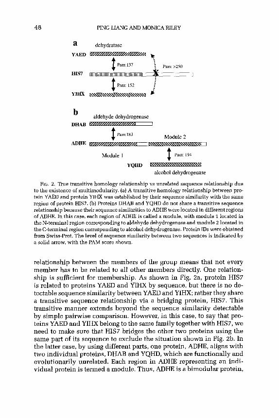
48 PING LIANG AND MONICA RILEY
a dehydratase
YAED - ,
$
: Pam:137
: : . Pam: >250
HIS7 vj x 1
Pam: 152 : :
YIHX e i
b aldehyde dehydrogenase
DHAB - I
Pam: 183
ADHE $ n- , Module 2
Module 1 t
Pam: 194
YQHD -
alcohol dehydrogenase
FIG. 2. True transitive homology relationship vs unrelated sequence relationship due to the existence of multimodularity. (a) A transitive homology relationship between pro- tein YABD and protein YIHX was established by their sequence similarity with the same region of protein HIS7. (b) Proteins DHAB and YQHD do not share a transitive sequence relationship because their sequence similarities to ADHE were located in different regions of ADHE. In this case, each region of ADHE is called a module, with module 1 located in the N-terminal region corresponding to aldehyde dehydrogenase and module 2 located in the C-terminal region corresponding to alcohol dehydrogenase. Protein IDS were obtained from Swiss-Prot. The level of sequence similarity between two sequences is indicated by a solid arrow, with the PAM score shown.
relationship between the members of the group means that not every member has to be related to all other members directly. One relation- ship is sufficient for membership. As shown in Fig. Za, protein HIS7 is related to proteins YAED and YIHX by sequence, but there is no de- tectable sequence similarity between YAED and YIHX; rather they share a transitive sequence relationship via a bridging protein, HIS7. This transitive manner extends beyond the sequence similarity detectable by simple pairwise comparison. However, in this case, to say that pro- teins YAED and YIHX belong to the same family together with HIS7, we need to make sure that HIS7 bridges the other two proteins using the same part of its sequence to exclude the situation shown in Fig. 2b. In the latter case, by using different parts, one protein, ADHE, aligns with two individual proteins, DHAB and YQHD, which are functionally and evolutionarily unrelated. Each region in ADHE representing an indi- vidual protein is termed a module. Thus, ADHE is a bimodular protein,

ANCESTRAL PROTEINS AND EVOLUTION 49
presumably as a result of gene fusion between an aldehyde dehydroge- nase and an alcohol dehydrogenase. Therefore, unlike a motif or a do- main, a module represents an independent individual functional pro- tein of independent ancestry representing a unit of protein evolution and cellular function (Riley and Labedan, 1997; Campbell and Baron, 1991; Baron et al., 1991; Hegyi and Bork, 1997). A module can exist as an individual protein, such as the E. coli proteins DHAB and YQHD, or as a part of larger proteins, such as ADHE. The situations for correspond- ing modules in different organisms are not necessarily the same. Unless proteins consisting of more than one module (multimodular) are iden- tified and each module is treated as a separate entity, false connections can be made. For instance, in the case of ADHE, without considering its multimodularity, two proteins, aldehyde dehydrogenase and alcohol dehydrogenase, would be incorrectly grouped into one family. Based on our experience, if no modules are identified and treated as separate en- tities from other modules in the same protein, over 70% of all E. coli paralogous proteins can be grouped into one large group, which is cer- tainly far from the truth.
Multimodularity of proteins has long been known. Many enzymes known to have more than one enzyme activity in a single protein are due mostly to the existence of multiple modules, such as ADHE, in Fig. 2b (Kessler et al., 1991). Another well-known example is FadB, which has NADH-dependent dehydrogenase activity at its C-terminal region and carries out three separate catalytic reactions-isomerization, hydration, and epimerization-in its N-terminal region, seemingly by a single ac- tive site, at least for the latter two activities (Yang et al., 1988; Muller- Newen and Stoffell993; McCormack, Teichmann, and Riley, submitted for publication). In addition to enzymes, multimodularity also exists for other types of proteins. Interesting examples are the phosphotransferase sytems (PTS), which both transport and phosphorylate carbohydrates. The EII part of these systems consists of at least three components (EII A/B/C), which can exist as individual proteins or fusion (multimodular) proteins of any two or all three of the components (Postma et al., 1996).
In recent work on the complete E. coli genomic sequence (Liang, Labe- dan, and Riley, submitted for publication), for the 2415 E. coli proteins which are paralogues, 2749 modules were identified, indicating the ex- istence of proteins that have more than one module, or are multimod- ular. In total, 286 proteins (11.8% of all paralogues) were identified as multimodular, among which 226 have modules in more than one par- alogous group and 86 have two or more modules in the same group, representing the occurrence of gene fusion and internal duplication, re- spectively. A few proteins have a combination of internal duplication and gene fusion. Table II lists examples for each of the three situations.

50 PING LIANG AND MONICA RILEY
TABLE II
SELECTED MULTIMODULAR PROTEINS
SW ID Module Start’] Enda Group IDb Function
MODF b0760-1 1 273 32
b0760-2 201 482 32
LPXD bolm_l 37 142 40
bolx-z 106 306 40
ILVB b3671-1 15 344 39 b3671-2 384 560 39
PTAA b0679w1 1 311 33
b0679-2 341 466 210
b0679-3 475 648 211
MALK b4ox-1 1 210 288
bdoz-2 179 291 289 MRCB bold%z 228 354 33
bolag-3 656 768 210 ADHE b1241-1 8 373 190
b1241-2 456 857 190 FADB b3846-1 8 206 15
b3846-2 314 643 323 DOGA b3692-1 8 121 383
b3692-2 206 587 341
HRSA bonl-1 16 178 223
bonl-2 384 492 33
bo731-3 468 628 33
YPIA b3486-1 82 602 139 b3486-2 832 1170 139 b3486-3 1221 1363 196 b3486-4 1364 1476 139
Transporters of ABC superfamily (ATP-binding component)
Transporters of ABC superfamily (ATP-binding component)
UDP-3-0-(3-hydroxymyistoyl)-glucosamine n-acyltransferase
UDP-3-0-(3-hydroxymyristoyl)-glucosamine n-acyltransferase
Acetolactate synthase I, large subunit Acetolactate synthase I, large subunit
Sugar-specific PTS: enzyme IIC, membrane component and sugar binding
Sugar-specific PTS: enzyme IIB, phosphorylation of sugar
Sugar-specific PTS: enzyme IIA, phosphotransferase EC 2.7.1.69
Maltose transport protein (ATP-binding component)
Repressor of ma1 operon Glycosyl transferase Transpeptidase Aldehyde reductase (dehydrogenase) Aldohol dehydrogenase NADH-dependent dehydrogenase Isomerase, hydratase, epimerase 2-Oxo-3-deoxygalactonate
6-phosphate aldolase Galactonate dehydratase
Sugar-specific PTS: enzyme IIA, phosphotransferase EC 2.7.1.69
Sugar-specific PTS: enzyme IIC, membrane component and sugar binding
Sugar-specific PTS: enzyme IIC, membrane component and sugar binding
Conserved protein Conserved protein Conserved protein, transmembrane domain Conserved protein
a The start and end position of the module. bArbitary group numbers for E. coli paralogous families.

ANCESTRALPROTEINSANDEVOLUTION 51
B. PARALOGOUSPROTEINFAMILIESIN E. coli
Paralogous protein families in E. coli had already been studied before the completion of the genome (Riley and Labedan, 1996,1997; Labedan and Riley, 1995a,b, 1999). A recent analysis was performed with the complete E. coli (K-12) genomic sequence (Blattner et al., 1997) and up- dated information on the functions of E. coli genes (Berlyn, 1998; Rudd, 2000; Riley and Serres, 2000; Liang, Labedan, and Riley, submitted for publication).
Among the 4289 protein-coding ORFs in E. coli, 2415 were identified as having at least one homologous partner in the same genome, and they are assembled into 611 sequence-related paralogous groups, while 1983 proteins do not match any other sequences in the genome at a level above the threshold we set and are, thus, called singles (nonpar- alogues). The major paralogous groups of proteins for each major type of function in E. coli are listed in Tables III-VI). Detailed information for the full list of paralogous groups and for the multimodular proteins, including the range of each module, is available at the GenProtEC web site (http://genprotec.mbl.edu).
We classified all genes of E. coli K-12 by the functions of their prod- ucts, both the experimentally known and the predicted functions, as summarized in Table VII. The distribution of genetic resources in E. coli is in three large functional classes and several smaller ones. The largest number of genes code for enzymes (34%), followed by the genes for transport functions (11%) and then the genes for regulatory processes (9%). Genes in the three categories together account for more than 55% of all genes and for more than 75% of genes with known products in E. coli K-12. Moreover, almost all of the largest paralogous groups fall into these three categories (Tables III-VI).
Interestingly, proteins in each of the three categories show distinctive characteristics. Enzymes tend to be clustered into smaller paralogous groups as indicated by the smallest average group size and the highest percentage (32%) as singles (nonparalogues). Single proteins that have no partner in E. coli can be considered as the only surviving members of preexisting paralogous groups or as former members of groups that have diverged too far from their former partners to allow the similarities to be detected today. Transporters and regulators are in far fewer groups than enzymes but they aggregate into much larger groups (Fig. 3) and have lower percentages as nonparalogues (17 and 20%, respectively) compared with enzymes. While regulators have the highest proportion of groups larger than seven (Fig. 3), the three largest paralogous groups are all transporters and they account for over 60% of all transporter proteins (Table IV). In addition, transporters have the highest percentage

TABLE III
MAJOR PARALOGOUS ENZYME FAMILIES IN E. coli
Group ID” Size* Group functionc
121 34 253 29 168 18 128 18
27 17 96 15
231 14 14 14 53 13
252 13 87 12
308 11
368 11 170 11
16 10 543 9
82 9
473 9 39 8
190 8 91 7 69 7
105 7 15 7
90 7 228 7 124 7 244 7
29 7 28 6
286 6 249 6 324 5
77 5 501 5 305 5
265 242 336
2 434 372
Mainly putative oxidative proteins Fe-S subunit of oxidoreductase NAD(P)-dependent oxidoreductases NAD(P)-dependent alcohol dehydrogenase ATP-dependent helicases Aldehyde dehydrogenases Methyltransferases Acyl transferases FAD/NAD(P) oxidoreductases Formate dehydrogenase/DMSO reductase Mainly P-type ATPases Dehydratasesldeaminase for cysteine, serine,
threonine, tryptophan Sugar kinases Oxidoreductases; NAD(P)-binding subunit Acetyl-CoA synthetases/ligases IS tranposase Oxidoreductases; all share an NAD(P)-linked
oxidoreductase domain Phage-related integrases/recombinases Acetolactate synthase (large unit) Dehydrogenases (iron-dependent) Acyl-CoA N-acyltransferases Aminotransferases Endoglucanasesldihydrodipicolinate synthases Dehydratases/racemases/CoA hydratase (involved
in metabolism of fatty acids) tRNA synthetases for asparagine and lysine Dehydratases/epimerase for NDP-glucoselmannose Acetyl-CoA synthetases Glutathione S-transferases Sugar kinases for ribulose, fuculose, xylulose, glycerol Phosphate &epimerase/aldolase for L-ribulose, fuculose, rhamnulose NADH/(P)-binding dehydrogenases Reductases Acyl-CoA transferases Decarboxylases for lysine, ornithine, arginine Phosphate epimeraseslsythases for ribulose, thiamine, and hexose Glucose-l-phosphate transferase (uridylyl, adenynyl,
thymidylyl, pyrodylyl) DNA-dependent helicases Pyruvate formate lyase-activating enzymes Oxidoreductases (for altronate, mannitol, mannonate;
mostly N-terminal] Transaldolases N-Acetylmuramoyl-1-alanine amidase Bisphosphate aldolases for tagatose, fructose
0 Arbitary number for E. coli paralogous groups. * Size excludes multimodules in the same group. c General function based on the known functions of the group members.
52

ANCESTRAL PROTEINS AND EVOLUTION 53
TABLE IV
MAJOR PARALOGOUS ‘BUNSPORTER FAMILIES IN E. coli”
Group ID Size Group function
521 94
32 83
33 68
158 39
524 31 278 12
68 12 223 12 530 11
34 11
201 11
532 10 526 9 467 7 529 7
193 7 356 6 160 6 531 6 172 5 523 5 210 4 378 4 343 4 405 4 229 4
19 4
Transport proteins (membrane component, mainly MFS superfamily)
Transport proteins of ABC superfamily (ATP-binding component)
Transport proteins of ABC superfamily (membrane component), also PTS
Mostly membrane component of transporter, regulators. Single hybrid motif
Transporter (mainly APC superfamily for amino acids) Transport proteins (SSS, DAACS family) Transport proteins @ND, GntP family) PTS transport proteins, IIA component Transport proteins of ABC superfamily
(membrane component) Transport proteins of ABC superfamily
(peripliasmic-binding) ABC transporters for amino acids
(periplasmic-binding component) Transport proteins (NCS2 and GntP families) Transport proteins (GPH, MFS family) Transport proteins (HAAAP family) Transport proteins (N-terminal; membrane
component; ABC, MFS family) Part of transport proteins (mainly DASS family) Putative transmembrane proteins Putative transport proteins Putative transport proteins Putative transport proteins MFS family of transport proteins Sugar-specific PTS system (IIB component) Sugar-specific PTS system (IIC component) DMT family of transport proteins NUP family of transport proteins ABC superfamily (periplasmic-binding component
for spermidine, molybdate, putrescine) Transporter proteins (BCCT and MATE family)
o Headings the fame as in Table III. (see footnotes a-c).
(18%) of multimodular proteins (both internal duplication and gene fu- sion), almost double the corresponding numbers for enzymes (8%) and regulators (8.6%), suggesting that transporter proteins are more often involved in gene fusion and internal gene duplication. Fused subunits of complex transporters are common.

54 PING LIANG AND MONICA RILEY
TABLE V
MAJOR PARALOGOUS REGULATOR FAMILIES IN E. coP
Group ID Size Group function
8 45 145 43 146 36
51 19 30 27 40 24
130 15 245 12 107 8 143 8 328 6
165 83
156 215 337
LysR-type transcription regulators Mainly LuxR/UhpA-type two-component regulators Kinase components of two-component regulators Mainly GntR-type transcription regulators AraC/XylS-type transcritional regulators Mainly GalR/LacI-type transcription regulators,
some periplasmic-binding component of ABC Mainly EBP transcription regulators DeoR-type transcription regulators LclR-type transcription regulators NagC/XylR-type transcription regulators Methyl-accepting chemotaxis proteins (N-terminal
of multimodular proteins) MerR-type transcription regulators LysR-type transcription regulators (mostly N-terminal) AsnC-type transcription regulators OmpR-type regulators MarR-type transcription regulators
o Headings the same as in Table III. (see footnotes a-c).
TABLE VI
MAJOR PARALOGOLJS FAMILIES IN E. coli FOR OTHER CATEGORIES~
Group ID Size Group function
59 30 Fimbrial proteins 139 15 Mainly conserved proteins, 2 flagellar-related
62 12 Periplasmic chaperones 61 11 Outer membrane proteins (N-terminal of group 60) 52 10 Mixed functions but mostly related to DNA 60 10 Outer membrane proteins (mostly C-terminal of group 61) 31 8 Unknown
155 6 Unknown 5 5 HSP70
472 5 Conserved proteins 238 5 Conserved proteins 218 5 Conserved proteins 283 5 Cytochrome
88 4 Conserved proteins 277 4 Conserved proteins
a Headings the same as in Table III. [see footnotes a-c).

ANCESTRAL PROTEINS AND EVOLUTION 55
TABLE VII
FUNCTION DISTRIBUTION OF ALL E. coli PROTEINS
Function Paralogues Singles Total (%)”
Transporter Regulator Factor Membrane Structure Carrier RNA Phage, IS Leader Cell process Unknown
Totalb
1030 410 312
62 320
39 8
91
11 368
2415
486 1518 (34.5) 86 496 (11.2) 84 396 (8.9) 69 131(3.0) 26 326 (7.4) 85 124 (2.8) 21 29 (0.7)
114 114 (2.6) 219 310 (7.0)
12 12 (0.3) 14 25 (0.6)
777 1145 (26.0)
1983 4403
0 Percentage in relation to OEWs. b The number excludes the overlap situations between the function categories.
In addition, as illustrated in Fig. 4, enzymes, transporters, and regu- lators showed different distribution patterns of the sequence similarity level between paralogues as measured by PAM scores. The sequence similarity between enzyme paralogues ranges from the threshold we used (PAM, 200; residue identity, ~25%) to near-identity (PAM, cl; residue identity, >98%), with a median PAM value of 149. Clearly en- zymes range widely in their degree of sequence conservation. Since many enzymes are found in small paralogous groups or pairs, it appears that either there were many ancestors or, more likely, many enzymes have diverged far from each other in their specialization, maintaining closeness to only a few others of similar sequence and function. Regu- lator paralogues have the lowest median PAM (137), but with very few at a very high sequence similarity level, indicating that very few recent duplications have occurred for genes in this category. Since regulators tend to belong to large groups, it appears that a few kinds of regulation mechanisms have been widely used. Interestingly, transporters have the highest median PAM value (165) among the three types of proteins, and the majority of transporter paralogues have a PAM value over 180, in- dicating that they require the least conservation for their function as transporters or that many transporters are very old paralogues. Like reg- ulators, there is not as much variety in types of transporters as in types of enzymes. These data suggest that different types of proteins, at least in the cases of enzymes, transporters, and regulators, have separate evolu- tion histories. To account for the differences in the sequence similarity

2 k 1
, ,:.
En
zym
es
b. Tr
ansp
orte
rs
C.
Reg
ulat
ors
19%
FIG
. 3.
The
frequ
ency
of
gro
up
size
s fo
r en
zym
es,
trans
porte
rs,
and
regu
lato
rs
in E
. co
li. T
he s
izes
of
para
logu
es
grou
ps
for
each
type
of
prot
eins
ar
e di
vide
d in
to
the
follo
win
g si
ze c
ateg
orie
s:
2, 3
, 4,
5, 6
, 7,
and
>i’.
Th
e pe
rcen
tage
of
gro
ups
in
each
siz
e ca
tego
ry i
n re
latio
n to
the
tot
al
num
ber
of g
roup
s in
eac
h fu
nctio
n ca
tego
ry i
s al
so i
ndic
ated
.

ANCESTRAL PROTEINS AND EVOLUTION 57
FIG. 4. Frequency of PAM scores for paralogous enzymes, transporters, and regulators. For each protein, only the best match as represented by the lowest PAM score was used. The frequency of PAM scores in each value range (every 10) is plotted for each of the three protein types.
level for the paralogues in the three types of proteins, it may be that transporters underwent gene duplication very early in evolution, while the gene duplication for most regulators happened at a later time and duplication for enzymes happened continuously over time. While many paralogous transporter proteins and enzymes are known to be very an- cient and existed in the LUCA (Gogarten, 1994; Makarova et al., 1999; Koonin and Bork, 1996; Mehta et al., 1993; Tomii and Kanehisa, 1998), there have been a few reports suggesting a later emergence of transcrip- tion regulators. Aravind and Koonin reported that the core transcription regulators in archaea and eukaryotes (TFIIB/TFB, TFIIE-(r, and MBF-l), and those in bacteria (the sigma factors) share no sequence similarity beyond the presence of a distinct HTH domain, and suggested that HTH domains might have been independently recruited for a role in trascription regulation in bacterial and archaeal/eukaryotic lineages (Aravind and Koonin, 1999). In another report, the derivation of eu- karyotic transcription regulators from ancient enzymatic domains was proposed (Aravind and Koonin, 1998).
C. RELATIONSHIPS OF PARALOGOUS PROTEIN FAMILY MEMBERS
The sequence relationships among the members of one family may differ from those in other families. Some families have a tight and mutu- ally connected sequence relationship, while others may have a smaller core set of members that are tightly related but the rest of the members

58 PING LIANG AND MONICA RILEY
a b
FIG. 5. Tight relationships among the four members of a group of transadolases. (a) Sequence similarity exists between each member and all of the other members as in- dicated by the solid lines and the PAM scores along the lines. (b) A possible phylogenetic tree based on the PAM distance matrix. Proteins are labeled by the first section of the Swiss-Prot ID.
are connected to only one or a few of the core members. As shown in Fig. 5a, each of the four members in a group of transaldolases shares sequence similarity to the other three members, although the similar- ity levels are not the same. In contrast, in a family containing oxidases, the four members have a much looser relationship in sequence (Fig. 6a). The degrees of similarity between YDIJ and GLCF, YHCU and GLCD; and GLCF and GLCD are below the threshold we set. The different anatomy of protein families no doubt reflects the fact that each family of proteins has experienced different evolution paths and has different require- ments of sequence conservation. Figures 5b and 6b represent the possi- ble evolution paths for the genes corresponding to the proteins in two groups.
Members of each of the paralogous groups almost always exhibit a clear relationship in functions, supporting evidence for the homology shared among the members of a paralogous family. This is true even in groups formed transitively so that not all members are related by our criteria for sequence similarity to all other members. An example is the group of oxidases discussed above (Fig. 6), in which all four members have “oxidase” as their shared function (including predicted function) and “FAD-binding domain” as their shared reaction type and structural property, despite the loose sequence relationships among these four proteins.

ANCESTRAL PROTEINS AND EVOLUTION 59
a - Pam:178 n YDIJ GLCD
Pa&249 Pam:221
,:.
i Pam:207
b
GLCD
YGCU
1 GLCF
FIG. 6. Loose relationships among the four members of a group of oxidases. (a) Se- quence similarity within the threshold exists only between three pairs of the members (indicated by solid lines). Dotted lines indicate that the sequence similarity is beyond the threshold. The PAM score for each pair of proteins is shown beside or on the line connecting the two proteins. [b) A phylogenetic tree based on the PAM distance matrix. Proteins are labeled by the first section of the Swiss-Prot ID.
For larger paralogous protein families, there is an observable rela- tionship between the functions and deeply branching subfamilies. The diagram for the members in a group of ATP-binding subunits of the mul- timeric ABC transporters demonstrates the detectable conservation for different types of substrates transported, such as amino acids, oligopep- tides, sugars, metals, and metal-containing proteins (Fig. 7). Evidence of functional divergence among the paralogous members in gaining speci- ficity in substrate binding, cellular location of function, or even new types of function in some exceptional cases provides us with abundant materials for studying the modes of functional divergence of proteins originally descended from a common ancestor by sequence divergence (Kerr and Riley, submitted for publication; Nahum and Riley, 2001).
D. PROTEINS AND PROTEIN FAMILIES SHARED BETWEEN E. coli AND A. fulgidus
Proteins shared by E. coli and A. fulgidus can be thought of as rep- resenting the proteins shared by the eubacterial and archaeal domains except for those horizontally transferred between the specific lineages related to these two species. Therefore, groups of these proteins with similar sequences probably also represent ancestral proteins at nodes

Iron-
cont
aini
ng
mol
ecul
e5
\
CC
MA

ANCESTRAL PROTEINS AND EVOLUTION 61
of evolutionary development prior to separation of the two domains. When conservative criteria (maximal PAM of 200 and minimal align- ment length of 100 residues) are used to detect similarity in amino acid sequence, 1535 (36%) E. coli proteins are orthologous to 1146 (48%) A. fulgidus proteins. These numbers increase to 2581 (60%) and 1565 (65%) for E. coli and A.fulgidus, respectively, when the criteria are loos- ened to a maximal PAM value of 250 and a minimal alignment length of 83 residues. These data suggest that in these two very distantly related genomes, up to 50% of their proteins still share detectable homology today, while each species also has a substantial portion of genes whose counterparts either do not exist in the other genome or have diverged to an undetectable distance.
Among the 1535 E. coli proteins that have counterparts in A. fulgidus, 1200 (78%) are on the E. coli paralogue list, while that number is 815 (71%) for A. fulgidus, indicating that the majority of these “universal” and likely essential proteins have a history of duplication. Among the 610 paralogous protein families identified in E. coli, 324 contain at least one orthologue in A. fulgidus, while the rest of the groups contain pro- teins that have no A. fulgidus homologue. Table VIII shows a list of large paralogous groups shared by E. coli and A. fulgidus, with the group sizes in both genomes indicated and the groups ordered by the group size in E. coli in a descending manner. All large E. coli groups have counterparts in A. fulgidus. Both genomes have membrane components of transporters as the largest group and the ATP-binding component of ABC transporters as the second largest group, but the order of the other groups by size is not the same in the two genomes. For example, the next largest group in A. fulgidus is the two-component regulators, while it is the LysR type of transcriptional regulators in E. coli.
Notably, although its genome size is only about 50% of the genome size of E. coli, A. fulgidus seems to have unusually large groups of acetyl- CoA synthetases/ligases as shown by the much larger group size than for the corresponding E. coli group (group 16 in Table VIII) and as reported from the genome sequencing project (Klenk et al., 1997). The same situa- tion is true for two additional acetyl-CoA related groups (not listed). The phyiological implications of the multiplicity of fatty acid metabolism are not yet understood. In addition, A. fulgidus seems to have relatively
FIG. 7. An unrooted tree for an E. coli paralogous group representing the ATP-binding components of ABC transporters. Among the 94 members, only the 59 proteins with experimental data on the substrates were included in the tree construction based on the PAM distance matrix to show the relationship between phylogenetic positions and the substrate binding specificity. Proteins are labeled with their Swiss-Prot IDS. Proteins with suffixes are partial-length modules.

62 PING LIANG AND MONICA RILEY
TABLE VIII
MAJOR TYPES OF PARALOCOUS FAMILIES SHARED BY E. coli AND A. fulgidus AND THEIR GROUP SIZES
Group IDa EC sizeb AF sizeC Group function
521
32
33
8
145
158
146 121
524
59 51 40
128
168
27 103
96 130
14
231
53
251
252
68 87
94 52
83 57
68 27
45 7
43 26
39 17
36 34 34 18
31 16
30 3 27 12 24 4
18 8
18 4
17 10 17 11 15 1 15 19
14 1
14 9
13 14
13 4
13 9
12 12 12 12
Transport proteins (membrane component; mainly MFS superfamily)
Transport proteins of ABC superfamily (ATP-binding component)
Transport proteins of ABC superfamily (membrane component; also PTS )
LysR transcription regulators; all have periplasmic-binding protein-like II domains
Mainly LuxR/UhpA-type two-component regulators; all have a CheY-like domain
Mostly membrane component of transporter, regulators
Kinase component of two-component regulators Mainly conserved proteins: many have no
SCOP assignments Transport proteins (mostly APC superfamily for
amino acids]; no SCOP assignments Fimbrial proteins, all have bacterial adhesins Mostly GntR-type regulators Mostly GalR/LcI-type transcription regulators/PBP;
all have periplasmic-binding protein-like I Oxidoreductases; all share NAD(P)-binding
Rossmann-fold domains and GroES-like Oxidoreductases; all share NAD(P)-binding
Rossmann-fold domains ATP-dependent helicases GTP-binding proteins Aldehyde dehydrogenases (NAD-dependent) Mostly EBP-type transcription regulators
with P-loop-containing nucleotide triphosphate hydrolases
Acyl transferases; all have trimeric LpxA-like domains
Methyltransferases; all have S-adenosyl-L- methionine- dependent methyltransferases
Oxidoreductases [mostly N-terminal); all have one or more FAD/NAD(P)-binding domains
Mixed functions (mostly membrane proteins related to cell division)
Dehydrogenases/reductases for DMSO, Na, formate, biotin sulfoxide
Transport proteins (RND, GntP family) P-type ATPase mostly; 6 have an SIS domain
(continues)

ANCESTRAL PROTEINS AND EVOLUTION 63
TABLE VIII-Continued
Group IDa EC sizeb AF size” Group function
223 12 2 PTS IIA component; share the IIA domain of mannitol-specific and ntr phosphotranspherase EII
DeoR-type transcription regulators (mostly N-terminal)
Transport proteins (SSS, DAACS family) Membrane protein (most for dehydrogenases) Transport proteins of ABC superfamily
(periplasmic-binding component) Oxidoreductases; all have NAD(P)-binding
Rossmann-fold domains ABC transport proteins (periplasmic-binding
component) with periplasmic-binding protein-like II domains
Dehydratases (cystine, serine, threonine, tryptophan) Sugar kinases; all have ribokinase-like domains Transport proteins of ABC superfamily
(membrane component) Acetyl-CoA synthetases/ligases; all have
Firefly luciferase-like Mixed; 3 dehydrogenases, 3 HSPs Outer membrane proteins (mostly C-terminal
of group 61) Transport proteins
245 12 2
278 7
34
12 11 11
14 5 1
170 11 4
201 11 6
308 368 530
9 4
14
11 11 11
16 10 26
52 10 a 60 10 1
532 10 7
a Group number for E. coli. b Group size for E. coli. c The number of proteins that have arthologues to the E. coli proteins in the group.
large groups of two-component transcriptional regulators but smaller groups of certain other types of transcriptional regulators, such as LysR, GalR/LacI, and DeoR, and it has a very small number of fimbrial pro- teins. The different characteristics for E. coli and A. fulgidus in the kinds and relative sizes of protein families are believed to reflect the different cell physiology, metabolism, and biology of these two genomes. An ex- tensive examination of these cases in the context of the physiology of these two genomes will provide useful information for improving our understanding of their biological natures.
E. PROTEINS UNIQUE TO E. coli AND A. fulgidus
Data from genome sequence projects so far have revealed that almost every genome has at least 20% of genes that do not match any other sequences in the current databases. These unmatched parts contribute a major proportion of unknown genes and impose a big challenge to

64 PINGLIANGANDMONICARILEY
genome annotation. However, these genes may be the key elements responsible for the distinctive characteristics of each type of organism. Therefore, it would be very crucial and interesting to understand the nature of these genes. Comparative genomics provides a powerful ap- proach to identify and study these genes. To collect the genes that are unique to a genome, it seems more rational to use more aggressive (loose) criteria for detecting homologues to ensure the true uniqueness of the identified genes. When the criteria of maximum PAM scores of 250 and minimum alignment lengths of 83 were used for detecting all ortho- logues in the 21 genomes listed in Table I, 105 E. coli proteins were found not to have any homologues among those genomes (Table IX). Twelve of the 105 are prophage genes, not bacterial in origin. Of the remaining 83, 72 (86%) have no match to the 21 microbial genomes, but among them are 7 “putative” ORFs with orthologues in other genomes. The remaining 65 are proteins related to a variety of functions, such as regulators and redox-related proteins. Understanding the contribution of unique genes to cell physiology will provide molecular information on the properties that differ between organisms.
It needs to be pointed out that the label “unique” is relative only to the specific set of genomes used in the analysis. The number of unique proteins drops when closely related species are included. It drops to 64 when Salmonella typhimurium genes are included (unpublished data), and we can assume that the list will be even shorter when Shigella and other E. coli species are included in the comparison. However, although the exact numbers of unique genes in each genome might be difficult to predict, it can be certain that the number will not drop to zero, because each differentiated species and strain is characterized by observable differences. In a practical vein, the small differences have value. Dif- ferences exist between nonpathogen and pathogen strains. This kind of information should help us to identify the virulent factors in the pathogen strains.
A comparable analysis for A. fulgidus against the other 21 genomes listed in Table 1 revealed that 60 protein-coding ORFs in this genome do not have any orthologues in the genomes on the list, and none of them has any known or predicted function information. We can expect that this fact will remain true for every genome, especially those organisms with little biology known.
F. THE MINIMUM NUMBER OF ANCESTRAL PROTEINS
A matter of ongoing interest is to figure out a minimal set of ancestral proteins necessary for generating all present-day proteins (Riley and Serres, 2000; Mushegian and Koonin, 1996; Mushegian, 1999; Maniloff,
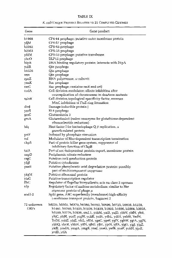
TABLE IX
E. coli UNIQUE PROTEINS RELATIVE TO 21 COMPLETED GENOMES
GeIle Gene product
b1998 YW b2362 b2363 yfdM ybc0 hipB YdfR b1556 rem rpoz ynaK racC cedA
minE
dinJ YmfJ grxC grxA
hfq
psiF rof chpS
tatA napD csgc y&J paaB
YkW yiaG flhC nip
araH-2
72 unknown ORFs
CP4-44 prophage; putative outer membrane protein CP4-57 prophage CPS-53 prophage CPS-53 prophage CPS-53 prophage; putative transferase DLP12 prophage DNA binding regulatory protein; interacts with HipA Qin prophage Qin prophage Qin prophage RNA polymerase, w subunit Rat prophage Rat prophage; contains recE and oriJ Cell division modulator; affects inhibition after
overreplication of chromosome in dnaAcos mutants Cell division topological specificity factor; reverses
MinC inhibition of FtsZ ring formation Damage-inducible protein J El4 prophage Glutaredoxin 3 Glutaredoxinl (redox coenzyme for glutathione-dependent
ribonucleotide reductase) Host factor I for bacteriophage Q/J replication, a
growth-related protein Induced by phosphate starvation Modulator of Rho-dependent transcription termination Part of proteic killer gene system, suppressor of
inhibitory function of ChpB Part of set-independent protein export, membrane protein Periplasmic nitrate reductase Putative curli production protein Putative cytochrome Putative phenylacetic acid degradation protein; possibly
part of multicomponent oxygenase Putative ribosomal protein Putative transcription regulator Regulator of flagellar biosynthesis; acts on class z operons Regulatory factor of maltose metabolism; similar to Ner
repressor protein of phage p Split gene, ABC superfamily (membrane) high-affinity
L-arabinose transport protein, fragment z
boz35, bosoz, b0370, b0380, b0392, bO609, b0725, b0816, b1228, b1481, b1760, b1825, b1836, b1839, b1903, b1936, b2084, b2658, b3100, b3776, b3836, smf_1, yahM, yaiB, yajD, ybbV, ybfN, ybi1, ybjC, ybjH, yceP, ycgW, ychH, yciN, ydbL, ydiH, yebW, YedN, MM, yehE, yf& yfcL yfM ygaC, ygeP, ygfl, yghW, ygiA, ygjN, yhbQ, yhcR, yhhK yifN, yjbD, yjbL, yjeN, yjfA, yjgG, yjgJ, yjgZ, ykE ymdA, ymgA, ymgB, ynaJ, yneG, ynfS, yoaE yohH, ypd1, yrdB, ytfA

66 PING LIANG AND MONICA RILEY
1996). Assembly of paralogous protein families for a large and suffi- ciently varied number of genomes provides a possibility to estimate the minimal number of ancestral genes. If molecular evolution were com- pletely transparent on the basis of sequence similarity, then the minimal number of ancestral proteins for E. coli would be the sum of the number of paralogous groups and the number of singles (nonparalogues). How- ever, the actual number of ancestral proteins represented by E. coli is probably much smaller and the exact number might not be possible to know on the basis of the primary sequence alone. Our current meth- ods for detecting sequence similarity may not be sensitive enough to bring all the true homologous members into groups. After many rounds of duplication and divergence, some genes and groups of genes have diverged to a point beyond the capability of the detection method we use. This means that it should be possible to cluster some of the E. coli paralogous groups into larger supergroups and some groups may be extended to include some proteins that are currently identified as sin- gles. The number of ancestors required for E. coli proteins will be redu- ced (1) when bridges between sequence-similar groups are found by sim- ilarity of motifs or by finding bridging sequences in other organisms and (2) when superfamilies of protein structure are used to extend the protein families by their similarity in structural properties.
G. THE LENGTH OF PRESENT-DAY PROTEINS AND MODULES: A HINT FOR THE SIZES OF ANCESTRAL PROTEINS
To examine the patterns for evolution by gene fusion and duplica- tion, we examined the distribution patterns of protein length accord- ing to the number of residues for all proteins and modules in each of the three domains based on their representatives listed in Table 1. At least two conclusions may be drawn from the data in Fig. 8. First, most ancestral proteins were about 100 residues in length. Proteins in all three domains seem to have selective ranges for their protein lengths, but all three domains share one range, from 100 to 130 residues, al- though eubacteria and eukaryotes have additional preferred ranges. In contrast, modules in all three domains demonstrate very uniform pref- erences of about 100 residues, suggesting that this is the optimal length range for unimodular proteins, which are probably the case for most an- cestral proteins. Second, the total protein length distribution patterns are not the same for the three domains of life. On average, eukaryotes (data limited to S. cerevisiae and C. elegans) have the longest protein length (443 amino acids), followed by eubacteria (320 amino acids), with archaea having the shortest protein length on average (288 amino acids). Eukaryotic proteins seem to show a trimodal distribution pattern

ANCESTRAL PROTEINS AND EVOLUTION 67
a. Archaea b. Bacteria
700 800 600 700
5 500 E 600
f 400 300
c % g
500 400 & 300 L 200 u 200
100 100 0 0
Protein Length (AA) Protein Length (AA)
c. S. cerevisiae and C. e/egans d. Modules
800 , W” 1 7000 (_ T1*coo
00000000000 0 9 + q? 9 d c$’ 6 6’ 2” 2%
Protein Length (AA) Protein Length (AA)
FIG. 8. Length distribution for proteins and modules. All proteins that are longer than 80 residues and their detected modules from the 22 genomes divided into the three do- mains of life. (a, b, c) The length frequency of proteins in Archaea, Eubacteria, and Eukarya, respectively. (d) The length frequency for modules in the three domains. Only the shortest lengths were used for modules which have alignment with more than one protein using slightly variable regions. The median values for proteins in each domain are indicated in the upper-right corners.
corresponding to unimodular, bimodular, and trimodular proteins at 110,230, and 350 residues, respectively, while eubacteria have only the first two and archaea have only the first one. We believe that the aver- age protein length in a genome is an indicator of the complexity of the genome and protein functions, and this number can be expected to be much higher for higher eukaryotes due to more frequent and complex gene fusions. In this sense, the archaeal domain seems to be the simplest in form among the three domains.
IV. Conclusions and Perspectives
The existence of paralogous groups of proteins of similar sequence and related function is entirely consistent with the proposal that pro- cesses of duplication followed by divergence have played a major role in the molecular evolution of proteins and genomes as shown in Fig. 9. It seems quite clear that limited kinds of proteins existed in the cells of

Spec
ies C
with
in g
enom
e
FIG
. 9.
A sc
hem
atic
ill
ustra
ting
sim
plifi
ed
patte
rns
of p
rote
in
evol
utio
n.
Two
ance
stra
l pr
otei
ns
(repr
esen
ted
by t
he d
iffer
- en
t sh
apes
) fro
m t
he l
ast
com
mon
an
cest
or
(LU
CA)
ha
ve e
volv
ed
diffe
rent
ly,
gene
ratin
g th
e se
t of
prot
eins
in
the
pre
sent
-day
sp
ecie
s A,
B, C
, D, a
nd E
. One
is v
ery
cons
erva
tive
(squ
ares
), wi
th
all
of it
s de
scen
dant
s sh
arin
g de
tect
able
se
quen
ce s
imila
rity
to e
ach
othe
r, wh
ile
the
othe
r [c
ircle
s)
has
been
act
ivel
y in
volv
ed
in d
uplic
atio
n wi
thin
ge
nom
es
and
dive
rgen
ce
at v
ario
us
poin
ts
over
the
who
le
evol
utio
n pa
th,
gene
ratin
g de
scen
dant
pr
otei
ns
that
m
ay
not
shar
e de
tect
able
se
quen
ce
sim
ilarit
y am
ong
the
para
logu
es
and
orth
olog
ues
(repr
esen
ted
by d
iffer
ent
shad
ing
patte
rns
of c
ircle
s).
Gen
e lo
ss o
ccur
red
in s
ome
linea
ges.

ANCESTRAL PROTEINS AND EVOLUTION 69
the LUCA, represented mainly by enzymes, transporters, and factors which were mostly unimodular and about 100 residues long. These proteins represent the core set of proteins that are involved in essential cellular functions, and they were used as the basic building blocks for generating all present-day large and complex multimodular proteins through internal gene duplication and gene fusion. Gene duplication within a genome has given rise to all paralogous families in present- day genomes and has led to the increase in the size and complexity of genomes, while duplication of genomes in the course of speciation has resulted in orthologous gene families and an increased number of species. Enzymes, transporters, and regulators seem to have experienced separate and distinctive evolution paths, with enzymes and transporters being relatively older than regulators.
Several biological processes complicate the analysis of the evolution process. Duplication of genes has continued over time. Through speci- ation and by duplication and divergence within individual genomes, proteins of any age have become ancestors and have continued to pro- duce variants. Some variants, continually evolving, finally, over time, have lost recognizable similarities, leaving these genes outside of any paralogous families. The same process also has occurred among genes entering different genomes due to the speciation process, in which case orthologues may have followed different evolutionary paths and may have gained different functions because of the different physiological environments of separated species. In other cases, an entire gene lineage may have been lost in certain species, making attempts to track ancestral relationships more challenging. Horizontal gene transfers among differ- ent genomes have provided sources of new function and, at the same time, have complicated the analysis of evolution processes (Doolittle, 1999; Hilario and Gogarten, 1993). Alien genes have been introduced into bacteria, mainly from phages, from transposons, and by chromoso- ma1 recombination with plasmids. Extended sequence divergence and alien genes seem likely to be the main factors responsible for the exis- tence of genes that are unique to certain phylogenetic lineages.
With the improving sequencing capability at industrial scales, we can expect that the number of completed genomes will increase very quickly. Experts are anticipating this number to be over 1000 genomes by the year 2005. As the number of sequenced genomes increases, the amount of comparative sequence data increases exponentially. To be able to manage and make full use of the astronomic amount of data, we are relying on more powerful computers with more memory and com- putation speed as well as more intelligent software (Kanehisa, 1998). One of the main tasks is to decipher the function of all genes based on information from traditional experimental biology, bioinformatics
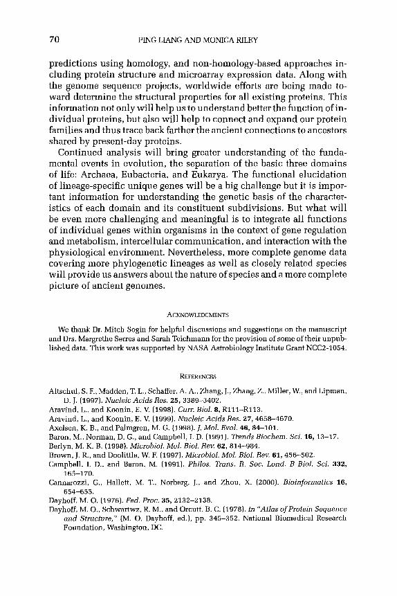
70 PING LIANG AND MONICA RILEY
predictions using homology, and non-homology-based approaches in- cluding protein structure and microarray expression data. Along with the genome sequence projects, worldwide efforts are being made to- ward determine the structural properties for all existing proteins. This information not only will help us to understand better the function of in- dividual proteins, but also will help to connect and expand our protein families and thus trace back farther the ancient connections to ancestors shared by present-day proteins.
Continued analysis will bring greater understanding of the funda- mental events in evolution, the separation of the basic three domains of life: Archaea, Eubacteria, and Eukarya. The functional elucidation of lineage-specific unique genes will be a big challenge but it is impor- tant information for understanding the genetic basis of the character- istics of each domain and its constituent subdivisions. But what will be even more challenging and meaningful is to integrate all functions of individual genes within organisms in the context of gene regulation and metabolism, intercellular communication, and interaction with the physiological environment. Nevertheless, more complete genome data covering more phylogenetic lineages as well as closely related species will provide us answers about the nature of species and a more complete picture of ancient genomes.
ACKNOWLEDGMENTS
We thank Dr. Mitch Sogin for helpful discussions and suggestions on the manuscript and Drs. Margrethe Serres and Sarah Teichmann for the provision of some of their unpub- lished data. This work was supported by NASA Astrobiology Institute Grant NCC2-1054.
REFERENCES
Altschul, S. F., Madden, T. L., Schaffer, A. A., Zhang, J., Zhang, Z., Miller, W., and Lipman, D. J. (1997). Nucleic Acids Res. 25, 3389-3402.
Aravind, L.,andKoonin,E. V (1998). Cum Biol. 8,Rlll-R113. Aravind, L., and Koonin, E. V. (1999). Nucleic Acids Res. 27, 4658-4670. Axelsen, K. B., and Palmgren, M. G. (1998). 1. Mol. Evol. 46, 84-101. Baron, M., Norman, D. G., and Campbell, I. D. (1991). Trends Biochem. Sci. l&13-17. Berlyn, M. K. B. (1998). Microbial. Mol. Biol. Rev. 62, 814-984. Brown, J. R., and Doolittle, W. F. (1997). Microbial. Mol. Biol. Rev. 61, 456-502. Campbell, I. D., and Baron, M. (1991). Philos. Trans. R. Sot. Land. B Biol. Sci. 332,
165-170. Cannarozzi, G., Hallett, M. T., Norberg, J., and Zhou, X. (2000). Bioinformatics 16,
654-655. Dayhoff, M. 0. (1976). Fed. Proc. 35,2132-2138. Dayhoff, M. O., Schwartwz, R. M., and Orcutt, B. C. (1978). In “Afks ofProtein Sequence
and Structure,” (M. 0. Dayhoff, ed.), pp. 345-352. National Biomedical Research Foundation, Washington, DC.

ANCESTRAL PROTEINS AND EVOLUTION 71
Doolittle, R. F. (1998). Nature 392, 339-342. Doolittle, W. F. (1999). Trends Cell Biol. 9, M5-M8. Feng, D. F., Cho, G., and Doolittle, R. F. (1997). Proc. Nat]. Acad. Sci. USA 94, 13028-
13033. Fitch, W. M. (1970). Syst. Zool. 19, 99-113. Furano, A.V. (1977). J. Biol. Chem. 252,2154-2157. Gogarten, J. P. (1994). J. Mol. Evol. 39, 541-543. Gogarten, J. P., and Olendzenski, L. (1999). Curr. Opin. Genet. Dev. 9, 630-636. Gogarten, J. P., Kibak, H., Dittrich, P., Taiz, L., Bowman, E. J., Bowman, B. J., Manolson,
M. F., Poole, R. J., Date, T., and Oshima, T. (1989). Proc. N&l. Acad. Sci. USA 88, 6661-6665.
Gonnet, G. H., Cohen, M. A., and Benner, S. A. (1992). Science 258,1443-1445. Gonnet, G. II., Hallett, M. T., Korostensky, C., and Bernardin, L. (2000). Bioinformatics
18,101-103. Hegyi, H., and Bork, P. (1997). J. Protein Chem. 16, 545-551. Hilario, E., and Gogarten, J. P. (1993). Biosystems 31, 111-119. Johanson, U., and Hughes, D. (1992). Gene 120,93-98. Kanehisa, M. (1998). Bioinformatics 14, 309. Iwabe, N., Kuma, K., Hasegawa, M., Osawa, S., and Miyata, T. (1989). Proc. Natl. Acad.
Sci. USA 86,9355-9359. Kessler, D., Leibrecht, I., and Knappe, J. (1991). FEBS Lett. 281, 59-63. Klenk, H. P., Clayton, R. A., and Tomb, J. F., et al. (1997). Nature 390, 364-370. Koonin, E. V., and Bork, P. (1996). Trends Biochem. Sci. 21, 128-129. Koonin, E. V., and Mushegian, A. R. (1996). Curr. Opin. Genet. Dev. 6, 757-762. Koonin, E. V., Aravind, L., and Kondrashov, A. S. (2000). Cell 101, 573-576. Labedan, B., and Riley, M. (1995a). Mol. Biol. Evol. 12, 980-987. Labedan, B., and Riley, M. (1995b). J, Bacterial. 177, 1585-1588. Labedan, B., and Riley, M. (1999). In “Organization of the Prokaryotic Genome,” (R. I,.
Charlebois, ed.), pp. 311-329. ASM Press, Washington, DC. Lander, E. S., and Weinberg, R. A. (2000). Science 287, 1777-1782. Li, W.-H., and Gram, D. (1991). “Fundamentals of Molecular Evolution.” Sinauer Asso-
ciates Sunderland, MA. Makarova, K.S., Aravind, L., Galperin, M. Y., Grishin, N. V., Tatusov, R. L., Wolf, Y. I., and
Koonin, E. V. (1999). Genome Bes. 9, 608-628. Maniloff, J. (1996). Proc. NatI. Acad. Sci. USA 93, 10004-10006. Mehta, P. K., Hale, T. I., and Christen, P. (1993). Eur. J. Biochem. 214, 549-561. Miller, D. L. (1978). Mol. Gen. Genet. 159, 57-6’2. Muller-Newen, G., and Stoffel, W. (1993). Biochemistry32, 11405-11412. Mushegian, A. (1999). Curr. Opin. Genet. Dev. 9, 709-714. Mushegian, A. R., and Koonin, E. V. (1996). Proc. Nat]. Acad. Sci. USA 93, 10268-10273. Nahum, L. A., and Riley, M. (2001). Divergence of Function in Sequence-Related Groups
of Escherichia coli Proteins. Genome Research. (in press). Ohno, S. (1970). “Evolution by Gene Duplication.” Springer-Verlag, Berlin. Ouzounis, C., Casari, G., Sander, C., Tamames, J,, andvalencia, A. (1996). Trends Biorech-
nol. 14,280-285. Palmgren, M. G., and Axelsen, K. B. (1998). Biochim. Biophys. Acta 1365, 37-45. Pennisi, E. (1999). Science 284, 1305-1307. Postma, P. W., Lengeler, J. W., and Jacobson, G. R. (1996). In “Escherichia coli and
Salmonella” (F. Neidhardt, I. R. Curtiss, J. Ingraham, et al., eds.), pp. 1149-1174. American Society for Microbiology, Washington, DC.
Ribeiro, S., and Golding, G. B. (1998). Mol. Biol. Evol. 15, 779-788.

72 PING LL4NG AND MONICA RILEY
Riley, M., and Labedan, B. (1996). In “Escherichia cob and Salmonella,” (F. Neidhardt, I. R. Curtiss, J. Ingraham, et al., eds.). American Society for Microbiology, Washington, DC.
Riley, M., and Labedan, B. (1997). 1. Mol. Biol. 268, 857-868. Riley, M., and Serres, M. H. (2000). Annu. Rev. Microbial. 54, 341-411. Rivera, M. C., Jain, R., Moore, J. E., and Lake, J. A. (1998). Proc. N&l. Acod. Sci. USA 95,
6239-6244. Sharp, P. M. (1991). I. Mol. Evol. 33, 23-33. Smith, T. F., and Waterman, M. S. (1981). 1. Mol. Biol. 147, 195-197. Tomii, K., and Kanehisa, M. (1998). Genome Res. 8, 1048-1059. Yang, S. Y., Li, J. M., He, X. Y., Cosloy, S. D., and Schulz, H. (1988). J. Bacterial. 170,
2543-2548.

Chromosome Packaging by Archaeal Histones
KATHLEEN SANDMAN AND JOHN N. REEVE
Department of Microbiology Ohio State University
Columbus, Ohio 43210
I. Introduction II. The DNA Packaging Problem
III. Nucleosomes A. The Eukaryal Nucleosome B. The Archaeal Nucleosome
IV. Nucleosomal DNA A. Length B. Topology C. Nucleosome Positioning
V. Transcription on Nucleosomal Templates VI. Histones
A. Archaeal Histones B. Natural Archaeal Histone Variants C. Nonhistone Histone Fold-Containing Proteins
VII. Evolutionary Considerations References
I. Introduction
Chromosomes are condensed in vivo, that is, they occupy signifi- cantly less volume than an equivalent amount of DNA free in solution. Physical and chemical forces promote the condensed state, but for bi- ological activity, condensed chromosomes must be structured so that genetic information can be accessed. Small, abundant proteins with positively charged surface residues positioned to interact with DNA are responsible for the organization of the condensed chromosome, but de- spite having a common function and similar biochemical features, there are several structurally distinct families of architectural chromosomal proteins. Many Bacteria employ members of the HU family, and most Eukarya have histones. In the Archaea, the distribution of chromosomal proteins parallels the primary phylogenetic subdivisions, with most Euryarchaeota having histones in common with the Eukarya, whereas several proteins, including Sac7 and homologues, are present in the Crenarchaeota. HU, histones, and Sac7 families apparently represent three solutions to the DNA compaction problem, and the existence of all three in prokaryotes suggests that all three have ancient origins.
75
ADVANCES IN APPLIED MICROBIOLOGY, VOLUm 50 Copyright Q 2001 by Academic Press
All rights of reproduction in any form reserved. 0065.2164/01 $35.00

76 KATHLEEN SANDMAN AND JOHN N. REEVE
This article focuses on histones and the complexes they form with DNA, known as nucleosomes. Prokaryotes and eukaryotes differ vastly in the size and complexity of their genomes, but both contain nucleo- somes. Here we discuss the versatility of these structures and emphasize the comparative simplicity of the archaeal system, which facilitates ex- perimental molecular structure-function-stability dissections.
II. The DNA Packaging Problem
As a consequence of the linear nature of DNA, without compaction the physical dimensions of the genetic information required to encode a living organism would vastly exceed the dimensions of a cell and, cer- tainly, of the subcellular region (nucleoid or nucleus), where the DNA is stored. Historically DNA compaction has been described in terms of linear reduction, but recently it has been argued that it is more appro- priately treated as a reduction in volume (Bloomfield, 1997; Holmes and Cozzarelli, 2000).
Bacterial and archaeal prokaryote cells have similar dimensions, rods and cocci with lengths of -1-2 pm, and volumes that can be estimated by modeling them as spheres (cocci) or rectangular prisms (bacilli), e.g., the volume of a coccus with l-pm diameter is -0.5 pm3 and an Es- cherichia co&sized bacillus of 1 x 1 x 2 pm has a volume of -2 pm3. Es- timating the volume of a chromosome is significantly more complicated. The common image of a DNA molecule as a rigid cylinder is invalid for molecules longer than -140 bp (50 nm). As the length of aDNA molecule increases, so does its flexibility and the number of alternative spatial configurations. Chromosome-sized molecules are typically modeled as random coils, and Trun and Marko (1998) calculated that the 4.6~Mbp (mega-base pair) E. coli chromosome should adopt a random coil volume of -10 pm3, much larger than the volume of the E. coli nucleoid. This example emphasizes clearly that DNA molecules must be condensed for accommodation within the confines of a nucleus or nucleoid.
Bloomfield (1996) has reviewed the forces that facilitate DNA conden- sation in vitro, and many of these also apply in viva. Multivalent cations, such as polyamines, neutralize otherwise repulsive negative charges along a DNA molecule, and the crowding effects of high concentrations of proteins and nucleic acids disrupt DNA-solvent interactions. Under these conditions, there are no chemical or physical impediments to DNA aggregation into densely packed structures with a volume easily accommodated intracellularly. The DNA packaging problem is, in fact, an issue not of how to confine a “too-large” chromosome but, rather, of how to preserve the conformational flexibility of this DNA molecule af- ter its packaging into an environment dominated by condensing forces.

CHROMOSOMEPACKAGINGBYARCHAEALHISTONES 77
Architectural chromosomal proteins solve the DNA packaging prob- lem by altering the linear path of DNA and preventing the spontaneous aggregation of the chromosome. Members of the bacterial HU family, as exemplified by the E. coli IHF protein, bend DNA and maintain the curve by binding to the resulting concave surface (Rice et al., 1996). In con- trast, the archaeal Sac7 bends and binds to the resulting convex surface (Robinson et al., 1998; Agback et al., 1998). Histones also bend and bind to the concave surface but are completely encircled by the DNA, which is held in a superhelix around a histone core in a nucleosome (Luger et al., 1997). It appears that the histone-based system of DNA packaging is most efficient (Sandman et al., 1998a). Histones block DNA aggrega- tion (Minsky et al., 1997) and, in so doing, organize the greatest length of DNA per unit mass of protein. This is supported by the observations of Li et al. (1999) that chromosome preparations from nucleosome-containing organisms, both from Archaea and Eukarya, are more resistant to aggre- gation in vitro than those from organisms lacking nucleosomes.
III. Nucleosomes
First, a brief description of the eukaryal nucleosome is provided, fol- lowed by a detailed description of the archaeal nucleosome, noting the similarities and differences between the two in the discussion of each feature of the DNA or histones.
A. THEEUKARYALNUCLEOSOME
X-ray analyses of crystals of the histone protein core (Arents and Moudrianakis, 1991) and of the entire histone-DNA complex (Luger et al., 1997) have provided detailed views of the eukaryal nucleosome core particle. The DNA molecule is wrapped 1.65 times around a histone core in a left-handed superhelix (Luger et al., 1997). The length of DNA in a single nucleosome, 145 bp (base pairs), is close to the persistence length of DNA (140 bp), the minimal length that can be circularized by the inherent flexibility of a DNA molecule. That the DNA is wrapped 1.65 times (88 bp per circumference) around the histone core empha- sizes the extent of distortion imposed on the DNA by histone binding in a nucleosome.
Eight histone polypeptides form the core of the nucleosome (Fig. I), and these are organized as heterodimers, HZA*HZB and H3sH4, with the central domain of each polypeptide (-65 amino acid residues) assuming the common histone fold (Arents and Moudrianakis, 1995) (Fig. 2). These four dimers form a protein superhelix, with positively charged residues on the surface of each dimer dictating the path of the

78 KATHLEEN SANDMAN AND JOHN N. REEVE
Eukaryal Eukaryal Archaeal Nucleosome Tetrasome Nucleosome
FIG. 1. Arrangement of the individual polypeptides [ovals) and DNA molecule in the eukaryal nucleosome core particle, the eukaryal tetrasome, and the archaeal nucleosome.
MELPIAPIGRIIKDAGAERVS I 10 10
DDARITLAKILEEMGRDIASE?AIICLARHA 30 40 50
L2 { a3 GRKTIKAEDIELAVRRFKK
60 69
FIG. 2. (A) Three-dimensional structure of the archaeal histone homodimer (HMfB)z as determined by X-ray crystallography (Decanniere et al., ZOOO), with the two monomers in different shades of gray. N indicates the paired N-termini and C indicates the paired C-termini. Alpha-helices and loop regions are indicated by (Y and L, respectively, and numbered as in the diagram in B. (B) Amino acid sequence of HMfB, with the secondary structural elements of the polypeptide indicated schematically above the sequence.

CHROMOSOME PACKAGING BY ARCHAEAL HISTONES 79
DNA in the nucleosome. As illustrated in Fig. 1, two H3qH4 dimers form the central tetramer by association of residues in each copy of H3, and one HZAeH2B dimer is attached on each side of this tetramer, by interactions between H4 and H2B, to complete the histone octamer.
B. THE ARCHAEAL NUCLEOSOME
Electron microscopy (EM) first hinted at the presence of a nucleoso- ma1 organization in prokaryotic Archaea. Chromosomes from Halobac- terium salinarium cells displayed the beads-on-a-string structure typical of eukaryal chromatin (Shioda et al., 1989), and proteinase and nuclease treatments subsequently revealed that these “beads” contained protein and could be released as individual particles by limited nucle- ase exposure (Takayanagi et al., 1992). EM visualization of complexes reconstituted in vitro using purified methanogen histones demonstrated the presence of similar structures and removal of the protein revealed that the DNA was constrained in loops, in a manner very reminiscent of eukaryal chromatin (Sandman et al., 1990). Subsequent biochemical studies have defined the structure and properties of the archaeal nucle- osome. These structures contain a histone tetramer (a dimer of dimers) circumscribed by -80 bp of DNA (Grayling et al., 1996; Pereira et al., 1997; Bailey et al., 1999) (Fig. 1). This structure is smaller than the eu- karyal nucleosome, but the near-identity of the protein folds (Starich ef al., 1996; Decanniere et al., 2000) and the mechanisms of DNA bind- ing and wrapping justify the designation archaeal nucleosomes rather than “nucleosome-like” structures. However, the most meaningful com- parison is not between the archaeal and the eukaryal nucleosomes but, rather, between archaeal nucleosomes and eukaryal tetrasomes (Pereira and Reeve, 1998; Sandman and Reeve, 2000), the structures formed by the eukaryal (H3.H4), tetramers and DNA (Alilat et al., 1999) (Fig. 1). These have the same histone:DNA stoichiometry and share DNA topol- ogy, DNA recognition, and nucleosome positioning features. Recently the tetrasome has been shown to play a biologically relevant role in transcription (see Section V).
IV. Nucleosomal DNA
A. LENGTH
The length of DNA in a nucleosome is determined by the number of histone dimers directing its path. The length of DNA directly in contact with an archaeal histone tetramer is 60-70 bp based on micrococcal nuclease (MN) protection assays, performed on archaeal nucleosomes assembled in vivo (Pereira et al., 1997) and in vitro (Grayling et al., 1997;

80 KATHLEENSANDMANANDJOHNN.RJXEVE
Sandman and Reeve, 1999). Similarly, 60-70 bp has been estimated as the length of DNA in a tetrasome based on EM measurements (Prunell, 1998) and MN protection experiments (Dong and van Holde, 1991). This is consistent with predictions based on the nucleosome core particle crystal structure, in which 27-28 bp is associated with each histone dimer, and 4 bp spans the space between the two dimers (Luger et al., 1997). In ligase-catalyzed circularization assays that used archaeal hi- stones to juxtapose the ends of short DNA molecules, a molecule of at least 88 bp was required for efficient circularization (Bailey et al., 1999), the length of DNA calculated to encircle the eukaryal nucleosome (see Section 1II.A). The difference between the two values, 60-70 bp from MN protection assays and 88 bp from circularization assays, can be ex- plained by proposing that the DNA protected from MN digestion is the DNA in direct contact with the histone tetramer but that this structure is horseshoe-shaped and the additional base pairs are needed to span the open end of the horseshoe (Pereira and Reeve, 1999).
The value of 146 bp that is commonly cited as the length of DNA in a eukaryal nucleosome core particle is also based on MN protection stud- ies. However, van Holde and Zlatanova (1999) have recently reviewed studies that reveal a variety of DNA lengths in the nucleosome core par- ticle, and they propose that the length of DNA present is dynamic and changes with chromosome activity. For example, Protacio et al. (1997) demonstrated that DNA at either end of the nucleosome transiently un- wraps, resulting in less than 146 bp organized by the histone octamer. Nucleosomes exist in arrays in vim, not as isolated particles, and the length of DNA organized by each nucleosome most likely varies with the properties of the local sequence, the transcriptional state of the DNA, and the association of linker histones and other chromosomal proteins.
B. TOPOLOGY
DNA is wrapped around the eukaryal histone octamer core in a left- handed supercoil, whereas the DNA wrapped around a histone tetramer, either archaeal (Musgrave et al., 1991; Sandman et al., 1994a) or eu- karyal (Hamiche et al., 1996), can be constrained in either a left-handed (negative) or a right-handed (positive) supercoil. The ability of histone tetramers to wrap DNA in either direction is explained by a reorienta- tion of the interface between the two dimers (Hamiche and Richard-Foy, 1998). The horseshoe-shaped tetramer is not planar, but twisted, and to change the direction of DNA wrapping, the two halves of the horse- shoe (each dimer) need only to move relative to each other, a movement made possible by swiveling around the axis of symmetry. Prune11 (1998) estimated that transition between the two forms requires very little en- ergy and could be triggered by DNA thermal fluctuations. The eukaryal

CHROMOSOME PACKAGING BY ARCHAEAL HISTONES 81
nucleosome core particle seems to be limited to negative DNA super- coiling, apparently because the addition of the two H2A. H2B dimers to the surface of the (H3 sH~)~ tetramer locks the structure in a left-handed supercoiling configuration and prevents interconversion between the two histone tetramer topologies (Hamiche et al., 1996).
The DNA wrapped around a nucleosome is overwound with a helical repeat of -10.1 bp, relative to B-form DNA in solution, which has a helical repeat of 10.4 bp. This value, and the potential energy conserved by the DNA constraint, is a feature of both the archaeal and the eukaryal nucleosomes (Pereira et al., 1997).
C. NUCLEOSOME POSITIONING
As a general chromosome packaging mechanism, nucleosomes must form regardless of the DNA sequence, but nevertheless, some sequences are preferentially incorporated into nucleosomes. Sequence features that result in preferential packaging in nucleosomes have been determi- ned using SELEX technology (Tuerk and Gold, 1990). Experiments using archaeal histones and either a random DNA population or mixed geno- mic sequences produced consistent results (Bailey et al., 2000). Seque- nces that conform to the general consensus of 5’-(A/T),NN(G/C),NN-3’ were most readily incorporated into archaeal nucleosomes, and these are the sequences that readily accept the alternating major and minor groove compressions imposed on the DNA by nucleosome assembly. In light of the fact that the constraints of DNA wrapping are con- served in archaeal and eukaryal nucleosomes (10.1 bp per turn, 88 bp per wrap), it is not surprising that essentially the same result was obtained previously when SELEX was used with eukaryal histone oc- tamers to identify sequences preferentially incorporated into eukaryal nucleosomes (Widlund et al., 1997; Lowary and Widom, 1998).
Evidence of the influence of nucleosomal packaging on DNA sequence selection in viva is present in genome sequences. Fourier transform anal- yses of the histone-selected SELEX fragments and some entire genome sequences have revealed the presence of AA (=TT), TA, and GC dinu- cleotides occurring nonrandomly at -lo-bp intervals (Thastrom et al., 1999; Bailey et al., 2000). Such dinucleotide repeats facilitate nucleo- some assembly, and their overabundance is observed only in genomes from organisms with a nucleosomal DNA organization, namely, Eukarya and Euryarchaeota.
V. Transcription on Nucleosomal Templates
DNA packaged in a nucleus or nucleoid must be accessible to en- zymes that use it as a substrate. Struhl (1999) described a significant

82 KATHLEEN SANDMAN AND JOHN N. REEVE
difference in promoter accessibility between Bacteria and Eukarya, at- tributable largely to the packaging of eukaryotic DNA in chromatin. In Bacteria there is a “nonrestrictive” ground state in which promoters are generally available to RNA polymerase, whereas in eukaryotes there is a “restrictive” ground state in which RNA polymerase cannot inter- act with promoters in the absence of activators and chromatin remod- eling complexes that counteract the repressive effects of nucleosomal packaging. Not considered by Struhl is the nucleosomal organization in Archaea, with transcription regulated by repression, as in Bacteria (Soppa et al., 1999). Apparently the archaeal nucleosome-based system of chromosome organization is not repressive per se, and the difference between the archaeal and the eukaryal nucleosomal organization pre- sumably reflects the use of histone tetramers versus histone octamers. The concept emerging is that the octamer-based nucleosome core parti- cle is a DNA packaging and storage structure, whereas the tetrasome (or the archaeal nucleosome) is the compacting structure in transcription- ally active chromatin. Briefly, the arguments supporting the presence of tetrasomes in transcribed regions of eukaryal chromosomes are as follows: (i) actively transcribed chromatin has less than stoichiometric amounts of histones HZA and HZB relative to histones H3 and H4 (Baer and Rhodes, 1983); (ii) HZA-HZB dimers exchange rapidly between the chromosome and the free histone pool during transcription, suggest- ing a loose association with the tetrasome (Louters and Chalkley, 1985; Jackson, 1990); (iii) defects in the SWI/SNF chromatin remodeling com- plex that is needed for gene activation and transcription are suppressed by HZA-HZB depletion (Hirschhorn et al., 1992); (iv) the transcrip- tion elongation factor FACT binds nucleosomes and removes HZA.HZB dimers to facilitate RNA polymerase passage (Orphanides et al., 1999); (v) transcription factor binding sites are accessed in vitro more readily in tetrasomes than in nucleosome core particles (Spangenberg et al., 1998); (vi) DNA organized into tetrasomes is far less compact and rigid than DNA in nucleosome core particles (Hansen and Wolffe, 1994; Tse et al., 1998); and (vii) in vitro transcription from (H3 sH~)~ templates is as effi- cient as transcription on naked DNA templates (Chirinos et al., 1999).
Gene expression is apparently correlated with loss of HZA-HZB dimers from the nucleosome core, leaving a tetrasome as the resulting template structure for transcription. The topological flexibility of the tetrasome then allows it to accommodate the supercoiling inherently associated with RNA polymerase passage (Liu and Wang, 1987) with- out dissociation from the DNA template. In higher eukaryotes, where transcriptional units may extend for tens of kilobase pairs, it is presum- ably very important that the tetrasome preserves chromosome packaging and has the flexibility needed to accommodate the mechanical events involved in transcription.

CHROMOSOME PACKAGING BY ARCHAEAL HISTONES 83
VI. Histones
Histones, the protein components of archaeal and eukaryal nucleo- somes, are defined by a common three-dimensional structure, the hi- stone fold (Arents and Moudrianakis, 1995) (Fig. 2). In this fold, -65 residues form three ol-helices (al, ~2, a3) separated by short b-strand loops (Ll, L2). Histone-fold monomers dimerize spontaneously, with the hydrophobic faces of the six helices interacting to form the core of the dimer structure. The /I strands pair to form short loops at each end of the dimer. Basic residues extend from the dimer surface to contact DNA at three sites, at each loop region and near the paired N-termini.
Archaeal histones are essentially only histone folds, whereas eukaryal histones have N- and C-terminal sequences that flank the histone fold. The N-terminal domains (“tails”) of the eukaryal histones range from 25 to 60 amino acid residues and protrude outside the nucleosome through the DNA minor groove (Luger et al., 1997). They provide targets for enzymes that add or remove acetyl, methyl, or phosphate groups, and it has been proposed (Strahl and Allis, 2000) that these covalent modifi- cations, which can exist in a very large number of combinations, act as a complex code that signals the state of chromosome condensation. Direct interactions between the histone tails extending from different nucleo- somes compact nucleosome arrays locally, and by modifying these tails, nucleosome arrays may be decondensed to facilitate transcription factor access.
A. ARCHAEAL HISTONES
The sequences of -30 archaeal histones are now known (http://www. biosci.ohio-state.edu/-microbio/Archaealhistones/index.html)andthey constitute a conserved family of proteins with 65-69 amino acid residues in sequences that exhibit limited but clear homology with the eukaryal nucleosome core histones. Archaeal histones and histone- encoding genes have been isolated from several members of the Euryar- chaeota, and genome sequences have revealed that archaeal histones are present in species throughout the Euryarchaeota except for Thermo- plasma acidophilum (Ruepp et al., 2000). To date, however, there is no evidence for histones in Crenarchaeota. Four high-resolution archaeal histone structures have been determined by NMR spectroscopy and by X-ray crystallography (Starich et al., 1996; Zhu et al., 1998; Decanniere et al., 2000). These four structures are very similar, and given the high level of sequence conservation in this protein family (Fig. 3), these structures should be reliable models for all archaeal histone structures.

HM
fB
HM
fA
HFo
B HT
zAl
HPyA
l M
J016
8 M
kaN
M
kaC
H
HbN
H
HbC
M
J164
7
J---a
I---J
J-
------
-----a
II----
------
-) J-
-a11
1---I
M
ELPI
APIG
RIIK
DA-
-GAE
RVS
DD
ARIT
LAKI
LEEM
GR
DIA
SEAI
KLAR
HAG
RKT
IKAE
DIE
LAVR
RFK
K*
MG
ELPI
APIG
RIIK
NA-
-GAE
RVS
DD
ARIA
LAKV
LEEM
GEE
IASE
AVKL
AKH
AGR
KTIK
AED
IELA
RKM
FK*
MEL
PIAP
IGR
IIKN
A--G
AER
VSD
DAR
EALA
KALE
EKG
ETIA
TEAV
KLAK
HAG
RKT
RL*
M
AELP
IAPI
DR
LIR
KA--
GAE
RVS
EDAA
KALA
EYLE
EYAI
EVG
KKAT
EFAR
HAG
RKT
VKAE
DVR
LAVK
A*
MG
ELPI
APW
RLI
RKA
--G
AER
VSEE
AAKI
LAEY
LEEY
AIEV
SKKA
VEFA
RH
AGR
KTIK
S*
MAE
LPVA
PFER
ILKK
A--G
AER
VSEA
AAEY
LAEA
VEEI
ALEI
AKEA
VELA
KHAK
RKT
VXVE
DIK
LALK
K*
MAV
ELPK
AAIE
RIF
RQ
GI-G
ERR
LSQ
DAK
DTI
YDFV
PTM
AEYS
~DAS
GKK
TLM
EEH
L~~V
L~EG
VED
D
GEL
FGR
ATVR
RIL
KRA-
-GIE
RAS
SDAW
LYN
KLIC
RAT
EELG
EKAA
EYAD
EM3R
KTVQ
GED
VEKA
ITYS
MPK
GG
EL*
MSV
ELPF
APW
SLIR
GH
A-G
DLR
VSAG
AAEE
LAR
RIQ
RH
GAI
LAW
AA
AAAR
EDG
RKT
LMAS
DFE
GIV
GPR
DD
TAPD
RR
G
DLA
LPVA
PVD
RIA
RLE
IDD
RFR
VSED
ARVA
LAG
VLEA
YAAD
IAD
GAA
VLAE
HAG
RR
TED
IQTW
TLVE
* M
LPKA
TVKR
IMKQ
H--
TDFN
ISAE
AWEL
CN
MLE
EIIK
ITTE
VAEQ
N~K
EGR
KTI~
DIK
QC
DD
ERLK
RKI
MEL
SER
TD~P
ILIK
EML~
ITSE
L*
FIG
. 3.
Alig
nmen
t of
arc
haea
l hi
ston
e se
quen
ces.
The
pos
ition
s of
the
a-he
lices
ar
e in
dica
ted
abov
e th
e al
ignm
ent.
An a
ster
isk
indi
cate
s th
e pr
esen
ce o
f a
stop
cod
on i
n th
e D
NA.
H
MfB
an
d H
MfA
-his
tone
s fro
m M
etha
noth
erm
us
ferv
idus
; H
FoB-
hist
one
from
Met
hono
bac-
te
rium
fo
rmic
icum
; H
TzAl
-his
tone
fro
m
Ther
moc
occu
s zi
lligi
i; H
PyAl
-his
tone
fro
m
Pyro
cocc
us
stra
in
GB-
3a;
MJO
l68
and
MJ1
647-
hi
ston
es
from
Met
hano
cocc
us
jann
osch
ii;
Mka
N,
Mka
C,
and
HH
bN,
HH
bC-h
isto
ne-fo
ld
sequ
ence
s of
the
N-
and
C-te
rmin
al
dom
ains
of
th
e hi
ston
es
from
Met
hano
pyru
s ka
ndle
ri an
d H
alob
acte
rium
st
rain
N
RC
l, re
spec
tivel
y.
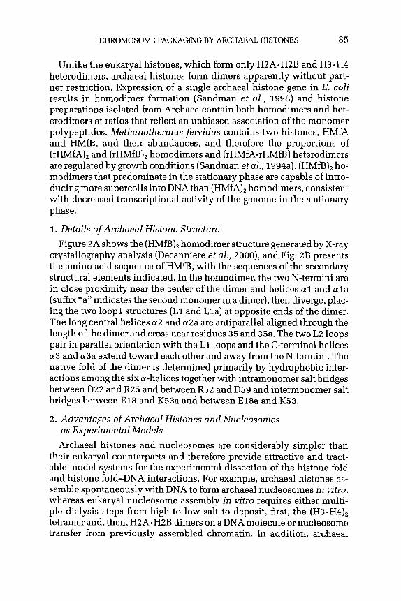
CHROMOSOME PACKAGING BY ARCHAEAL HISTONFS 85
Unlike the eukaryal histones, which form only H2AsH2B and H3sH4 heterodimers, archaeal histones form dimers apparently without part- ner restriction. Expression of a single archaeal histone gene in E. coli results in homodimer formation (Sandman et al., 1998) and histone preparations isolated from Archaea contain both homodimers and het- erodimers at ratios that reflect an unbiased association of the monomer polypeptides. Methanothermus fervidus contains two histones, HMfA and HMfB, and their abundances, and therefore the proportions of (rHMfAh and (rHMfB), homodimers and (rHMfA-rHMfB) heterodimers are regulated by growth conditions (Sandman et al., 1994a). (HMfBh ho- modimers that predominate in the stationary phase are capable of intro- ducing more supercoils into DNA than (HMfA), homodimers, consistent with decreased transcriptional activity of the genome in the stationary phase.
1. Details of Archaeal Histone Structure
Figure 2A shows the (HMfB)2 homodimer structure generated by X-ray crystallography analysis (Decanniere et al., 2000), and Fig. 2B presents the amino acid sequence of HMfB, with the sequences of the secondary structural elements indicated. In the homodimer, the two N-termini are in close proximity near the center of the dimer and helices al and ala (suffix “a” indicates the second monomer in a dimer), then diverge, plac- ing the two loop1 structures (Ll and Lla) at opposite ends of the dimer. The long central helices a2 and o12a are antiparallel aligned through the length of the dimer and cross near residues 35 and 35a. The two L2 loops pair in parallel orientation with the Ll loops and the C-terminal helices a3 and a3a extend toward each other and away from the N-termini. The native fold of the dimer is determined primarily by hydrophobic inter- actions among the six a-helices together with intramonomer salt bridges between D22 and R25 and between R52 and D59 and intermonomer salt bridges between El8 and K53a and between E18a and K53.
2. Advantages of Archaeal Histones and Nucleosomes as Experimental Models
Archaeal histones and nucleosomes are considerably simpler than their eukaryal counterparts and therefore provide attractive and tract- able model systems for the experimental dissection of the histone fold and histone fold-DNA interactions. For example, archaeal histones as- semble spontaneously with DNA to form archaeal nucleosomes in vitro, whereas eukaryal nucleosome assembly in vitro requires either multi- ple dialysis steps from high to low salt to deposit, first, the (H3.H4)2 tetramer and, then, H2A. H2B dimers on a DNA molecule or nucleosome transfer from previously assembled chromatin. In addition, archaeal

86 KATHLEEN SANDMAN AND JOHN N. REEVE
nucleosomes can be generated using a single recombinant archaeal his- tone preparation purified from E. coli. The addition of such recombinant histone preparations to DNA results in the formation of homotetramer- containing archaeal nucleosomes (Bailey et al., 1999), and site-directed mutagenesis of the cloned histone gene with subsequent expression in E. coli results in the availability of single histone variants that can be assayed for histone-fold stability and archaeal nucleosome formation. Many of the archaeal histones are from hyperthermophilic archaea, and purification of these stable recombinant proteins from E. coli is often as simple as boiling the crude extract to denature the host proteins and then recovering the histone of interest in a single heparin affinity step (Sandman et al., 2001). Crystallization and data collection by NMR over periods of several days are also greatly facilitated by the availability of such stable proteins.
3. Variants of rHMfB Given the experimental advantages, we have undertaken a detailed
investigation of archaeal histones and the archaeal nucleosomes assem- bled from homotetramers of rHMfI3, the recombinant (r) version of HMfB from M. fervidus, and from rHMfB variants generated by site-directed mutagenesis. An alignment of the amino acid sequences of archaeal histone family members (Fig. 3) identifies residues that are highly con- served and regions that accept residue variability. Differences among the archaeal histones and between archaeal and eukaryal histones have been investigated by replacing rHMfB residues with residues present in other histones. Alanine residues have also been introduced to deter- mine the consequences of the presence of this relatively unreactive side chain and oppositely charged or similarly charged residues have been substituted to probe electrostatic interactions (Table I).
After genetic construction, the first assay performed on every his- tone variant is to confirm its synthesis in E. coli. Most, but not all, of the rHMfl3 variants do accumulate in E. coli, and those that do not are presumed to be grossly misfolded and therefore subject to rapid degradation. DNA binding by rHMfI3 variants has been studied by an electrophoretic mobility shift assay (EMSA) performed using an agarose gel and a long DNA molecule [4.5 kb (kilo-base pairs)] that assays for the assembly of complexes that migrate faster than histone-free DNA (Sandman et al., 1990) and by a traditional EMSA in which the mobility of a shorter DNA fragment is retarded through a polyacrylamide gel by histone binding (Grayling et al., 1997). Ligase-mediated circularization of short DNA molecules (Bailey et al., 1999) and changes in the circu- lar dichroism signal of DNA at 275 nm (Soares et al., 2000) have also been used to assay the DNA binding of HMfB variants. Measurements

CHROMOSOME PACKAGING BY ARCHAEAL HISTONES 87
TABLE I
LIST AND PROPERTIES OF rHMfl3 VARIANTS GENERATED BY SITE-DIRECTED MUTAGENESIS
Variant(s) Category” Nucleosome formationb Additional phenotypes/notes
MlG EZD EZK L31 L3C P4C P4S 15V A6P P7C 18V
RlOG RloS, RlOK
IllL Kl3R K13E K13T Dl4E Dl4K
D14K/R37E D14N
D14N/R37E A15P A15S G16K
A171, Al7V E18D, E18P
E18K
AV cs cs cs cs cs cs cs cs cs cs cs cs AV AV AV AV AV AV AV AV AV cs cs cs cs AV EV
E18K/G51K EV
RlSS, Rl91, R19Q Rl9G vzoc V2OD v201 s21c
S21A, SZlT R25K 126E
T27A I31V 131A
cs CS, EV
cs cs AV cs cs AV
AV, ST AV
AV, ST AV, ST
+ N-Terminal structural variability f + + - f + +
- +
f + + - + + f f + +
+
- + +
+
Recombinant protein not detected
Recombinant protein not detected
Higher salt required for stability Higher salt required for stability
Recombinant protein not detected
Recombinant protein not detected
Greater affinity for DNA than wild-type histone
Greater affinity for DNA than wild-type histone
f -
Recombinant protein not detected + + i f f + + + 5°C decrease in T”
(continues)

88 KATHLEEN SANDMAN AND JOHN N. REEVE
TABLE I-Continued
Variant(s) Nucleosome
Categorya formationb Additional phenotypes/notes
131Y/M35Y M35C
AV
M35K M35Y G36A R37E R37K R37L R37Q D38E D38T I39A I39V A431 I44V
L46A, L461, L46Q, L46C, L46V
L46F L46S R48K H49A H49D
H49E, H49K G51A G51K
AV, ST AV, ST AV, ST AV, ST
AV
AV AV
AV cs
AV, ST cs
cs cs AV cs EV
cs AV, EV
R52K R52H, R52Q, R52A
K53R, K53T K53E
T54K, T541, T54R, T54C T54S, T54V, T54Y
155L, I55T 155M 155C 155V K56R K56E
K56Q, K561 E58S
cs cs cs cs cs
CS cs cs
AV, CS cs cs cs
AV, ST
D59A, DSSE, D59N cs 160A, 160G cs
+ +
+ + +
+ f f + +
-
+ zt
+
+ * 5 f
+
-
f - -
f -
+ + -
+
Introduction of aromatic residues Monomers can be cross-linked by
oxidation of these cysteine residues
18°C decrease in T”
5°C increase in T” 6°C decrease in T”
8°C increase in T” 6°C increase in 7’” Recombinant protein not detected
Recombinant protein not detected
Recombinant protein not detected
Recombinant protein not detected Greater affinity for DNA than
wild-type histone
Recombinant protein not detected
Recombinant protein not detected
Recombinant protein not detected Greater affinity for DNA than
wild-type histone; 5°C increase in T”
Recombinant protein not detected Recombinant protein not detected

CHROMOSOME PACKAGING BY ARCHAEAL HISTONES 89
TABLE I-Continued
Variant(s) Categorya Nucleosome formationb Additional phenotypes/notes
16OV AV, CS, ST E61A cs E61K cs
E61V, E61Q E61R
LGZI, L62V, L62A, L62W, L62R, L62C
L62M L62Y L62Q
V64R R66M F67A K68* K68E
K68E/K69* K69* K69E
cs cs cs
cs cs cs
AV AV ST AV
AV AV AV
+ + f
+ * zt
+ 5
Higher salt required for stability DNA bands are retarded in
agarose EMSA
Recombinant protein not detected
DNA bands are retarded in agarose EMSA
f
I
5°C decrease in T” Recombinant protein not detected
f f + f
8°C decrease in T”
* AV, mimics natural variation within the acchaeal histone family: EV, mimics natural variation between archaeal and eukaryal histones; ST, constructed to investigate histone-fold stability; CS, con- structed to investigate conservation of secondary or tertiary structure.
b +, indistinguishable from wild type; f, nucleosome formation but somewhat impaired: -, no nucleosome formation.
of the number of supercoils introduced and the direction of the super- coiling that results from nucleosome formation (Musgrave et al., 1991) by rHMfB variants on a closed circular DNA molecule have also been made. Both circular dichroism and differential scanning calorimetry have been used to quantitate the effects of amino acid residue substi- tution on the stability of the native fold of (rHMfB)2 dimers (Li et al., 1998).
Substitutions in the Hydrophobic Core of the (HMfBI, Histone Dimer. Hi&one-fold monomers are held together to form a dimer primarily by hydrophobic interactions between residues on the hydrophobic face of each amphipathic helix. As illustrated in the sequence alignment (Fig. 3), a hydrophobic side chain is present every three or four residues in each helix, although there is no strict residue conservation. For ex- ample, in al, positions 5, 8, 11, and 12 are occupied predominantly

90 KATHLEEN SANDMAN AND JOHN N. REEVE
by isoleucine, valine, or leucine, and changing these residues individ- ually to other hydrophobic residues results in histone variants that are indistinguishable from the wild type (Table I) (Soares et al., 2000).
In contrast, residue 55, located in L2 adjacent to DNA contacting residues, is most often a valine and, in a few cases, isoleucine or leucine. Substitutions in HMfB at 155 illustrate the importance of the hydropho- bic packing in this region of the molecule. Only the 155V change is tolerated in the rHMfB context; all others result in histone variants that exhibit reduced DNA binding (Table I) (Soares et al., 2000).
rHMfB has only one aromatic residue, phenylalanine 67, and this has precluded the use of spectroscopic techniques to quantitate this protein. The related archaeal histones HPyAl (Sandman et al., 1994b) and HTzAl (Ronimus and Musgrave, 1996), from Pyrococcus strain GB-3a and Thermococcus zilligii, respectively, have tyrosine residues at positions 31 and 35 within the hydrophobic core (Fig. 3), and sub- stituting tyrosine into rHMfB at positions 31 and 35 resulted in stable homodimers with spectral properties expected for a protein with four aromatic residues.
Structural Stability of Archaeal Histone Dimers. A comparison of rHMfB from M. fervidus [optimal temperature (T,,,) for growth of 83”C] with rHFoB from M. formicicum (Tort for growth of 45°C) (Darcy et al., 1995) revealed that, although the two histones share 78% sequence iden- tity (Fig. 3), they unfold at temperatures that differ by more than 30°C (Li et al., 1998). The 13 residue differences between HFoB and HMfB were introduced individually into HMfB, and the stability of the re- sulting HMfB variants was determined by following melting transitions using circular dichroism spectroscopy (Li et al., 2000b). The largest decrease in stability resulted from M35K substitution, and in recip- rocal experiments, replacing residue K35 in rHFoB with methionine, tyrosine, or phenylalanine increased the stability. A comparison of the high-resolution structures of HMfB and HFoB dimers reveals the pres- ence of two solvent-accessible cavities in rHFoB dimers that are filled in rHMfB dimers (Zhu et al., 1998). These cavities are adjacent to residues 36 and 36a, near the center of the histone dimer, where a2 and a2a cross. Also, surrounding these cavities and contributing to the differ- ences in thermal stability is the residue at position 31 (I in HMfB but A in HFoB). Overall it is clear that bulky hydrophobic residues that fill cavities more completely within the hydrophobic core make a substan- tial contribution to the higher stability of archaeal histone dimers from hyperthermophiles, although some additional stability is conferred by an increased number of surface-located electrostatic interactions (see DNA-Contacting Residues, below).

CHROMOSOME PACKAGING BY ARCHAEAL HISTONES 91
DNA-Contacting Residues. Structural studies (Starich et al., 1996; Decanniere et al., 2000) have identified surface-located basic residues that are positioned appropriately to interact with DNA. Near the N-terminus a hydrogen-bonded proline tetrad motif (P4/P4a and P7/P7a) unique to the archaeal histones orients the RlO side chain appropria- tely so that imino groups can make an electrostatic interaction with phosphates on each strand of the bound DNA. DNA contacts are also made in both loop regions, by residues R19, K53a, and T54a and R19a, K53, and T54, supported by the ElGK53a and E18a-K53 salt bridges and anchored to the hydrophobic core by V20lWOa and 155/155a.
Not surprisingly, these residues, RlO, R19, K53, and T54, are abso- lutely conserved in all archaeal histones (Fig. 3) and have clear ho- mologues in the eukaryal histones. Changing these residues results in rHMfB variants with significantly impaired abilities to form nucleo- somes based on gel shift assays (Table 1) (Soares et al., 2000). As these EMSAs require nucleosome persistence throughout extended periods of electrophoresis, the ligase-mediated circularization assay was used to determine if these histone variants might transiently form nucleosomes. Several rHMfB variants that gave a negative EMSA result were still able to facilitate the circularization of an 88bp DNA fragment, demonstrat- ing that they did still bind and bend DNA in solution (Soares et al., 2000). In fact, construction of an HMfB variant that was totally inca- pable of DNA binding required introducing residue substitutions at all three DNA binding sites, namely, in al, Ll, and L2. These results under- score the cooperative nature of DNA binding within a nucleosome and that loss of DNA binding at one histone-DNA contact point, even re- peated four times around a homotetramer, does not necessarily prevent nucleosome formation.
A rHMfB-R37E variant was generated to address the unique occur- rence of an arginine at position 37 in HMfB (Fig. 3). Based on the agarose EMSA, this variant did not form nucleosomes (Decanniere et al., ZOOO), but as residue 37 is located on the surface of ~2, and distant from the three DNA contacting sites, this suggested that additional factors were influencing nucleosome formation. Based on the crystal structure of HMfB, R37 forms surface-located salt bridges with E33 and D14a, and these influence the orientation of E2a, which in turn positions RlOa correctly for DNA binding (Decanniere et al., 2000). Disruption of this network of ionic interactions by substituting glutamic acid for arginine at position 37 apparently therefore has a domino effect that ultimately interferes with DNA binding.
The surface-localized electrostatic interactions also contribute to sta- bility in rHMfB dimers. The R37E change destabilized rHMfB and a re- ciprocal E37R change stabilizes rHFoB (Li et al., 2009b). Although the

92 KATHLEEN SANDMAN AND JOHN N. REEVE
magnitude of the increase in stability conferred by these charged residue substitutions was less than observed with cavity-filling hydrophobic changes, they nevertheless confirmed the importance of surface ionic interactions for fold stability.
Conserved Structures. Many of the invariant residues in the archaeal histone family play important structural roles. They are needed to main- tain the histone fold and do not tolerate substitutions. For example, alanine-glycine pairs are present at the C-termini of (~1 and a2 that terminate these helices (Aurora et al., 1994), and most substitutions at these positions result in variants that do not accumulate in E. coli (Table I). A notable exception is the rHMfE!-G51K variant that was de- signed to mimic the eukaryal histones and the archaeal histones from Methanococcusjannaschii. This HMfR variant has an increased affinity for DNA (Soares et al., 2000).
A salt bridge is formed between R52 and D59 in all archaeal histones and between the structurally homologous residues in all eukaryal his- tones. Substitutions of alanine, glutamic acid, or glutamine at position D59 resulted in variants that did not accumulate in E. coli, and of the four substitutions attempted at R52, only the conservative rHMfR-R52K variant accumulated in E. coli. Still, the variant did not form nucleo- somes as judged by the agarose EMSA (Table I).
The N-terminal sequence ELPhAP (h indicates a hydrophobic residue) is conserved in all the archaeal histones. These residues position RlO to contact DNA, by forming the proline tetrad structure and by electrostatic interactions mediated through E2, although residue substitutions sur- prisingly revealed that the presence of a proline at positions 4 and 7 is not absolutely required for DNA binding (Decanniere et al., 2000). This proline-rich sequence is not found in the eukaryal histones, which have much longer N-terminal tail domains and which must, therefore, use a different structural mechanism to orient the side chains of arginines in al toward the DNA.
Dimer-dimer In teractions. The similarity between the archaeal and the eukaryal histone dimers predicts that the molecular basis for tetramer formation is also similar. As illustrated in the crystal structure of the eukaryal nucleosome core (Luger et al., 199i’) the two copies of H3 in H3.H4 dimers interact (Fig. 1) to assemble the (H3.H4h tetramer by forming a four-helix bundle that contains the C-terminal regions of two ~2’s and the N-terminal regions of two ~3’s. A histidine residue in each copy of H3 makes a buried hydrogen bond to an aspartic acid resi- due, and additional stability is conferred by hydrophobic interactions.

CHROMOSOME PACKAGING BY ARCHAEAL HISTONES 93
Homologues of all residues responsible for these interactions are con- served in HMfEt, namely, H49, D52, L46, and L62. Of these, H49 and D52 are present in all archaeal histones and L46 and L62 are highly conserved but occasionally are replaced by other large hydrophobic residues. As noted above (see Conserved Structures), D52 cannot be substituted in rHMfB, but surprisingly, substitutions are acceptable at positions 46, 49, and 62 (Table I), resulting in HMfB variants that are capable of nucleosome formation (Soares et al., 2000).
B . NATURAL ARCHAEAL HISTONE VARIANTS
The genome of i’kf. jannaschii encodes six histone-fold proteins (Bult et al., 1996), five predicted to have typical archaeal histone features and one, encoded by MJ1647, that should have an additional C-terminal N-residue domain (Fig. 4). The histone fold of MJ1647 has many of the invariant features described above, but conspicuously absent are homologues of P7, R19, and H49. To evaluate these variations, both the full-length MJ1647 and a deletion encoding only the histone-fold region (MJ1647A) were expressed in E. coli (Li et al., 2000a). Both resulting recombinant proteins were found to be capable of nucleosome forma- tion, although archaeal nucleosomes formed by the full-length MJ1647 were not as stable as nucleosomes formed by MJ1647A during prolonged electrophoresis. M. jannaschii is a hyperthermophile (Tort for growth of 85”C), and thermal unfolding measurements were therefore made on both recombinant proteins. The presence of the C-terminal domain clearly increased the overall stability of the protein and the standard- state (1M) midpoint unfolding temperature (T”) calculated for MJ1647 was above 120”C, the highest value observed for any archaeal histone to date. The somewhat paradoxical instability of the MJl647-containing nucleosome compared with the extreme stability of the MJ1647 dimers
HMfB
H4 1 MJ1647 Mka/HHb
FIG. 4. The organization of the histone-fold domains in archaeal and eukaryal histones. Hatched boxes represent the three cr-helices in a histone-fold domain. The open box repre- sents an amino acid sequence in MJ1647 predicted to form an u-helix with no recognizable homology in other histone fold-containing proteins.

94 KATHLEENSANDMANANDJOHNN.REEVE
suggests that this polypeptide may not normally form homodimers but, rather, functions as a partner in a heterodimeric complex with one of the other five histones in M. jannaschii.
Methanopyrus kandleri contains a histone that is approximately twice the size of the typical archaeal histone (Slesarev et al., 1998), and Halobacterium sp. NRC1 is predicted to encode such a histone (Fig. 4) (Ng et al., 2000). These appear to have resulted from gene fusions and actually contain two tandem histone-fold motifs. Based on the Ii’-kDa molecular weight determined for the histone purified from M. kandleri, this “dimeric” archaeal histone is not proteolytically cleaved in viva but must fold to form a structure that is effectively a histone-fold dimer stabilized by intramolecular interactions.
C. NONHISTONE HISTONE FOLD-CONTAININGPROTEINS
The histone fold is present as a structural element in several eukaryal proteins in addition to the nucleosome core histones (Table II). These proteins have a variety of functions, ranging from atypical core histones found in a subset of nucleosomes to subunits of transcription factors (TATA-binding protein associated factors; TAFs) and DNA polymerase. In all cases, the histone folds in these proteins exist in dimer configura- tions, consistent with the requirement of dimer formation for histone- fold stability. The most conserved feature is that all these proteins have three a-helices with hydrophobic surfaces which mediate dimerization. Mutagenesis studies of the hydrophobic residues of hTAFrr20 have con- firmed their importance for dimer formation (Gangloff et al., 2000).
Analyses of the sequences of these nonhistone histone fold-containing proteins reveal that although the hydrophobic core needed for dimeriza- tion is conserved, the residues in the archaeal histones and the eukaryal core histones that specify the path of the DNA around the nucleosome are largely absent. Studies of the histone fold-containing TAFs suggested that a histone octamer-like complex may be present within transcrip- tion factor TFIID (Xie et al., 1996), but subsequent structural studies have not documented such an arrangement. The dTAFtt60-dTAFrr40 het- erodimer has been shown to form a tetramer that binds specifically to a conserved downstream promoter element (Burke and Kadonaga, 1997), but residues that contact the DNA have not been identified.
TAFs organized as specific heterodimers are found as components of several large, multisubunit transcription complexes, including the general transcription factor TFIID, and the SAGA and PCAF histone acetyltransferase complexes (reviewed by Green, 2000). The histone folds within these TAFs appear to aid in assembly of these large com- plexes rather than providing DNA binding domains.

TABL
E II
EUKA
RYA
L HIS
TON
E-FO
LD PR
OTE
INS
Hist
one-
fold
pa
ir”
Hist
one
fold
ba
sed
on
Bind
s D
NAb
St
ruct
ure/
func
tion
Refe
renc
e(s)
yCSE
4. H
4 hC
ENP-
A . H
4 yH
ZA.Z
.HZB
yH
AP3.
HAP
5 hN
F-YB
.NF-
YC
hCBF
-A . C
BF-C
yT
AF60
. TAF
17
hTAF
80 .T
AF31
dT
AF60
. TAF
40
yTAF
68. N
D
hTAF
LO.T
AF13
5 dT
AF30
a .N
D
yTAF
40. T
AF19
hT
AF28
.TAF
18
dTAF
3Ofi .
ND
yY
DR
1. B
UR
G
hDr1
. DR
AP
hNC
Zcy-
NC
2p
dhN
Czf
f-NC
2b
yTAF
68. A
DA1
yS
PT3
(intra
mol
ecul
ar)
yDPB
3 .D
PB4
hp12
.p17
hp
ls.p
17
dCH
RAC
14 .C
HR
AClG
Sequ
ence
hom
olog
y Ye
s
Sequ
ence
hom
olog
y Se
quen
ce ho
mol
ogy
ND
N
O
Crys
tal s
truct
ure
Sequ
ence
hom
olog
y
Crys
tal s
truct
ure
Sequ
ence
hom
olog
y
Sequ
ence
hom
olog
y Se
quen
ce ho
mol
ogy
Sequ
ence
hom
olog
y
Sequ
ence
hom
olog
y
Yes
ND
ND
NO
ND
N
O
ND
Yes
Subs
titut
es fo
r H3
.H4
in c
entro
mer
e ch
rom
atin
Subs
titut
es fo
r HZA
. H2B
in a
sub
set o
f nuc
leos
omes
R
ecog
nize
s CC
AAT
box
in p
rom
oter
s; b
inds
to T
BP
Com
pone
nts
of T
FIID
and
his
tone
ace
tyltr
ansf
eras
e
Com
pone
nts
of T
FIID
Com
pone
nts
of T
FIID
Rep
ress
es an
d ac
tivat
es tr
ansc
riptio
n
Com
pone
nts
of h
isto
ne a
cety
ltran
sfer
ase
Com
pone
nt o
f his
tone
ace
tyltr
ansf
eras
e C
ompo
nent
s of
DN
A po
lym
eras
e E
Com
pone
nts
of c
hrom
atin
rem
odel
ing
com
plex
Palm
er e
t 01.
(199
1)
sto1
er e
t al.
(199
5)
Sant
iste
ban
et a
l. (2
000)
M
anto
vani
(19
99)
Xie
et a
l. (1
996)
O
gryz
ko e
t al.
(199
8)
Burk
e an
d Ka
dona
ga (1
997)
G
angl
off e
t 01.
(~00
0)
Birc
k et
al.
(199
8)
Mer
mel
stei
n et
al.
(199
6)
Kim
et a
l. (2
000)
W
illy
et a
l. (2
000)
Gan
glof
f et a
l. (2
000)
Bi
rck
et a
l. (1
998)
Li
et a
l. (Z
OO
Oc)
Poot
et d
(200
0)
Cor
ona
et a
L(20
00)
a Th
e or
igin
of t
he h
isto
ne-fo
ld p
airs
is in
dica
ted
by y
(ye
ast),
h (h
uman
), d
01 (D
roso
phila
). b N
D, n
ot d
eter
min
ed.
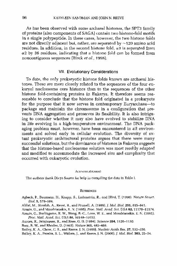
96 KATHLEEN SANDMAN AND JOHN N. REEVE
As has been observed with some archaeal histones, the SPT3 family of proteins (also components of SAGA) contain two h&one-fold motifs in a single polypeptide. In these cases, however, the two histone folds are not directly adjacent but, rather, are separated by -120 amino acid residues. In addition, in the second histone fold, a3 is separated from a2 by 36 residues, indicating that a histone fold can be formed from noncontiguous sequences (Birck et al., 1998).
VII. Evolutionary Considerations
To date, the only prokaryotic histone folds known are archaeal his- tones. These are more closely related to the sequences of the four eu- karyal nucleosome core histones than to the sequences of the other histone fold-containing proteins in Eukarya. It therefore seems rea- sonable to conclude that the histone fold originated in a prokaryote for the purpose that it now serves in contemporary Euryarchaea-to package and maintain the chromosome in a configuration that pre- vents DNA aggregation and preserves its flexibility. It is also intrigu- ing to consider whether it may also have evolved to stabilize DNA in life evolving in a high-temperature environment. The DNA pack- aging problem must, however, have been encountered in all environ- ments and solved early in cellular evolution. The diversity of ex- tant prokaryotic architectural proteins argues that there were many successful solutions, but the dominance of histones in Eukarya suggests that the histone-based nucleosome solution was most readily adapted and modified to accommodate the increased size and complexity that occurred with eukaryotic evolution.
ACKNOWLEDGMENT
The authors thank Divya Soares for help in compiling the data in Table I.
REFERENCES
Agback, P., Baumann, H., Knapp, S., Ladenstein, R., and Hard, T. (1998). Nature Sfruct. Biol. 5, 579-584.
Alilat, M., Sivolob, A., Revet, B., and Prunell, A. (1999). 1. Mol. Biol. 291, 815-841. Arents, G., and Moudrianakis, E. N. (1995). Proc. N&l. Acad. Sci. USA 92, 11170-11174. Arents, G., Burlingame, R. W., Wang, B.-C., Love, W. E., and Moudrianakis, E. N. (1991).
Proc. Natl. Acad. Sci. USA 88,10148-10152. Aurora, R., Srinivasan, R., and Rose, G. D. (1994). Science 264, 1126-1130. Baer, B. W., and Rhodes, D. (1983). Nature 301, 482488. Bailey, K. A., Chow, C. S., and Reeve, J. N. (1999). Nucleic Acids Res. 27, 532-536. Bailey, K. A., Pereira, S. L., Widom, J., and Reeve, J. N. (2000). 1. Mol. Biol. 303, 25-34.

CHROMOSOME PACKAGING BY ARCHAEAL HISTONES 97
Birck, C., Poch, O., Romier, C., Ruff, M., Mengus, G., Lavigne, A.-C., Davidson, I., and Moras, D. (1998). Cell 94, 239-249.
Bloomfield, V. A. (1996). Cum Opin. Struct. BioJ. 6, 334-341. Bloomfield, V. A. (1997). Biopolymers 44, 269-282. Bult, C. J., White, O., Olsen, G. J., Zhou, L., Fleischmann, R. D., and Sutton, G. G., et al.
(1996). Science 273,1058-1073. Burke, T. W., and Kadonaga, J. T. (1997). Genes Dev. 11, 3020-3031. Chirinos, M., Hernandez, F., and Palacian, E. (1999). Arch. Biochem. Biophys. 370, 222-
230. Corona, D. F. V., Budde, A., Deuring, R., Ferrari, S., Varga-Weisz, P. D., Wilm, M., Tamkun,
J. W., and Becker, P. B. (2000). EMBOJ. 19, 3049-3059. Darcy, T. J., Sandman, K., and Reeve, J. N. (1995). J. Bacterial. 177, 858-860. Decanniere, K., Babu, A. M., Sandman, K., Reeve, J. N., and Heinemann, II. (2000). J. Mol.
BioJ. 303, 35-47. Dong, F., and van Holde, K. E. (1991). Proc. N&J. Acad. Sci. USA 88, 10596-10600. Gangloff, Y.-G., Werten, S., Romier, C., Carre, L., Poch, O., Moras, D., and Davidson, I.
(2000). Mol. Cell. BioJ. 20, 340-351. Grayling, R. A., Sandman, K., and Reeve, J. N. (1996). Adv. Protein Chem. 48,437-467. Grayling, R. A., Bailey, K. A., and Reeve, J. N. (1997). Extremophiles 1, 79-88. Green, M. (2000). Trends Biochem. Sci. 25, 59-63. Hamiche, A., and Richard-Foy, H. (1998). J. BioJ. Chem. 273, 9261-9269. Hamiche, A., Carot, V., Alilat, M., DeLucia, F., O’Donohue, M.-F., R&et, B., and Prunell, A.
(1996). Proc. NatJ. Acad. Sci. USA 93, 7588-7593. Hansen, J. C., and Wolffe, A. P. (1994). Proc. NatJ. Acad. Sci. USA 91, 2339-2343. Hirschhorn, J. N., Brown, S. A., Clark, C. D., and Winston, F. (1992). Genes Dev. 6, 2288-
2298. Holmes, V. F., and Cozzarelli, N. R. (2000). Proc. Natl. Acad. Sci USA 97, 1322-1324. Jackson, V. (1990). Biochemistry29, 719-731. Kim, S., Cabane, K., Hampsey, M., and Reinberg, D. (2000). Mol. Cell. Biol. 20,2455-2465. Li, J.-Y., Arnold-Schulz-Gahman, B., and Kellenberger, E. (1999). Microbiology 145, l-2. Li, W.-T., Grayling, R. A., Sandman, K., Edmondson, S., Shriver, J. W., and Reeve, J. N.
(1998). Biochemistry 37, 10563-10572. Li, W.-T., Sandman, K., Pereira, S. L., and Reeve, J. N. (2000a). Extremophiles 4,43-51. Li, W.-T., Shriver, J. W., and Reeve, J. N. [2OOOb). J. Bacterial. 182, 812-817. Li, Y., Pursell, Z. F., and Linn, S. (2000~). J. BioJ. Chem. 275, 23247-23252. Liu, L. F., and Wang, J. C. (1987). Proc. AJatJ. Acad. Sci. USA 84, 7024-7027. Louters, L., and Chalkley, R (1985). Biochemistry 24, 3080-3085. Lowary, P. T., and Widom, J. (1998). J. Mol. BioJ. 276,19-42. Luger, K., Mader, A. W., Richmond, R. K., Sargent, D. F., and Richmond, T. J. (1997).
Nature 389, 251-260. Mantovani, R. (1999). Gene 239, 15-27. Mermelstein, F., Yeung, K., Cao, J., Inostroza, J. A., Erdjument-Bromage, H., Eagelson, K.,
Landsman, D., Levitt, P., Tempst, P., and Reinberg, D. (1996). Genes Dev. 10, 1033- 1048.
Minsky, A., Ghirlando, R., and Reich, 2. (1997). I. Theor. BioJ. 188, 379-385. Musgrave, D. R., Sandman, K. M., and Reeve, J. N. (1991). Proc. N&J. Acad. Sci. USA 88,
10397-10401. Ng, W. V., Kennedy, S. P., Mahairas, G. G., Berquist, B., and Pan, M., et al. (2000). Proc.
Natl. Acad. Sci. USA 97,12176-12181. Ogryzko, V. V., Kotani, T., Zhang, X., Schlitz, R. L., Howard, T., Yang, X.-J., Howard, B. H.,
Qin, J., and Nakatani, Y. (1998). Cell 94, 35-44.

98 KATHLEEN SANDMAN AND JOHN N. REEVE
Orphanides, G., Wu, W.-H., Lane, W. S., Hampsey, M., and Reinberg, D. (1999). Nature 400,284-288.
Palmer, D. K., O’Day, K., Trong, H. L., Charbonneau, H., and Margolis, R. L. (1991). Proc. Natl. Acad. Sci USA 88, 3734-3738.
Pereira, S. L., Grayling, R. A., Lurz, R., and Reeve, J. N. (1997). Proc. N&l. Acad. Sci. USA 94,12633-12637.
Pereira, S. L., and Reeve, J. N. (1998). Extremophiles 2, 141-148. Pereira, S. L., and Reeve, J. N. (1999). I. Mol. Biol. 289, 675-681. Poot, R. A., Dellaire, G., Hiilsmann, B. B., Grimaldi, M. A,, Corona, D. F. V., Becker, P. B.,
Bickmore, W. A., and Varga-Weisz, P. D. (2000). EMBO J. 19, 3377-3387. Protacio, R. U., Polach, K. J., and Widom, J. (1997). J. Mol. Biol. 274, 708-721. Prunell, A. (1998). Biaphys. J. 74, 2531-2544. Rice, P. A., Yang, S., Mizuuchi, K., and Nash, H. A. (1996). Cell 87, 1295-1306. Robinson, H., Gao, Y.-G., McCrary, B. S., Edmondson, S. P., Shriver, J. W., and Wang,
A.H.-J. (1998). Nature 392, 202-205. Ronimus, R., and Musgrave, D. R. (1996). Biochim. Biophys. Acta 1307, 1-7. Ruepp, A., Graml, W., Santos-Martinez, M.-L., Koretke, K. K., Volker, C., Mewes, H. W.,
Frishman, D., Stocker, S., Lupas, A. N., and Baumeister, W. (2000). Nature 407, 508- 513.
Sandman, K., and Reeve, J. N. (1999). J. Bacterial. 181, 1035-1038. Sandman, K., and Reeve, J. N. (2000). Arch. Microbial. 173, 165-169. Sandman, K., Krzycki, J. A., Dobrinski, B., Lurz, R., and Reeve, J. N. (1990). Proc. Natl.
Acad. Sci. USA 87,5788-5791. Sandman, K., Grayling, R. A., Dobrinski, B., Lurz, R., and Reeve, J. N. (1994a). Proc. N&J.
Acad.Sci. USA 91,12624-12628. Sandman, K., Perler, F. B., and Reeve, J. N. (199413). Gene 150, 207-208. Sandman, K., Pereira, S. L., and Reeve, J. N. (1998a). Cell. Mol. Life Sci. 54, 1350-1364. Sandman, K., Zhu, W., Summers, M. F., and Reeve, J. N. (1998b). In “Thermophiles: The
Key to Molecular Evolution and the Origin of Life?” (J. Weigel and M. W. W. Adams, eds.), pp. 243-253. Taylor & Francis, London.
Sandman, K., Bailey, K. A., Pereira, S. L., Soares, D., Li, W.-T., and Reeve, J. N. (2001). Archaeal histones and nucleosomes. Methods Enzymol. 334, 116-129.
Santisteban, M. S., Kalashnikova, T., and Smith, M. M. (2000). Cell 103, 411422. Shioda, M., Sugimori, K., Shiroya, T., and Takayanagi, S. (1989). J. Bacterial. 171, 4514-
4517. Slesarev, A. I., Belova, G. I., Kozyavkin, S. A., and Lake, J. A. (1998). Nucleic Acids Res.
26,427430. Soares, D. J., Sandman, K., and Reeve, J. N. (2000). J. Mol. Biol. 297, 39-47. Soppa, J. (1999). Mol. Microbial. 31, 1295-1305. Spangenberg, C., Eisfeld, K., Stiinkel, W., Luger, K., Flaus, A., Richmond, T. J., Truss, M.,
and Beato, M. (1998). J. Mol. Biol. 278, 725-739. Starich, M. R., Sandman, K., Reeve, J. N., and Summers, M. F. (1996). J. Mol. Biol. 255,
187-203. Stoler, S., Keith, K. C., Curnick, K. E., and Fitzgerald-Hayes, M. (1995). Genes Dev. 9,
573-586. Strahl, B. D., and Allis, C. D. (2000). Nature 403,41-45. Struhl, K. (1999). Cell 98, 14. Takayanagi, S., Morimura, S., Kusaoke, H., Yokoyama, Y., Kano, K., and Shioda, M. (1992).
J. Bacterial. 174, 7207-7216. Thastrom, A., Lowary, P. T., Widlund, H. R., Cao, H., Kubista, M., and Widom, J. (1999).
J. Mol. Biol. 288, 213-229.

CHROMOSOME PACKAGING BY ARCHAEAL HISTONES 99
Trun, N. J., and Marko, J. F. (1998). ASM Newslett. 64, 276-283. Tse, C., Fletcher, T. M., and Hansen, J. C. (1998). Proc. Nat]. Acad. Sci. USA 95, 12169-
12173. Tuerk, C., and Gold, L. (19901. Science 249, 505-510. van Holde, K., and Zlatanova, J. (1999). BioEssays 21, 776-780. Widlund, H. R., Cao, H., Simonsson, S., Magnusson, E., Simonsson, T., Nielsen, P. E.,
Kahn, J. D., Crothers, D. M., and Kubista, M. (1997). 1. Mol. Biol. 267, 807-817. Willy, P. J., Kobayashi, R., and Kadonaga, J. T. (2000). Science 290, 982-984. Xie, X., Kokubo, T., Cohen, S. L., Mirza, II. A., Hoffmann, A., Chait, B. T., Roeder, R. G.,
Nakatani, Y., and Burley, S. K. (1996). Nature 360, 316-322. Zhu, W., Sandman, K., Lee, G. E., Reeve, J. N., and Summers, M. F. (1998). Biochemistry
37,10573-10580.

DNA Recombination and Repair in the Archaea
ERICA M. SEITZ, CYNTHTA A. HASELTINE, AND
STEPHEN C. KOWALCZYKOWSKI Sections of Microbiology and of Molecular and Cellular Biology
Center for Genetics and Development University of California, Davis Davis, California 956163665
I. Introduction II. Recombinational Repair
A. An Overview of Homologous Recombination B. Generation of DNA Breaks C. Initiation of Homologous DNA Recombination: DSB End Processing D. DNA Pairing and Strand Exchange E. Single-Stranded DNA Binding Proteins F. Additional Proteins Involved in DNA Strand Exchange G. Holliday-Junction Cleaving Enzymes H. Summary: Archaeal Recombinational Repair
III. DNA Repair Pathways A. Direct DNA Damage Reversal B. Base Excision Repair C. Nucleotide Excision Repair D. Mismatch Repair E. Flap Endonuclease Protein Homologues F. Translesion DNA Synthesis and Mutagenesis
G. Summary: DNA Repair Mechanisms in the Archaea References
I. introduction
DNA is subjected daily to considerable environmental and endoge- nous damage, which challenges both the integrity of the essential information that it contains and its ability to be transferred to fn- ture generations. All cells, however, are prepared to handle damage to the genome through an extensive DNA repair system, thus un- derscoring the importance of this process in cell survival. The Ar- chaea represent a rather diverse group of organisms, including many members who thrive under conditions that would be lethal for most bacteria and eukaryotes. These conditions, such as extreme temper- atures, also present a new challenge to the Archaea and to their genomes, reinforcing the need to possess an efficient DNA repair system (DiRuggiero et al., 1999; Grogan, 2000). This, and the fact that the Archaea are a largely unexplored domain of life prompted
101 ADVANCES IN APPLIED MICROBIOLOGY, VOLUME 50
Copyright 0 2001 by Academic Press All rights of reproduction in any form reserved.
0065.2164/01$35.00

102 ERICA M. SEITZ ET AL.
interest in the types of DNA repair mechanisms that operate within this domain.
Studies carried out in bacteria, especially in Escherichia coli, or in eukaryotes, particularly in the yeast Saccharomyces cerevisiae, re- vealed much of what is known about these processes. These studies showed that DNA repair occurs by several pathways (Lindahl and Wood, 1999); these include reversal of DNA damage, excision of damaged nu- cleotides (nucleotide excision repair or NER) or bases (base excision re- pair or BER), excision of misincorporated nucleotides (mismatch repair or MMR), and recombinational repair (Friedberg et al., 1995). Although relatively little was known about DNA repair in Archaea, the recent sequencing of several archaeal genomes permitted the identification of structural homologues of many proteins involved in these different pathways. In this article, we review the most important features of DNA repair learned from studies of organisms such as E. coli and S. cerevisiae. In particular, we emphasize the elements which have been conserved throughout evolution, either at the level of global mechanisms or at the level of the protein effecters. We apply this knowledge to the third do- main of life, the Archaea, and review what is known about DNA repair in this domain of life, with a specific emphasis on recombinational repair.
II. Recombinational Repair
One of the most serious types of damage that can be inflicted on the genome is a DNA break either in a single strand or in both strands of DNA [a double-stranded DNA (dsDNA) break; DSB]. DNA breaks of any type pose a particularly significant problem to the cell because they challenge the integrity of the DNA molecule and can lead, if not repaired, to loss of information, gross chromosomal rearrangements, and chromosome missegregation. Because of these potentially lethal consequences, both bacterial and eukaryal organisms have mechanisms for repairing this type of DNA lesion, although the manner by which each repairs the lesion differs. In the Bacteria, this type of damage is remedied primarily by the process of homologous DNA recombina- tion (Kowalczykowski et al., 1994; Kuzminov, 19X2), whereas in the Eukarya, the DSB is repaired by either homologous recombination or nonhomologous end joining (NHEJ) (Paques and Haber, 1999; Sung et al., 2000). Recombination involves pairing of the damaged DNA with a homologous partner to copy any lost information from the homologue, thereby accurately repairing the DSB, whereas NHEJ involves ligation of the DSB without the need for significant homology, thus being inherently error-prone. Here we focus on DSB repair by homologous recombination, as NHEJ appears to be a uniquely eukaryal process.

DNARFiCOMBINATIONANDREPAIRINTHEARCHAEA 103
A. ANOVERVIEWOFHOMOLOGOUSRECOMBINATION
Homologous DNA recombination is a primary means for the repair of DSBs. Although the general mechanism is similar in bacteria and eukaryotes, the proteins that are involved in this process differ (Fig. 1). Figure 1 depicts the DSB repair model (Resnick, 1976; Szostak et al., 1983) and the likely proteins that act at each step. After DSB forma- tion, both ends of the break are resected to create single-stranded DNA (ssDNA), which then invades a homologous dsDNA molecule. After DNA strand invasion occurs, the 3’ ends of the invading strands serve as primers for the initiation of nascent DNA synthesis, which leads to the formation of two Holliday junctions that are cleaved in one of two orientations to generate two types of recombinant molecules (Fig. 1).
Biochemical studies have revealed the function of many enzymes that participate in the process of homologous recombination. In E. coli, it was determined that the process of homologous recombination involves the action of more than 25 different proteins (Kowalczykowski et al., 1994). Figure 1 shows some of the enzymes from E. coli and S. cerevisiae that act at each step in this process (Kowalczykowski et al., 1994; Paques and Haber, 1999) and for which there are, or may be, either structural or functional homologues in the Archaea. The first step in the homol- ogous DNA recombination pathway is an initiation or processing step, which involves processing of the broken DNA molecule so that a region with a partially ssDNA character is generated. This processing can be accomplished through the action of DNA helicases, nucleases, or both. The next step corresponds to the search for the homologous target DNA molecule, which is immediately followed by the exchange of their DNA strands. This step is accomplished by DNA strand exchange proteins, which bind to the ssDNA that was generated previously. The resultant nucleoprotein filament is the active form of these proteins, which acts both in the homology search process and in the invasion of the recip- ient DNA molecule. The consequence of this initial pairing event is a region of newly paired or heteroduplex DNA, which is also known as a joint molecule (Kowalczykowski and Eggleston, 1994). The third step involves the reciprocal exchange of the two DNA strands, creating a four-stranded structure known as a Holliday junction. The regions of heteroduplex DNA are extended by protein-promoted branch migration, which involves the action of either the DNA strand exchange protein or a specialized DNA helicase. The final step involves symmetric cleav- age of the Holliday junction in one of two orientations by a Holliday junction-specific endonuclease to produce one of two alternative re- combinant products (Kowalczykowski et al., 1994; West, 1994a,b; White et al., 1997; Lilley and White, 2006). Despite differences between the

3 5’
5’
3
+ 5’
3’
3’
5’
E. c
oli
S. ce
revi
siue
Sp
oil
Arch
aea
Spoi
l’
___
-_-
Mre
ll/Ra
dSO
/Xrs
2 M
relU
Rad
50
SgsP
Sg
slZJ
RadS
l R
PA
Rad
55/5
7 R
ad54
R
ad52
-_
_
Rad
A RP
AISS
B R
adB
Rad
543
_--
__-
?S
Cccl
(m
itocb
ondr
ial)
Hjr/
Hje6
Initi
atio
n
Proc
essi
ng
Hei
icas
e and
/or
nucl
ease
R
ecBC
D
SbcC
D
Rec
Q
~I
3’ -
3’
- 5’
H
omol
ogou
s Pai
ring
& D
NA
Stra
nd
EXC
ha?l
@?
1 R
ecA-
like
prot
ein
and
Rec
A SS
B 3’
5’
Acce
ssor
y pro
tein
s __
_ __
_ R
ecO
(R)
Rec
F(R
)
DN
A Sy
nthe
sis
& An
neal
ing
Bran
ch m
igra
tion
prot
eins
R
uvAB
R
ecG
D
NA
Het
erod
uple
x Ex
tens
ion
; y-
--y
;
3’---
- -
5’
Res
olut
ion
Res
olva
se
3’---
- Sp
liced
’
’ Pa
tche
d 5’
3’
Ruv
C

DNA RECOMBINATION AND REPAIR IN THE ARCHAEA 105
well-studied bacterial (namely, E. coli) and eukaryal systems (namely, S. cerevisiae), these basic steps remain mostly conserved.
1. Bacterial Homologous DNA Recombination
E. coli possesses two pathways for the repair of DNA strand breaks (Kowalczykowski et al., 1994; Kuzminov, 1999): the RecBCD pathway, which repairs DSBs; and the RecF pathway, which repairs primarily single-strand gaps but can repair DSBs as well. Both of these pathways for recombinational repair depend on the action of the RecA protein. In the RecBCD pathway, the RecBCD helicase/nuclease both processes the DSB to create ssDNA and loads RecA protein onto this ssDNA in anticipation of DNA strand exchange. In the RecF pathway, RecQ helicase processes the broken DNA molecule to produce ssDNA, and the RecO and RecR proteins aid in loading RecA protein onto the ssDNA by mediating the removal of ssDNA binding (SSB) protein (Umezu et al., 1993; Harmon and Kowalczykowski, 1998; Kuzminov, 1999).
2. Eukaryal Homologous DNA Recombination
Homologous DNA recombination is studied in the Eukarya most extensively with the yeast, S. cerevisiae, but recent studies in mammals demonstrate the commonality of this eukaryotic process (Paques and Haber, 1999). As discussed later, some parallels can be drawn between the yeast and the bacterial systems, but for the most part, the system in yeast exists as a more complex process. The repair of DSBs by homol- ogous recombination requires members of the yeast RAD52 epistasis group, which consists of RAD50, RAD51, RAD52, RAD54, RAD55, RAD57, RAD59, MREZI, XRSZ, and RDH54/TID1 genes (Game, 1993; Paques and Haber, 19%). The function of the proteins encoded by these genes has been studied both genetically and biochemically, but the precise function of some proteins is not yet fully understood (Fig. 1).
3. Archaeal Homologous DNA Recombination
The genome sequences of several archaeons has made it possible to identify structural homologues of many proteins involved in the process of homologous DNA recombination. In addition, some of these proteins
FIG. 1. Mechanism for double-stranded DNA break repair by homologous recombina- tion, and the proteins involved. Shown are the proteins that are either known or proposed to act in each step of this process in E. coli, S. cerevisiae, and the Archaea. Notes: rThe archaeal Spoll protein is a subunit of TopoVI, and a direct role in DSB formation is not clearly defined. ‘A role for Sgsl in initiation is unclear. 3 Assignment is based only on se- quence homology. 4 The Rad54 protein is not a structural homologue of either the RuvAB or the RecG protein; however, it will promote DNA heteroduplex extension (J. Solinger et al, in press). s? refers to the fact that an activity has been found in human cells but the respon- sible protein is unknown. “Hje refers to an activity only; the protein has not been identified.

106 ERICA M. SEITZ ET AL.
have been studied biochemically, and there is some genetic evidence supporting the role of these genes in archaeal homologous DNA recom- bination. Evidence for stimulation of chromosomal marker exchange in the hyperthermophilic archaeon Sulfolobus QC~~OCQ~~Q~~US provides evidence for DNA repair, conjugation, and homologous recombina- tion processes in these organisms (Schmidt et al., 1999). Figure 1 and Table I present mainly the proteins involved in this process for which homologues have been found in the Eukarya and Archaea. For the most part, the proteins identified in the Archaea show greater structural and, in some cases, functional, similarity to eukaryal proteins than to their bacterial counterparts (Fig. 1).
B. GENERATION OF DNA BREAKS
DNA breaks can occur either in a single DNA strand, creating ssDNA gaps, or in both strands, DSBs. There are many routes for production of ssDNA gaps or DSBs, but DNA replication is a major mechanism for converting ssDNA lesions into larger gaps or DSBs (Kogoma, 1997; Kuzminov, 1999; Kowalczykowski, 2000; Michel, 2000). As illustrated in Fig. 2, ssDNA gaps can be created if a blocking lesion is not removed by repair processes prior to the arrival of the DNA replication machinery. If the lesion is on the lagging strand template, then Okazaki fragments cannot be joined; if the lesion is on the leading strand, then the replication fork halts and may initiate farther down- stream. In either case, a region of single-stranded, unreplicated DNA is created. Lesions having the ability to halt the progression of replicative DNA polymerases are numerous and include the well-studied 64 thymine photoproducts and cyclobutane pyrimidine dimers caused by ultraviolet (UV) light (Edenberg, 1976).
DSBs can arise from several sources. Exogenously, DSBs are caused by ionizing radiation such as X-rays or y rays or by various radiomimetic chemicals. Endogenously, DSBs can be created directly by reactive oxygen species and can also arise as a consequence of replicating a nicked DNA template (Fig. 2). Indeed, if a DNA replication fork encoun- ters an interruption (nick or ssDNA gap) in one of the two DNA strands, this interruption will be converted to a DSB (Kuzminov, 1999; Paques and Haber, 1999; Kowalczykowski, 2000). Nicks in DNA can result from numerous sources, including unsealed Okazaki fragments on the lagging strand and incision of a damaged DNA strand by another repair system, such as either nucleotide or base excision repair. DSBs can also be created as a consequence of the replication apparatus stalling or halting. Stalling can occur, for example, due to the presence of a chemical imper- fection in the DNA or a protein complex tightly bound to DNA, either of which can block the progression of the fork. The stalled DNA replication

TABL
E I
ARC
HAE
ALR
ECO
MBI
NAT
ION
PRO
TEIN
HO
MO
LOG
UES
~
Rad
A pa
ralo
gue
Mre
ll R
ad50
Xr
s2
RPA
ISSB
R
adA
Rad
B O
ther
R
ad52
R
ad54
’ R
ad59
Sp
oll
Hjc”
A. f
ulgi
dus
Hal
obac
teriu
m
M.
jann
asch
ii M
. th
erm
oaut
otro
phic
um
E! a
byss
ii l?
furio
sus
19 ho
rikos
hii
A. p
erni
x S.
sol
fata
ricus
+ -
+ -
+ -
+ -
+ -
+ -
+ -
+ -
+ -
-t-
+ -
- +
+ -
- +
+ ?d
-
+ +
- -
+ +
- -
+ +
- -
i- +
- -
+ +
- -
+ +
- -
? -
? - -
- -
? ? -
? -
? -
+e
-
+ +
+ ?
+ +
+ +
+ ?
+ +
+ +
+ +
+ +
a Th
e po
tent
ial
reco
mbi
natio
n pr
otei
n ho
mol
ogue
s fro
m n
ine
fully
se
quen
ced
arch
aeal
gen
omes
ar
e re
pres
ente
d.
A +
indi
cate
s th
e pr
esen
ce
of a
sing
le
hom
olog
ous
prot
ein
sequ
ence
, wh
ile
a -
repr
esen
ts
the
failu
re
to d
etec
t a
hom
olog
ue.
A ?
is s
hown
fo
r si
ngle
pr
otei
n se
quen
ces
wher
e a
hom
olog
ue
may
be
pres
ent,
but
suffi
cien
tly
high
le
vels
of
hom
olog
y to
per
mit
conf
iden
t as
sign
men
t ar
e no
t ap
pare
nt.
An R
rep
rese
nts
the
pres
ence
of
an
RPA-
like
stru
ctur
al
prot
ein
hom
olog
ue,
while
an
S re
pres
ents
th
e pr
esen
ce
of a
n SS
B-lik
e st
ruct
ural
pr
otei
n ho
mol
ogue
. b
A si
ngle
pro
tein
se
quen
ce
with
lim
ited
hom
olog
y to
Rad
54 w
as i
dent
ified
in
eac
h of
the
org
anis
ms
indi
cate
d by
a ?
. ’
A Ho
llida
y-ju
nctio
n en
donu
clea
se
activ
ity
dist
inct
fro
m
Hjc
was
fou
nd
S. s
hiba
tae
and
S. s
olfa
taric
us
and
is c
alle
d Hj
e.
d A
sing
le p
rote
in
sequ
ence
was
ide
ntifi
ed
with
ho
mol
ogy
to R
ad55
. e
This
ho
mol
ogue
is
bas
ed o
n se
quen
ce
sim
ilarit
y on
ly.

A. D
oubl
e-st
rand
ed D
NA
Brea
ks
,“:
5’
qiz&
y
5’-
- 3’
3’
- -
5’
3’
5’
+ 5’
- 3’
-
B. S
ingl
e-st
rand
ed D
NA
gaps
5’
3’
3’-
+ 5’
3’
3’
5’
Repl
icat
ion
I I
of li
near
ch
rom
osom
e
2:
3’
5’
5’
3’
3’
:: 3’
5’
FIG
. 2.
Sing
le-s
trand
ed
DN
A ga
ps o
r do
uble
-stra
nded
D
NA
brea
ks a
re f
orm
ed
by D
NA
dam
agin
g ag
ents
an
d by
DN
A re
plic
atio
n th
roug
h th
e le
sion
. D
epic
ted
is th
e pr
oduc
tion
of (
A) a
dou
ble-
stra
nded
D
NA
brea
k,
form
ed
eith
er b
y D
NA
dam
agin
g ag
ents
dire
ctly
or
by
repl
icat
ion
thro
ugh
a ni
cked
te
mpl
ate,
an
d (B
) a
sing
le-
stra
nded
D
NA
gap,
for
med
by
rep
licat
ion
stop
ping
at
the
les
ion.
Bo
th s
sDN
A ga
ps a
nd d
sDN
A br
eaks
can
be
repa
ired
by h
omol
ogou
s re
com
bina
tion.
[A
dapt
ed
from
Ko
wal
czyk
owsk
i (Z
OO
O).]

DNA RECOMBINATION AND REPAIR IN THE ARCHAEA 109
forks must be restarted for the replication of the genome to be com- pleted. This restart can be achieved through the introduction of a DSB at the regressed replication fork, followed by recombination-dependent replication (Kogoma, 1997; Michel et al., 1997; Kuzminov, 1999; Paques and Haber, 1999; Kowalczykowski, 2000; Marians, 2000; Michel, 2000).
In addition to these general mechanisms for DSB formation, DSBs in the Eukarya are also produced in a programmed and specific manner. For example, in meiotic cells, DSBs are enzymatically introduced during the initiation phase of meiosis, to ensure the crossing-over of homologues needed for their faithful segregation (Keeney et al., 1997; Haber, 2OOOa,b).
1. DSBs in the Bacteria In E. coli, DNA replication initiates at the chromosomal origin, OriC,
and progresses bidirectionally along the two arms of the circular chro- mosome toward the replication terminus. The majority of these replica- tion forks encounters an obstacle to their progression, leading to their stalling (Kogoma, 1997; Michel et al., 1997; Kuzminov, 1999). These obstacles can be chemical lesions, DNA-bound protein complexes, or secondary DNA structure. Regardless of the obstacle, complete replication of the chromosome requires the origin-independent restart of the stalled replication fork. DNA recombination is responsible for this restart (Kogoma, 1996). Recent studies indicate that the first step in this process involves regression of the replication fork by reannealing of the two newly synthesized DNA strands after replication fork arrest. This creates an X-shaped Holliday junction that contains one accessible dsDNA end (Postow et al., 2000; Flores et al., 2001). The RecG protein, a DNA helicase involved in homologous recombination, can catalyze such Holliday junction formation by replication fork reversal (McGlynn and Lloyd, 2000). At this stage, this intermediate can be processed in either of two ways. The RecBCD enzyme, an enzyme involved in the initiation of DNA recombination in bacteria (see below), is a dsDNA nuclease that acts on the DSB created at the Holliday junction (which was formed by replication fork reversal) and starts degrading the DNA. This nucleolytic action effectively shortens the two newly synthesized strands and allows the replication fork to move back from the point where it initially stalled, giving it another opportunity to progress past the previous block after it reinitiates. Alternatively, the regressed replication fork/Holliday junction can be recognized and cleaved by the RuvABC complex to produce a DSB (Michel et al., 1997; Seigneur et al., 1998). The RuvAB complex is involved in the branch migration of Holliday junctions, and RuvC is an endonuclease that specifically cleaves these junctions, as discussed in more detail below. The DSB is then repaired by homologous recombination and is used to restart

110 ERICA M. SEITZ ET AL.
replication through the action of the PriA protein, which links recom- bination and replication restart (Kogoma, 1996,1997; Kowalczykowski, 2000; Marians, 2000; Michel, 2000; Sandler and Marians, 2000).
2. DSBs in the Eukarya
The importance of the above findings is underlined by the fact that sites which are known to block DNA replication in mitotic eukaryal cells promote chromosomal instability due to an increased frequency of homologous recombination, suggesting that the relationship between replication blockage and recombination-dependent replication fork restart is universal (Rothstein et al., 2000). In yeast cells undergoing meiosis, DSBs have long been observed to coincide with known meiotic recombination hot spots (Nicolas et al., 1989; Sun et al., 1989; Debrauwere et al., 1999). These meiotic DSBs were mapped at nu- cleotide resolution along the entire length of chromosome III and were found to cluster in intergenic promoter-containing intervals, but their occurrence did not require transcription (Baudat and Nicolas, 1997; Borde et al., 1999). Because some breaks were found to have the Spoll protein covalently linked to the 5’ ends of the break sites (Liu et al., 1995; Keeney et al., 1997), it was hypothesized that this protein is the endonu- clease responsible for the formation of the meiotic DSB. Mutation of a conserved tyrosine residue in this protein (the residue that attacks the phosphodiester bond and results in a transient covalent DNA-protein complex) eliminated the DSBs and meiotic recombination (Bergerat et al., 1997). Following this discovery, Spoll homologues were dis- covered in Schizosaccharomyces pombe, Drosophila melanogaster, Caenorhabditis elegans, and Mus musculus and were found to be essential for meiotic recombination (Dernburg et al., 1998; McKim and Hayashi-Hagihara, 1998; Celerin et al., 2000; Cervantes et al., 2000). In mice, knockouts of the SpolZ gene result in drastic gonadal abnormalities due to defective meiosis, and this gene is additionally required for meiotic synapsis (Baudat et al., 2000; Romanienko and Camerini-Otero, 2000). Overall, these studies demonstrate that homol- ogous DNA recombination during meiosis is initiated by the formation of specific DSBs. Recent results demonstrate that the formation of these breaks in yeast is carefully controlled by the cell and is coupled to the last round of meiotic DNA replication (Borde et al., 2000).
3. Spoll in the Archaea An archaeal type II topoisomerase from the hyperthermophile Sul-
folobus shibatae that showed homology to the S. cerevisiae Spoll pro- tein was discovered and is referred to as topoisomerase VI (TopoVI) (Bergerat et al., 1994,1997). TopoVI is a type II topoisomerase, and these enzymes help regulate DNA topology during transcription, replication,

DNA RJXOMBINATION AND REPAIR IN THE ARCHAEA 111
and recombination by catalyzing DNA strand transfer through transient DSBs. This particular topoisomerase is composed of two subunits, A and B, and defines a new family of topoisomerases. The A subunit showed significant homology to the Spoll protein in S. cerevisiae and to the Spoll homologue in S. pombe, the Recl2 protein. Upon inspection of the nine fully sequenced archaeal genomes, we identified several addi- tional homologues, and Fig. 3 shows an alignment of these proteins from eight archaeal organisms. A Spoll protein homologue was not found in P furiosus. Overall, these proteins share 28-35% similarity to the S. cerevisiae Spoll protein, and each has five conserved DNA gyrase motifs, labeled I-V (Figs. 3 and 4). The S. shibatae TopoVI can relax both positive and negative supercoils and has a strong decatenase ac- tivity, implying a function in the maintenance of chromosome topology (Bergerat et al., 1997).
C. INITIATION OF HOMOLOGOUS DNA RECOMBINATION: DSB END PROCESSING
After the formation of a DSB, processing of the DNA ends must occur to create a suitable substrate for the next step in homologous recombi- nation, which is catalyzed by a DNA strand exchange protein (Fig. 1). In E. coli, the RecBCD enzyme is responsible for this end-processing event (for review, see Kowalczykowski et al., 1994; Kuzminov, 1999; Arnold and Kowalczykowski, 1999), but in the Eukarya and Archaea the mechanism by which this initial processing event occurs is largely unknown. There are, however, enzymes involved in some aspect of DNA end processing that are homologous between the Eukarya and the Archaea; these are the Rad50 and Mrell proteins (Paques and Haber, 1999; Sung et al., ZOOO), which, interestingly, also share homology with a DNA nuclease in E. coli, comprised of the SbcC and SbcD proteins (Sharples and Leach, 1995).
The RecBCD enzyme is not the only protein capable of initiating re- combination in E. coli. In a recJ3C- sbcBC- background, recombination proceeds by an alternate pathway known as the RecF pathway. In the absence of the RecBCD enzyme, another helicase, RecQ, processes the DSB (Clark and Sandler, 1994; Mendonca et al., 1995). Interestingly, the Eukarya also have structural homologues of the RecQ helicase; in S. cerevisiae it is the Sgsl protein, and it also affects recombination, but its precise function is unclear (Gangloff et al., 1994; Watt et al., 1995). In humans, there are five proteins that in their conserved helicase domains show significant amino acid similarity to the E. coli RecQ helicase: Blm, Wrn, RecQL, RecQ4, and RecQ5 (Puranam and Blackshear, 1994; Seki et al., 1994; Ellis et al., 1995; Yu et al., 1996; Kitao et al., 1998; Shen and Loeb, 2000). Mutations at the BLM, WRN, and RECQ4 loci lead to Bloom’s, Werner’s, or Rothmund-Thomson

ApeSpoll IhDPERGI LYI ssospoll I
c!on..n.u. . . . . . . . . . . . . . . . . . ,......... . . . . ..g..p ,.....l.,. n......... . ..g..k..x
I
COP..D.O. . . . ..I.... .1.I...Yk.
FIG. 3. Multiple alignment of archaeal Spoll protein homologues. Sequences were as follows: A. fulgidus (Am), gi2649657; Halobacterium sp. NRC-l (Halo), gi10580448; M. jannoschii (Mja), mj0369; M. thermoautofrophicum (Mth), gi2622109; r! abyssii (Pab), gi5458027; J? horikoshii (Pho), ph1563; A. pernix (Ape), gi5104364; and S. sol- fataricus (Sso), bacO4-042. The sequences were aligned using MULTALIN at http:// www.toulouse.inra.fr/multalin.html. Highly conserved residues are shaded in dark gray, while moderately conserved residues are shaded in light gray. DNA gyrase motifs I-V are indicated.

DNA RlXOMBINATION AND REPAIR IN THE ARCHAEA 113
A. fulgidus
Halobacterium
2 2 k!
M. jannaschii
5 & M. thermo.
P. abyssii
P. horikoshii
g d A. pernix 2 i! iY S. solfataricus V
S. cerevisiae
FIG. 4. Schematic representation of archaeal Spoll protein homologues. Also shown, for comparison, is the S. cerevisiae spoil protein. DNA gyrase motifs I-V are indicated.
syndromes, respectively, which are rare, inherited diseases that result in DNA replication abnormalities and genomic instability (Kitao et al., 1999a,b; Chakraverty and Hickson, 1999; Shen and Loeb, 2000). Inter- estingly, a member of the RecQ helicase family was identified in the crenarchaeote A. pernix (Kawarabayasi et al., 1999).
1. Bacterial RecBCD-like Enzymes
DNA processing in wild-type E. coli is carried out by the RecBCD enzyme, a heterotrimeric protein complex that possesses DNA he- licase activity, as well as dsDNA and ssDNA exonuclease activities (Kowalczykowski et al., 1994; Arnold and Kowalczykowski, 1999; Kuzminov, 1999). The exonuclease activity of the RecBCD enzyme ini- tially degrades DNA in a preferential 3’-to-5’ direction (Fig. 5). This destructive activity is regulated by the interaction of the RecBCD en- zyme with an eight-nucleotide DNA hot-spot sequence called x (Lam et al., 1974; Smith et al., 1980; Dixon and Kowalczykowski, 1993; Anderson and Kowalczykowski, 1997a; Bianco and Kowalczykowski, 1997). When the RecBCD enzyme encounters a properly oriented x site, the Y-to-5 exonuclease activity is attenuated, while a weaker 5’-to-3’ exonuclease is activated (Fig. 5). Since the helicase activity

114 ERICA M. SEITZ ET AL.
..,GCTGGTGG --... ,,,.
..,. ..’ ,,, . . . . “-
zc RecBCD
DNA unwinding & asymmetric degradation
I X-recognition Attenuation of 3’-5’ nuclease Up-regulation of S-3’ nuclease
.J
+
.-----
FIG. 5. RecBCD helicase/exonuclease activity is regulated by the recombination hot spot, x. The RecBCD enzyme enters the DSB and both unwinds and degrades the DNA (the 3’-strand is degraded more extensively than the 5’-strand). Recognition of x (5’- GCTGGTGG-3’) is followed by both attenuation of the 3’-5’ nuclease activity and a switch in the polarity of nuclease degradation (to 5’-3’), resulting in degradation of the opposite DNA strand. Also (not shown), the RecA protein is loaded by the RecBCD enzyme onto the x-containing strand. [Adapted from Anderson and Kowalczykowski (199i’a).]
is unaffected, these changes result in a switch in polarity of DNA strand degradation: before x, the RecBCD enzyme preferentially de- grades the Y-ending strand, whereas after encountering a x site, the RecBCD enzyme degrades the s/-ending strand (Fig. 5) (Dixon and Kowalczykowski, 1993; Anderson and Kowalczykowski, 1997a). This processing results in a DNA molecule containing a 3’-ssDNA overhang, onto which the RecBCD enzyme also facilitates the loading of the RecA protein. The RecA nucleoprotein filament then promotes ho- mologous pairing and DNA strand exchange (Anderson and Kowal- czykowski, 1997b). Indeed, this facilitated loading of the RecA pro- tein by the RecBCD enzyme is essential to the RecBCD-mediated recombination pathway (Arnold and Kowalczykowski, 2000). Func- tional homologues of the RecBCD enzyme exist in other bacteria, and although their mechanism of action differs somewhat, the net effect is to process DSBs into 3’-tailed ssDNA (Chedin et al., 2000).

DNA RECOMBINATION AND REPAIR IN THE ARCHAEA 115
There is no known homologue of the RecBCD enzyme in either the Eukarya or the Archaea at this time, but a structural homologue of the SbcCD enzyme of E. coli exists in both of these phylogenetic domains (Fig. 1) (Connelly et al., 1999). The SbcC and SbcD proteins form a complex that possesses ATP-independent ssDNA endonuclease and ATP-dependent dsDNA exonuclease activities (Connelly and Leach, 1996; Connelly et al., 1997). The SbcC protein contains an ATP-binding motif, and the SbcD protein contains a nuclease domain. This complex can also recognize and cleave DNA hairpins (Connelly et aI., 1998, 1999; Cromie et aI., 2000).
2. E. coli RecQ Helicase The RecQ helicase is responsible for processing DSBs in the absence
of a functional RecBCD enzyme, and it functions in the RecF pathway of recombination. Null mutations in recQ, in combination with other mutations, result in a loo-fold reduction in homologous recombina- tion proficiency and cause an increase in sensitivity to UV irradiation (Nakayama et al., 1984, 1985). RecQ is a 3’-to-5’ DNA helicase that can initiate homologous recombination either at a DSB or at ssDNA regions (Lanzov et al., 1991; Lloyd and Buckman, 1995) and can unwind a vari- ety of DNA substrates, including intermediates formed by homologous pairing events (Harmon and Kowalczykowski, 1998). RecQ helicase, in the presence of RecA and SSB proteins, can also initiate homologous recombination in vitro (Harmon and Kowalczykowski, 1998). Another function for RecQ helicase comes from evidence that it acts together with topoisomerase III to control recombination (Harmon et al., 1999).
3. Eukaqml Sgsl Helicase The S. cerevisiae Sgsl helicase is a member of the RecQ helicase
family that is involved in the segregation of chromosomes, control of aging, and regulation of recombination. Mutation of SGSZ results in premature aging in yeast cells and the accumulation of extrachro- mosomal rDNA circles (Gangloff et al., 1994; Watt et al., 1995, 1996; Sinclair and Guarente, 1997; Saffi et al., 2000). The Sgsl protein is also a 3’-to-5’ helicase (Bennett et al., 1998). Additionally, like the E. coli system, the Sgsl protein interacts with S. cerevisiae Top0111 to control recombination events (Gangloff et al., 1994; Bennett et al., 2000; Duno et al., 2000; Fricke et al., 2000).
Five additional members of the RecQ helicase family exist in hu- mans, and three are responsible for causing diseases, known as Werner’s, Bloom’s, and Rothmund-Thomson syndromes (Ellis et al., 19%; Yu et al., 1996; Kitao et Ql., 1998,1999a,b). These diseases are characterized by the premature onset of aging and an increased incidence of chromo- somal abnormalities (Epstein and Motulsky, 1996; Lindor et al., 2000).

116 ERICA M. SEITZ ET AL.
4. Archaeal Sgsl Helicase
A putative Sgsl protein homologue exists in the crenarchaeote A. pernix (gi5105033) (Kawarabayasi et al., 1999). Searching the rest of the fully sequenced archaeal genomes has not yet resulted in convinc- ing Sgsl protein homologues. The A. pernix Sgsl protein homologue is similar in size to the S. cerevisiae Sgsl protein and shows 42% simi- larity to the S. cerevisiae Sgsl protein and 47% similarity to the E. coli RecQ protein in the region containing the helicase domains.
5. Eukaryal MREl 1 /RAD50/ XRS2 (NBSl) Proteins
The genes involved in DNA end processing in S. cerevisiae are called RAD50, MREZ 1, and XRSZ, and their gene products form a complex. This complex is involved in many DNA repair processes, which include homologous recombination, nonhomologous end joining, telomere maintenance, and the generation of DSBs in meiosis (Paques and Haber, 1999; Sung et al., 2000). The Rad50 protein shows homology to the E. coli SbcC protein, while the Mrell protein shows homology to the E. coli SbcD protein (Sharples and Leach, 1995). The Rad50 protein is a member of a family of proteins called the structural maintenance of chromosomes (SMC) family (Hirano, 1999). This protein has ATP-dependent DNA binding and partial DNA unwinding activities (Raymond and Kleckner, 1993). Several mutations near the nucleotide binding site additionally cause defects in meiotic but not in mitotic DSB repair (Alani et al., 1990). The Mrell protein is homologous to a family of phosphodiesterases (Ogawa et al., 1995). In accordance with this fact, both the S. cerevisiae and the human Mrell proteins have ssDNA endonuclease activity and a 3’-to-5’ exonuclease activity (Furuse et al., 1998; Paul1 and Gellert, 1998; Usui et al., 1998). The Mrell and Rad50 proteins from humans and yeast form a complex, which results in enhanced exonuclease activity. These proteins, like the bacterial SbcD protein, specifically require manganese for activation of nuclease activity (Furuse et al., 1998). ‘Processing of DSBs during meiotic recombination is dependent on the nuclease activity of Mrell, which is proposed to remove the DSB-promoting protein, Spoll, from the 5’ terminus of the DSB to which it is covalently attached (Sung et al., 2000). The Rad50/Mrell complex interacts with a third protein called Xrs2. This interaction takes place via the Mrell subunit (Johzuka and Ogawa, 1995), although the role of Xrs2 in changing the function of the Mrell/Rad50 complex remains undefined.
In humans, the Rad5O/Mrell complex interacts with a third protein, called p95 or NBS1 (named due to its involvement in Nijemegen break- age syndrome) (Dolganov et al., 1996). Although this third subunit appears to be analogous to the yeast Xrs2 protein, there is essentially

DNA RECOMBINATION AND REPAIR IN THE ARCHAEA 117
no sequence homology between these two proteins (Petrini, 1999). This third protein confers upon the complex the ability to open DNA hairpins efficiently, as well as an ATP-dependent endonuclease activity that acts on 3’-ssDNA tails adjacent to a duplex region (Paul1 and Gellert, 1999). This complex can also unwind duplex DNA to a limited extent, causing strand separation that is stimulated by ATP (Paul1 and Gellert, 1999).
6. Archaeal RAD50/MREI 2 Proteins
Rad50 and Mrell protein homologues exist in at least nine archa- eons to date (Table I and Figs. 6-8). The archaeal Rad50 proteins share 30-38% similarity with the S. cerevisiae Rad50 protein and 5-13% sim- ilarity with E. coli SbcC protein and have conserved Walker-A and -B domains (Fig. 6). We also identified archaeal Mrell protein homologues in each of the fully sequenced genomes available; these share ZO-25% similarity with the S. cerevisiae Mrell protein and 8-20% similarity with E. coli SbcD protein. The archaeal Mrell proteins all contain the four domains that were proposed to be essential for nuclease activity (I-IV in Figs. 7 and 8). A homologue of either the Xrs2 or the NBS1 sub- unit has not yet been detected, raising the possibility that the Archaea lack this third subunit.
Mrel 2 (pfh/lreZ 1) and Rad50 (pfRad50) from the euryarchaeote Pyro- coccus furiosus were recently cloned, and their gene products purified (Hopfner et al., 2000a). This Mrell homologue, the pfMrel1 protein, showed sequence similarity with other members of the Mrell protein family and had 29% identity and 42% similarity with the human Mrell protein in the conserved N-terminal domains of the two proteins. The pfMrel1 protein, alone, digests ssDNA in a Mn2+-dependent manner. The pfRad50 gene is located next to the pjMre2 1 gene in the r! furiosus genome, which is similar to the genetic organization of the E. coli sbcC and sbcD genes. The pfRad50 protein displays only 19% homology to the human Rad50 protein, although the key residues of the Walker-A and -B ATP binding motifs are conserved between the pfRad50 protein and other members of this protein family (Hopfner et al., 2000a).
The pfMrel1 and pfRad50 proteins form a stable complex (pfMREll/ RadSO), which can digest linear plasmid DNA in an ATP-dependent manner. pfMREll/RadSO shows 3’-to-5’ ssDNA exonuclease activity, and this activity is ATP dependent, like the bacterial SbcCD com- plex and the eukaryal MrelURad50 complex. These activities were ob- served at elevated temperatures of 50°C (Hopfner et al., 2000a). The high-resolution X-ray crystal structures of the ATP-bound and ATP- free Rad50 catalytic domains were determined for pfRad50. The two Rad50 catalytic domains associate in an ATP-dependent manner and form a putative DNA binding groove at the interface of this interaction

FIG. 6. Multiple alignment of archaeal Rad50 protein homologues. Sequences were as follows: A. fulgidus (Afu), gi2649562; Halobacterium sp. NRC-l (Halo), gilO580117; M. jannaschii (Mja), mj1322; M. fhermooutotrophicum (Mth), gi2621615; E! abyssii (Pab),
118

n&zerp. nlENs@EK” RNL------- -SIEEY& E”KE&X ‘IIEEF”RLS XI*‘I----EH LDRXRYX I%
. ..*.1..1. . . . ..I.... .,........ . . . . . ..I.. ..,,...... .1...,.... . . . . ..I....
535458643; P. furiosus (Pfu), orf 1474; J? horikoshii (Pho), gi3257342; A. pernix (Ape), gi5103499; and S. solfataricus (Sso), bac26-052. The sequences were aligned using MULTALIN at http://www.toulouse.inra.fr/multalin.html. Highly conserved residues are shaded in dark gray, while moderately conserved residues are shaded in light gray. The two conserved Walker-A and -B ATP binding domains are indicated as A and B.
119

120 ENCA M. SEITZ ET AL.
*peMrel1 WKIVAGFEHA GTSPG‘PFEG PLDL*XIARE YYXIPLNhAS TII”DLRG1\A AEXDELRYRE IL$TLATWS SIonrell DrrrQTvosL STTLPRDKOL DKIIIEYLTK YEX6SEDEAN LILm4lKH”E SDEI”BLIL1( -Rl.TG”NL
CO”~.r.#“S . . . . . . . . . . . . . . .._.. ..,...,._. . . . . . . . . ..__ . ..__. . . . . . . . . . .
FIG. 7. Multiple alignment of archaeal Mrell protein homologues. Sequences were as follows: A.fulgidus (Afu), G69378; Halobacterium sp. NRC-l (Halo), gi10580116; A4. jan- naschii (Mja), B64465; M. thermoautofrophicum (Mth), E69171; P abyssii (Pab), E75103; P furiosus (Pfu), orf1475; F! ho&o&ii (Pho), D71083; A. pernix [Ape), E72765; and S. solfataricus (Sso), bac26-053. The sequences were aligned using MULTALIN at http:// www.toulouse.inra.fr/multalin.html. Highly conserved residues are shaded in dark gray, while moderately conserved residues are shaded in light gray. Conserved nuclease do- mains I-IV as described for the Mrell family are indicated.

DNA RECOMBINATION AND REPAIR IN THE ARCHAEA 121
Halobacterium 1 -1 387
M. jannaschii 1-i 366
M. thermo. 1 111 I 587
P. abyssii I-]423
P. horikoshii 1~1413
, P. furiosus IV 428
S. cerevisiae 1 I I I 692
I II III \
nuclease domains
FIG. 8. Schematic representation of archaeal Mrell protein homologues. Also, shown, for comparison, is the S. cerevisiae Mrell protein. Conserved nuclease domains I-IV are indicated.
(Fig. 9). This suggests that the Rad50 protein may regulate DNA binding and release after DNA end processing through its association with the Mrell protein (Hopfner et al., 2OOOb). The fact that the Archaea possess both a MrelVRad50 protein homologue and a Spoll protein homologue suggests that this group of organisms may both form and process DSBs more similarly to the Eukarya than to the Bacteria.
D. DNA PAIRING AND STRAND EXCHANGE
Perhaps the most crucial step in homologous recombination is that of homologous pairing and DNA strand exchange (Fig. 10) (Kowalczykowski and Eggleston, 1994; Bianco et al., 1998; Kuzminov, 1999). The first archaeal recombination protein identified was a DNA strand exchange protein. This protein was discovered based upon its homology to both the bacterial and the eukaryal DNA strand exchange proteins, although it displayed more homology to the eukaryal DNA strand exchange protein (Sandler et al., 1996). In the Bacteria, the role of

122 ERICA M. SEITZ ET AL.
i
FIG. 9. Structure of the catalytic domain of the PfzzriosusRad50 protein. (A) The bilobal ABC type ATPase fold of the Rad.50 protein catalytic domain, which is created by associ- ation of the N-terminal and C-terminal ATPase segments of Rad50 protein. The Walker-A and -B motifs, as well as other important catalytic domains, are indicated. (B) Electron micrograph of the elongated rods of the 600-residue coiled-coil domain of the Rad.50 pro- tein homodimer. The scale bar is 10 nm. (C) Proposed structure of a Rad50 homodimer. (Courtesy of J. A. Tainer, Scripps Research Institute.)
homologous pairing and DNA strand exchange is fulfilled by the RecA protein (Bianco and Kowalczykowski, 1999). In the Eukarya, the RadSl protein, which is homologous to the RecA protein, assumes this role (Ogawa et al., 1993), and in the Archaea, this DNA strand exchange step is mediated by the RadA protein (Seitz et al., 1998).
1. Bacterial DNA Strand Exchange: The RecA Protein Pioneering work on the E. coli RecA protein helped to define its
role as the prototypical DNA strand exchange protein. The recA gene was originally isolated in E. coli over 30 years ago as a mutation re- sponsible for a dramatic reduction in recombination levels, and its in- volvement was eventually established for almost all pathways of bac- terial recombination (Clark and Margulies, 1965). Subsequently, the

DNA RECOMBINATION AND REPAIR IN THE ARCHAEA 123
3' 5’
ssDNA binding q i protein 1
3'- 5' -3'
5’ 3'
5’ - 3'
“mediator protein”
5' - 3'
DNA strand 0
exchange 6@J 1
protein 3' 5' 5' 3' - 3'
Displacement of ssDNA binding proteins and “mediator” proteins
5'
FIG. 10. Biochemical mechanism for the homologous pairing and DNA strand exchange step of homologous recombination. Shown is the DNA strand exchange protein-mediated homologous pairing event between a dsDNA molecule with a DSB and an intact target DNA molecule. After processing of the DSB, ssDNA tails are created, to which a ssDNA binding protein binds. To bind the ssDNA, the DNA strand exchange protein must then displace the ssDNA binding protein; this replacement is aided by mediator or exchange proteins. Next the DNA strand exchange protein catalyzes a homology search and pairs the two DNA molecules. The opposite end of the DSB, after processing, pairs either by the same process or by annealing of the displaced ssDNA in the joint molecule with the repair of ssDNA in the DSB. After DNA strand invasion, the 3’ end serves as a primer for DNA replication (dashed line).

124 ERICA M. SEITZ ET AL.
RecA protein was found to possess many biochemical activities: ss- and dsDNA-dependent ATPase, DNA- and ATP-dependent coprotease, ATP- stimulated DNA annealing and ATP-dependent DNA strand exchange activities (Radding, 1989; Cox, 1999; Bianco and Kowalczykowski, 1999). After initial processing of the DSB ends by the RecBCD or RecQ enzymes (Anderson and Kowalczykowski, 1997b; Harmon and Kowalczykowski, 1998), the RecA protein begins a search for homol- ogy and catalyzes the pairing and exchange of a DNA strand between each of the two DNA molecules (Fig. 10). RecA protein-mediated ho- mologous pairing and DNA strand exchange occur through a series of distinct steps: presynapsis, synapsis, and DNA heteroduplex extension. During presynapsis, the RecA protein binds to ssDNA in a stoichiomet- ric fashion, with one RecA monomer bound per three nucleotides of ssDNA. The RecA protein interacts with ssDNA in a nonspecific, coop- erative manner but does display a preference for binding and pairing DNA sequences rich in G and T residues (Tracy and Kowalczykowski, 1996). RecA protein assembly on ssDNA is polar and occurs in a 5’-to-3’ direction to yield a continuous right-handed helical nucleoprotein fila- ment of RecA protein termed the “presynaptic complex” (Stasiak et al., 1984; Egelman and Stasiak, 1986; Stasiak and Egelman, 1986, 1994). Formation of this presynaptic complex occurs much more readily in the presence of a single-stranded DNA binding protein, the SSB pro- tein. Because the RecA protein binds poorly to dsDNA, the presence of secondary structure in ssDNA impedes the formation of a contiguous RecA protein filament. The SSB protein removes this block by disrupt- ing the secondary structure and is subsequently displaced by the RecA protein. Removal of this ssDNA secondary structure permits contiguous filament formation by the RecA protein (Kowalczykowski and Krupp, 1987). The formation of the active RecA nucleoprotein filament typically depends on the presence of a cofactor such as ATP or dATP, and in this ATP-bound form, the RecA protein is in a state that has a high affinity for binding to DNA. The RecA protein hydrolyzes ATP at a rate (kcat) of 25- 30 min-*. Although this ATP hydrolysis is not required for the homolo- gous pairing and DNA strand exchange step, it is important in converting the RecA protein from a high-affinity ATP-bound form to an ADP-bound form that has a low affinity for DNA (Kowalczykowski, 1991). This al- lows the RecA protein both to bind tightly to DNA and to dissociate read- ily from DNA. Within the filament lies the ssDNA molecule, which has been extended by binding of the RecA protein to 1.5 times the axial spac- ing of regular B-form DNA (Stasiak et al., 1981; Egelman and Stasiak, 1986, 1998; Stasiak and Egelman, 1986, 1984; Egelman and Yu, 1989).
During the synaptic step of this process, the RecA nucleoprotein filament catalyzes the search for homology within another dsDNA

DNA RECOMBINATION AND REPAIR IN THE ARCHAEA 125
molecule and exchanges DNA strands between the two molecules. First, the RecA filament makes a series of random, nonhomologous contacts with the target duplex DNA molecule before finding the homologous se- quence. Next, the RecA protein catalyzes the exchange of DNA strands, producing a joint molecule. Subsequent to the formation of this joint molecule, the heteroduplex DNA can be extended by the RecA protein through a branch migration step that occurs in only one direction (5’ to 3’ relative to the displaced ssDNA) (Cox and Lehman, 1981); however, in viva, the RuvAB proteins likely assume this function (West, 1997). The SSB protein also plays a second function in DNA strand exchange at this postsynaptic step, by binding to the displaced ssDNA strand and preventing RecA protein-dependent reinvasion of the duplex DNA molecule by the displaced strand (Kowalczykowski et al., 1994).
2. Eukaryal DNA Strand Exchange: The Rad51 Protein The existence of a RecA protein homologue in the Eukarya was
discovered almost 10 years ago (Shinohara et al., 1992). Mutants of S. cerevisiae were isolated on the basis of their sensitivity to ionizing radiation and their inability to undergo meiosis. Of the corresponding genes, studies showed that a rad51 null mutant is defective in both mi- totic and meiotic recombination and is impaired in DSB repair (Game, 1993). Additionally, it was found that the Rad51 protein showed a strong amino acid similarity to the RecA protein (Shinohara et al., 1992). The Rad51 protein possesses many of the same biochemical activities as the RecA protein: stoichiometric binding to DNA (one Rad51 protein monomer per three nucleotides of DNA), ssDNA-dependent ATPase activity, and catalysis of DNA strand exchange (Sung, 1994). The Rad51 protein also forms a right-handed helical nucleoprotein filament on DNA, similar to that of the RecA protein (Ogawa et al., 1993). Inter- esting differences do exist between these two homologues, however: the Rad51 protein hydrolyzes ATP at a much slower rate (0.7 min-l), has a greater affinity for dsDNA binding, and catalyzes DNA strand exchange much less efficiently, even in the presence of the eukaryotic SSB protein, replication protein-A (RPA), than the RecA protein. Rad51 protein-promoted DNA strand exchange is almost entirely dependent on the presence of a ssDNA binding protein, in contrast to the RecA protein-promoted reaction (Sung and Robberson, 1995; Sugiyama et al., 1997). The ready binding of Rad51 protein to dsDNA poses a unique problem, in that it blocks DNA strand exchange in vitro (Sung and Robberson, 1995). Interestingly, the Rad51 protein also shows a pairing bias that is opposite to that of the RecA protein (Mazin et al., 2OOOb), suggesting that the biochemical properties of the two nucleoprotein filaments may be different. Additionally, the Rad51 protein interacts

126 EFXA M. SEITZ ET AL.
with other members of the RAD52 epistasis group, some of which stimulate activities of the Rad51 protein (Sung et al., 2000) (Fig. 10; see below).
3. Archaeal DNA Strand Exchange: The RadA Protein A role for the RadA protein (Sandler et al., 1996) in DNA repair via ho-
mologous recombination came from genetic analysis showing that dele- tion of the radA gene in Haloferax volcanii (Woods and Dyall-Smith, 1997) resulted in an archaeon that exhibited a decreased growth rate and an increased sensitivity to DNA damaging agents such as UV irradiation and ethylmethane sulfonate (EMS). The RecA protein ho- mologue from the hyperthermophilic crenarchaeote Sulfolobus solfa- taricus was the first to be purified and studied biochemically (Seitz et al., 1998). It shares many of the same biochemical characteristics of the RecA and Rad51 proteins: the RadA protein is a DNA-dependent ATPase, forms a helical nucleoprotein filament on DNA (Fig. 11) and catalyzes DNA strand exchange. The RadA protein also binds ssDNA with the same stoichiometry as do the RecA and Rad51 proteins, one RadA monomer per three nucleotides of DNA, and it shows a pref- erence for binding to and pairing DNA sequences that are rich in G and T residues (Seitz and Kowalczykowski, 2000). These biochemical
RadA protein RecA protein
10nm
FIG. 11. Nucleoprotein filaments of RecA and RadA proteins imaged by atomic force microscopy. Shown are complexes of the RadA and RecA proteins assembled on pBR322 dsDNA in the presence of the ATP analogue, ADP . Al. F4. As shown, the RadA protein forms a right-handed helical structure that is similar to the structure formed by the RecA protein. [Adapted from Seitz et al. (1998).]

DNA RECOMBINATION AND REPAIR IN THE ARCHAEA 127
activities were seen only at elevated temperatures, close to those at which S. solfataricus thrives. The nucleoprotein filament formed by the archaeal RadA protein is the same right-handed helical structure formed by the E. coli RecA and the S. cerevisiae Rad51 proteins (Egelman and Stasiak, 1986; Ogawa et al., 1993; Seitz et al., 1998) (Fig. 11). The RadA protein’s biochemical activities seem more akin to those of the Rad51 protein, however, in that the rate of ATPase activity is rather low (kcat = 0.2 min-l), and the efficiency of DNA strand exchange is also rather poor (Seitz et al., 1998).
The RadA proteins from other hyperthermophilic archaeons, Desul- furococcus amylolyticus, Pyrobaculum islandicum, and I? furiosus, pos- sess similar biochemical activities, also at elevated temperatures (Kil et al., 2000; Komori et al., 2000b; Spies et al., 2000). Figure 12 shows an alignment of nine archaeal RadA protein sequences, demonstrating the extensive sequence conservation; the well-conserved Walker-A and -B nucleoside triphosphate binding motifs are indicated. In accord with its biochemical similarity to the eukaryal Rad51 protein, the amino acid sequences show that the archaeal RadA proteins are structurally more closely related to the eukaryal Rad5 1 protein (34-42 % identical and 5 3- 63% similar) than to their bacterial counterpart (l4-17% identical and 25-31% similar). Domain analysis of the RadA protein from I? furiosus demonstrates that the C-terminal portion of the protein, which contains the central core domain (Domain II), possesses DNA-dependent ATPase activity and DNA strand exchange activity, although much reduced in comparison to those of the native RadA protein. Addition of the miss- ing N-terminal peptide to the C-terminal portion restored RadA protein activity to 60% of the wild-type level as measured by ATPase and DNA strand exchange activities, which suggests that the N terminus is needed for the protein to achieve the proper structure for optimal activity (Komori et al., 2000a).
E. SINGLE-STRANDED DNA BINDING PROTEINS
As stated previously, DNA strand exchange takes place in essentially three stages. During the steps of presynapsis and postsynapsis, ssDNA binding proteins help to alleviate ssDNA secondary structure and to prevent reinvasion of the displaced single strand of DNA after synapsis, respectively (Kowalczykowski et al., 1994). These functions are fulfilled in bacteria by the ssDNA binding (SSB) protein and in eukaryotes by replication protein-A (RPA) (Fig. 13). Several ssDNA binding proteins have also been identified in the Archaea. Although single-stranded DNA binding proteins are conserved throughout the Archaea, Bacteria, and Eukarya, their protein architectures are quite different.

128 ERKA M. SEITZ ET AL.
COaS.“*U. . . . . . . . . . . . . . . . . ..Pl d.d....... . ..Vk.“T~V .I‘..l*.h*?s l..vd....l. . . ..“....V
COIL*.D.“* hilgti..t.R fyLklc.*g.* IvaI1.D.Ph Ip.g~*rrf.i t..Di.d... .
FIG. 12. Multiple alignment of archaeal RadA protein homologues. Sequences were as follows: A. fulgidus (Afu), gi2649602; Halobacterium sp. NRC-l (Halo), gi10581871; M. jannaschii (Mja), gi2146708; M. thermoautotrophicum (Mth), gi.2622493; I? abyssii (Pab), gi7448305; I! furiosus (Pfu), gi3560537; R horikoshii (Pho), gi3256652; A. pernix (Ape), gi5103509; and S. solfataricus (Sso), gi2129447. The sequences were aligned using MULTALIN at http://www.toulouse.inra.fr/multalin.html. Highly conserved residues are shaded in dark gray, while moderately conserved residues are shaded in light gray. The two conserved Walker-A and -B domains are indicated as A and B.

DNA RECOMBINATION AND REPAIR IN THE ARCHAEA 129
Prokaryotic SSB
Archaeal SSB/RPA
f
E. coli s. solfahlricus
A. pernix GlXle
ampliication I
AC uisition of C-terminal a an N-temdnal domains
I zo M. jannaschii
M. thmwaulomphicum
Intramolecular recombination
between Ll and D I
Reintegration in the genome I
A.fulgidus HaIahacterium
P. abysii P. furiosus
\ P. horikoshii
I
Separationof the two genes
Zll Eukaryotic RPA s. cerevisiue
RPA70 RPA32 RPA14
FIG. 13. A model for the evolutionary relationship between the single-stranded DNA binding proteins. Shown is a possible scheme for the evolution of the heterotrimeric eukaryal RPA protein from the single subunit of the bacterial and archaeal SSB proteins. The path illustrated is the simplest and does not necessarily imply the actual evolutionary mechanism. [Adapted from Chedin et al. (1998a).]
1. The Bacterial SSB Protein The E. coli SSB protein is important in the processes of replication,
recombination, mutagenesis, transposition, repair, and response to DNA damage (Meyer and Laine, 1990). This protein binds preferentially and cooperatively to ssDNA (Lohman and Ferrari, 1994). The E. coli SSB protein is encoded by a single gene, while the active form of the protein is a homotetramer in which each monomer contains one ssDNA binding domain (Lohman and Ferrari, 1994). During the process of homologous recombination, the SSB protein is involved in stimulation of RecA protein-mediated DNA strand exchange and in protecting ssDNA from nucleolytic degradation (Kowalczykowski et al., 1994; Anderson and Kowalczykowski, 1998).
2. The Eukaryal RPA The eukaryal RPA complex is composed of three distinct subunits
(Gomes and Wold, 1995, 1996; Wold, 1997). The large subunit of this protein, RPA70, has several domains. The N terminus mediates inter- actions between RPA and many cellular proteins, while the middle

130 ERICA M. SEITZ ET AL.
region contains two functional and homologous ssDNA binding sites. The C terminus is involved in interactions with the other subunits of this heterotrimeric complex and also contains a zinc-finger domain, which is important for RPA function (Wold, 1997). RPA3.2 carries a third functional ssDNA binding site and is phosphorylated in a cell cycle-dependent manner (Bochkareva et al., 1998). Finally, the smallest subunit, RPA14, has an additional ssDNA binding domain. Although the bacterial and eukaryal proteins have completely different protein architectures and share little homology overall, a significant amount of homology is found between their ssDNA binding domain motifs. For example, the ssDNA binding domain A of the RPA70 subunit shows similarity to the E. coli SSB protein. This homology also extends to phage-encoded SSB’s and, now, to the archaeal ssDNA binding proteins (Philipova et al., 1996; Chedin et al., 1998b; Kelly et al., 1998; Haseltine, 2001; Wadsworth and White, 2001).
3. Archaeal ssDNA Binding Proteins A ssDNA binding protein was initially found by sequence analysis
in each of three archaeons: Methanococcus jannaschii, Methanobac- terium thermoautotrophicum, and Archaeoglobus fulgidus (Fig. 13 and Table I) (Chedin et al., 1998b). These proteins are homologous to the eukaryal RFAZ gene, which corresponds to the RPA70 subunit, the largest subunit of the RPA heterotrimeric complex. Interestingly, the ssDNA binding proteins discovered in these three archaeons possessed completely different architectures from either the SSB protein or RPA (Chedin et al., 1998b; Kelly et al., 1998). The euryarchaeal M. jannaschii and M. thermoautotrophicum proteins exist as a single polypeptide chain and encompass four ssDNA binding domains in tandem, all of which show homology to each other (Fig. 13). Additionally, these ssDNA binding domains contain amino acids that are conserved in the eukaryal RPA70 subunit and are known to make contacts with DNA. Furthermore, a strongly conserved zinc-finger domain was also found within these proteins. This finding implies that these proteins function as a single subunit that does not require multimerization, as in the case of the SSB protein, or association with other subunits, as in the case of the eukaryal RPA.
Investigation into other members of the Archaea, however, revealed ssDNA binding proteins with varied architectures (Chedin et al., 1998b). For example, in A. fulgidus, a protein containing two subunits with two DNA binding domains in each was discovered. The second subunit also contained a putative zinc-finger motif. This organization proved to be true for Pyrococcus Qbysii, Pyrococcus horikoshii, l? furiosus, and Halobacterium sp. NRC-l as well (Fig. 13). Finally, the genomes

DNA RECOMBINATION AND REPAIR IN THE ARCHAEA 131
of Aeropyrum pernix and S. SO~~Q~Q~~CUS, two members of the Cre- narchaeota, possess proteins with a completely different architecture (Haseltine, 2001; Wadsworth and White, 2001). These proteins contain a single subunit with a single ssDNA binding domain and an acidic C terminus, which are hallmarks of an E. coli SSB protein-like struc- ture. This suggests that the ssDNA binding proteins from members of the Crenarchaeota and Euryarchaeota must have diverged early in evo- lution and that representatives of each type of ssDNA binding protein still exist in members of the Archaea.
The ssDNA binding proteins from A4. jannaschii (Kelly et al., 1998; E. M. Seitz and S. C. Kowalczykowski, unpublished observation) and, most recently, S. solfataricus (Haseltine, 2001; Wadsworth and White, 2001) were purified. Both proteins show ssDNA binding activity at el- evated temperatures, but neither stimulate the ATPase activity or DNA strand exchange activities of the RadA protein. Since secondary struc- ture is not stable in ssDNA at elevated (7%80°C) temperatures, there may be little need for an SSB protein in the presynaptic step of archaeal recombination. Consequently, these ssDNA binding proteins might be needed only for postsynaptic steps.
F. ADDITIONAL PROTEINS INVOLVED IN DNA STRAND EXCHANGE
During the process of DNA strand exchange, the RecA, Rad51, and RadA proteins may encounter obstacles that prevent them from binding to ssDNA or from efficiently completing the DNA strand exchange or DNA heteroduplex extension step. In some instances, ssDNA binding proteins can actually serve as competitors to binding of the DNA strand exchange proteins to ssDNA. This competition is overcome by “media- tor” proteins that can facilitate the binding of the DNA strand exchange protein to ssDNA (Fig. 10). In E. co& the RecF, RecO, and RecR pro- teins serve this function by facilitating binding of the RecA protein to a SSB protein-coated ssDNA gap (Umezu et al., 1993; Webb et al., 1997; Kuzminov, 1999). While there is no structural homologue of either the RecF, the RecO, or the RecR protein in the Eukarya, two factors, the Rad52 protein and Rad55/57 proteins, help the Rad51 protein to over- come the competition imposed by the binding of RPA to ssDNA (Paques and Haber, 1999; Sung et al., 2000). The Rad55/57 proteins share homol- ogy to the Rad51 protein and are, therefore, referred to as Rad51 protein paralogues. Homologues of the RecF, RecO, RecR or Rad52 protein have not been identified in the Archaea. However, there exists a RadA pro- tein paralogue, the RadB protein (Komori et al., 20OOb), whose function is unclear, but it may also serve a “mediator” role during DNA strand exchange.

132 ERICA M. SEITZ ET AL.
1. Recombination Mediator/DNA Annealing Proteins a. Bacterial RecFOR Proteins. In both the Bacteria and the Eukarya, there exist proteins that aid the DNA strand exchange protein, In wild- type E. coli, the need for these “accessory” proteins is revealed when the DNA lesion is a daughter strand gap, whose repair occurs via the RecF pathway of recombinational repair (Horii and Clark, 1973; Kuzminov, 1999). In this pathway, three proteins facilitate aspects of RecA nucleo- protein filament formation: RecF, RecO, and RecR (Fig. 1) (Kolodner et al., 1985). In the course of daughter strand gap repair, SSB protein is the first protein to bind to the ssDNA within the gap. To facilitate the exchange of RecA protein for SSB protein, the RecOR protein complex binds to the SSB protein-ssDNA complex and facilitates the polymer- ization of the RecA protein filament at the expense of the SSB protein- coated ssDNA. The RecA protein can now pair the ssDNA gap with a homologous sequence to permit repair of the ssDNA gap. In this capac- ity, the RecO and -R proteins help both to direct the RecA protein to the gap and to displace the SSB protein that is coating the ssDNA. The RecF protein forms a complex with the RecR protein, and this complex binds randomly to dsDNA to stop RecA nucleoprotein filament exten- sion (Webb et al., 1997). The RecO protein can also anneal complemen- tary ssDNA (Luisi-DeLuca and Kolodner, 1994) and, in fact, can anneal ssDNA that is complexed with the SSB protein (N. Kantake, M. V. V M. Madiraju, T. Sugiyama, and S. Kowalczykowski, in preparation). To date, no structural homologues of RecF, RecO, or RecR have been uncov- ered in eukaryal or archaeal organisms, although these proteins are con- served throughout the Bacteria; however, functional homologues exist.
b. The Eukalyal Rad52 Protein. The importance of S. cerevisiae RAD52 in recombination is underscored by the fact that null mutations in RAD52 eliminate the cell’s ability to carry out all homologous recom- bination events (Game, 1993; Rattray and Symington, 1994). RAD52 has therefore been implicated in multiple recombination pathways: ho- mologous recombination, ssDNA annealing (SSA), and break-induced replication (BIR) (Paques and Haber, 1999; Sung et al., 2000). The Rad52 protein bears no structural homology to any known recombination fac- tors in the Bacteria; however, it appears to be a functional homologue of the RecO(R) protein. Additionally, no Rad52 protein homologues have been identified in the Archaea.
The Rad52 protein binds ssDNA and mediates DNA strand anneal- ing between two homologous DNA molecules; this activity is stimu- lated by the presence of RPA bound to the DNA (Mortensen et al., 1996; Shinohara et al., 1998; Sugiyama et al., 1998). The Rad52 protein binds to DNA by forming ring-shaped multimers (Shinohara et al., 1998; Van

DNA RECOMBINATION AND REPAIR IN THE ARCHAEA 133
Dyck et al., 1999) and binds to ssDNA with a higher affinity than to ds- DNA (Mortensen et al., 1996; Van Dyck et al., 1999). The Rad52 protein forms a complex with the Rad51 protein, as shown by immunoprecip- itation (Sung, 1997b). The Rad52 protein is also able to form a com- plex with RPA or with RPA-ssDNA complexes (Shinohara et al., 1998; Sugiyama et al., 1998). During DNA strand exchange, the Rad52 protein is able to overcome the inhibition to Rad51 protein posed by the binding of RPA to ssDNA (New et al., 1998; Shinohara et al., 1998). While the Rad52 protein can bind ssDNA, it does not displace RPA from ssDNA; rather it mediates an efficient exchange between the Rad51 protein and RPA (Sung, 199713; New et al., 1998; Shinohara and Ogawa, 1998). The mechanism by which the Rad52 protein carries out this role as “medi- ator” may be through its ability to target the Rad51 protein to ssDNA, although presently the exact mechanism is not entirely clear.
2. Rad51 and RadA Protein Paralogues a. Eukaryal Rad55/57 Proteins (Rad51 Protein Paralogues). Addi- tional members of the yeast RAD52 epistasis group function in conjunc- tion with the Rad51 protein, and some of these members exist in archaeal genomes. Two proteins in S. cerevisiae show limited homology to both the RecA and the Rad51 proteins and are called the Rad55 and Rad57 proteins (Sung et al., 2000). The homology between these proteins and either the RecA or the Rad51 protein resides mainly in the sequence motifs that are involved in nucleoside triphosphate binding. In yeast, mutations in these genes result in cells that are cold-sensitive for both recombination and sensitivity to ionizing radiation. The recombination defect of a rad55 rad57 double mutant is no greater than that of either single mutation alone, which suggests an epistatic relationship between the two genes (Lovett and Mortimer, 1987). The two proteins interact with one another, as evidenced by yeast two-hybrid experiments and coimmunoprecipitation (Johnson and Symington, 1995). The Rad55/57 complex aids the RadEil protein in forming a more continuous filament on ssDNA that is complexed with RPA during the presynaptic step of DNA strand exchange (Sung, 1997a).
Human cells contain five Rad51 paralogues of unknown function, known as XRCCZ, XRCCS, RadSlB, Rad51C, and Rad51D. These human Rad51 paralogues are all mitotically expressed (Albala et al., 1997; Rice et al., 1997; Cartwright et al., 1998a,b; Dosanjh et al., 1998; Liu et al., 1998), and share 20-30% amino acid homology with the human Rad51 protein and with each other. The XRCCZ and XRCC3 genes are impor- tant for chromosome stability in mammalian cells (Fuller and Painter, 1988; Tucker et al., 1991; Cui et al., 1999), and XRCCZ and XRCC3 are

134 ERICA M. SEITZ ET AL.
important for efficient repair of DSBs by homologous recombination (Cui et al., 1999; Pierce et al., 1999). Additionally, these five human Rad51 paralogues interact with one other (Schild et al., 2000).
b. Archaeal RadA Protein Paralogues. The Archaea possess proteins homologous to RadA protein as well, and they may serve the same sort of presynaptic role in homologous recombination as demonstrated for Rad55/57 (Fig. 1). The RadA protein paralogue in the Archaea is referred to as RadB. Figure 14 shows an alignment of nine RadB pro- teins and the conserved Walker-A and -B motifs. These RadB proteins differ from RadA in two ways: first, the RadB proteins are smaller than the RadA protein, lacking both an N- and a C-terminal extension (Fig. 15). Second, while the sequences are homologous, they share only about 30-40% similarity with the RadA protein. In addition, there is a difference between the euryarchaeal and the crenarchaeal RadB protein sequences. The crenarchaeal RadB proteins show more sequence similarity to the E. coli RecA protein and, in fact, cannot be identified through a Blast search with the S. cerevisiae Rad51 protein sequence. Figure 16a shows an alignment between the E. coli RecA protein and the RadB proteins from the crenarchaeotes S. solfataricus and A. pernix. The crenarchaeal RadB protein is truncated on both the N and the C termini in comparison to the RecA protein but shows 25-27% amino acid similarity over the entire protein. Conversely, RadB proteins from euryarchaeotes show more sequence similarity to the S. cerevisicle Rad51 protein (Bult et al., 1996; Klenk et al., 1997; Smith et al., 1997; Kawarabayasi et al., 1998; Komori et al., 2000b). Figure 16b shows an alignment of euryarchaeal RadB proteins with the S. cerevisiae Rad51 protein. These euryarchaeal RadB proteins share 38-54% amino acid similarity, across the entire protein, to the Rad51 protein.
The radB gene from p furiosus was cloned and its gene product purified (Komori et al., 2000b). This protein possesses a weak DNA- independent ATPase activity, and, interestingly, a higher affinity for binding to ssDNA than does the RadA protein. The RadB protein inhibits RadA protein-promoted D-loop formation under all conditions examined. Inhibition is also seen in RadA protein-promoted DNA strand exchange unless the RadB protein is added after the RadA protein is allowed to bind the ssDNA. Electron microscopy reveals that the RadB protein forms a filamentous structure on ssDNA. The RadB protein did not show any interaction with the RadA protein, which differs from the situation with Rad51 and Rad55/57. Interestingly, this protein coimmunoprecipitates with the Hjc enzyme from P furiosus, a Holliday junction-resolving enzyme (see below), and the RadB protein inhibited Holliday junction cleavage by the Hjc protein. The fact that the RadB

AfuRadB MjaRadB MthRadB PabRadS PfuFadB PhoRadB
PkodlRadB
ApeRadB SSDR&B
MQR
MKKLRDMGEN MRASKYEAKG
MLNT MKGSKSEVQG
RR1 MTL ELL MVL
ML
NYSLV D IPGM MVS ILDF
.__.....I. . . ..tq.... d.llqGq... g.. yGpp gtGKt.....
AfuRadB MjaRadB MthRadB PabRadB PfuRadB PhoRadB
PkodlRadB
ApeRadB SsoRadB
FSNVNYKVY LENHIIYNAF ADSIIVFEPS LSKFIIFEPN LEKFVIFEPM LSKFIIFEPM LSRFILFTPS
ERQGLFAWD TSGIGEAA KKER IEKKLI-IID W---EKE DQWS
.*.er..q.a . ..q...... . . . . . . . . . . .f.aq...i. . . ...".... . . j.l-. jtw.,. .
141 AfuRadB DDRKQ-IKIK ;;;a% DEANKNIMLN
-EGNA-SSFN PabRadB ---GSRD--H VE PfuRadB ---GSRE--Y GE PhoP.adB ---GSKN--Y GE
PkodlRadB ---ENRSGLI AE
ApeRadB RVAVDSVSTL YLAKP SILV- --SQVSVTER SsoRadB RLVIDSVSAL FLDKP IYAT- --SQYAITTS
COIISWISU~ . . . . . .._.. ..l..ql..l l..rrk..la v..tnq.... ..d....... p.qq..lay. .k.i.rle..
AfuRadB MjaRadB MthRadB PabRadS PfuRadB PhoRadB
PkodlRadB
; !80
KPENGEENI D
SsoRadB I IVYPDK VVRIGRRVSI E IVLKGR LEERRKDYTL PEKVKRKIIE SGKKTEEELK
COllS~llSUS . ..*...ll.a .l&h?z...~ g..v..tt1t ..gi...... . . . . . . . . . . . . . . . . . . . . . . . . . . .
FIG. 14. Multiple alignment of archaeal RadB protein homologues. Sequences were as follows: A. fulgidus (Afu), gi-2648436; M. jannaschii (Mja), mj0254; i”vf. thermoou- totrophicum (Mth), gi-2622824; r! abyssii (Pab), gi5457551; F! furiosus (Pfu), orf527; P horikoshii (Pho), 833256505, I! KODl (Pkod), gi6009935; A. pernix (Ape), gi5105190; and S. solfataricus (Sso), ~62-008. The sequences were aligned using MULTALIN at http:/ /www.toulouse.inra.fr/multalin.html. Highly conserved residues are shaded in dark gray, while moderately conserved residues are shaded in light gray. The two conserved Walker- A and -B domains are indicated as A and B.

136 ERICA M. SEITZ ET AL.
Euryarchaea
Crenarchaea
Euryarchaea
Crenarchaea
/A. fulgidus
Halobaeterium
hf. jannaschii
M. thermo. P. abyssii P. fiuiosus
P. horikoshii
A. pernix
L s. solfataricus
‘A. fulgidus H&bacterium
M. jannaschii M. thermo.
P. abyssii
P. furiosus P. horikoshii A. pernix s. so(falillicus
RadSl
Dmcl
RadB
1 I I .%M
1 I El 354 b
Domain I Domain U 1 I A 35.1
FIG. 15. Schematic representation of the archaeal RadB proteins compared to the RadA proteins and to the RecA/Rad51 proteins. Shown for comparison are the S. cerevisiae proteins, Rad51 and Dmcl, and the E. coli protein, RecA. RadA proteins are approximately 100 amino acids longer than RadB proteins at the N terminus (Domain I). RadB proteins consist primarily of a central core domain (Domain II). The two conserved Walker-A and -B domains are indicated as A and B.
protein did not stimulate any RadA protein activity could be due to the fact that, to function properly, it must form a heterodimer with an- other unknown protein, like the S. cerevisiae Rad55/57 protein complex (Komori et al., 2060b).
3. Rad54 Proteins a. The Yeast Rad54 Protein. Another member of the RAD52 epistasis group, the Rad54 protein, was shown in S. cerevisiae to enhance Rad51 protein function during the synaptic phase of DNA strand exchange (Petukhova et al., 1999; Mazin et al., 2000a; Van Komen et al., 2000). This protein belongs to a group of proteins known as the SwiZ/SnfZ family, which are involved in a variety of chromosomal processes (Eisen et al., 1995). The Rad54 protein has dsDNA-dependent ATPase activity,

DNA RECOMBINATION AND REPAIR IN THE ARCHAEA 137
and it can induce a conformational change in dsDNA, which is man- ifest as a change in the linking number of covalently closed dsDNA (Petukhova et al., 1999; Tan et al., 1999). The Rad54 protein interacts with the Rad51 protein in both yeast two-hybrid and in vitro analyses (Petukhova et al., 1998), and the Rad54 protein stimulates, by more than IO-fold, Rad51 protein-dependent homologous DNA pairing (Petukhova et al., 1999; Mazin et al., 2000a; Van Komen et al., 2000).
b. Archaeal Rad54 Protein Homologues. A putative Rad54 protein homologue exists in the crenarchaeote S. solfataricus (Table I and Figs. 16 and 17). The S. solfataricus Rad54 homologue shows conser- vation of the seven helicase motifs that are found in the yeast Rad54 protein, and it is about 30 amino acids longer than the yeast protein. Figure 17 shows an alignment of the S. solfataricus Rad54 protein with the S. cerevisiae Rad54 protein, and the conserved helicase motifs are la- beled. Also indicated are conserved leucine residues that may constitue a leucine zipper motif. Figure 18 is a schematic comparison of these two proteins. The S. solfataricus Rad54 protein lacks the nuclear localiza- tion signal (NLS) of the S. cerevisiae Rad54 protein but has 47 and 25% amino acid similarity and identity, respectively, to the first 200 amino acids immediately following the yeast Rad54 NLS. This 200-amino acid region makes the Rad54 protein family distinct from other Swi2/ Snf2 DNA-dependent ATPases (Kanaar et al., 1996). Additionally, the S. solfataricus Rad54 protein has a conserved leucine zipper motif that is found in the S. cerevisiae Rad54 protein. Homologues of the Rad54 protein cannot be identified unequivocally in other archaeons due to weak sequence conservation, and currently there is no biochemistry available for any putative archaeal Rad54 protein.
G. HOLLIDAY-JUNCTION CLEAVING ENZYMES
When first proposed, the Holliday model for recombination en- visioned that exchange of both single strands of dsDNA with a homologous duplex DNA would produce a four-way junction, termed the Holliday (1964) junction. This four-way Holliday-junction is central to many models of homologous recombination, and physical evidence for this junction in meiotic recombination was demonstrated (Schwacha and Kleckner, 1995). The formation of this four-way junction is followed by branch migration, which includes the progres- sive exchange of base-pairing between the homologous duplex DNA molecules (West, 1992; White et al., 1997). Cleavage of this junction by the introduction of two symmetric phosphodiester cleavages (Fig. 19) in one of two possible orientations results in two possible recombinant

ERICA M. SEITZ ET AL. 138
(a)
ApaadB SsoRadB EcoRecA
ApeRadB SSOR.XlB EcoRecA
..V...pad. v......k.. . . ..G..r.. .ds.vs.l.l .Kpa.ark.s ..Lkrvln.. ..ti...sq.
ApeRad8 SsoRadB EcoRecA
211 280 S A P
con~(IMUZI ..Tt.g..g. gveh.adgiI R......eGE lv...r...i .kmr.t.h.. . ..efeI..g .gIv..g.lv
ApeRadB SSORSdB EcoRecA
281 350 R E KRKIIE TEEELK D GAJVYSY IGQGKANA TAWLKDNPET AKEIEKKVRE LLLSNPNSTP DFSVDDSEGV
ApeRadB SSORdB EcoRecA
FIG. 16
MAIDENKQKA JJiAALGQIEK QFGKGSIMRL GEDRS
. . . . . . . . . . . . . . . . . . . . . . . . . . . . . . . . . . . ..V.t .sl'Gil..D. .l..GGiP.g .." 1.0.~
tGKti+lq. ia.glXaG.p c.fv..Ee.. d.v...a.qf gH~..ey... .l....dAl. ..i.aa..
..g....l.e k........g .k........ __,......_ . . . . . . . . . . . . . . . . . . . . . . . . . . . . . .
351
AEXNEDF
. . . . . .
Multiple alignment of RadB protein homologues. (a) Alignment of crenar- chaeal RadB proteins with E. coli RecA protein. Sequences were as follows: A. pernix (Ape), gi5105190; S. solfataricus (Sso), ~62008; and E. coli (Eco), gi1789051. (b) Align- ment of euryarchaeal RadB proteins with S. cerevisiae Rad51 protein. Sequences were as follows: A. fulgidus (Afu), gii2648436; M. jannaschii (Mja), mj0254; M. thermoautotroph- icum (Mth), gi-2622824; P abyssii (Pab), gi5457551; I? furiosus (Pfu), orf527; P horikoshii (Pho), gi3256505, P. KODl (Pkod), gi6009935; and S. cerevisiae (See), gi603333. The sequences were aligned using MULTALIN at http://www.toulouse.inra.fr/multalin.html. Highly conserved residues are shaded in dark gray, while moderately conserved residues are shaded in light gray. The two conserved Walker-A and -B domains are indicated as A and B.
DNA products: spliced, which results in exchange of genetic markers; and patched, which results in heteroduplex DNA but no exchange of the flanking genetic markers.
The branch migration step (Fig. 1) can be catalyzed by a DNA strand exchange protein; however, in E. coli two proteins, RuvA and RuvB, which form the heterodimer called RuvAB, promote particularly
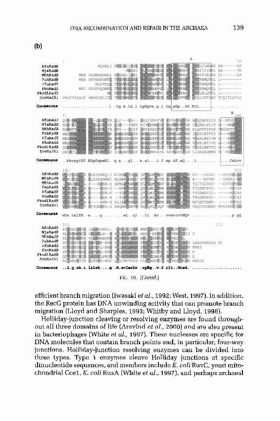
DNA RECOMBINATION AND REPAIR IN THE ARCHAEA 139
AfuRadE MjaRadB MthRadB PabRadB PfURadR PhoRadEs
PkodlRadB
AfuRadE Mj aFL%dB MthRadB PabRadB PfuRadl? PhoRadB
PkodlRadB SeeRadS
consensus
AfuRad.S Mj aRadB MthRadB PabRadB PfuRadB PhoRadB
PkodlRadB SceRadSl
1
PMGF YTAADF
MQRMLI
LRDMGENRRI
70 N*-----j(& NA------W pp-----m L------ L--------- L--------- L---------
TCQIPLDIGG
. . . . . . . ..I . . . . . . . ..l .fq.k.ld.l LgGgvs.g.i tq yoP..tG Ktt..... . . . . . . . . . . .
SKLKTIVD GSLKKTVC
LLDAAAQMMS
..LlotayiDT WgfSpKl. q.a...gl.. s.al...i.f ep.df.eQ.. .i........ .I....f.LWj
141 FT---DVGS- R----Ey,q.- YSITGDDGR- YYDSNTNTL- YYDSNSGIL- YHDSNSNSL- HFDSRTEMT- VAQVDGGMAF
-__-- NPDPK
Comensus vm.talYK. e....g.... . . ..sL..ql ..Ll..Ar.. mnvivtNQv . . . . . . . . . . . . . . ..p.Gg
AfuRadB MjaRadB MthRadB PabRadB PfUFadB PhoRadB
PkodlRadB SeeRad
LKAKPE KNED K E REEDE
272
:NGEE NI
consensus ..l.y.sk.i ltlek . . ..q .R.avlerhr .rpBq..v.f rIt..Giad. . . . . . . . . . . . . . . . . . . . . .
FIG. 16. (Contd.)
efficient branch migration (Iwasaki et al., 1992; West, 1997). In addition, the RecG protein has DNA unwinding activity that can promote branch migration (Lloyd and Sharples, 1993; Whitby and Lloyd, 1998).
Holliday-junction cleaving or resolving enzymes are found through- out all three domains of life (Aravind et al., 2000) and are also present in bacteriophages (White et al., 1997). These nucleases are specific for DNA molecules that contain branch points and, in particular, four-way junctions. Holliday-junction resolving enzymes can be divided into three types. Type 1 enzymes cleave Holliday junctions at specific dinucleotide sequences, and members include E. coli RuvC, yeast mito- chondrial Cccl, E. coli RusA (White et al., 1997), and perhaps archaeal

140 ERICA M. SEITZ ET AL.
SceRadS4:
SsoRad54:
189
356
DNKEEESKKMIKSTQEKDNINKEKNSQEERPTQRIGRHPALMTNGVRNKP--LRE-LL 244 D EEE K++ + + +E+ Q+I + + +K 'LRE LL
SceRad54:
SsoRad54:
245
415
DISEEEFMKLVSENRTIVELGGNLVEIOEKSLQKIKDLLYKIKSKKIDKIDILRESLL 414
I GDSENSAE--NKKKFASVPVVIDPKLAKI-LRPHQVEG---VRFLNRGA GD E + E ++ + +++p K LRP+Q++G +RF+N+ GOIEINOELLORLRGNKSFQLLEPYNIKANLRPYQIKGFSWNRFMNKLG
IA SceRad54:
SsoRad54:
298 GLGKTLQCI MWTLLRQGPQGKRLID NELIKWLGPNTLTPLA 356 EL Kt
EELSKFAPHLRFAVFH 527
SceRadSI:
SsoRad54:
II 357 VDGKKSSMGGGNTTVSQAIHAWAQAQGRNIVKP@ISYEl@RNVDC@NC
DK+ + +++ CY L R+ +LK 528 EDRSKI@EDYD---------------------II@TYAVI@DT-RLK@,
III SceRad54:
SsoRad54:
LKNGOSLTFTALOSISC RRVILSGTPIQNDLSEY LLSFSNPGLLGSRAE 474 +KN + F A+ + lRi$+++F NPGLLGS +E IKNPQTKIFKAVKELKS YRIALTGTPIENKVDDL IMTFLNPGLLGSYSE 624
SceRad54:
SsoRad54:
475 FRKNFENPILRGRDAOATDKEITKGEAQLQKLSTIVSKFIIRRT--NDILAKYLPCKY 531 F++ F PI +G D KE +I2 I+S FI+RRT + +LP K
625 FKSKFATPIKKG---DNMAKE---------ELKAIISPFILRRTKYDKAIINDLPDKI 671
SceRad54:
SsoRad54:
532 EHVIFVNLKPLQNELYNKLIKSREVKKVVKGVGGSQPLRAIGI-------LKKLCNHP 583 E ++ NL p Q +Y EV+ + + ++ G+ LK++ +HP
672 ETNVYCNLTPEQAAMYKA-----EVENLFNNIDSVTGIKRKGMILSTLLKLKQIVDHP 725
SceRad54:
SsoRad54:
584 NLLNFEDEFODEOD-----LELPODYNMPGSKARDVQTKYSAKFSILERFLHKIKTES 637 +E+ ++ GK + T++ I+ +K E
SceRad54:
SsoRad54: AVL +
SceRad54:
SsoRad54:
V VI 697 LSSKAGGCGINL~GANRLILMDPDRtNPAAOQQALARVWkKKDCFIYRFISTGTIE 755
LS KAGG GINL ANRtI D PA + QA RV+ GQ ++ +++ IS GT+E 835 LSVKAGGFGINL SANRVIHFDRW IGQTRNVIVHXLISVGTLE 893
SceRad54:
SsoRad54:
756 EKIFQRQSMKMSLSSCVVDAKEDVERLFSSDNLRQLFQ 794 SKI Q + K SL ++ + + s++ LR++ +
894 EKIDQLLAFKRSLFKDIISSGOSWITELSTEELRKVIE 932
FIG. 17. Comparison of the S. solfatoricus Rad54 protein homologue with S. cerevisiae Rad54. Protein sequences (Sso Rad54 homologue sh13a6224-662&064 and See Rad54 protein gi6321275) were aligned using BLAST at http://www.ncbi.nlm.nih.gov/BLAST/
The seven helicase domains characteristic of Swi2/Snf2 DNA-dependent ATPases are indicated, although the homology in motif IV is weak. Identical residues are represented by the single-letter amino acid code, while highly conserved residues are indicated by a +. Residues that may constitute a leucine zipper motif are circled.
Hjc (Kvaratskhelia and White, 2000a). This sequence requirement is probably important to limit cleavage to the Holliday junction. Type 2 enzymes, on the other hand, which include the bacteriophage enzymes T4 endo VII and T7 endo I, have little or no substrate specificity. These endonucleases can cleave a wide variety of other DNA structures, such
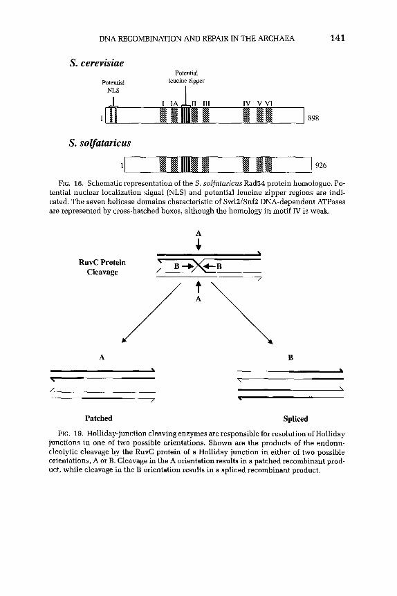
DNA RECOMBINATION AND REPAIR IN THE ARCHAEA 141
S. cerevisiae
Potential NLS
Potential leucine zipper
I
IV v VI
1 898
S. solfataricus
1 926
FIG. 18. Schematic representation of the S. solfataricus Rad54 protein homologue. Po- tential nuclear localization signal (NLS) and potential leucine zipper regions are indi- cated. The seven helicase domains characteristic of SwiZ/SnfZ DNA-dependent ATPases are represented by cross-hatched boxes, although the homology in motif IV is weak.
i
RuvC Protein Cleavage
A B \ *
\ \ / \
/ \
Patched Spliced
FIG. 19. Holliday-junction cleaving enzymes are responsible for resolution of Holliday junctions in one of two possible orientations. Shown are the products of the endonu- cleolytic cleavage by the RuvC protein of a Holliday junction in either of two possible orientations, A or B. Cleavage in the A orientation results in a patched recombinant prod- uct, while cleavage in the B orientation results in a spliced recombinant product.

142 ERICA M. SEITZ ET AL.
as three-way junctions, bulged duplexes, mismatches, and cisplatin adducts (White et al., 19%‘). The third type of Holliday-junction resolvases is defined by a newly discovered archaeal Hje enzyme. Like type 1, this enzyme shows substrate specificity, but like type 2, it does not exhibit sequence specificity for cleavage (Kvaratskhelia and White, 2OOOb). Although these Holliday-junction resolving enzymes show the same type of specificity for binding to and cleaving four-way junctions, at the amino acid level these proteins show little or no conservation. Indeed, while a Holliday-junction cleaving activity is detected in yeast nuclei and mammalian extracts, no proteins have been assigned to this activity as of yet (Constantinou et al., 2001).
1. The Bacterial RuvC Protein
The E. coli RuvC protein is the prototypic Holliday-junction cleaving enzyme (Bennett and West, 1996; Shah et al., 1997, West, 1997; Eggle- ston and West, 2000). The crystal structure of RuvC was determined at atomic resolution and demonstrates that the catalytic center, compris- ing four acidic residues, lies at the bottom of a cleft that fits a DNA duplex (Ariyoshi et al., 1994a,b). The RuvC protein specifically binds four-way Holliday junctions as a dimer and cleaves the strands in a magnesium- and homology-dependent manner. The ssDNA nicks made by RuvC are symmetric; they are found in strands of similar polarity, ex- clusively on the 3’ site of thymine residues. Strand cleavage by the RuvC dimer occurs in a sequence-specific manner, and the optimal sequence for cleavage is (A-T)TTJ(C >G-A) (Fogg et al., 1999).
2. Archaeal Holliday-Junction Cleaving Enzymes
The first archaeal Holliday-junction cleaving activity was detected in the hyperthermophilic archaeon, l? furiosus; the gene was cloned, and the protein was subsequently purified (Komori et al., 1999). This protein, named Hjc (for Holliday-junction cleavage), introduces sym- metrically related nicks into two DNA strands of similar polarity, as observed with the E. coli RuvC enzyme and other known resolvases. This P furiosus Hjc enzyme resolves Holliday junctions by introduc- ing paired cuts, 3’ to the point of strand exchange, without discernible sequence specificity. The l? furiosus Hjc protein does not share any se- quence similarity with any of the other known resolvases, although this sequence is highly conserved in the genomes of other archaeons (Table I and Fig. 20). P furiosus Hjc protein cleaves the recombination interme- diates that are formed by the E. coli RecA protein as efficiently as does the E. coli RuvC enzyme (Komori et al., 1999).

DNA RECOMBINATION AND REPAIR IN THE ARCHAEA 143
1 80 AfuHjc PFPC MjaHjc MRR ---- Mthejc KKPL PfuHjc __-- PhoHjc ____
ApeHjc M SRSP." PGQVL SsoHjc MN REmPI REQAE
COLVSlS”S . ..m.rkG.. .ERel.k.L. ..GfAviR.. as.q”..... .pD.vAg.gg ..l.iE.K.. k......Y.. . . . . ..l.ef
81 147 AfuHjc MjaHjc NthHjc PfuHjc LE GKS Phosjc LE "GEPK
ApeHjc SsoHjc FL
Consenms ar.fGg.p.1 a.kf.g.... f.....l..t . ..nfvid.. . . . . . ..dl. .v.g...... .d...
FIG. 20. Multiple alignment of archaeal Holliday-junction cleavage protein homo- logues. Sequences were as follows: A. fulgidus (Afu), gi2648580; A4. jannaschii (Mja), gi2496010; A4 thermoautotrophicum (Mth), gi2622382; P furiosus (Pfu), gi5689160; P horikoshii (Pho), gi5689160; A. pernix (Ape), gi5104108; and S. solfataricus (Sso), gi6015898. The sequences were aligned using MULTALIN at http://www.toulouse. inrafr/multalin.html. Highly conserved residues are shaded in dark gray, while mod- erately conserved residues are shaded in light gray. The E! aerophilzzm homologue is not shown because the genome sequence has not been publicly released.
The S. solfataricus Hjc protein was identified based on homology to the P. furiosus Hjc protein and showed 34% amino acid sequence iden- tity to this protein. Additional homologues of the Hjc enzyme were iden- tified in the archaea shown in Fig. 20, plus Pyrobaculum aerophilum. These proteins show 35% amino acid identity between them, including 13 totally conserved residues that may function in binding the catalytic metal ions (Fig. 20). This conserved catalytic metal ion binding domain was identified previously in several restriction enzymes and is part of the active site of the type II restriction enzyme EcoRV (Kvaratskhelia et al., 2000). Domain analysis of the P furiosus Hjc enzyme also re- vealed the importance of several residues that confer enzymatic activity to this protein, three of which were found to be conserved in the motif found in type II restriction endonuclease family proteins (Komori et al., 2000a). The S. solfataricus Hjc enzyme binds specifically to four-way DNA junctions in a Mg ‘+-dependent manner, cleaves the junction 3’ to the center of the junction, and may show some sequence specificity for cleavage (Kvaratskhelia and White, 2000a).
Another archaeal Holliday-junction resolving enzyme, Hje (for Holliday-junction endonuclease), was found in two members of the crenarchaeota, S. solfataricus and S. shibatae (Table I) ( Kvaratskhelia and White, 2OOOb). The partial purification of these enzymes showed

144 ERICA M. SEITZ ET AL.
that these endonucleases resolve Holliday junctions in a Mg’+- dependent manner by introducing paired nicks in opposing strands, thereby releasing nicked duplex DNA products. Further experiments showed that the Hje protein does not show sequence specificity for junction cleavage, suggesting that Hje does not belong to the type 1 class of sequence-specific junction resolving enzymes, such as the E. coli RuvC and yeast mitochondrial Cccl proteins. The Hje proteins do not cleave three-way junctions as does the T4 endonucleaseVI1 enzyme but do discriminate between the continuous and the exchanging strands of the four-way DNA junction to a greater extent than any other known Holliday-junction cleavage enzyme (Kvaratskhelia and White, 2OOOb). The archaeal Hje enzyme may therefore use this type of discrimination for recognition and resolution of Holliday junctions to achieve speci- ficity without having to rely on the local nucleotide sequence, like the RuvC enzyme. The Hje enzyme introduces a new class of Holliday- junction resolving enzymes that is unlike any of the previously studied enzymes (Kvaratskhelia and White, 2000a). The S. solfataricus Hje enzyme produces a cleavage pattern completely different from that of the Hjc enzyme, which suggests that there are two Holliday-junction resolving enzymes in this archaeon (Kvaratskhelia and White, 2000a).
H. SUMMARY: ARCHAEAL RECOMBINATIONAL REPAIR
The process of homologous DNA recombination in the Archaea has only just begun to be explored. This nascent analysis has been greatly fa- cilitated by the relatively recent sequencing of several different archaeal genomes, since the ability to perform genetic screens in these organisms is still rather difficult due to unusual growth requirements, as well as the inability to transform genetically many members of this group.
The picture emerging for this process in the Archaea is one that shows much more similarity to the pathway of eukaryal homologous DNA re- combination than to that of bacterial recombination. Homologues of the eukaryal Spoll protein, which is involved in the creation of DSBs in meiosis, exist in nearly all members of the Archaea, although it is unclear at this point whether this protein plays a direct role in the initi- ation of homologous recombination in the Archaea, since it is a subunit of topoisomerase VI. The lack of a bacterial RecBCD enzyme homologue to process the DSB suggests that there is a different initiation or DNA end processing mechanism in the Archaea. Homologues of another eu- karyal/bacterial nuclease complex that can process DNA ends are, how- ever, found in the Archaea: the Rad50 and Mrell proteins (Fig. 1 and Table I). Although their precise role in recombination is uknown, per- haps in conjunction with a DNA helicase, appropriate DSB processing

DNA RECOMBINATION AND REPAIR IN THE ARCHAEA 145
can be effected. Interestingly, there also exists at least one example of an archaeal homologue of the RecQ/Sgsl helicase family. Therefore, related mechanisms of DSB processing are likely for the Archaea and Eukarya.
The archaeal homologous DNA strand exchange protein, RadA, clearly shows more homology to the eukaryal Rad51 protein rather than to the bacterial RecA protein, both structurally and functionally. The fact that RadA protein homologues exist in more than 14 archaeons illustrates the importance of this protein in archaeal cellular function, and given the ubiquity of the Rad51 and RecA proteins, all Archaea are expected to have a RadA homologue.
The Archaea also possess an interesting family of ssDNA binding proteins, which likely serves an important function in the processes of DNA replication, recombination, and repair. These proteins are also more similar at the sequence level to the eukaryal RPA, but they display very diverse structural forms. The euryarchaeal proteins closely resemble RF’A in that they also incorporate a zinc binding domain within the protein; however, these proteins exist in one- or two-subunit structural variants, rather than the three-subunit quaternary structure of RPA (Fig. 13). In contrast, however, the crenarchaeal protein resembles the bacterial SSB protein in structural form (a single ssDNA binding domain with an acidic tail, which assembles into a tetramer) while retaining sequence similarity to the binding domains of eukaryal RPA.
The existence of RAD.52 epistasis group homologues in the Archaea also substantiates this similarity to the eukaryal process. These homo- logues include members, known as RadB proteins, that bear similarity to RecA or Rad51 proteins but are distinct from RadA proteins. RadB pro- teins, which are RadA protein paralogues, may be homologues of Rad55 or Rad57 proteins. A putative Rad54 protein homologue is also present.
Finally, Holliday-junction resolvases exist in the Archaea. While these enzymes do not show homology to any known resolvases, they are able to bind to four-way Holliday junctions and promote their cleavage in a Mg ‘+-dependent manner, as shown for all other Holliday-junction cleaving enzymes. The Hjc enzyme, present in most archaeons, is a Holliday-junction resolving enzyme, which may show some sequence specificity for cleavage. The Hje enzymes seem to define their own class of Holliday-junction resolvases, in that they do not display any sequence specificity for cleavage of the Holliday junction but do discriminate between stacked four-way junctions that contain continuous and those that contain exchanging strands, which is different from any Holliday-junction resolving enzyme known to date. Until the identification of the eukaryal Holliday-junction resolvases responsible for this step in homologous recombination, it is impossible to say whether the archaeal resolvases resemble eukaryal resolvases.

146 ERICA M. SEITZ ET AL.
Thus, the archaeal system does seem to represent a “simpler” version of the complex eukaryal process, but with unique features, and with some features that bear resemblance to those of Bacteria.
III. DNA Repair Pathways
All living cells have many mechanisms for repairing the various types of DNA damage encountered (Lindahl and Wood, 1999). The multiple pathways employed can be divided into several distinct groups: direct reversal of DNA damage, which chemically reverses DNA damage; base excision repair (BER), which removes the damaged base; nucleotide excision repair (NER), which removes lesions in oligonucleotide form; mismatch repair (MMR), which corrects mispaired bases in DNA: and bypass pathways, which involve specialized DNA polymerases that can insert residues opposite damaged sites so that DNA replication can con- tinue. In this article, we focus mainly on the pathways where homo- logues have been identified or studied in the Archaea. These processes include direct reversal of DNA damage, NER, and BER (Fig. 21). Toward the end of the article we discuss what is known in the other
me- ?”
5’ G 3’ 3’ c 5’
5’ G 3’ 3’ c 5’
Direct Reversal
5’ ” 3’ 3’7-5’
DNA glycosylase
Base Excision Repair
5, T-c% 3, 3’ A G 5’
UvrABCD Rad l-10
3’ 3’ A G 5’
Nucleotide Excision Repair
FIG. 21. Three DNA repair pathways common to all phylogenetic domains. Direct reversal chemically reverses the modification and includes the removal of a methyl group from O”-methylguanine. Base excision repair corrects modifications, such as the incorpo- ration of a uracil residue, by removing a single base. Nucleotide excision repair involves the removal of intact nucleotides, such as a T-C pyrimidine dimer; the lesion is excised as an oligonucleotide, whose length differs for bacterial and eukaryal NER systems.

DNA RECOMBINATION AND REPAIR IN THE ARCHAEA 147
pathways of MMR and error-prone DNA repair in this phylogenetic domain.
A. DIRECT DNA DAMAGE REVERSAL
The first DNA repair mode to be discovered was photoreactivation of DNA (Friedberg et aI., 1995). Photoproducts in DNA are created by exposure to UV radiation at wavelengths near the absorption maxi- mum of DNA. To repair the major photoproduct formed, a pyrimidine dimer, organisms have a photoreactivation system to reverse the base damage directly. Photoreactivation is a light-dependent process involv- ing the enzyme-catalyzed monomerization of cis-syn-cyclobutyl pyrim- idine dimers (Fig. 22), and the enzymes that catalyze the photoreactiva- tion of pyrimidine dimers in DNA are referred to as DNA photolyases or photoreactivating enzymes (Friedberg et al., 1995). This activity is widely distributed in nature and exists in the Bacteria, Eukarya, and Archaea (Friedberg et al., 1995; DiRuggiero et al., 1999; Grogan, 2000).
1. Photolyase Photolyase is able to split dimers using visible light as the source of
energy. This enzyme is able to absorb visible or near-UV light because it contains a photochemically active chromophore (reduced FAD) as well as another chromophore which transduces the absorbed energy to the FAD cofactor. In bacteria, such as E. coli, the phrB gene encodes the DNA photolyase; in lower eukaryotes, such as S. cerevisiae, this gene is referred to as PHRI. The E. coli and S. cerevisiae photolyases contain 5,10-methenyltetrahydrofolate (MTHF) as the second chromophore and have an absorption maximum at 380 nm (Sancar et al., 1987; Johnson et al., 1988). However, the Gram-positive bacterium Streptomyces griseus and the cyanobacterium Anacystis nidulans contain 8-hydroxy- 5-deazaflavin as a second chromophore, which has an absorption maximum at 440 nm (Eker et al., 1981, 1990; Yasui et al., 1988; Sack et al., 1998). Photoreactivation activity has been detected in four ar- chaeons in vivo: H. halobium, M. thermoautotrophicum, S. solfataricus, and S. acidocaldarius (Fig. 22) (Grogan, 2000). The DNA photolyase from M. thermoautotrophicum was purified and characterized and was found to have an absorption maximum at 440 nm (Kiener et al., 1989).
2. DNA Alkyltrcmsferases Another mechanism of DNA damage repair occurs in response to
certain mutagenic alkylating agents, which react with DNA to pro- duce both 0-alkylated and N-alkylated products. 06-alkylguanine and 04-alkylthymine are potentially mutagenic lesions because they can
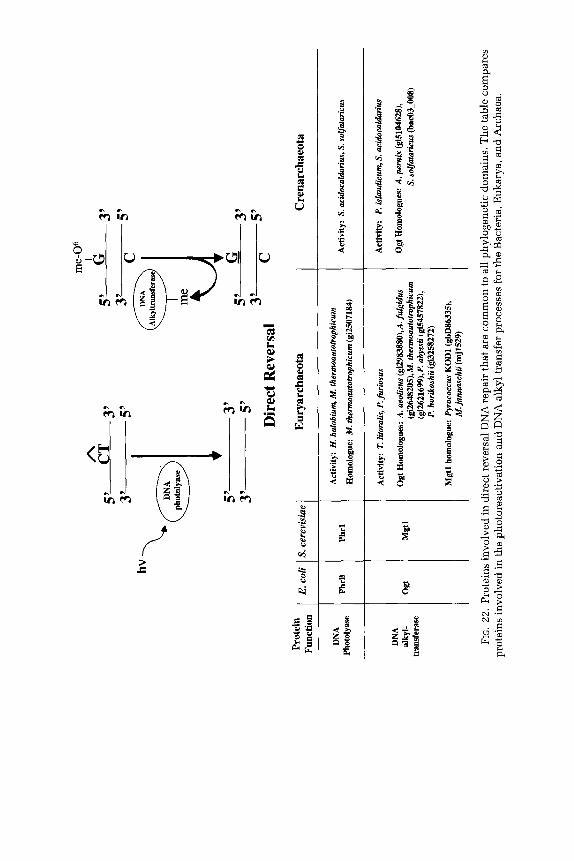
Prot
ein
DN
A Ph
otol
yase
DN
A dk
yl-
trans
fera
se
hv
me-
O6
3’
.
5’
5’
G
3’
Dire
ct R
ever
sal
3’
c 5’
E. c
oli
: Y. ce
revi
siut
PhrB
Ph
rl Mst
l
Eury
arch
aeot
a
Activ
ity:
H. h
alob
iuq
M.
ther
nroa
utot
ioph
icum
Hom
olog
ue:
M. t
herm
oaut
otro
phic
um (
gi25
0718
4)
Activ
ity:
T. lit
oral
is, P
. furio
sus
Ogt
Ho
mol
ogue
s:
A. a
eolic
us (g
i298
3880
),A.fi
l~id
us
(gi2
648X
IS),M
. th
erm
oaut
otm
phic
um
(gi2
6216
99),P
. ab
yssi
i (gi
5457
822)
, P.
hor
ikos
hii
(gi3
2582
72)
Mgt
l ho
mol
ogue
: Py
roco
ccus
KO
DI
(gbD
8633
5).
M. j
anna
whii
(mj1
529)
i
Cren
arch
aeot
a
Activ
ity:
S. a
cido
cald
wius
, S.
sol
fata
ricus
Activ
ity:
P. Q
land
icum
, S. a
cido
cald
ariu
s
Ogt
Hom
olog
ues:
A. p
erk
@51
0462
8),
S. s
o&tm
icus
(b
ac03
-008
)
FIG
. 22
. Pr
otei
ns
invo
lved
in
dire
ct
reve
rsal
D
NA
repa
ir th
at a
re c
omm
on
to a
ll ph
ylog
enet
ic
dom
ains
. Th
e ta
ble
com
pare
s pr
otei
ns
invo
lved
in
the
pho
tore
activ
atio
n an
d D
NA
alky
l tra
nsfe
r pr
oces
ses
for
the
Bact
eria
, Eu
kary
a,
and
Arch
aea.

DNA RECOMBINATION AND REPAIR IN THE ARCHAEA 149
mispair during semiconservative DNA synthesis. The DNA repair pro- tein, O”-alkylguanine DNA alkyltransferase (ATase), functions by trans- ferring the problematic alkyl groups from the 0” position of guanine and the O4 position of thymine to a cysteine residue at the active site of the protein (Foote et al., 1980; Olsson and Lindahl, 1980). This irreversible process results in the stoichiometric inactivation of the protein.
The E. coli enzyme that is responsible for transferring methyl groups from the O6 position of O”-methylguanine was originally called O6- methylguanine DNA methyltransferase, but it is also known as Ada due to its importance in the adaptive response to alkylation damage (Friedberg et al., 1995). This protein is able to recognize methyl groups and larger alkyl groups as substrates. E. coli possesses an additional protein, however, called Ogt (a DNA alkyltransferase encoded by the o@ gene), which transfers the alkyl groups from 04-methylthymine and 06-methylguanine to a cysteine residue in the ATase (Goodtzova et al., 1997). The protein responsible for 06-alkylguanine DNA alkyl- transferase activity in S. cerevisiae is the product of the MGTZ gene and is known as Mgtl protein. This protein shows conservation with the E. coli Ada and Ogt proteins and with the human and mammalian Mgtl proteins as well (Xiao and Samson, 1992).
In the Archaea, DNA alkyltransferases and DNA methyltransferases were found in several members. The protein MGMT (for O”- methylgua- nine DNA methyltransferase) was isolated from the hyperthermophilic archaeon Pyrococcus sp. KODl and possesses methyltransferase activity at temperatures as high as 90°C (Leclere et al., 1998). Additionally, alkyltransferase activity was detected in cell extracts from two eur- yarchaeotes, Thermococcus litoralis and I? furiosus, and two crenar- chaeotes, S. acidocaldarius and E! islandicum. The principal activity of these extracts resembled that of the E. coli Ogt protein (Skorvaga et al., 1998). Subsequent analysis of sequenced archaeal genomes revealed Ogt homologues also in A. aeolicus, A. fulgidus, A. pernix, M. thermoau- totrophicum, M. jannaschii, I? abysii, I? horikoshii, and S. solfataricus (Fig. 23) (Grogan, 2009). Figure 23 shows an alignment of eight archaeal Ogt protein homologues with the bacterial Ogt protein from T maritima. These proteins all have a conserved methyl-acceptor cysteine residue. The conservation of these alkyltransferases throughout evolution sug- gests a strong need for this function, which is most likely due to the toxic and mutagenic consequences of this type of DNA damage.
B. BASE EXCISION REPAIR
Base excision repair (BER) involves the removal of nonbulky DNA le- sions such as uracil, thymine glycols and hydrates, and 8-oxo-guanine in essentially two steps (Fig. 24). First, a DNA glycosylase releases the

150 ERICA M. SEITZ ET AL.
cormermm ..*..... . ..g...Kk. ue.g.... . . . . . . . . . . . . . . . . . . . . . . . . . . . . . . . . .
FIG. 23. Multiple alignment of Ogt protein homologues. Sequences were as follows: Aquifex aeolicus (Aae), gi2983880; A. fulgidus (Afu), gi2648205; M. jannaschii (Mja), mj1529; M. thermooutotrophicum (Mth), gi2621699; P abyssii (Pab), gi5457822; P horikoshii (Pho), gi3258272; A. pernix (Ape], gi5104628; S. solfatoricus (Sso), bac03-008; and Thermotoga maritima (Tmar), gi4981422. T maritima is a member of the Bacteria. The sequences were aligned using MULTALIN at http://www.toulouse.inra.fr/multalin.html. Highly conserved residues are shaded in dark gray, while moderately conserved residues are shaded in light gray. The methyl acceptor cysteine is marked by an asterisk.
base by cleaving the glycosidic bond that connects the base to the de- oxyribose. Next, the abasic sugar (apurinic/apyrimidinic (AP) site) is released by the combined actions of AP lyase and AP endonucleases (Friedberg et al., 1995; Sancar, 1996; Wood, 1996).
1. DNA Glycosylases DNA glycosylases recognize only a certain form of base damage, such
as a specific inappropriate base (e.g., uracil) or a specific base mispairing.
FIG. 24. Proteins involved in base excision repair (BER) that are common to all phylo- genetic domains. The table compares proteins involved in BER for the Bacteria, Eukarya, and Archaea, showing conserved homologues of a uracil DNA glycosylase, a mismatch gly- cosylase, an 8-oxoguanine DNA glycosylase, and an apurinic nuclease. Notes: ‘Although reported as UDG homologues (Sandigursky and Franklin, 2000), these sequences are an- notated in their respective genomes as DNA polymerase homologues. zThis protein has also been suggested to be a novel mismatch glycosylase (Horst and Fritz, 1996; Begley et al., 1999) and has been categorized here as a MutY homologue for simplicity.

s---y
- 3’
3’
G
5’
Prot
ein
Func
tion
Euca
rya
DNA
glyc
osyla
se
AP
endo
nucle
ase U 4
5’
c
3’
3’
G
5’
Base
Exc
isio
n Re
pair
Eury
arch
aeot
a Cr
enar
chae
ota
;;l:s
z.
1 UD
G
1 UD
G
1 Ac
tivity
: A.
ji&i
dus,
P.
iskm
dicu
m P
. fwio
sus,
T.
lito
ralis
Ac
tivity
: S.
shib
atae
, S. s
o&ta
ricus
Ho
mol
ogue
s:
A.fu
lgid
us
(gi2
6482
43)‘,
P.
aby
ssii
(gi3
2578
96)‘,
P.
hor
ikosh
ii ki5
4581
17)’
Hom
olog
ue:
A. p
erni
.x
(gi5
1040
69)’
Nth
___
Mis
mat
ch
Glyc
osyla
se
Hom
olog
ues:
A.
fulg
idus
@
X488
61),
Halo
bact
eriu
m
(gilO
S801
85),
M. ja
nnas
chii
(mj1
434)
, hf
. the
rmof
orm
icic
um (
gi23
2205
), P.
ner
ophi
lum
, P. a
byss
ii (gi
S458
097)
, P.
fim
iosu
s (o
rfl41
1),
P. h
oriko
shii
(gi3
2579
23)
Hom
olog
ue:
S. s
olfa
fatiu
s (~
04-0
06)
Mut
Y ---
Ho
mol
ogue
: Ha
loba
&ri&
(g
i105
8100
9),
M.
thhe
rmoa
utof
roph
icum
MIG
(gi
2621
835)
2 Ho
mol
ogue
: A.
per
nix
(gj5
1045
42)
I 8.
oxog
uani
ne
DNA
OXO
G
OtZ
Sl
Activ
ity:
M. ja
nnax
hii
(MO
gg)
Gly
cosy
lase
Ho
mol
ogue
: M
. jan
mwh
ii (g
i283
3558
)
Endo
IV
Ho
mol
ogue
s:
M. j
anm
zwhi
i (m
j161
4),
M. t
hem
zoau
totm
phic
um (g
i892
8109
)
Hom
olog
ue:
M. t
herm
oaut
otro
phic
um (
gi26
2261
2)

152 ERICA M. SEITZ ET AL.
DNA glycosylases were first identified in E. coli but are ubiquitous in nature. Generally speaking, DNA glycosylases are small, single-subunit proteins that have no cofactor requirement. These enzymes recognize the presence of damaged or mismatched bases and catalyze the breakage of the glycosyl bond between the base and the DNA sugar-phosphate backbone. Some of these enzymes have an associated AP lyase activity that produces Y-a&unsaturated aldehyde and 5’-phosphate products (McCullough et al., 1999). Glycosylase action, or the loss of purines or pyrimidines, results in the production of a common intermediate, the AP site. These sites are further processed by the AP endonucleases or AP lyases that cleave the phosphodiester bond either 5’ or 3’ to the AP site, respectively. This site is then processed further to yield a 3’-OH suitable for polymerization and ligation (Sancar, 1996).
a. Uracil DNA Glycosylases. Deamination of cytosine results in the formation of a uracil base. Since uracil will base-pair with adenine, cy- tosine deamination results in a transition mutation from G-C to A-T, if the uracil-containing strand is used as a replication template (Fried- berg et al., 1995). DNA glycosylases that excise uracil or thymine at the N-glycosidic bond can be classified into two major types according to amino acid sequence and function. The first type is uracil DNA glycosy- lase (UDG), which excises uracil from both ss- and dsDNA (U/G and U/A mispairs). This type of enzyme does not, however, excise thymine from T/G mismatches. UDG is found in all organisms, and there is 56% amino acid sequence identity between E. coli UDG and human UDG (Olsen et Al., 1989; Krokan et al., 1997). The second type of DNA glycosylase includes a mismatch-specific uracil DNA glycosylase (MUG), found in E. coli and Serratia marcescens, and thymine DNA glycosylase (TDG) from humans (Neddermann et al., 1996). MUG and TDG recognize the mismatched basepairs in dsDNA and remove both mismatched uracil and thymine. TDG recognizes and repairs U/G and T/G mispairs equally, while MUG is mostly U/G mispair specific. MUG has 32% amino acid identity with the central part of human TDG.
A uracil DNA glycosylase (UDG) was first described based on protein activity in the archaea S. shibatae, S. solfataricus, l? islandicum, I? furiosus, and T. litorolis (Fig. 24) (Koulis et Al., 1996). Subsequent to this discovery, a UDG from the archaeon A. fulgidus was isolated (Sandigursky and Franklin, 2000). These enzymes showed biochemical characteristics similar to those of the E. coli enzyme, as well as the same enzyme from the thermophilic bacterium T maritima (Sandigursky and Franklin, 1999). This archaeal UDG enzyme can remove uracil opposite guanine, as would occur in DNA after cytosine deamination. However, this glycosylase was not able to remove thymine from a similar substrate containing a T-G base pair, which is similar to the

DNA RECOMBINATION AND REPAIR IN THE ARCHAEA 153
activity of the T. maritima UDG (Sandigursky and Franklin, 1999). Additional homologues of this protein exist in l? horikoshii, P. abysii, and A. pernix and were identified based on amino acid sequence homology (Fig. 24) (Sandigursky and Franklin, 2000).
b. Mismatch Glycosylases. A mismatch glycosylase (Mth-MIG) that shows functional similarity to MUG/TDG glycosylases was discovered encoded on the cryptic plasmid pV1 of M. thermoautotrophicum (Fig. 24). Mth-MIG processes U/G and T/G but not U on a single strand of DNA (Horst and Fritz, 1996; Begley et al., 1999). Mth-MIG shows little amino acid similarity to MUG/TDG and UDG but shows significant sequence similarity to the [4Fe-4S]-containing Nth/MutY DNA glycosylase fam- ily, which catalyzes N-glycosylic reactions on DNA substrates other than U/G and T/G mispairs and which are conserved in both the Bacteria and the Eukarya. These types of DNA glycosylases include DNA endonucle- ase III (Nth; thymine glycol DNA glycosylase), MutY DNA glycosylase (A/G-specific adenine glycosylase), UV endonuclease (UV endo), and methylpurine DNA glycosylase II (MpgII). The unique structural and functional characteristics of Mth-MIG suggest that it is a new type of U/ G and T/G mismatch-specific glycosylase. Another putative homologue of this protein was identified in the archaeon M. jannaschii based upon sequence homology to endonuclease III (Fig. 24) (Begley et al., 1999).
An additional DNA glycosylase with significant sequence homology to [4Fe-4Sl-containing Nth/MutY DNA glycosylases was discovered in the hyperthermophilic archaeon l? aerophilum (Fig. 24) (Yang et al., 2000). This protein, Pa-MIG, shows 34% amino acid identity to the M. thermoformicicum Mth-MIG protein and 30% amino acid identity to the E. coli MutY protein. This protein also has amino acid residues that are generally conserved in the [4Fe-4S]-containing Nth/MutY DNA gly- cosylase family (Lu and Fawcett, 1998; Yang et al., 2000). The Pa-MIG protein also has a conserved tyrosine residue that is conserved among all Nth proteins and is critical for associated AP lyase activity. Biochem- ically, the Pa-MIG protein processes both U/G and T/G mismatches and may have a weak AP lyase activity associated with the enzyme, as does the E. coli MutY enzyme. This protein could also process T/7,8- dihydro+oxoguanine (GO) and U/GO substrates but could not process A/G and A/GO mispairs, which are substrates for the MutY protein, or G/G and G/GO mispairs. Members of this Nth/MutY/MIG/MpgII/UV endoglycosylase superfamily can also be found in A. pernix, A. fulgidus, M. jannaschii, and I? horikoshii (Yang et al., 2000). Figure 25 shows an alignment of nine archaeal members of this DNA glycosylase family. The conserved lysine residue within the Nth protein family is indicated, and the cysteine residues involved in the [4Fe-4S] binding cluster are also indicated. M. thermoformicicum Mth-MIG is not indicated due

AfuEabIII Halmr1 MjaEeJIII k%3lmxbIII ~111 PfIBKbIII s.s&h&III MOJPAETI-
- . . . . . . . . . . . . . . . . . . . . . . . . . . . . . . . . . . . . . . . . . . . . . . . . . . . . . . . . . . . . ,......... . . . . . . ...1
81 160 ApzMlty EwyRuIMp(D
Hald4ltY Ixf-TInm
cxammmu . . . . . . . . . . ..*.... 9. ..lva.iluq .t.d..v.n . ..l...... . . ..lma.e . . ..e..... ..g.y..Wt
AfumbIII mm11 MjaRxbIII ~111 -1II pfumQ111 SsaEnQIII
Qoauyl .lh.a..i. a.y.g..... p......... .dL.l.miE *.tkl.vl. .a.g...... ..vD%r.R ..Rl....g.
241 . . l . 320
WY - Ytm
Halmty GIRDEIXIPM -v
A?zuFmbIII iiRdmmn Halohck11m- MjambIII jxlmKma Pakaxk311 F&%GAsw -111 ApwL19sII?Ev pfun?doIII ApnmPEm ssamd3111 VmMIKysy
- . . . . . . . . . . . ..l..l.P. . . . . . . ..a . . ..b&..c . ..w... .Spl.e.c.. . . . . . . . . . . . . . . . . . . . .
FIG. 25. Multiple alignment of archaeal MutY and endonuclease III protein ho- mologues. Sequences were as follows: A. pernix (Ape), gi5104542; Holobocterium (HaloMutY), gi10581009; A. fulgidus (Afu), gi2648861; Halobacterium (HaloEndoIII), gi10580185; M. jannoschii (Mja), mj1434; I! obyssii (Pab), 815458097; P furiosus (Pfu), orfl411; I! horilcoshii (Pho); gi3257923; and S. solfotaricus (Sso), gi3257923. The se- quences were aligned using MULTALIN at http://www.toulouse.inra.fr/multalin.html. Highly conserved residues are shaded in dark gray, while moderately conserved are residues are shaded in light gray. The conserved lysine residue within the Nth family is marked with an asterisk. The strictly conserved aspartic acid residue is indicated by an “x.” The cysteine residues involved in binding the [4Fe-4S] cluster are marked with dots.

DNA RECOMBINATION AND REPAIR IN THE ARCHAEA 155
to the incompletion of this genome sequencing project at this date, and I! aerophilum is not indicated due to restrictions on obtaining the se- quences. The archaeal MIG family is remotely related to the human MBD4 thymine glycosylase (Pa-MIG shows 21% amino acid identity in the glycosylase domain to human MBD4 protein), which also repairs T/G and U/G mismatches in dsDNA. The C-terminal catalytic domain of the human MBD4 protein shows homology to E. coli endonuclease III and MutY proteins (Petronzelli et al., 2000).
2. 8-Oxoguanine DNA Glycosylases
Another member of the DNA glycosylase family that has a homologue in the Archaea is 8-oxoguanine DNA glycosylase (Gogos and Clarke, 1999). 8-Oxoguanine (oxoG) is caused by oxidizing agents or ionizing radiation and can be highly mutagenic if not repaired properly. DNA glycosylases that are specific for this oxoG type of lesion were discov- ered throughout the Bacteria and Eukarya, although they do not appear to belong to the same family. The eukaryal oxoG DNA glycosylases of yeast and mammals (Ogg 1 protein in S. cerevisiae and humans) belong to a protein sequence-related family of DNA glycosylases whose mem- bers have a wide range of specificities. The bacterial enzymes, however, such as the E. coli MutM enzyme (or Fpg), make up their own dis- tinct family that share sequence conservation, require zinc for activity, and have a strong S-elimination activity (Girard et al., 1997). An oxoG DNA glycosylase was identified, based on sequence homology to the DNA glycosylase superfamily, in the euryarchaeote M. jannaschii, and its gene product purified (Fig. 24). This protein, called mjOgg, is dis- tantly related to other known oxoG-specific enzymes belonging to the same glycosylase superfamily and shows no greater sequence homology with the eukaryal Oggl protein than other members. mjOgg shows DNA glycosylase activity and a specificity for oxoG. This enzyme also has an associated DNA lyase activity (Gogos and Clarke, 1999).
3. AP Endonucleases
The AP endo/endonuclease IV family is another class of enzymes in- volved in BER that have putative representatives in the Archaea, based on sequence analysis. Homologues have been found in M. jannaschii and M. thermoautotrophicum (Fig. 24). Following the release of free, damaged, or inappropriate bases by DNA glycosylases, AP sites are produced. The repair of these lesions is initiated by AP endonucle- ases, which catalyze the incision of DNA exclusively at AP sites, and this prepares the DNA for subsequent excision, repair synthesis, and DNA ligation. Endonuclease IV, encoded by the nfo gene in E. coli, cat- alyzes the formation of ssDNA breaks at sites of base loss in duplex DNA.

156 ERICA M. SEITZ ET AL.
Endo IV attacks phosphodiester bonds 5’ to the sites of base loss in DNA, leaving 3’-OH groups. The bacterial endo IV protein is a homologue of eukaryotic apurinic endonucleases (Aravind et al., 1999). Additionally, a homologue of E. coliNfi, or endonuclease V, was tentatively identified, based on sequence homology, in A4. thermoautotrophicum (Fig. 24) (Aravind et al., 1999). These putative protein homologues have yet to be studied biochemically.
C. NUCLEOTIDE EXCISION REPAIR
Another ubiquitous repair pathway is the nucleotide excision repair (NER) pathway (Friedberg et al., 1995; Sancar, 1996). During NER, dam- aged bases such as pyrimidine dimers and (64) photoproducts are en- zymatically excised from DNA as intact nucleotides that are a part of an olignonucleotide fragment (Fig. 26). There are two excision mecha- nisms. One is via an endonuclease-exonuclease mechanism, where an endonuclease makes an incision at a phosphodiester bond either 5’ or 3’ to the lesion, and then an exonuclease digests the damaged strand past the lesion. The second mechanism involves the action of an exci- sion nuclease (excinuclease), which incises the phosphodiester bonds on either side of the lesion and at some distance away from the lesion, to excise the lesion in a nucleotide fragment of a unique length. The fragment and UvrC protein are then released by the action of a DNA helicase (UvrD protein, or helicase II, in E. coli) (Fig. 26) (Friedberg et Ql., 1995; SanCar, 1996).
NER has been characterized in detail in both the Bacteria and the Eukarya, where the damage to the DNA is excised by the combined actions of several proteins in an ATP-dependent manner. The multisub- unit complex that comprises the excinuclease in E. coli is made up of the UvrA, UvrB, and UvrC proteins (Sancar, 1996). The UvrA protein functions in recognizing the site of DNA damage, while the UvrB and UvrC proteins catalyze the excision reaction, hydrolyzing the eighth phosphodiester bond on the 5’ side of the damaged base or bases and the fourth or fifth phosphodiester bond on the 3’ side of the damaged base or bases. This leads to the excision of the lesion in the form of a 12- to 13-nucleotide fragment (Sancar, 1996). The UvrD protein (helicase II) then releases the oligonucleotide fragment as well as the DNA-bound UvrC protein. The eukaryal excinuclease incises the 20th~25th phos- phodiester bond 5’ and the 3rd-8th phosphodiester bond 3’ to the lesion to generate 24- to 32-nucleotide fragments (Fig. 26). This NER system, however, involves the action of many more proteins than the bacterial process and is, thus, much more complex. None of the protein subunits that make up the eukaryal excinuclease show any significant homology

Bact
eria
: Eu
cary
a:
Euca
rva
--.
Hom
olog
ue:
M.
ther
moa
utot
roph
icum
(M
T443
)
--_
Hom
olog
ue:
M.
them
zoau
totro
phic
um
(MT4
42)
Radl
Ho
mol
ogue
s: A
.fulg
idus
(A
F026
4), M
. janm
whii
(MJ1
505)
, M
. the
rmoa
utot
roph
icum
(M
T141
51
Ikid
2 Ho
mol
ogue
s: A
. ful
gidu
s (A
F026
41, M
. the
rmoa
utoh
ophi
cum
(M
T163
3),
I’. u
byss
ii (PA
BlS7
7)
Rad
3 __
_ Ho
mol
ogue
: P.
abys
sii
(PAB
2385
) I
FIG
. 26
. Pr
otei
ns
invo
lved
in
nuc
leot
ide
exci
sion
re
pair
(NER
) th
at
are
com
mon
to
all
phyl
ogen
etic
do
mai
ns.
The
tabl
e co
mpa
res
prot
eins
in
volv
ed
in N
ER f
or t
he B
acte
ria,
Euka
rya,
an
d Ar
chae
a,
show
ing
the
cons
erve
d ex
cinu
clea
ses
invo
lved
in
thi
s pr
oces
s.
3’
* G
5’
Uvr
ABC
D
Rad
l-10
, Rad
Z,
24-3
2 nt f
r&en
t 5’
T
C
39
Nuc
leot
ide
Exci
sion
Rep
air

158 ERICA M. SEITZ ET AL.
to the bacterial enzyme. The eukaryal system, however, is conserved throughout the Eukarya (Wood, 1996).
When the Archaea were explored for the presence of NER activity, the activity was found to be more similar to that of the bacterial system. The first experiments, using a cell extract from A4. thermoautotroph- icum, demonstrated the release of an oligomer containing the lesion that was lo-11 nucleotides in length (Ogrunc et al., 1998). This find- ing paralleled the results with the purified E. coli excinuclease, which released a 12mer fragment, whereas the mammalian excinuclease re- leased a 27-mer fragment. The archaeal reaction was ATP dependent, in accordance with the behavior of both the bacterial and the eukaryal ex- cinucleases. This archaeon also has UvrA and UvrB homologues, based on sequence homology (Fig. 26) (Grogan, 2000).
The mechanism of NER seems to differ, however, for other members of the Archaea, and homologues of the eucaryal NER system were detected. These include homologues of Radl, Rad2, Rad3, Rad25, and Rad27, as well as mouse ERCCl and human XP-F proteins (Fig. 26) (Aravind et al., 1999; Grogan, 2000). In the Eukarya, two nucleases are used to create the dual incisions during NER. In S. cerevisiae, the nucleases are the Rad2 protein and the Radl-10 protein complex (Game, 1993, 2000; Prakash and Prakash, 2000). The Radl and RadlO proteins form a complex that has a ssDNA endonuclease activity which cleaves 3’-ended ssDNA at the junction with duplex DNA (Radl-10). The Rad2 protein also has ssDNA endonuclease activity. Homologues of the yeast Radl protein were uncovered in the archaea M. jcmnaschii, A. fulgidus, and M. thermoautotrophicum, although none was found in bacteria (Aravind et al., 1999). All of the nucleases from this Radl family of proteins contain a conserved ERKX$D motif and a conserved aspartate residue. The archaeal homologues predict, interestingly, an N-terminal helicase domain that is normally inactive in eukaryotes (Aravind et al., 1999). Putative homologues of Rad2 were identified in I? abysii, A. fulgidus, and M. thermoautotrophicum (Fig. 26) (Aravind et Al., 1999; Grogan, 2000). Two helicases in S. cerevisiae, Rad3 and Rad25, are also involved in NER. These helicases are responsible for creating a bubble structure during NER (Prakash and Prakash, 2000), and a homologue of the Rad3 helicase was identified in p abysii (Fig. 25) (Grogan, 2000).
D. MISMATCH REPAIR
Both bacterial and eukaryal organisms can repair mismatched DNA base pairs. Mismatches arise by several mechanisms, including errors generated during the process of DNA replication, the formation of

DNA RECOMBINATION AND REPAIR IN THE ARCHAEA 159
heteroduplex DNA as part of the recombination process, and through the deamination of 5-methylcytosine. This type of modified base can be found in the DNA of many organisms, from bacteria to eukaryotes. Deamination causes the conversion of a G-5-mC base pair to a G-T base pair (Friedberg et al., 1995; Yang, 2000).
The basic enzymology of the major MMR processes is very similar in bacteria and eukaryotes. MMR in E. coli has been studied extensively and occurs via a methyl-directed MutHLS system. MutS protein initi- ates this process by binding, as a homodimer, to base-base mismatches and loop insertions-deletions that may have arisen due to polymerase misincorporation and slippage errors, respectively. This MutS repair complex then recruits a MutL protein homodimer, which activates the endonuclease activity of MutH. The ATP binding and hydrolysis activ- ities of MutS and MutL proteins may cause conformational changes to regulate binding to mismatches and subsequent interactions with other factors such as MutH. Once MutH is activated, its endonuclease activity is directed to incise the newly replicated DNA strand at hemimethylated sites formed after the passage of the replication fork. The nicked strand is then unwound by the activity of helicase II and degraded back past the mismatch, either by 5’-to-3 or by 3’-to-5’ exonucleases, and repair synthesis fills in the resulting gap (Modrich, 1991; Yang, 2000).
Unlike the system in E. coli, S. cerevisiae has six MutS protein ho- mologues, which are referred to as MutS homologue (MSH) proteins (Kolodner and Marsischky, 1999). In yeast, MMR begins with a MSH2 protein recognizing the mismatch and forming a heterodimer with either a MSH3 or a MSH6 protein to bind the mismatches; each of the latter provides specificity for the type of error that is recognized (Eisen, 1998; Kolodner and Marsischky, 1999). The roles of the other MutS homo- logues in yeast are not as well understood. MSHl protein is involved in MMR in mitochondrial DNA, although the function of this protein has not yet been completely characterized (Chi and Kolodner, 1994). The MSH4 and MSH5 proteins are not involved in MMR but, instead, func- tion during meiotic crossing-over and chromosome segregation (Pochart et al., 1997). Mismatch recognition and repair mechanisms in humans and other higher eukaryotes show similarity to those that exist in yeast (Fishel and Wilson, 1997; Kolodner and Marsischky, 1999).
The Archaea, so far, have been shown to possess only a single MutS protein homologue (Eisen, 1998; Aravind et al., 1999). The putative MutS protein homologue was detected in only one member of the Archaea, M. thermoautotrophicum (Eisen, 1998), based on sequence homology to the E. coli MutS protein; however, this MutS protein ho- mologue was shown to group closer to a subgroup of MutS protein homologues that includes MSH4 and MSH5, which are chromosome

160 ERICAM.SEITZETAL.
crossover and segregation proteins (Eisen, 1998). There is no biochem- ical characterization of this protein as of yet.
E. FLAPENDONUCLEASEPROTEINHOMOLOGUES
DNA structures containing single-stranded branches or “flaps” are found as intermediates of DNA replication, recombination, or repair (DeMott et al., 1996; Bambara et al., 1997). Degradation of these flap structures during these processes is carried out by a protein known as FEN-l (flap endonuclease-1). This protein possesses 5’-to-3’ exonucle- ase activity and can act as an endonuclease for 5’ ssDNA flaps. FEN-l protein homologues were discovered in several members of the Archaea: A. fulgidus, I? furiosus, M. jannaschii, and l? horikoshii (Hosfield et al., 1998a; Rao et al., 1998; Matsui et al., 1999). These proteins show a high level of sequence homology with the human FEN-l protein; the M. jannaschii FEN-l homologue shows 76% amino sequence similarity, and the homologues from A. fulgidus and l? furiosus show 72 and 74% amino sequence similarity, respectively. The A. fulgidus, P furiosus, M. jcmnaschii, and l? horikoshii FEN-l protein homologues were purified, and they show specificity for flap DNA structures (Hosfield et al., 1998a; Rao et al., 1998; Matsui et al., 1999). The FEN-l protein from P furio- sus was crystallized, and the structure was determined (Hosfield et al., 1998b).
F. TRANSLESION DNA SYNTHESIS AND MUTAGENESIS
In the bacterium E. coli, mutagenesis that occurs after exposure to DNA-damaging agents requires a distinct system (the SOS-induced mu- tagenesis system), which processes DNA damage in an error-prone man- ner. Several genes in E. coli are regulated by the SOS system, and two of these are error-prone DNA polymerases: UmuD’,C, which is also re- ferred to as PolV (Tang et al., 1999; Goodman, 2000); and DinB, which is referred to as PolIV (Wagner et al., 1999). Homologues of the E. coliDinB protein were discovered in S. cerevisiae, C. elegans, M. musculus, and H. sapiens (Gerlach et al., 1999; Woodgate, 1999). In yeast, the Rad30 protein is homologous to both the UmuC and the DinB proteins and is a DNA polymerase (DNA pol r]) that can replicate thymine dimers in template DNA (Johnson et al., 1999). Additionally, a human homologue of yeast Rad30 (Xeroderma pigmentosum variant; XPV) shows activi- ties similar to those of the yeast pol q (Masutani et al., 1999a; Masutani et al., 1999b).
A DinB/UmuC protein homologue was identified by sequence analy- sis in the archaeon, S. solfataricus (Kulaeva et al., 1996). This protein

DNA RECOMBINATION AND REPAIR IN THE ARCHAEA 161
homologue shows 32% sequence similarity to the DinB protein and 22% sequence similarity to the UmuC protein. Additionally, DNA mutagene- sis induced by exposure to UV radiation was detected in the pyTococcus species of archaea (Watrin and Prieur, 1996). Biochemical characteriza- tion of this archaeal protein homologue is yet to be reported.
G. SUMMARY: DNA REPAIR MECHANISMS IN THE ARCHAEA
As discussed above, recombinational repair in the Archaea shares more orthologous protein components with the eukaryal system than with the bacterial system, based on the similarities with many compo- nents of the yeast RAD52 epistasis group.
However, the comparison of other DNA repair pathways has not pro- duced a simple conclusion. Proteins involved in the direct reversal of DNA damage are similar in both bacteria and eukaryotes, and the ar- chaeal protein homologues show similarities to both as well. The ar- chaeal DNA alkyltransferases, however, show homology to the bacterial Ogt protein.
The archaeal DNA glycosylases involved in BER show homology to both bacterial and eukaryal enzymes, a consequence of the fact that many bacterial DNA glycosylases are also conserved in the Eukarya. The archaeal UDG protein displays both biochemical and sequence similar- ity to bacterial UDG proteins. The Archaea have a mismatch glycosylase with homology to the Nth/MutY/MIG/MpgIT/UV endoglycosylase su- perfamily, which is also conserved in both the Bacteria and the Eukarya. An archaeal8-oxoguanine DNA glycosylase exists in M. jannaschii, but the sequence of this enzyme differs greatly from those of both its eu- karyal and its bacterial counterparts. Finally, the members of the AP endo/endonuclease IV family in the Archaea are similar in sequence to the bacterial proteins.
In the case of NER, the archaeal proteins show similarities in some cases to the bacterial proteins and in other species to the eukaryal pro- teins. An activity was identified in M. thermoautotrophicum that mim- ics the action of the UvrABCD proteins, and UvrA and B protein homo- logues exist, based on sequence similarity, in this archaeon. However, in other archaea, protein homologues of the eukaryal NER machinery were detected.
Less information is available on the processes of mismatch repair and error-prone DNA repair in this third domain of life. So far, only one MutS homologue was found; although this homologue was dis- covered based on sequence homology to the E. coli MutS protein, it groups closer to a subgroup that includes eukaryal MutS protein homo- logues. Another protein involved in DNA replication, recombination,

162 ERICA M. SEITZ ET AL.
and repair, FEN-l protein, has homologues in several archaea, and these show a high degree of sequence homology with the human FEN-l pro- tein. Finally, a homologue of a bacterial protein involved in error-prone DNA replication, DinBLJmuC, was found in just one member of the Archaea.
In conclusion, it appears that the Archaea possess proteins involved in DNA repair that are similar to both bacterial and eukaryal compo- nents and some proteins that are only distantly related to either. For this reason, it is difficult to classify the entire archaeal domain as be- ing “more” bacterial or eukaryal in its means for repairing damage to its DNA. Further investigation into the processes by which the Archaea are able to repair DNA damage will reveal mechanisms by which this unique domain of life deals with the classic problem of DNA damage and should lend insight into the evolution of DNA repair processes.
ACKNOWLEDGMENTS
We thank the following members of the Kowalczykowski lab for providing comments on the manuscript: Piero Bianco, Carole Bornarth, Joel Brockman, Frederic Chedin, Mark Dillingham, Naofumi Handa, Alex Mazin, Jim New, and Yun Wu. We also thank Dr. John A. Tainer for providing the Rad50 protein structural figure. This work was supported by NIH Training Grant GM07377 to E.M.S., NSF Postdoctoral Fellowship in Microbial Biology 0074380 to C.A.H., and NIH Grants GM62653 and GM 41347 and Human Frontiers Science Program Grant HFSP-RG63 to S.C.K.
REFERENCES
Alani, E., Padmore, R., and Kleckner, N. (1990). Cell 61,419-436. Albala, J. S., Thelen, M. P., Prange, C., Fan, W., Christensen, M., Thompson, L. H., and
Lennon, G. G. (1997). Genomics 46,476-479. Anderson, D. G., and Kowalczykowski, S. C. (1997a). Genes Dev. 11, 571-581. Anderson, D. G., and Kowalczykowski, S. C. (1997b). Cell 90, 77-86. Anderson, D. G., and Kowalczykowski, S. C. (1998). J. Mol. Biol. 282, 275-285. Aravind, L., Walker, D. R., and Koonin, E. V. (1999). Nucleic Acids Res. 27, 1223-1242. Aravind, L., Makarova, K. S., and Koonin, E. V. (2000). Nucleic AcidsRes. 28, 3417-3432. Ariyoshi, M., Vassylyev, D. G., Iwasaki, H., Fujishima, A., Shinagawa, H., and Morikawa,
K. (1994a). J. Mol. Bid. 241, 281-282. Ariyoshi, M., Vassylyev, D. G., Iwasaki, H., Nakamura, H., Shinagawa, H., and
Morikawa, K. (1994b). Cell 78, 1063-1072. Arnold, D. A., and Kowalczykowski, S. C. (1999). In “Encyclopedia of Life Sciences.”
Nature Publishing Group, London, www.els.net. Arnold, D. A., and Kowalczykowski, S. C. (2000). J, Biol. Chem. 275, 12261-12265. Bambara, R. A., Murante, R. S., and Henricksen, L. A. (1997). J. Biol. Chem. 272, 4647-
4650. Baudat, F., and Nicolas, A. (1997). Proc. N&l. Acad. Sci. USA 94, 5213-5218. Baudat, F., Manova, K., Yuen, J. l?, Jasin, M., and Keeney, S. (2000). Mol. Cell 6, 989-998. Begley, T. J., Haas, B. J., Noel, J,, Shekhtman, A., Williams, W. A., and Cunningham, R. P.
(1999). Curr. Biol. 9, 653-656.

DNA RECOMBINATION AND REPAIR IN THE ARCHAEA 163
Bennett, R. J., and West, S. C. (1996). Proc. Nat]. Acad. Sci. USA 93, 12217-12222. Bennett, R. J., Sharp, J. A., and Wang, J. C. (1998). J. Biol. Chem. 273, 9644-9650. Bennett, R. J., Noirot-Gras, M. F., and Wang, J. C. (2000). J. Eiol. Chem. 275, 26898-26905. Bergerat, A., Gadelle, D., and Forterre, P. (1994). J. Biol. Chem. 269, 27663-27669. Bergerat, A., de Massy, B., Gadelle, D., Varoutas, P. C., Nicolas, A., and Forterre, P. (1997).
Nature 386,414-417. Bianco, P. R., andKowalczykowski, S. C. (1997). Proc. Natl. Acad. Sci. USA 94,6706-6711. Bianco, P. R., and Kowalczykowski, S. C. (1999). In “Encyclopedia of Life Sciences.”
Nature Publishing Group, London, www.els.net. Bianco, P. R., Tracy, R. B., and Kowalczykowski, S. C. (1998). Front. Biosci. 3, D570-D603. Bochkareva, E., Frappier, L., Edwards, A. M., and Bochkarev, A. (1998). J. Biol. Chem.
273,3932-3936. Borde, V., Wu, T. C., and Lichten, M. (1999). Mol. Cell. Biol. 19, 4832-4842. Borde, V., Goldman, A. S., and Lichten, M. (2000). Science 290, 806-809. Bult, C. J., White, O., Olsen, G. J., Zhou, L., Fleischmann, R. D., Sutton, G. G., Blake, J. A.,
Fitzgerald, L. M., Clayton, R. A., and Gocayne, J. D. (1996). Science 273, 1058-1073. Cartwright, R., Dunn, A. M., Simpson, P. J., Tambini, C. E., and Thacker, J. (1998a). Nucleic
Acids Res. 26,1653-1659. Cartwright, R., Tambini, C. E., Simpson, P. J., and Thacker, J. (1998b). Nucleic Acids Res.
26,3084-3089. Celerin, M., Merino, S. T., Stone, J. E., Menzie, A. M., and Zolan, M. E. (2000). EMBO J,
19,2739-2750. Cervantes, M. D., Farah, J. A., and Smith, G. R. (2000). Mol. Cell 5, 883-888. Chakraverty, R. K., and Hickson, I. D. (1999). Bioessays 21, 286-294. Chedin, F., Noirot, P., Biaudet, V., and Ehrlich, S. D. (1998a). Mol. Microbial. 29, 1369-
1377. Chedin, F., Seitz, E. M., and Kowalczykowski, S. C. (1998b). Trends Biochem. Sci. 23,
273-277. Chedin, F., Ehrlich, S. D., and Kowalczykowski, S. C. (2000). J. Mol. Biol. 298, 7-20. Chi, N. W., and Kolodner, R. D. (1994). J, Biol. Chem. 269, 29984-29992. Clark, A. J., and Margulies, A. D. (1965). Proc. Natl. Acad. Sci. USA 53,451-459. Clark, A. J., and Sandler, S. J. (1994). Crit. Rev. Microbial. 20, 125-142. Connelly, J. C., and Leach, D. R. (1996). Genes Cells I, 285-291. Connelly, J. C., de Leau, E. S., Okely, E. A., and Leach, D. R. (1997). J. Biol. Chem. 272,
19819-19826. Connelly, J. C., Kirkham, L. A., and Leach, D. R. (1998). Proc. Natl. Acad. Sci. USA 95,
7969-7974. Connelly, J. C., de Leau, E. S., and Leach, D. R. (1999). Nucleic Acids Res. 27, 1039-1046. Constantinou, A., Davies, A. A., and West, S. C. (2001). Cell 194, 259-268. Cox, M. M. (1999). Prog. Nucleic Acid Res. Mol. Biol. 63, 311-366. Cox, M. M., and Lehman, I. R. (1981). Proc. Nat]. Acad. Sci. USA 78, 6018-6022. Cromie, G. A., Millar, C. B., Schmidt, K. H., and Leach, D. R. (2000). Genetics x54,513-522. Cui, X., Brenneman, M., Meyne, J., Oshimura, M., Goodwin, E. H., and Chen, D. J. (1999).
Mutat. Res. 434, 75-88. Debrauwere, H., Buard, J., Tessier, J., Aubert, D., Vergnaud, G., and Nicolas, A. (1999).
Nature Genet. 23, 367-371. DeMott, M. S., Shen, B., Park, M. S., Bambara, R. A., and Zigman, S. (1996). J, Biol. Chem.
271,30068-30076. Dernburg, A. F., McDonald, K., Moulder, G., Barstead, R., Dresser, M., and Villeneuve,
A. M. (1998). Cell 94, 387-398. DiRuggiero, J., Brown, J. R., Bogert, A. P., and Robb, F. T. (1999). J. Mol. Evol. 49,474-484.

164 ERICA M. SEITZ ET AL.
Dixon, D. A., and Kowalczykowski, S. C. (1993). Cell 73, 87-96. Dolganov, G. M., Maser, R. S., Novikov, A., Tosto, L., Chong, S., Bressan, D. A., and Petrini,
J. H. (1996). Mol. cell. Biol. 16,4832-4841. Dosanjh, M. K., Collins, D. W., Fan, W., Lennon, G. G., Albala, J. S., Shen, Z., and Schild,
D. (1998). Nucleic Acids Res. 26, 1179-1184. Duno, M., Thomsen, B., Westergaard, O., Krejci, L., and Bendixen, C. (2000). Mol. Gen.
Genet. 264, 89-97. Edenberg, H. J. (1976). Biophys. 1, 16, 849-860. Egelman, E. H., and Stasiak, A. (1986). I. Mol. Biol. 191, 677-697. Egelman, E. H., and Stasiak, A. (1988). 1. Mol. Biol. 200, 329-349. Egelman, E. H., and Yu, X. (1989). Science 245, 404-407. Eggleston, A. K., and West, S. C. (2000). I. Biol. Chem. 275, 26467-26476. Eisen, J. A. (1998). Nucleic Acids Res. 26,4291-4300. Eisen, J. A., Sweder, K. S., and Hanawalt, P. C. (1995). Nucleic Acids Res. 23, 2715-2723. Eker, A. P., Dekker, R. H., and Berends, W. (1981). Photochem. Photobiol. 33, 65-72. Eker, A. P., Kooiman, P., Hessels, J. K., and Yasui, A. (1990). I. Biol. Chem. 265, 8009-
8015. Ellis, N. A., Groden, J., Ye, T. Z., Straughen, J., Lennon, D. J., Ciocci, S., Proytcheva, M.,
and German, J. (1995). Cell 83, 655-666. Epstein, C. J., and Motulsky, A. G. (1996). Bioessays 18, 1025-1027. Fishel, R., and Wilson, T. (1997). Curr. Onin. Genet. Dev. 7, 105-113. Flores, M. J., Bierne, H., Ehrlich, S. D., and Michel, B. (2001). EMBO J. 20, 619-629. Fogg, J. M., Schofield, M. J., White, M. F., and Lilley, D. M. (1999). Biochemistry 38,
11349-11358. Foote, R. S., Mitra, S., and Pal, B. C. (1980). Biochem. Biophys. Res. Commun. 97,654-659. Fricke, W. M., Kaliraman, V., and Brill, S. J. (2000). J. Biol. Chem. (in press). Friedberg, E. C., Walker, G. C., and Siede, W. (1995). “DNA Repair and Mutagenesis.”
ASM Press, Washington DC. Fuller, L. F., and Painter, R. B. (1988). Mutat. Res. 193, 109-121. Furuse, M., Nagase, Y., Tsubouchi, H., Murakami-Murofushi, K., Shibata, T., and Ohta, K.
(1998). EMBOJ. 17,6412-6425. Game, J. C. (1993). Semin. Cancer Biol. 4, 73-83. Game, J. C. (2000). Mutat. Res. 451,277-293. Gangloff, S., McDonald, J. P., Bendixen, C., Arthur, L., and Rothstein, R. (1994). Mol. Cell.
Biol. 14, 8391-8398. Gerlach, V. L., Aravind, L., Gotway, G., Schultz, R. A., Koonin, E. V., and Friedberg, E. C.
(1999). Proc. Nutl. Acod. Sci. USA 96, 11922-11927. Girard, P. M., Guibourt, N., and Boiteux, S. (1997). Nucleic Acids Res. 25, 3204-3211. Gogos, A., and Clarke, N. D. (1999). J. Biol. Chem. 274, 30447-30450. Comes, X. V., and Wold, M. S. (1995). J. Biol. Chem. 270, 4534-4543. Comes, X. V., and Wold, M. S. (1996). Biochemistry35, 10558-10568. Goodman, M. F. (2000). Trends Biochem. Sci. 25, 189-195. Goodtzova, K., Kanugula, S., Edara, S., Pauly, G. T., Moschel, R. C., and Pegg, A. E. (1997).
J. Biol. Chem. 272, 8332-8339. Grogan, D. W. (2000). Trends Microbial. 8, 180-185. Haber, J. E. (2000a). Mufat. Res. 451, 53-69. Haber, J. E. (2OOOb). Curr. Opin. Cell. Biol. 12, 286-292. Harmon, F. G., and Kowalczykowski, S. C. (1998). Genes Dev. 12, 1134-1144. Harmon, F. G., DiGate, R. J., and Kowalczykowski, S. C. (1999). Mol. Cell 3, 611-620. Haseltine, C. A., and Kowalczykowski, S. C. (2001). Submitted for publication. Hirano, T. (1999). Genes Dev. 13, 11-19.

DNA RECOMBINATION AND REPAIR IN THE ARCHAEA 165
Holliday, R. (1964). Genet. Res. 5,282-304. Hopfner, K. P., Karcher, A., Shin, D., Fairley, C., Tamer, J. A., and Carney, J. P. (2000a).
J. Bacterial. 182,6036-6041. Hopfner, K. P., Karcher, A., Shin, D. S., Craig, L., Arthur, L. M., Carney, J. P., and Tainer,
J. A. (2OOOb). Cell 101, 789-800. Horii, Z.-I., and Clark, A. J. (1973). J. Mol. Biol. 80, 327-344. Horst, J. P., and Fritz, H. J. (1996). EMBO J. 15, 5459-5469. Hosfield, D. J., Frank, G., Weng, Y., Tainer, J. A., and Shen, B. (1998a). J. Biol. Chem. 273,
27154-27161. Hosfield, D. J., Mol, C. D., Shen, B., and Tainer, J. A. (1998b). Cell 95,135-146. Iwasaki, H., Takahagi, M., Nakata, A., and Shinagawa, H. (1992). GenesDev. 6,2214-2220. Johnson, J. L., Hamm-Alvarez, S., Payne, G., Sancar, G. B., Rajagopalan, K. V., and Sancar,
A. (1988). Proc. NatI. Acad. Sci. USA 85, 2046-2050. Johnson, R. D., and Symington, L. S. (1995). Mol. Cell. Biol. 15,4843-4850. Johnson, R. E., Prakash, S., and Prakash, L. (1999). Science 283,1001-1004. Johzuka, K., and Ogawa, H. (1995). Genetics 139,1521-1532. Kanaar, R., Troelstra, C., Swagemakers, S. M., Essers, J., Smit, B., Franssen, J. H., Pastink,
A., Bezzubova, 0. Y., Buerstedde, J. M., Clever, B., Heyer, W. D., and Hoeijmakers, J. H. (1996). Curr. Biol. 6, 828-838.
Kawarabayasi, Y., Sawada, M., and Horikawa, H. et al. (1998). DNA Res. 5, 55-76. Kawarabayasi, Y., Hino, Y., and Horikawa, H., et al. (1999). DNA Res. 6,83-101,145-152. Keeney, S., Giroux, C. N., and Kleckner, N. (1997). Cell 88, 375-384. Kelly, T. J., Simancek, P., and Brush, G. S. (1998). Proc. Nat]. Acad. Sci. USA 95, 14634-
14639. Kiener, A., Husain, I., Sancar, A., and Walsh, C. (1989). J, Biol. Chem. 264, 13880-
13887. Kil, Y. V., Baitin, D. M., Masui, R., Bench-Osmolovskaya, E. A., Kuramitsu, S., and Lanzov,
V. A. (2000). J. Bacterial. 182, 130-134. Kitao, S., Ohsugi, I., Ichikawa, K., Goto, M., Furuichi, Y., and Shimamoto, A. (1998).
Genomics 54,443-452. Kitao, S., Lindor, N. M., Shiratori, M., Furuichi, Y., and Shimamoto, A. (1999a). Genomics
61,268-276. Kitao, S., Shimamoto, A., Goto, M., Miller, R. W., Smithson, W. A., Lindor, N. M., and
Furuichi, Y. (199913). Nature Genet. 22, 82-84. Klenk, H. P., Clayton, R. A., and Tomb, J. F., et al. (1997). Nature 390, 364-370. Kogoma, T. (1996). Cell 85, 625-627. Kogoma, T. (1997). Microbial. Mol. Biol. Rev. 61, 212-238. Kolodner, R. D., and Marsischky, G. T. (1999). Curr. Opin. Genet. Dev. 9, 89-96. Kolodner, R., Fishel, R. A., and Howard, M. (1985). J. Bacterial. 163, 1060-1066. Komori, K., Sakae, S., Shinagawa, H., Morikawa, K., and Ishino, Y. (1999). Froc. NotI.
Acad. Sci. USA 96,8873-8878. Komori, K., Miyata, T., Daiyasu, H., Toh, H., Shinagawa, H., and Ishino, Y. (2000a). J. Biol.
Chem. 275,33791-33797. Komori, K., Miyata, T., DiRuggiero, J., Holley-Shanks, R., Hayashi, I., Cann, I. K., Mayanagi,
K., Shinagawa, H., and Ishino, Y. (2000b). J. Biol. Chem. 275, 33782-33790. Koulis, A., Cowan, D. A., Pearl, L. H., and Savva, R. (1996). FEMS Microbial. Lett. 143,
267-271. Kowalczykowski, S. C. (1991). Annu. Rev. Biophys. Biophys. Chem. 20, 539-575. Kowalczykowski, S. C. (2000). Trends Biochem. Sci. 25, X56-165. Kowalczykowski, S. C., and Eggleston, A. K. (1994). Anna. Rev. Biochem. 63, 991-1043. Kowalczykowski, S. C., and Krupp, R. A. (1987). J, Mol. Biol. 193, 97-113.

166 ERICA M. SEITZ ET AL.
Kowalczykowski, S. C., Dixon, D. A., Eggleston, A.K., Lauder, S.D., and Rehrauer, W. M. (1994). Micro&l. Rev. 58, 401-465.
Krokan, H. E., Standal, R., and Slupphaug, G. (1997). Biochem. J. 325, 1-16. Kulaeva, 0. I., Koonin, E. V., McDonald, J. P., Randall, S. K., Rabinovich, N., Connaughton,
J. F., Levine, A. S., and Woodgate, R. (1996). Mutat. Res. 357, 245-253. Kuzminov, A. (1999). Microbial. Mol. Biol. Rev. 63, 751-813. Kvaratskhelia, M., and White, M. F. (2000a). 1. Mol. Biol. 297, 923-932. Kvaratskhelia, M., and White, M. F. (2OOOb). 1, Mol. Biol. 295, 193-202. Kvaratskhelia, M., Wardleworth, B. N., Norman, D. G., and White, M. F. (2000). J. Biol.
Chem. 275,25540-25546. Lam, S. T., Stahl, M. M., McMilin, K. D., and Stahl, F. W. (1974). Genetics 77,425-433. Lanzov, V., Stepanova, I., and Vinogradskaja, G. (1991). Biochimie 73, 305-312. Leclere, M. M., Nishioka, M., Yuasa, T., Fujiwara, S., Takagi, M., and Imanaka, T. (1998).
Mol. Gen. Genet. 256, 69-77. Lilley, D. M., and White, M. F. (2000). Proc. Natl. Acad. Sci. USA 97, 9351-9353. Lindahl, T., and Wood, R. D. (1999). Science 286, 1897-1905. Lindor, N. M., Furuichi, Y., Kitao, S., Shimamoto, A., Arndt, C., and Jalal, S. (2000). Am.
J. Med. Genet. 90, 223-228. Liu, J., Wu, T. C., and Lichten, M. (1995). EMBO J. 14,4599-4608. Liu, N., Lamerdin, J. E., Tebbs, R. S., Schild, D., Tucker, J. D., Shen, M. R., Brookman,
K. W., Siciliano, M. J., Walter, C. A., Fan, W., Narayana, L. S., Zhou, Z. Q., Adamson, A. W., Sorensen, K. J., Chen, D. J., Jones, N. J., and Thompson, L. H. (1998). Mol. Cell 1, 783-793.
Lloyd, R. G., and Buckman, C. (1995). Genetics 139, 1123-1148. Lloyd, R. G., and Sharples, G. J. (1993). EMBO J. 12, 17-22. Lohman, T. M., and Ferrari, M. E. (1994). Annu. Rev. Biochem. 63, 527-570. Lovett, S. T., and Mortimer, R. K. (1987). Genetics 116, 547-553. Lu, A. L., and Fawcett, W. P. (1998). J. Biol. Chem. 273, 25098-25105. Luisi-DeLuca, C., and Kolodner, R. D. (1994). 1. Mol. Biol. 236, 124-138. Marians, K. J. (2000). Trends Biochem. Sci. 25, 185-189. Masutani, C., Araki, M., Yamada, A., Kusumoto, R., Nogimori, T., Maekawa, T., Iwai, S.,
and Hanaoka, F. (1999a). EMBO J. l&3491-3501. Masutani, C., Kusumoto, R., Yamada, A., Dohmae, N., Yokoi, M., Yuasa, M., Araki, M.,
Iwai, S., Takio, K., and Hanaoka, F. (1999b). Nature 399, 700-704. Matsui, E., Kawasaki, S., Ishida, H., Ishikawa, K., Kosugi, Y., Kikuchi, H., Kawarabayashi,
Y., and Matsui, I. (1999). J. Biol. Chem. 274, 18297-18309. Mazin, A. V., Bornarth, C. J., Solinger, J. A., Heyer, W. D., and Kowalczykowski, S. C.
(2000a). Mol. Cell 6, 583-592. Mazin, A. V., Zaitseva, E., Sung, P., and Kowalczykowski, S. C. (2OOOb). EMBO J. 19,
1148-1156. McCullough, A. K., Dodson, M. L., and Lloyd, R. S. (1999). Annu. Rev. Biochem. 68,
255-285. McGlynn, P., and Lloyd, R. G. (2000). Cell 101, 35-45. McKim, K. S., and Hayashi-Hagihara, A. (1998). Genes Dev. 12, 2932-2942. Mendonca, V. M., Klepin, H. D., and Matson, S. W. (1995). J. Bacterial. 177, 1326-1335. Meyer, R. R., and Laine, P. S. (1990). Microbial. Rev. 54, 342-380. Michel, B. (2000). Trends Biochem. Sci. 25, 173-178. Michel, B., Ehrlich, S. D., and Uzest, M. (1997). EMBO J. 16, 430-438. Modrich, P. (1991). Annu. Rev. Genet. 25, 229-253. Mortensen, U. H., Bendixen, C., Sunjevaric, I., and Rothstein, R. (1996). Proc. Natl. Acad.
Sci. USA 93,10729-10734.

DNA RECOMBINATION Ah, ” I-1 AIR IN THE ARCHAEA 167
Nakayama, H., Nakayama, K., Nakayama, R., Irino, N., Nakayama, Y., and Hanawalt, P. C. (1984). Mol. Gen. Genet. 195,474-480.
Nakayama, K., Irino, N., and Nakayama, H. (1985). Mol. Gen. Genet. 200, 266-271. Neddermann, P., Gallinari, P., Lettieri, T., Schmid, D., Truong, O., Hsuan, J. J., Wiebauer,
K., and Jiricny, J. (1996). J. Biol. Chem. 271, 12767-12774. New, J. H., Sugiyama, T., Zaitseva, E., and Kowalczykowski, S. C. (1998). Nature 391,
407-410. Nicolas, A., Treco, D., Schultes, N. P., and Szostak, J. W. (1989). Nature 338, 35-39. Ogawa, H., Johzuka, K., Nakagawa, T., Leem, S. H., and Hagihara, A. H. (1995). Adv.
Biophys. 31,67-76. Ogawa, T., Yu, X., Shinohara, A., and Egelman, E. H. (1993). Science 259, 1896-1899. Ogrunc, M., Becker, D. F., Ragsdale, S. W., and Sancar, A. (1998). J. Bacteriol. 180, 5796-
5798. Olsen, L. C., Aasland, R., Wittwer, C. U., Krokan, H. E., and Helland, D. E. (1989). EMBO
J. 8,3121-3125. Olsson, M., and Lindahl, T. (1980). J. Biol. Chem. 255, 10569-10571. Paques, F., and Haber, J. E. (1999). Microbial. Mol. Biol. Rev. 63, 349-404. Paull, T. T., and Gellert, M. (1998). Mol. Cell 1, 969-979. Paull, T. T., and Gellert, M. (1999). Genes Dev. 13, 1276-1288. Petrini, J. H. (1999). Am. J. Hum. Genet. 64, 1264-1269. Petronzelli, F., Riccio, A., Markham, G. D., Seeholzer, S. H., Stoerker, J., Genuardi, M.,
Yeung, A. T., Matsumoto, Y., and Bellacosa, A. (2000). J. Biol. Chem. 275, 32422- 32429.
Petukhova, G., Stratton, S., and Sung, P. (1998). Nature 393,91-94. Petukhova, G., Van Komen, S., Vergano, S., Klein, H., and Sung, P. (1999). J. Biol. Chem.
274,29453-29462. Philipova, D., Mullen, J. R., Maniar, H. S., Lu, J., Gu, C., and Brill, S. J. (1996). Genes Dev.
10,2222-2233. Pierce, A. J., Johnson, R. D., Thompson, L. H., and Jasin, M. (1999). Genes Dev, 13, 2633-
2638. Pochart, P., Woltering, D., and Hollingsworth, N. M. (1997). J. Biol. Chem. 272, 30345-
30349. Postow, L., Ullsperger, C., Keller, R. W., Bustamante, C., Vologodskii, A. V., and Cozzarelli,
N. R. (2001). J. Biol. Chem. 276, 2790-2796. Prakash, S., and P&ash, L. (2000). Mutat. Res. 451,13-24. Puranam, K. L., and Blackshear, P. J. (1994). J. Biol. Chem. 269, 29838-29845. Radding, C. M. (1989). Biochim. Biophys. Acta 1008,131-145. Rao, H. G., Rosenfeld, A., and Wetmur, J. G. (1998). J. Bacterial. 180, 5406-5412. Rattray, A. J., and Symington, L. S. (1994). Genetics 138, 587-595. Raymond, W. E., and Kleckner, N. (1993). Nucleic Acids Res. 21, 3851-3856. Resnick, M. A. (1976). J. Theor. Biol. 59, 97-106. Rice, M. C., Smith, S. T., Bullrich, F., Havre, P., and Kmiec, E. B. (1997). Proc. Natl. Acad.
Sci. USA 94,7417-7422. Romanienko, P. J., and Camerini-Otero, R. D. (2000). Mol. Cell 6, 975-987. Rothstein, R., Michel, B., and Gangloff, S. (2000). Genes Dev. 14, I-IO. Sack, S. Z., Liu, Y., German, J., and Green, N. S. (1998). Clin. Exp. Immunol. 112,248-254. Saffi, J., Pereira, V. R., and Henriques, J. A. (2000). Curr. Genet. 37, 75-78. Sancar, A. (1996). Annu. Rev. Biochem. 65,43-81. Sancar, G. B., Smith, F. W., and Heelis, P. F. (1987). J, Biol. Chem. 262, 15457-15465. Sandigursky, M., and Franklin, W. A. (1999). Curr. Biol. 9, 531-534. Sandigursky, M., and Franklin, W. A. (2000). J. Biol. Chem. 275, 19146-19149.

168 ERICA M. SEITZ ET AL.
Sandler, S. J., and Marians, K. J. (2000). 1. Bacterial. 182, 9-13. Sandier, S. J., Satin, L. H., Samra, H. S., and Clark, A. J. (1996). Nucleic Acids Res. 24,
2125-2132. Schild, D., Lio, Y., Collins, D. W., Tsomondo, T., and Chen, D. J. (2000). J. Biol. Chem. 275,
16443-16449. Schmidt, K. J., Beck, K. E., and Grogan, D. W. (1999). Genetics 152, 1407-1415. Schwacha, A., and Kleckner, N. (1995). Cell 83, 783-791. Seigneur, M., Bidnenko, V., Ehrlich, S. D., and Michel, B. (1998). Cell 05,419~430. Seitz, E. M., and Kowalczykowski, S. C. (2000). Mol. Microbial. 37, 555-560. Seitz, E. M., Brockman, J. P., Sandier, S. J., Clark, A. J., and Kowalczykowski, S. C. (1998).
Genes Dev. 12,1248-1253. Seki, M., Miyazawa, H., Tada, S., et al. (1994). Nucleic Acids Res. 22, 4566-4573. Shah, R., Cosstick, R., and West, S. C. (1997). EMBO J. 18, 1464-1472. Sharples, G. J., and Leach, D. R. (1995). Mol. Microbial. 17, 1215-1217. Shen, J. C., and Loeb, L. A. (2000). Trends Genet. 16, 213-220. Shinohara, A., and Ogawa, T. (1998). Nature 301,404-407. Shinohara, A., Ogawa, H., and Ogawa, T. (1992). Cell 80, 457-470. Shinohara, A., Shinohara, M., Ohta, T., Matsuda, S., and Ogawa, T. (1998). Genes Cells 3,
145-156. Sinclair, D. A., and Guarente, L. (1997). Cell 01, 1033-1042. Skorvaga, M., Raven, N. D., and Margison, G. P. (1998). Proc. Natl. Acad. Sci. USA 95,
6711-6715. Smith, D. R., Doucette-Stamm, L. A., Deloughery, C., et al. (1997). J. Bacterial. 179, 7135-
7155. Smith, G. R., Kunes, S. M., Schultz, D. W., Taylor, A., and Triman, K. L. (1980). In
“Mechanistic Studies of DNA Replication and Genetic Recombination” B. Alberts, ed. pp. 927-931. Academic Press, New York.
Spies, M., Kil, Y., Masui, R., Kato, R., Kujo, C., Ohshima, T., Kuramitsu, S., and Lanzov, V. (2000). Eur. J. Biochem. 267, 1125-1137.
Stasiak, A., and Egelman, E. H. (1986). Biochem. Sot. Trans. 14, 218-220. Stasiak, A., and Egelman, E. H. (1994). Bxperientia 50, 192-203. Stasiak, A., Di Capua, E., and Koller, T. (1981). J. Mol. BioJ. 151, 557-564. Stasiak, A., Stasiak, A. Z., and Koller, T. (1984). Cold Spring Harbor Symp. Quant. Biol.
40, 561-570. Sugiyama, T., Zaitseva, E. M., and Kowalczykowski, S. C. (1997). J. Biol. Chem. 272, 7040-
7945. Sugiyama, T., New, J. H., and Kowalczykowski, S. C. (1998). Proc. Natl. Acad. Sci. USA
D5,604D-6054. Sun, H., Treco, D., Schultes, N. P., and Szostak, J. W. (1989). Nature 338, 87-90. Sung,P (1994). Science 265,1241-1243. Sung, P. (1997a). Genes Dev. ll, llll-1121. Sung, P. (1997b). J. Biol. Chem. 272, 28104-28197. Sung, P., and Robberson, D. L. (1995). Cell 82, 453-461. Sung, P., Trujillo, K. M., and Van Komen, S. (2000). Mutat. Res. 451, 257-275. Szostak, J. W., Orr-Weaver, T. L., Rothstein, R. J., and Stahl, F. W. (1983). Cell 33, 25-35. Tan, T. L., Essers, J., Citterio, E., Swagemakers, S. M., de Wit, J., Benson, F. E., Hoeijmakers,
J. H., and Kanaar, R. (1999). Curr. Biol. 0, 325-328. Tang, M., Shen, X., Frank, E. G., O’Donnell, M., Woodgate, R., and Goodman, M. F. (1999).
Proc. Nat]. Acad. Sci. USA 06, 8919-8924. Tracy, R. B., and Kowalczykowski, S. C. (1996). Genes Dev. 10, 1890-1903.

DNA RECOMBINATION AND REPAIR IN THE ARCHAEA 169
Tucker, J. D., Jones, N. J., Allen, N. A., Minkler, J. L., Thompson, L. H., and Carrano, A. V. (1991). Mutat. Res. 254, 143-152.
Umezu, K., Chi, N. W., and Kolodner, R. D. (1993). Proc. Nat]. Acad. Sci. USA 90, 3875- 3879.
Usui, T., Ohta, T., Oshiumi, H., Tomizawa, J., Ogawa, H., and Ogawa, T. (1998). Cell 95, 705-716.
Van Dyck, E., Stasiak, A. Z., Stasiak, A., and West, S. C. (1999). Nature 398, 728-731. Van Komen, S., Petukhova, G., Sigurdsson, S., Stratton, S., and Sung, P. (2099). Mol. Cell
6,563-572. Wadsworth, R. I., and White, M. F. (2001). Nucleic Acids Res. 29, 914-920. Wagner, J., Gruz, P., Kim, S. R., Yamada, M., Matsui, K., Fuchs, R. P., and Nohmi, T. (1999).
Mol. Cell 4, 281-286. Watrin, L., and Prieur, D. (1996). Curr. Microbial. 33, 377-382. Watt, P. M., Louis, E. J., Borts, R. H., and Hickson, I. D. (1995). Cell 81, 253-260. Watt, P. M., Hickson, I. D., Borts, R. H., and Louis, E. J. (1996). Genetics 144, 935-945. Webb, B. L., Cox, M. M., and Inman, R. B. (1997). Cell 91, 347-356. West, S. C. (1992). Annu. Rev. Biochem. 61, 603-640. West, S. C. (1994a). Cell 76, 9-15. West, S. C. (1994b). Ann. N.y Acod. Sci. 726, 156-163; discussion, 163-154. West, S. C. (1997). Annu. Rev. Genet. 31, 213-244. Whitby, M. C., and Lloyd, R. G. (1998). J. Biol. Chem. 273, 19729-19739. White, M. F., Giraud-Panis, M. J., Pohler, J. R., and Lilley, D. M. (1997). J, Mol. Biol. 269,
647-664. Wold, M. S. (1997). Annu. Rev. Riochem. 66, 61-92. Wood, R. D. (1996). Annu. Rev. Biochem. 85, 135-167. Woodgate, R. (1999). Genes Dev. 13, 2191-2195. Woods, W. G., and Dyall-Smith, M. L. (1997). Mol. Microbial. 23, 791-797. Xiao, W., and Samson, L. (1992). Nucleic Acids Res. 20, 3599-3606. Yang, H., Fitz-Gibbon, S., Marcotte, E. M., Tai, J, H., Hyman, E. C., and Miller, J. H. (2000).
J. Bacterial. 182, 1272-1279. Yang, W. (2000). Mutat. Res. 460, 245-256. Yasui, A., Takao, M., Oikawa, A., Kiener, A., Walsh, C. T., and Eker, A. P. (1988). Nucleic
AcidsRes. 16,4447-4463. Yu, C. E., Oshima, J., Fu, Y. H., Wijsman, E. M., Hisama, F., Alisch, R., Matthews, S.,
Nakura, J., Miki, T., Ouais, S., Martin, G. M., Mulligan, J., and Schellenberg, G. D. (1996). Science 272, 258-262.

Basal and Regulated Transcription in Archaea
J~RG SowA Institute for Microbiology
Biocentre Niederursel J. W Goethe University Frankfurt
D-60439 Frankfurt, Germany
I. Introduction II. Promoter Elements
A. The TATA Box B. The Transcription Factor B Recognition Element C. The Initiator Element D. Additional Basal Promoter Elements
III. Transcription Initiation Factors A. The TATA Box Binding Protein B. Transcription Factor B C. Further Transcription Initiation Factors
IV. Preinitiation Complex Formation and Polarity of Transcription V. The Different Phases of Transcription: From Initiation to Termination
A. Preinitiation and Beyond B. Elongation C. Termination
VI. RNA Polymerase A. RNA Polymerase Composition B. Structures and Interactions of Subunits C. RNA Polymerase Structure and Mechanism
VII. Archaeal Histones and Nucleosomes VIII. Regulation of Gene Expression
IX. Global Regulation of Gene Expression A. Multiplication of Initiation Factors in Haloarchaea B. Modified Versions of Initiation Factors C. TBP Interacting Proteins D. DNA Structure E. Differential Nucleosome Formation F. Global Regulators
X. A Bioinformatic View on Archaeal Transcription XI. Gene-Specific Transcriptional Regulation
XII. Concluding Remarks and Outlook References
I. Introduction
About 20 years ago it was recognized that the Archaea form a third domain of life, apart from the Bacteria and Eukarya (former names: archaebacteria, eubacteria, and eukaryotes). Many archaeal species
171 ADVANCES IN APPLIED MICROBIOLOGY, VOLUME 50
Copyright 0 2001 by Academic Press All rights of reproduction in any form reserved.
0065-2164/01 $35.00

172 JtiRG SOPPA
were isolated from environments extreme in temperature, pH value, salt concentration, or pressure, therefore the Archaea were often thought to be “ancient extremophiles” which today cannot compete with the more modern Bacteria and Eukarya in “normal” habitats. However, during recent years newly developed methods of molecular ecology have opened the possibility of monitoring the microbial composition of ecosystems without the requirement of a cultivation method. It turned out that the Archaea are widespread and can be discovered in oceans, freshwater lakes, sediments, and soils around the world and have also been found as endosymbionts (Pace, 1997). In addition, these studies showed that the diversity is much higher than anticipated on the basis of the isolated species, and a new taxon on the kingdom level could even be defined (Barns et al., 1996). Unfortunately these mesophilic species could not be cultivated until now and thus today cannot be used for biochemical, physiological, or molecular genetic characterization. Phylogenetic analyses based on 16s rDNA sequences indicated that the Archaea form a monophyletic group separated from the Bacteria and Eukarya (Woese et al., 1990). Rooting of the universal tree of life using families of duplicated paralogous genes indicated that the Archaea and Eukarya are sister groups and share a common ancestor to the exclusion of the Bacteria. In congruence with this view, archaeal and eukaryotic information processing systems were found to be more closely related to one another than to the bacterial counterpart, and the basal transcrip- tion machinery is a prominent example. On the other hand, the Archaea share a variety of metabolic pathways with the Bacteria to the exclusion of the Eukarya, challenging the view of solely vertical inheritance. During the last few years a large number of microbial genome sequences have become available and indicated that genomes, at least in the past, have been very flexible and that horizontal gene transfer has been or still is much more frequent than previously anticipated (Doolittle, 1999). This would make the conservative species concept, which includes a common history of organisms with (most of) their genes, obsolet for the early history of life and possibly even for today’s microorganisms.
A further problem is that it might be impossible to reconstruct the correct phylogenetic relationships of domains based on sequence data recovered from recent organisms because long branch attraction hinders recovering the correct topology and because many sites might be satu- rated with mutations and therefore be uninformative concerning early evolution (Phillipe and Forterre, 1999). These considerations could lead to the view that it might not be meaningful to delineate features of the domain of the Archaea, e.g., the basal transcription apparatus, and com- pare them with the counterparts of the other two domains.
However, the following (and other) evidence indicates that the Archaea are a monophyletic group united by a variety of common

TRANSCRIPTION IN ARCHAEA 173
features: (I) analysis of the fraction of shared orthologues using 13 whole-genome sequences showed that the Archaea, Bacteria, and Eukarya are three separated groups (Snel eta]., 1999); (2) the membranes of Archaea are composed of ether lipids of glycerol and isoprenoid al- cohols, while the other two domains have ester lipids of glycerol and fatty acids (de Rosa et al., 1986); (3) the glycerol component of the lipids has a different stereochemistry in the Archaea compared to the other two domains (Koga et al., 1998); and (4) as detailed in this arti- cle, the Archaea have a common basal transcription apparatus, devi- ating from the bacterial counterpart and being similar, but not iden- tical, to the eukaryotic model. In general, it has been shown that “informational genes” encoding products which take part in cellu- lar information processing, e.g., replication, transcription, and trans- lation, are not easily being laterally transferred between species, in contrast to “operational genes” encoding elements of metabolic path- ways (Jain et al., 1999). An explanation for this finding could be that the elements of information processing pathways have many inter- actions and are even involved in multicomponent complexes such as the ribosome. The transfer of a single element would be prohibi- ted because it would not function in the network of specific physical interactions in the new host, e.g., a bacterial 0 factor cannot functionally interact with eukaryotic polymerase subunits or with archaeal promoter elements. In this view, informational genes including the genes for the basal transcriptions apparatus would be vital for the definition of species and phylogenetic comparisons. In agreement with this view it has been hypothesized that the last common ancestor was not a single cell but the gene pool of a population and that early in evolution genes were freely exchangable (Woese, 1998). Only the development of higher fidelity and higher specificity in replication, transcription, and translation, meaning the invention of complexes of proteins of increasing size, would have been the prerequisite for the formation of “modern” organisms with an increasing fraction of vertically transmitted genes. The genes encoding elements of cellular information processing would have been the first to lose their lateral interchangability and, again, would be most typical for phylogenetic groups (Woese, 1998).
These considerations should underscore that the basal transcription apparatus is central to cellular life. It is required for the transcription of all genes of a cell and thus is at the start of expressing the genotype of a cell into a phenotype. Furthermore, regulation of expression often takes place at the level of transcription initiation, therefore the basal transcription apparatus must interact not only with basal promoter ele- ments, but also with global and gene-specific regulators.
Soon after it was discovered that the Archaea form a third group of organims distinct from the Bacteria, it became clear that they do

174 JtiRG SOPPA
not follow the bacterial paradigm but have a multicomponent RNA polymerase similar to that of the Eukarya. Knowledge about the archaeal transcription apparatus has increased tremendously since then and different aspects have regularly been summarized (recent reviews: Soppa, 1999a; Bell and Jackson, 1998; Soppa et al., 1998; Reeve et al., 1997; Thomm, 1996; Langer et al., 1995; Brown et al., 1989). Important progress has been made recently which has not been summarized be- fore, e.g., on the polarity determinants of transcription, transcription elongation, in vitro systems for transcriptional regulation, RNA poly- merase structure, nucleosomes, and bioinformatic processing of genome sequences.
The basal transcription apparatus is comprised of cis-acting promoter elements, transcription initiation factors which recognize these ele- ments, a multicomponent polymerase, and factors required for elon- gation and termination. In vivo, the basal transcription apparatus has to interact with nucleosome-bound instead of naked DNA. Transcriptional regulators have to interact with elements of the basal transcription appa- ratus to inhibit or enhance transcription. This article gives an overview of the current understanding of the basal transcription apparatus and transcriptional regulation in the Archaea, with an emphasis on new developments.
II. Promoter Elements
Early experiments on archaeal transcription initiation resting on a limited set of stable RNA genes from methanogens revealed that the Archaea do not follow the bacterial paradigm and that the canonical -10 and -35 regions do not exist (With et al., 1986). In 1988 the idea was put forward that archaeal promoters might be similar to eukaryotic poly- merase II promoters and might include a TATA box (Reiter et al., 1988; Thomm and With, 1988). As at that time no archaeal transformation system was available which would have allowed promoter characteriza- tion in vivo, efforts were concentrated on establishing in vitro transcrip- tion systems to open the possibility of identifying and analyzing the components necessary for specific transcription initation (Frey et al., 1990; Htidepohl et al., 1990; Knaub and Klein, 1990). Today, in vitro transcription systems for several species of methanogens and sulfolob- ales are available, including a defined homologous system composed of purified RNA polymerase and transcription factors from Sulfolobus acidocaldarius (Bell and Jackson, 200Ob), and a system for the hyper- thermophilic species Pyrococcusfuriosus was even established working at 80°C (Hethke et al., 1996). No such system has yet been established for a haloarchaeal species. However, until very recently haloarchaea
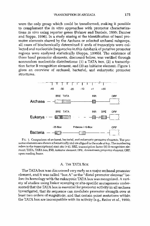
TRANSCRIPTION IN ARCHAEA 175
were the only group which could be transformed, making it possible to complement the in vitro approaches with promoter characteriza- tions in vivo using reporter genes (Palmer and Daniels, 1995; Danner and Soppa, 1996). In a study aiming at the identification of basal pro- moter elements shared by the Archaea or selected archaeal subgoups, all cases of biochemically determined 5’ ends of transcripts were col- lected and nucleotide frequencies in this databank of putative promoter regions were analyzed statistically (Soppa, 1999b). The existence of three basal promoter elements, discussed below, was verified through nonrandom nucleotide distributions: (1) a TATA box, (2) a transcrip- tion factor B recognition element, and (3) an initiator element. Figure 1 gives an overview of archaeal, bacterial, and eukaryotic promoter structures.
I 1 I ’ I ’ I 1 I ‘v- -40 -30 -20 -10 +I +30
BRE TATA INR ORF
Archaea 3-t
BRE TATA INR DPE ORF
Bacteria -35-Box Pribnow-/-1 O-Box ORF
FIG. 1. Comparison of archaeal, bacterial, and eukaryotic promoter elements. The pro- moter elements are shown schematically and are aligned to the scale at top. The numbering refers to the transcriptional start site (+I). BRE, transcription factor (II) B recognition ele- ment; TATA, TATA box; INR, initiator element; DPE, downstream promoter element; ORF, open reading frame.
A. THE TATA Box
The TATA box was discovered very early as a major archaeal promoter element, and it was called “box A” or the “distal promoter element” be- fore its homology with the eukaryotic TATA box was recognized. A vari- ety of studies using linker scanning or site-specific mutagenesis under- scored that the TATA box is essential for promoter activity in all archaea investigated, that its sequence can modulate promoter strength over at least two orders of magnitude, and that certain point mutations within the TATA box are incompatible with its activity (e.g., Reiter et al., 1990;

176 JtiRG SOPPA
Hausner et al., 1991; Hain et al., 1992; Palmer and Daniels, 1995; Danner and Soppa, 1996). The TATA box is recognized by an archaeal TATA box binding protein (TBP) which is homologous to the eukaryotic transcrip- tion factor, and crystallization of protein DNA complexes has shown that TBP contacts 8 bp (base pairs) of DNA, defining the length of this element (see Section IV). In agreement with the fact that the Archaea have only one RNA polymerase instead of the three eukaryotic enzymes, the TATA box is required for transcription of all types of genes, protein-coding genes as well as rRNA and other stable RNA genes. The TATA box is op- timally centered around -26/--27, and several lines of evidence suggest that its spacing to the transcription initation site is somewhat flexible and can vary by one or two positions around the ideal value: (1) the intro- duction of small deletions or insertions 3’ of the TATA box did not alter the start site selection in an in vitro Sulfolobus transcription system, while variation of three nucleotides or more shifted the start site (Reiter et al., 1990); (2) a considerable fraction of natural archaeal promoter se- quences fits the TATA box consensus only if they are shifted by one or two nucleotides; and (3) the same is true for haloarchaeal promoter se- quences selected after random mutagenesis (Danner and Soppa, 1996).
Three systematic studies were performed to characterize the sequence requirements of an archaeal TATA box experimentally. In the first study all single-nucleotide changes at seven positions of a rRNA TATA box of Sulfolobus shibatae were constructed and the resulting promoter strengths of the 21 mutants and the wild type were quantitated using an in vitro transcription system (Hain et al., 1992). In the second study all single-nucleotide changes at 11 positions of a tRNA promoter box from Haloferax volcanii were constructed and the promoter strengths of the 33 mutants were compared to the wild-type sequence in vivo using a transcriptional reporter gene (Palmer and Daniels, 1995). Both studies showed that at several positions two or even three different nu- cleotides are compatible with a high promoter strength, whereas other nucleotides at the same positions diminish promoter activity. In the third study 14 nucleotides around the TATA box of a protein-encoding gene of H. volcanii were randomized, resulting in a library of about lOa plasmids, and sequences with a high promoter activity in vivo were se- lected making use of a marker gene. The TATA box sequences recovered after the selection were A/T-rich, but a variety of sequences with a high promoter activity in viva was found (Danner and Soppa, 1996). The experimentally observed high variability of the TATA box sequence is in excellent agreement with the mode of interaction between the TATA box and the TATA box binding protein described below (Section 1II.A).
Another approach is the analysis of natural promoters with known transcriptional start sites (Soppa, 1999b). It underscored the variability

TRANSCRIPTIONINARCHAEA 177
of TATA box sequences but also revealed statistical preferences for certain nucleotides at different positions and indicated that the opti- mal TATA box sequence is not identical for different archaeal groups, i.e., TTTTTAAA was found for Crenarchaeota (mostly sulfolobales), TTTATATA was found for methanogens, and NTTTWWWN was found for halophiles (W = A or T; N = A or T or G or C). The consensus sequen- ces derived from natural promoters fit well the results of the experimen- tal studies on haloarchaea and sulfolobales (Fig. 1). No comparison is possible for methanogens, because no systematic TATA box characteri- zation has been reported until now.
B. THETRANSCRIPTIONFACTOR B RECOGNITIONELEMENT
The existence of the transcription factor B element (BEE) upstream of the TATA box was discovered in the Archaea and Eukarya simultane- ously. Qureshi and Jackson (1998) chose a strong Sulfolobus promoter for a detailed characterization and found that the region upstream of the TATA box is important for its promoter strength. The region increases the strength of a weaker promoter if it is fused to its TATA box. A mu- tant analysis confirmed that the region three to six nucleotides upstream of the TATA box is important for promoter function in an in vitro tran- scription system, and several nucleotides upstream and two nucleotides downstream of the TATA box gave signals in in vitro protection assays using the ternary complex of DNA, TBP, and transcription factor B (TFB). SELEX experiments were used to verify that the archaeal TFB selects specific sequences from a randomized pool of DNA fragments, proving a sequence-specific interaction between TFB and several nucleotides on both sides of the TATA box. Most prominent were an A three nucleotides and a G six nucleotides upstream of the TATA box, but unequal distribu- tions after selection were also obvious for additional positions, albeit to a lesser extent. An important observation was that TBP alone does not select for DNA fragments with a high promoter strength from a random library, while the combination of TBP and TFB does, underscoring that BRE is indeed an important basal transcription element for Sulfolobus and that it has an important role in transcription initiation. Until now the BRE has been investigated systematically only using the Sulfolobus system, but the data are in agreement with earlier circumstantial obser- vations, e.g., that a purine-rich sequence upstream of the TATA box is important for the strength of a haloarchaeal promoter in vivo (Palmer and Daniels, 1995) or that replacement of the region upstream of the TATA box of the GDH gene diminished transcription to 6% in an in vivo transcription system of Pyrococcus furiosus (Hethke et al., 1996). Analysis of natural promoters revealed the preference for two A’s at

178 JORGSOPPA
positions -33/-34 and indicated that a BRE is important for transcrip- tion initiation in all the archaea in viva in the context of the whole transcription apparatus, the presence of general DNA binding proteins, and at the physiological conformeric state of chromosomal DNA (Soppa, 1999b). However, it still remains to be determined whether different ar- chaeal groups have different BRE consensus sequences and how many and which nucleotides upstream and downstream of the TATA box in- flucence transcription initiation in the different systems.
The BRE was also found to be a basal promoter element in eukary- otes. Lagrange et al. (1998) used the human TFIIB to select for DNA fragments with an affinity for this transcription factor from a random fragment library. A nonrandom sequence distribution in the selected subpopulation was found at eight positions upstream of the TATA box, and the consensus sequence -38 SSRCGCC -32 was derived (S = G or C; R = A or G). Mutations were introduced at positions -37 and -34 and it was verified that they decreased TFIIB binding as well as in vitro transcription efficiency, proving that the BRE is a bona fide basal promoter element in the eukaryotic system. In the binary system of TFIIB and DNA, cross-linking of TFIIB was observed only at positions upstream of the TATA box, while in the ternary system of DNA, TFIIB and TBP nucleotides both upstream and downstream of the TATA box could be cross-linked to TFIIB, showing (1) that the primary sequence- specific binding site of eukaryotic TFIIB is also upstream of the TATA box and (2) that TBP-induced bending of the TATA box DNA is a pre- requisite to allow TFIIB to contact nucleotides on both sides of the TATA box (see Section IV).
In summary, the newly discovered BRE is a basal promoter element in the Archaea and Eukarya, the consensus sequences are different in the only two experimentally analyzed systems, i.e., human and Sulfolobus, and the importance of the BRE upstream of different genes in different species remains to be clarified.
C. THE INITIATORELEMENT
The third archaeal basal promoter element is the initiator element around the transcription initiation site itself. Using an in vitro transcrip- tion system of Sulfolobus, it was shown that mutations at the initiator element are less severe than TATA box mutations, that upon changing the spacing between the two elements the TATA box overides the initia- tor element control, and upon insertions or deletions of three or more nucleotides transcription starts at a new artificial “initiator element” around the optimal distance to the TATA box (Reiter et al., 1990).

TRANSCRIPTIONINARCHAEA 179
The analysis of natural promoters indicates that an initiator element is also present throughout the Archaea but that it might be of different importance for transcription initiation for different groups, i.e., it is hardly detectable in haloarchaeal sequences and very pronounced at the initiation sites of methanogens and sulfolobales (Soppa, 19Wb). No systematic studies to characterize the initiator element have been performed until now, the sequence requirements have not been deter- mined experimentally, and it is not known which protein recognizes it and at which stage of the initiation process it is read out. Recently specifically labeled DNA fragments were cross-linked to the proteins in the initiation complex of Pyrococcus, and it was shown that the large subunits A’ and B are in the vicinity of the initiator element, while A’ is not (Bartlett et al., 2000).
D. ADDITIONALBASALPROMOTERELEMENTS
Apart from the three elements mentioned, no further basal promoter element has been found in archaeal systems. It should be explicitly noted that there is no indication for the presence of a “downstream promoter element” (DPE), which is found in a variety of eukaryotic pro- moters and in Drosophila is as widely used as the TATA box (Kutach and Kadonaga, 2000). This is in agreement with the model that the eukary- otic DPE is contacted by a TBP-associated factor (TAF) and that TAFs are not present in the Archaea.
III. Transcription Initiation Factors
In the Bacteria, promoter recognition is accomplished by a complex of core RNA polymerase with a tightly bound CT factor which is lost during elongation. In the Eukarya, promoter elements are first recog- nized by transcription factors, which subsequently recruit the poly- merase. Factors involved in initiation at polymerase II promoters up- stream of protein-encoding genes are TFIIA, TFIIB, TFIID, TFIIE, TFIIF, and TFIIH, most of them complexes of several different polypeptides. In the Archaea, it was discovered about 10 years ago that purified RNA polymerase cannot initiate transcription in vitro accurately by itself, but transcription factors must be added (Frey et al., 1990; Hiidepohl et al., 1990), and thus archeal transcription initiation is similar to the eukaryotic model. Using a variety of approaches including biochemical isolation, immunoprecipitation with specific antibodies, and genome sequence analysis, it became clear that the main and probably the only transcription initiation factors in the Archaea are the TATA box binding
180 JijRG SOPPA
protein (TBP) and TFB, a homologue of the eukaryotic factor TFIIB (as archaea have only one RNA polymerase, the “II” is omitted when archaeal proteins are named in accordance with eukaryotic polymerase II-related proteins).
A. THE TATA Box BINDING PROTEIN
In the Eukarya, TBP is the most general transcription factor involved in transcription initiation with all three RNA polymerases, acting in complexes with different polypeptides at the three types of promoters. At polymerase II promoters, TBP binding to the TATA box is the ini- tial step in promoter recognition. In vitro, TBP alone is sufficient for transcription initiation in concert with TFIIB and polymerase; in viva it acts in a complex called TFIID, in which it is bound to TBP-associated factors (TAFs).
After the first TBP sequences were discovered in the Archaea (e.g., Marsh et al., 1994; Rowlands et al., 1994), it soon became clear that TBP is widely distributed and is a basal archaeal transcription factor. Today, more than 20 sequences are known. In general, each archaeal genome contains a single TBP gene (exceptions are discussed in Sec- tion IX). Archaeal TBPs consist of about 180 amino acids and are highly conserved; the similarities between different archaeal TBPs range be- tween 40 and 55% (throughout this article similarity means the fraction of identical amino acids). TBPs are comprised of two repeats of about 90 amino acids, thus it seems that the recent tbp gene arose by an an- cient gene duplication. The similarities of the first to the second repeats are slightly higher than 40%, indicating that archaeal TBPs are highly symmetrical proteins and can be regarded as “internal dimers” (Fig. 2).
Eukaryotic TBPs share with their archaeal homologues the two re- peat domains of about 90 amino acids and, in addition, contain an N-terminal extension which is very variable both in length and in se- quence throughout the Eukarya. The function of this N-terminal domain is not known. Remarkably, the similarity of the first and the second re- peats of eukaryotic TBPs is only 26%, about 1.5% lower than for archaeal TBPs. Probably eukaryotic TBPs have evolved further from the ances- tral symmetric state because they have to interact with a larger variety of different transcription factors, e.g., with TAFs to form TFIID. The simi- larities between archaeal TBPs and the homologous parts of eukaryotic TBPs range between 20 and 40%. Within the Eukarya, the degree of con- servation is very different; it is extremely high in higher Eukarya, but between higher and lower Eukarya the degree of conservation is about the same as between the Archaea and Eukarya, e.g., 27% between Homo sapiens and Plasmodium falciparum.

TRANSCRIPTION IN ARCHAEA 181
A. The TBP of Pyrococcus woesei
B. Comparison of TBPs
Archaea (D+E) - <420’-\ E-rich (K+W
FIG. 2. The TATA box binding protein. [A) Structure of the TATA box binding pro- tein of Pyrococcus woesei [PDB entry number 1PCZ (DeDecker et al., 1996)]. To visual- ize the internal symmetry of the protein, the first and second repeat domains (compare text) are shown in different shades of gray. (B) Comparison of archaeal and eukaryotic TBPs. The domain structure is shown schematically. Average values of the fraction of identical amino acids are indicated. The acidic C-terminal extension is present only in a subgroup of archaea (see text). The excess of negatively charged amino acids (positive value) or positively charged amino acids (negative value) is depicted on the right. aa, amino acids.
The alignment of archaeal and eukaryotic TBP sequences and con- struction of a phylogenetic tree revealed that they form two distinct monophyletic groups. The similarities of the first repeats of archaeal TBPs are only slightly greater to the first than to the second repeats of eukaryotic TBPs. Therefore it seems that the gene duplication had al- ready occurred in the last common ancestor of the Archaea and Eukarya but that the fusion gene had not evolved far at the time that the two lineages split and that the protein at that time was still nearly abso- lutely symmetrical. It seems safe to assume that the cognate DNA ele- ment, the precursor of the TATA box, had perfect dyad symmetry at that time.
Severeal structures have been determined for eukaryotic or archaeal TBPs alone or in complex with DNA, TF(II)B, or TFIIA (e.g., Tsai and Sigler, 2000; Littefield et al., 1999; Kosa et al., 1997; DeDecker et al.,

182 J6RG SOPPA
1996; Kim and Burley, 1994; Nikolov et al., 1995; Chasman et al,, 1993). Not unexpectedly, it turned out that the structure of the archaeal protein from Pyrococcus woesei is very similar to the structures of TBPs (C-terminal domains) from yeast, Arubidopsis thalinana, and Homo sapiens. TBP from all sources is a saddle-shaped protein of a//3 structure with a high twofold symmetry also for the eukaryotic proteins (Fig. 2). The concave underside of the protein binds 8 bp of DNA, thereby creating a high distortion of the TATA box. Two pair of pheny- lalanines, which are absolutely conserved in archaeal and eukaryotic TBPs, intercalate between the first and the last 2 bp of the TATA box. The central 6 bp adopt a conformation which was unprecedented and is today called “TATA-DNA” (Guzikevich-Guerstein and Shakked, 1996). It is somewhat related to A-DNA, and a molecular dynamic simulation suggests that TATA-DNA has the intrinsic property of adopting an A-DNA-like conformation, which might be an intermediate in complex formation (Flatters et al., 1997). The TATA-DNA conformation is char- acterized by very wide and shallow minor grooves, which interact with the concave underside of TBP. Most of the protein-DNA interactions are nonpolar and hydrophobic. TBP binding to the minor groove and the absence of electrostatic interactions with bases indicate that TBP recognizes the deformability of the TATA box more than its specific sequence. The minor groove is chemically more monotonous than the major groove, therefore many DNA binding proteins with a broad sequence specificity bind to the minor groove. This is in good agreement with the sequence variability of archaeal TATA boxes described above. It also fits to a study in which one eukaryotic TBP was cocrystallized with 10 sequence modifications of a TATA box fragment, and the structure of the protein, the path of the DNA in the complexes, and the protein- DNA interfaces were found to be identical in all cases (Patikoglou et al., 1999). On both sides of the TATA box the DNA is in the canonical B-DNA conformation. Nearly all of the amino acids at the DNA binding surface of TBP are absolutely conserved in the Archaea and Eukarya. In eukaryotic TBPs, the amino acids which are conserved in the first and second repeat domain (26%) are mostly confined to the DNA binding domain, while the sequence identity at equivalent positions of the con- vex upper surface is low. In contrast, the convex surface of archaeal TBPs contains many identical amino acids at equivalent positions of the two repeat domains, in accordance with the higher primary se- quence conservation of the two domains in archaeal TBPs (42%). This unequal distribution of conserved residues at different surfaces of the protein structure strengthens the above-mentioned conclusion that the reason for the different degree of conservation is the higher number

TRANSCRIPTIONINARCHAEA 183
of interaction partners of eukaryotic TBPs. Therefore it cannot be ex- pected that archaeal TBPs can functionally replace eukaryotic TBPs in factor-dependent transcription. However, it has been reported that human and yeast TBPs can functionally replace the archaeal protein in an in vitro transcription system (Wettach et al., 1995), underscor- ing the high structural similarity of archaeal and eukaryotic TBPs. Two differences between archaeal and eukaryotic TBPs have already been mentioned, i.e., the presence of an additional N-terminal domain in the eukaryotic proteins and the higher degree of internal symmetry of the ar- chaeal proteins. A third difference is that archaeal TBPs have an excess of acidic amino acids, whereas eukaryotic TBPs have an excess of basic amino acids. It will be interesting to unravel what the consequences of these differences are. The TBPs of three groups of Archaea, i.e., Pyrococcus, Thermococcus, and Sulfolobus, contain a short C-terminal extension that is not present other archaeal sequences. The extension contains a run of five or six glutamic acid resisues (sometimes inter- rupted by a single glycine or leucine residue) and thus is very acidic. Its conservation in several species indicates that it is important for func- tion, either for binding another transcription factor or as a target for a posttranslational modification regulating function. This is the first ex- ample of the specialization of a highly conserved basal transcription factor in some archaeal species; further examples are discussed below (Section IX).
B. TRANSCRIPTIONFACTORB
At eukaryotic polymerase II promoters the next factor to bind after TBP is TFIIB, before additional transcription factors and the polymerase are recruited. Archaea harbor a homologue to TFIIB, which is called TFB. Sequence analysis of a prepublished sequence led to the first discovery of an archaeal TFB in 1992 (Ouzounis and Sander, 1992); today more than 20 TFB sequences are known. In general, archaeal genomes contain a single tfa gene; some exceptions are discussed in Section IX.
Archaeal and eukaryotic TF(II)B’s consist of three domains: an N- terminal region-TF(II)B N-comprised of about 100-12~1 amino acids; and a C-terminal region-TF(II)B c-comprised of two repeats of about 90 amino acids (see Fig. 3). The degree of conservation is substan- tially higher for archaeal TFBs than for archaeal TBPs, i.e., the simi- larities are 50-60%. Similarly to the TBPs, the similarities of the two repeat domains are higher for the archaeal TFBs (25-37%) than for the eukaryotic TFIIBs (ll-25%). In addition, the similarities of the

184 J6RG SOPPA
A. TFBC of Pyrococcus woesei
6. Comparison of TFBsiTFIIBs
Archaea
FIG. 3. Transcription factor B. (A) Structure of the C-terminal domain of transcription factor B (TFBc) of Pyrococcus woesei. The structure of TFBc was determined in a ternary complex together with TBP and a BRE- and TATA box-containing DNA fragment [PDB entry number lD3U (Littlefield et al., 1999)]. The N-terminal domain of TFB was not included in structure determination. To visualize the internal symmetry of TFBc, the two repeats are shown in different shades of gray. (B) Comparison of archaeal TFBs with eukaryotic TFIIBs. The domain structure of TF(II)B is shown schematically. Average values of the fraction of identical amino acids are indicated. aa, amino acids.
N-terminal domains are much higher within the Archaea (50-60%) than within the Eukarya (3040%). Again, the interaction of the eukaryotic protein with a higher number of global and gene-specific transcrip- tion factors compared to the archaeal TFB can easily explain these observations.
The similarities between archaeal TFBs and eukaryotic TFIIBs are between 20 and 30% and thus slightly lower than between archaeal and eukaryotic TBPs. The first repeat of the archaeal TFBs is on aver- age only 8% more similar to the first than to the second repeat of the eukaryotic TFIIBs, which indicates that the two repeats of the TF(II)B precursor were also nearly identical at the time that the two lineages separated.
It was discovered simulatiously in archaea and eukaryotes that TF(II)B is a sequence-specific DNA binding protein and recognizes a

TRANSCRIPTION IN ARCHAEA 185
transcription factor B recognition element (BRE) upstream of the TATA box (see above; Lagragne et al., 1998; Qureshi and Jackson, 1998). The ability to recognize the BRE is localized in the C-terminal domain of TF(II)B, because the binding affinities of TF(II)B and TF(II)Bc to several wild-type and mutant BREs are virtually identical (Bell and Jackson, 2090b; Lagrange et al., 1998), and TFBc is able to select specific se- quences from a random DNA fragment library (Qureshi and Jackson, 1998). At that time these findings were very surprising since the struc- ture determination of TBP/TF(II)B/DNA ternary complexes from both an archaeon (Kosa et al., 1997) and a eukaryote (Nikolov et al., 1995) had not revealed any base-specific DNA contacts of TF(II)Bc. However, the DNA used for crystallization was rather short and did not contain a BRE, and it turned out that this had inhibited the formation of base- specific contacts. The ternary complexes of the archaeal and the eukary- otic systems were recrystallized using DNA fragments containing BREs derived from biochemical data, and in both cases sequence-specific DNA contacts of TF(II)B were obvious in the new structures (see Fig. 4).
FIG. 4. The ternary preinitiation complex. Structure of the preinitiation complex, com- prised of TBP and TFB from Pyrococcus wosei and a BRE- and TATA box-containing DNA fragment [PDB entry number 1D3U (Littlefield et al., 1999)). TBP is shown at the top, in darker gray. TFB is shown at the left, in lighter gray. The DNA is shown in wire-frame representation. Note the asymmetric binding of TFB to the C-terminal (left) repeat of TBP, the highly distorted path of the DNA at the concave underside of TBP, and the TFB in- teractions with DNA on both sides of the TATA box, which are more intense and base specific at the BRE upstream [left) of the TATA box.

186 JijRGSOPPA
The TFB of Pyrococcus woeseii makes contacts several nucleotides up- stream of the TATA box, with positions -3 and -6 being the strongest specificity determinants, and it makes contacts with phosphates up to nine positions upstream and six positions downstream of the TATA box (Littlefield et al., 1999). The strong bending of the DNA at the TATA box, brought about by the TBP-DNA interactions (see above), is a pre- requisite for the ability of TFB to contact both sides of the TATA box. A prominent example is a single amino acid, arginine 188 of Pyrococcus TFB, which contacts phosphates on both sids of the TATA box in the ternary complex. These TFB contacts stabilize the TBP-induced con- formational deformation of the DNA. Essentially identical results were obtained using human TFIIBc, human TBPc, and the adenovirus major late promoter (Tsai and Sigler, 2000). In the human system, an additional base-specific TFIIBc contact was detected five positions downstream of the TATA box, which had not been anticipated before. Both the archaeal and the eukaryotic TF(II)Bc’s make the base-specific contacts upstream of the TATA box with a helix-turn-helix (HTH) motif localized in the second repeat domain (helix H5’), which binds to the major groove of the DNA. This corrobarates an earlier prediction based on a primary se- quence analysis and is in excellent aggrement with the observation that mutations in this helix influence the recognition of a BRE base pair three positions upstream of the TATA box (Lagrange et al., 1998). The DNA in- teractions of the TF(II)Bc HTH motif are reminiscent of the mode of bind- ing of previously analyzed bacterial and eukaryotic HTH DNA binding proteins. The binding ot TF(II)Bc to the major groove upstream and the minor groove downstream of the TATA box agrees very well with SELEX experiments which had shown that the major determinants for TFB recognition are located upstream of the TATA box (Qureshi and Jackson, 1998). The exclusive binding to the major groove in the upstream re- gion leaves the minor groove untouched, and thus additional proteins, e.g., regulatory factors, could bind there simultaneously with TF(II)B (Tsai and Sigler, 2000).
The structure of TFIIBc was first resolved by NMR spectroscopy and it was revealed that the two repeats have a structure similar to the cyclin A fold (Bagby et al., 1995). The X-ray structures of archaeal and eukaryotic TF(II)Bc’s in the ternary complex established that the structure of the two repeats is virtually identical in contact with TBP and DNA in the crystallized form compared to the solution structure (Tsai and Sigler, 2000; Littlefield et al., 1999; Kosa et al., 1997; Nikolov et al., 1995). However, the two repeats are arranged differently from one another in the solution structure than in the ternary complex. The comparison indicated that upon TF(II)B binding to the TBP-DNA binary complex, an en bloc rotation of the two cyclin repeats of about 100” is necessary

TRANSCRIPTIONINARCHAEA 187
to allow the formation of the contacts with the major groove upstream of the TATA box and the minor groove downstream of the TATA box (Tsai and Sigler, 2000).
While the C-terminal domain of TF(II)B mediates DNA binding, the N-terminal domain is involved in polymerase recruitment and in late stages of the initiation process (see Section IV). The structure of part of the the N-terminal TF(II)B domain was first solved for the archaeal protein from Pyrococcus furiosus (Zhu et al., 1996) and later for the human protein (Chen et al., 2000). It contains a zinc ribbon motif of three antiparallel B strands, a motif that has also been found in other proteins involved in transcription, e.g., the elongation factor TF(II)S and the polymerase subunit RPBS. A model containing the structure of the resolved part of the N-terminal domain, the structure of the C-terminal domain, and a simulation of the remaining part of the protein indicated that the N-terminal domain could reach position -1 and is in agreement with the functional studies described below that TF(II)B influences open complex formation and promoter clearance.
C. FURTHER TRANSCRIPTION INITIATION FACTORS
Faithful transcription initiation has been accomplished using TBP, TFB, and purified RNA polymerase of several archaea, which could be taken as an indication that archaea contain no further transcription initi- ation factors. However, in some eukaryotic in vitro transcription systems TBP and TFB are also sufficient for initiation, while that is clearly not the case in viva. Therefore the archaeal in vitro systems do not prove the lack of further basal transcription initiation factors. While from a philosophical point of view the absence of a factor cannot be proven for obvious reasons, the following paragraphs list a variety of arguments that the Archaea indeed use a much smaller set of basal transcription initiation factors than the Eukarya.
In an elegant set of experiments the factor requirement for transcrip- tion initiation of Sulfolobus shibatae was analyzed (Qureshi et al., 1997). An in vitro transcription system using crude cell extract was established. Immunodepletion and TBP, TFB, and polymerase verified that all three components are essential for transcription and, because in each case the other two components were not coprecipitated, that they are not stably associated in the cell extract. Supplementation of the de- pleted extracts with recombinant TBP or TFB or with a highly purified polymerase restored the activity in the respective depleted extracts. Gel filtration gave no indication that either of the two factors was associated with another protein, and the purified polymerase did not contain any nonpolymerase subunits. Thus there was no indication for the presence

188 JijRG SOPPA
or the need for a transcription factor apart from TBP and TFB on several promoters used.
Today about 10 archaeal genome sequences are available (the genome sequences can be addressed and analyzed using the program PEDANT at http://pedant.mips.biochem.mpg.de). None contains recognizable ho- mologues of TAFs or of subunits of the eukaryotic factors TFIIA, TFIIE, TFIIF, and TFIIH. However, all archaeal genomes contain a gene encod- ing a protein of 200 amino acids with similarity to the N-terminal half of the a-subunit of TFIIE. The eukaryotic TFIIE is heterotetramer of e& composition. Two of its functions are to recruit TFIIH to the promoter and to promote the polymerase-phosphorylating activity of TFIIH. Both functions seem to be unnecessary in the Archaea (see below). But TFIIE is not well characterized in the Eukarya, and recently it has been shown that TFIIEa alone can influence TBP binding to DNA (Yokomori et al., 1998). It contains a zinc ribbon motif that is required for that function and that is conserved in the known eukaryotic TFIIEa! (CXXCX2,CXXC). The presence of the zinc ribbon is also conserved in the archaeal pro- teins, but its sequence is more variable (CX24CX,4-15CXXC). While the sequence similarity and the existence of the zinc ribbon motif open the possibility that the archaeal protein is a homologue of TFIIEo and a third basal transcription factor, experimental evidence for its expres- sion, its function, or its involvement in transcription initiation is still lacking.
There are more arguments for the absence of an archaeal TFIIH ho- mologue than the lack of recognizable sequence similarity in genome sequences. The function of TFIIH in the Eukarya is to phosphorylate the largest RNA polymerase subunit at its C-terminal domain (CTD). The underphosphorylated polymerase is recruited to the promoter, and CTD phosphorylation by TFIIH or other kinases is a prerequisite for the transition from the initiation to the elongation phase of transcription. Archaeal RNA polymerases do not contain this CTD and thus a phos- phorylation switch does not seem to operate. This could be underscored by the experimental evidence that the /3-v phosphoanhydride bond of ATP, which is required during eukaryotic CTD phosphorylation, is not required for transcription in an in vitro system of Sulfolobus (Bell et al., 1998). Therefore the Archaea apparently do not require a TFIIH homo- logue and, in turn, also do not need the TFIIH recruiting and stimulating activities of TFIIE.
IV. Preinitiation Complex Formation and Polarity of Transcription
The available data indicate that the archaeal preinitiation complex consists of a DNA fragment containing the BEE, TATA box, and initiation

TRANSCRIPTION IN ARCHAEA 189
element, the transcription factors TFB and TBP, and RNA polymerase. The involvement of the TFEa homologue, for which no experimen- tal data exist, cannot be ruled out, but no additional factors seem to be involved. For some time the factors which determine the polarity of transcription were thoroughly discussed. TBPs from yeast and from Arabidobsis thaliana were found to bind in an oriented manner to the TATA box in crystallized binary complexes (J. L. Kim et al., 1993; Y. Kim et al., 1993), and it was thought that deviations from perfect symmetry in the repeat domains would cause a preference for one orientation. How- ever, it was shown that in solution yeast TBP binds to two TATA boxes in both orientations and that the preference for one of them is only mod- est (Cox et al., 1997). Therefore even in the Eukarya the TBP-TATA box interaction cannot determine polarity, and this can safely be assumed to be even more true for the Archaea, which harbor a TBP of much higher symmetry. Indeed, in an in vitro transcription system the switch of TATA box orientation had no influence on the direction of transcription (Bell et al., 1999). The discovery of the BEE in the Archaea and Eukarya explained how asymmetical TFB binding to the convex upper side of only the C-terminal TBP repeat in combination with sequence-specific binding upstream of the TATA box could orient the two transcription factors faithfully on the DNA in the ternary complex and, through asym- metrical binding of RNA polymerase, could lead to the observed high fidelity in the direction of transcription. It was indeed verified in an in vitro transcription system from Sulfolobus that switching the DNA sequences surrounding the TATA box, changing exclusively the BEE orientation, and leaving the orientation of the TATA box and the initia- tion element untouched resulted in a complete reversal of the direction of transcription (Bell et al., 1999). Similarly, yeast TBP, which in solu- tion was shown to bind to the TATA box of the adenovirus major late promoter in both orientations, became more than 99% unidirectional in the quaternary complex together with TFIIB and TFIIA (Kays and Schepartz, 2000).
The question of polarity was complicated for some time by the obser- vation that TBP and TFB in the archaeal ternary complex (Kosa et al., 1997) had an opposite orientation compared to several ternary structures determined with eukaryotic proteins (e.g., Nikolov et al., 1995) and that, in none of the complexes, could base-specific DNA contacts of TF(II)B could be observed. As mentioned above, the DNA fragments used for crystallization did not contain a BRE, which had not yet been discov- ered at that time, and recrystallization (Littlefield et al., 1999; Tsai and Sigler, 2000) nicely verified the biochemical evidence, i.e., that ternary complex orientation is identical in the Archaea and Eukarya and that a much larger surface of TF(II)B than observed previously interacts with
190 JdRG SOPPA
DNA, including several contacts in the major groove with bases of the BRE.
The structure of the archaeal ternary complex indicates that binding of TBP and TFB to promoter DNA is cooperative (compare Fig. 4). While to my knowledge this has not been quantitated, it is in agreement with many observations that the affinity of TBP and TFB for DNA is rather low and that ternary complex formation is observed at protein and DNA concentrations which would not allow binding of the individual pro- teins (e.g., Qureshi and Jackson, 1998).
In all available structures of archaeal and eukaryotic ternary com- plexes TF(II)Bc was used for crystallization, while the N-terminal do- main was omitted. The affinities of binding to the ternary complexes are about the same for Sulfolobus solfataricus TFBc and TFB, indi- cating that the N-terminal domain does not play an important role in DNA binding. However, in contrast to the C-terminal domain, only the full-length protein can recruit RNA polymerase into a quaternary complex, giving evidence for a direct interaction of the N-terminal do- main with the polymerase (Bell and Jackson, 2OOOb). An interaction of TFB with the polymerase could also be shown for Methanobacterium thermoautotrophicum, and, in contrast to the Sulfolobus system, in this case it was detected in the absence of TBP and DNA (Darcy et al., 1999). Size fractionation of a crude cell extract revealed that TFB and polymerase cosedimented in a sucrose gradient, and furthermore, poly- merase could be coimmunoprecipitated with anti-TFB antibodies, in- dicating a stable interaction in solution. Several experiments indicate that eukaryotic TFIIB is also involved in polymerase recruitment, e.g., only full-length TFIIB, in contrast to TFIIBc, was shown to be active in an in vitro transcription system (Malik et al., 1993) and a mutant analysis showed that the N-terminal region of yeast TFIIB is important for polymerase interaction (Bangur et al., 1997). Using the Sulfolobus system it was shown that archaeal TFB has additional functions in the process of transcription initiation apart from polarity determination and polymerase recruitment (Bell and Jackson, 2000). A point mutation at a highly conserved position in the N-terminal domain immediately down- stream of the zinc ribbon motif was constructed and found to be undis- tinguishable from the wild-type protein in ternary complex formation, polymerase recruitment, and even open complex formation. Neverthe- less, the wild type and mutant differed considerably in the amounts of transcripts formed in an in vitro system. The differences were sensitive to the NTP concentrations as well as to the sequence of the initiator element at the transcriptional start site. These data indicate that TFB also plays a role in late stages of transcription initiation, e.g., during promoter clearence.

TRANSCRIPTIONINARCHAEA 191
V. The Different Phases of Transcription: From Initiation to Termination
A. PREINITIATIONANDBEYOND
Knowledge about the different steps in the initiation process from archaeal RNA polymerase binding to the TBP/TFB/DNA ternary com- plex until a stable elongation complex is reached is very scarce. It has been shown that, in contrast to eukaryotic systems, in Sulfolobus TBP, TFB, and polymerase are sufficient for open complex formation and that neither additional transcription factors nor ATP hydrolysis is nec- essary (Bell et al., 1998; Bell and Jackson, 2000b). The region from -12 to -1 becomes sensitive to the single strand-specific reagent potassium permanganate, and upon the addition of three ribonucleotide triphos- phates, this sensitive region can be chased several positions into the transcribed region until the lack of the fourth ribonucleotide triphos- phate stalls the complex (Bell et al., 1998). These data agree well with the length of the transcription bubble within the eukaryotic polymerase.
In the Eukarya, after open complex formation there is a phase of abortive transcription, in which small RNAs of 2 to 10 nucleotides are synthesized and released from the complex, until productive promoter clearence occurs and polymerase II enters the elongation phase (Keene and Luse, 1999). Whether this takes place during archaeal transcription initiation as well has not been determined. The similarities of archaeal and eukaryotic RNA polymerases and the advances in polymerase structure determination, described below, make it tempting to spec- ulate that this is a shared feature. As pointed out above, the analysis of an archaeal TFB mutant has made clear that at least one additional step has to be accomplished between open complex formation and elongation. The existence of different stages during the initiation pro- cess was also observed in the mammalian system, where it was shown that the DNA region around +40 is required transiently, after open complex formation and synthesis of the first ~10 phosphodiester bonds and before the transcripts have reached a length of more than about 14 nucleotides. It was concluded that “promoter escape requires that the RNA polymerase II transcription complex undergoes a critical structural transition” (Dvir et al., 1997).
B. ELONGATION
The presence of an archaeal homologue to the eukaryotic transcrip- tion elongation factor TFIIS was first predicted based upon an open reading frame of Sulfolobus acidocaldarius discovered accidentally downstream of a gene for a RNA polymerase subunit (Langer and Zillig, 1993). This view was challenged after the discovery of an orthologue in

192 J6RG SOPPA
Thermococcus celer and the hypothesis was put forward that it might instead be a small polymerase subunit (Kaine et al., 1994). The determi- nation of several archaeal genome sequences revealed that this gene is conserved in the Archaea and enabled a sequence analysis of the emerg- ing protein family (Soppa, 1999a; Hausner et al., 2000). Several lines of evidence indicated that it is more likely a polymerase subunit than an elongation factor: (1) the archaeal proteins have a size of about 100-130 amino acids, comparable a eukaryotic polymerase subunit conserved in polymerases I, II, and III (called RPA12.2, RPBS, and RPCll), while eu- karyotic TFIIS is about 300 amino acids long; (2) the degree of similarity is on average greater to the polymerase subunits than to TFIIS; (3) the archaeal protein and the polymerase subunits contain two zinc binding motifs, while eukaryotic TFIIS contains only one; and (4) the gene is localized near polymerase subunit genes in S. acidocaldariius.
Despite this circumstantial evidence, biochemical characterization of the Methanococcus thermolithotrophiscus protein revealed that it is a bona fide archaeal elongation factor and not a polymerase subunit (Hausner et al., 2000). The archaeal protein will be called aTFS to point out its limited similarity to eukaryotic TFIIS. aTFS is not present in the purified polymerase, and size fractionation of a crude cell extract showed that it has an elution profile distinct from that of the polymerase and thus was not separated from the polymerase in the course of pu- rification. It was shown that aTFS exhibits some activities which are similar to the activities of eukaryotic TFIIS: (1) aTFS induces a cleav- age activity of the RNA polymerase at stalled elongation complexes, (2) this activity removes predominantly dinucleotides from the 3’ end of the nascent transcript as the polymerase retracts, and (3) the amount of arrested elongation complex is reduced and the amount of full-length runoff transcript is enhanced by aTFS in an in vitro transcription system. These data verify that the conserved archaeal aTFS is an elongation fac- tor, which enables the polymerase to resume transcription at sites where it was stalled or arrested.
The sequence similarities indicate a common ancestry of aTFS, the three polymerase subunits RPA12.2, RF’B9, and RPCll, and TFIIS, which had aquired an additional N-terminal domain. It seems that the four eukaryotic proteins have specialized during evolution, e.g., poly- merase III has a factor-independent cleavage activity, which depends on the subunit IWCll. A RPCll-deficient polymerase III is unable to release dinucleotides at stalled elongation complexes, but the activity can be restored by the subsequent addition of RPCll, which, however, is not able to integrate into the polymerase and thus, in this artificial situation, has retained the ability to act as an elongation factor (Chedin

TRANSCRIPTION IN ARCHAEA 193
et al., 1998). As Hausner et al. (2000) point out, the polymerase II sub- unit RPB9 deviates from the other four proteins in the region of the zinc ribbon motif retained in TFIIS and, specifically, has lost a glutamic acid residue which has been shown to be important for function in TFIIS (Jeon et al., 1994) and RFCll (Chedin et al., 1998). In agreement with this, a polymerase II deficient in this subunit is still competent of TFIIS-induced transcript cleavage, indicating that RPB9 is not involved in this reaction. It is tempting to speculate that the precursor protein evolved as an elongation factor in the common ancestor of the Archaea and Eukarya, increasing the efficiency of transcription. Archaeal aTFS would have retained this function, while the eukaryotic protein became integrated into the polymerase before its specialization into the three derivatives. In this model polymerase II would have reintroduced the use of the elongation factor, but in a larger version, thereby allowing subunit RPB9 to acquire a new function.
C. TEFuv~INATIoN
The mechanism of termination of transcription in the Archaea is hardly understood. Early results of transcriptional termination studies were collected by Brown and colleages (1989), and not much has been added to that picture in the last decade. The determination of the 3’ ends of a variety of mRNAs or stable RNAs from different archaeal species has often located the termination site within or very close to an oligo(dT) tract. In general, “staggered termination” was observed, i.e., transcrip- tion terminated not on a single nucleotide, but in a closely defined region on a few up to more than 10 nucleotides. However, no common struc- ture for an “archaeal terminator” could be defined until now, because the oligo(dT) tract was located either downstream of a putative stem-loop structure, similar to bacterial p-independent terminators, or upstream of a putative stem-loop structure directly following the 3’ end of a gene, or without a putative stem-loop structure in its vicinity. The construc- tion of mutants verified for the oligo(dT) tract of the tRNA”“’ gene of Methanococcus thermolitothrophicus that it is essential for termination (Thomm et al., 1994). Deletion of the last two thymidine residues of the wild-type sequence 5’ TTTTAATTTT 3’ severely decreased the ter- mination efficiency, and deletion of two additional thymidine residues abolished termination at this site completely. However, a deletion up- stream of the oligo(dT) tract, within the tRNA gene, also resulted in a reduced termination efficiency and enhanced readthrough, indicating that the oligo(dT) tract is a required but not a sufficient determinant for termination. In a second study a reporter gene was used to verify that

194 J6RG SOPPA
the T-rich element 5’ TTATTCTTT 3’ acts in viva as an efficient termi- nator of transcription in Holoferux volcanii and that both the inversion of the element and the mutation of the last two thymidines to guanosines totally abolish its activity (Thompson et al., 1999). In this case 88 bp was transferred into the nonnative surrounding of a reporter gene from yeast and thus this region appeared to to be the sole determinant for termination. The presence of inverted repeats close to the T-rich ele- ment was not reported, and a nearby direct repeat was shown not to be important for termination. At the moment it appears as if no uniform archaeal termination signal exists, therefore further studies are needed to characterize the complete termination structures for different classes of genes in different archaea.
VI. RNA Polymerase
A. RNA POLYMERASE COMPOSITION
Very soon after it was proposed that the Archaea are a third domain of organisms distinct from the Bacteria and Eukarya, it became clear that ar- chaeal polymerases do not follow the bacterial paradigm. Bacterial RNA polymerases contain only four different subunits and have the compo- sition o&?‘w. In contrast to bacteria, eukaryotes harbor three different polymerases with specialized functions, which are multicomponent en- zymes of 12 to 17 subunits. By 1983 it was clear that archaea contain only one type of polymerase, that RNA polymerases of several archaea of diverse subgroups are comprised of at least 10 subunits, and that, on the basis of not only the composition but also the immunological crossreac- tivity, archaeal polymerases are more closely related to their eukaryotic than to their bacterial counterparts (Huet et al., 1983). Hence RNA poly- merases were the first component of the basal transcription apparatus for which the archaeal-eukaryotic relationship was discovered, and in fact this observation triggered much of the subsequent work described above. Zillig’s group had a huge impact on the field in the following decade, and the reader is referred to earlier reviews summarizing the results obtained until the mid-1990s, which included isolation of the genes for the larger subunits of a variety of archaea, cloning of the genes for 8 subunits (of about 12 detected) of the Sulfolobus enzyme, phyloge- netic reconstruction of polymerase subunit relationships, and introduc- tion of a factor-dependent in vitro transcription system (Langer et al., 1995; Zillig et al., 1992,1993). The results clearly underscored the rela- tionship of the archaeal polymerase and the eukaryotic polymerase II.
The best-characterized archaeal RNA polymerases are the enzymes from Sulfolobus acidocaldarius (Langer et al., 1995) and the enzyme

TRANSCRIPTION IN ARCHAEA 195
recently isolated from Methanobacterium thermoautotrophicum (Darcy et al., 1999). Table I shows a comparison of the subunit compositions of the two archaeal enzymes with the eukaryotic polymerase from yeast and the bacterial enzyme from E. coli. While it has been known for a long time that the three largest subunits of the polymerase-called A, B, and D-are conserved in the Archaea and have homologues in the three eukaryotic polymerases and in the bacterial enzyme, the follow- ing examples illustrate the difficulties in determining the exact num- ber and identities of the small subunits. The enzyme isolated from M. thermoautothrophicum contains two subunits-F and P-which had not been detected in the genome sequence: one had not been recognized as a polymerase subunit, and the other had not been annotated as an open reading frame (ORF). The reasons were the only modest sequence similarity of F to the eukaryotic RPB4 subunit and the fact that P is smaller than the homologous RPB12 and consists of fewer than 50 amino acids, thus escaping ORF-finding routines. F had been isolated with the polymerase from Sulfolobus, but its being a real subunit was questioned because it was easily lost during purification and in vitro transcription was possible without it (Langer et al., 1995), and P had not been found as
TABLE I
SUBUNIT COMPOSITION OF ARCHAEAL, BACTERIAL, AND EUKARYOTIC POLYMERASES
Yeast MW Methanobacterium Complex Sulfolobus polymerase II &Da) thermoautotrophicum’l in vitrob acidocaldariusC E. coli
Rpbl 191.6 Rpb2 138.8 Rpbs 35.3 Rpb4 25.4 Rpb5 25.1 Rpbti 17.9 Rpb7 19.1 Rpb8 16.5 Rpb9 14.3 Rpblo 8.3 Rpbll 13.6 Rpblz 7.7
A’, A” B’, B” D F H K E’, E”” - - Ne L P
A’, A” B
X D a Fd H K E - -
X N X L a X -
cI The polymerase of iW. thermooutotrophicum was isolated by Darcy et al. (1999). b Subunits of the Methanococcus jonnaschii polymerase which form a complex in vitro (Werner
et al., 2000). c The polymerase of Sulfolobus acidocaldorius was isolated by Langer et al. (1995). d Subunit F was present in the isolated polymerase, but the gene was not cloned by Langer et al.
(1995). e Subunits E” and N were not found in the isolated polymerase but are predicted from the genome
sequence.

196 J6RG SOPPA
part of the Sulfolobus enzyme. Nevertheless, it is clear that F and P are archaeal polymerase subunits, because (1) they are isolated consistently in stochiometric amounts with the Methanobacterium enzyme, (2) the genes as well as their genomic localization are conserved in the Archaea, (3) homologous subunits of eukaryotic polymerase II are known, (4) het- erologously produced P of Methanococcus jonnaschii can form a qua- ternary complex with three other subunits (Eloranta et al,, 1998), and (5) the yeast homologue of F-RPB4-is also less tightly bound to the polymerase than other subunits and an RPB&deficient enzyme is func- tional (see below). Contrary to the above example, two subunits have not been identified in the purified Methanobacterium enzyme which had been predicted to be present based on the genome sequence. But these subunits-E” and N-are very small and could well have been included in the isolated complex but overlooked due to technical limitations. N can safely be assumed to be an archaeal polymerase subunit because it is conserved in the Archaea, it has a eukaryotic orthologue, and N from M. jannaschii is part of the above-mentioned quaternary complex. The situation is somewhat more difficult with E”, a polypeptide of about 60 amino acids. It is conserved in the Archaea but not in the Eukarya, and the only reason for its designation as a polymerase subunit is that the Sulfolobus polymerase contains a fusion protein which combines E’ and E” into a single polypeptide.
In summary, of the 12 subunits of the yeast polymerase II, 10 seem to be present in archaeal polymerases. The large subunit A is generally represented by 2 polypeptides (A’ and A”), and the same is true in many cases for the large subunit B and for the small subunit E; therefore the archaeal enzyme consists of 11-13 polypeptides. The large subunits are shared by all archaeal, eukaryotic, and bacterial enzymes, whereas ar- chaeal and eukaryotic polymerases share six small subunits which do not have counterparts in the bacterial enzyme. With the exception of E”, no archaeal polymerase subunit has been discovered which does not have a eukaryotic homologue. Archaeal polymerases do not seem to contain counterparts for eukaryotic subunits RPB8 and RPB9. There is no indication of a RPB8 homologue in the Archaea, and in the structure of the eukaryotic polymerase, EPB8 is located at the periphery of the en- zyme complex (Cramer et al., ~OOO), therefore it might well be missing in the archaeal polymerase. RPB9 is the subunit with similarity to aTFS and TFIIS, and since aTFS has been proven not to be a polymerase sub- unit in Methanoscoccus (Hausner et al., 2000) and Methanobacterium (Darcy et al., 1999), the RPB9 homologue seems to be present but a transcription factor in Archaea (see above), which, however, would be associated with the polymerase during elongation phase. The structure of the C-terminal domain of aTFS, containing one of the two zinc ribbon

TRANSCRIPTION IN ARCHAEA 197
motifs, has been determined for the protein from Thermococcus ceder (Wang et al., 1998). The structure could be used to determine the po- sition of RPB9 in the eukaryotic polymerase II structure (Cramer et al., ZOOO), underscoring the similarity of the two proteins.
It remains to be determined whether variations of this general view exist in different archaeal or eukaryotic species or in one species un- der different conditions. In yeast, it has been shown that subunit RPB4 is essential for growth at high and low temperatures, but that at inter- mediate temperatures an RPB4 deletion strain is viable, albeit with a reduced growth rate (Woychik and Young, 1989). Isolated yeast poly- merase II contained only substoichiometric amounts of subunits RPB4 and RPB7. The polymerase isolated from a RPB4 deletion strain con- tained neither of them and was compromised in vitro only in initiation, and not in elongation of transcription (Fu et al., 1999). The RPB4 ho- mologous archaeal subunit F was found not to bind as tightly to the polymerase as other subunits (Langer et al., 1995), therefore it seems at least possible that the participation of one or a few of the smaller subunits might be variable.
B. STRUCTURES AND INTERACTIONS OF SUBUNITS
The structures of two archaeal polymerase subunits and of a TFS have been resolved recently. Two structures were obtained for sub- unit H, which is homologous to RPB5 (Thiru et al., 1999; Yee et al., 2000). Structure determination of RPB5 from yeast (Todone et al., 2000) proved that the result of Yee et al. is correct. Subunit H consists of a four-stranded /3 sheet that is flanked on both sides by an w-helix, giv- ing the overall structure an unprecedented mushroom shape. Archaeal H subunits are about 70 amino acids long, while eukaryotic RBP5’s contain an additional N-terminal extension of about 140 amino acids. It turned out that RPB5 has a bipartite structure; the C-terminal part forms a separate structural domain, which is very similar to the archaeal H-subunit structure. The eukaryote-specific N-terminal sequences fold into a second distinct protein domain which makes relatively few di- rect contacts with the C-terminal conserved structural domain (Todone et al., 2000). A yeast mutant with a single amino acid change in the N-terminal part is unaffected in basal transcription but is defective in transcriptional activation (Miayo and Woychik, 1998), and the inter- action of the N-terminal domain with a viral transcriptional activator has been reported (Cheong et al., 19%). Thus it seems that the main function of the additional N-terminal domain in eukaryotic RPB5 is the interaction with transcriptional activators which are not present in the Archaea. Determination of the polymerase II structure has revealed that

198 JijRGSOPPA
the C-terminal domain of eukaryotic RPB5, which corresponds to H, is in contact with DNA, while the N-terminal domain is not (Cramer et al., 2000).
Structure determination of the WBlO homologue subunit N from M. thermoautothrophicum revealed that it consists of an unprecedented zinc bundle, i.e., three cr-helices stabilized by a zinc ion, which is co- ordinated to four cysteine residues contained in an atypical motif of the conseneus sequence CXXCX,CC (Mackereth et al., 2000). The struc- ture of the archaeal subunit was helpful in a recent reconstruction of yeast polymerase II structure, which included the use of high-resolution structures of different subunits of different species of archaea, bacteria, and eukaryotes (Cramer et al., 2000). It can be taken as reinforcement for this approach that a major part of the archaeal and the eukaryotic protein is interchangeable. Hybrid proteins were constructed from the Sulfolobus and the yeast protein, and a version that contained a ma- jority of archaeal-derived amino acids was found to function in yeast in vim, while an additional replacement of a eukaryotic motif led to a deficiency in polymerase I (but not II) assembly (Gadal et al., 1999).
The functional equivalence of archaeal and eukaryotic subunits was corrobarated by two further studies. Using isolated eukaryotic subunits in vitro, it was found that a ternary RPBs3-11-10 complex and a qua- ternary RF'Bs3-11-10-12 complex can be formed. First, it was shown that the equivalent D-L-N subunit ternary complex forms in vitro from heterologously produced Methanococcus jannaschii proteins (Eloranta et al., 1998); later this was also verified for the quaternary D-L-N-P complex (Werner et al., 2000). Furthermore, a specific interaction that had been observed between the human homologues RPB4 and RPB7 also occurs in an equivalent manner between the archaeal subunits F and E (Werner et al., 2000). In addition, in this case the binding partners could even be swapped across the domain boundaries, i.e., archaeal F can form a specific complex with human RPB7. Taken together, these data indicate that not only the subunits but also their contacts are highly conserved in archaea and eukaryotes, which is of special importance for the D-L-N-P complex because it represents the initial structural framework onto which the large subunits assemble to form a functional polymerase.
C. RNA POLYMERASE STRUCTUREANDMECHANISM
In the last 2 years major breakthroughs have been accomplished in understanding the structures of the bacterial polymerase and eukaryotic polymerase II. For details the reader is referred to recent publications (Cramer et al., 2000; Naryshkin et al., 2000; Opalka et al., 2000; Mooney

TRANSCRIPTION IN ARCHAEA 199
and Landick, 1999; Poglitsch et al., 1999; Fu et al., 1999; Zhang et al., 1999). Due to its great similarity to the eukaryotic enzyme in terms of subunit composition as well as subunit interactions, the archaeal polymerase can safely be assumed to be of a similar structure; therefore the major conclusions of these studies are summarized here. Special emphasis is placed on the publication by Cramer et al. (2000), in which a 20-year-long attempt to understand eukaryotic RNA polymerase II culminates. A backbone model of yeast polymerase at a 3-A resolution is presented, and high-resolution structures of small subunits from the Archaea, Bacteria, and Eukarya are used to localize the yeast homologues in the polymerase model (see Fig. 5). The model includes 10 of the 12 subunits, because RPB4 and RPB7 were found to be under- represented in enzyme preparations, inhibited crystal formation, and were therefore omitted. The two large subunits form distinct masses with a cleft between them. At the base the large subunits are anchored by a subassembly of RPB3, RPBlO, RPBll, and RPB12. As described above, these four subunits form a quaternary complex in vitro, which is also built by the archaeal orthologues. RPB3 and RPBll include regions which are similar to the bacterial a-subunit, and the elucidation of the structure of the bacterial polymerase from Thermus aquaticus at a 3.3-A resolution (Zhang et al., 1999) has placed the (Ye dimer at a position equivalent to that of the eukaryotic quaternary subassembly. The large subunits of the bacterial enzyme, /I and /3’, are assembled on the a2 plat- form. In general, the bacterial and eukaryotic (and probably the archaeal) polymerases share a DNA- and RNA-contacting core, which is sur- rounded by eukaryotic (and archaeal-)-specific elements. RBPS, which is found only in the eukaryotic enzyme, has a very peripheral position.
The eukaryotic enzyme possesses two jaws, composed of domains of the large subunits and eukaryotic-/archaeal-specific subunits. About 20 bp of downstream DNA (that has not yet been transcribed) can be placed between the jaws in the canonical B-DNA conformation. The synopsis of several approaches indicates that parts of the polymerase are mobile and the polymerase can exist in an open conformation necessary for initation and termination and a conformation important for elongation, in which the polymerase closes on the DNA, explaining the high processivity of the enzyme. The DNA is melted near the active site, which contains a tightly bound Mg ion. The linear extension of the path of the downstream DNA into the enzyme is inhibited by a “wall” made of part of RPB2; the DNA is not only melted but also tilted, and the upstream DNA leaves the enzyme at an approximately 90” angle to the downstream DNA. Near the active site about 8 bp of the DNA is melted and forms the “transcription bubble,” in which the template strand is base-paired with the 3’ end of the nascent RNA. Two funnels

JijRG SOPPA
FIG. 5. The RNA polymerase. A backbone model of 10 of 12 subunits of eukaryotic RNA polymerase II was constructed by Cramer et al. (2000) (PDB entry number 1ENO) with the inclusion of predetermined high-resolution structures of subunits from the Archaea, Bacteria, and Eukarya. RBP4 and RBP7, which are conserved in the Archaea and Eukarya but not present in the Bacteria, were not part of the crystallized complex. (A) The whole structure is shown. The subunits RBP3, RBPlO, RBPll, and RBP12 are localized at the right end of the complex and are colored black (the four eukaryotic subunits as well as their archaeal homologues form a quaternary complex in vitro). The two large subunits are shaded in light gray, RBP2 (upper part) a little bit darker than RPBl (lower part).

TRANSCRIPTION IN ARCHAEA 201
in the polymerase were observed which could accommodate a single- stranded nucleic acid, and it seems that the nascent RNA (about nu- cleotides 9 to 18 from the 3' end) leaves the enzyme at a place near the DNA entry site and helps to lock the polymerase in the closed con- formation. A hole leads to the active site from the “underside” of the polymerase, and it was concluded that the substrates (rNTP’s) reach the active site through this opening. A second role has been ascribed to this hole. The polymerase is able to backtrack on the template to over- come obstacles encountered during the process of trancription. While the transcription bubble moves backward, the 3’ end of the RNA be- comes liberated, and it could be accommodated in the substrate hole before it is liberated by TFIIS.
The similarity of the subunit compositions and their interactions in- dicates that most of the conclusions based on the polymerase II struc- ture can be generalized to the archaeal enzyme. This view is reinforced by a recent study in which base-specifically labeled DNA fragments were cross-linked to the archaeal polymerase of P. fursiosus (Bartlett et al., 2000). This approach enabled determination of the contacts of the large subunits (A, A”, B) with specific DNA positions in the initi- ation complex. The results were in full agreement with the structural data discussed above, again underscoring the similarity of the archaeal polymerase and the eukaryotic polymerase II.
VII. Archaeal Histones and Nucleosomes
Until now the interaction of the basal transcription apparatus with naked DNA has been discussed, but at least in Euryarchaeota it is con- fronted with nucleosome-bound DNA in vivo. Knowledge about ar- chaeal histones and nucleosomes has increased dramatically during recent years, due mainly to the work of Reeve and colleages, who con- centrated on the A4ethanothermus fervidus proteins. Several reviews and exciting new findings have been published recently (e.g., Sandman and Reeve, 2000; Bailey et al., 2000; Decanniere et al., 2000; Soares et al., 2000; Pereira and Reeve, 1999) and an up-to-date picture is given in another article in this book (by Sandman and Reeve). In short, AL?. fervidus has the coding capacity for two histone orthologues, called HmfA and HmfB, which can form homodimers as well as heterodimers.
The subunits RBPS, RBP6, RBPB, and RBP9 are shaded in dark gray, and the localization of RBPB and RBP9 is indicated. (B) Same as A, but the archaeal-leukaryotic-specific subunits RBPS, RBPG, RBPB, and RBP9 are omitted. The universal subunits RPBl and RBPz are shown as well as the four small complex-forming subunits, which build a platform for the large subunits similar to the (~2 dimer in the bacterial polymerase (Zhang et al., 1999).

202 J6RG SOPPA
Archaeal nucleosomes are comprised of histone tetramers instead of the octamers found in eukaryotic nucleosomes, but eukaroytic nucle- osomes contain a tetrameric core of histones H3 and H4, which, in a variety of aspects, resembles the archaeal nucleosome. The structure of archaeal histones contains the histone fold known from eukaryotic histones, but the extension of the eukaryotic histones required for re- versible acetylation is not present. As first proposed by Ouzounis and Kyrpides (1996) based on sequence comparisons, the histone fold is also found in some eukaryotic transcription factors, linking DNA pack- aging and regulation of transcription. About 60 nucleotides of DNA are wrapped around archaeal nucleosomes and are protected from nuclease digestion. The nucleosomes recognize positioning signals, which include T-rich oligomers spaced at regular intervals of about 10 bp. Whole-genome analysis has revealed long-range oscillations (se- quence periodicities) with a phase of 10 bp for eukaryotes and histone- containing euryarchaea and 11 bp for bacteria and Crenarchaeota (Herzel et al., 1998; Bailey et al., 2000), which can be taken as an argu- ment that nucleosomes are regularly spaced in archaea, that nucleoso- ma1 packaging of DNA influenced whole-genome evolution, and that the data on M. fervidus histones can be generalized to other euryarchaeal species.
Studies on eukaryotic nucleosomes have revealed great differences between DNA packaged into nuclesomes with histone octamers and DNA packaged into (H3-H4),-tetramers; e.g., the latter is much more accessible to transcription factor binding and as a template for tran- scription (Tse et al., 1998; Hansen and Wolffe, 1994). Presently it is not known how the archaeal basal transcription apparatus can cope with DNA packaged into nucleosomes, e.g., (1) whether both initiaton and elongation, neither, or only one of the processes is influenced and (2) how transcription efficiency, velocity, and processivity are effected. The use of nucleosome-bound DNA in an in vitro transcription system is badly needed to tackle these and related questions, and with the avail- ability of in vitro transcription systems for a growing number of species, the recombinant large-scale production of archaeal histones and the def- inition of nucleosome positioning signals, such a system has come into reach.
The Crenarchaeota analyzed to date do not contain histones and the analyses of whole-genome sequence periodicities indicate that DNA is not packaged in a comparable way to euryarchaeal nucleosomes. Never- theless, Crenarchaeota contain small basic DNA binding proteins, which have been shown to colocalize with DNA inside the cell (Bohrmann et al., 1990, 1994), therefore in Crenarchaeota the basal transcription apparatus also has to cope with protein-bound DNA in viva

TRANSCRIPTION IN ARCHAEA 203
VIII. Regulation of Gene Expression
The expression of genetic information can be regulated at many levels, from the initiation of transcription to the reversible covalent modifica- tion, the allosteric regulation of enzymatic activity, and the differential subcellular localisation of proteins. In bacteria, gene expression is reg- ulated mostly at the level of transcription, while many mechanisms are realized in eukaryotes. In contrast to the major advances in the de- scription of the basal transcription apparatus, the understanding of gene regulation in archaea is only in its beginning. As archaea share with bac- teria a lifestyle as free-living single-cellular organisms with a relatively short generation time, and thus constantly have to grow and multiply, it can be envisaged that transcriptional regulation will be a prominent mechanism of regulation of gene expression. Transcriptional regulation means that the activity of the basal transcriptional apparatus has to be influenced-either inhibited or induced-at single genes, at regulons comprised of several or many coordinately regulated genes, or on the whole-genome level. Sections IX and XI therefore give an overview of the current understanding of global transcriptional regulation and of the examples of gene-specific transcriptional regulation currently under investigation.
IX. Global Regulation of Gene Expression
A. MULTIPLICATION OF INITIATION FACTORS IN HALOARCHAEA
While most archaeal genomes harbor single genes encoding TBP and TFB, haloarchaea were found to posses multiple-up to eight-alleles. This was first discovered by Daniels’ group, working with Huloferux volcanii (Thompson et al., 1999; Reeve et al., 1997), and whole-genome sequencing verified that it is also true for Halobacterium salinarum (Baliga et al., 2000; Oesterhelt et (II., unpuslished). Theoretically more than 40 combinations of TBPs and TFBs are possible in H. salinarum, and it is tempting to speculate that transcription from various classes of promoters could be regulated differentially by the expression level of individual initiation factor isoforms. Thus it seems that haloarchaea would have transformed the basal transcription apparatus into a variant which is simultaneously capable of initiation and global regulation of transcription. Two observations corroborate this interpretation: (1) tran- scription of one of six tfb alleles of H. volcanii was found to be six- to eightfold induced in response to a heat shock (Thompson et al., 1999), and (2) it has been shown that the heat-shock control elements in the promoters of three chaperonin genes of H. volcanii are located in a small region directly upstream and downstream of the TATA box (Thompson

204 JiiRG SOPPA
and Daniels, 1998), i.e., exactly where TFB binds during transcription initiation (see above). Taken together, it seems that H. volcanii would possess a dedicated “heat-shock TFB” and use it to control expression of the heat-shock regulon. It will be interesting to study whether and how the genomes of haloarchaea are organized into different regulons by initiation factor isoforms and how differential promoter recognition is realized at the molecular level.
B. MODIFIED VERSIONS OF INITIATION FACTORS
Comparative analysis of primary sequences of initiation factors in- dicates that there may exist modified versions of initiation factors with specialized functions (Soppa, 1999a). One example is Pyrococcus horikoshii, which contains two tfb genes. The first codes for a bona fide TFB, while the second deduced TFB deviates considerably from standard TFBs. It consists of only the two repeat domains, while the N-terminal domain is lacking totally. As it has been shown that the N- terminal domain is inportant for polymerase recruitment (see above), this TFB version cannot fulfill its normal function in transcription ini- tiation. It could well be that it is important for transcription at a special subclass of promoters, possibly in concert with a specific regulator re- placing the function of the N-terminal domain. The second example was found in 29. sdinarum. One of its deduced TBPs deviates from a bona fide TBP in several respects: (1) its overall similarity to other haloar- chaeal TBPs is much lower than normal (40 instead of 80%), (2) the internal symmetry is lower than in other TBPs (27% instead of about 40%), (3) two of the four phenylalanines which intercalate between the first and the last TATA box base pairs are replaced by tyrosines, and (4) some additional residues are changed which are conserved in all other TBPs. Therefore, it might well be that this TBP functions only at a small subset of promoters. This would be analogous to the situa- tion in higher eukaryotes, where TBP-related factors (TRFs) have been described. In Drosophila, TBP and TRF are 56% identical, but TRF is important for development and functions only at a small subset of genes in vivo (Hansen et al., 1997).
C. TBP INTERACTING PROTEINS
Circumstantial evidence indicates that some archaea may contain TBP interacting proteins which could be involved in regulation or other transcription-coupled processes. The genomes of A. fulgidus, Aeropyrum pernix, l? abyssei, l? horikoshii, and S. shibatae contain a gene coding for a putative protein with similarity to a eukaryotic protein

TRANSCRIPTIONINARCHAEA 205
called TIP49 (aTBP interacting protein of 49-kDa molecular mass). The rat protein was cloned based on its affinity to TBP, and the rat and the human proteins were shown to interact with TBP in nuclear extracts (Kanemaki et al., 1997; Makino et al., 1998; Kurokawa et al., 1999). Archaeal and eukaryotic proteins are well conserved, and in fact the similarities of the archaeal proteins to the human protein is even greater than the similarity of the human protein to the yeast protein. The func- tion of this emerging protein family is not understood. The proteins are about 450 amino acids long, contain sequence motifs which indicate their ability to bind ATP/GTP (Walker box A and Walker box B), and show a limited similarity to the bacterial helicase RuvB.
A TBP interacting protein was isolated from cell extract of Pyrococcus kodakaraensis using affinity chromatography (Matsuda et (II., 1999). The gene was cloned; the deduced protein has a molecular mass of 25.5 kDa and therefore is called TIP26. Heterologously produced TIP 26 was shown to bind to TBP in vitro with a Kn of about 1.3 pM. TBP binding to a TATA box was inhibited by TIP26, indicating that it may be a negative regulator of transcription initiation. TIP26-encoding genes are also present in the genomes of P: abyssei and P horikoshii, but not in the other archaeal genomes, therefore this putative regulator seems to be limited to the genus of Pyrococcus.
D. DNA STRUCTURE
It might well be that DNA topology is involved in global regulation of gene expression in the Archaea, similar to the situation in the Bacteria, where it was found that the superhelical density of the chromosome can influence transcription. Using an in vitro transcription system of the hyperthermophilic archaeon Sulfolobus shibatae, it has been shown that negatively supercoiled, relaxed, and positively supercoiled tem- plates can be transcribed at the physiological temperature of 75”C, while only negatively supercoiled DNA can be transcribed at lower tempera- tures (Bell et al., 1998). Transcription with the latter templates at low temperatures was inhibited at the initiation step, therefore it was con- cluded that thermal energy is required for promoter melting by RNA polymerase. The natural state of DNA in hyperthermophilic archaea is relaxed or positively supercoiled, therefore temperature changes in the environment should influence the transcription initiation frequency and efficiency. In agreement with this idea, it has indeed been found that hyperthermophilic archaea change the positive superhelical density of their DNA in response to a cold shock or heat shock (Lopez-Garcia and Forterre, 1999, ZOOO), which in itself is an indication that DNA topology and transcription are not independent.

206 J6RGSOPPA
In haloarchaea, more than 20 Z-DNA-containing genomic frag- ments were isolated making use of an anti-Z-DNA antibody (Kim and DasSarma, 1996). These regions could well influence transcription if they were localized in promoter regions. For the bacterioopsin gene it was verified that its transcription is influenced by the superhelicity of the DNA and that a non-B-DNA conformation, probably Z-DNA, is involved in transcriptional regulation (Yang et al., 1996).
E. DIFFERENTIAL NUCLEOSOME FORMATION
As mentioned, euryarchaeota contain histones which package the DNA into nucleosomes. There are several lines of evidence which in- dicate that differential packaging of chromosomal DNA occurs, and it can be assumed that this would influence transcription on a global scale. (1) It was shown that the two histones of M. fervidus-HmfA and HmfB-are differentially produced, HmfA is the major histone dur- ing exponential growth, while both are present equally in the station- ary phase (Sandmann et al., 1994). The structures of the two histones are very similar but not identical (Decanniere et al., ZOOO), therefore it could well be that the two homodimers and the heterodimer, which have been shown to form in viva, package DNA slightly differently. (2) A more pronounced difference between the exponential and the sta- tionary phase has been observed with H. salinarum. The DNA of early exponential cells was found to be protein-free, in contrast to the DNA of stationary phase cells, which was packaged into regular nucleosome- like structures (Takayanagi et al., 1992). (3) The discovery that a pre- dicted heat-shock control element is present in most promoters of archaeal histone genes (Gelfand et al., 2000) indicates that chromosomal packaging changes in response to a heat shock, which can be envisaged to result in global changes in transcription. (4) In M. fervidus it was shown that nucleosomes are not positioned arbitrarily but that nucle- osome positioning signals exist, and the analysis of genome periodic- ities made it likely that nucleosome positioning is a common feature of Euryarchaeota (Bailey et al., 2000). Sequence-specific positioning of nucleosomes in promoter regions could influence transcription and make it responsive to differential packaging and/or the activity of addi- tional transcription factors.
E GLOBALREGULATORS
The analysis of archaeal genomes reveals the presence of many puta- tive DNA binding proteins of several families (see Section X), some of which will most likely turn out to be global regulators of transcription.

TRANSCRIPTION IN ARCHAEA 207
In recent years work on archaeal members of the Lrp family of DNA binding proteins has started, especially using proteins from Pyrococcus and Sulfolobus. The Lrp family is widely distributed in bacteria, and the best-studied example, Lrp from E. coli, is a global regulator which induces or represses the expression of nearly 80 genes. E. coli Lrp-the leucine-responsive protein-controls primarily genes encoding prod- ucts involved in amino acid metabolism and transport, and Lrp activity at many, but not all, promoters can be influenced by leucine availability. E. coli Lrp represses the transcription of its coding gene in a leucine- independent manner. Pyrococcus Lrp was shown to bind to the pro- moter of its own gene at a region overlapping the TATA box and thereby to regulate its transcription negatively (Brinkman et al., 2000). Similarly, Sulfolobus Lrp was shown to bind to its own promoter in the TATA box region, a mutant analysis revealed that a helix-turn-helix (HTH) motif is important for its function, and the abundance of the 11-p transcript and its accumulation in the stationary phase suggest that Lrp may in- deed be a global regulator (Enoru-Eta et al., 2000; Napoli et al., 1999; Charlier et al., 1997). However, none of the target genes of these ar- chaeal Lrp’s (apart from the feedback control) have been identified yet, and thus further studies are required to verify that they are (global) reg- ulators of transcription and identify the metabolic pathways under their control.
A negative selection procedure was used to isolate mutants of H. volcanii which have lost the ability to grow anaerobically via ni- trate respiration. A variety of genes was identified to be essential for nitrate-respirative growth, among them one gene encoding a regulator of the Lrp family (Wanner and Soppa, 1999, unpublished). This is the first example of the identification of an archaeal regulator based on its function in viva. This haloarchaeal Lrp family member seems to control an anaerobic respiration regulon and thus could well be a global regula- tor, but in this case the protein is not yet available and its DNA binding capacity is unknown. Taken together, experimental data are emerging that archaeal members of the Lrp family are regulators of transcription, and a more detailed characterization can be expected soon.
X. A Bioinformatic View on Archaeal Transcription
The advent of whole-genome sequencing opened the possibility of generating new information by data mining, and this bioinformatic approach is rapidly gaining power with the increasing number of genome sequences available for comparative analyses. Three recent studies are mentioned which focus on the analysis of archaeal transcrip- tion. Kyrpides and Ouzounis (1999) analyzed the four archaeal genome

208 J6RG SOPPA
sequences completed at that time (from Archaeoglobus fulgidus, M. jannaschii, M. thermoautotrophicum, and Pyrococcus horikoshii) for the presence of transcription-related genes and compared the deduced gene products with proteins from the Bacteria and Eukarya. They found only 12 transcription-related proteins which are shared by the Archaea and Eukarya to the exclusion of the Bacteria: TBP, TFB, aTFS, TFEa, seven RNA polymerase subunits, and histones. In general, each of the four genomes contained one orthologue of each gene; in some cases sev- eral paralogues were present. These data indicate the absence of typi- cal eukaryotic-like transcriptional regulators in the Archaea. Fourteen genes were identified which are present in the Archaea, Bacteria, and Eukarya and were called universal genes, including, of course, the two large polymerase subunits. In this automatic approach the similarity of the bacterial o-subunit to the archaeal and eukaryotic subunits was not considered high enough to classify the gene as universal. Surprisingly, “histone deacetylases” were included in this group, which clearly have to have another substrate in archaea, which contain only the core his- tone fold, and bacteria, which lack histones totally. It was hypothesized that the reversible (de-)acetylation of a DNA binding protein might be an ancient mechanism of regulating gene expression, and it will be inter- esting to see whether this turns out to be true and to identify the archaeal and bacterial substrates. A total of 168 proteins falling into 24 families was found to be shared by the Archaea and Bacteria to the exclusion of the Eukarya. Contrary to the above-mentioned classes, in this case the distribution in the four archaeal genomes was quite variable. Many of the proteins were bacterial transcriptional regulators, indicating that in archaea a “eukaryotic-like” basal transcription apparatus acts in concert with “bacterial-like” regulatory proteins.
Aravind and Koonin (1999) analyzed the same four archaeal genomes for the presence of likely DNA binding proteins and classified them into families. They also found that archaea contain bacterial-like helix- turn-helix (HTH) proteins and revealed that the concentration of HTH proteins in archaeal and bacterial genomes (fraction corrected for genome size) is about the same. Interestingly, it was found that in archaea the HTH domains are fused to a great variety of other do- mains, from replication system components to metabolic enzymes, and it will be interesting to investigate whether these are really DNA binding proteins which integrate various cell cycle and metabolic signals into a gene-regulatory network. It was also found that archaea harbor in their genomes genes coding for two other putative DNA binding domains, i.e., the Arc/MetJ domain and the zinc ribbon, The concentration of theses genes was found to be considerably higher in archaeal than in bacterial genomes. Again, these results should induce experimental examination.

TRANSCRIPTION IN ARCHAEA 209
Gelfand et al. (2000) aimed at the prediction of transcription regula- tory sites in the Archaea. They found that an iterative profile search us- ing only one genome was unsuccessful but that a comparative genomic analysis using initial profile “seeds” derived from experimental data al- lowed the prediction of the regulatory signals and members of several regulons. One example was the heat-shock regulon. Starting from some characterized archaeal heat-shock genes, a search profile was derived which, in combination with the the phylogenetic knowledge about con- served orthologue groups, allowed the prediction of several families of archaeal heat shock-induced proteins. Among them were two families of chaperonins, which could have been expected, but the identification of the archaeal histones as putative heat shock-induced proteins was a surprise. The prediction corroborates the hypothesis that histones might be involved in transcriptional regulation (see above) and pro- poses a specific and unanticipated condition under which they might be differentially produced and influence DNA packaging.
These examples illustrate, on one hand, the power of bioinformatic approaches and, on the other hand, that experimental studies are needed to test their hypotheses or to generate starting points for analysis. Clearly, the integration of bioinformatics with genetics, biochemistry, and the emerging techniques of functional genomics would, and hope- fully will, deepen our understanding of regulatory hierachies and net- works in the Archaea.
Xl. Gene-Specific Transcriptional Regulation
The investigation of gene-specific regulation in the Archaea is still in its beginning, but it is a rapidly developing field. A variety of model systems for regulatory studies has been established in recent years, which are summarized in Table II (only one reference is listed for each system and research group). A survey of these studies exceeds the scope of this review, but a few new developments will be mentioned.
In two cases it has been possible to unravel the mechanism of action of an archaeal regulatory protein in vitro, and two principles were found. Bell et al. (1999a) characterized MDRl from Archaeoglobus fulgidus, a homologue of the bacterial metal-dependent transcriptional regula- tor DtxR. The m&-Z gene is the first gene of an operon also containing three genes encoding subunits of an ABC transporter, which probably is involved in metal import. All four genes are cotranscribed, and tran- scription initiates at the m&l promoter. It was shown that transcription is inhibited by the presence of several metal ions in vivo. Heterologously produced MDRl binds to three operator sites of its own promoter in vitro and thereby inhibits in vitro transcription. Interestingly, MDRl

TABL
E II
RECE
NTRE
PORT
SONT
FCAN
SCRI
PTIO
NALR
EGUL
ATIO
NINA
UCHA
EA
Fact
or
Spec
ies
Regu
late
d ge
nes/
path
way
Si
gnal
(s)
Ref
eren
ce
Dtx
R
Brp,
Bat
G
vpE
ArcR
Br
p, B
at
? ? TFB?
Lr
p-fa
m.
Th
? ? ? ? Lrp
? Lrp
Lrp
Lrp
Lrp
?
Arch
aeog
lobu
s fu
lgid
us
Halo
bact
eriu
m
salin
arum
Ha
loba
cter
ium
sa
linar
um
Halo
bact
eriu
m
salin
arum
Ha
loba
cter
ium
sa
linar
um
Halo
fero
x m
edite
rrane
i Ha
lofe
rax
volc
anii
Halo
fera
x vo
lcan
ii Ha
lofe
rax
volc
anii
Met
hano
bacf
eriu
m
ther
moa
utot
roph
icum
M
etha
noba
cter
ium
th
erm
oaut
otro
phic
um
Met
hano
cocc
us
mar
ipal
udis
M
etha
noco
ccus
m
arip
alud
is
Met
hano
cocc
us
voita
e M
etha
nosa
rcin
a ba
rcer
i Py
roco
ccus
fu
riosu
s Py
roco
ccus
fu
riosu
s Su
lfolo
bus
acid
ocal
dariu
s Su
lfolo
bus
acid
ocal
dariu
s Su
lfolo
bus
solfa
taric
us
Sulfo
lobu
s so
lfata
ricus
Su
lfolo
bus
solfa
taric
us
ABC
tra
nspo
rter
oper
on
hop
girp
gen
es
arcA
CB
hop
Halo
cin
H4
TFB
isof
orm
C
hape
roni
n Ni
trate
re
spira
tion
fmdE
CB
mrc
and
mrt
gene
s, e
.g.
nif
gene
s gl
nA
frc a
nd v
hc g
enes
ni
fHD
K2
Lrp
1am
A op
eron
Lr
p Lr
p Lr
P Lr
p m
alA,
1a
cS
Met
al
ions
02
, lig
ht
Gro
wth
ph
ase
Argi
nine
02
, lig
ht
Gro
wth
ph
ase
Hea
t sh
ock
Hea
t sh
ock
? W
HZ N-s
ourc
e N
-sou
rce
Se
N-s
ourc
e ? Ce
llobi
ose
? ? ? Car
bon
sour
ce
Bell
et a
l. (2
000a
) Ba
liga
and
Das
Sarm
a (2
000)
Kr
iiger
et
al.
(199
8)
Rue
pp
and
Sopp
a [1
996)
G
ropp
et
al.
(199
5)
Che
ung
et a
l. (1
997)
Th
omps
on
et a
l. (1
9%)
Thom
pson
an
d Da
niel
s (1
998)
W
anne
r an
d So
ppa
(199
9)
Hoc
hhei
mer
et
al.
(199
9)
Mor
gan
et a
l. (1
997)
Ke
ssle
r et
al.
(199
8)
Coh
en-K
upie
c ef
al.
(199
9)
Sorg
enfre
i et
al.
(199
7)
Chi
en
et a
l. (1
998)
Br
inkm
an
et a
l. (Z
OO
O]
Voor
hors
t et
al.
(199
9)
Enor
u-Et
a (2
000)
Na
poli
(199
9)
Bell
and
Jack
son
(200
0)
Char
lier
et a
l. (1
997)
Ha
selti
ne
et a
l. 11
999)

TRANSCRIPTIONINARCHAEA 211
binds downstream of the BRE and TATA box in the region of -18 to +67 and does not prevent the binding ot TFB and TBP but inhibits the recruitment of RNA polymerase and thereby transcription initita- tion. By in vivo cross-linking of proteins and DNA, the in vitro data were verified, i.e., that MDRl binds its own promoter in the presence, but not in the absence, of free metal ions. Bell and Jackson (2000) also investigated the mechanism of action of Lrsl4, a Sulfolobus protein of the Lrp protein family (see above). They showed that Lrsl4 is a dimer in solution and binds to multiple sites in its own promoter. In contrast to the example descibed above, the Lrsl4 binding sites over- lap with the BRE and TATA box, and it was found that the binding of Lrsl4 and that of TFB/TBP are mutually exclusive and therefore Lrsl4 inhibits transcription by preventing preinitiation complex formation.
In the majority of cases listed in Table II transcriptional regulation is mediated by a “bacterial-type” regulator with a HTH DNA binding motif. An exception is the GvpE protein, which was found to be an activator of transcription of gas vesicle gene clusters of several haloarchaeal species (Kruger et al., 1998). It contains a coiled-coil (leucine zipper) motif, which is very similar to an equivalent motif in the eukaryotic GCN4 protein. The coiled-coil motif is probably necessary for dimerization and it was found to be essential for GvpE function in vivo. As an activator of transcription, GvpE should interact with a component of the basal transcription apparatus, which remains to be identified.
Additional examples emerge which indicate that the presence of an activator is necessary for transcription to occur. The in vitro transcrip- tion system of M. thermoautotrophicum faithfully produces transcripts from a histone-gene promoter but not from promoters of several genes encoding enzymes involved in methanogenesis (Darcy et al., 1999). As mentioned, a 23. volcanii mutant was isolated which had lost the abil- ity to grow via nitrate respiration, and the mutation was tracked to a member of the Lrp family, which accordingly should act as an activator in vivo (Wanner and Soppa, unpublished).
Two examples have been reported indicating that transcription initia- tion is possible in the absence of one or both of the basal initiation factors described above. (1) It was found that a BRE is not involved in transcrip- tion of the bacterioopsin gene of H. sdinarum in vivo and thus initiation should occur without TFB participation (Baliga and Das Sarma, 2000). In this case, the presence and location of an upstream activator sequence are essential for transcription, thus it seems that the TFB function in initiation has been replaced by a gene-specific activator. (2) Using an in vitro transcription system of Sulfolobus it was found that a minor promoter of the tfb gene is recognized by the polymerase alone and transcription is not stimulated further by the addition of TBP and TFB

212 JbRG SOPPA
(Qureshi et&., 1997), while the major promoter is factor dependent. One explanation seems to be that this specialized promoter ensures that a basal level of TFB is constantly produced and a possible negative feed- back loop-i.e., that a decrease in TFB level would result in an even further decrease in TFB production-is circumvented.
XII. Concluding Remarks and Outlook
This article hopefully has shown that the areas exploring basal tran- scription and transcriptional regulation in the Archaea are developing rapidly and major advances have been made recently. A variety of im- portant questions which were under controversial discussion 2 years ago, e.g., the polarity determinants of transcription in the Archaea and Eukarya (see Soppa, 1999a), has now been clearly resolved. Exciting new findings have been reported on different steps in the initiation phase of transcription and on the factor requirements during elongation. A major breakthrough has been reached in understanding the structure of RNA polymerase, which is based on the combined effort of many groups working with single subunits, subunit complexes, whole enzymes, and mutant derivatives in vitro and in vivo. Characterization of the regu- lation of transcription in the Archaea is still not very evolved, but it can be expected to take off soon based on numerous new developments in recent years, e.g., the increasing availability of genome sequences, the establishment of new and defined in vitro transcription systems, a highly efficient transformation system for methanogens, the introduc- tion of many model systems for regulatory studies [see Table II), and genomewide mutagenesis approaches.
In my opinion, important challenges for archaeal transcription re- search in the coming years concern the following questions and areas.
1. Until now attention has been focused mainly on the preinitiation phase of transcription. The subsequent phases of transcription, from open complex formation to termination, and their factor re- quirement should be characterized.
2. The currently available structures of the bacterial polymerase and eukaryotic polymerase II will hopefully be refined. This will al- low us to reveal more clearly the similarities and differences of the archaeal polymerase versus the enzymes of the other two do- mains and will help to establish the high-resolution structure of the archaeal polymerase. Together, successful efforts in these areas would help us to understand the molecular mechanisms of differ- ent activities of the polymerase, from DNA melting to proofreading and antitermination.

TRANSCRIPTION IN ARCHAEA 213
3. Until now archaeal transcription in vitro has been characterized exclusively using naked DNA. It will be interesting to study how the basal transcription apparatus can cope with nucleosome-bound DNA and how constitutive and regulated transcription will be affected.
4. The interaction of global and gene-specific regulators with the basal transcription apparatus should be studied in a variety of cases. This is more trivial in the case of negative regulators, which just inhibit different components of the basal transcription apparatus from recognizing DNA elements, but will be very interesting in the case of positive regulators such as GvpE.
5. The methods of functional genomics should and will be estab- lished, i.e., transcriptome and proteome analyses, but also genome- wide molecular genetic approaches. This will allow us to identify the targets of global and gene-specific regulators and thereby to gain insight into regulatory hierachies and networks in archaeal transcriptional regulation.
6. It will be important to study several species of diverse archaeal groups to be able to advance from single examples to generalizable rules.
7. It would be especially fruitful if research on archaeal model species advanced to allow the integration of the power of different ap- proaches; i.e., ideally an in vitro transcription system allowing the study of regulation with a nucleosome-bound template should be available for a species with a highly efficient transformation system and advanced molecular genetics, for which-of course- the genome sequence should be known, functional genomic meth- ods should be introduced, cell biological methods allowing the real-time analysis of the intracellular localization of diverse con- stituents should be applicable, and proteins and protein complexes should hasten to form highly ordered crystals. Even though this will not come into being tomorrow, the integration of different ap- proaches to study a single species in silica, in vitro, and, last but also first, in vivo will certainly deepen our understanding of basal and regulated transcription.
REFERENCES
Aravind, L., and Koonin, E. V. (1999). Nucleic Acids Res. 27,4658-4670. Bagby, S., Kim, S., Maldonada, E., Tong, K. I., Reinberg, D., and Ikura, M. (19%). Cell 82,
857-867. Bailey, K. A., Per&a, S. L., Widom, J., and Reeve, J. N. (2000). I. Mol. Biol. 303,
25-34. Baliga, N. S., and DasSarma, S. (2000). Mol. Microbial. 36, 1175-1183.
214 JijRG SOPPA
Baliga, N., GOO, Y., Ng, W., Hood, L., Daniel& C., and DasSarma, S. (2000). Mol. Microbial. 36,1184-1185.
Bangur, C. S., Pardee, T., and Ponticelli, A. S. (1997). Mol. Cell. Bio]. 17, 6784-6793. Barns, S. M., Delwiche, C. F., Palmer, J. D., and Pace, N. R. (1996). Proc. N&l. Acad. Scj.
USA93,9188-9193. Bartlett, M. S., Thomm, M., and Geiduschek, E. P. (2000). Nature Sfruct. Biol. 7, 782-785. Bell, S. D., and Jackson, S. P. (1998). Trends Microbial. 6, 222-228. Bell, S. D., and Jackson, S. P. (2000a). 1. Biol. Chem. 275, 31624-31629. Bell, S. D., and Jackson, S. P. (2000b). I, Biol. Chem. 275, 12934-12940. Bell, S. D., Jaxel, C., Nadal, M., Kosa, P. F., and Jackson, S. P. (1998). Proc. N&l. Acad. Sci.
USA 95,15218-15222. Bell, S. D., Kosa, P. L., Sigler, P. B., and Jackson, S. P. (1999). Proc. Nut]. Acad. Sci. USA
96,13662-13667. Bell, S. D., Cairns, S. S., Robson, R. L., and Jackson, S. P. (2000). Mol. Cell. 4, 971-982. Bohrmann, B., Arnold-Schulz-Gahmen, B., and Kellenberger, E. (1990). 1. Struct. Biol.
104,112-119. Bohrmann, B., Kellenberger, E., Arnold-Schulz-Gahmen, B., Sreenivas, K., Suryanarayana,
T., Stroup, D., and Reeve, J. N. (1994). I. Struct. Biol. 112, 70-78. Brinkman, A. B., Dahlke, I., Tuininga, J. E., Lammers, T., Dumay, V., de Heus, E., Lebbink,
J. H., Thomm, M., de Vos, W. M., and van Der Oost. J. (2000). 1, Biol. Chem. 275, 38160-38169.
Brown, J. W., Daniels, C. J., and Reeve, J. N. (1989). Crit. Rev. Microbial. 16, 287-338. Charlier, D., Roovers, M., Thia-Toong, T.-L., Durbecq, V., and Glansdorff, N. (1997). Gene
201,63-68. Chasman, D. I., Flaherty, K. M., Sharp, P. A., and Kornberg, R. D. (1993). Proc. NatI. Acad.
Sci. USA 90,8174-7478. Chedin, S., Riva, M., Schultz, P., Sentenac, A., and Carles, C. (1998). Genes Dev. 12,3857-
3871. Chen, H. T., Legault, P., Glushka, J., Omichinski, J, G., and Scott, R. A. (2000). Protein Sci.
9,1743-1752. Cheong, J. H., Yi. M., Lin, Y., and Murakami, S. (1995). EMBO J. 14, 143-150. Cheung, J., Danna, K. J., O’Connor, E. M., Price, L. B., and Shand, R. F. (1997). J. Bncteriol.
179,548-551. Chien, Y., Helmann, J. D., and Zinder, S. H. (1998). J. Bczcteriol. 160, 2723-2728. Cohen-Kupiec, R., Marx, C. J., and Leigh, J. A. (1999). J. Bacterial. 181, 256-261. Cox, J. M., Hayward, M. M., Sanchez, J. F., Gegnas, L. D., van der Zee, S., Dennis, J. H.,
Sigler, P. B., and Schepartz, A. (1997). Proc. N&l. Acad. Sci. USA 94, 13475-13480. Cramer, P., Bushnell, D. A., Fu, J., Gratt, A., Maier-Davis, B., Thompson, N. E., Burgess, R.
R., Edwards, A. M., David, P. R., and Kornberg, R. D. (2000). Science 286, 640-649. Danner, S., and Soppa, J. (1996). Mol. Microbial. 19, 1265-1276. Darcy, T. J., Hausner, W., Awery, D. E., Edwards, A. M., Thomm, M., and Reeve, J. N.
(1999). J. Bacterial. 181, 4424-4429. Decanniere, K., Babu, A. M., Sandman, K., Reeve, J. N., and Heinemann, U. (2000). J. Mol.
Biol. 303, 35-47. DeDecker, B. S., O’Brien, R., Fleming, P. J., Geiger, J. H., Jackson, S. P., and Sigler, P. (1996).
J. Mol. Biol. 264, 1072-1084. de Rosa, M., Gambacorta, A., and Gkiozzi, A. (1986). Microbial. Rev. 50, 70-80. Doolittle, W. F. (1999). Science 284, 2124-2128. Dvir, A., Tan, S., Conaway, J. W., and Conaway, R. C. (1997). J. Biol. Chem. 272, 28175-
28178. Eloranta, J. J., Kato, A., Teng, M. S., and Weinzierl, R. 0. (1998). Nucleic Acids Res. 26,
5562-5567.

TRANSCRIPTION IN ARCHAEA 215
Enoru-Eta, J., Gigot, D., Thia-Toong, T. L., Glansdorff, N., and Charlier, D. (2000). 1. Bac- teriol.182,3661-3672.
Flatters, D., Young, M., Beveridge, D., and Lavery, R. (1997). J. Biomol. Struct. 14,757-765. Frey, G., Thomm, M., Brtidigam, B., Gohl, H. P., and Hausner, W. (1990). Nucleic Acids
Res.18,1361-1367. Fu, J., Gnatt, A. L., Bushnell, D. A., Jensen, G. J., Thompson, N. E., Burgess, R. R., David,
P. R., and Kornberg, R. D. (1999). Cell 98, 799-810. Gadal, O., Shpakovski, G. V., and Thuriaux, P. (1999). J. Biol. Chem. 274, 8421-8427. Gelfand, M. S., Koonin, E. V., and Mironov, A. A. (2000). Nucleic Acids Res. 28,695-705. Gropp, F., Gropp, R., and Betlach, M. C. (1995). Mol. Microbial. 16, 357-364. Guzikevich-Guerstein, G., and Shakked, Z. (1996). Nature Struct. Biol. 3, 32-37. Hain, J., Reiter, W.-D., Hiidepohl, U., and Zillig, W. (1992). Nucleic Acids Res. 20, 5423-
5428. Hampsey, M. (1998). Microbial. Mol. Biol. Rev. 62,465-503. Hansen, J. C., and Wolffe, A. P. (1994). Proc. NatI. Acod. Sci. USA 91, 2339-2343. Hansen, S. K., Takada, S., Jacobson, R. H., Lis, J. T., and Tijan, R. (1997). Cell 91, 71-83. Haseltine, C., Montalvo-Rodriguez, R., Bini, E., Carl, A., and Blum, P. (1999). J. Bacterial.
181,3920-3927. Hausner, W., Frey, G., and Thomm, M. (1991). J. Mol. Biol. 222,495-508. Hausner, W., Lange, U., and Musfeldt, M. (2000). 1. Viol. Chem. 275, 12393-12399. Herzel, H., Weiss, O., and Trifonov, E. (1998). J. Biomol. Struct. Dyn. 16, l-5. Hethke, C., Geerling A. C. M./Hausner, W., de Vos, W. M., and Thomm, M. (1996). Nucleic
Acids Res. 24,2369-2376. Hochheimer, A., Hedderich, R., and Thauer, R. K. (1999). Mol. Microbjol. 31, 641-650. Hiidephol, U., Reiter, W. D., and Zillig, W. (1990). Proc. Nat]. Acad. Sci. USA 67, 585x-
5855. Huet, J.. Schnabel, R., Sentenac, A., and Zillig, W. (1983). EMBO J. 2,1291-1294. Jain, R., Rivera, M. C., and Lake, J. A. (1999). Proc. Nat]. Acad. Sci. USA 96, 3801-3806. Jeon, C., Yoon, H., and Agarwal, K. (1994). Proc. Nat]. Acad. Sci. USA 91,9106-9110. Kadonaga, J. T. (1998). Cell 92, 307-313. Kaine, B. P., Mehr, I. J., and Woese, C. R. (1994). Proc. Natl. Acad. Sci. USA 91,
3854-3856. Kanemaki, M., Makino, Y., Yoshida, T., Kishimoto, T., Koga, A., Yamamoto, K., Yamamoto,
M., Moncollin, V., Egly, J. M., Muramatsu, M., and Tamura, T. (1997). Biochem. Biophys. Res. Commun. 235, 64-68.
Kays, A. R., and Scheparts, A. (2000). Chem. Biol. 7,601-610. Keene, R. G., and Luse, D. (1999). J. Biol. Chem. 274,11526-11534. Kessler, P. S., Blank, C., and Leigh, J. A. (1998). J. Bacterial. 160,1504-1511. Kim, J. M., and Dassarma, S. (1996). J. Biol. Chem. 271, 19724-19731. Kim, J. L., and Burley, S. K. (1994). Struct. Biol. 1, 638-652. Kim, J. L., Nikolov, D. B., and Burley, S. K. (1993). Nature 365, 520-527. Kim, Y., Geiger, J. H., Hahn, S., and Sigler, P. B. (1993). Nature 365, 512-520. Knaub, S., and Klein, A. (1990). Nucleic Acids Res. 16, 1441-1446. Koga, Y., Kyuragi, T., Nishihara, M., and Sone, N. (1998). J. Mol. Evol. 46, 54-63. Kosa, P. F., Ghosh, G., DeDecker, B. S., and Sigler, P. B. (1997). Proc. Nat]. Acad. Sci. USA
94,6042-6047. Kruger, K., Hermann, T., Armbruster, V., and Pfeifer, F. (1998). J. Mol. Biol. 279, 761-771. Kurokawa, Y., Kanemaki, M., Makino, Y., and Tamura, T. A. (1999). DNA Seq. 10, 37-42. Kutach, A. K., and Kadonaga, J. T. (2000). Mol. Cell. Biol. 20,4754-4764. Kyrpides, N. C., and Ouzounis, C. A. (1999). Proc. Not]. Acod. Sci. uSA 96, 8545-8550. Lagrange, T., Kapanidis, A. N., Tang, H., Reinberg, D., and &right, R. H. (1998). Genes
Dev. 12,34-44.

216 JORG SOPPA
Langer, D., and Zillig, W. (1993). Nucleic Acids Res. 21, 2251-2252. Langer, D., Hain, J., Thuriaux, P., and Zillig, W. (1995). Proc. N&l. Acad. Sci. USA 92,
5768-5772. Littlefield, O., Korkhin, Y., and Sigler, P. B. (1999). Proc. N&l, Acad. Sci. USA 96, 13668-
13673. Lopez-Garcia, P., and Forterre, P. (1999). Mol. Microbial. 33, 766-777. Lopez-Garcia, P., and Forterre, P. (2000). Bioessays 22, 738-746. Machereth, C. D., Arrowsmith, C. H., Edwards, A. M., and McIntosh, L. P. (2000). Proc.
N&l. Acad. Sci. USA 97,6316-6321. Makino, Y., Mimori, T., Koike, C., Kanemaki, M., Kurokawa, Y., Inoue, S., Kishimoto,
T., and Tamura, T. (1998). Biochem. Biophys. Res. Commun. 245, 819-823. Malik, S., Lee, D. K., and Roeder, R. G. (1993). Mol. Cell. Biol. 13, 6253-6259. Marsh, T. L., Reich, C. I., Whitelock, R. B., and Olsen, G. J, (1994). Proc. Nafl. Acad. Sci.
USA 91,4180-4184. Matsuda, T., Morikawa, M., Haruki, M., Higashibata, H., Imanaka, T., and Kanaya, S.
(1999). FEBS Lett. 457, 38-42. Miayo, T., and Woychik, N. A. (1998). hoc. N&l. Acad. Sci. USA 95, 15281-15286. Mooney, R. A., and Landick, R. (1999). Cell 98, 687-690. Morgan, R. M., Pihl, T. D., Nolling, J., and Reeve, J. N. (1997). /. Bacterial. 179, 889-898. Napoli, A., van der Oost, J., Sensen, C., Charlebois, R. L., Rossl, M., and Ciaramella, M.
(1999). J. Bocteriol. 181, 1474-1480. Naryshkin, N., Revyakin, A., Kim, Y., Mekler, V., and Ebright, R. H. (2000). Cell 101,
601-611. Naryshkina, T., Rogulja, D., Golub, L., and Severinov, K. (2000). 1. Biol. Chem. 275,
31183-31190. Nikolov, D. B., Chen, H., Halay, E. D., Usheva, A. A., Hisatake, K., Lee, D. K., Roeder, R.
G., and Burley, S. K. (1995). Nuture 377, 119-128. Opalka, N., Mooney, R. A., Richter, C., Severinov, K., Landick, R., and Darst, S. A. (2000).
Proc. Natl. Acad. Sci. USA 97, 617-622. Ouzounis, C. A., and Kurpides, N. C. (1996). 1. Mol. Evol. 42, 234-239. Ouzounis, C., and Sander, C. (1992). Cell 71, 189-190. Pace, N. R. (1997). Science 276, 734-740. Palmer, J. R., and Daniels, C. J. (1995). J. Bacterial. 177, 1844-1849. Patikoglou, G. A., Kim, J. L., Sun, L., Yang, S. H., Kodadek, T., and Burley, S. (1999). Genes
Dev. 13, 3217-3230. Pereira, S. L., and Reeve, J. N. (1999). J, Mol. Biol. 269, 675-681. Philippe, H., and Forterre, P. (1999). 1. Mol. Evol. 49, 509-523. Poglitsch, C. L., Meredith, G. D., Gnatt, A. L., Jensen, G. J., Chang, W. H., Fu, J., and
Kornberg, R. D. (1999). Cell 98, 791-798. Qureshi, S. A., and Jackson, S. (1998). Mol. Cell 1, 389-400. Qureshi, S. A., Bell, S. D., and Jackson, S. P. (1997). EMBOJ. 16, 2927-2936. Reeve, J. N., Sandman, K., and Daniels, C. J. (1997). Cell 89, 999-1002. Reiter, W. D., Palm, P., and Zillig, W. (1988). Nucleic Acids Res. 16, 1-19. Reiter, W. D., Hiidepohl, U., and Zillig, W. (1990). Proc. Nafl. Acod. Sci. USA 87, 9509-
9513. Rowlands, T., Baumann, P., and Jackson, P. (1994). Science 264, 1326-1329. Ruepp, A., and Soppa, J. (1996). J. Bacterial. 178, 4942-4947. Sandman, K., and Reeve, J. N. (2000). Arch. Microbial. 173, 165-169. Sandman, K., Grayling, R. A., Dobrinski, B., Lurz, R., and Reeve, J. N. (1994). Proc. Natl.
Acad. Sci. USA 91,12624-12628. Snel, B., Bork, P., and Huynen, M. A. (1999). Nature Genet. 21, 108-110.

TRANSCRIPTION IN ARCHAEA 217
Soares, D. J., Sandmann, K., and Reeve, J. (2000). 1. Mol. Biol. 297, 39-47. Soppa, J, (1999a). Mol. Microbial. 31, 1295-1305. Soppa, J. (1999b). Mol. Microbial. 31, 1589-1601. Soppa, J.. and Link, T. A. (1997). Eur. J. Biochem. 249, 318-324. Soppa, J., Link T. A., z. Miihlen, A., Ruepp, A., and Vatter, P. (1998). In “Microbiology
and Biogeochemistry of Hypersaline Enviroments” (A. Ohren, ed.), pp. 249-263. CRC Press, Boca Raton, FL.
Sorgenfrei, O., Miiller, S., Pfeiffer, M., Sniezko, I., and Klein, A. (1997). Arch. Microbial. 167,189-195.
Takayanagi, S., Morimura, S., Kusaoke, H., Yokoyama, Y., Kano, K., and Shioda, M. (1992). J. Bacferiol. 174, 7207-7216.
Thiru, A., Hodach, M., Eloranta, J, J., Kostourou, V., Weinzierl, R. O., and Matthews, S. (1999).J Mol. Biol. 287, 753-760.
Thomm, M. (1996). FEMS Microbial. Rev. 18, 159-171. Thomm, M., and With, G. (1988). Nucleic Acids Res. 16, 151-163. Thomm, M., Hausner, MT., and Hethke, C. (1994). Syst. Appl. Microbial. 16, 648-655. Thompson, D. K., and Daniels, C. J. (1998). Mol. Microbial. 27, 541-551. Thompson, D. K., Palmer, J. R., and Daniels, C. J. (1999). Mol. Microbial. 33, 1081-1092. Todone, F., Weinzierl, R. O., Brick, P., and Onesti, S. (2000). Proc. Nat]. Acad, Sci. USA
97,6306-6310. Tsai, F. T., and Sigler, P. B. (2000). EMBO J. 19, 25-36. Tse, C., Fletcher, T. M., and Hansen, J. C. (1998). Proc. Natl. Acad. Scj. USA 95, 12169-
12173. Voorhorst, W. G., Gueguen, Y., Geerling, A. C., Schut, G., Dahlke, I., Thomm, M., van der
Oost, J., and de Vos, W. (1999). J, Bacterjol. 181, 3777-3783. Wang, B., Jones, D. N. M., Kaine, B. P., and Weiss, M. A. (1998). Structure 6, 555-569. Wanner, C., and Soppa, J. (1999). Genetics 152, 1417-1428. Werner, F., Eloranta, J. J., and Weinzierl, 0. J. (2000). Nucleic Acids Res. 28,4299-4305. Wettach, J., Gohl, H. P., Tschochner, H., and Thomm, M. (1995). Proc. Nat]. Acod. Scj.
USA 92,472-476. With, G., Hummel, H., Jarsch, M., Bar, U., and Bock, A. (1986). Nucleic Acjds Res. 14,
2459-2479. Woese, C. (1998). Proc. Nafl. Acad. Scj. USA 95, 6854-6859. Woese, C., Kandler, O., and Wheelis, M. L. (1990). Proc. Nat]. Acad. Scj. USA 67, 4576-
4579. Woychik, N. A., and Young, R. A. (1989). Mol. Cell. Biol. 9, 2854-2859. Yang, C. F., Kim, J. M., Molinari, E., and Dassarma, S. (1996). J. Bacterjol. 178, 840-845. Yee, A., Booth, V., Dharamsi, A., Engel, A., Edwards, A. M., and Arrowsmith, C. H. (2000).
Proc. NatI. Acad. Sci. USA 97, 6311-6315. Yokomori, K., Verrijzer, C. P., and Tijan, R. (1998). Proc. Nat]. Acad. Sci. USA 95, 6722-
6727. Zhang, G., Campbell, E. A., Minakhin, L., Richter, C., Severinov, K., and Darst, S. A.
(1999). Cell 98, 811-824. Zhu, W., Zen& Q., Colmgelo, C. M., Lewis, M., Summers, M. F., and Scott, R. A. (1996).
Nature Sfruct. Biol. 3,122-124. Zillig, W., Palm, P., Langer, D., Klenk, H. P., Lanzendorfer, M., Hii&p&l, U., and Hain, J.
(1992). Biochem. Sot. Symp. 1992,79-88. zillig, W., Palm, P., Klenk, H. P., Langer, D., Hiidepohl, U., Hain, J., Lanzendorfer, M., and
Ho~z, I. (1993). III “The Biochemistry of Archaea” (M. Kates, D. J. Kushner, and A. T. Matheson, eds.), pp. 367-391. Elsevier, New York.

Protein Folding and Molecular Chaperones in Archaea
MICHEL R. LEROUX
Department of Molecular Biology and Biochemistry Simon Fraser University
Burnaby, British Columbia, Canada VSA IS6
I. Introduction II. Molecular Chaperones
A. Definition and Principle of Action B. Molecular Chaperone Families C. Distribution of Chaperones across the Three Domains
III. Protein Folding in Vitro versus in Vivo A. Folding in the Test Tube B. Cotranslational and Molecular Chaperone-Assisted Protein Folding
IV. Ribosome-Bound Molecular Chaperones: Trigger Factor and NAC A. Trigger Factor B. Nascent Polypeptide-Associated Complex C. Other Possible Ribosome-Associated Chaperones
V. The Hsp70 Chaperone System A. Hsp70: A Nearly Ubiquitous Molecular Chaperone System 8. Role in de Novo Protein Folding and Other Cellular Processes C. The Archaeal Hsp70 System
VI. Prefoldin A. Two Paths of Discovery B. Classification and Distribution of Prefoldin Proteins C. Function of Prefoldin in Eukaryotes and Archaea D. Structure and Functional Mechanism of Prefoldin
VII. Chaperonins A. Two Phylogenetically Distinct Chaperonin Families B. Cellular Functions of Chaperonins
VIII. Small Heat-Shock Proteins A. Function of Small Hsps B. Structure of Small Hsps C. Chaperones with the Immunoglobulin Fold
IX. HsplOO/Clp/AAA ATPase Chaperones: Disassembly, Folding, Unfolding, and Role in Degradation A. The HsplOOlClp Family B. Archaeal AAA ATPases
X. Hsp9o XI. Protein Translocation
A. The Set-Dependent Translocation System B. The SRI’-Dependent Translocation System
XII. Protein Folding Catalysts A. Peptidyl-Prolyl Isomerase B. Protein Disulfide Isomerase
XIII. Protein Folding in Extremophiles and the Protective Function of Molecular Chaperones
219 ADVANCES IN APPLIED MICROBIOLOGY, VOLUME 50
Copyright 0 2001 by Academic Press All rights of reproduction in any form reserved.
0065.2164/01$35.00

220 MICHEL R. LEROUX
A. Adaptation to Extreme Environments B. General Molecular Chaperone Requirements C. Oxidative Stress
XIV. Future Outlook References
I. Introduction
The central dogma of molecular biology, DNA + RNA -+ Protein, underscores two biological processes (transcription and translation) that are fundamental to life. Simply stated, an organism’s genomic blueprint (DNA] is transcribed into messenger RNA molecules, which are in turn translated into the proteins required for performing the bulk of a cell’s functions. This chapter presents a comparative view of how archaea carry out the final step in protein formation, which is protein folding and assembly. Related aspects of cellular information processing employed by archaea are addressed in other chapters in this text. These include transcription, DNA repair and recombination and, protein turnover.
The biogenesis of proteins comprises folding and assembly, events that were originally presumed to be spontaneous but are now known in many cases to be assisted by families of proteins termed molecular chaperones. The transport of proteins to and across biological mem- branes is also an essential step in protein biogenesis that can be assisted by chaperones. Following the useful lifetime of a protein, degradation is an obligatory event which ensures that the proteome functions at peak efficiency; it is notable that a conserved family of chaperones has recently emerged as important constituents, or cohorts, of proteolytic degradation machineries. Perhaps not surprisingly, chaperones are also involved in a number of housekeeping cellular processes involving the regulation of protein function. Molecular chaperones thus play impor- tant roles in the birth, life, and death of a significant fraction of an organism’s protein repertoire.
In Sections II and III, a broad overview of well-characterized mole- cular chaperone families and their cellular functions is presented, covering the three domains of life (Bacteria, Archaea, and Eukarya). In subsequent sections, in-depth descriptions of the structures and functions of a variety of representative molecular chaperones are given, with special emphasis placed on eukaryotic-like chaperone families that exist in archaea.
II. Molecular Chaperones
A. DEFINITION AND PRINCIPLE OF ACTION
Historically, the term “molecular chaperone” was first coined to de- scribe a function ascribed to nucleoplasmin, a protein rich in basic

ARCHAEAL MOLECULAR CHAPERONES 221
charges that prevents unproductive associations between histones and DNA (Laskey et al., 1978). This basic functional characteristic of nucle- oplamin, which parallels that of a human chaperone ensuring proper interactions between different people, was later recognized by John Ellis to comprise a diverse family of unrelated proteins (Ellis, 1987; Ellis and Hemmingsen, 1989). Presently, well over 50 types of molecular chaper- ones and chaperone cofactors are known. Their cellular functions are as varied as their primary sequences and structures and include assisting de RCJVO protein folding and assembly, preventing protein aggregation and renaturing proteins under stress conditions, facilitating the trans- port of proteins to different cell locations and across membranes, un- doing protein aggregates, promoting protease-dependent protein degra- dation by unfolding, and modulating the conformation of a variety of signalling and other proteins (Beissinger and Buchner, 1998; Bukau and Horwich, 1998; Ellis and Hartl, 1999; Feldman and Frydman, 2000; Hartl, 1996; Horwich et al., 1999; Leroux and Hartl, 2000a; Saibil, 2000).
Remarkably, the wide breadth of functions demonstrated by chaper- ones stems from variations on one basic, common mechanistic princi- ple: the ability to interact with and stabilize surfaces that require proper intra- or intermolecular self-assembly. Intramolecular self-assembly refers to the folding of the protein to the native state, whereas inter- molecular self-assembly denotes protein interactions with like or un- like proteins to form homo- or hetero-oligomeric complexes, respec- tively (Ellis and van der Vies, 1991). Most often, although not always, the transiently exposed surfaces arise from the exposure of hydropho- bic residues that are normally buried within the folded, native proteins. As chaperones are by definition not part of the final protein structure or assembly, a variety of chaperone mechanisms for disengaging from the protein substrates is utilized. In a large number of cases (e.g., for Hsp70, Hsp90 and chaperonins), the hydrolysis of nucleotides plays an important role, but interactions with cofactors or transfers to other chaperones for further processing may also be required. For example, both prefoldin and small heat-shock protein chaperones stabilize only nonnative proteins and require ATP-dependent chaperones for folding (Leroux and Hartl, 2000a). Some cofactors not only enhance the folding efficiency of the chaperone but also may possess chaperone (substrate- binding) functions on their own [e.g., the Hsp40/DnaJ family of I&p70 cofactors (Cyr et al., 1994; Kelley, 1998)].
Peptidyl-prolyl isomerase (PPI) and protein disulfide isomerase (PDI) proteins catalyze the slow cis-trans isomerization of prolyl bonds and formation of correct disulfide bonds in nonnative polypeptides, respec- tively (Ferrari and Soling, 1999; Gothel and Marahiel, 1999; Maruyama and Furutani, 2000; Schiene and Fischer, 2000). The PPI and PDI families therefore assist protein folding but are typically categorized

222 MICHEL R. LEROUX
separately from molecular chaperones under the label “protein fold- ing catalysts” (Gething and Sambrook, 1992). This designation can be somewhat arbitrary, however, since a growing number of family mem- bers (e.g., bacterial trigger factor and DsbG) exhibit both catalytic ac- tivity (PPI and PDI activities, respectively) and a general ability to bind and stabilize nonnative structures in proteins (Wang and Tsou, 1998).
Aside from their general mechanism of action, perhaps the most im- portant consideration regarding molecular chaperones is their scope of action: Do they bind and stabilize a large spectrum of nonnative polypeptides (as does Hsp70, which interacts with nascent chains, and small heat-shock proteins, which bind a variety of proteins unfolded because of cellular stress, for example)? Or are they specific for only one or a few types of substrates, as is the case with the collagen-specific chaperone Hsp47 and the bacterial PapD/FimC periplasmic chaperones, which assist the folding and assembly of pili structures? For many differ- ent chaperones, shedding light into the question of substrate specificity remains a top priority.
B . MOLECULAR CHAPERONE FAMILIES
The exposure of cells to stress conditions such as nonphysiolog- ical temperatures and proteotoxic agents (heavy metals, for exam- ple) causes protein denaturation and a concomitant upregulation in the expression of the so-called stress or heat-shock proteins (Hsps) (Hightower, 1980; Lindquist and Craig, 1988; Morimoto et al., 1997). This phenomenon, termed the stress or heat-shock response, is ob- served in all organisms, including archaea (Cavicchioli et al., 2000; Cannio et al., 2000; Conway de Macario and Macario, 2000; Macario et al., 1999b). Most of the prominent Hsps, named according to their molecular weights, have been identified and found to be bona fide molecular chaperones. Indeed, the recent discovery of a novel and un- usual redox-activated chaperone, Hsp33, was made possible by the char- acterization of heat-inducible Escherichia coli genes previously identi- fied in a genomic expression screen (Chuang and Blattner, 1993; Jakob et al., 1999). The best-studied stress-inducible chaperone families in- clude-small Hsps (12-42 kDa), Hsp60 (chaperonin) and the HsplO co- factor, Hsp70 and its chaperone cofactor Hsp40, Hsp90 and associ- ated cofactors, and members of the HsplOO/Clp/AAA ATPase family (Beissinger and Buchner, 1998; Leroux and Hartl, 2000a; Saibil, 2000). As would be expected for a diverse group of proteins that perform numerous housekeeping functions, some chaperones are present dur- ing normal growth conditions and their expression may or may not

ARCHAEALMOLECULARCHAPERONES 223
be upregulated during stress conditions; those that are strictly stress- inducible are likely to play critical functions in organism survival un- der adverse situations. A list of the major molecular chaperone families and brief descriptions of their functions is presented in Table I. The role of various chaperones in de nova protein folding is summarized schematically in Fig. 1.
The number of molecular chaperones that exist within cells is likely to be significantly larger than we are presently aware of, as judged by the latest unearthing of prefoldin, a chaperone first identified in Saccharomyces cerevisiae but also present in archaea (Geissler et al., 1998; Leroux et al., 1999), and clusterin, a eukaryotic protein with signif- icant sequence similarity to small Hsps (Wilson and Easterbrook-Smith, 2000). Perhaps most remarkably, various related members of a protein family (AAA ATPases), found in all three domains and known to be asso- ciated with protein complexes involved in proteolytic degradation, are also capable of functioning as molecular chaperones. The latest series of discoveries that have brought this to our attention-that chaperones can be involved in both degradation and folding, two essentially oppo- site cellular processes-would be viewed as paradoxical were it not for the increasing realization that chaperones play essential quality con- trol roles in cells (Brodsky and McCracken, 1999; Ellgaard et al., 1999; Gottesman et al., 1997; Horwich et al., 1999; Suzuki et al., 1997).
C. DISTRIBUTIONOFCHAPERONESACROSSTHETHREEDOMAINS
Given that the processes of protein folding and assembly, transport, protection from stress, and degradation play essential roles in all organ- isms, it might be expected that the molecular chaperones which carry out these functions would be highly conserved in all three domains of life. With the materialization of complete genomic DNA sequences for several eukaryotes and countless bacteria and archaea, it has now become possible to catalog the full complement of known molecular chaperones in whole organisms. Table I reveals one important aspect of molecular chaperones: few molecular chaperones are as widely rep- resented as some housekeeping proteins (e.g., ribosomal subunits and various proteins involved in translation). Some of the functions car- ried out by chaperones either may be highly specialized (and thus not needed in all organisms) or are functionally redundant with members from different or related chaperone families. Alternatively, uncharacter- ized proteins that display a weak sequence similarity to, or which are not homologous to known chaperone families, may possess important and perhaps complementary chaperone functions (Leroux and Hartl, 2000a).

TABL
E I
DIS
TRIB
UTI
ON
OF
MO
LEC
ULA
R C
HAP
ERO
NES
IN
TH
E TH
REE
DO
MAI
NS
OF
LIFE
Prot
ein fa
mily
Pr
opos
ed func
tion(
s)
Arch
aea
cyt0
s01
Bact
eria
Eu
kary
a Cy
toso
l Pe
ripla
sm
Cyto
sol
Mito
chon
. Ch
loro
pl.
ER
Chap
eron
es
ltigg
er fa
ctor
(T
F)
NAC
E;:
* H
sp70
/Dna
K
Hsp
40/D
naJ
Grp
E
Pref
oldi
n
Cha
pero
nin
Hsp
lO
Rib
osom
e-bo
und ch
aper
one an
d pe
ptid
yl-p
roly
l isom
eras
e. Coo
pera
tes
with
Dna
K dur
ing d
e IIO
VO prot
ein
fold
ing
Rib
osom
e-bo
und fa
ctor
that
inte
ract
s with
na
scen
t chai
ns an
d reg
ulat
es the
targ
etin
g of s
igna
l sequ
ence
-bea
ring
prot
eins
to th
e ER
Bi
nds li
near
hydr
opho
bic se
gmen
ts in
nonn
ativ
e pro
tein
s and
assi
sts fo
ldin
g in
an A
TP- a
nd co
fact
or-d
epen
dent
m
anne
r Bi
nds n
onna
tive p
rote
ins a
nd st
imul
ates
AT
P hyd
roly
sis o
f Hsp
70/D
naK
AT&A
DP
nucl
eotid
e exc
hang
e fact
or fo
r H
sp70
lDna
K O
ctop
us-s
hape
d hexa
mer
ic co
mpl
ex
stab
ilize
s nonn
ativ
e pro
tein
s with
its
coile
d-co
il “ten
tacl
es” fo
r sub
sequ
ent
fold
ing b
y cha
pero
nin
Doub
le-ri
ng stru
ctur
es wi
th ce
ntra
l cavi
ties
med
iate
the A
TP-d
epen
dent
re
fold
ing o
f no
nnat
ive p
olyp
eptid
es
Gro
up I c
hape
roni
n cofa
ctor
. Bow
l-sha
ped
hept
amer
caps
chap
eron
in cy
linde
r du
ring f
oldi
ng
Cofa
ctor
s A-E
Spec
ifica
lly invo
lved
in th
e pos
tcha
pero
nin
a-N
AC
Dna
K (on
ly in
so
me a
rch&
Dna
J (onl
y in
som
e arc
haea
) G
rpE (
only
in
som
e arc
haea
) Pr
efol
dinl
Gim
C
Ther
mos
ome
(Gro
up II
chap
eron
in)
-
TF
Dna
K
Dna
J
Grp
E -
Gro
EL (G
roup
I ch
aper
onin
)
Gra
ES
-
-
ap-N
AC
Hsp
70
Hsp
40
Grp
E
Pref
oldi
nl
Gim
C
TRiC
lCC
T (G
roup
II ch
aper
onin
) -
Cofa
ctor
s A-E
Mito
chon
H
sp70
Mito
chon
. H
sp40
M
itoch
on.
GTE
-
Hsp
60 (G
roup
I ch
aper
onin
)
Hsp
lO
Chlo
ropl
. H
sp70
Bi
P
Chlo
ropl
. ER
Hsp
40
Hsp
40
Chlo
ropl
. Grp
E G
rpE
-
RuB
P (G
roup
I ch
aper
onin
) -
cpna
o

Smal
l Hs
ps
Hsp9
0
AAA
ATPa
ses
Hsp3
3
Calre
ticul
in
K ul
PapD
iFim
C
SecB
Pr
otei
n ex
port
chap
eron
e
Fold
ing
Cata
lyst
s Pe
ptid
yl-p
roly
l is
omer
ase
WI)
Prot
ein
disu
lfide
is
omer
ase
PPIa
ses
cata
lyze
th
e sl
ow
cis-
tram
is
omer
izat
ion
of p
roly
l bo
nds
PDIa
ses
assi
st c
orre
ct
disu
lfide
bo
nd
form
atio
n in
pro
tein
s
Larg
e sp
heric
al
asse
mbl
ies
prev
ent
prot
ein
aggr
egat
ion.
Co
oper
ate
with
ot
her
chap
eron
es
(Hsp
70ic
hape
roni
nsl
for
prot
ein
fold
ing
Abun
dant
ch
aper
one
play
s va
rious
ro
les,
in
clud
ing
refo
ldin
g un
der
stre
ss
cond
ition
s,
mod
ulat
ion
of p
rote
in
conf
orm
atio
n.
Func
tions
wi
th
a va
riety
of
cof
acto
rs
in e
ukay
otes
He
xam
eric
ch
aper
one
com
plex
es
that
di
spla
y bo
th
fold
ing
and
unfo
ldin
g ac
tiviti
es.
Ofte
n co
uple
d to
pro
teas
e fo
r pr
otei
n de
grad
atio
n Ch
aper
one
activ
ated
by
“re
dax
switc
h”
unde
r co
nditi
ons
of o
xida
tive
stre
ss
Inte
gral
m
embr
ane
chap
eron
e pr
omot
es
fold
ing
and
olig
omer
izat
ion
of
glyc
opro
tein
s ER
lum
en
chap
eron
e is
inv
olve
d in
gl
ycop
rote
in
fold
ing
Perip
lasm
ic
chap
eron
e st
abili
zes
and
dire
cts
the
asse
mbl
y of
pili
su
buni
ts
to
the
mem
bran
e
Smal
l Hs
p
VAT,
Len
, PA
N,
othe
rs - - -
FKBP
m
embe
rs
mos
tly
?IxA
: ot
hers
wi
th
a th
iore
doxi
n do
mai
n
Ib
HtpC
ClpA
IBIX
, Le
n,
FtsH
, Hs
lU,
etc.
Hsp3
3
SecB
FKBP
, cy
clop
hilin
, pa
rvul
in
Thio
redo
xins
Smal
l Hs
ps,
a-cr
ysta
llin
Mito
chon
. sm
all
Hsp
Chlo
ropl
. sm
all
Hsp
Hsp9
0 -
Grp
941G
p96
Vario
us
Yta,
Ym
e in
fa
mili
es
S. c
erev
isio
e (s
ee t
ext)
-
PapD
/Fim
C
SurA
Va
rious
fa
mili
es
Cycl
ophi
lin
D
Dsb
mem
bers
Th
iore
doxi
ns
-
FtsH
rel
ated
-
- - Ca
lnex
in
- Ca
lretic
ulin
- -
Cycl
ophi
lin
Cycl
ophi
lin
rela
ted
rela
ted
- Va
rious
m
embe
rs

226
Bacteria
MICHEL R. LEROUX
Archaea Eukarya
I P&in/
GimC
Themlosome CCTiTRiC
FIG. 1. Molecular chaperones are involved in de nova protein folding in the three do- mains of life. In bacteria, two cooperating molecular chaperones interact with a significant proportion of nascent polypeptide chains: trigger factor (TF), a peptidyl-prolyl isomerase with the ability to interact with unfolded proteins, and the Hsp70 system (DnaK, including its cofactors DnaJ and GrpE). The nascent chains may fold upon release from the ribosome or interact with the chaperonin system consisting of GroEL and its cofactor GroES. In eu- karyotes, nascent chains first bind the ribosome-bound nascent polypeptide-associated complex (NAC), which mediates the targeting of preproteins to the endoplasmic reticu- lum but may also have a general ability to stabilize nascent chains. The Hsp70 chaperone system (including Hsp40, the eukaryotic homologue of DnaJ, and various other Hsp70 cofactors) binds to a wide spectrum of nascent chains. Prefoldin/GimC is a chaperone involved in the biogenesis of actins, tubulins, and perhaps other proteins. Incompletely folded actins and tubulins, as well as a variety of other proteins, are folded by CCT/TRiC, presumably with the assistance of prefoldin. In archaea, a NAC-like protein subunit also exists, but its function and ability to bind the ribosome have not been demonstrated. Many archaea lack an Hsp70 system (outlined in the figure], but all possess prefoldin, which may play an Hsp70-like function during de ROVO folding by transferring nascent polypeptides to the chaperonin (thermosome) for folding to the native state.

ARCHAEAL MOLECULAR CHAPERONES 227
I. Archaeal Chaperones Archaea universally harbor only a limited number of chaperone fam-
ilies, namely, small Hsp(s), chaperonin, AAA ATPases, prefoldin, NAC (nascent polypeptide-associated complex), and folding catalysts (FKBP- type peptidyl-prolyl isomerase and disulfide isomerases). The most in- triguing omission from this list is the Hsp70 family of chaperones, which is absent from many archaea but is found in the cytosol of all bacteria and eukaryotes, often in multiple copies, and in eukaryotic mitochondria (mt-Hsp70), chloroplasts (cp-Hsp70), and endoplasmic reticulum (BiP) (Table 1). Interestingly, some archaeal chaperones-such as the chaper- onin termed “thermosome,” prefoldin, NAC, and some AAA ATPases- are distinctly eukaryal-like and either have no representative members in bacteria or have a more distant homologue, as in the case of the chap- eronin (Horwich and Willison, 1993; Gutsche et al., 1999; Leroux et al., 1999; Zwickl et al., 1999). In contrast to eukaryotes (see below), how- ever, archaea appear to encode a rather restricted set of members from each chaperone family (Macario and Conway de Macario, 2001). One notable exception is that the euryarchaeote Halobacterium sp. NRC-l encodes four small Hsps (most have one or two) (Ng et al., 2000). Many chaperone complexes are homooligomeric or heterooligomeric, how- ever; an example of this is the double-ring chaperonin, which is assem- bled from one type of subunit in Methcmococcus jcmnaschii, two sub- units in Methanobacterium thermoautotrophicum, and probably three subunits in Sulfolobus (Archibald et al., 1999; Gutsche et al., 1999).
Nearly all sequenced archaeal genomes possess a complete, or com- mon, core of molecular chaperones. A previously-believed exception, discussed later, is that the genome of Aeropyrum pernix (Kawarabayasi et al., 1999) encodes only one of the two known subunits of ar- chaeal prefoldin; it does, however, lack a homologue of the Lon chaperone-protease. In several cases, one or more (but not all) archaeal species have apparently acquired chaperone(s) via lateral gene transfers (e.g., bacterial ClpA, an HsplOO family member, Hsp70 and its cofac- tors, the bacterial protein export chaperone SecB, and various folding catalysts).
2. Bacterial Chaperones Bacteria are, like archaea, prokaryotes-and both might be expected
to boast very similar intracellular processes and thus an equally similar ensemble of molecular chaperone families. Interestingly, bacteria typi- cally have all known archaeal chaperones except prefoldin and encode some chaperones that are ubiquitous in eukaryotes, such as an Hsp70 system (including DnaJ and GrpE cofactors) and HtpG, a homologue

228 MICHEL R. LEROUX
of the Hsp90 chaperone. Trigger factor, a PPI and chaperone, is com- mon to all bacteria and has no counterpart in archaea or eukaryotes (Hesterkamp and Bukau, 1996a). In addition, several other bacterial- specific chaperones include the periplasmic PapD/FimC chaperone in- volved in pilus biogenesis in gram-negative pathogens, Hsp33, various folding catalysts, and SecB (Driessen et al., 1998; Jakob et al., 1999; Sauer et al., 2000). Bacteria thus appear to have a richer selection of chaperones than archaea, but it should be noted that there is more vari- ability in the families present in bacteria compared to those found in archaea. For instance, many bacteria do not encode PapD/FimC, Hsp33, small Hsp, HtpG, SecB, or certain protein folding catalysts; strikingly, at least one lacks a chaperonin system which is essential in E. coli (Glass et al., 2000).
3. Eukaryotic Chaperones As a whole, eukaryotes not only possess the largest number of differ-
ent molecular chaperone families, but also have the highest count of or- thologues and cofactors (Pahl et al., 1997). Among the highly conserved eukaryotic chaperones are cytosolic and organellar Hsp70 and chap- eronin proteins, Hsp90 and numerous associated chaperone cofactors, small Hsps, prefoldin, AAA ATPases, NAC, and various protein folding catalysts (Table I). The size of certain chaperone families is remarkable: for example, S. cerevisiae has 14 Hsp70 proteins, plants, and the ne- matode Caenorhabditis elegans have 15 or more small Hsps. Similarly, eukaryotes have experienced a tremendous expansion in the number of chaperone cofactors. The relative abundance of molecular chaperone families and cochaperones is likely to support the more diversified cel- lular processes existing in eukaryotes (Kelley, 1998; Rassow et al., 1995). Eukaryotes also have organellar chaperones that either are specific to the domain [e.g., calnexin and calreticulin in the endoplasmic reticulum, which are involved in the folding and assembly of glycoproteins) or are of endosymbiotic origin (e.g., Hsp70 and cofactors, the Hsp60/HsplO chaperonin system and AAA ATPases) (Leroux and Hartl, 2000a).
The subunit complexity of many oligomeric chaperones is also strik- ingly greater in eukaryotes than in prokaryotes. For example, prefoldin contains six different subunits and the cytosolic chaperonin, termed CCT or TRiC, contains eight related proteins (the archaeal counterparts typically have two and one or two subunits, respectively). In the case of the latter two chaperones, the increase in complexity is likely to be necessary for accomodating specialized functions, namely, the folding of actins and tubulins, which are not present in prokaryotes (reviewed by Leroux and Hartl, 2000b; see also Section VI.B.4). The presence of

ARCHAEAL MOLECULAR CHAPERONES 229
multiple members of a chaperone family, especially as often occurs in eukaryotes, is not necessarily an indication of functional redundancy, however. Differences in gene expression (whether spatial, temporal, or conditional), in combination with variable substrate specificities can make seemingly redundant chaperones perform different functions (Leroux and Hartl, 2000a). A noteworthy example is that some C. elegans small Hsps are expressed only under heat-shock conditions, some are expressed only during certain developmental stages, and the tissue distribution of the various members varies significantly (Ding and Candido, 2000; Leroux et al., 1997a; Linder et al., 1996; Stringham et al., 1992).
4. Molecular Chaperone Requirements and Genomic Complexity
From comparing the genomes of eukarya, archaea, and bacteria, it would appear as though there is not one single molecular chaperone family that is strictly conserved across all domains, except perhaps for a member of the PDI family (see Section XI1.B). While Hsp70, small Hsps, and various related members of the AAA ATPase family are highly conserved, not all sequenced genomes encode these chaperones. Chap- eronins, which assist de novo protein folding, are found in all eukary- otes and archaea. Up until now, GroEL and GroES chaperonin systems were also believed to be absolutely conserved in bacteria. Its absence in the recently sequenced genome of the mucosal bacterial pathogen Ureaplasma urealyticum poses a mystery that will need to be clarified (Glass et al., 2000). Given the abundance of different molecular chaper- one families in yeast and multicellular eukaryotes, it will be extremely interesting to scrutinize the sequence of the smallest known eukaryotic genome (2.9 million base pairs), that of the parasitic microsporidian Encephalitozoon cuniculi (Vivares and Metenier, 2000)-in search of a minimal set of chaperones in a compact eukaryotic genome. It should be noted, however, that the chaperone requirements of organisms does not appear to correlate with the complexity, or size, of the genome. For example, the number of different chaperones encoded by the rel- atively small Thermoplasma acidophilum archaeal genome [1.56 Mb (Ruepp et al., 2OOO)l is essentially identical to that of Halobacterium sp. [2.57 Mb (Ng et al., ZOOO)]. Similarly, the smallest genome for a prokary- ote, that of the bacterium Mycoplasma genitalium [0.58 Mb (Fraser et al., 1995)], encodes the same chaperones as those of Chlamydia trachomatis [almost twice the genome size at 1.05 Mb (Stephens et al., 1998)]. On the other hand, Rickettsiaprowazekii [l.lO Mb (Andersson et al., 1998)] also includes HtpG and Hsp22 (a small Hsp). In conclusion, it appears likely that there is sufficient functional overlap in the known (and presently

230 MICHEL R. LEROLJX
unknown) chaperones to make it unnecessary for organisms to have an all-encompassing set of molecular chaperone families.
III. Protein Folding in Vitro versus in Viva
The term protein folding refers to the process a polypeptide un- dergoes to acquire a native conformation and thus biological activity. From a theoretical perspective, folding within a reasonable time span can be understood only by the fact that a limited number of common structural motifs, mainly B-sheets, cr-helices, and loops, form and self- assemble cooperatively and hierarchically to make up the final product (Dinner et al., 2000). These concepts, combined with emerging views of protein folding occurring along “folding landscapes,” adequately resolve Levinthal’s paradox, which puts forth that it would take an astronomical amount of time for proteins to sample all possible con- formations to reach the native conformation (Levinthal, 1968; Karplus, 1997). Because of the technical challenges involved in studying protein folding in viva, most of our knowledge regarding protein folding is derived from in vitro biochemical and biophysical experiments on model proteins (Radford, 2000).
A. FOLDING IN THE TEST TUBE
A seminal discovery, made in the early 1970s by Christian Anfinsen, was instrumental in propulsing the field of protein folding forward. By showing that ribonuclease A could be unfolded in a denaturant and subsequently refolded to a fully functional state by dilution in buffer, he demonstrated that the information contained within a polypeptide chain is sufficient to specify its native conformation (Anfinsen, 1973). In other words, protein folding and assembly could be assumed to occur without any external factors. Such spontaneous refolding of a protein in the test tube is a remarkable tool which biochemists and biophysicists use to understand folding principles, and such studies have led to the concept of protein folding intermediates and molten globules (nonna- tive proteins with significant tertiary structure), for example (Kuwajima and Arai, 2000; Rumbley et al., 2001). Although numerous proteins can be refolded under appropriate conditions in vitro (e.g., at low protein concentrations and temperatures), unfolded proteins expose hydropho- bic amino acids that make them highly prone to associate incorrectly both inter- and intra-molecularly, resulting in misfolding and aggre- gation (Gething and Sambrook, 1992). The importance of two critical events that facilitate the folding of newly made polypeptides in the cell started to be fully appreciated a mere decade before the turn of the

ARCHAEAL MOLECULAR CHAPERONES 231
century: the folding at the cotranslational level, and the participation of molecular chaperones (Ellis and van der Vies, 1991).
B. COTRANSLATIONAL AND MOLECULAR CHAPERONE-ASSISTED PROTEIN FOLDING
Proteins synthesized on the ribosome (or extruded through a mem- brane) must, because of physical constraints, fold in a gradual and se- quential manner (i.e., cotranslationally). Secondary structure elements likely form spontaneously and self-assemble to create the subdomains and larger domains of fully synthesized proteins (Hartl, 1996; Kolb et al., 1995). Because such secondary and supersecondary structures expose hydrophobic residues that are normally shielded when present in the native tertiary (and quaternary) conformation of the protein, off-pathway misfolding and aggregation events are possible, or perhaps even likely, in the crowded cellular milieu (Bukau et al., 1996; Ellis and Hartl, 1999; Leroux and Hartl, 2000a). Indeed, the intracellular concentration of macromolecules (preexisting and nascent proteins, as well as nucleic acids) is estimated as 340 mg/ml in E. coli, and such crowding is predicted to increase the propensity for nonnative proteins to undergo off-pathway folding events (Ellis and Hartl, 1996; Minton, 2000). The ability of molecular chaperones transiently to bind and shield nonnative structures in proteins is ideally suited to assist the folding process: it prevents premature misfolding and aggregation of the nascent protein until it is entirely made and can fold to the native state (Fig. 1). Similarly, native proteins undergoing denaturation will have a strong tendency to aggregate unless they can be stabilized by chaperones and be permitted to enter a productive folding pathway (e.g., see Schroder et al., 1993). There is now also evidence that chaperones of the HsplOO/Clp family can rescue proteins that have aggregated as a consequence of cellular stress (Glover and Lindquist, 1998; Motohashi et al., 1999; Zolkiewski, 1999; Goloubinoff et aI., 1999; see also Section 1X.A). Evidently, characterizing the structures and in vitro functions of different families of molecular chaperones can assist us in determining how protein folding occurs in vivo.
IV. Ribosome-Bound Molecular Chaperones: Trigger Factor and NAC
Some chaperones are bound to the ribosome in close proximity to the polypeptide exit tunnel and are thus perfectly positioned to stabilize nascent chains and assist their folding in a timely manner (Bukau et al., 1996; Hartl, 1996; Rassow and Pfanner, 1996). Two such

232 MICHEL R. LEROUX
factors associated with ribosomes are trigger factor and the nascent polypeptide-associated complex (NAC).
A. TRIGGER FACTOR
All bacterial genomes sequenced thus far encode a ribosome- associated protein, known as trigger factor (TF), that is not found in archaea or eukaryotes (Hesterkamp and Bukau, 1996a; Stoller et al., 1995). Being an FKBP-like peptidyl prolyl isomerase with an ability to bind nonnative proteins, TF is both a classical protein folding cata- lyst and a molecular chaperone (Hesterkamp and Bukau, 1996b; Scholz et al., 1997; Stoller et al., 1996). Although the importance of TF in catalyzing the cis-trans isomerization of proline residues in viva has not been conclusively demonstrated, this chaperone has been shown to bind nascent polypeptides (Hesterkamp et al., 1996; Valent et al., 1995), and more recent studies suggest that TF cooperates with another nascent-chain binding chaperone, DnaK, to stabilize nascent chains and assist de nova protein folding (Deuerling et al., 1999; Teter et al., 1999) (Fig. 1). Although TF and DnaK are each dispensible for E. coli viabil- ity, disruption of both leads to synthetic lethality. This suggests that their functions, which are both directed toward assisting the folding of nascent chains, are complementary and partially overlapping (reviewed by Pfanner, 1999). Archaea lack a TF homologue, although all encode at least one FKBP-type PPI (Section XII.A), and it has been noted that those archaea which do possess an Hsp70 chaperone system also have a PPI of the cyclophilin family (see Maruyama and Furutani, 2000, and references within). The recently sequenced T acidophilum genome rep- resents an exception to this apparent codistribution of archaeal PPI and DnaK, however (Ruepp et al., 2000). Regardless, it is unclear whether archaeal PPI(s) are associated with the ribosome like TF, and it is pos- sible that a ribosome-associated eukaryotic-like factor (NAG) may play an analogous function in archaea.
B. NASCENT POLYPEPTIDE-ASSOCIATED COMPLEX
Eukaryotes, but not bacteria, encode a heterodimeric (UP) protein complex that is known as the nascent polypeptide-associated complex, or NAC (Wiedmann et al., 1994). Its reported functions are to prevent sig- nal recognition particle (SRP) binding to inappropriate nascent chains (i.e., those lacking a signal peptide) and prevent ribosomes translat- ing nonsecretory polypeptides from interacting with the ER membrane (Section X1.B) (reviewed by Rassow and Pfanner, 1996). Because it is

ARCHAEAL MOLECULAR CHAPERONES 233
the first protein complex to interact with nascent chains after extrusion from the ribosome, it may also have a chaperone function comparable to TF in assisting de nova protein folding (Wang et al., 1995).
The existence of an archaeal protein complex homologous to NAC has not been generally recognized, although one subunit was reported to exist in close proximity to a T acidophilum zinc-containing ferro- doxin (Cosper et al., 1999). Sequence homology searches using various eukaryotic CX- and B-NAC protein sequences reveal that the eukaryotic 01 subunit of NAC has a highly conserved protein homologue in archaea (e.g., the hypothetical proteinMTH177 from M. thermoautotrophicum is an a-NAC subunit). The /3-NAC subunit either is not encoded in archaeal genomes or has diverged sufficiently to escape detection. An excellent study by the Wiedmann laboratory (Beatrix et al., 2000) describing the properties of eukaryotic NAC revealed that although the separate sub- units contribute to nascent-chain binding, and the a! subunit alone can act to prevent ribosome binding to the ER membrane, both subunits are required for preventing SRP interactions with the nascent chain. Nev- ertheless, a-NAC can interact with nucleic acids on its own, including ribosomal RNA (Beatrix et al., 2000). It is thus conceivable that archaeal a-NAC may function either alone or in association with a yet unrecog- nized partner in protein targeting and possibly in stabilizing nascent chains. Since the a-NAC subunit contains a domain with homology to the Hsp40/DnaJ chaperone and interacts with Hsp70 in eukaryotes (Beatrix et al., 2000), eukaryotic and archaeal NAC may cooperate with the Hsp70 system (if present) during de nova protein folding in a manner similar to the synergistic action of TF with DnaK.
C. OTHER POSSIBLE RIBOSOME-ASSOCIATED CHAPERONES
The small heat-shock protein HrpA of a Mycobacterium, a chaperone that also has archaeal homologues (Section VIII), apparently interacts with ribosomes (Tabira et al., 1998), but this represents the only known example of such an association between this family of chaperones and ribosomes. Interestingly, S. cerevisiae possess one class of Hsp70 chap- erone, Ssb, that is tightly associated with ribosomes (Pfund et al., 1998). The physiological and functional significance of this is clear, given that ribosome binding is mediated by zuotin, a known Hsp40/DnaJ cofac- tor of Hsp70 (Yan et (II., 1998). Nonetheless, there is no evidence that Hsp70 systems form an integral part of the protein synthesis machin- ery in other eukaryotes, bacteria, or archaea that possess Hsp70. Since Hsp70 interacts with nascent polypeptide chains (see below), a close proximity to the polypeptide exit tunnel may not be obligatory.

234 MICHELR.LEROUX
V. The Hsp70 Chaperone System
The Hsp70 molecular chaperone family has received a tremendous amount of attention, due to its important and diverse functions under both normal and cellular stress conditions (Morimoto et al., 1997; Schrijder et al., 1993). Its role in folding newly synthesized proteins in the eukaryotic and bacterial cytosol, as well as in eukaryotic organelles and endoplasmic reticulum, is largely believed to be essential (Bukau and Horwich, 1998; Craig et al., 1995; Hart& 1996; Rothman, 1989). Yet the recent sequencing of several archaeal genomes is challenging our present view of the importance of an Hsp70 system in de nova protein folding pathways.
A. Hs~70:A NEARLYUBIQUITOUS MOLECULARCHAPERONE SYSTEM
Searching for DnaK/Hsp70 homologues in fully sequenced archaeal genomes thus far reveals that four of seven archaea lack this mole- cular chaperone: Archaeoglobulus fulgidus, M. jannaschii, Pyrococ- cus horikoshii, and A. pernix are devoid of DnaK, whereas Halobac- terium sp., M. thermoautotrophicum, and T acidophilum possess one. In contrast, all eukaryotes contain multiple copies of Hsp70, and all sequenced bacterial genomes sport at least one DnaK homologue and its two known obligatory cofactors, DnaJ and GrpE. As expected, ar- chaea that have DnaK invariably have DnaJ and GrpE homologues. It is striking that many of the above-mentioned archaea live in similar en- vironments and that all are from the Kingdom Euryarchaeota (except for A, pernix, which is a crenarchaeote)-thus, the lack or presence of DnaK is not indicative of phylogenetic or environmental differences in the archaeal domain. The proposal that DnaK may have been acquired by some archaea via lateral gene transfers with bacteria therefore seems highly probable (Gribaldo et al., 1999). Indeed, archaeal Hsp70 homo- logues are more closely related to the bacterial family of chaperones than they are to eukaryotic cytosolic homologues (Gupta and Golding, 1993). Given the importance of Hsp70 in de nova folding pathways, its absence in many archaea has correctly been described as paradoxical (Macario and Conway de Macario, 1999). The question is, can this paradox be resolved?
B. ROLEIN de NOVOPROTEINFOLDINGANDOTHERCELLULARPROCESSES
Hsp70 was first shown in 1990 by the Welch group to bind a sub- stantial fraction of nascent polypeptide chains. Based on the previous finding that Hsp70 was implicated in posttranslational protein assembly

ARCHAEAL MOLECULAR CHAPERONES 235
and protein translocation across membranes, the authors proposed that Hsp70 facilitates the proper folding of newly made proteins (Beckmann et al., 1990). Numerous additional reports of eukaryotic Hsp70 bind- ing to proteins cotranslationally were subsequently published (Eggers et al., 1997; Frydman et al., 1994; Nelson et al., 1992; Pfund et al., 1998; Thulasiraman et oil., 1999), and after doubts that bacterial DnaK func- tioned in this capacity (e.g., see Fenton and Horwich, 1997), two papers describing the interaction of DnaK with nascent chains were published (Deuerling et al., 1999; Teter et al., 1999). In addition, both eukary- otic and bacterial Hsp70 proteins play important roles in maintaining the structural integrity of proteins under stress conditions by prevent- ing aggregation and refolding (Arsene et al., 2000; Hartl, 1996). Other assigned functions for Hsp70 include roles in protein translocation, degradation of unstable proteins and regulation of signaling molecule functions (Clarke, 1996; Hayes and Dice, 1996; Helmbrecht et al., 2000; Mayer and Bukau, 1998; Morimoto et al., 1997).
Hsp70 proteins consist of two major domains, namely, an ATPase do- main that is structurally similar to actin (Bork et al., 1992; Flaherty et al., 1990; Harrison et al., 1997; Sriram et al., 1997) and a peptide binding domain (Zhu et al., 1996). Hsp70/DnaK interacts with substrate proteins by binding short extended segments displaying a core of three to five hydrophobic amino acids (Blond-Elguindi et al., 1993; Rudiger et al., 1997; Zhu et al., 1996). The structures of the peptide binding domain with and without bound peptide have been solved (Morshauser et al., 1999; Zhu et al., 1996). The hydrophobic peptide is bound in an ex- tended conformation within a B-sandwich subdomain that is closed off by side loops and an a-helical “lid” (Fig. 2A). The affinity of substrates for DnaK is modulated by conformational changes in the ATPase do- main (reviewed by Bukau and Horwich, 1998; Mayer and Bukau, 1998). When charged with ATP, DnaK has a relatively high on and off rate of substrate binding and release, which corresponds to an opening of the a-helical segment (Palleros et al., 1993; Schmid et al., 1994). DnaJ, a cofactor of DnaK, is itself a molecular chaperone that can transfer sub- strates to DnaK (Langer et al., 1992) (Fig. 2B). The crystal structure of Hsc20 (a DnaJ homologue) has now been determined (Cupp-Vickery and Vickery, 2000). The protein contains two separate domains, a J domain representative of DnaJ/Hsp40 proteins and a protein-binding domain that may act as a scaffold to facilitate delivery of proteins to Hsc66, a DnaK homologue. DnaJ/Hsp40 has the additional role of accelerating the ATP hydrolysis rate of DnaK, locking the bound substrate in the cleft (reviewed by Kelley, 1999; Mayer and Bukau, 1998). Substrate release then depends on GrpE, the DnaK nucleotide exchange factor (Harrison et al., 1997).

MICHEL R. LEROUX 236
Chaperoni
d n
FIG. 2. Structure of a DnaK-peptide complex and functional cycle of DnaKIHsp70 sys- tems. (A) Hsp70 consists largely of peptide binding and ATPase domains separated by a short linker region. The DnaK-peptide structure shows the peptide containing a hy- drophobic core (NHs-NRLLLTG-COOH) tightly bound in a p-sandwich subdomain bor- dered by loops and an a-helical “lid” segment. (B) DnaJ, itself a molecular chaperone, transfers nonnative proteins to the ATP-bound, substrate-accepting form of DnaK. DnaJ promotes DnaK ATP hydrolysis, which transduces information to the peptide binding do- main and increases its affinity for the substrate. GrpE, a nucleotide exchange factor, allows the substrate to leave DnaK, either in a folded conformation or for subsequent folding by Hsp70 again or other chaperones (e.g., a chaperonin).

ARCHAEALMOLECULARCHAPERONES 237
On the whole, the functional cycle and substrate binding ability of Hsp70 is consistent with its role in protein folding, where it transiently interacts with and stabilizes short hydrophobic stretches in nascent or denatured proteins-allowing the substrate to fold to the native state or transferring it for further folding by a chaperonin (Fig. 2B).
C. THE ARCHAEALHSP~O SYSTEM
As with its eukaryal and bacterial counterparts, the archaeal Hsp70 system from Methanosarcina mazei is induced under stress (heat-shock) conditions, possibly indicating a role in preventing the aggregation and refolding of denatured proteins (Lange et al., 1997; Macario et al., 1991). The strict cooccurrence of archaeal DnaK with DnaJ and GrpE confirms that the chaperone system is functional (Conway de Macario et al., 1994). Other than these observations, little is known about the cellu- lar function(s) of archaeal Hsp70; in fact, its absence in many archaeal clades suggests that it plays a nonessential, albeit probably beneficial function in this domain (Gribaldo et al., 1999). A further implication is that either its function is not required for stabilizing nascent chains or another cellular protein(s) has(have) a similar function(s) in archaea (Macario and Conway de Macario, 1999). While the first possibility can- not be discounted, the recent analysis of a novel protein complex termed prefoldin/GimC provides evidence that a different, conserved archaeal chaperone may assist de ROVO protein folding in a manner that is con- sistent with an Hsp70-like function.
VI. Prefoldin
A. WOPATHSOFDISCOVERY
Despite the availability of complete genome sequences from various organisms, numerous open reading frames that may encode chaperones await characterization, either after selection based on sequence simi- larity (where, to a large degree, function can be anticipated) or after unforeseen discovery in genetic, expression, or biochemical screens. A novel molecular chaperone family was recently uncovered by such gene-hunting screens.
Using S. cerevisiae harboring a mutation in v-tubulin, the Schiebel laboratory searched for genes whose disruption lead to synthetic lethal- ity (Geissler et al., 1998). As could be expected, two genes encod- ing spindle-pole body proteins known to interact with y-tubulin were isolated. Five additional genes encoding a family of coiled-coil pro- teins were also found. Biochemical and genetic analyses of the GIM

238 MICHELR.LEROUX
proteins, an acronym for Genes Involved in Microtubule biogenesis, revealed that they assemble into a common heterooligomeric complex and are involved in the biogenesis of tubulins and probably also actins. A genetic interaction with CCT was also observed, suggesting poten- tial chaperone-like functions in cytoskeletal protein folding/assembly (Geissler et al. 1998).
Shortly thereafter, the Cowan laboratory reported on their biochemi- cal isolation of a bovine testis protein chaperone that could form a sta- ble binary complex with unfolded actin and tubulin and release them to the cytosolic chaperonin CCT for folding (Vainberg et al., 1998). Re- markably, the heterooligomeric complex contained orthologues of the five yeast GIM proteins plus one additional related protein that was later observed in the purified yeast GIM complex (Siegers et al., 1999). The apparent necessity for the chaperone-bound actin to be transferred to CCT for folding suggested a function and a name for the complex: prefoldin (PFD), a play-on-word for pre(before)-folding (Vainberg et al., 1998).
B. CLASSIFICATION AND DISTRIBUTION OF PREFOLDINPROTEINS
1. Eukaryotes All eukaryotes examined to date possess six PFD/GIM proteins,
named PFDl to -6 or GIMl to -6 (Geissler et al., 1998; Leroux et al., 1999; Siegers et al., 1999; Vainberg et al., 1998). Orthologues of all six genes are found in S. cerevisiae, C. elegans, Drosophila melanogaster, Arabidopsis thaliana, and mammals. For consistency, a unified PFD nomenclature is used here.
2. Archaea Inspection of archaeal genome sequences reveals the presence of two
types of PFD subunits: one is most closely related to eukaryotic PFD5, and the other is most similar to PFDG. Sequence comparisons and phylo- genetic analyses show that the six eukaryal PFD proteins can be grouped into two separate classes, a! and /?. PFD3 and -5 belong to the a! class, whereas PFDl, -2, -4, and -6 all fall into the /3 class (Fig. 3A) (Leroux et al., 1999). Based on this grouping, archaeal PFD5 was named PFDa, and PFD6 is referred to as PFDB. Interestingly, M. jannaschii contains two divergent PFDa subunits, similar to the situation in eukaryotes (Leroux et al., 1999). Crystallographic, biochemical, and genetic studies on archaeal PFD confirmed the classification as well as the structural and functional relatedness of archaeal and eukaryal PFD complexes (see below).

ARCHAEAL MOLECULAR CHAPERONES 239
B a class N C
B class N+--------“--“-.s*sm.a.s.+->C coiled p-turns duplicated coiled coils p-turn coils
p class subunit
C a class subunit
C'"
/ coiled coil
FIG. 3. Classification, domain structures, and tertiary structures of prefoldin subunits. (A) Primary sequence and phylogenetic analyses reveal that archaeal and eukaryal pre- foldin subunits can be grouped into two separate classes (a and #?). Archaea generally possess one member from each class (PFDcr and PFDj?). Eukaryotic prefoldin has two members of the o class and four members of the b class. A schematic representation shows the organization of archaeal and eukaryal prefoldin subunits into a hexameric complex. (B) Secondary structure predictions demonstrate that all prefoldin subunits possess a cen- tral core of B-turns that is flanked by long, uninterrupted coiled coils. o-class subunits contain an additional B-turn involved in dimerization. [C) The crystal structures of ar- chaeal prefoldin subunits agree well with the secondary structure predictions and show that the coiled coil regions form intramolecularly.

240 MICHEL R. LEROUX
3. Bacteria An a-class PFD gene (PFDS) is present in Aquifex aeolicus, but
Thermotoga maritima, another thermophilic bacterium, lacks a coding region homologous to PFD. Given that PFD is not present in any other bacteria, it seems likely that the Aquifex sequence was acquired via lateral gene transfer (Leroux et al., 1999). It is somewhat perplexing that its coding region not only has been retained, but has not degenerated relative to archaeal and eukaryal PFD5; it appears to encode a functional subunit, but without a #?-class subunit, it would not be expected to be functional as a chaperone (see Section VI.D.3). Contrary to the previous report that A, pernix lacked an a-class PFD subunit (Leroux et al., 1999), this organism does appear to encode a divergent PFDu protein, lending further proof that a functional archaeal complex requires at least one a-class, and one B-class subunit. The possibility that an individual PFD subunit support a chaperone function in A. aeolicus must be investigated before any firm conclusions are drawn.
4. Cooccurrence of Prefoldin and Group II Chaperonins and the Emergence of Eukaxyotes
As alluded to previously, CCT and PFD share the task of assisting the biogenesis of actins and tubulins and appear to do so collabora- tively. The significance of this complementarity in function is further highlighted by the fact that PFD and Group II chaperonins (CCT in the eukaryotic cytosol and thermosome in archaea) coexist strictly within the same cellular compartments (Leroux et al., 1999; Vainberg et al., 1998). Furthermore, the expansion in the number of eukaryotic PFD subunits parallels that of CCT. There are six PFD subunits and eight CCT subunits in eukaryotes, compared to typically two PFD subunits and one or two thermosome subunits in archaea. The increase in subunit complexity of eukaryotic CCT is likely to be required for actin and tubulin folding, a function not supported by the homooligomeric bacterial GroEL/GroES chaperonin system (Leroux and Hartl, 2000b; Tian et al., 1995). Similarly, archaeal PFD cannot replace eukaryotic prefoldin in vivo (Leroux et al., 1999).
Because of the essential role of CCT as well as PFD in cytoskeletal protein biogenesis, it is tempting to speculate that the coevolution and subunit diversification of CCT and PFD may have been instrumental to the emergence of an ur-eukaryote from its progenitor. Since a cytoskele- ton is one of the most important and defining attribute of eukaryotes compared with prokaryotes, it can be argued that without archaeal- like chaperones (PFD and the CCT-related thermosome), eukaryotes may never have come into existence (Leroux and Hartl, 2000b; see also Willison, 1999). On the other hand, it is widely accepted that eukaryotes

ARCHAEAL MOLECULAR CHAPERONES 241
were the joint product of a symbiotic event between an archaeum and a bacterium (Doolittle, 1999). It is thus noteworthy that most bacteria contain both FtsA and FtsZ, cell-division proteins that are structurally related to actin and tubulin, respectively (Lowe and Amos, 1998; van Den Ent and Lowe, 2000). Archaea, on the other hand, apparently do not make use of FtsA, although nearly all have an FtsZ homologue [A. pernix is an exception (Faguy and Doolittle, 1999)]. Thus, the trans- fer of bacterial FtsA to an ur-eukaryotic genome that already possessed an FtsZ homologue (perhaps from archaea) may have been a critical piece of the puzzle needed to evolve the first ‘modern’ eukaryotic cell (Faguy and Doolittle, 1998).
C. FUNCTION OF PREFOLDIN IN EUKARYOTES AND ARCHAEA
Most of the in vivo functional data on PFD have been obtained from studies of the S. cerevisiae chaperone complex, with additional infor- mation derived from cell-free translation studies. Conversely, essen- tially all of the biochemical and structural studies have made use of archaeal PFD, which represents a simplified model system amenable to structure-function studies.
1. Eukaryotic Prefoldin: A Specialized Molecular Chaperone?
Disruption of individual PFD genes most noticeably cause cy- toskeletal protein phenotypes, including hypersensitivity to the micro- tubule depolymerizing drug benomyl and to the actin inhibitor latrun- culin A, as well as osmotic and cold sensitivity (Geissler et al., 1998; Vainberg et al., 1998). Similar phenotypes are also observed with many temperature-sensitive mutants of CCT subunits at the restrictive tem- perature (reviewed by Stoldt et al., 1996). Unlike the lethality observed with yeast CCT gene disruptions (Ursic and Culbertson, 1991; Chen et al., 1994), however, yeast survive null mutations in one or more PFD subunits (Geissler et al., 1998; Vainberg et al., 1998; Siegers et al., 1999).
In an effort to understand the in vivo function of PFD, the folding pathway of actin was characterized with respect to folding kinetics and interaction with chaperones in CCT or PFD mutant backgrounds. The folding of actin can be monitored by performing “pull-downs” with beads cross-linked to deoxyribonuclease I (DNase I), which tightly binds folded actin (Lazarides and Lindberg, 1974). Pulse-chase experiments showed that newly synthesized actin from yeast and mammalian cells folds with a half-time of approximately 1 min (Siegers et al., 1999). Because CCT binds and releases newly made actin with the same half- time, the actin must be in a native or near-native conformation when it is released from the chaperonin. Heterologous expression of a GroEL mutant “trap” that can bind nonnative actin (and other proteins) but not

242 MICHEL R. LEROUX
release it has no effect on the folding of newly made actin (Siegers et al., 1999; Thulasiraman et al., 1999). In contrast, newly synthesized actin made under nonpermissive conditions in a temperature-sensitive CCT mutant, or in the presence of an amino acid analogue (which causes protein misfolding), can be seen to interact with the GroEL chaperonin trap (Siegers et al., 1999; Thulasiraman et al., 1999). These findings suggest that in normal circumstances, the chaperonin can specifically sequester nonnative forms of the actin from the bulk cytosol, preventing off-pathway folding reactions that would make folding inefficient.
What happens to actin biogenesis when PFD function is abrogated? The result is that as with mutant CCT, the chaperonin trap can bind to newly produced actin (Siegers et al,, 1999). DNase I binding studies further revealed that the rate of actin folding was slowed about five- fold, directly demonstrating that PFD enhances the efficiency of actin folding in vivo. Importantly, the rate of actin release from CCT was di- minished to the same extent (Siegers et al., 1999). Because both yeast and mammalian PFD are able to interact physically with CCT (Siegers et al., 1999; Vainberg et al., 1998), the above results imply that PFD behaves as a cofactor of the eukaryotic chaperonin in ways that are sim- ilar to how GroES acts as a cofactor of GroEL: PFD sequesters nonna- tive proteins from the cytosol in conjunction with the chaperonin, and it accelerates the folding efficiency of the ATP-dependent, cylindrical chaperone. Whether PFD caps CCT during folding, in effect redirecting nonfolded forms of the chaperonin substrates back to the chaperonin, or accelerates the chaperonin ATPase activity, is unknown and must be further investigated.
Although PFD appears to function in close cooperation with the chap- eronin, it appears likely that it can also interact directly with stalled, truncated actin and tubulin nascent chains produced in a rabbit retic- ulocyte translation system somewhat before CCT binds to these chains (Hansen et al., 1999). Thus, it is likely that PFD interacts with its sub- strates in a cotranslational manner, as does Hsp70/DnaK, and then trans- fers the nonnative proteins to a chaperonin for folding. The same study also provided evidence that PFD is a specialized chaperone that inter- acts strictly with actins and tubulins, although it may assist the biogene- sis of other target proteins. One likely candidate is the tumor suppressor von Hippel-Lindau (VHL), which interacts with PFD and requires CCT for its biogenesis (folding/assembly) (Tsuchiya et al., 1996).
2. Archaeal Prefoldin: A General Chaperone Like Hsp70?
If eukaryotic PFD facilitates the biogenesis of actins and tubulins in cooperation with CCT, what is PFD doing in archaea? This is a fair question: prokaryotic genomes do not encode these cytoskeletal proteins, and thus archaeal PFD must interact with a different set of

ARCHAEAL MOLECULAR CHAPERONES 243
cellular proteins. One possible target would be the tubulin homologue, FtsZ, but newly made h/f. thermoautotrophicum FtsZ does not appear to interact with PFD from the same organism in an in vitro translation system (unpublished observation). Prefoldin does bind a wide variety of denatured proteins in vitro, however (Leroux et al., 1999). Denatured lysozyme, mitochondrial rhodanese, actin, T acidophilum glucose de- hydrogenase, and firefly luciferase can be prevented from aggregating, or can form complexes, with archaeal PFD. This suggests that the archaeal chaperone may have the ability to recognize a broad range of nonnative proteins in the cell (Leroux et al., 1999). It will be important to analyze what the physiological substrates of PFD (and the chaperonin) are in archaea.
There is an intriguing parallel between the function of Hsp70 and that of PFD during de nova protein folding. Both chaperones interact with nascent chains and can release incompletely folded polypeptides to a chaperonin for folding to the native state (Hansen et al., 1999; Leroux et al., 1999; Vainberg et al., 1998). The similarity does not extend to their mechanism of action, as Hsp70 binds and releases hydrophobic peptide fragments in an ATP- and cofactor-dependent reaction cycle. There is no evidence that PFD uses nucleotides for binding or releas- ing its substrates. Moreover, it is possible that the PFD hexamer does not bind to short extended hydrophobic segments of nonnative pro- teins as Hsp70 does but, rather, interacts with either whole proteins or subdomains of larger proteins (Hansen et al., 1999; Siegert et al., 2000; see also below). Regardless of their precise functional mechanism, the overall contribution of Hsp70 and PFD to a chaperonin-dependent pro- tein folding pathway may be similar. Indeed, this hypothesis may help to explain the apparent paradox that the otherwise ubiquitous Hsp70 chaperone system is not present in all archaea, as PFD may replace it. Where both archaeal DnaK and PFD coexist, as is the case in eukary- otes, their functions may overlap, perhaps in a manner similar to that of DnaK and the ribosome-bound chaperone trigger factor. Exploring the relative contribution of DnaK and PFD to de nova protein folding in archaea will necessitate disrupting either gene, or both, simultane- ously. Such genetic manipulations should eventually become possible (Tumbula and Whitman, 1999).
D. STRUCTUREANDFUNCTIONALMECHANISMOFPREFOLDIN
1. Structural Model for Archaeal and Eukcqal Prefoldin Producing a basic model for the quaternary structure of PFD was
greatly facilitated by the fact that the two archaeal subunits (CX and /?) could be produced in E. coli and assembled into a functional complex

244 MICHEL R. LEROUX
in vitro by simply mixing the two proteins together (Leroux et al., 1999). The complex formed has, by size exclusion chromatography, the same apparent molecular weight (-200 kDa) as do native archaeal PFD and eukaryotic PFD. The archaeal a! and fi subunits in the complex were found to be at a 1:~ ratio, and the molecular mass of the complex was determined to be 84 kDa by sedimentation velocity analysis. Thus, ar- chaeal PFD is an aZb4 hexamer (Leroux et al., 1999), and the pres- ence of any significant amount of mixed oligomeric complexes (e.g., as/?& was ruled out by mass spectrometric analysis of the native com- plex (Fandrich et al., 2000). Cross-linking analyses revealed that the archaeal hexamer is apparently assembled from PFD/I monomers that interact with a PFDa! dimer “core” (Leroux et al., 1999). These data are consistent with a model in which eukaryotic and archaeal PFD complexes contain a dimer core of a-class subunits (PFD3 and -5, or PFDa) flanked by four B-class subunits (PFDl, -2, -4 and -6, or PFD/?) (Fig. 3A).
2. Structure of Archaeal Prefoldin Sequence analysis and secondary structure predictions strongly sug-
gested that all PFD subunits are built of a central #?-strand containing region flanked by two coiled coils of approximately the same length (Fig. 3B) (Leroux et al., 1999). Elucidating the crystal structure of A4. thermoautotrophicum PFD bore out these predictions (Siegert et al., 2000). The a-class subunit has two B-hairpins, one more than the B-class subunit. This extra p-hairpin, believed to have been duplicated during evolution from the neighboring strands, couples another sub- unit to form the PFDa dimer core. From these p-hairpins extend two unbroken a-helices that form coiled coils 60-70 A in length (Fig. 3C). Coiled coils are found in many other proteins, such as tropomyosin and DNA polymerase I (see review by Lupas, 1996) and possess a degenerate heptad repeat (Q-g) with usually hydrophobic residues in the Q and d positions.
The PFD hexamer has a singular quaternary structure not previously observed in other molecular chaperones or protein complexes in gen- eral (Fig. 4). The overall shape is best described as that of a jellyfish or octopus with six “tentacles.” The body, or core, of the complex con- sists of a unique double b-barrel structure formed by the /?-hairpins of all six subunits. From this base protrude six tentacle-like coiled coils formed by the joining of the N- and C-terminal a-helical regions. The coiled coils make contacts with residues from the /?-barrel near the base but are otherwise structurally independent from each other. Indeed, the packing of PFD in the crystal provides evidence that the coiled coils are likely to have significant flexibility in solution (Siegert et al., 2000).

ARCHAEAL MOLECULAR CHAPERONES 245
double p-barrel domain
Coiled ’ coils
View from View of the ‘top’ the inside surface
FIG. 4. Quaternary structure of prefoldin from the archaeum M. fhermooutotrophicum. The overall organization of the hexameric complex is unique and is best described as that of a jellyfish or octopus with six “tentacles.” The body, or core, of the complex is an oligomerization domain of eight B-turns forming a double p-barrel structure. The two p-strands labeled with white stars allow e-subunit dimerization; other B-strands form the rest of the p-barrel (as shown in the “view from the ‘top”’ structure). From this core protrude six o-helical coiled coils that are well solvated with hydrophilic residues and are structurally independent of each other. A molecular surface representation of the cavity formed by the coiled coils (lower-right structure) is shown.
3. Molecular Mechanism of Prefoldin
Many chaperones expose hydrophobic residues that are involved in binding to apolar side chains in nonnative substrate proteins (Fenton et al., 1994; Xu et al., 2000; Zhu et al., 1996). Since archaeal PFD binds a wide range of unfolded proteins, any apolar patches on PFD might

246 MICHEL R. LEROUX
reflect nonspecific binding sites. Molecular surface representations of the PFD structure show that most of the solvent-accessible surface of PFD is hydrophilic in character (Siegert et al., 2000). There are, however, two areas where hydrophobic patches are exposed: one is at the bottom of the “cavity” formed by the six coiled coils (Fig. 4; “view of the inside surface”), and the others are near the distal ends of the coiled coils, where partial unwinding of the helices exposes part of the hydrophobic interface.
Truncating the distal regions (approximately four helical turns) of the four PFDB coiled coils completely abrogates the ability of the chaperone complex to bind and stabilize an unfolded protein (Siegert et al., 2000). Similarly, truncation of the two PFDa coiled coils (leav- ing the PFDa subunits intact) results in a severe reduction in chap- erone function. These observations suggest that PFD makes use of multisubunit (multivalent) interactions of the coiled-coil end regions with unfolded proteins. Although the functional mechanism of PFD is unique, chaperonins are also believed to stabilize nonnative pro- teins on their apical domains through interactions with multiple sub- units (Farr et al., 2000; Llorca et al., 1999a, 2000). As with CCT, which has eight types of apical domain surfaces on which actins, tubulins, and other proteins can interact specifically, eukaryotic PFD has six subunits that could expose actin- and tubulin-specific binding sites. The expected flexibility of the tentacles in solution may allow PFD to adapt to protein substrates of varying sizes or conformations. In- deed, archaeal PFD can bind and stabilize a small denatured protein (lysozyme, 14 kDa), as well as unfolded firefly luciferase, a large, 62-kDa protein.
In conclusion, PFD appears to interact with and stabilize newly syn- thesized proteins before transfering them to a chaperonin for subsequent folding to the native state. Although eukaryotic PFD appears to inter- act with and cooperate with the chaperonin during the folding reaction, there is, so far, no evidence that archaeal PFD interacts with the archaeal chaperonin (Leroux et al., 1999).
VII. Chaperonins
Chaperonins belong to the Hsp60 family of molecular chaperones, a group of -60 kDa proteins that typically assemble into double-ring toroidal structures and are involved in assisting protein folding and as- sembly (Bukau and Horwich, 1998; Gutsche et al., 1999; Hartl, 1996; Sigler et al., 1998). A protein complex known as Rubisco subunit Binding Protein, or RuBP, was the first chaperonin to be characterized (reviewed by Ellis and Hemmingsen, 1991). It was only in 1988,

ARCHAEALMOLECULARCHAPERONES 247
however, that Hemmingsen et al. (1988) uncovered a striking similarity between RuBP and the bacterial protein, GroEL, and coined the term “chaperonin.” Although much of what is known today about chaper- onins is based on studies of the chaperonin system from E. coli, there is now strong evidence that the Group II chaperonins from archaea and eukarya, which are closely related, probably function in a markedly different manner.
A. T~OPHYLOGENETICALLYDISTINCTCHAPERONIN FAMILIES
1. Group I Chaperonins
Together with the mitochondrial homologue Hsp60, GroEL and RuBP are classified under the unifying category of Group I chaperonins (Willison and Kubota, 1994). All have a common bacterial origin, and an oligomeric structure consisting of heptameric rings, and function in cooperation with a saucer-shaped cofactor, GroES or HsplO, that covers the chaperonin cylindrical cavity during the ATP-driven protein fold- ing reaction (Fig. 5, top) (reviewed by Braig, 1998; Fenton and Horwich, 1997; Hartl, 1996; Sigler et al., 1998). Of these, the E. coli GroEL and GroES chaperonin system has emerged as the archetype for understand- ing chaperonin-mediated protein folding.
2. Group II Chaperonins
A few years after the recognition of chaperonins as a novel chap- erone family, the archaeal chaperonin counterpart was discovered by accident; upon lysing the thermophilic archaeum Pyrodictium occulturn and examining its contents by electron microscopy, double- ring toroids that were shown to possess an ATPase activity and were speculated to be chaperonin-like archaeal protein complexes, were ob- served (Phipps et al., 1991). The sequence ofthis protein was not known, but that of another related toroidal ATPase from Sulfolobus shibatae was determined and found to possess the ability to bind unfolded proteins, as expected for a molecular chaperone (Trent et al., 1991). Surprisingly, it showed limited sequence homology to Group I chaperonins but was more closely related to the eukaryotic protein t-complex polypeptide-1 (TCP-1).
The function of the tcp-z gene, found in lower eukaryotes to mam- mals, was not clear at the time, although it was shown to be involved in microtubule-mediated processes in S. cerevisiae and speculated that it may participate in the assembly of microtubules (Ursic and Culbert- son, 1991). Three separate publications in 1992 then reported that the tcp-z gene product was part of a heterooligomeric chaperonin complex

Gro
EUG
roES
po
iype
ptid
e
C-d
&.
R
prot
rusi
on
1 f-2
,
isol
ated
ap
ical
\ d
omai
n
It.
inte
rmed
iate
11 do
mai
n
equa
toria
l do
mai
n

ARCHAEAL MOLECULAR CHAPERONES 249
capable of assisting the folding of actin and tubulin (Frydman et al., 1992; Gao et al., 1992; Lewis et al., 1992). The archaeal and eukary- otic cytosolic chaperonins are now commonly called thermosome and CCT (chaperonin-containing TCP-1; also termed TRiC, TCP-1 ring com- plex, and c-cpn), respectively. Some surprises came with the discov- eries of thermosome and CCT: the rings contained eight or nine sub- units, rather than seven for Group I chaperonins, and CCT was built from a total of eight related protein subunits (GroEL is homo-oligomeric) (reviewed by Gutsche et al., 1999). Archaeal thermosome contains one or two subunits, although a third subunit may be present in Sulfolobus chaperonins (Archibald et al., 1999). Thermosome subunits are approxi- mately 40% identical to members of the CCT family (Kubota et al., 1994). These observations indicate that there is another type of chaperonin (termed Group II) in the archaeal and eukaryal cytosols (Willison and Kubota, 1994). So far, only the crystal structure of the thermosome from 7: acidophilum has been solved (Ditzel et al., 1998) (Fig. 5, bottom).
A captivating story will likely emerge from studies on a novel, thermosome-related chaperonin in humans. Recently, mutations in a previously uncharacterized chaperonin gene were found to be linked to the McKusick-Kaufman (MKKS) and Bardet-Biedl (BBS) syndromes, which together predispose individuals to several ailments, including retinal dystrophy, polydactyly (additional fingers and toes), diabetes, and cardiomyopathy (Katsanis et al., 2000; Slavotinek et al., 2000; Stone et al., 2000). The oligomeric structure, cellular function, and role of the Group II chaperonin in developmental pathways affecting MKKS and BBS patients will need to be clarified.
B. CELLULAR FUNCTIONS OF CHAPERONINS
2. Structure and Function of the GroELJGroES (Group I) Chaperonin System
The basic functional mechanism of Group I chaperonins has been resolved largely as a result of observing the structural dynamics of
FIG. 5. Structural similarities and differences among a Group I chaperonin system, GroELlGroES from E. coli (top), and an archaeal Group II chaperonin (cr,Whermosome) from 7: acidophilum (bottom). The first two structures on the left represent top and side views of the two types of chaperonins, respectively. The two structures on the right are single chaperonin subunits in the closed and open conformations, respectively. In the case of GroEL, the closed structure has GroES bound to it, whereas the open structure is apo-GroEL. In the thermosome, the closed structure has so-called “protrusions” covering the central cavity, whereas in the open form, the protrusions face up and leave the cavity open (only the apical domain structure is available).

250 MICHEL R. LEROUX
GroEL during the different stages of its ATP- and GroES-driven folding cycle, as well as by site-directed mutagenesis, biochemical, and bio- physical studies (reviewed by Fenton and Horwich, 1997; Hartl, 1996; Sigler et Q]., 1998). Crystal structures of apo-GroEL, of GroEL bound to a non-hydrolyzable ATP analogue, and of the active GroEL/GroES protein folding complex (Boisvert et al., 1996; Braig et al., 1994; Xu et al., 1997) define two basic states of the chaperonin: an acceptor state, which can bind nonnative substrates, and a protein folding complex that forms a closed folding compartment (Fig. 5). In contrast to DnaK, which binds short hydrophobic segments in nascent polypeptides, GroEL interacts with polypeptides that are fully synthesized and are best de- scribed as molten globules (i.e., incompletely folded, compact proteins which display a significant amount of secondary and tertiary structure) (Hayer-Hart1 et al., 1994; Martin et al., 1991). The chaperonin binding site has been mapped to the apical domain, which exposes hydropho- bic residues that line the opening of the cylinder (Fenton et al., 1994) (Fig. 5). The acceptor state can be either apo-GroEL or GroEL having ADP and GroES bound on the trans side of the chaperonin (the GroEL ring opposite the substrate binding cis ring) (Sigler et al., 1998) (Fig. 6). After polypeptide binding, seven ATP molecules occupy the cis equa- torial (ATPase) domains of the chaperonin, and this is followed by the coaxial docking of the GroES heptameric ring to the cis ring. Large con- formational changes in the cis ring accompany ATP and GroES binding, resulting in the displacement of the bound substrate to the interior of a cavity (Fig. 6).
The large en bloc conformational changes in the chaperonin, an up- ward movement and rotation of the apical domain within the bilobal protein, produce an interior surface that is considerably more hy- drophilic compared to its original state (Xu et al., 1997). The polar microenvironment provided by the chaperonin, in effect comparable to creating a situation of infinite protein dilution where aggregation is prevented, is believed to be conducive for protein folding. While the se- questration of aggregation-prone folding intermediates may be the pri- mary driving force behind chaperonin-assisted protein folding, there is now also evidence that a partial unfolding of the polypeptide or rear- rangement of protein domains may contribute to the folding process by “unscrambling” kinetically-trapped folding intermediates (Coyle et al., 1999; Shtilerman et al., 1999).
Much of what is known about the function of Group I chaperonins is derived from studies using various model substrates which are not typically found in bacteria. Recently, an effort to catalog some of the endogenous (natural) substrates of GroEL has been made. Large-scale

ARCHAEAL MOLECULAR CHAPERONES 251
Group I folded protein
ATP, G&S on tfzms ring)
substrate-bound to cis ring
Group II
epa state
ATP/GroES-bound folding-active state state (ATP to ADP transition)
apo state
ATP
ATP-bound stata ADP’PI hydmfysis state ADP-bound state (substrate-accepting
state?) 4
(open conformation) (prasumed folding-active state) (substrata-awapting
A?P state?)
I
FIG. 6. Functional cycles of Group I and Group II chaperonins. For simplicity, only the cis rings of the two chaperonins are shown; in the case of Group II chaperonins, the confor- mational changes occurring in the opposite (trans) ring are unclear. Group I chaperonins (e.g., E. coli GroEL) bind polypeptides via mostly hydrophobic residues lining the cavity, either in the apoprotein (ape) state or with ADP and GroES bound in the trans ring. ATP and GroES binding causes large conformational changes and results in the displacement of the bound substrate into a hydrophilic cavity conducive for protein folding. Following ATP hydrolysis and binding of ATP and GroES on the trans ring, the protein is released in a folded or near-native form. A tentative ATPase cycle of Group II chaperonins, as de- duced from studies on the thermosome, is shown at the bottom. The apo and ADP-bound states of the chaperonin are likely to be substrate-accepting states (substrate not shown). Upon binding ATP, the thermosome appears to enlarge in size, probably as a result of an “opening” of the apical domains and protrusions. Closure of the chaperonin cavity by the protrusions is likely to occur during the hydrolysis of ATP (represented as ADP*Pi), after which Pi is released and the chaperonin is ready for another round of folding.
immunoprecipitations of GroEL-substrate complexes led to the identi- fication of -50 proteins involved in varied cellular processes and pro- vided further evidence that about 10% of all E. coli proteins exploit the chaperonin for their biogenesis (Houry et al., 1999; see also Ewalt et al., 1997). Experimentally identifying some of the natural substrates for all known chaperones, as was done for GroEL and DnaK as well (Mogk et al., 1999), may help us understand not only the specificity of chaper- ones but also what structural determinants make certain proteins more likely to require chaperone assistance. For example, a motif consisting of two or more czp-folds was found in many of the GroEL substrates, suggesting that such motif-bearing proteins may be more prone to mis- folding or aggregating when produced de novo (Houry et al., 1999).

252 MICHELR.LEROUX
2. The Group II Chaperonin System Most Group II chaperonins accommodate eight subunits in each ring
and, thus, display eightfold symmetry in the crystal structure or un- der electron microscopic examination (Ditzel et al., 1998; Llorca et al., 1999b). Sulfolobus thermosomes possess nine-membered rings (Marco et al., 1994), consistent with the recent finding that the organism has three thermosome proteins, likely assembling in a (a/?~&, arrangement (Archibald et al., 1999). The most surprising difference between Group I and Group II chaperonins is that the latter proteins possess an insertion in their apical domain (Bosch et al., 2000; Ditzel et al., 1998; Klumpp et al., 1997). The crystal structure of the isolated apical domain from the 1: acidophilum thermosome revealed a helical protrusion from the apical domain that is clearly absent in Group I chaperonins (Fig. 5, com- pare top and bottom structures) (Klumpp et al., 1997). The suggestion that the protrusion could serve as a gate mimicking the presence of a GroES-like cofactor was correct, as the crystal structure of the whole a/3-thermosome from the same organism revealed the same protrusion closing the chaperonin chamber in an iris-like fashion (Ditzel et al., 1998) (Fig. 5). Interestingly, the different crystal structures of the ther- mosome cz and B apical domains, in addition to the closed thermosome structure, show that the protrusions exhibit some conformational vari- ability (Bosch et al., 2000). Overall, the built-in lid of Group II chap- eronins provides further evidence that chaperonin function is closely linked to their ability to sequester nonnative substrates completely from the bulk cytosol. As with GroEL, the inside of the thermosome cavity is hydrophilic and is thus conducive to protein folding (Ditzel et al., 1998).
a. Conformational Cycle of Group II Chaperonins. The eukaryotic cy- tosolic chaperonin CCT is predicted to have the same overall fold as the thermosome except for the fact that it is built from eight related subunits. All CCT subunits appear to possess the protrusions seen in the thermosome (Klumpp et al., 1997). Clearly, the archaeal Group II chaperonin is an excellent model system for understanding CCT func- tion. Apart from a number of structural studies performed with CCT (see Carrascosa et al., 2001; Liou and Willison, 1997; Llorca et al., 1998,1999b; reviewed by Gutsche et al., 1999), much of what is known about the conformational changes that occur during its ATPase cycle has been obtained from the analysis of the ‘I acidophilum thermo- some (Gutsche et al., ZOOOa-c). An outline of the ATP-driven thermo- some cycle is shown in Fig. 6 (bottom). Little information regarding substrate-thermosome interactions is available. It is likely that the apo- or ADP-bound chaperonin can interact with substrates, based on studies

ARCHAEAL MOLECULAR CHAPERONES 253
of the eukaryotic cytosolic chaperonin (Melki and Cowan, 1994; Melki et al., 1997). Upon ATP binding, a marked enlargement of the chaper- onin cylinder appears to take place (Gutsche et al., 2000a). The closed conformation of the chaperonin, which is likely to be the active-folding state, has been suggested to occur immediately after the hydrolysis of the y phosphate in ATP and before release of the phosphate (Gutsche et al., 2000a) (Fig. 6). The large change in conformation resulting from the cleavage of the bond has no parallel in Group I chaperonins, al- though the latter’s active-folding state coincides with ATP hydrolysis (Sigler et al., 1998) (Fig. 6).
b. A Different Polypeptide Binding Site? While the apical domain ex- tensions present in Group II chaperonins are highly suggestive of a GroES-like function in cytosolic compartments lacking GroES, their pro- posed role in substrate binding, based on their hydrophobic character (Ditzel et al., 1998; Klumpp et al., 1997), remains to be demonstrated experimentally. This proposition stems from the observation that the hy- drophobic apical domain residues of GroEL, which mediate substrate binding, are not conserved and are not all hydrophobic in both thermo- some and CCT subunits (Ditzel et al., 1998; Klumpp et al., 1997). In the case of the eukaryotic chaperonin, an electron microscopic reconstruc- tion of CCT bound to nonnative actin shows that the binding site is likely to be below the apical domain protrusions (Llorca et al., 1999a) (i.e., be- low the protrusion as seen in the open conformation of the isolated thermosome apical domain) (Fig. 5). Furthermore, CCT-substrate inter- actions would not be expected to involve only hydrophobic residues given the apparent specificity of the interactions with actins and tubu- lins (Leroux and Hartl, 2000b). It has also been shown that polar, as well as charged residues, play a role in substrate binding to GroEL (e.g., see Chen and Sigler, 1999).
There are at present few substrate binding studies published for the thermosome that provide valuable information regarding its mode of in- teraction with nonnative proteins or refolding mechanism. In one study, Guagliardi et al. (1995) found that the Sulfolobus solfataricus archaeal chaperonin possesses two surfaces (I and II) that are spatially separated and are differentially affected by ATP binding-in this case, surface I is apparently hidden, whereas surface II is open for substrate binding. Whether the protrusions form one of these surfaces remains to be seen. Similarly, there is at present no information on thermosome substrates, although the same thermosome was shown to prevent the aggregation of various proteins in vitro, as well as promote the folding of these proteins (Guagliardi et al., 1994). Thermosomes from other organisms have also been shown to possess the ability to prevent the aggregation of various

254 MICHELR.LEROUX
proteins (reviewed by Gutsche et al., 1999). The relative abundance of the archaeal chaperone suggests that it may be important in folding a large number of cellular proteins. CCT, the eukaryotic homologue, was once believed to fold only actins and tubulins, but there is growing ev- idence that its substrate repertoire is not limited to these cytoskeletal proteins and that it may interact with the same proportion of proteins as does GroEL in bacteria (reviewed by Leroux and Hartl, 20OOb).
VIII. Small Heat-Shock Proteins
Small Hsps have at times been referred to as “junior chaperones” (Jakob and Buchner, 1994), perhaps not only because of their lower molecular weights, but because their mechanism of action is less well studied and understood relative to that of their “senior” counterparts, Hsp70 and chaperonins. This designation is not justified, as they are highly ubiquitous chaperones, forming a diverse family of 12 to 4%kDa proteins that are present in the eukaryotic cytosol and organelles (mitochondria and chloroplasts), in most bacteria, and in all archaea ex- amined to date (Table 1).
A. FUNCTIONOFSMALL HSPS
1. Eukaryotic and Bacterial Small Hsps The common in vitro functional attribute of small Hsps from the
three domains is their ability to bind and stabilize efficiently proteins that are unfolded and would otherwise embark on an aggregation path- way (Chang et al., 1996; Horwitz, 1992; Jakob et al., 1993; Kim et al., 1998b; Leroux et al., 1997b). In the cell, their function is important in protecting organisms under stress conditions and contributing to ther- motolerance (adaptation to more severe stresses) (Landry et al., 1989; Rollet et al., 1992; Yeh et al., 1994). Not surprisingly, many small Hsps (although not all) are highly induced under stress conditions (e.g., see Stringham et al., 1992). Eukaryotic small Hsps such as Hsp25, Hsp27, and a-crystallins appear to perform additional, specific functions. For example, a-crystallins play a critical role in maintaining the structural and functional integrity of the eye lens (Horwitz, ZOOO), and mutations in the gene not only lead to cataract formation but also to desmin-related myopathy as a result of the accumulation of aggregated desmin (an in- termediate filament) in muscles (reviewed by Clark and Muchowski, 2000).
While the ability of small Hsps to protect nonnative proteins from aggregating is well established, their involvement in refolding proteins

ARCHAEAL MOLECULAR CHAPERONES 255
is controversial (Chang et al., 1996; Leroux et al., 199713). Complicat- ing matters further, there is evidence that a small Hsp (aB-crystallin) may use ATP to enhance this process (Muchowski and Clark, 1998; Muchowski et al., 1999a), but such an intriguing possibility will re- quire substantiation by examining this property in a wide range of fam- ily members. Paradoxically, ATP inhibits the ability of a plant small Hsp to refold citrate synthase (Smykal et al., 2000)-the very same substrate that was shown to be more efficiently folded in the pres- ence of ATP by human aB-crystallin (Muchowski and Clark, 1998). The genome sequence of the archaeum T. acidophilum has been re- ported to encode three small Hsps, including one (Ta0437) containing an ATPase domain homologous to E. coli, ArsA, the catalytic component of a pump that extrudes arsenite and antimony from the cytosol (Ruepp et al., 2000). Inspection of the protein sequence, however, reveals no homology to the ol-crystallin domain, signifying that it is not a bona fide small Hsp.
A plausible mechanism for the function of small Hsps has recently been suggested. Three independent research groups found that the refolding of substrates bound to small Hsps necessitated additional components, in the form of an Hsp70 chaperone and possibly also a chaperonin. Thus, denatured firefly luciferase bound to plant Hspl8 is efficiently refolded only in the presence of rabbit reticulocyte lysate (a rich source of chaperones) and ATP (Lee et al., 1997), mammalian Hsp25-bound substrate is reactivated in the presence of Hsp70 and ATP (Ehrnsperger et al., 1997), and a bacterial small Hsp (IbpB) re- leases bound substrate to the DnaK/DnaJ/GrpE chaperone system, after which folding to the native state requires GroEL/GroES (Veinger et al., 1998). The cellular function of small Hsps could therefore be to cap- ture and stabilize efficiently a large pool of denatured proteins that can be subsequently reactivated following the cellular stress (Leroux and Hartl, 2000a). In cases where unfolding proteins escape such a protec- tive mechanism, Nature has also evolved a means to recover the function of the aggregated proteins (see Section 1X.A).
2. Archaeal Small Hsp Function There is only one publication that addresses the function of an ar-
chaeal small Hsp, a 16.5-kDa protein from M. jannaschii (Kim et al., 1998b). Consistent with the known function of small Hsps, Hsp16.5 can suppress the aggregation of a wide range of proteins under heat-shock conditions: two purified proteins (single-chain monellin and citrate syn- thase) and the majority of proteins from an E. coli extract were protected from precipitating at high temperatures in the presence of the chaper- one but not in its absence. Assuming that archaeal small Hsps also need

256 MICHELR.LEROUX
to cooperate with an Hsp70 chaperone system to refold bound proteins, what happens in archaea lacking this ATP-dependent chaperone sys- tem? One possibility is that archaeal proteins are transferred to ther- mosome for folding or that another type of chaperone can be used. To address this question, it would be interesting to purify a refolding factor from archaea lacking Hsp70 using an assay that consists of a refoldable substrate bound to a small Hsp.
B. STRUCTLJREOFSMALL HSPS
The majority of small Hsps examined to date form large protein com- plexes that have highly polydisperse oligomeric states. The complexes range between about 200 and 800 kDa and form roughly globular struc- tures as judged by electron microscopic examination (Behlke et al., 1991; Haley et al., 2000). The overall organization of all small Hsps is identical and can be divided into three domains (Caspers et al., 1995) (Fig. 7A). The N-terminal domain is poorly conserved between family members and is highly variable in size. The central region of small Hsps is invariably the o-crystallin domain, an -85-amino acid region that displays strong sequence conservation (Ingolia and Craig, 1982). The C-terminal region of small Hsps is often termed an “extension” and, as with the N-terminal domain, varies in size and is highly divergent in sequence.
The large sizes and dynamic structures of small Hsps have made them extremely difficult targets for structural analysis by X-ray crystallogra- phy or NMR. For example, attempts to solve the structure of an appar- ently monomeric small Hsp failed (Leroux et al., 1997a, unpublished observation), and the structure of a seemingly well-defined nonameric small Hsp from a Mycobacterium has not been reported (Chang et al., 1996). However, not long after the sequence of the first archaeal small Hsp was revealed by sequencing the genome of M. jannaschii, the first crystal structure of a small Hsp was reported (Kim et al., 1998a) (Figs. 7B and C). What likely made this feat possible is that Hsp16.5 is a homoge- neous multimer whose structure is “locked” into a crystallizable form below the optimal growth temperature of the thermophilic host (60°C). Similarly, the crystallization of M. thermoautotrophicum PFD (Siegert et al., 2000) was likely possible because of the same reason.
Hsp16.5 assembles into a spherical structure containing 24 subunit monomers. The protein complex displays two-, three-, and fourfold symmetry and displays six square and eight triangular windows (two views of the complex are shown in Figs. 7B and C). The inside of the sphere is occupied with the N-terminal region immediately preceed- ing the conserved a-crystallin domain (Fig. 7D), although it is likely

ARCHAEAL MOLECULAR CHAPERONES 257
FIG. 7. Small heat-shock protein domain organization and quaternary structure. (A) All members of the family possess a conserved central region about 85 residues in length termed the cr-crystallin domain. This domain is flanked by an N-terminal domain of variable size required for multimerization and by a nonconserved C-terminal extension. (B) View of the small Hsp spherical cage built of 24 subunits, shown along the threefold axis of symmetry. (C) Small Hsp oligomer shown along the fourfold axis of symmetry. (D) View of the small Hsp dimeric building block in a surface cutaway view of the oligomer. The N- and C-terminal regions face the interior and exterior of the spherical complex, respectively.
to be disordered and was not observed in the crystal structure. This N-terminal region is involved in multimerization, as its removal from C. elegans Hsp16-2 and a-crystallin results in smaller complexes of sig- nificantly smaller sizes (dimers-tetramers) (Leroux et al., 199713; Merck et al., 1992). The C-terminal extensions of individual archaeal small Hsp monomers are exposed on the surface of the oligomer, where they make contacts with neighboring subunits (Fig. 7D). The role of this extension in stabilizing the protein complex appears fairly certain, although there

258 MICHELR.LEROUX
are mixed reports as to whether it plays a crucial function in binding unfolded proteins (Fernando and Heikkila, 2000; Leroux et al., 199713).
The highly conserved a-crystallin domain is clearly important for sub- strate binding, however (Bova et oil., 1999; Dai et al., 2000; Muchowski et al., 1999b). Studies aimed at identifying substrate binding sites, such as locating where the binding of the hydrophobic probe bis- ANS occurs, suggest that a number of regions in this domain may be involved in binding to hydrophobic regions in nonnative proteins (reviewed by Leroux and Hartl, 2000a). The ratio of small Hsp monomer to substrate protein is 1:l for the archaeal chaperone (Kim et al., 1998b), as well as for a-crystallin, bacterial, plant, and nematode small Hsps (Chang et al., 1996; Das et al., 1996; Lee et al., 1997, and unpublished observations). Together, the above findings are consistent with small Hsps binding proteins on the denaturation pathway mainly through surface interactions with the a-crystallin domain (Haslbeck et al., 1999; Lee et al., 1997), stabilizing proteins for subsequent refolding by ATP- dependent chaperones.
C. CHAPERONESWITHTHEIMMUNOGLOBULINFOLD
It should be noted that the structure of Hsp16.5 monomers resem- bles the immunoglobulin fold and is also related to that of the bacterial periplasmic chaperones FimC/PapD (Holmgren and Branden, 1989; Kim et al., 1998b). The two chaperones are clearly not homologous, how- ever. The periplasmic chaperones do not form large oligomeric struc- tures, and unlike the general chaperone function of small Hsps, they have evolved the highly specialized task of chaperoning pilus subunits to their site of assembly on the outer membrane (reviewed by Sauer et al., 1999). The functional mechanism of FimC/PapD chaperones is now well understood and is very elegant: their immunoglobulin-like structure lacks a peripheral /?-strand that accepts a donor strand from a pilus subunit and releases it to the pilus structure for assembly with another /?-strand (Choudhury et al., 1999; Sauer et al., 1999). Interest- ingly, the small Hsp dimeric building block (Fig. 7D) also includes an extension which contacts a neighboring subunit via a /?-strand inter- action (Saibil, 2000). Remarkably, the secreted mammalian chaperone termed clusterin appears to interact with a large number of extracellular protein substrates and contains a region with limited sequence homol- ogy to the a-crystallin domain (Wilson and Easterbrook-Smith, 2000). Although the structure of clusterin, a 75 to BO-kDa heterodimeric pro- tein containing a coiled-coil region, is strikingly different from that of small Hsps, its mechanism of action may be similar.

ARCHAEAL MOLECULAR CHAPERONES 259
IX. Hspl OO/Clp/AAA ATPase Chaperones: Disassembly, Folding, Unfolding, and Role in Degradation
The AAA, or closely related AAA+, family of ATPases plays impor- tant roles in a multitude of cellular processes (for recent reviews, see Langer, 2000; Pate1 and Latterich, 1998; Vale, 2000). The AAA ATPases (ATPases Associated with different cellular Activities) form a diverse family of usually hexameric ring complexes found in all three domains of life. They share one or two evolutionary conserved -250-amino acid sequences that contain Walker A and B motifs essential for ATP bind- ing and hydrolysis. Numerous members have now been shown to pos- sess molecular chaperone activity, an intriguing feature given that many (although not all) are also associated with protease complexes.
A. THE HsP~OO/CLP FAMILY
S. cerevisiae encodes a hexameric molecular chaperone of the HsplOO family (Hspl04) that apparently has few close homologues except for Arabidopsis thaliana HsplOl (Lee et al., 1994), although the sequence- and structure-related HslU and Clp members (e.g., ClpA, ClpB, and ClpX) are found in most bacteria and are also present in eukaryotes (Porankiewicz et al., 1999). Susan Lindquist’s laboratory first reported the importance of Hspl04 in thermotolerance or, more specifically, in protecting yeast from thermal and other types of cellular stresses (Sanchez and Lindquist, 1990; Sanchez et al., 1992). Her group later showed that the in viva function of Hspl04 function is not to prevent protein aggregation during stresses (as do small Hsps, Hsp70, and chap- eronins) but, rather, is to mediate the disassembly/resolubilization of ag- gregated proteins (Glover and Lindquist, 1998; Parse11 et al., 1994). The Hsp70 chaperone system is required in such protein “revivals” (Glover and Lindquist, 1998). Importantly, this cooperation between chaperones is also observed with bacterial ClpB, which can resolubilize aggregated proteins in cooperation with the DnaK chaperone system (Goloubinoff et al., 1999; Mogk et al., 1999; Motohashi et al., 1999; Zolkiewski, 1999). Furthermore, two additional Clp members, ClpA and ClpX, are known to facilitate the disassembly of various protein complexes into active monomers in a manner analogous to how HsplO4 and ClpB disassem- ble aggregated proteins and are, thus, classified as molecular chaperones [see Schirmer et al. (1996) and Porankiewicz et al. (1999) and references within].
ClpA and ClpX also associate in a symmetry-mismatched fashion with a heptameric protease termed ClpP (Grimaud et al., 1998). Together, the

260 MICHELR.LEROUX
chaperone-protease complexes are involved in the degradation of vari- ous substrates (Horwich et al., 1999; Wickner et al., 1999). The emerging view as to how this occurs is that the chaperone binds to the protein to be degraded and, in an ATP-dependent process, promotes its unfolding and threading into the cylindrical protease compartment for degrada- tion (Singh et al., 1999, 2000; Weber-Ban et al., 1999). As discussed below, such a function is similar to that of the proteasomes of archaea and eukaryotes. Crystal structures of a bacterial HsplOO family chap- erone, HslU, have recently been solved in a complex with HslV, its proteasome-like partner (see Fig. 8 for the structure) (Bochtler et al., 2000; Sousa et al., 2000).
Oddly, archaea appear to lack HsplOO/Clp family members (HsplOO, Clp, or HslU). This is somewhat surprising, given that all sequenced archaeal genomes contain SppA, a ClpP-like protease (Bolhuis et al., 1999). Similarly, archaeal genomes contain the HslV protease but not HslU, its chaperone partner, It is thus likely that other AAA ATPases present in archaea fulfill the chaperone function in such chaperone- protease systems.
B. ARCHAEAL AAA ATPASES
1. Proteasome Activating Nucleotidase (PAN) The major protease of eukaryotic cells, termed proteasome, specifi-
cally degrades proteins that are covalently bound to ubiquitin, a pro- tein tag that is absent from prokaryotes. The 26s proteasome consists of a 20s protease particle assembled from four stacked heptameric rings formed from seven different a! and B subunits (1~~j3~j3,a~). The first pro- teasome structure to be solved (Lowe et al., 1995), from T acidophilum, is closely related to its eukaryotic counterpart (Groll et al., 1997) ex- cept for its simpler subunit composition (it has single a! and fi subunits) (Fig. 8). The eukaryotic 19s regulatory particle, associated with both ends of the 20s core, is the site where ubiquitylated proteins bind and are presumed to be unfolded and threaded into the degradation cham- ber (Ferrell et al., 2000). It includes six AAA ATPases (Rpt 1-6) that are likely to form a ring complex. These proteins, believed to have unfolding activity, were recently shown also to exhibit chaperone-like activity in suppressing the aggregation and refolding of denatured citrate synthase (Braun et al., 1999).
A homologue with approximately 45% sequence identity to the six eu- karyotic Rpt subunits also exists in archaea (Zwickl et al., 1999). The ini- tial characterization of Rpt from M . jannaschii revealed that it associates with the archaeal proteasome and greatly stimulates its proteolytic

!! P7
i P7
T ac
idop
hilu
m p
rote
asom
e Hs
lU A
AA A
TPas
e
pote
ntia
l su
bstra
te
T: ac
idop
hiiu
m V
AT
N-te
rmin
al d
omai
n FI
G. 8
. Ar
chae
al
prot
easo
me
and
AAA
ATPa
se m
olec
ular
ch
aper
ones
. Th
e pr
otea
som
e fro
m
Z oc
idop
hihm
co
nsis
ts
of f
our
stac
ked
hept
amer
ic
rings
with
th
e co
re B
sub
units
in
volv
ed
in p
rote
olys
is.
Bact
eria
l H
slU
is
the
first
Hsp
lOO
/AAA
AT
Pase
stru
ctur
e to
be
solv
ed.
This
pro
tein
as
sem
bles
int
o a
hexa
mer
, as
with
m
ost
or a
ll AA
A AT
Pase
s, a
nd a
ssoc
iate
s wi
th
a pr
otea
se
term
ed
Hsl
V in
a m
anne
r si
mila
r to
tha
t in
whi
ch
the
arch
aeal
PA
N c
hape
rone
as
soci
ates
wi
th
the
prot
easo
me.
Th
e N
MR
st
ruct
ure
of V
AT,
an A
AA
ATPa
se c
hape
rone
fro
m
T ac
idop
hilu
m,
reve
als
a gr
oove
on
the
sur
face
of
the
mol
ecul
e (h
ighl
ight
ed)
that
is
pos
itive
ly
char
ged
and
has
been
pro
pose
d by
Col
es e
t al
. (1
888)
to
repr
esen
t th
e su
bstra
te
bind
ing
site
. T
acid
ophi
lum
la
cks
PAN
, an
d th
us
the
VAT
hexa
mer
or
its
rel
ated
ho
mol
ogue
, VA
T-Z,
may
rep
lace
it.

262 MICHEL R. LEROUX
activity (Wilson et al., 2000; Zwickl et al., 1999). It was termed PAN, for Proteasome-Activating Nucleotidase (Zwickl et al,, 1999). A recent study from the Goldberg laboratory demonstrated that as with the 19s regulatory particle, PAN has the ability to prevent the aggregation of unfolded proteins as well as to promote their refolding (Benaroudj and Goldberg, 2000). The authors suggest that this chaperone function may occur independently of the proteasome. In association with the pro- teasome, PAN can also unfold proteins in an ATP-dependent manner, as with the ClpAP and ClpXP chaperone-protease complexes, and this process is linked to substrate degradation by the proteasome (Benaroudj and Goldberg, 2000).
2. Valosine-Containing Protein (VCP)-like ATPase (VAT) Interestingly, ?: acidophilum does not contain PAN, raising the possi-
bility that another chaperone may play a similar, or redundant, function (Ruepp et al., 2000). One of the candidates is VAT or its close homologue, VAT-2 (Golbik et al., 1999). Archaeal VAT is related to yeast Cdc48 and mammalian p97, AAA ATPases involved in membrane fusion processes and ubiquitin-dependent degradation, and to an unwinding factor that functions in DNA replication (reviewed by Pate1 and Latterich, 1998; Vale, 2000). VAT has both foldase and unfoldase activities, depending on the activity of the ATPase, which can be modulated by altering the Mg2+ concentration (Golbik et al., 1999).
The crystal structure of the hexameric p97 AAA ATPase ring complex has been solved (Zhang et al., ~OOO), and cryoelectron microscopy has revealed that upon ATP binding, substantial conformational changes to the complex occur (Rouiller et al., 2000). The NMR structure of the N-terminal domain of VAT, lacking the two AAA ATPase domains, has also been solved (Fig. 8) (Coles et al., 1999). The kidney-shaped N-terminal domain structure is nearly identical to that of the homol- ogous domains in p97, NSF, and Set 18 [the latter two proteins are eukaryotic AAA ATPases (Babor and Fass, 1999; May et al., 1999)]. The authors propose that a positively charged cleft formed along the surface of the N-terminal domain, which has chaperone activity on its own, may be the substrate binding site (highlighted in the VAT structure in Fig. 8).
3. Lon Protease Two proteases containing AAA ATPase domains, Lon and FtsH, are
found in bacteria and eukaryotic organelles (Suzuki et al., 1997). The single polypeptide chains are likely to possess both molecular chaper- one activity (mediating the disassembly, folding, and oligomerization of proteins) as well as their confirmed proteolytic activity (Suzuki et al.,

ARCHAEAL MOLECULAR CHAPERONES 263
1997). All archaeal genomes sequenced to date, except that of A. pernix, encode Lon homologues. There is no evidence of FtsH homologues, which are associated with membranes and mediate the destruction of membrane (and possibly other) proteins (Langer, 2000).
In conclusion, protease-associated AAA ATPases are molecular chap- erones that likely serve two opposed cellular functions using one com- mon mechanism to accomplish their goals. ATP binding/hydrolysis is used to drive conformational rearrangements in the hexamer, and this leads to either the partial or the total unfolding of the tethered protein substrate. Such an action can cause the disassembly of protein aggre- gates and their folding, either with or without the participation of other molecular chaperones. Conversely, unfolding can facilitate the trans- fer of the bound protein to the linked protease for degradation. The molecular basis for sorting out the fate of the substrates, an apparent polypeptide quality control mechanism which is very poorly under- stood, should prove to be a fascinating area for future research. The use of simpler archaeal model systems for these studies could prove to be highly valuable.
X. Hsp90
Hsp90 is an abundant chaperone present in bacteria and in the cy- tosol and endoplasmic reticulum of eukaryotes. It has been studied ex- tensively in eukaryotes, where its diverse functions include modulating the conformation of signaling proteins, assisting protein folding under stress conditions, and facilitating protein degradation (see the excellent review by Csermely et al., 1998). In E. coli, the Hsp90 homologue known as HtpG appears to play an important role in stress tolerance (possibly in collaboration with other chaperones, such as DnaK and ClpB) (Tanaka and Nakamoto, 1999; Thomas and Baneyx, 1998; Thomas and Baneyx, 2000). The complete absence of an HspSO-like chaperone in archaea is thus somewhat surprising. On the other hand, small Hsps, thermo- some, prefoldin, and AAA ATPase chaperones may perform analogous functions in protecting archaea from cellular stress. An absolutely es- sential role for HtpG in extreme environments is unlikely, as the hyper- thermophilic bacteria A. aeolicus and T maritima do not encode this chaperone (Deckert et al., 1998; Nelson et al., 1999).
Xl. Protein Translocation
A large fraction of the proteins synthesized in the cytosol must tra- verse lipid bilayers to reach their final destination. These extracyto- plasmic proteins are normally translocated across a proteinaceous core

264 MICHEL R. LEROUX
complex consisting of evolutionarily conserved components. Further- more, a cellular machinery must be present to ensure that the proteins are maintained in a translocation-competent (extended) conformation for translocation (reviewed by Schatz and Dobberstein, 1996). There are two major translocation machineries found in the three domains, a Sec- dependent system and a signal recognition particle (SRP)-dependent system.
A. THE SEC-DEPENDENT T~ANSLOCATION SYSTEM
In bacteria, SecY, SecE, and SecG form the core, integral mem- brane components of the translocation apparatus. In the archaeal do- main, these three components are more closely related to their eukary- otic homologues than they are to the bacterial proteins (Eichler, 2000; Pohlschroder et al., 1997). Indeed, cross-domain comparisons proved to be helpful in identifying two of the proteins: no apparent homologues of bacterial SecY and SecG could be found in archaea, although proteins with sequence similarity to the eukaryotic SecGla and Sec61p homo- logues were uncovered (Pohlschroder et al., 1997).
In the Set-dependent pathway of bacteria, a molecular chaperone known as SecB targets newly synthesized preproteins to SecA, an ATPase which associates with SecYEG (Driessen et al., 1998). The re- cently solved crystal structure of SecB revealed a homotetramer with a hydrophobic groove along its surface and an acidic region at the ex- treme end of the molecule that is likely to be the SecA binding site (Xu et al., 2000). Since many archaea do not possess an Hsp70 sys- tem that may function analogously to SecB, it is surprising that archaea also lack the SecB chaperone (only M. jannaschii has one, apparently obtained via lateral gene transfer). Furthermore, bacterial SecA func- tions as a translocation ATPase that provides the energy during protein translocation (Lill et al., 1989). In eukaryotes, various Hsp70 homo- logues accomplish this energy-dependent function (Schatz and Dob- berstein, 1996). It is thus highly intriguing that archaea also lack SecA (Eichler, 2000; Pohlschroder et al., 1997). The search for an archaeal pro- tein with a SecA or Hsp70-like function in translocation could reveal a novel protein(s) that may be conserved in bacteria and/or eukaryotes and be essential in archaea.
B. THE SRP-DEPENDENT T~ANSLOCATION SYSTEM
A second protein translocation apparatus operates in all organ- isms (Eichler, 2000; Pohlschrijder et al., 1997). In eukaryotes, nascent polypeptides bearing an endoplasmic reticulum (ER) signal sequence

ARCHAEAL MOLECULAR CHAPERONES 265
are targeted to the ER membrane via the signal recognition particle (SIG’). Eukaryotic SRP is a ribonucleic acid-protein complex that binds the nascent polypeptide, causing the ribosome to arrest translation, and interacts with its membrane-bound receptor (Bui and Strub, 1999). The bacterial equivalents of this system are simpler and comprise Ffh, a ho- mologue of the 54kDa SRP subunit and FtsY, a homologue of the SRP receptor a! subunit (Dalbey and Kuhn, 2000).
Archaea possess at least two components homologous to eukaryal SRP, namely, SRI’19 and SRP54 (Althoff et al., 1994; Moll et al., 1999). In addition, all sequenced archaea possess SRP receptors (a subunit). Together, the archaeal SRP-dependent translocation system is more similar to its eukaryotic counterpart than it is to the bacterial system (Pohlschrbder et al., 1997). This should not be surprising, given that the discrimination between bacterial SRP and Set-dependent pathways invoves the selective recognition of nascent chains by SRP and trig- ger factor, the ribosome-bound molecular chaperone (Beck et al., 2000). TF is not present in archaea (see Section IV.A), but a subunit of NAC, which is similarly involved with SRP in discriminating nascent chains that lack or contain a targeting signals in eukaryotes, is also present in archaea (Section 1V.B). Analyzing the minimal protein translocation ma- chinery of archaea may thus be extremely useful for our understanding of the eukaryotic translocation system.
XII. Protein Folding Catalysts
A. PEPTIDYL-PROLYL ISOMERASE
Three classes of peptidyl-prolyl isomerases (PPIases), cyclophilin, FK506 binding protein (FKBP), and parvulin, have been characterized in eukaryotes and bacteria (Maruyama and Furutani, 2000). Trigger fac- tor is a chaperone and PPI with limited sequence homology to FKBP (Callebaut and Mornon, 1995). Similarly, the periplasmic parvulin of E. coli, SurA, also has the ability to interact with nonnative proteins (Behrens et al., 2001). Thus, some PPIases have both the ability to cat- alyze prolyl residue cis-trans isomerization and to function as a “clas- sical” molecular chaperone.
In archaea, FKBP-type PPIases are ubiquitous, parvulin-type PPI- ases are absent, and only two members of the cyclophilins are known [one from Halobacterium cutirubrum has been characterized and M. thermoautotrophicum encodes a homologue (reviewed by Maruyama and Furutani, ZOOO)]. Although the role of archaeal PPIases remains obscure, it is noteworthy that an archaeal FKBP has been found to exhibit chaperone-like activity in refolding denatured mitochondrial

266 MICHEL R. LEROUX
rhodanese, a commonly used substrate for molecular chaperone as- says (prevention of aggregation and renaturation) (Furutani et al., 2000). Interestingly, the chaperone activity of the 17-kDa protein from M. thermoautotrophicum was found to be independent of its PPI activity (Furutani et al., 2000), mirroring what has been observed with E. coli TF (Scholz et al., 1997). Whether such archaeal chaperones interact with nascent chains, or function mostly under physiological conditions or cellular stresses, remains to be seen.
B . PROTEIN DISULFIDE ISOMERASE
Protein disulfide bond formation is a rate-limiting step in protein fold- ing that is catalyzed by protein disulfide isomerases (PDIases). These protein folding catalysts form a diverse family of proteins found in both prokaryotes and eukaryotes (Ferrari and Soling, 1999). The crys- tal structure of a PDI member from the hyperthermophilic archaeon Py~ococcus furiosus revealed a structural similarity to eukaryotic PDI, having two domains containing the thioredoxin fold (Ren et al., 1998). Archaea possess a variety of proteins containing thioredoxin domains (e.g., TrxA, which is one of the few chaperones conserved in all se- quenced genomes), but few biochemical studies on archaeal PDI pro- teins have been reported. In one important study, it was shown that a PDI from M. junnaschii showed greatly enhanced thermotolerance and somewhat higher specific activity relative to its E. coli homologue (Lee et al., 2000). Interestingly, an E. coli PDI, DsbG, has recently been shown to prevent the aggregation of misfolded proteins (Shao et al., 2000). At least two archaea (A. fulgidus and H&bacterium sp. NRC-l) also have DsbG homologues, apparently acquired via lateral gene transfer events. As with archaeal PPIases, the in viva function of PDIases has not been demonstrated and it is not known whether they are essential cellular components. Clearly, establishing genetic systems for performing gene knockouts in archaea remains an achievable goal that will provide essen- tial insights into the importance of cellular proteins involved in protein folding.
XIII. Protein Folding in Extremophiles and the Protective Function of Molecular Chaperones
A. ADAPTATION TO EXTREME ENVIRONMENTS
Hyperthermophilic, halophilic (salt-loving), and psychrophilic (cold- loving) proteins are naturally adapted to the cellular milieu inside

ARCHAEAL MOLECULAR CHAPERONES 267
their host. Surprisingly, only relatively minor changes in the amino acid compositions and structures of proteins are required to adapt pro- teins to such extreme conditions (reviewed by Scandurra et al., 2000). For example, proteins from thermophilic archaea may possess any one (or more) of the following characteristics relative to their mesophilic counterparts: a somewhat more compact and hydrophobic core, shorter solvent-exposed loops, and slightly larger polar surface areas. On the other hand, the stability of native structures in extreme environments can be tremendously affected by single amino acid substitutions. With this in mind, what happens when a particular protein is not in a native, stably folded state in an inhospitable cellular environment?
B. GENERAL MOLECULAR CHAPERONE REQUIREMENTS
The question as to whether proteins found in extremophiles re- quire additional safeguarding from molecular chaperones seems sensi- ble (Ladenstein and Antranikian, 1998). Proteins may be more prone to misfold and aggregate under exceptionally inclement conditions. Molecular chaperones have been shown to be key players in an organ- ism’s defense against environmental insults (Morimoto et al., 1997). The types of agents which induce a stress response are highly varied (e.g., temperatures significantly higher or lower than the optimal growth tem- perature of the organism, heavy metals, free oxygen radicals, and other reactive substances) but have one universal characteristic: they are pro- teotoxic and can cause protein denaturation and aggregation (Ananthan et al., 1986). Chaperones have the unique ability to stabilize and refold denatured proteins, as well as to rescue protein aggregates (Leroux and Hartl, 2000a).
Interestingly, however, the overall molecular chaperone complement of extremophilic archaea does not appear to be specifically geared for handling nonnative proteins compared with mesophilic organisms. Ar- chaea do possess a stress response, however (reviewed by Conway de Macario and Macario, 1994, 2000; Macario et al., 1999). In one striking case, a chaperone has been found to be highly induced under heat-shock conditions. Remarkably, the thermosome from l? occulturn was found to consitute up to almost 75% of the total cellular proteins at the heat- shock temperature of 108% (Phipps et al., 1991). The expression of the chaperonin is not induced to such high levels in other archaea, how- ever, arguing against the possibility that protection from this family of chaperones is an absolute requirement in extremophilic archaea. More- over, archaea often lack Hsp70, which plays an important role under stress conditions in bacteria and eukaryotes, and archaeal PFD does not

268 MICHEL R. LEROUX
appear to be heat inducible (unpublished observations). In these cir- cumstances, small Hsps and AAA ATPases may play an important role in protecting archaea from stress conditions, especially given that they are absolutely conserved.
C. OXIDATIVE STRESS
The exposure of cells to reactive oxygen species, such as hydrogen peroxide (H,O,) and oxygen radicals, can lead to protein damage (e.g., inappropriate disulfide bond formation, side chain oxidation) and thus instability and aggregation in the cell (McDuffee et al., 1997). Heat stress itself results in oxidative stress, perhaps as a consequence of the thermal denaturation of antioxidant proteins (Davidson et al., 1996). In bacteria, a 3%kDa stress-induced protein, Hsp33, was recently demonstrated to be a molecular chaperone activated under oxidizing cellular conditions (Jakob et al., 1999). Homology searches reveal that this protein is rela- tively well conserved in bacteria, with homologues also present in the II: maritima and A. ueolicus thermophiles, but is absent from eukaryotes and archaea. It is conceivable that other archaeal molecular chaperones may protect archaeal proteins from terminal inactivation due to oxida- tive damage. As a first line of defense during oxidative stress, archaea also possess enzymes that can eliminate reactive oxygen species [i.e., superoxide dismutase, catalase and peroxidase (Cannio et al., 2000)). Interestingly, a catalase-peroxidase bifunctional enzyme is shared be- tween bacteria and archaea, presumably as a result of lateral gene trans- fer (Faguy and Doolittle, 2000).
XIV. Future Outlook
Archaea possess many eukaryotic-like proteins or protein complexes whose structures have been solved. This has been the case for a few molecular chaperones, such as the Group II chaperonin, PFD, and the small Hsp. Some archaeal protein complexes have no direct counter- parts in bacteria and are more readily amenable to structure-function studies than their eukaryotic homologues because of their simpler sub- unit structures. Thus, as more eukaryotic proteins involved in protein biogenesis and degradation are uncovered, it will be fruitful to look to- ward the abundance of sequenced archaeal genomes in search of related proteins, for both structural and functional characterization. We can also look forward to novel developments in archaeal genetic methodologies to determine the functions of the proteins within the organism itself, as opposed to simply in vitro. On the whole, the combined analysis and comparison of archaeal, eukaryotic, and bacterial homologues not only

ARCHAEAL MOLECULAR CHAPERONES 269
helps to shed light on specific proteins themselves, but also provides us with a broader perspective on how organisms have evolved similar (or different) biological systems to support fundamental cellular processes.
REFERENCES
Althoff, S., Selinger, D., and Wise, J. A. (1994). Nucleic Acids Res. 22, 1933-1947. Ananthan, J., Goldberg, A. L., and Voellmy, R. (1986). Science 232, 522-524. Andersson, S. G., Zomorodipour, A., Andersson, J. O., Sicheritz-Ponten, T., Alsmark, U. C.,
Podowski, R. M., Naslund, A. K., Eriksson, A. S., Winkler, H. H., and Kurland, C. G. (1998).Nature 396,133-140.
Anfinsen, C. B. (1973). Science 181, 223-230. Archibald, J. M., Logsdon, J. M., and Doolittle, W. F. (1999). Curr. Viol. 9, 1053-1056. Arsene, F., Tomoyasu, T., and Bukau, B. (2000). Int. I. Food Microbial. 55, 3-9. Babor, S. M., and Fass, D. (1999). Proc. Natl. Acad. Sci. USA 96, 14759-14764. Beatrix, B., Sakai, H., and Wiedmann, M. (2000). J. Biol. Chem. 275, 37838-37845. Beck, K., Wu, L. E, Brunner, J., and Muller, M. (2000). EMBO J. 19, 134-143. Beckmann, R. P., Mizzen, L. E., and Welch, W. J. (1990). Science 248, 850-854. Behlke, J., Lutsch, G., Gaestel, M., and Bielka, H. (1991). FEBS Left. 288,119-122. Behrens, S., Maier, R., de Cock, H., Schmid, F. X., and Gross, C. A. (2001). EMBO J. 20,
285-294. Beissinger, M., and Buchner, J. (1998). Biol. Gem. 379, 245-259. Benaroudj, N., and Goldberg, A. L. (2000). Nature Cell Biol. 2, 833-839. Blond-Elguindi, S., Cwirla, S. E., Dower, W. J., Lipshutz, R. J., Sprang, S. R., Sambrook,
J. F., and Gething, M. J. (1993). Cell 75, 717-728. Bochtler, M., Hartmann, C., Song, H. K., Bourenkov, G. P., Bartunik, H. D., and Huber, R.
(2000). Nature 403,8OO-805. Boisvert, D. C., Wang, J., Otwinowski, Z., Horwich, A. L., and Sigler, P. B. (1996). Nature
Struct. Biol. 3,170-177. Bolhuis, A., Matzen, A., Hyyrylainen, H. L., Kontinen, V. P., Meima, R., Chapuis, J.,
Venema, G., Bron, S., Freudl, R., and van Dijl, J. M. (1999). J. Biol. Chem. 274, 24585- 24592.
Bork, P., Sander, C., and Valencia, A. (1992). hoc. Natl. Acad. Sci. USA 89, 7290-7294. Bosch, G., Baumeister, W., and Essen, L. 0. (2000). J. Mol. Biol. 301, 19-25. Bova, M. P., Yaron, O., Huang, Q., Ding, L., Haley, D. A., Stewart, P. L., and Horwitz, J.
(1999). Proc. N&l. Acad. Sci. USA 98, 6137-6142. Braig, K. (1998). Curr. Opin. Struct. Biol. 8, 159-165. Braig, K., Otwinowski, Z., Hegde, R., Boisvert, D. C., Joachimiak, A., Horwich, A. L., and
Sigler, P. B. (1994). Nature 371, 578-586. Braun, B. C., Glickman, M., Kraft, R., Dahlmann, B., Kloetzel, P. M., Finley, D., and
Schmidt, M. (1999). Nature Cell Biol. 1, 221-226. Brodsky, J. L., and McCracken, A. A. (1999). Semin. Cell. Dev. Biol. IO, 507-513. Bui, N., and Strub, K. (1999). Biol. Gem. 380, 135-145. Bukau, B., Hesterkamp, T., and Luirink, J. (1996). Trends Cell Biol. 6, 480-486. Bukau, B., and Horwich, A. L. (1998). Cell 92, 351-366. Callebaut, I., and Mornon, J. P. (1995). FEBS Letf. 374, 211-215. Cannio, R., Fiorentino, G., Morana, A., Rossi, M., and Bartolucci, S. (2000). Front. Bjoscj.
5, D768-D779. Carrascosa, J. L., Llorca, O., and Valpuesta, J. M. (2001). Micron 32,43-50.

270 MICHEL R. LEROUX
Caspers, G. J., Leunissen, J. A., and de Jong, W. W. (1995). J. Mol. Evol. 40, 238-248. Cavicchioli, R., Thomas, T., and Curmi, P. M. (2000). Extremophiles 4, 321-331. Chang, Z., Pr imm, T. P., Jakana, J., Lee, I. H., Serysheva, I., Chiu, W., Gilbert, H. F., and
Quiocho, F. A. (1996). J. Biol. Chem. 271, 7218-7223. Chen, L. L., and Sigler, P. B. (1999). Cell 99, 757-768. Chen, X., Sullivan, D. S., and Huffaker, T. C. (1994). Proc. Natl. Acad. Sci. USA 91,9111-
9115. Choudhury, D., Thompson, A., Stojanoff, V., Langermann, S., Pinkner, J., Hultgren, S. J.,
and Knight, S. D. (1999). Science 285, 1061-1066. Chuang, S. E., and Blattner, F. R. (1993). J. Bacterial. 175, 5242-5252. Clark, J. I., and Muchowski, P. J. (2000). Curr. Opin. Struct. Biol. 10, 52-59. Clarke, A. R. (1996). Curr. Opin. Struct. Biol. 8, 43-50. Coles, M., Diercks, T., Liermann, J., Groger, A., Rockel, B., Baumeister, W., Koretke, K. K.,
Lupas, A., Peters, J., and Kessler, H. (1999). Curr. Biol. 9, 1158-1168. Conway de Macario, E., Dugan, C. B., and Macario, A. J. (1994). J. Mol. Biol. 240,95-1o1. Conway de Macario, E., and Macario, A. J. (1994). Trends Biotechnol. 12, 512-518. Conway de Macario, E., and Macario, A. J. (2000). Front. Biosci. 5, D780-D786. Cosper, N. J.. Stalhandske, C. M., Iwasaki, H., Oshima, T., Scott, R. A., and Iwasaki, T.
(1999). J. Biol. Chem. 274, 23160-23168. Coyle, J. E., Texter, F. L., Ashcroft, A. E., Masselos, D., Robinson, C. V., and Radford, S. E.
(1999). Nature Struct. Biol. 6, 683-690. Craig, E., Ziegelhoffer, T., Nelson, J.. Laloraya, S., and Halladay, J. (1995). Cold Spring
Harbor Symp. Quant. Biol. 60,441-449. Csermely, P., Schnaider, T., Soti, C., Prohaszka, Z., and Nardai, G. (1998). Pharmacol.
Ther. 79, 129-168. Cupp-Vickery, J. R., and Vickery, L. E. (2000). J. Mol. Biol. 304, 835-845. Cyr, D. M., Langer, T., and Douglas, M. G. (1994). Trends Biochem. Sci. 19, 176-181. Dai, H., Mao, Q., Yang, H., Huang, S., and Chang, Z. (2000). J. Protein Chem. 19, 319-326. Dalbey, R. E., and Kuhn, A. (2000). Annu. Rev. Cell Dev. Biol. 16, 51-87. Das, K. P., Petrash, J. M., and Surewicz, W. K. (1996). J. Biol. Chem. 271,10449-10452. Davidson, J. F., Whyte, B., Bissinger, P. H., and Schiestl, R. H. (1996). Proc. Nat]. Acad.
Sci. USA 93,5116-5121. Deckert, G., Warren, P. V., Gaasterland, T., Young, W. G., Lenox, A. L., Graham, D. E.,
Overbeek, R., Snead, M. A., Keller, M., Aujay, M., Huber, R., Feldman, R. A., Short, J. M., Olsen, G. J., and Swanson, R. V. (1998). Nature 392, 353-358.
Deuerling, E., Schulze-Specking, A., Tomoyasu, T., Mogk, A., and Bukau, B. (1999). Nature 400,693-696.
Ding, L., and Candido, E. P. M. (2000). Biochem. J. 351,13-17. Dinner, A. R., Sali, A., Smith, L. J., Dobson, C. M., and Karplus, M. (2000). TrendsBiochem.
Sci. 25, 331-339. Ditzel, L., Lowe, J., Stock, D., Stetter, K. O., Huber, H., Huber, R., and Steinbacher, S.
(1998). Cell93,125-138. Doolittle, W. F. (1999). Science 284, 2124-2129. Driessen, A. J., Fekkes, P., and van der Wolk, J. P. (1998). Curr. Opin. Microbial. 1, 216-
222. Eggers, D. K., Welch, W. J., and Hansen, W. J. (1997). J. Biol. Chem. 272, 19645-19648. Ehrnsperger, M., Graber, S., Gaestel, M., and Buchner, J. (1997). EMBOJ. 16, 221-229. Eichler, J. (2000). Eur. J. Biochem. 267, 3402-3412. Ellgaard, L., Molinari, M., and Helenius, A. (1999). Science 286, 1882-1888. Ellis, R. J. (1987). Nature 328, 378-379.

ARCHAEAL MOLECULAR CHAPERONES 271
Ellis, R. J., and Hartl, F. U. (1996). FASEBJ. 10, 20-26. Ellis, R. J., and Hartl, F. U. (1999). Curr. Opin. Struct. BioJ. 9, 102-110. Ellis, R. J., and Hemmingsen, S. M. (1989). Trends Biochem. Sci. 14, 339-342. Ellis, R. J., and van der Vies, S. M. (1991). Annu. Rev. Biochem. 60, 321-347. Ewalt, K. L., Hendrick, J. P., Houry, W. A., and Hartl, F. U. (1997). Cell 90, 491-500. Faguy, D. M., and Doolittle, W. F. (1998). Curr. Biol. 8, R338-R341. Faguy, D. M., and Doolittle, W. E (1999). Curr. Biol. 9, R883-R886. Faguy, D. M., and Doolittle, W. F. (2000). Trends Genet. 16, 196-197. Fandrich, M., Tito, M. A., Leroux, M. R., Rostom, A. A., Hartl, F. U., Dobson, C. M., and
Robinson, C. V. (2000). Proc. Nat]. Acad. Sci. USA 97,14151-14155. Farr, G. W., Furtak, K., Rowland, M. B., Ranson, N. A., Saibil, H. R., Kirchhausen, T., and
Horwich, A. L. (2000). Cell 100, 561-573. Feldman, D. E., and Frydman, J. (2000). Curr. Opin. Struct. Biol. 10, 26-33. Fenton, W. A., and Horwich, A. L. (1997). Protein Sci. 6, 743-760. Fenton, W. A., Kashi, Y., Furtak, K., and Horwich, A. L. (1994). Nature 371, 614-619. Fernando, P., and Heikkila, J. J. (2000). Cell Stress Chaperones 5, 148-159. Ferrari, D. M., and Soling, H. D. (1999). Biochem J. 339, l-10. Ferrell, K., Wilkinson, C. R., Dubiel, W., and Gordon, C. (2000). Trends Biochem. Sci. 25,
83-88. Flaherty, K. M., DeLuca-Flaherty, C., and McKay, D. B. (1990). Nature 346, 623-628. Fraser, C. M., Gocayne, J. D., White, O., et al. (1995). Science 270, 397-403. Frydman, J., Nimmesgern, E., Erdjument-Bromage, H., Wall, J. S., Tempst, P., and Hartl,
F. U. (1992). EMBO J. 11,4767-4778. Frydman, J., Nimmesgern, E., Ohtsuka, K., and Hartl, F, U. (1994). Nature 370, 111-117. Furutani, M., Ideno, A., Iida, T., and Maruyama, T. (2000). Biochemistry 39, 2822. Gao, Y., Thomas, J. O.,Chow,R. L.,Lee, G. H., andCowan,N. J. (1992). Cell69,1043-1050. Geissler, S., Siegers, K., and Schiebel, E. (1998). EMBO J. 17, 952-966. Gething, M. J., and Sambrook, J. (1992). Nature 355, 33-45. Glass, J. I., Lefkowitz, E. J., Glass, J. S., Heiner, C. R., Chen, E. Y., and Cassell, G. H. (2000).
Nature 407, 757-762. Glover, J. R., and Lindquist, S. (1998). Cell 94, 73-82. Golbik, R., Lupas, A. N., Koretke, K. K., Baumeister, W., and Peters, J. (1999). Biol. C&m.
380,1049-1062. Goloubinoff, P., Mogk, A., Zvi, A. P., Tomoyasu, T., and Bukau, B. (1999). Proc. NatI. Acad.
Sci. USA 96,13732-13737. Gothel, S. F., and Marahiel, M. A. (1999). Cell. Mol. Life Sci. 55, 423436, Gottesman, S., Wickner, S., and Maurizi, M. R. (1997). Genes Dev. 11, 815-823. Gribaldo, S., Lumia, V., Creti, R., Conway de Macario, E., Sanangelantoni, A., and Cam-
marano, P. (1999). J. Bacterial. 181,434443. Grimaud, R., Kessel, M., Beuron, F., Steven, A. C., and Maurizi, M. R. (1998). J. BioJ. Chem.
273,12476-12481. Groll, M., Ditzel, L., Lowe, J., Stock, D., Bochtler, M., Bartunik, H. D., and Huber, R. (1997).
Nature 386,463471. Guagliardi, A., Cerchia, L., Bartolucci, S., andRossi, M. (1994). Protein Sci. 3,1436-1443. Guagliardi, A., Cerchia, L., and Rossi, M. (1995). J. Biol. Chem, 270, 28126-28132. Gupta, R. S., and Golding, G. B. (1993). J. Mol. Evol. 37, 573-582. Gutsche, I., Essen, L. O., and Baumeister, W. (1999). J. Mol. Bial. 293, 295-312. Gutsche, I., Holzinger, J., Rossle, M., Heumann, H., Baumeister, W., and May, R. P. (2000a).
Curr. Biol. 10, 405408. Gutsche, I., Mihalache, O., and Baumeister, W. (2OOOb). I. Mol. Biol. 306, 187-196.

272 MICHEL R. LEROUX
Gutsche, I., Mihalache, O., Hegerl, R., Typke, D., and Baumeister, W. (20o0c). FEBS Lett. 477,278~282.
Haley, D. A., Bova, M. P., Huang, Q. L., McHaourab, H. S., and Stewart, P. L. (2000). J. Mol. Biol.298, 261-272.
Hansen, W. J., Cowan, N. J., and Welch, W. J. (1999). 1. CellBiol. 145, 265-277. Harrison, C. J., Hayer-Hard, M., Di Liberto, M., Hartl, F., and Kuriyan, J. (1997). Science
276,431-435. Hartl, F. LJ. (1996). Nature 381, 571-579. Haslbeck, M., Walke, S., Stromer, T., Ehrnsperger, M., White, H. E., Chen, S., Saibil, H. R.,
and Buchner, J. (1999). EMBO J. 18,6744-6751. Hayer-Hartl, M. K., Ewbank, J. J., Creighton, T. E., and Hartl, F. U. (1994). EMBO J. 13,
3192-3202. Hayes, S. A., and Dice, J. F. (1996). J. Cell Biol. 132, 255-258. Helmbrecht, K., Zeise, E., and Rensing, L. (2000). Cell Prolif 33, 341-365. Hemmingsen, S. M., Woolford, C., van der Vies, S. M., Tilly, K., Dennis, D. T., Georgopou-
10s C. P., Hendrix, R. W., and Ellis, R. J. (1988). Nature 333, 330-334. Hesterkamp, T., and Bukau, B. (1996a). FEBS Left. 389, 32-34. Hesterkamp, T., and Bukau, B. (1996b). FEBS Lett. 385, 67-71. Hesterkamp, T., Hauser, S., Lutcke, H., and Bukau, B. (1996). Proc. N&l. Acad. Sci. USA
93,4437-4441. Hightower, L. E. (1980). J. Cell. Physiol. 102,407-427. Holmgren, A., and Branden, C. I. (1989). Nature 342, 248-251. Horwich, A. L., Weber-Ban, E. U., and Finley, D. (1999). Proc. N&l. Acad. Sci. USA 96,
11033-11040. Horwich, A. L., and Willison, K. R. (1993). Philos. Trans. R. Sot. Lond. B. Biol. Sci. 339,
313-325; discussion 325-326. Horwitz, J. (1992). Proc. N&l. Acad. Sci. USA 89,10449-10453. Horwitz, J. (2000). Semin. Cell. Dev. BioJ. 11, 53-60. Houry, W. A., Frishman, D., Eckerskorn, C., Lottspeich, F., and Hartl, F. U. (1999). Nature
402,147-154. Ingolia, T. D., and Craig, E. A. (1982). Prac. Natl. Acad. Sci. USA 79, 2360-2364. Jakob, U., and Buchner, J. (1994). Trends Biochem. Sci. 19, 205-211. Jakob, U., Gaestel, M., Engel, K., and Buchner, J. (1993). J. Biol. Chem. 268, 1517-1520. Jakob, U., Muse, W., Eser, M., and Bardwell, J. C. (1999). Cell 96, 341-352. Karplus, M. (1997). Fold Des 2, S69-S75. Katsanis, N., Beales, P. L., Woods, M. O., Lewis, R. A., Green, J. S., Parfrey, P. S., Ansley,
S. J., Davidson, W. S., and Lupski, J. R. (2000). Nature Genet. 26, 67-70. Kawarabayasi, Y., Hino, Y., Horikawa, H. et al. (1999). DNA Res. 6, 83-101, 145-152. Kelley, W. L. (1998). Trends Biochem. Sci. 23, 222-227. Kelley, W. L. (1999). Curr. Biol. 9, R305-R308. Kim, K. K., Kim, R., and Kim, S. H. (1998a). Nature 394,595-599. Kim, R., Kim, K. K., Yokota, H., and Kim, S. H. (1998b). Proc. Natl. Acad. Sci. USA 95,
9129-9133. Klumpp, M., Baumeister, W., and Essen, L. 0. (1997). Cell 91, 263-270. Kolb, V. A., Makeyev, E. V., Kommer, A., and Spirin, A. S. (1995). Biochem. Cell. Biol. 73,
1217-1220. Kubota, H., Hynes, G., Came, A., Ashworth, A., and Willison, K. (1994). Curr. Biol. 4,
89-99. Kuwajima, K., and Arai, M. (2000). In “Frontiers in Molecular Biology. Mechanisms
of Protein Folding” (R. Pain, Ed.), 2nd ed., pp. 138-174. Oxford University Press, Oxford.

ARCHAEAL MOLECULAR CHAPERONES 273
Ladenstein, R., and Antranikian, G. (1998). Adv. Biochem. Eng. Biotechnol. 61, 37-85. Landry, J., Chretien, P., Lambert, H., Hickey, E., and Weber, L. A. (1989). 1. Cell Biol. 109,
7-15. Lange, M., Macario, A. J., Ahring, B. K., and Conway de Macario, E. (1997). Curr. Microbial.
35,116-121. Langer, T. (2000). Trends Biochem. Sci. 25, 247-251. Langer, T., Lu, C., Echols, H., Flanagan, J., Hayer, M. K., and Hartl, F. U. (1992). Nature
356,683~689. Laskey, R. A., Honda, B. M., Mills, A. D., and Finch, J. T. (1978). Nature 275, 416-420. Lazarides, E., and Lindberg, U. (1974). Proc. Nat]. Acad. Sci. USA 71, 4742-4746. Lee, Y. R., Nagao, R. T., and Key, J. L. (1994). Plant Cell 6, 1889-1897. Lee, G. J., Roseman, A. M., Saibil, H. R., and Vierling, E. (1997). EMBO J. 16, 659-671. Lee, D. Y., Ahn, B. Y., and Kim, K. S. (2000). Biochemistry39, 6652-6659. Leroux, M. R., and Hartl, F. U. (2000a). In “Frontiers in Molecular Biology. Mechanisms
of Protein Folding” (R. Pain, Ed.), 2nd ed., pp. 364-405. Oxford University Press, Oxford.
Leroux, M. R., and Hartl, F. U. (2666b). Curr. Biol. 10, R260-R264. Leroux, M. R., Ma, B. J., Batelier, G., Melki, R., and Candido, E. P. (1997a). J, Biol. Chem.
272,12847-12853. Leroux, M. R., Melki, R., Gordon, B., Batelier, G., and Candido, E. P. (1997b). I. Biol. Chem.
272,24646-24656. Leroux, M. R., Fandrich, M., Klunker, D., Siegers, K., Lupas, A. N., Brown, J. R., Schiebel,
E., Dobson, C. M., and Hartl, E U. (1999). EMBO J. 16, 6730-6743. Levi&al, C. (1968). J. Chim. Phys. 65,44-45. Lewis, V. A., Hynes, G. M., Zheng, D., Saibil, H., and Willison, K. (1992). Nature 356,
249-252. Lill, R., Cunningham, K., Brundage, L. A., Ito, K., Oliver, D., and Wickner, W. (1989).
EMBO J. 6,961-966. Linder, B., Jin, Z., Freedman, J. H., and Rubin, C. S. (1996). J. Biol. Chem. 271, 30158-
30166. Lindquist, S., and Craig, E. A. (1988). Annu. Rev. Genet. 22, 631-677. Liou, A. K., and Willison, K. R. (1997). EMBO J. 16, 4311-4316. Llorca, O., Smyth, M. G., Marco, S., Carrascosa, J. L., Willison, K. R., and Valpuesta, J. M.
(1998). J. Biol. Chem. 273, 10091-10094. Llorca, O., McCormack, E. A., Hynes, G., Grantham, J., Cordell, J., Carrascosa, J. L.,
Willison, K. R., Fernandez, J. J., and Valpuesta, J. M. (1999a). Nature 402, 693-696. Llorca, O., Smyth, M. G., Carrascosa, J. L., Willison, K. R., Radermacher, M., Steinbacher,
S., and Valpuesta, J. M. (1999b). Nature Struct. Biol. 6, 639-642. Llorca, O., Martin-Benito, J., Ritco-Vonsovici, M., Grantham, J., Hynes, G. M., Willison,
K. R., Carrascosa, J. L., and Valpuesta, J. M. (2000). EMBO J. 19, 5971-5979. Lowe, J., and Amos, L. A. (1998). Nature 391, 203-206. Lowe, J., Stock, D., Jap, B., Zwickl, P., Baumeister, W., and Huber, R. (1995). Science 266,
533-539. Lupas, A. (1996). Trends Biochem. Sci. 21, 375-382. Macario, A. J., and Conway de Macario, E. (1999). Genetics 152, 1277-1283. Macario, A. J., and Conway de Macario, E. (2001). Front. Biosci. 6, 262-283. Macario, A. J., Dugan, C. B., and Conway de Macario, E. (1991). Gene 106, 133-137. Macario, A. J., Lange, M., Ahring, B. K., and Conway de Macario, E. (1999). Microbial.
Mol. Biol. Rev. 63, 923-967. Marco, S., Urena, D., Carrascosa, J. L., Waldmann, T., Peters, J,, Hegerl, R., Pfeifer, G.,
Sack-Kongehl, H., and Baumeister, W. (1994). FEBS Lett. 341, 152-155.

274 MICHEL R. LEROUX
Martin, J., Langer, T., Boteva, R., Schramel, A., Horwich, A. L., and Hartl, F. U. (1991). Nature 352, 36-42.
Maruyama, T., and Furutani, M. (2000). Front. Biosci. 5, D821-D836. May, A. P., Misura, K. M., Whiteheart, S. W., and Weis, W. I. (1999). Nat. Cell. Biol. 1,
175-182. Mayer, M. P., and Bukau, B. (1998). Biol. Chem. 379, 261-268. McDuffee, A. T., Senisterra, G., Huntley, S., Lepock, J. R., Sekhar, K. R., Meredith, M. J.,
Borrelli, M. J., Morrow, J. D., and Freeman, M. L. (1997). 1. Cell Physiol. 17X,143-151. Melki, R., and Cowan, N. J. (1994). Mol. Cell Biol. 14, 2895-2904. Melki, R., Batelier, G., Soulie, S., and Will iams, R. C., Jr, (1997). Biochemistry36, 5817-
5826. Merck, K. B., De Haard-Hoekman, W. A., Oude Essink, B. B., Bloemendal, H., and De Jong,
W. W. (1992). Biochim. Biophys. Acta 1130, 267-276. Minton, A. P. (2000). Curr. Opin. Struct. Biol. 10, 34-39. Mogk, A., Tomoyasu, T., Goloubinoff, P., Rudiger, S., Roder, D., Langen, H., and Bukau,
B. (1999). EMBOJ. 18, 6934-6949. Moll, R., Schmidtke, S., and Schafer, G. (1999). Eur. J. Biachem. 259, 441448. Morimoto, R. I., Kline, M. P., Bimston, D. N., and Cotto, J. J. (1997). Essays Biochem. 32,
17-29. Morshauser, R. C., Hu, W., Wang, H., Pang, Y., Flynn, G. C., and Zuiderweg, E. R. (1999).
J. Mol. Biol. 289, 1387-1403. Motohashi, K., Watanabe, Y., Yohda, M., and Yoshida, M. (1999). Proc. N&l. Acad. Sci.
USA 96, 7184-7189. Muchowski, P. J., and Clark, J. I. (1998). Proc. Nafl. Acad. Sci. USA 95, 1004-1009. Muchowski, P. J., Hays, L. G., Yates, J. R., 3rd, and Clark, J. I. (1999a). 1. Biol. Chem. 274,
30190-30195. Muchowski, P. J., Wu, G. J., Liang, J. J., Adman, E. T., and Clark, J. I. (1999b). J. Mol. Biol.
289,397-411. Nelson, K. E., Clayton, R. A., Gill, S. R., et al. (1999). Nature 399, 323-329. Nelson, R. J., Ziegelhoffer, T., Nicolet, C., Werner-Washburne, M., and Craig, E. A. (1992).
Cell 71, 97-105. Ng, W. V., Kennedy, S. P., Mahairas, G. G., et al. (2000). Proc. Nat]. Acad. Sci. USA 97,
12176-12181. Pahl, A., Brune, K., and Bang, H. (1997). Cell Stress Chaperones 2, 78-86. Palleros, D. R., Reid, K. L., Shi, L., Welch, W. J., and Fink, A. L. (1993). Nature 365,
664-666. Parsell, D. A., Kowal, A. S., Singer, M. A., and Lindquist, S. (1994). Nature 372,475478. Patel, S., and Latterich, M. (1998). Trends Cell Biol. 8, 65-71. Pfanner, N. (1999). Curr. Biol. 9, R7204724. Pfund, C., Lopez-Hoyo, N., Ziegelhoffer, T., Schilke, B. A., Lopez-Buesa, P., Walter, W. A.,
Wiedmann, M., and Craig, E. A. (1998). EMBO J. 17, 3981-3989. Phipps, B. M., Hoffmann, A., Stetter, K. O., and Baumeister, W. (1991). EMBO J. 10,1711-
1722. Pohlschroder, M., Prinz, W. A., Hartmann, E., and Beckwith, J, (1997). Cell 91, 563-566. Porankiewicz, J., Wang, J., and Clarke, A. K. (1999). Mol. Microbial. 32, 449458. Radford, S. E. (2000). Trends Biochem. Sci. 25, 611-618. Rassow, J., and Pfanner, N. (1996). Curr. Biol. 6, 115-118. Rassow, J., Voos, W., and Pfanner, N. (1995). Trends Cell Biol. 5, 207-212. Ren, B., Tibbelin, G., de Pascale, D., Rossi, M., Bartolucci, S., and Ladenstein, R. (1998).
Nature Struct. Biol. 5, 602-611.

ARCHAEAL MOLECULAR CHAPERONES 275
Rollet, E., Lavoie, J. N., Landry, J., and Tanguay, R. M. (1992). Biochem. Biophys. Res. Commun. 185,116-120.
Rothman, J. E. (1989). Cell 59, 591-601. Rouiller, I., Butel, V. M., Latterich, M., Milligan, R. A., and Wilson-Kubalek, E. M. (2000).
Mol. Cell8, 1485-1490. Rudiger, S., Germeroth, L., Schneider-Mergener, J., and Bukau, B. (1997). EMBO J. 16,
1501-1507. Ruepp, A., Graml, W., Santos-Martinez, M. L., Koretke, K. K., Volker, C., Mewes, H. W.,
Frishman, D., Stocker, S., Lupas, A. N., and Baumeister, W. (2000). Nature 407, 508- 513.
Rumbley, J., Hoang, L., Mayne, L., and Englander, S. W. (2001). Proc. N&l. Acod. Sci. USA 98,105-112.
Saibil, H. (2000). Curr. Opin. Struct. Biol. 10, 251-258. Sanchez, Y., and Lindquist, S. L. (1990). Science 248, 1112-1115. Sanchez, Y., Taulien, J., Borkovich, K. A., and Lindquist, S. (1992). EMBO J. 11, 2357-
2364. Sauer, F. G., Futterer, K., Pinkner, J. S., Dodson, K. W., Hultgren, S. J,, and Waksman, G.
(1999). Science 285, 1058-1061. Sauer, F. G., Knight, S. D., Waksman, G. J., and Hultgren, S. J. (2000). Semin. Cell. Dev.
Biol. 11,27-34. Scandurra, R., Consalvi, V., Chiaraluce, R., Politi, L., and Engel, P. C. (2000). Front. Biosci.
5, D787-D795. Schatz, G., and Dobberstein, B. (1996). Science 271, 1519-1526. Schiene, C., and Fischer, G. (2000). Curr. Opin. Struct. Biol. 10,40-45. Schirmer, E. C., Glover, J. R., Singer, M. A., and Lindquist, S. (1996). Trends Biochem.
Sci. 21, 289-296. Schmid, D., Baici, A., Gehring, H., and Christen, P. (1994). Science 263, 971-973. Scholz, C., Stoller, G., Zarnt, T., Fischer, G., and Schmid, F. X. (1997). EMBO J. 16, 54-58. Schroder, H., Langer, T., Hartl, E U., and Bukau, B. (1993). EMBO J. 12,4137-4144. Shao, F., Bader, M. W., Jakob, U., and Bardwell, J, C. (2000). J. Biol. Chem. 275, 13349-
13352. Shtilerman, M., Lorimer, G. H., and Englander, S. W. (1999). Science 284, 822-825. Siegers, K., Waldmann, T., Leroux, M. R., Grein, K., Shevchenko, A., Schiebel, E., and
Hartl, E U. (1999). EMBO J. 18, 75-84. Siegert, R., Leroux, M. R., Scheufler, C., Hartl, F, U., and Moarefi, I. (2000). Cell 108,
621-632. Sigler, P. B., Xu, Z., Rye, H. S., Burston, S. G., Fenton, W. A., and Horwich, A. L. (1998).
Annu. Rev. Biochem. 67, 581-608. Singh, S. K., Guo, F., and Maurizi, M. R. (1999). Biochemistry38, 14906-14915. Singh, S. K., Grimaud, R., Hoskins, J. R., Wickner, S., and Maurizi, M. R. (2000). Proc.
Natl. Acod. Sci. USA 97, 8898-8903. Slavotinek, A. M., Stone, E. M., Mykytyn, K., Heckenlively, J. R., Green, J. S., Heon, E.,
Musarella, M. A., Parfrey, P. S., Sheffield, V. C., and Biesecker, L. G. (2000). Nature Genet. 26, 15-16.
Smykal, P., Masin, J., Hrdy, I., Konopasek, I., and Zarsky, V. (2000). Plant J, 23, 703- 713.
Sousa, M. C., Trame, C. B., Tsuruta, H., Wilbanks, S. M., Reddy, V. S., and McKay, D. B. (2000). Cell103,633-643.
Sriram, M., Osipiuk, J., Freeman, B., Morimoto, R., and Joachimiak, A. (1997). Structure 5,403-414.

276 MICHEL R. LEROUX
Stephens, R. S., Kalman, S., Lammel, C., Fan, J., Mar&e, R., h-avid, L., Mitchell, W., Qlinger, L., Tatusov, R. L., Zhao, Q., Koonin, E. V., and Davis, R. W. (1998). Science 282,754-759.
Stoldt, V., Rademacher, F., Kehren, V., Ernst, J. F., Pearce, D. A,, and Sherman, F. (1996). Yeast12,523-529.
Staller, G., Rucknagel, K. P., Nierhaus, K. H., Schmid, F. X., Fischer, G., and Rahfeld, J. U. (1995). EMBOJ. 14,4939-4948.
Stoller, G., Tradler, T., Rucknagel, K. P., Rahfeld, J. U., and Fischer, G. (1996). FESS I,&. 384,117-122.
Stone, D. L., Slavotinek, A., Bouffard, G. G., Banerjee-Basu, S., Baxevanis, A. D., Barr, M., and Biesecker, L. G. (2000). Nature Genet. 25, 79-82.
Stringham, E. G., Dixon, D. K., Jones, D., and Candido, E. P. (1992). Mol. Bjol. Cell 3, 221-233.
Suzuki, C. K., Rep, M., Maarten van Dijl, J., Suda, K., Grivell, L. A., and Schatz, G. (1997). Trends Biochem. Sci. 22, 118-123.
Tabira, Y., Ohara, N., Kitaura, H., Matsumoto, S., Naito, M., and Yamada, T. (1998). Res. Microbial. 149, 255-264.
Tanaka, N., and Nakamoto, H. (1999). FEBS Lett. 458, 117-123. Teter, S. A., Houry, W. A., Ang, D., Tradler, T., Rockabrand, D., Fischer, G., Blum, P.,
Georgopoulos, C., and Hartl, F. U. (1999). Cell 97, 755-765. Thomas, J. G., and Baneyx, F. (1998). I. Bacterial. 180, 5165-5172. Thomas, J. G., and Baneyx, F. (2000). Mol. Microbial. 36, 1360-1370. Thulasiraman, V., Yang, C. F., and Frydman, J. (1999). EMBOJ. 18, 85-95. Tian, G., Vainberg, I. E., Tap, W. D., Lewis, S. A., and Cowan, N. J. (1995). Nature 375,
250-253. Trent, J. D., Nimmesgern, E., Wall, J. S., Hartl, F. U., and Horwich, A. L. (1991). Nature
354,490-493. Tsuchiya, H., Iseda, T., and Hino, 0. (1996). Cancer Res. 56, 2881-2885. Tumbula, D. L., and Whitman, W. B. (1999). Mol. Microbial. 33, l-7. Ursic, D., and Culbertson, M. R. (1991). Mol. Cell Biol. 11, 2629-2640. Vainberg, I. E., Lewis, S. A., Rommelaere, H., Ampe, C., Vandekerckhove, J., Klein, H. L.,
and Cowan, N. J. (1998). Cell 93, 863-873. Vale, R. D. (2OOO).J. CellBiol. 150, F13-F19. Valent, Q. A., Kendall, D. A., High, S., Kusters, R., Oudega, B., and Luirink, J. (1995).
EMBO J. 14,5494-5505. van Den Ent, F., and Lowe, J. (2000). EMBO J. 19,5300-5307. Veinger, L., Diamant, S., Buchner, J., and Goloubinoff, P. (1998). J. Biol. Chem. 273,11032-
11037. Vivares, C. P., and Metenier, G. (2000). Curr. Opin. Microbial. 3, 463-467. Wang, C. C., and Tsou, C. L. (1998). FEBS Lett. 425, 382-384. Wang, S., Sakai, H., and Wiedmann, M. (1995). J. Cell Biol. 130, 519-528. Weber-Ban, E. U., Reid, B. G., Miranker, A. D., and Horwich, A. L. (1999). Nature 401,
90-93. Wickner, S., Maurizi, M. R., and Gottesman, S. (1999). Science 286, 1888-1893. Wiedmann, B., Sakai, H., Davis, T. A., and Wiedmann, M. (1994). Nature 370, 434-
440. Willison, K. R. (1999). In “Molecular Chaperones and Protein Folding Catalysts”
(B. Bukau, Ed.), Vol. 1, pp. 555-571. Hardwood Academic, London. Willison, K. R., and Kubota, H. (1994). In “The Biology of Heat Shock Proteins and Mole-
cular Chaperones” (R. I. Morimoto, A. Tissieres, and C. Georgopoulos, Eds.), pp. 299- 312. Cold Spring Harbor Laboratory Press, Cold Spring Harbor, NY.

ARCHAEAL MOLECULAR CHAPERONES 277
Wilson, H. L., Ou, M. S., Aldrich, H. C., and Maupin-Furlow, J. (2000). I. Bocteriol. 182, 1680-1692.
Wilson, M. R., and Easterbrook-Smith, S. B. (ZOOO]. Trends Biochem. Sci. 25, 95-98. Xu, Z., Horwich, A. L., and Sigler, P. B. (1997). Nature 388, 792-798. Xu, Z., Knafels, J. D., and Yoshino, K. (2000). Nature Struct. Biol. 7, 1172-1177. Yan, W., Schilke, B., Pfund, C., Walter, W., Kim, S., and Craig, E. A. (1998). EMBO J. 17,
48094817. Yeh, K. W., Jinn, T. L., Yeh, C. H., Chen, Y. M., and Lin, C. Y. (1994). Biotechnol. Appl.
Biochem. 19,4149. Zhang, X., Shaw, A., Bates, P. A., Newman, R. H., Gowen, B., Orlova, E., Gorman, M. A.,
Kondo, H., Dokurno, P., Lally, J., Leonard, G., Meyer, H., van Heel, M., and Freemont, P. S. (2000). Mol. Cell 6, 1473-1484.
Zhu, X., Zhao, X., Burkholder, W. F., Gragerov, A., Ogata, C. M., Gottesman, M. E., and Hendrickson, W. A. (1996). Science 272,1606-1614.
Zolkiewski, M. (1999). J. Biol. Chem. 274, 28083-28086. Zwickl, P., Ng, D., Woo, K. M., Klenk, H. P., and Goldberg, A. L. (1999). J. Biol. Gem. 274,
26008-26014.

Archaeal Proteasomes: Proteolytic Nanocompartments of the Cell
JULIE A. MAUPIN-FURLOW, STEVEN J. KACZOWKA, MARK S. Ou, AND HEATHER L. WILSON
Department of Microbiology and Cell Science University of Florida
Gainesville, Florida 32612 -0700
I. Introduction II. 20s Proteasomes
A. Architecture B. Distribution and Types of 20s Proteasome Subunits C. Mechanism of Peptide Bond Hydrolysis D. Assembly and Processing of 20s Proteasomes E. Chemical Inhibitors of Proteasome Activity
III. Protease-Associated Regulators A. Energy-Dependent Regulators B. Non-AAA+ Regulators of Proteasomes
IV. Targeting Substrates for Degradation A. Ubiquitin-Dependent Pathways B. Ubiquitin-Independent Pathways
V. Proteasomes in the Cell A. Role of the Proteasome in Eukaryotes B. Proteasome Function in Eubacteria and Archaea
VI. Additional Energy-Dependent Proteases of the Archaea VII. Summary and Perspective
References
I. Introduction
Energy-dependent proteolysis is not only vital to the elimination of defective proteins but also central to the specific regulation of essential functions in both procaryotic and eukaryotic cells. In eukaryotes, pro- cesses regulated by energy-dependent proteases include cell cycle con- trol, transcription, metabolism, differentiation, DNA repair, responses to stress, apoptosis, and the generation of peptides presented on ma- jor histocompatibility complex class I molecules to the immune sys- tem (Peters, 1994; Hilt and Wolf, 1996; Coux et al., 1996; Ciechanover et al., 2000). Similarly in eubacteria these proteases have central roles in systems including stress responses, sporulation, cell division, re- striction modification, mRNA stability, swarming, and biofilm forma- tion (Gottesman, 1999). The roles of energy-dependent proteases in the archaea are also presumed to be integral to cell survival but remain to be established.
279 ADVANCES IN APPLIED MICROBIOLOGY, VOLUME 50
Copyright 0 2001 by Academic Press All rights of reproduction in any form reserved.
00652164101 $35.00

280 JULIE A. MAUPIN-FURLOW ET AL.
There is only a small group of energy-dependent proteases known, and they classify into four general families based on the primary se- quence of their proteolytic active sites. Proteases classifying to the first family appear to have a single threonine residue active site at the amino terminus. They include the 20s proteasomes found in eukarya, archaea, and Gram-positive actinomycetes, all of which are presumed to associate with energy-dependent regulatory components. The simpler eubacterial HslUV protease, composed of the HslU ATPase and HslV protease, is also included in the proteasome family. The Clp family includes the ClpAP and ClpXP proteases, in which ClpP, a serine pro- tease, associates with either ClpA or ClpX ATPases of differing substrate specificities. The Lon family members have ATPase and serine protease domains on the same polypeptide and have little primary sequence sim- ilarity to the serine active site of the Clp proteases. The FtsH family con- sists of membrane-associated Zn2+-dependent metalloproteases with ATPase domains on the same polypeptide. Homologues from all four of these families are fairly widespread among the eubacteria and play critical roles in the cytosol and organelles of eukaryotes. Based on genome sequences, however, FtsH and Clp proteases are not found in the archaea. Instead, the proteasome may be responsible for energy- dependent degradation of the majority of proteins in the archaeal cy- tosol. An archaeal Lon-related serine protease is apparently anchored in the cell membrane and may function in the energy-dependent hydrol- ysis of hydrophobic and/or membrane-associated proteins (Maupin- Furlow et al., 2000; Ruepp et al., 2000).
Although these four families of energy-dependent proteases share lim- ited primary sequence identity, they appear to have converged to a self- compartmentalized quaternary structure or nanocompartment (Lupas etal., 1997a; Aravind and Ponting, 1998). Detailed structural analysis re- veals that the proteolytic active sites of Clp and proteasome family mem- bers are isolated in a cylindrical chamber and are separated from the cy- tosol or membrane by narrow, ring-shaped openings no wider than the width of an a-helix (Lowe et al., 1995; Wang et al., 1997; Bochtler et al., 1997; Groll et al., 1997). Likewise, ring-shaped structures have been observed for FtsH (Shotland et al., 1997). Thus, the degradation of bio- logical substrates is presumed to require additional energy-dependent proteins or protein domains to mediate substrate recognition, unfold- ing, and translocation into the protease compartment. Many of these energy-dependent components that participate in proteolysis may also have independent roles as chaperones. This is believed to provide a proofreading step following initial binding of protein substrates to en- sure the proper selection of proteins destined for processive hydrolysis (Wickner et al., 1999).

PROTEASOMESTRUCTUREANDFUNCTION 281
This review focuses on the structure and function of proteasomes, with special emphasis on those found in the archaea. The relationships of proteasomes to other energy-dependent proteases including the Clp, FtsH, and Lon protease families are integrated into the discussion. In addition, the architecture, distribution, catalytic mechanism, and as- sembly of the core 20s proteasome and associated regulatory proteins are included. Recent findings on how cells target proteins for degrada- tion and what roles energy-dependent proteases play in cell physiology are also communicated.
II. 20s Proteasomes
A. ARCHITECTURE
Proteasomes are composed of a proteolytic core, the 20s proteasome, which associates with ATPase regulatory components to form an energy- dependent protease complex. In eukaryotes, the 19s cap regulators are ATPase complexes that associate with 20s proteasomes to form the 26s proteasome. Similarly the archaeal PAN or proteasome-activating nucleotidase, which is related to the ATPase subunits of the 1% cap, appears to associate with and stimulate protein hydrolysis by 20s pro- teasomes in the archaea (Zwickl et al., 1999; Wilson et al., 2000). The overall architecture of the 20s proteasome core is highly conserved from bacteria to human (Dahlmann et al., 1989). It is cylindrical, with a length of 15 nm and a diameter of 11 to 12 nm, and consists of four stacked rings with a central channel (Fig. 1). 20s proteasomes are composed of 28 sub- units that are related in primary sequence and classify into two groups: a- and B-type (Coux et al., 1994). Immunoelectron microscopy and aver- age projection image analysis reveal that 20s proteasomes have a seven- fold symmetry with a-type subunits located in the two outer rings and ,&type subunits forming the two inner rings with each ring composed of seven subunits (Grizwa et al., 1991; Lupas et al., 1994). The number of different subunits that form 20s proteasomes varies among organ- isms. The majority of archaeal and eubacterial 20s proteasomes have fairly simple compositions of single a! and ,!I subunits. In contrast, eu- karyal20S proteasomes are composed of seven a-type and seven B-type subunits. Each eukaryal subunit is located at a defined position in the heptameric rings to form 20s complexes with twofold symmetry (Kopp et al., 1993; Kopp et al., 1997). Immunoelectron microscopy and protein cross-linking studies coupled with recent biochemical analysis using N-ethylmaleimide suggest that the arrangement of the different subunits of the human 20s proteasome is similar to that of yeast (Kopp et al., 1997; Hochstrasser et al., 1999). X-ray crystal structures of 20s proteasomes

282 IULIE A. MAUPIN-FURLOW ET AL.
FIG. 1. Transmission electron micrograph of 20s proteasomes from the archaeon Haloferux volcanii. Typical four-stacked rings are visible in the side-on views. The cen- tral channel is visible in the end-on views. The particles, negatively stained with uranyl acetate, have a length of 15 nm and a diameter of 12 nm. Bar, 100 nm.
from the archaeon Thermoplasma acidophilum and eukaryote Saccha- romyces cerevisiae have been determined to 3.4- and 2.4-w resolution, respectively (Lowe et al., 1995; Groll et al., 1997). This detailed struc- tural information provides tremendous insight into the mechanism of protein degradation mediated by these elaborate complexes and enables predictions to be made on the atomic structure of related 20s protea- somes such as those from the archaeon Methanosarcina thermophila (Fig. 2).
Ironically, the ?: acidophilum 20s proteasome is similar in overall structure to the molecular chaperone GroEL that promotes protein fold- ing (Zwickl et al., 1990; Weissman et al., 1995). Both have sevenfold rotational symmetry around a large central cavity that is accessible only through an entry port at each end of the particle. There are fundamen- tal differences, however, between these two complexes. For GroEL, the hydrophobic substrate-binding sites are immediately interior to 4.5nm channel openings at each end of the cylinder (Braig et al., 1994; Boisvert et al., 1996; Xu et al., 1997). These dimensions may protect GroEL from self-association while enabling nonnative polypeptides to enter the chamber and bind sites used to refold proteins. The archaeal 20s proteasome, in contrast, has only 1.3-nm openings at each end that ap- pear to protect most folded proteins from entering the central cavity that harbors the proteolytic active sites (Lowe ef al., 1995).

PROTEASOME STRUCTURE AND FUNCTION 283
FIG. 2. Predicted structure of the Methanosarcina thermophila 20s proteasome. The image is an atomic model based on the crystal structure coordinates of the 20s proteasomes from Thermoplasma acidophilum and yeast. The active sites involved in peptide bond hydrolysis are indicated in black. Computational results were obtained using software programs from Molecular Simulations Inc. (http://www/msi.com), and from the Swiss Institute of Bioinformatics (http:l/www.expasy.ch).
In general, the yeast 20s proteasome is similar in overall structure to that of the archaeal 20s proteasome. Both contain three inner cav- ities including two antechambers of 59 nm3 and a central chamber of 84 nm3 (Hegerl et al., 1991; Lowe et al., 1995; Groll et al., 1997). The antechambers are each formed by single (Y- and B-rings and have 2.2-nm openings to the central chamber formed by the two B-rings. The central chamber harbors the 6 to 14 active sites responsible for hydrolysis of peptide bonds. In vitro 20s proteasomes are able to hydrolyze unfolded

284 JULIE A. MAUPIN-FURLOW ET AL.
proteins in an energy-independent mechanism. It is unclear how these unfolded substrates are translocated in the absence of ATP from the e- ring openings to the active sites formed by the /3 subunits that are 8 to 10 nm away. The role of the antechambers also remains to be determined since other self-compartmentalized proteases such as ClpP and HslV do not have antechambers yet still function in the cell (Wang et al., 1997; Bochtler et al., 1997). It is possible that clusters of hydrophobic residues lining the antechamber may preserve the substrate in an unfolded state and direct the polypeptide chain to the active site clefts of the central chamber (Baumeister et al., 1998). The antechambers may also play a role in restricting access to the central chamber of the 20s proteasome (Bochtler et al., 1999).
Although there are numerous similarities between the archaeal and the yeast 20s proteasomes, there are also detectable differences in struc- ture (Lowe et al., 1995; Groll et al., 1997; Baumeister et al., 1998). In con- trast to the simpler archaeal enzyme, the yeast 20s proteasome forms a larger number of intersubunit contacts that may facilitate the rather com- plicated assembly of 14 different subunits. There are also side windows of 1.3 nm in the yeast structure that may be used for rapid discharge of degradation products; these windows are not apparent in the archaeal enzyme. Most notably the amino termini of the yeast u subunits appear to form tightly closed constrictions or axial gates that appear to block the substrate entry ports at the ends of the 20s proteasome cylinder. In contrast, the a! amino termini of the archaeal2OS proteasome are disor- dered and do not block the axial openings. Thus it was proposed that activators, known to associate with yeast 20s proteasomes, promote an open gate conformation for protein substrate entry. Consistent with this mechanism, 19s cap (PA700) and 11s (PA28, PA26, or REG) regulatory complexes interact with the ends of a variety of eukaryal 20s protea- somes and stimulate the hydrolysis of peptides and proteins (Ma et al., 1992; Dubiel et al., 1992; Adams et al., 1998a; Strickland et al., 2000). A recent crystal structure analysis of 11s regulators in complex with a 20s proteasome suggests that activation loops of the regulator induce conformational changes to open the axial gates formed by the a! sub- units of 20s proteasomes (Whitby et al., 2000). Furthermore, eukaryal 20s proteasomes purified in the presence of glycerol are often in a latent state and require heat, chaotrophic agents, or hydrostatic pressure for activation; presumably these agents mediate a conformational change that opens the axial gates (McGuire et al., 1989; Weitman and Etlinger, 1992; Arribas et al., 1994; Gardrat et al., 1999).
Analysis of yeast 20s proteasomes using atomic force microscopy (AFM), however, suggests that there is no need for the binding of additional proteins to open the axial gates of yeast 20s proteasomes

PROTEASOMESTRUCTUREANDFUNCTION 285
(Osmulski and Gaczynska, 2000). Although the majority (75%) of the gates exist in a closed state, the remaining complexes are in an open gate conformation. The addition of a chymotrypsin-like peptide substrate [i.e., N-succinyl-leu-leu-val-Amc (Amc), 7-amino+methyl-coumarinl shifts the equilibrium of uninhibited 20s proteasomes towards the open conformation. Thus, it is possible that the detection of only the closed form in the crystal structure is due to the thermodynamic preference and/or abundance of the “idle” state. It appears that the axial gates of the yeast 20s proteasome are continuously undergoing conformational changes. Both the presence of unblocked catalytic sites and cleavable substrate shift the equilibrium of the axial gates to the open state. The regulatory complexes may lock the gates into the open position. Inter- estingly, ligand-induced conformational changes are also evident in the side view of the yeast 20s proteasomes as observed by AFM, although the rationale for these changes remains to be determined (Osmulski and Gaczynska, 2000). Plasticity in structure is also evident in the rabbit 20s proteasome based on the stimulatory effects of hydrostatic pressure on the caseinolytic activity of the enzyme (Gardrat et al., 1999).
The narrow openings for substrate access to the active sites of 20s proteasomes reveal that additional enzymes or cofactors are proba- bly needed to unfold substrate proteins prior to hydrolysis. In fact, most native proteins are resistant to hydrolysis by 20s proteasomes in vitro (Wenzel and Baumeister, 1993; Dick et al., 1994; Wenzel and Baumeister, 1995), whereas polypeptides unfolded by heat (Dahlmann et al., 1992; Maupin-Furlow et al., 1998; Wilson et al., 2000), oxidative damage (Fagen et al., 1986; Pacifici et al., 1989; Sacchetta et al., 1990; Wenzel and Baumeister, 1993; Pacifici et al., 1993), or disulfide bond re- duction (Mykles and Haire, 1991; Cardozo et al., 1992; Dick et al., 1994; Wenzel and Baumesister, 1995) are usually degraded. Covalent attach- ment of Znm-diameter Nanogold particles to unfolded insulin prevents its degradation by 20s proteasomes, and electron micrographs reveal that this bulky label blocks insulin from entering the proteasome chan- nel (Wenzel and Baumeister, 1995). It appears that the dimensions of the cylinder openings protect the cell from the random and nonspecific hydrolysis of proteins. These dimensions may also restrict the size of products that are released from the complex to short polypeptides of 3 to 30 amino acids which have little secondary structure (Akopain et al., 1997; Kisselev et al., 1998).
B. DISTRIBUTIONAND~~PES OF ~OSPROTEASOME SUBUNITS
20s proteasomes appear to be universally distributed among the ar- chaea and eukarya. In contrast, genuine eubacterial proteasomes have

286 JULIE A. MALJPIN-FURLOW ET AL.
been identified only in the high-G+C Gram-positive actinomycetes, which may have acquired the gene(s) by horizontal transfer. All 20s proteasome subunits are related and classify into either CY- or p-type su- perfamilies based on their relationship to the single Q! and B subunits of the T acidophilum 20s proteasome, the first to be completely sequenced (Zwickl et al., 1992a; Coux et al., 1994). This simple two-subunit (a#~) 20s proteasome composition appears to be widespread among other archaea including Archaeoglobus fulgidus, Methanobacterium ther- moautotrophicum AH, Methanococcus jannaschii, Methanosarcina thermophila, and Halobacterium sp. NRC-l (Maupin-Furlow et al., 2000). Single a/3-20s proteasomes are also common among the actino- mycetes including Frankia strain ACN14a/+s-R, Mycobacterium smeg- matis, Mycobacterium tuberculosis, Streptomyces coelicolor, and most Rhodococcus species (De Mot et al., 1999).
There are exceptional prokaryotes, however, that synthesize up to four 20s proteasome subunits related to either the CI- or the p-type subfamilies. The eubacterium Rhodococcus erythropolis strain N186/21 synthesizes a single 20s proteasome composed of two a-type and two B-type subunits in which the paralogs are over 80% identical (Tamura et al., 1995; Ziihl et al., 1997a). Several archaea including Haloferax volcanii, Aeropyrum pernix, and two P~~~CO~CUS species encode three 20s proteasome subunits related to either the a- or the #I-type proteins (Maupin-Furlow et al., 2000). The archaeal paralogs are only 42 to 57% identical, suggesting that they probably have separate roles and do not serve redundant functions in these organisms. These significant differ- ences in primary sequence also suggest that gene duplication events that may have generated these paralogs occurred earlier in the archaea than in R. erythropolis.
Analysis of 20s proteasomes isolated from H. volcanii reveals that a single /I subunit and two a-type subunit paralogs are synthesized in this halophilic archaeon. Purification of native 205 proteasomes in combination with homologous production of epitope-tagged derivatives of the individual subunits in H. volcanii reveals that at least two dif- ferent 20s proteasomes are synthesized. These include complexes of al, ~2, and p subunits as well as complexes of ai and j3 subunits (Wilson et al., 1999; Kaczowka and Maupin-Furlow, unpublished). Based on genome sequence, several Pyrococcus species including I? abysii, P horikoshii, and i? furiosus appear to synthesize a single a-type and two /I-type proteasomal proteins (Kawarabayasi et al., 1998; Heilig, 2000) (P furiosus unfinished genome, http://www.ncbi.nlm.nih. gov/Microb-blast/credits/2261.html). Both /?-type proteins are probably cleaved to generate mature proteins with a conserved amino-terminal threonine residue that forms the proteolytic active site. Interestingly,
PROTEASOME STRUCTURE AND FUNCTION 287
20s proteasomes purified from r! furiosus are composed of a single type of cx and ,5’ subunit (cx/?x) based on protein sequence analysis of SDS-PAGE-separated proteins (Bauer et al., 1997). Whether or not the second @ subunit (B2) is synthesized and incorporated into active 20s proteasomes as cr~lp2 or aj?2 complexes remains to be determined. The archaeon A. pernix also appears to produce a single a- and two /?-type proteasomal proteins based on genome sequence (Kawarabayasi et al., 1999). Interestingly, only one of two /3 proteins is likely to be processed to expose the active-site amino-terminal threonine residue involved in peptide bond hydrolysis. It is still unclear why some prokaryotes pro- duce multiple 20s proteasomes of different subunit composition or 20s proteasomes of greater subunit complexity than others.
The eukaryal 20s proteasomes are highly complex, with seven a-type and seven p-type proteins assembling into an ordered complex of twofold symmetry (Kopp et al., 1993, 1997). Nomenclature for these subunits has recently been adopted (e.g., ct!l to (r7 and bl to /?7). The complete yeast genome sequence and biochemical studies of isolated proteasomes suggest that lower eukaryotes produce a single 20s pro- teasome of 14 different subunits (Heinemeyer et al., 1994). Sequence analysis of the Arabidopsis genome reveals a set of 23 genes encoding 20s proteasome subunits that classify into 14 groups corresponding to the al to a7 and /I1 to 87 subunits from yeast. In contrast to yeast, where each of the 14 subunits is encoded by a single gene, many of the Arabidopsis subunits (i.e., al to a6, b2 to 84) have a corresponding par- alogue with greater than 90% identity. It is speculated that these highly identical isoforms serve redundant functions, which may reflect the ab- solute necessity of these gene products for development and cellular responses to environmental changes (Fu et al., 1999).
Isoforms of /?-type subunits have also been identified in higher eu- karyotes including mammals and zebrafish; however, they are only 70% identical and evidence suggests that they serve different roles in the cell (Tanaka and Kasahara, 1998; Murray et al., 1999). 20s proteasomes of different subunit composition have been purified from higher eukary- otes including a general housekeeping proteasome that is constitutively produced in most mammalian cell lines and an immunoproteasome that is produced at high levels in cells involved in the immune system as well as other cell types after IFN-I/ induction (Tanaka and Kasahara, 1998; Murray et al., 1999). Differences between these two 20s proteasomes include replacement of the constitutive fil, 82, and /?5 subunits by the IFN-y-inducible Bli, /?Zi, and #?5i to generate a proteasome with altered peptidase and protease activities. The constitutive Bl, b2, and 85 sub- units are likely required for general proteolysis, whereas the inducible isoforms may stimulate the digestion of foreign proteins for presentation

288 JULIE A. MAUPIN-FURLOW ET AL.
by the MHC class I pathway to the immune system (Driscoll et al., 1993; Gaczynska et al., 1993; Brown et al., 1993; Friih et al., 1994). Two dis- tinct forms of j3li have been identified in isolated 20s proteasomes; one corresponds to the “inactive” primary translation product of the gene, and the other to the active form that is processed at the amino terminus (Martinez and Monaco, 1993).
Most eubacteria do not synthesize 20s proteasomes and instead have an HslV protease that is a self-compartmentalized Ntn-threonine hy- drolase and a divergent member of the ,&type proteasome subfamily (Rohrwild et al., 1996; Yoo et al., 1997; Lupas et al., 1997b). HslV is a cylindrical homooligomer of two stacked hexameric rings with pu- tative substrate openings of about 0.7 nm at each end of the cylin- der axis (Bochtler et al., 1997). Proteasomal o-type proteins have not been identified in most eubacteria including those whose genomes have been completely sequenced (De Mot et al., 1999). Instead, a six- to seven-membered-ring HslU ATPase interacts directly with HslV and is required for the hydrolysis of peptide and protein substrates in an ATP-dependent reaction (Rohrwild et al., 1996). The occurrence of HslUV and 20s proteasomes among all organisms appears to be mutu- ally exclusive, and some organisms such as the cyanobacterium Syne- chocystis sp. PCC6803 as well as the eubacterial pathogens Mycoplasma genitalium, Mycoplasma pneumoniae, Chlamydia trachomatis, and Treponema pallidum do not encode either protease (De Mot et al., 1999).
C. MECHANISM OF PEPTIDE BOND HYDROLYSIS
Small synthetic peptide substrates are often used to characterize the peptidase activities of purified 20s proteasomes. Eukaryal 20s pro- teasomes have three major peptidase activities including hydrolysis of peptide bonds carboxyl to hydrophobic, basic, and acidic amino acid residues [chymotrypsin-like, trypsin-like, and postglutamyl pep- tide hydrolyzing (PGPH) or caspase-like activities, respectively] (Rivett, 1993; Orlowski et al., 1993; Cardozo, 1993). Mutagenesis and cocrystal- lization of the yeast 20s proteasome with specific inhibitors suggest that fil, Bz, and 85 subunits are responsible for the caspase-, trypsin-, and chymotrypsin-like activities, respectively (Heinemeyer et al., 1993; Arendt and Hochstrasser, 1997; Groll et al., 1997; Dick et al., 1998). The catalytic role of the remaining four B subunits, if any, is unclear. The 20s proteasomes isolated from many eukaryotes have two addi- tional peptidase activities including cleavage between small neutral amino acids and after branched chain residues (SNAAP and BrAAP activities) (Orlowski et al., 1993; Mykles, 1996). Mutations in the Bl

PROTEASOME STRUCTURE AND FUNCTION 289
subunit of yeast (Dick et al., 1998) and inhibition of 20s proteasomes isolated from bovine pituitary with 3&dichloroisocoumarin (Cardozo et al., 1999) strongly suggest the involvement of the PGPH activity in the cleavage of BrAAP substrates. Mammalian 20s proteasomes also un- dergo a significant change in subunit composition after IFN-y induction, which results in replacement of the constitutive “active” fi subunits with the three B-subunit paralogs, Bli (LMP2), B2i (LMPlO/MECL-l), and fi5i (LMP7), which are also catalytically active (Glynne et al., 1991; Martinez and Monaco, 1991; Ortiz-Navarrete et al., 1991; Nandi et al., 1996; Hisamatsu et al., 1996). This alters the cleavage specificity of 20s proteasomes (Driscoll et al., 1993; Gaczynska et al., 1993,1996; Eleuteri et al., 1997; Orlowski et al., 1997; Cardozo and Kohanski, 1998) and may facilitate the generation of antigenic peptides (Fehling et al., 1994; Van Kaer et al., 1994; Cerundolo et al., 1995; Sibille et al., 1995). Peptidase activities of 20s proteasomes are further enhanced and modified by association with the IFN-y-inducible 11s regulatory complex (Ustrell et al., 1995; Groettrup et al., 1996; Mykles, 1996).
Prokaryotic 20s proteasomes are relatively simple in the types of peptides hydrolyzed compared to those of eukaryotes. Most have only chymotrypsin-like peptidase activity including those of the eubacte- ria Rhodococcus erythropolis (Tamura et al., 1995), Streptomyces coeli- color (Nagy et al., 1998), Frankia strain ACN14a/ts-r (Pouch et al., 2000) as well as the archaea T acidophilum (Dahlmann et al., 1992) and H. volcanii (Wilson et al., 1999). The 20s proteasomes of the methanoar- chaea Methanosarcina thermophila (Maupin-Furlow et al., 1998) and Methanococcus jannaschii (Wilson et al., 2000) are somewhat unusual, with high levels of both chymotrypsin- and caspase-like peptidase ac- tivities. Only low levels of trypsin-like peptidase activity have been ob- served for archaeal proteasomes (Akopian et al., 1997; Maupin-Furlow et al., 1998; Wilson et al., 2000). A few of the archaeal enzymes are stim- ulated by SDS (Akopian et al., 1997; Maupin-Furlow et al., 1998) but none appear to purify in a latent state, suggesting that the openings of the a-rings do not significantly restrict access of peptides and unfolded proteins to the B-subunit active sites.
It should be noted that analysis of the peptide products generated dur- ing the degradation of proteins by 20s proteasomes reveals redundant cleavage patterns which do not appear to correlate with preferences for these synthetic peptide substrates (Wenzel et al., 1994; Dick et al., 1994; Leibovitz et al., 1994, 1995; Ehring et al., 1996). Typically the peptide substrates have a scissile bond that is an amide bond between an amino acid and an aromatic amine (e.g., Amc, 7-amino-4-methyl-coumarin), which differs structurally from true peptide bonds. Thus, peptidase ac- tivities are useful in providing insight into how small peptides bind and

290 JULIE A. MAUPIN-FURLOW ET AL.
are hydrolyzed by the active sites formed by the B-type subunits but do not fully reflect the mechanisms of protein hydrolysis.
Several B-type subunits of 20s proteasomes belong to the amino- terminal (Ntn) hydrolase family (Brannigan et al., 1995) which also includes HslV (Bochtler et al., 1997), penicillin acylase (Duggleby et al., 1995), glutamine-phosphoribosyl-pyrophosphate amidotrans- ferase (Smith et al., 19&l), lysosomal aspartyl glucosaminidase (Oinonen et al., 1995), asparagine synthase (Larsen et al., 1999), and glucosamine-6-phosphate synthase (Isupov et al., 1996). The common feature of this family is the generation of an amino-terminal residue that is used as an active-site nucleophile in hydrolysis and that is ex- posed after autocatalytic removal of a propeptide (Seemtiller et al., 1996; Maupin-Furlow et al., 1998). Among the B-type proteasomal subunits undergoing such maturation, an amino-terminal Thr is exposed which has a y-oxygen that acts as the nucleophile and an a-amino group that is the likely proton acceptor in the hydrolysis of peptide bonds (Lowe et al., 1995; Fenteany et al., 1995; Seemtiller et al., 1995a; Groll et al., 1997; Maupin-Furlow et al., 1998). In addition, a salt bridge formed by conserved Lys33 and Asp/Glulr/ residues of the mature b subunit may also be involved in accepting the Thrl side-chain proton through a charge-relay system.
The 20s proteasomes are classified as Ntn hydrolases as determined by mutagenesis, inhibitor, and crystallography studies. Initially, all Ser, Cys, and His residues of the 7Y acidophilum 20s proteasome were systematically modified to Ala with no change in protease activity (Seemtiller et al., 1995b). This revealed that the 20s proteasome was not an unusual type of serine protease as suggested previously by in- hibitor studies (Mason, 1990; Dahlmann et al., 1992; Djaballah et al., 1992; Orlowski et al., 1993). Further evidence for the mechanism of pro- teolysis came from two simultaneous studies on the T acidophilum 2OS proteasome. The amino-terminal Thr of the B subunit was modified to an Ala, which resulted in abolishment of the chymotrypsin-like peptidase activity (Seemtiller et al., 1995a), and the 20s proteasome was cocrys- tallized with a peptide aldehyde, acetyl-leu-leu-norleucinal, which bound to the active site and inhibited peptidase activity (Lowe et al., 1995). The results of these studies were in agreement with the finding that a hydrolyzed form of the antibiotic lactacystin, clasto-lactacystin B-lactone, covalently bound to the amino-terminal Thr residues of a sub- set of B-type subunits to inhibit several peptidase activities of a mam- malian 20s proteasome (Fenteany et al., 1995; Dick et al., 1996). Amino acid sequence alignment of eukaryal p-type subunits predicts that only three of the seven constitutive p-type subunits are “active” and harbor the residues involved in peptide bond hydrolysis (Seemtiller et al., 1995a). Cocrystallization of the yeast 20s proteasome with peptidase

PROTEASOME STRUCTURE AND FUNCTION 291
inhibitors suggests that the caspase-, trypsin-, and chymotrypsin-like activities are mediated by the fil, j32, and 85 subunits, respectively (Groll et al., 1997). These results are consistent with the previous in viva mutagenesis studies that suggested that pairs of active and “inactive” p-type subunits cooperate to catalyze the three peptidase activities (Heinemeyer et al., 1991, 1993; Hilt et al., 1993; Enenkel et al., 1994; Chen and Hochstrasser, 1996; Gueckel et al., 1998; Dick et al., 1998). In contrast, the methanoarchaeal20S proteasomes have 14 identical B sub- units catalyzing both chymotrypsin- and caspase-like peptide activities (Maupin-Furlow et al., 1998; Wilson et al., 2000).
There is a tendency to assume that identical active B subunits of pro- teasomes have the same kinetic constants and activities; however, there are several studies that suggest otherwise. For example, the two ac- tive sites of the eukaryal enzyme that cleave PGPH substrates have dif- ferent biochemical properties with sigmoidal kinetics consistent with positive cooperativity. One site has a much higher affinity for substrate than the other site, and these two catalytic components respond differ- ently to metal ions and peptide aldehydes (Arribas and Castafio, 1990; Orlowski et al., 1991; Djaballah and Rivett, 1992). Likewise, the two chymotrypsin-like peptidase activities of rat liver 20s proteasomes can be distinguished based on their different susceptibilities to inhibition by peptidyl chloromethyl ketones (Djaballah et al., 1992; Reidlinger et al., 1997). In general, subunit interactions appear to modify the kinetics of substrate binding as well as the specificity of the proteolytic active sites within the proteasome complex.
As discussed above, it is suspected that the amino group of Thrl is the primary proton acceptor of 20s proteasomes in the hydrolysis of peptide bonds, but this remains to be directly determined. The Lys33 residue is near this catalytic nucleophile but is predicted to be protonated at physiological pH. Additionally, Lys33 is not needed for the trypsin-like peptidase activity of the Methanosarcina 20s proteasome, suggesting that it is not essential for peptide bond hydrolysis (Maupin-Furlow et al., 1998). In contrast, amino-a-acetylation of propeptide-deleted @ subunits inhibits peptidase activity in yeast, providing the most definitive evidence that the Thrl a-amino group is involved in the cat- alytic mechanism of the proteasome (Arendt and Hochstrasser, 1999). Water molecules may also play a role in proteolysis as a proton shuttle between the amino-terminal Thr y-oxygen and the a-amino group based on their location in the yeast 20s proteasome crystal structure (Bochtler et al., 1999).
Based on these studies, a mechanism similar to serine proteases is envisioned. It is likely that the hydroxyl group of the active-site threo- nine residue initiates hydrolysis by attacking the carbonyl of the pep- tide bond. This results in the formation of a tetrahedral intermediate

292 JULIE A. MAUPIN-FURLOW ET AL.
that collapses into an acyl-enzyme intermediate and releases the pep- tide product generated downstream of the cleavage site. Then nucle- ophilic attack of the acyl-enzyme intermediate by water yields free proteasome enzyme, and the second peptide product upstream of the cleavage site is released. Why threonine functions as the active-site nu- cleophile in proteasomes was recently investigated by comparing the kinetics of ThrlSer, ThrlCys, and ThrlAla mutants to the unmodi- fied proteasome. Based on this and earlier studies it appears that the ThrlCys and ThrlAla mutants are completely inactive in peptide and protein degradation. The ThrlSer mutant, in contrast, is relatively unaf- fected in cleavage of peptides synthesized with aromatic amine reporter groups (e.g., Amc) but diminished severalfold in hydrolysis of proteins, peptide esters, and short polypeptides (Seemtiller et al., 1995a; Maupin- Furlow et al., 1998; Kisselev et al., 2000). Thus, the rate-limiting step in a proteasome with a serine nucleophile appears to be the collapse of the tetrahedral intermediate into the acyl-enzyme since the group amino to the cleavage site in the substrate is so influential in hydrolytic ac- tivity of the ThrlSer mutant proteasome. The additional methyl group of the threonine-vs-serine side chain is somehow necessary to support the rapid rates of protein breakdown catalyzed by the proteasome. Fur- thermore, in contrast to the ThrlSer mutant, the rate-limiting step in proteolysis by the wild-type proteasome appears to be the entry of the protein into the proteasome chamber or translocation of substrate be- tween active sites, since the rate of peptide bond cleavage decreases with increasing chain length of unfolded polypeptide (Kisselev et al., 2000).
Proteasomes appear to hydrolyze unfolded polypeptides via a pro- cessive mechanism in the cell, i.e., they completely degrade a sub- strate protein to oligopeptides before attacking another protein molecule (Akopian et al., 1997; Kisselev et al., 1999a). The majority of proteins are likely hydrolyzed by processive mechanisms in the cell to avoid the accumulation of partially digested proteins and peptide fragments that would otherwise be harmful. Processive degradation appears to be an intrinsic feature of 20s proteasomes and may be accounted for by a trapping of substrate inside the cylinder. In vitro, 20s proteasomes alone hydrolyze relatively small, unfolded substrate proteins by a pro- cessive mechanism in the absence of ATP or cofactors, This has been demonstrated using a fluorescent derivative of bovine B-casein, a 209- amino acid globular protein containing five or six residues of fluores- cein isothiocyanate (FITC) per molecule, as a substrate protein (Akopian et al., 1997; Kisselev et al., 1999a). The T acidophilum 20s protea- some as well as rabbit muscle 26s and 20s proteasomes were incubated with a molar excess of FITC-labeled casein and the fluorescent peptide products of the reaction were analyzed by reversed-phase HPLC. These enzymes each generated a specific pattern of products independent of

PROTEASOME STRUCTURE AND FUNCTION 293
time, which suggests a processive mechanism of degradation. Although association of the eukaryal 20s proteasome with the 1% cap in the presence of ATP modified the pattern of peptides generated, it did not influence processive hydrolysis of FITC-labeled casein (Kisselev et al., 1999a). The kinetics of hydrolysis of the 30-amino acid insulin B-chain by 20s proteasomes isolated from human red blood cells (Dick et al., 1991) and enolase 1 by yeast 20s proteasomes (Mathew and Morimoto, 1998) support this model. The related Lon and ClpAP proteases of E. coli also degrade proteins processively; however, unlike 20s proteasomes, the mechanism appears to be linked to ATP hydrolysis (Goldberg, 1992; Maurizi, 1992; Thompson et al., 1994). Interestingly, some proteins are not degraded by 20s proteasomes in a processive mechanism and in- stead are cleaved into large fragments. This may occur in vivo during the cotranslational processing of NF-KB ~105 and related NF-KB ~100 to the smaller p50 and p52 proteins (Palombella et al., 1994; Sears et al., 1998; Lin et al., 1998; Coux and Goldberg, 1998; Orian et al., 1999; Heusch et al., 1999). Furthermore, in vitro degradation of unfolded S-carboxamidomethylated lysozyme of 129 amino acids by bovine 20s proteasomes is nonprocessive, with the release of intermediate frag- ments of masses greater than 7 kDa (about 60 amino acids) that are later hydrolyzed to smaller peptides of 6 to 20 amino acids (Wang et al., 1999a). Thus, processive hydrolysis of some protein substrates may be more efficient when 20s proteasomes are associated with energy- requiring complexes such as the eukaryal 19s cap, archaeal PAN, or eubacterial ARC (see discussion of AAA+ proteins). Consistent with this possibility, the estimated duration of 6 min for the in vitro degra- dation of one molecule of the 436-amino acid enolase by one yeast 20s proteasome is similar to that for other 20s proteasomes, but the rate is far lower than the expected physiological rates (Nussbaum et al., 1998).
The exact determinant of size and type of peptide products generated by 20s proteasomes remains unclear. Originally it was noted that the average length of products synthesized during hydrolysis of proteins by the T acidophilum 20s proteasome was between seven and nine amino acids, which is in good agreement with the distance between the prote- olytic active sites of 2.8 nm (Wenzel et al., 1994). Thus, the spacing be- tween the multiple active sites was proposed to act as a molecular ruler in determining the length of the peptide product (Lowe et al., 19%; Baumeister and Lupas, 1997; Groll et al., 1997). However, when 20s proteasomes are incubated with a molar excess of substrate protein and the relative amounts of peptide products are determined at a constant rate of degradation, the individual products range from 3 to 30 amino acids and their sizes fit a log-normal distribution (Kisselev et cd., 1998, 1999a). A comparison of yeast 20s proteasomes with six, four, or two ac- tive sites reveals that the average fragment lengths for peptide products

294 JULIEA.MAUPIN-FURLOWETAL.
does not depend on the number of active sites in the chamber. These re- sults provide strong evidence that the distance between the active sites does not influence the peptide product length (Nussbaum et al., 1998). Thus, the dimensions of the axial gates formed by the a-ring may play a role in determining the size of products released from 20s protea- somes or, alternatively, the active B subunits preferentially bind certain lengths of polypeptides to generate the observed size distribution of products. Interestingly, regulatory components that associate with 20s proteasomes appear to influence product size. In vitro digestion of p- casein by human 20s and 26s proteasomes reveals that overlapping but distinct sets of peptide products are generated, with the 26s pro- teasome producing shorter cleavage products than the 20s proteasome (Emmerich et al., 2000).
There is substantial evidence that 20s proteasomes do not cleave at random; instead cleavage is highly specific and allosterically regu- lated. In general, 20s proteasomes appear to cleave protein substrates with a preference for proline at position 4 (P4) and leucine at Pl up- stream from the cleavage site. A preference for Pl' amino acids that promote B-turns is also observed (Nussbaum et al., 1998; Dick et al., 1998; Emmerich et al., 2000). There may also be an ordered, cycli- cal “bite-chew” mechanism of protein degradation that determines the type of products generated based on studies demonstrating allosteric control of the peptidase sites of rabbit muscle 20s proteasomes. The chymotrypsin-like site appears to be involved in the initial cleavage or “bite” of proteins followed by a stimulation of caspase-like activ- ity which appears to facilitate “chewing” of the polypeptide fragments (Kisselev et al., 1999b). This j3- to /3-subunit allostery, coupled with the demonstrated interdependence of the substrate binding and cleaving processes, may be linked to the allosteric transition of the axial gates formed by the a! subunits from closed to open conformation after the addition of peptide substrates to uninhibited yeast 20s proteasomes (Dorn et al., 1999; Osmulski and Gaczynska, 2000). However, recent data based on the complex kinetic effects of Ritonavir on 20s protea- somes challenge the bite-chew model and suggest a kinetic “two-site modifier” model. The model suggests that substrates or effecters may bind the active sites as well as a second noncatalytic modifier site, and this binding would influence the types of products generated by protea- somes (Schmidtke et al., 2000).
D. ASSEMBLYANDPROCESSINGOF ZOSPROTEASOMES
The assembly pathway of 20s proteasomes is not fully elucidated; however, current models suggest some distinct differences among the

PROTEASOME STRUCTURE AND FUNCTION 295
three domains (Gerards et al., 1998a). One major difference is the number of B-type subunits cleaved at the amino terminus during mat- uration of the proteolytic complex. All of the B-type subunits of 20s proteasomes characterized from prokaryotes are processed to expose an active-site Thrl residue. An exception may be the crenarchaeon Aeropyrum, which appears to encode both active and inactive ,&type subunits based on the genome sequence (Kawarabayasi et al., 1999). In contrast, the yeast 20s proteasome contains two unprocessed subunits, 83 and 84, and five processed subunits, including the “inactive” g6 and 87 as well as the “active” j31, /?2, and 85 (Kimura et al., 2000). The processing sites of these B subunits (pl, 82, and 85-87) are conserved among yeast, plants, and mammals (Fu et al., 1999). An additional dif- ference is the length and sequence of the /3 propeptide. Some p-type proteins do not even have propeptides as seen for many eubacterial HslV proteases, whereas others have rather large propeptides, i.e., the 75 residue propeptide of the yeast fi5 protein. Interestingly, higher eu- karyotes use alternative leader exons to influence the length of some propeptides including those of the Bli and b5i proteins (Friih et al., 1992; Martinez and Monaco, 1993).
Proteolytic exposure of the amino terminus of p-type proteasomal subunits is autocatalytic and most commonly occurs at a conserved Gly-Thr bond (Chen and Hochstrasser, 1996; Seemtiller et al., 1996; Schmidtke et al., 1996; Ztihl et al., 199713; Maupin-Furlow et al., 1998; Ditzel et al., 1998). Exceptions include the autocatalytic processing of /?6 and 87 proteins of yeast at His-Gln and Asn-Thr bonds, respectively (Kimura et al., 2000). The common fold of the Ntn hydrolase family (Brannigan et al., 1995) suggests that B-type proteins are processed by an intramolecular mechanism due to the conformational strain at the cleavage site created by an unusual p-sheet structure. In yeast, matu- ration of b proteins to active subunits was examined by mutagenesis, which suggested that it is independent of the presence of other active B subunits and occurs by intrasubunit autolysis (Groll et al., 1999). During removal of the p propeptide, the active-site amino group of Thrl of the mature protein is not yet exposed in the precursor to accept protons for peptide bond hydrolysis. A catalytic water molecule may instead abstract a proton from the hydroxyl group of the Thr residue of the pre- cursor (Bochtler et al., 1999), consistent with crystal structures of Ntn hydrolases (Duggleby et al., 1995; Groll et al., 1997). This would pro- mote nucleophilic attack by the Thr y-oxygen on the carbonyl carbon of the preceding Gly-Thr bond of the p precursor. This bond is ultimately hydrolyzed to release the propeptide and expose the active-site Thr as the amino-terminal residue of the mature protein. In contrast, the in- active b6 and 87 subunits that are intermediately processed are likely

296 JULIE A. MAUPIN-FURLOW ET AL.
cleaved by an intermolecular mechanism mediated by the active /3 sub- units (Nussbaum et al., 1998). In vitro studies with the I: acidophilum 20s proteasome suggest that archaeal fi proteins may also mature by an intermolecular event (Seemtiller et al., 1996). Mixing wild-type fi proteins with B mutants, which alone are not processed, results in pro- cessing of up to 90% of the mutant B protein in recombinant 20s protea- somes synthesized in E. coli (Seemiiller et al,, 1996). Interestingly, the conserved Gly residue amino to the cleavage site of the propeptide ap- pears to be more important for propeptide removal than the Thr residue carboxyl to this site (Seemiiller et al., 1996).
The exact role(s) of the propeptide at the amino terminus of the p-type precursor proteins of 20s proteasomes is not fully understood. Although no direct evidence is available, the B propeptide is widely believed to protect the cell from unregulated hydrolysis of intracellular proteins that would otherwise occur if 20s proteasome assembly intermediates were exposed in the cytosol. The fi propeptide is apparently needed for proper folding of the j3 precursor and may act as a molecular chaperone that protects the /I protein against degradation or premature multimer- ization similar to the propeptides of elastase, a-toxin, and lipase (Braun and Tommassen, 1998). The yeast ,!3 propeptides also protect the active- site amino-terminal Thr from modification prior to incorporation into 20s proteasomes as described below.
Many proteasome-like HslV proteases do not even have a propep- tide and instead rely upon a methionine aminopeptidase to expose an active-site Thrl residue. However, unlike genuine 20s proteasomes, HslV displays only weak peptidase activity and weak binding of in- hibitors in the absence of the ATPase regulatory component, HslU (Yoo et al., 1996; Rohrwild et al., 1996; Huang and Goldberg, 1997). HslU and ATP are apparently needed to modify allosterically the geometry of the HslV active site to increase the binding affinity for peptides and proteins (Bogy0 et al., 1997). Thus, in the absence of the regulatory HslU ATPase, assembly intermediates of HslV probably have limited activity in the nonspecific, unregulated hydrolysis of proteins in the cell.
The eubacterial ,!3 proteasomal propeptides are relatively large, rang- ing from 49 to 65 residues. They are needed in cis or trans for productive assembly of 20s proteasomes in vitro as well as in recombinant E. coli (Ziihl et al., 1997b). Likewise, the yeast 85 propeptide of 75 amino acid residues is essential for incorporation of the /35 subunit into active 20s proteasomes and can be supplied in truns (Chen and Hochstrasser, 1995, 1996). Thus, many /I propeptides appear to facilitate folding of the pre- cursor or are necessary for appropriate contacts with other subunits or proteins for efficient 20s proteasome assembly. Removal of the propep- tide from the yeast /ll, 82, and j35 residues by mutagenesis prior to

PROTEASOME STRUCTURE AND FUNCTION 297
20s proteasome assembly suggests that another critical function of the ,!I propeptide is protection of the amino-terminal catalytic Thr residues against cotranslational amino-a-acetylation (Arendt and Hochstrasser, 1999). Surprisingly, many archaeal B propeptides are relatively short, ranging from 6 to 11 residues, and are dispensable in the assembly of 20s proteasomes in vitro as well as in recombinant E. coli (Zwickl et al., 1994; Seemtiller et al., 1996; Maupin-Furlow et al., 1998; Wilson et al., 2000). Even the 49-residue /3 propeptide of N. volcanii is not essential for in vitro assembly of 20s proteasomes from individual al and /3 sub- units dissociated in low salt (Wilson et al., 1999). Thus, the archaeal #I propeptide appears to have a minimal, if any, role in the actual assembly mechanism of individual subunits into active 20s proteasomes outside the archaeal cell. Instead, the archaeal fi propeptide may be needed for proper folding, stabilization, and/or protection of the amino terminus of the /3 subunit when synthesized in vivo as suggested by a recent study in H. volcanii (Kaczowka and Maupin-Furlow, unpublished).
The order and mechanism of incorporation of individual subunits into active 20s proteasomes are still unclear. In the eubacteria, the CX- and B-type proteins alone remain monomeric but, when mixed together, form half (a7B7) and full (~~~/3~/3~ CY~) 20s proteasomes (Ziihl et al., 1997b). Mature, correctly processed j3 subunits are not detected in these half- proteasomes, suggesting that cleavage of the p propeptide occurs dur- ing the joining of the half-proteasomes to form active 20s proteasomes (Ziihl et al., 199i’b). The cx and /3 proteins of the eubacteria must interact to initiate half-complex formation, and the rate-limiting step of assem- bly appears to be the subsequent processing of the ,!I propeptide after dimerization of the two half-proteasomes (Ziihl et al., 1997b). Unlike the archaea and eukarya, the genes encoding the eubacterial 20s pro- teasome a! and B proteins are in tandem and appear to be cotranscribed from the same operon, which may ensure equimolar synthesis of these subunits for 20s proteasome assembly (Tamura et al., 19%).
Like the eubacteria, the archaeal p proteins alone are inactive even in the absence of propeptides and form only monomers, dimers, trimers, or aggregates (Zwickl et al., 1994; Maupin-Furlow et al., 1998; Wilson et al., 2000). However, in contrast, archaeal a! subunits spontaneously self-assemble into single and/or double-stacked heptameric rings when produced in recombinant E. coli (Zwickl et al., 1994; Maupin-Furlow et al., 1998; Wilson et al., 2000). Mutagenesis of archaeal a subunits reveals that the amino-terminal a-helix, which is absent in B-type pro- teins, is important for a-ring and 20s proteasome complex formation (Zwickl et al., 1994). The archaeal a-ring appears to provide the scaf- folding for autoassembly of the o-ring in the formation of active 20s proteasomes (Zwickl et al., 1994; Maupin-Furlow et al., 1998). Whether

298 JULIE A. MAUPIN-FURLOW ET AL.
additional maturation factors are necessary to stimulate either eubacte- rial or archaeal2OS proteasome assembly in viva is unclear.
Assembly of eukaryal 20s proteasomes is more complicated, with 14 different subunits located at specific positions in either the a- or the /?-ring. Some eukaryal cx subunits including the human a7 (HsC8) and Trypanosoma brucei a5 proteins self-assemble into single, double, and even four-stacked protein rings when produced in recombinant E. coli (Gerards et al., 1997; Yao et al., 1999). The human a7 protein also induces heteroligomeric ring formation when synthesized with a1 (PROS27) and a6 (PROSSO) that are adjacent to a7 in the axial ring of 20s proteasomes (Gerards et al., iwab). Similar to archaea, the amino terminus of eukaryal a-type subunits is essential for incorporation of these subunits into 20s proteasomes (Seelig et al., 1993). It remains to be determined whether all seven eukaryal a subunits spontaneously as- semble into a distinct ring or whether the a subunits require interaction with B-type proteins or additional factors for proper oligomerization. In viva, eukaryal assembly intermediates of 13s to 16s have been identified which contain at least six a-type and several b-type proteins (Frentzel et al., 1994; Pate1 et al., 1994; Yang et al., 1995; Schmidtke et al., 1997). Thus, a-ring formation may precede B protein incorporation, although further studies are needed. Unprocessed and partially processed forms of B-type proteins have been observed in 13s to 16s half-proteasome complexes including the bli, B5i, and g6 proteins (Frentzel et al., 1994; Yang et al., 1995; Schmidtke et al., 1996; Nandi et al., 1997). In addi- tion, putative maturation factors are associated with half-proteasomes including Umpl and Hsc73, an Hsp70 family member (Schmidtke et al., 1997; Schmidt et al., 1997; Ramos et al., 1998). Umpl appears to influ- ence the conformation of @ propeptides and to ensure proper autocat- alytic processing of the /?-type subunits in yeast. Umpl then becomes trapped inside the assembled, active 20s proteasome for destruction (Ramos ef al., 1998). The human Umpl homologue appears to serve a similar role and is IFN-y inducible (Burri et al., 2000). Proteasome maturation requires the supply of newly translated proteins, but it is unknown whether this is a need for newly synthesized subunits or a maturation factor(s) that are otherwise rate limiting (Frentzel et al., 1994).
E. CHEMICAL INHIBITORS OF PROTEASOME ACTIVITY
Pharmacological inhibitors of proteasomes have been available since 1994 and are used extensively for in vivo studies. Several types of proteasome inhibitors have been analyzed and include peptide alde- hydes, vinyl sulfones, lactacystin and its derivative clasto-lactacystin

PROTEASOME STRUCTURE AND FUNCTION 299
/I-lactone, and peptide boronates. Many of these inhibitors are used in viva since they readily enter many cell types. However, selective inhibitors of lysosomal and Ca’+- activated proteases are needed as con- trols in eukaryotic cell studies, since many proteasome inhibitors also influence cathepsin and calpain activities (Lee and Goldberg, 1998). In viva application of inhibitors has only recently been initiated in the archaea and may provide some insight into archaeal proteasome func- tion (Ruepp et al., 1998).
Peptide aldehydes (e.g., Cbz-ile-glu(O-tBu)-ala-leucinal (PSI), Cbz- leu-leu-leucinal (MG132), Cbz-leu-leu-norvalinal (MG115), and acetyl- leu-leu-norleucinal (MGlOl), where Cbz is N-benzyloxycarbonyl] are substrate analogues which inhibit the transition state during hydroly- sis of peptide bonds carboxyl to hydrophobic or acidic residues (Rock et al., 1994; Figueiredo-Pereira et al., 1994; Lee and Goldberg, 1996). The inhibitors form reversible hemiacetyl complexes with the active- site amino-terminal threonine hydroxyl groups, thus inhibiting the pro- teolytic activity of the proteasome without influencing its ATPase or isopeptidase activities. Lactacystin and its derivative clasto-lactacystin /3-lactone are natural products that were originally isolated from the actinomycetes (Omura et al., 1991). Lactacystin forms a B-lactone in aqueous solutions. The B-lactone is the active form of the inhibitor and functions as a pseudosubstrate that is covalently linked to the active site Thrl hydroxyl groups of the proteasome (Fenteany et al., 1995; Dick et al., 1996). Recently peptides containing either a carboxyl-terminal vinyl sulfone (e.g., Cbz-leu-leu-leu-viny1 sulfone) or boronate groups (e.g., Cbz-leu-leu-leu-boronate) have been developed and are potent in- hibitors that covalently modify the Thrl active sites of the proteasome (Bogy0 et al., 1997; Adams et al., 1998b).
Potential therapeutic applications of proteasome inhibitors include reducing the production of inflammatory mediators through their abil- ity to block the activation of NF-KB by the proteasome. Furthermore, the use of proteasome inhibitors for cancer therapy appears promis- ing since these compounds induce apoptosis in rapidly dividing trans- formed cells while seeming to reduce apoptosis in other cells (Lee and Goldberg, 1998; Adams et al., 2000).
I I I. Protease-Associated Regulators
A. ENERGY-DEPENDENTREGULATORS
Self-compartmentalized proteases alone typically catalyze only the hydrolysis of unfolded polypeptides and short peptides and are un- able to degrade most native or aggregated proteins. Instead, separate

300 JULIE A. MAUPIN-FIJRLOW ET AL.
ATPase (nucleotide triphosphatase, NTPase) domains or complexes are needed to activate the hydrolysis of folded and aggregated proteins by these proteases (Schmidt et al., 1999). The ATPase component is likely to function as a reverse chaperone to unfold and disaggregate protein substrates and to facilitate their entry into the chamber harboring the proteolytic active sites.
1. The AAA+ Supe$amily The majority of NTPases involved in energy-dependent proteolysis
classify to the AAA family (ATPases associated with various cellu- lar activities) or AAA+ superfamily (Neuwald et al., 1999). The Rpt (yegulatory particle triple-A) subunits of the 19s cap of the eukaryal26S proteasome, archaeal PAN (proteasome-activating nucleotidase), eubac- terial ARC (ATPase ring-forming complex), ClpX, ClpA, HslU, Lon, and FtsH proteins are all proposed to be involved in proteolysis and are included in this superfamily. Molecular chaperones such as the S. cere- visiae Hspl04 protein and other enzymes mediating protein:protein in- teractions are also related. The AAA proteins are P-loop nucleotidases with at least one characteristic 250 residue ATP-binding domain which includes a Walker A motif (G-X,-GXGKT), predicted to be involved in coordination of MgZf and formation of hydrogen bonds with nucleotide triphosphates including the /3- and y-phosphates. A modified Walker B motif or “DEAD” box is also located in this domain and is predicted to be involved in Mg 2+ binding and ATP hydrolysis (Walker et al., 1982; Saraste et al., 1990). A second region of homology or SRH motif [(T/S)- (N/S)-X5-DXA-X,-R-X,-RX-(D/E)] that distinguishes AAA+ proteins from the broader family of Walker-type ATPases is conserved. The SRH motif is part of the interface between neighboring subunits in the crystal structure of the NEM-sensitive fusion protein (NSF) (Lenzen et al., 1998; Yu et al., 1998). Mutations of the SRH motif of FtsH suggest that it may play an intermolecular catalytic role that is ATP hydrolysis dependent (Karata et al., 1999).
The substrate-binding domain(s) of the unfoldase components of pro- teases is (are) not fully elucidated. AAA+ proteins contain a carboxyl- terminal helical extension, distal to the ATPase domain(s), which has been implicated in substrate selection and discrimination (SSD) (Levchenko et al., 1997a; Smith et al., 1999). Carboxyl-terminal portions of the AAA domains of eubacterial Lon and Clp proteases are necessary for binding and degradation of substrates in vitro (Smith et al., 1999). However, deletion of this region in Ymelp (FtsH orthologue) does not influence the binding of unfolded substrates in vivo (Leonhard et al., 1999). A recent crystal structure of the HslUV protease suggests that the

PROTEASOME STRUCTURE AND FUNCTION 301
carboxyl terminus of the HslU ATPase may not be substrate accessible and may instead bind between the subunits of the HslV protease (Sousa et al., 2000). This would be consistent with the recent finding that the addition of an epitope tag to the carboxyl terminus of an Rpt (ATPase) subunit of the 19s cap regulator inhibits assembly of the 26s protea- some in yeast (Verma et al., 2000). Modification of the amino-terminal coiled-coil domain found in many AAA+ proteins influences bind- ing of substrate proteins to the yeast Ymel-AAA (FtsH-like) protease (Leonhard et al., 1999). However, amino-terminal modification also in- fluences a variety of other activities including nucleotide hydrolysis by Rpt2 of the 26s proteasome and related archaeal PAN (Lucero et al., 1995; Wilson et al., 2000), subunit interaction of the 26s proteasome (Richmond et al., 1997), and autodegradation of ClpA by the ClpAP protease (Seol et al., 1995). Furthermore, the amino-terminal coiled-coil domain is not needed for stimulation of @casein hydrolysis by the archaeal PAN-20s proteasome (Wilson et al., 2000).
2. Proteasome-Associated AAA Proteins In the eukaryotic cell, complexes containing AAA proteins are known
to associate with 20s proteasomes and include the 19s cap (PA700) and a 300~kDa modulator. Of these, the 19s cap, composed of at least 17 or 18 subunits, associates with 20s proteasomes to form the energy- dependent 26s proteasome involved in the degradation of folded “na- tive” proteins often covalently tagged with ubiquitin. The 19s regulator is thought to perform a number of functions in this process includ- ing recognition of polyubiquitinated substrates, recycling of ubiquitin, opening of the axial gates of the 20s proteasomes, and unfolding and translocation of substrate proteins into the proteolytic compartment of the 20s proteasome. Biochemical and genetic studies have been used to analyze the subunit organization of the asymmetrical 1% caps (Ferrell et al., 2000). Six of the 19s subunits, Rptl to Rpt6, are AAA proteins and the remaining non-ATPase subunits are designated Rpnl to Rpnl2 (regulatory particle non-ATPase) in yeast (Finley et al., 1998).
Recently, the 19s cap was separated into Lid and Base domains and characterized in vitro (Glickman et al., 1998). The Lid is needed for recognition of ubiquitinated proteins and does not directly interact with the 20s core. The Base is the energy-dependent component that recog- nizes, unfolds, and feeds model “native” protein substrates such as ca- sein into the proteolytic chamber of 20s proteasomes. The Base directly associates with the outer a-rings of the 20s core and is composed of eight subunits including the six AAA proteins (Rptl to Rpt6). Thus, it is spec- ulated that these six Rpt proteins form a hexameric ring that interfaces

302 JULIE A. MALJPIN-FURLOW ET AL.
the heptameric a-ring of the 20s core to form a symmetry mismatch simi- lar to ClpAP protease (Beuron et al., 1998). Three-dimensional electron microscopy suggests that there is a flexible linkage or “wagging-type movement” between the 19s caps and the 20s core that may be a con- sequence of this symmetry mismatch (Walz et al., 1998).
The assembly of 19s caps with 20s proteasomes is not well under- stood in comparison with the biogenesis of 20s proteasomes. In a variety of eukaryotes, a 300~kDa modulator is involved in stimulating assembly of the 26s proteasome from 19s and 20s particles via an ATP-dependent mechanism. The modulator is composed of the Rpt4 and Rpt5 ATPase subunits of the 19s cap and an additional protein, p27 (DeMartino et al., 1996; Adams et al., 1997; Watanabe et al., 1998; Hastings et al., 1999). The exact role of the modulator in this process is unknown but may be an intermediate involved in insertion of the Rpt subunits to complete the maturation of immature or damaged 19s complexes.
Orthologs of eukaryal Base domain AAA+ (Rpt) proteins are pre- dicted from the archaeal genome sequence and include the PAN pro- tein (proteasome-activating nucleotidase). Interestingly, not all archaea encode PAN proteins including T acidophilum (Ruepp et al., 2000) and Pyrobaculum aerophilum (Zwickl et al., 1999). It is speculated that these organisms may use alternative AAA+ proteins with closer relationships to the FtsH/CDC48 and Lon families for activation of proteasome-mediated protein degradation in the cell. In addition, the halophilic archaea including Halobacterium NRC-l (Ng et al., 2000) and Holoferax volcanii (Maupin-Furlow, unpublished results) encode two PAN paralogs with over 70% identical amino acid residues. It is possi- ble that this adds flexibility to the repertoire of substrates that are de- graded by the proteasome similar to the related ClpA and ClpX ATPases that associate with the ClpP protease in eubacteria (see discussion below).
The A4. jonnaschii PAN protein has been purified and is an irregular ring-shaped ATPase and CTPase complex with a diameter similar to that of 20s proteasomes (Zwickl et al., 1999; Wilson et al., 2000). In vitro, PAN associates with the ends of 20s proteasomes in the presence of ATP much like the 19s cap complexes of eukaryotes (Wilson et al., 2000) and activates the hydrolysis of substrate proteins such as bovine fi-casein in the presence of either ATP or CTP (Zwickl et al., 1999; Wilson et al., 2000). The amino-terminal 73 amino acids modify the specificity of nucleotides hydrolyzed by PAN and are not necessary for activation of /3-casein degradation (Wilson et nl., 2006). Interestingly, 5’-adenylyl B,y-imidophosphate (AMP-PNP) is not hydrolyzed by PAN yet supports PAN-dependent activation of B-casein degradation as de- termined by the generation of new a-amino groups using fluorescamine

PROTEASOME STRUCTURE AND FUNCTION 303
(Wilson et al., 2000). AMP-PNP does not support the conversion of /3-[14C]-casein into acid-soluble products (Zwickl et al., 1999). This sug- gests that the binding of a nonhydrolyzable nucleotide analogue to PAN supports a single or limited number of cleavages by 20s proteasomes of substrate protein into large, acid-precipitable products much like the Lon protease of E. coli (Edmunds and Goldberg, 1986).
The ARC protein of the actinomycetes is predicted to activate pro- teolysis by 20s proteasomes similar to the archaeal PAN protein. ARC is a divergent member of the AAA+ family, which in R. erythropolis is encoded by a separate operon but spatially linked to genes encoding the a2 and 82 subunits of the eubacterial2OS proteasome (Lupas et al., 1997b; Wolf et al., 1998). The ARC protein purifies as a complex of single and double-stacked hexameric rings that hydrolyzes nucleotides including ATP, CTP, and ADP (Wolf et al., 1998). ARC does not appear to activate protein degradation or associate with the 2~1s proteasome in vitro; however, the association constant for these isolated proteins may be too low to detect specific interactions outside the cell (Wolf et al., 1998).
3. AAA Protein Structure and Mechanism Recent biochemical advances are providing detailed insight into the
structure and mechanism of the AAA+ proteins. Many of these proteins form ring-like structures that are likely to be physiologically advanta- geous in the catalysis of protein folding and degradation. The ring may provide a compartment to isolate the substrate protein in a central cav- ity away from other proteins in the cytosol. This architecture may also facilitate cooperative interactions between the substrate and a high local concentration of binding sites (Horwich et al., 1999).
X-ray crystal structures of several members of the AAA+ family have been determined including the 6’ subunit of DNA polymerase III clamp loader (Guenther et al., 1997), the hexamerization domain D2 of the NSF vesicle-fusion protein (Lenzen et al., 1998; Yu et al., 1998), and the HslU nucleotide triphosphatase in the presence and absence of the HslV protease (Bochtler et al., 2000; Sousa et al., 2000). The 3.4-A crys- tal structure of the H. influenzae HslUV protease is consistent with the solution structure of the active complex derived from small-angle X-ray scattering data (Sousa et al., 2000). Cryo-electron micrograph three-dimensional reconstructions of the 26s proteasome (Walz et al., 1998), ClpAP (Beuron et al., 1998), and VAT (CDC48) (Rockel et al., 1999) as well as structural analysis of the amino-terminal domain of VAT via nuclear magnetic resonance (Coles et al., 1999) also provide insight into the overall AAA+ protein structure. This, in combination with bio- chemical studies on Walker-type NTPases, has led to the development

304 JULIE A. MAUPIN-FURLOW ET AL.
of models to explain how AAA+ proteins function (Schmidt et al., 1999; Vale, 2000; Sousa et al., 2000).
It appears that the AAA+ proteins classify to a broad group of mechanoenzymes that use similar nucleotide-dependent conforma- tional changes to mediate many biological processes (e.g., proteolysis, protein folding, RNA unwinding). Based on current structural models, it appears that the nucleotide providing energy binds in regions that promote significant conformational changes in the AAA+ proteins. The nucleotide-binding site for the NSF D2 domain is at the interface of neighboring subunits. This arrangement is likely to promote the ob- served ATP-dependent association of the subunits into hexameric rings and facilitate intersubunit cooperativity (Lenzen et al., 1998; Yu et al., 1998). In HslUV the nucleotide binds at an interface formed by the car- boxy1 and amino domains of HslU, which are joined by an intermediate hinge. This nucleotide-binding region also associates with the apical helices of HslV. Thus, ATP-binding to HslU appears to induce a shift in the apical helices of HslV that propagates to the threonine active site, consistent with the ATP-dependent assembly and activation of HslUV (Sousa et al., 2000).
The conformational changes of AAA+ proteins are most likely medi- ated by the presence or absence of the y-phosphate of the NTP (Lorsch and Herschlag, 1998). In general, conformational changes in Walker- type NTPases are mediated by contacts between residues of the enzyme and the NTP y-phosphate as well as the Mg2+ chelated to the fi- and y-phosphates (Smith and Rayment, 1996; Vale, 1996). A key residue involved in the conformational changes is probably the invariant aspar- tate (D) that is the first residue of the DEAD box (the Walker B motif) which binds Mg2+ via a water molecule. After hydrolysis of the NTP and release of the inorganic phosphate, the Mg2+ moves, causing conforma- tional changes in the enzyme. In addition, the conserved lysine of the Walker A motif as well as other residues in direct contact with the /?- and y-phosphates of the bound NTP becomes disrupted after nucleotide hydrolysis. The /3- and y-phosphates of the nucleotide interact with an arginine finger motif, similar to the Ras-RasGAP complex (Ahmadian et al., 1997). This may provide a mechanism for transducing structural alterations through ATP binding/hydrolysis to mediate intersubunit co- operativity and communication within the regulatory nucleotidase ring and protease for complex assembly, substrate affinity, protein unfolding, and protein translocation.
When AAA ATPases associate with proteases such as 20s protea- somes and ClpP, they appear to have an internal cavity and a central pore in the hexameric ring which forms a passage that is continuous

PROTEASOME STRUCTURE AND FUNCTION 305
FIG. 3. Comparison of the energy-dependent ClpAP protease (left) to the 26s protea- some (right). The self-compartmentalized proteolytic chambers (20s proteasome and ClpP protease) and energy-dependent AAA+-regulatory complexes (19s cap or PA790 regulator and ClpA ATPase) are indicated. The 19s cap Lid and Base domains are also labeled. The Base domain is composed of eight subunits including the six AAA+ proteins, which are proposed to form a hexmeric ring similar to ClpA. (Figure adapted with permission from S. Wickner, M. R. Maurizi, and S. Gottesman.)
with the central channel of the protease (Fig. 3). This suggests that as the substrate protein becomes unfolded it passes through the central cavity of the AAA+ complex. Then it travels through a passage formed by a combination of the pore of the AAA+ ring and the narrow openings located at either end of the protease cylinder.
Similarly to molecular chaperones such as GroEL/ES, a first approxi- mation for energy-dependent proteolysis incorporates a minimum two- state model with ATP- versus ADP-bound enzyme. The ATP-bound state may involve association of the regulatory unfoldase with the protease as well as an increased affinity of the unfoldase for the polypeptide ligand. Denaturation of substrate protein, however, may require the en- zyme to cycle through at least one additional conformational state that is readily accessed by hydrolysis of ATP to the ADP-bound enzyme. The unfolding of substrate is likely to occur in the ring-like structure of the reverse chaperone. The conformational switch due to ATP hydrolysis may be similar to opening a trap door, which joins the unfoldase to the proteolytic chamber and allows the substrate to enter. Alternatively, the

306 JULIE A. MAUPIN-FURLOW ET AL.
unfoldase may actively thread the extended polypeptide chain through the narrow passage of the protease while the protease is actively pulling the substrate into the proteolytic chamber. In either case, it is likely that multiple turnovers of the nucleotide are required for the unfoldase both to denature and to translocate the protein substrate into the proteolytic cylinder (Horwich et al., 1999).
The protein-unfolding activities of a variety of AAAf proteins asso- ciated with proteases have been demonstrated in vitro by monitoring the fluorescence of derivatives of green fluorescent protein (GFP). Ini- tial studies tested the action of ClpA on GFP-SsrA, a stable monomeric form of GFP with an ll-amino acid nonpolar carboxyl-terminal recog- nition peptide (SsrA-tag; see section on targeting substrates for degrada- tion). Unfolding was examined in the presence and absence of a GroEL “trap” mutant that captures but does not release nonnative forms of GFP. These studies demonstrate that ClpA catalyzes ATP-dependent protein unfolding and that this unfolding is global as determined using deuterium-hydrogen exchange (Weber-Ban et al., 1999). A more recent study demonstrates that ClpA binds specifically to a GFP-RepA protein fusion in a reaction that requires ATP binding, not hydrolysis, and that ATP hydrolysis is needed to unfold the fusion protein. In addition, ClpA binds globally unfolded proteins and releases these substrates only in the presence of hydrolyzable ATP, even when ClpA is in association with ClpP. These results reveal that there are two ATP-requiring steps for proteolysis: (1) the initial protein unfolding step and (2) the translo- cation of unfolded substrate into the proteolytic chamber (Hoskins et al., 20OOa). Interestingly, ClpXP with translocated substrate trapped within the proteolytic chamber retains the ability to unfold additional sub- strate proteins (Singh et al., 2000). ClpX also catalyzes the release of substrates that are trapped in ClpP complexes in which the active site serine is inactivated by chemical modification (Kim et (II., 2000). More recent studies with the archaeal PAN demonstrate that it too catalyzes the ATP-dependent unfolding of GFP-SsrA and can be linked to degra- dation by 20s proteasomes (Benaroudj and Goldberg, 2000).
4. Chaperone Activities of AAA Proteins
The same energy-dependent AAA+ complexes promoting the unfold- ing and translocation of proteins for degradation may also function as molecular chaperones that promote protein folding in the cell. This dual activity would provide a proofreading step following the initial binding of substrate proteins, enabling the cell to distinguish among proteins destined for refolding, disaggregation, or destruction (Wickner et al., 1999). The number of ATPase complexes participating in prote- olysis and protein folding in the cell is unclear (Schmidt et al., 1999).

PROTEASOME STRUCTURE AND FUNCTION 307
Often when putative “unfoldases” are separated from the protease com- ponent in vitro, they catalyze the reverse reaction by promoting protein folding and preventing protein aggregation similar to molecular chaper- ones (Glickman et al., 1998; Hoskins et al., 1998; Leonhard et al., 1999; Pak et al., 1999; Braun et al., 1999).
It is currently unknown whether these in vitro results are consistent with reactions that occur in the cell; however, there is some evidence for chaperone activities mediated by these AAA+ proteins in viva. In eu- bacteria, the ClpX ATPase acts in the specific disassociation of proteins into active monomers, such as the phage MuA transposase tetramer that is disassembled from the DNA after recombination (Mhammedi-Alaoui et al., 1994). Likewise the ClpA ATPase may also act as a chaperone in the transition of inactive bacteriophage RepA dimers to monomers that reinitiate Pl phage DNA replication in E. coli (Wickner et al., 1994; Hoskins et al., 1998; Pak et al., 1999). Furthermore, mutations in ftsH cause improper orientation of some proteins in the cytoplasmic mem- brane that are overcome by high-level synthesis of molecular chaperones in E. coli (Shirai et al., 1996). Abnormal assembly of penicillin bind- ing protein 3 in the membrane has also been reported for ftsH mutants (Ogura et al., 1991). Proper assembly of membrane proteins including the F, domain of the proton ATPase and cytochrome c oxidase also re- quires active FtsH in E. coli and yeast (Arlt et al., 1996; Akiyama et al., 1996a; Leonhard et al., 1999).
The 19s cap of the eukaryal 26s proteasome, which contains six Rpt (AAA+) subunits, may also participate in maintaining the quality con- trol of proteins by functioning as an unfoldase and a molecular chap- erone. Recently, the 19s cap and the eight-subunit Base domain, which contains the six Rpt (AAA+) proteins, have been shown to inhibit the ag- gregation of incompletely folded, nonubiquitinated proteins and to pro- mote the refolding of denatured proteins (e.g., citrate synthase) to their native state in vitro (Braun et al., 1999; Schmidt et al., 1999; Strickland et al., 2000). This chaperone activity appears also to operate in viva by playing a nonproteolytic role in nucleotide excision repair to disassem- ble or rearrange the repair complex in yeast (Russell et al., 1999). The exact mechanism of the refolding and disaggregation activity of the 19s cap ATPases is unclear. The stoichiometric ratios of enzyme-to-substrate protein needed for activity, ATP dependence, and detection of unfolded substrate:enzyme intermediates differ somewhat among current studies (Braun et al., 1999; Strickland et al., 2000).
Recent in vitro evidence suggests that the archaeal PAN protein may also reduce the aggregation of denatured proteins and refold certain denatured enzymes. These processes appear to require ATP binding but not ATP hydrolysis (Benaroudj and Goldberg, 2000). However, studies

308 JULIEA.MAUPIN-FURLOWETAL.
have not been performed to confirm the chaperone activity of the PAN protein in viva.
5. Additional Nonproteolytic Roles for Proteasome-Associated AAA Proteins ATPase (Rpt) proteins of the 19s cap are also found in other com-
plexes that may regulate transcription and other cellular processes. It is possible that these proteins function independently of the protea- some as ATP-dependent molecular chaperones and mediate conforma- tional changes in substrate proteins. Interestingly, Rpt6 (SUGl, Tripl, S8) rescues mutations in the GAL4 transcriptional activating domain (Swaffield et al., 1992). Rpt6 also copurifies with a RNA polymerase II holoenzyme complex that responds to the GAL-VP16 and GCN4 tran- scriptional activators and interacts with the activating domains of these transcription factors in vitro (Kim et al., 1994; Hengartner et al., 1995). Furthermore, Rpt6 alone interacts with at least two components of the transcriptional machinery that belong to the TFIID complex (i.e., TBP and hTAFrr30) (Swaffield et al., 1995; vom Baur et al., 1996). Rpt6 also appears to mediate ligand-dependent transcriptional activity of several nuclear receptors including those which bind thyroid hormone, estro- gen, and vitamin D3 (vom Baur et al., 1996). Thus, Rpt6 may function as a linker between the transcriptional enhancer binding proteins and the basal transcriptional complex. Whether Rpt6 is present in the cell in free form and/or functions with other complexes in addition to the 26s proteasome remains to be determined.
B. NON-AAA+ REGULATORSOFPROTEASOMES
There are several non-AAA+ complexes that associate with 20s pro- teasomes including 11s regulators (PA28, PA26, or REG), various in- hibitors, and several heat shock protein (Hsp) complexes reflecting the central role of proteasomes in a variety of pathways.
The 11s regulators are IFN-y inducible (Realini et al., 1994; Ahn et al., 1995) heptamers that exist in ring-like particles including het- erooligomers of RR& and REGB as well as a homooligomer of REGy (Realini et al., 1997; Zhang et al., 1999). In the absence of ATP, the 11s regulators can bind to either one or both ends of 20s proteasomes and stimulate peptidase activity up to so-fold, with little effect on protein degradation (Ma et al., 1992; Dubiel et al., 1992; Realini et al., 1997). They behave as positive allosteric activators that activate certain pep- tidase activities by decreasing the K, and reducing the positive coop- erativity of substrate binding, with minimal changes in the V,,. These

PROTEASOME STRUCTURE AND FUNCTION 309
kinetic findings suggest that the rate of peptide cleavage in 20s protea- somes is limited by peptide substrate uptake or release, and association of 11s regulators improves this rate of peptide translocation (Stohwasser et al., 2000). The structural basis for activation was recently investigated by cocrystallization of the yeast 20s proteasome associated with two trypanosome 11s regulators (Whitby et al., 2000). At 3.4-w resolution, the B subunits of the 20s proteasome were essentially unchanged after binding the regulators, suggesting that there was not an alteration in con- formation at the catalytic centers. Instead, the 11s regulators opened the 20s axial gates, consistent with activation of peptidase activities. There- fore, the 11s regulator may act to stimulate release of peptide products, while the 19s caps recognize, unfold, and translocate substrate proteins into the 20s proteasome catalytic chamber. This model is consistent with the observation that 11s and 19s regulators can bind simultane- ously to separate ends of the same 20s proteasome (Hendil et al., 1998). The 11s regulators may also facilitate the generation of longer peptide ligands for MHC class I molecules by reducing the processivity of 20s proteasomes.
Proteasome inhibitors include the eukaryal CF-2, which affects both peptidase and proteinase activities. Inhibition appears to be linked to the ubiquitination state of CF-2. Surprisingly, CF-2 is identical to S- aminolevulinic acid dehydratase (ALAD), the second enzyme in the pathway of heme biosynthesis. The domains for each enzymatic activ- ity (i.e., proteasome inhibition and ALAD) are separate based on differ- ences in Zn2+ dependence. This suggests that these activities either are physiologically related or represent an example of gene sharing sim- ilar to the lens crystalline family (Volker et al., 1993). The eukaryal PI31 (proteasome inhibitor of 31,000 Da) also inhibits the peptidase and proteinase activities of the 20s proteasome. The PI31 protein has a proline-rich carboxyl terminus that forms a random coil and is re- quired for proteasome inhibition (McCutchen-Maloney et al., 2000). A 200-kDa homooligomer of 50-kDa subunits has also been described that noncompetitively inhibits proteolysis and chymotrypsin-like pep- tidase activity (Li et al., 1991). The /?-amyloid protein is also an in- hibitor that has been shown to enter the central channel of mammalian 20s proteasomes (Gregori et al., 1997). Hsp90 often copurifies with the proteasome; it inhibits the chymotrypsin-like peptidase activity of the proteasome but also protects proteasome peptidase activities against ox- idative damage (Tsubuki et al., 1994; Conconi et al., 1996). Interestingly, in screening for molecules interacting with archaeal 20s proteasomes, an inhibitor of Ca2+-dependent proteinase activity was partially purified but not characterized (Ehlers et al., 1997). In general, the mechanisms
310 JULIE A. MAUPIN-FURLOW ET AL.
of inhibition for all of these proteins remain unclear. It is possible that the inhibitors cap the axial pores of the proteasome or that a segment of the inhibitor may enter the proteasomal channel similar to a plug. Alternatively, the inhibitor may bind to the proteasome at sites distant from the terminal rings and promote conformational changes that influ- ence proteasome-substrate interactions prior to or following substrate entry into the proteolytic chamber.
IV. Targeting Substrates for Degradation
Many questions still remain as to how the cell regulates the sudden and specific targeting of a fully functional protein for degradation by energy-dependent proteases such as the proteasome. The known mech- anisms of protein targeting can be categorized into ubiquitin-dependent and -independent regulatory pathways.
A. UBIQUITIN-DEPENDENT PATHWAYS
In eukaryotes, the majority of short-lived proteins are targeted for degradation by conjugation with ubiquitin, a 76-residue protein, which provides a signal for recognition by regulatory 19s caps of 26s pro- teasomes (Laney and Hochstrasser, 1999; Ciechanover et al., 2000). Thus, the specificity of substrate protein recognition is primarily at the level of the ubiquitin conjugation system. Ubiquitination is an energy- dependent process that occurs through the covalent attachment of the carboxyl terminus of ubiquitin to s-amino groups of Lys residues of the target protein. The process is initiated by the ubiquitin-activating enzyme (El), which hydrolyzes ATP, forming an adenylated enzyme in- termediate that forms a thioester-linked complex with ubiquitin. Then, through a transthiolation reaction, ubiquitin is transferred to one of many distinct ubiquitin carrier/conjugating enzymes (E2). Ubiquitin may be linked to the substrate protein either directly by an E2 enzyme or, more often, requiring assistance from one of many substrate-specific ubiquitin protein ligases (E3) (Jackson et al., 2000). Multiubiquitin chains are then formed on the target proteins by the conjugation of addi- tional ubiquitin moieties to one of seven lysine residues in the ubiquitin molecules already attached to the target protein. Substrates ligated to multiubiquitin chains in which the carboxyl terminus of one ubiquitin is linked to the Lys48 of the previously attached ubiquitin are preferen- tially degraded by 26s proteasomes (Chau et al., 1989; Thrower et al., 2000). Often an additional factor, E4, assists in the elongation of these chains for recognition by the proteasome (Koegl et al., 1999). Numerous deubiquitination enzymes (DUBS) have been identified, and it is likely

PROTEASOME STRUCTURE AND FUNCTION 311
that the pathways of ubiquitination and deubiquitination compete in the cell. Thus, selectivity of proteolysis by the proteasome would de- pend strongly on the exact combination of ubiquitination and deubiq- uitination enzymes present in the cell at any given time (D’Andrea and Pellman, 1998; Wilkinson, 2000).
There is an extensive array of E3 ligases involved in recognizing sub- strate degrons, specific sequence motifs associated with protein hydrol- ysis by 26s proteasomes. Each degron has its own E3, thus allowing numerous independent pathways for regulation of protein degradation. Degrons include proline-glutamate/aspartate-serine-threonine (PEST) sequences (Rechsteiner and Rogers, 1996), destabilizing amino-terminal residues (Varshavsky, 1997), hydrophobic regions (Beal et al., 1998), glycine-rich regions (GRR) (Lin and Ghosh, 1996), destruction boxes of mitotic cyclins (Glotzer et al., 1991; Luca et al., 1991), and De@ degradation signals (Johnson et al., 1998). In some instances the de- struction of degron-containing substrates is facilitated by the presence of functional nuclear localization sequences (NLS). Presumably, the NLS concentrates some substrates to the vicinity of the endoplasmic reticulum-nuclear envelope where specific E2/E3 proteins such as UbcS and Ubc7 are located (Johnson et al., 1998; Hochstrasser et al., 1999). This is consistent with the finding that rapid degradation of a Degl- containing fusion protein is observed only when the reporter is imported into the nucleus (Lenk and Sommer, 2000).
Recognition of the degron by the E3 enzyme can be regulated by several other mechanisms (Pickart, 2000). Interaction of E3 with the substrate protein can be altered through covalent modifications of the substrate including phosphorylation (Fuchs et al., 1998) and glycosyla- tion (Su et al., 1999; Chung et al., 2000) as well as noncovalent modifi- cations such as interaction with other proteins. Unfolding of a protein, which exposes sequences or nonnative structures recognized by E3, may activate ubiquitin conjugation. Changes in the levels of E3 expression can also regulate substrate recognition In addition, the activity of E3’s is regulated by external signals through allosteric activation by small lig- ands. For example, the E3 known as Ubrlp or E3a, which is involved in the N-end rule pathway, is activated by specific dipeptides that acceler- ate the proteasome-mediated degradation of a transcriptional repressor of a di-/tripeptide transporter in yeast (Turner et al., 2000). Allosteric regulation may also be involved in the proteasome-mediated turnover of methylglutaryl-CoA reductase and fructose bisphosphatase, which is stimulated by metabolites (Gardner and Hampton, 1999; Schule et al., 2000). Interestingly, E3’s may also facilitate hydrolysis of the target protein by directly interacting as an E3:substrate complex with the 26s proteasome (Xie and Varshavsky, 2000).

312 JULIE A. MAUPIN-FURLOW ET AL.
Ubiquitin-like modifier proteins (Ubls) and ubiquitin-domain pro- teins (UbDs) are emerging as further controls that regulate the ubiquitin- proteasome system (Liakopoulos et al., 1998; Hochstrasser et al., 1999). Ubl proteins resemble ubiquitin in their amino acid sequences and are covalently attached to target proteins similar to ubiquitin; however, their physiological roles differ (Hochstrasser, 2000a). After Ubl modification, a target protein can change its subcellular location or its interactions with other proteins/DNA. Ubls such as the small ubiquitin-related mod- ifier (SUMO) are conjugated to proteins that are also targets for ubiqui- tination (e.g., I-KBCX, c-Jun, ~53) and appear to modulate the stability and/or activity of these proteins (Desterro, et al., 1998; Muller et al., 2000). Several E3 ligases are also modified by SUM0 or the related-to- ubiquitin 1 (RUBl) protein (Buschmann et al., 2000; Read et al., 2000). This modification appears to trigger a conformational change and/or limit self-ubiquitination of E3’s, which is necessary for optimal ubiqui- tination of the target protein.
UbD proteins bear protein domains that are related to ubiquitin but are otherwise unrelated to each other in sequence (Jentsch and Pyrowolakis, 2000; Hochstrasser, 2000a). About 30 UbD proteins have been identified, and many play an important role in protein folding/degradation by link- ing pathways to functions of the proteasome. For example, RAD23 is a UbD that is required for nucleotide excision repair and interacts with the ubiquitin-binding RpnlO subunit of the 19s cap of the 26s pro- teasome (Schauber et al., 1998; Hiyama et al., 1999). This interaction appears to target RAD23 for either remodeling by the 19s ATPase sub- units or degradation by the 26s proteasome. Several UbDs including BAG-l have affinities for the Hsp70 chaperones and may recruit these chaperones to the proteasome to couple protein folding with proteolysis (Liiders et al., 2000).
Not all ubiquitinated proteins are targeted for degradation by the 26s proteasome. Many plasma membrane proteins such as cell surface re- ceptors, transporters, and channels are ubiquitinated in response to ligand binding (Hicke 1999). This often triggers endocytosis and sub- sequent degradation of these proteins in the vacuole or lysosome. In yeast, L28, which is a component of the large subunit of the ribosome, is the most abundant ubiquitinated protein. Modification of L28 with Lys63-linked ubiquitin chains is strongly dependent on the phase of the yeast cell cycle. Removal of ubiquitin from L28 reduces the activity and stability of the ribosome, suggesting that ubiquitination has a role in facilitating protein translation (Spence et al., 2000). Likewise several hi- stones modified by ubiquitin are stable in vim; their incorporation into nucleosomes is proposed to alter the local chromatin structure, result- ing in increased transcriptional activity, replication, and meiosis (Nickel

PROTEASOME STRUCTURE AND FUNCTION 313
et al., 1989; Pham and Sauer, 2000; Robzyk et al., 2000). Interestingly, ligand binding also appears to determine the fate of some ubiquitinated proteins as demonstrated by an experiment with a modified dihydrofo- late reductase (mDHFR) bearing a destabilizing amino-terminal residue. Ligand binding to mDHFR was found to inhibit its degradation but not ubiquitination, suggesting that the folded state of the enzyme is stabi- lized in the ligand-bound form (Johnston et al., 1995).
To date, ubiquitin targeting systems have not been identified in prokaryotes including those whose genomes have been completely se- quenced; however, distant cousins of the ubiquitin system do exist. There are several parallels between the activation of ubiquitin and the biosynthesis of sulfur-containing coenzymes including thiamin and molybdopterin (Rajagopalan, 1997; Taylor et al., 1998; Hochstrasser, 2OOOb). For example, the synthesis of the thiazole moiety of thiamin requires This, a short polypeptide with a carboxyl-terminal Gly-Gly dipeptide similar to ubiquitin. Like the ubiquitin system, the ThiF sulfurtransferase protein is related to El enzymes and catalyzes the adenylation of This. The ThiS-COAMP then reacts with cysteine or a cysteine-derived sulfur donor to generate This-COSH, which is in- volved in subsequent sulfur transfer reactions in thiamine biosynthe- sis. Other prokaryotic relationships to the ubiquitin system include the El-like MccB enzyme, which catalyzes the phosphoramide linkage of a modified adenylate to the carboxyl terminus of an antibiotic protein, mi- crocin C7, found in many enteric bacteria (Gonzalez-Pastor et al., 1995). In addition, cyanobacteria synthesize an El-like HesA protein required for nitrogen fixation that is encoded in the same operon as the ferre- doxin HesB, which has structural similarities to ubiquitin (Borthakur et al., 1990). HesB-like proteins are also required for iron-sulfur cluster assembly in many other prokaryotic organisms. Thus, it has been spec- ulated that ubiquitin and ubiquitin-like proteins with their activating enzymes may have evolved from protein-based sulfide-donor systems or systems involved in iron-sulfur cluster assembly. The E3 ligases and other components of ubiquitin systems may be more recent evolutionary developments.
Interestingly, many eubacterial and viral systems appear to interact with and regulate the ubiquitin systems of eukaryotic cells. For exam- ple, the virulence effector YopJ of the eubacterium Yersinia is translo- cated via a type III secretory system into animal host cells. YopJ then disrupts the universal signaling pathways of animal hosts and, thus, pre- vents phagocytosis and induction of the host immune response. YopJ has recently been identified as a cysteine protease that is envisioned to cleave the isopeptide bond that links the carboxyl termini of Ubls to the Lys residues of host proteins (Orth et al., 2000). YopJ may also

314 JULIE A. MAUPIN-FURLOW ET AL.
process ubiquitin-like protein precursors so that the carboxyl-terminal Glyresidue is available for covalent modification of target proteins. Like- wise, evidence suggests that enteric organisms suppress the immune response of host cells by blocking the ubiquitination of 1~Ba and its subsequent degradation by the proteasome (Neish et al., 2000).
B. UBIQUITIN-INDEPENDENT PATHWAYS
1. Ubiquitin-Independent Systems in Eukaryotes There are now several examples of the ubiquitin-independent degra-
dation of substrate proteins mediated by the eukaryal proteasome (Pickart, 1997). The prototype is the turnover of ornithine decarboxylase (ODC), which catalyzes the rate-limiting step in polyamine biosynthesis, a pathway directly linked to cell proliferation (Hayashi and Murakami, 1995). The noncovalent interaction of ODC with the protein factor an- tizyme triggers ODC degradation by the proteasome (Murakami et al., 1992; Li and Coffino, 1993; Hayashi et al., 1996). Some proteasome substrates, such as IKBCX and c-Jun, are subject to bimodal targeting in which the signal-induced turnover is mediated by ubiquitin and basal turnover is ubiquitin independent (Treier et al., 1994; Jariel-Encontre et al., 1995; Krappmann et al., 1996). Also, several nonubiquitinated proteins bind directly to proteasome subunits including the viral Tax protein, which binds to two 20s core subunits (Rousset et al., 1996); the HBx protein of hepatitis B virus, which interacts with a proteaso- ma1 a-type subunit (Fischer et al., 1995); the ~55 tumor necrosis factor receptor, which binds to a non-ATPase 19s cap protein (Boldin et al., 1995); and an ankyrin repeat protein which binds to an Rpt (AAA) 19s cap protein (Dawson et al., 1997). The binding of scavenger proteins (e.g., ankyrin-repeat proteins) to substrate proteins appears to signal proteasome-mediated degradation independent of the ubiquitin path- way (Dawson et al., 1997). Some Rpt subunits of the proteasome also form alternative complexes that do not contain the ubiquitin recogni- tion proteins of the 19s cap and appear to recognize and modulate the folding state of protein substrates in the absence of ubiquitin (Hastings et al., 1999). Whether the above examples of ubiquitin-independent in- teraction with proteasome proteins are exceptions or commonly used in eukaryotic cells for proteasome-mediated hydrolysis is unclear.
2. Ubiquitin-Independent Systems in Prokaryotes Similar to eukarya, the compartmentalized proteolytic active sites of
prokaryotic energy-dependent proteases are rather nonspecific. How- ever, in contrest to the vbiguitin system, the unfoldase that associates

PROTEASOME STRUCTURE AND FUNCTION 315
with the energy-dependent protease appears to be directly involved in the recognition of substrate and may also decide between protein destruction and refolding. Some evidence suggests that stimulation of proteolysis may also require accessory proteins to modify covalently the substrate or substrate-associated protein, dock the substrate to the unfoldase/protease complex, or mediate conformational changes in the substrate or unfoldase. Examples of these mechanisms include phos- phorylation of RssR as well as the SsrA-tagging system stimulated by SspB (see discussion below).
The eubacterial unfoldases or their accessory proteins recognize dif- ferent motifs in their substrates including residues that are not usually surface-exposed in native proteins, For example, improperly folded or unfolded proteins generally have an increased exposure of hydrophobic residues on their surface that are often recognized for degradation by proteases. This is demonstrated by the addition of amino acid ana- logues or puromycin, which increases the intracellular concentration of abnormal proteins and stimulates energy-dependent proteolysis (Goff and Goldberg, 1985). Likewise, the synthesis of certain “foreign” or modified proteins that do not properly fold in the cell stimulates their degradation (Huang et al., 2000). Thermal stability of a protein can also influence its turnover rate; those that are most thermally stable have the longest intracellular half-lives, and vice versa (Parse11 and Sauer, 1989). Energy-dependent proteases in the membrane, such as E. coli FtsH, are also able to sense the folding state of protein substrates typically located in the membrane (Akiyama et al., 1998). FtsH catalyzes the selective degradation of subunits of the Set translocase and F, sector of the proton ATPase when they fail to associate with other subunits of their respective complexes (Kihara et al., 1995; Akiyama et al., 1996a,b). Thus, it is likely that residues that are normally hidden from the surface of native proteins, which subsequently become exposed, can then be recognized as substrate by energy-dependent proteases for degradation.
Although ubiquitin-like proteins are not associated with tagging sub- strate proteins in the prokaryotes, there are specific substrate motifs and even tags recognized by unfoldases and/or accessory proteins that ac- tivate degradation pathways in the eubacteria. Often these recognition sequences are located at the amino or carboxyl terminus of a protein sub- strate. The addition of amino-terminal arginine, lysine, leucine, pheny- lalanine, tyrosine, or tryptophan residues to B-galactosidase confers short half-lives (t2 min) that are dramatically increased in a clpA mu- tant. The same test protein modified with other amino-terminal residues is highly stable, with a half-life of more than 10 h. This suggests that ClpAP protease is necessary for the recognition of substrates to be de- graded by the N-end rule in eubacteria (Varshavsky, 1992). Components

316 JULIE A. MAUPIN-FURLOW ET AL.
of the N-end rule have not been reconstituted in vitro, suggesting that additional factors are necessary for function. Furthermore, the pathway has not been extended to physiological substrates.
There are, however, examples of amino-terminal sequences playing a critical role in facilitating proteolysis in viva. ClpA recognizes N- terminal signals in RepA in the vicinity of amino acids 10 to 15 (- LYADIE-) (Hoskins et al., 2000b). ClpA also recognizes HemA, glutamyl- tRNA reductase, through sites in the amino-terminal 18 amino acids (MTLLALGINHKTAPVSLR-) (Wang et al., 1999b). Residues located be- tween 15 and 18 (-FPLF-) and 26 and 29 (-FPSP-) of the amino terminus of UmuD, a component of the SOS mutagenesis pathway, are recognized by Lon for degradation (Gonzalez et al., 1998). Furthermore, deletion of the first 18 amino acids of a bacteriophage h0 replication protein deriva- tive (MTNTAKILNFGRGNFAGQ-) blocks its degradation by the ClpXP protease, whereas deletions in the carboxyl terminus do not influence protein stability (Gonciarz-Swiatek et al., 1999). Interestingly, ClpX still retains its capacity to bind the amino-terminal deletion derivative of ho, apparently through an internal motif, without efficiently promoting its energy-dependent binding to ClpP protease. Thus stable binding of sub- strate polypeptides to AAA+ unfoldases does not appear to be sufficient to insure proteolysis.
The most elaborate system identified in eubacteria is the SsrA-tagging system, which is used to mark an incomplete nascent polypeptide chain for destruction when it becomes stalled on the ribosome due to the generation of 3’-truncated mRNA, rare codons within the mRNA, or under conditions which promote tRNA scarcity (e.g., starvation) (Tu et al., 1995; Keiler et al., 1996; Roche and Sauer, 1999). Recent evi- dence suggests that premature transcription termination resulting from a strong DNA-binding protein that binds within an open reading frame may also be linked to the SsrA-tagging system (Abo et al., 2000). The ssrA gene encodes a hybrid tmRNA that resembles tRNA-ala at the 5’- end and mRNA encoding a lo-residue peptide (ANDENYALAA) with a stop codon at the ?-end. This tmRNA enters the unoccupied P site of a stalled ribosome complex and adds the alanine from the SsrA- tRNA structure; it then switches to translation of the SsrA-mRNA, re- sulting in the addition of a total of 11 nonpolar residues to the nascent chain. In vitro studies suggest that SsrA-tagged proteins are recognized for destruction by the ClpAP and ClpXP proteases and, to a lesser extent, by the FtsH protease (Gottesman et al., 1998; Herman et al., 1998). However, in vivo the ClpAP and FtsH proteases do not appear to have a role in degrading SsrA-tagged substrates (Levchenko et al., 2000). Instead, a ribosome-associated SspB protein is required to bind SsrA-tagged proteins and enhance recognition of these proteins for

PROTEASOME STRUCTURE AND FUNCTION 317
destruction by the ClpXP protease. Modification of the terminal Ala- Ala residues of the SsrA tag to Asp-Asp significantly reduces this sub- strate recognition and degradation (Levchenko et al., 2000). An addi- tional component of the system is a 160-residue SmpB protein that specifically binds SsrA-RNA (Karzai et al., 2000). SmpB appears to be critical for a step(s) after the charging of SsrA-RNA with alanine but prior to the transpeptidation reaction that couples this alanine to the growing peptide chain. Together these studies reveal that the elaborate SsrA system provides a means for the cell rapidly to iden- tify damaged proteins for destruction while still associated with the ribosome.
The extreme carboxyl terminus of a protein itself can also serve as a recognition signal for substrate protein degradation; the determinants are often nonpolar residues similar to the SsrA tag. Examples include the carboxyl-terminal lo-residue sequence of MuA (-LEQNRRKKAI), which is required for recognition and disassembly of tetrameric MuA by ClpX (Levchenko et al., 199i’b). The carboxyl-terminal eight amino acids of SulA (-KIHSNLYH) is sufficient for degradation by Lon protease (Zaiss and Belote, 1997). The Mu vir repressor protein is recognized by ClpXP protease for degradation by a carboxyl-terminal motif of seven amino acids (-FMNRKVL). A single amino acid substitution (N to D) in this motif is sufficient to prevent degradation by the Clp protease. Fusion of the Mu vir carboxyl-terminus to CcdA, a substrate normally degraded only by Lon, targets the protein for degradation by both Clp and Lon proteases. However, similar experiments with the naturally stable CcdB protein had no effect, suggesting that certain carboxyl-terminal motifs are not sufficient to target all proteins for degradation (Laachouch et al., 1996).
Another pathway for signaling protein degradation in enterics in- volves a two-component response regulator, RssB. In its phosphorylated form RssB binds gs (RpoS) and specifically targets this general stress re- sponse 0 factor for degradation by ClpXP. Stress apparently influences the affinity of RssB for os by modulating the rate of RssB phosphoryla- tion (Becker et al., 1999).
Molecular chaperones and cochaperones such as the DnaK/DnaJ/ Hsp70 and GroEL/GroES families are essential for the selective break- down of some protein substrates by energy-dependent proteases. For example, in E. coli a nonsecreted form of alkaline phosphatase that lacks a signal sequence fails to fold in the cytosol and is rapidly de- graded by ClpAP and Lon proteases. DnaJ is required for the degrada- tion of this modified phosphatase and appears to promote the formation of protease-substrate complexes (Huang et al., 2000). Likewise DnaK may sequester cr3’ from the RNA polymerase core (afiB’) and make it

318 JULIE A. MAUPIN-FURLOW ET AL.
accessible for FtsH protease degradation, whereas g32 in association with the core polymerase is stabilized (Blaszczak et al., 1999). Fur- thermore, repeated cycles of substrate protein binding and release from GroEL appear to facilitate degradation by the Clp proteases, which is presumed to solubilize protein aggregates for access to the Clp ATPases (Kandror et al., 1999).
Little is known about how substrate proteins are recognized for degra- dation by proteasomes in the archaea or actinomycetes. However, in analogy to the ubiguitin-independent Clp, Lon, and FtsH, it is likely that unfoldase components of proteasomes (e.g., PAN and ARC) are in- volved in this recognition. In vitro studies suggest that the archaeal PAN regulator is able to recognize substrates such as B-casein that lack any appreciable secondary structure and to stimulate degradation of these substrates by the proteasome in the presence of either ATP or AMP-PNP, a nonhydrolyzable analogue of ATP (Zwickl et al., 1999; Wilson et al., 2000). Based on genome sequence, the SsrA-tag system is not present in the archaea; however, this nonpolar tag is recognized in vitro by the archaeal PAN protein to stimulate ATP-dependent degradation by the 20s proteasome (Benaroudj and Goldberg, 2000).
V. Proteasomes in the Cell
A. ROLEOFTHEPROTEASOMEINELJKARYOTES
Protein degradation is an essential process involved in many aspects of cell growth, development, and environmental responses. In eukary- otes, the ubiquitin-proteasome pathway is linked to numerous cell processes including cell division, class I antigen processing, signal transduction, development, DNA repair, heat shock responses, heavy metal resistance, and degradation of abnormal proteins (Teichert et al., 1989; Jentsch, 1992; Lee and Goldberg, 1996). The central role of protea- somes is highlighted by yeast mutagenesis studies, which demonstrate that many subunits of the 26s proteasome are essential for life (Hilt and Wolf, 1996; Rubin et al., 1998). The ubiquitin-proteasome system is required for the targeted destruction of specific proteins as well as the bulk turnover of proteins. In mammalian cells, the 26s proteasome has the tremendous responsibility of degrading the majority of cytosolic proteins [Lee and Goldberg, 1998) and about 30% of newly synthesized proteins (Schubert et al., 2000). Likewise in yeast more than 50% of nascent proteins with an amino-terminal degron are cotranslationally degraded (Turner and Varshavsky, 2000). Thus, the folding of nascent proteins appears to be in kinetic competition with cotranslational degradation mediated by the ubiquitin-proteasome pathway.

PROTEASOME STRUCTURE AND FUNCTION 319
The rate-limiting step in the ubiquitin-proteasome pathway is as- sumed to be the elaborate and highly regulated conjugation of polyu- biquitin chains to the substrate as discussed above. However, the subsequent degradation of ubiquitin conjugates by the proteasome is not simply a rapid and unregulated process. The cell appears to mod- ulate the intracellular distribution, content, composition, and activity of ZOS proteasomes as well as their associated regulatory components (e.g., the 19s cap).
I. The Subcellular Location of Proteasomes In eukaryotic cells, proteasomes are abundant and evenly distributed
in the cytoplasm, throughout the nuclear matrix, and around the nucle- olus (Peters et al., 1994; Reits et al., 1997; Rivett, 1998). Populations of proteasomes are also found associated with the endoplasmic reticulum, intermediate filaments, and centrosome (or microtubule-organizing cen- ters) (Grossi de Sa et al., 1988; Rivett et al., 1992; Palmer et al., 1994, 1996; Arcangeletti et al., 1997; Wigley et al., 1999). They also can be found concentrated at the nuclear envelope-ER network (Wilkinson et al., 1998; Enenkel et al., 1998). There are several mechanisms that may govern the intracellular distribution of 20s proteasomes. Adaptor proteins such as the 19s and 11s complexes may guide 20s proteasomes to different locations. The incorporation of different CY- or p-type subunit isoenzymes and/or covalent modification of proteasomal subunits may also modify the distribution. Proteasomes may also be tethered to var- ious substrate proteins located in different subcellular compartments (e.g., misfolded proteins associated with ER membranes) (Rivett, 1998; Hirsch and Ploegh, 2000).
2. Phosphorylation of Proteasome Subunits Many subunits of the proteasome 20s core and regulatory complexes
possess putative nuclear localization signals as well as tyrosine and serine/threonine phosphorylation sites that may regulate the activity and subcellular location of the proteasome. Casein kinase II appears to mediate the phosphorylation of some of these subunits. The kinase copurifies with human erythrocyte proteasomes and mediates in vitro phosphorylation of a SO-kDa proteasomal subunit (Ludemann et al., 1993). In addition, casein kinase II phosphorylates the (r6 subunit from rice proteasomes; however, the phosphorylation site of a6 is not con- served among different organisms (Umeda et al., 1997). Purified human placental and rat liver proteasomes contain a2 subunits with phospho- tyrosine and phosphothreonine residues, while the a3, 015, a7, and p6 subunits have phosphoserine residues (Wehren et al., 1996). The phos- phorylation of a3 and a7 has been confirmed by immunoprecipitation

320 JULIE A. MAUPIN-FURLOW ET AL.
of proteasomes labeled with 32P in vivo. A serine residue within a po- tential cGMP-dependent phosphorylation site (-RRYDSR-) was labeled within the a3 subunit. Phosphoserine residues were identified in the a7 subunit (Ser-243 and Ser-250) which were no longer phosphory- lated by casein kinase II after mutation to Ala (Castatio et al., 1996; Mason et al., 1996). The phosphorylation sites of a3 and a7 are lo- cated at the ends of the proteasome cylinder, suggesting that covalent modification of these subunits may influence interaction of 20s protea- somes with regulatory components (Bose et al., 1999). Both NLS and tyrosine phosphorylation sites are important for nuclear translocation of the a2 subunit in viva (Benedict and Clawson, 1996). Subunits of the 11s regulator U/B complex and y homooligomer are all phospho- rylated at serine residues. Dephosphorylation of these subunits elimi- nates the ability of the 11s regulators to stimulate and modify the pep- tidase activities of the proteasome (Li et al., 1996). Several subunits of the 19s caps are also phosphorylated in vivo including the Rpt2 (S4) ATPase (Mason et al., 1998), which is highly related to the archaeal PAN protein,
3. Protein and mRNA Levels of Eukaryal Proteasomes The overall content and individual populations of 20s core and reg-
ulatory subunits of the proteasome differ based on growth and environ- mental conditions. These changes are likely to reflect the adaptations of the cell to particular environmental conditions. The most fluid and dy- namic changes appear in the subunit composition for non-ATPase com- ponents of the 26s proteasome, which has made these proteins difficult to characterize. There are also transient associations of 26s proteasome subpopulations with other proteins, which may shuttle substrates to the 26s proteasome (Seeger et al., 1997). It should be noted that an in- crease in the intracellular levels of proteasome mRNAs does not always parallel an increase in the levels of the corresponding subunits. This suggests that the production of proteasome protein may be regulated posttranscriptionally (e.g., unassembled subunits are rapidly degraded and/or translation is regulated).
Typically ubiquitin and components of ubiquitin-dependent proteol- ysis are induced in response to various stresses including desiccation, heat shock, heavy metals, and infection (Vierstra, 1996). The most es- tablished changes in proteasome composition are after IFN-y induction. The levels of the Bli, /?2i, and B5i subunits increase and replace their constitutive counterparts in the 20s core, resulting in modification of the peptidase activities of the proteasome. The levels of 11s regulator proteins are also increased in the presence of IFN-y, resulting in the stimulation of peptide hydrolysis. There are also general differences in

PROTEASOME STRUCTURE AND FUNCTION 321
the p-subunit composition of 20s proteasomes in different subcellular compartments. The /X2 protein is enriched in nuclear proteasomes but low in microsomes, whereas the #?li is relatively low in the nuclei and somewhat enriched in the microsomes (Palmer et al., 1996).
In yeast, the heavy metal cadmium appears to increase the levels of 84, a5, Rpnll, and Rpt5 proteins over three- to fivefold based on pro- teomics (Vido et al., 2000). Yeast DNA chip analysis suggests that the mRNA transcripts of the Rpnl (non-ATPase) and /31 subunits of the 26s proteasome are increased 5- to IO-fold after exposure to DNA-damaging agents (Jelinsky and Samson, 1999). Growth-dependent changes in the levels of 26s proteasome protein content and activity are also observed in yeast, with increases occurring in the stationary phase (Fujimuro et al., 1998). Recent analysis using mild heat shock conditions in yeast suggests that there is a rapid reorganization of the cellular distribution of proteasomes and possible covalent modification of the al and a6 sub- units. However, no changes in the levels of 2OS/26S proteasome proteins were evident after this mild heat shock (Kuckelkorn et al., 2000).
Changes in the levels, subunit composition, and tissue distribution of 2OS/26S proteasome proteins also occur during development and pro- grammed cell death (Klein et al., 1990; Hashimoto et al., 1996). This is observed for Drosophila species that express genes encoding alter- native isoforms of the a4 subunit exclusively in the germline of the testes during spermatogenesis (Yuan et al., 1996; Belote et al., 1998). Likewise, in Manduca an eightfold increase in the absolute amounts of proteasome proteins is detected and parallels the synthesis of four new proteasome subunits in intersegmental muscles during programmed cell death (Jones et al., 1995; Dawson et al., 1995). Increases in the levels of a molting hormone prevent the occurrence of programmed cell death and also prevent the rise in the levels of an Rpt ATPase subunit in this organ- ism (Low et al., 1997). During fetal development in rats, heterogeneous proteasome populations are found throughout the cell, and subpopula- tions of proteasomes are found preferentially in the apical domain of hepatocytes (Briane et al., 1992).
Glucose starvation and hypoxia appear to induce the nuclear accu- mulation of proteasome proteins and increase peptidase activities in tumor cells (Ogiso et al., 1999). Similarly, the levels of proteasome pro- teins are increased in breast cancer cells and in neighboring normal cells (Bhui-Kaur et al., 1998). This response may enhance the ability of cancer cells to respond to stress under adverse conditions. In contrast, carbon starvation does not appear to change the amount of 20s proteasome protein levels or location in plants. Instead, an increase in vacuolar en- doprotease activity is responsible for the increased degradation of bulk proteins in these cells (Brouquisse et al., 19%).

322 JULIEA.MAUPIN-FURLOWETAL.
Proteasome levels also appear to influence how a cell ages. Typically aged cells accumulate increased levels of oxidized and glycated pro- teins as well as proteins modified with the lipid peroxidation product 4-hydroxy-2-nonenal. Cell aging and replicative senescence decrease proteasome activity and subunit levels, which may explain the ob- served accumulation of oxidized and abnormal proteins in these cells (Petropoulos et al., 2000). Complementing this observation, the gene expression of 2OS, 26S, and 11s subunits was found to be significantly elevated in cells from centenarians compared to the normal aging popu- lation (Chondrogianni et al., 2000). Analysis of the subunit composition of 20s proteasomes from normal and aging cells by 2D-PAGE suggests that four of these subunits (~3, a4, a5, and 84) may be altered by post- translational modification after aging (Bulteau et al., 2000). This may influence the observed changes in activity and/or subunit levels of the proteasome.
B. PROTEASOMEFUNCTIONINEUBACTEIUAANDARCHAEA
The proteasome-related HslUV protease, found in most eubacte- ria, is encoded by the heat shock-inducible hslUV operon (Chuang et al., 1993). In vitro HslUV functions as an ATP-dependent protease and protein-activated ATPase toward denatured substrates (Seol et al., 1997). In vivo HslUV is involved in the overall proteolysis of misfolded proteins in E. coli (Missiakas et al,, 1996) and regulates the heat shock re- sponse by controlling the hydrolysis of g32 (RpoH) in a synergistic mech- anism with other ATP-dependent proteases (Kanemori et al., 1997). A transient increase in a32 then activates transcription of heat shock genes in E. coli. The HslUV protease also suppresses the SOS-mediated inhi- bition of cell division by its ability to degrade SulA, an inhibitor of the FtsZ cell division protein (Khattar, 1997).
Little is known about the biological significance of 20s proteasomes or AAA+ proteins in the archaea or actinomycetes. The role of 20s protea- somes in protein degradation has not been demonstrated in prokaryotic cells, and substrate proteins have not been identified. In analogy to the eukaryal proteasome it is possible that the prokaryotic enzyme plays an active role in mediating stress responses of the cell. In general, most purified proteasomes are stable at temperatures above the optimum for growth and are resistant to damage by oxidizing agents (Wagner and Margolis, 1993; Wilson et al., 1999; Andersson et al., 1999). This sug- gests that the enzyme complex is stable and may function after exposure to stress in the cell.
In the actinomycetes, the levels of the 20s proteasome subunits do not increase after cells are exposed to heat shock (De Mot et al., 1999). In

PROTEASOME STRUCTURE AND FUNCTION 323
addition, two-dimensional electrophoresis reveals that the ARC (AAA+) protein is expressed in a relatively small amount, and its levels are inde- pendent of the growth temperature of the organism from 4 to 34°C (Wolf et al., 1998). Knockout mutations of (II- and p-type 20s proteasomal genes (prcAB) in the eubacterium Mycobacterium smegmatis result in no discernible phenotypic changes including response to stresses such as heat shock, nutrient depletion, ethanol shock, heavy metals, amino acid analogues, and osmotic shock (Knipfer and Shrader, 1997). Even M. smegmatis prcAB ion double mutants display wild-type growth rates and stress tolerances (Knipfer et al., 1999). Based on genome sequences, the actinomycetes do not synthesize HslUV proteases; however, it is possible that other proteases such as Clp or distantly related Lon homo- logues compensate for the loss of Lon and 20s proteasome function in these mutant strains. This compensation for loss of protease function has been observed for eukaryotic cells adapted for growth in the pres- ence of proteasome inhibitors. These adapted cells exhibited enhanced tripeptidyl peptidase II (TPPII) activity catalyzed by a large rod-shaped protease that appears to substitute for some metabolic functions of the proteasome (Geier et al., 1999). In E. coli, the HslUV protease is able to hydrolyze substrates typically degraded by the Lon protease (Wu et al., 1999).
Many archaea grow optimally in what are considered to be “extreme” environments such as high/low temperature, high/low pH, high salin- ity, high pressure, and absence of oxygen. However, even extremophiles exhibit stress responses and acquire thermotolerance after brief expo- sure to heat shock temperatures (Trent et al., 1994; Macario et al., 1999). To investigate the role of the archaeal proteasome in stress responses, 7: acidophilum cells were incubated with a tripeptide vinyl sulfone inhibitor that inactivated about 80% of the 20s proteasome B subunits through covalent modification of the amino-terminal Thr active-site nu- cleophile (Ruepp et al., 1998). This partial inhibition of 20s proteasome activity significantly reduced the ability of cells to overcome heat shock. These results suggest that as active 20s proteasomes become limiting, T. acidophilum is unable to overcome an increased level of heat- damaged proteins. It is likely that these proteins formed intracellular aggregates or inclusion bodies that inhibited cell growth. Analysis of H. volcanii reveals that during normal growth the levels of the al pro- teasome protein are constitutive, whereas the levels of the ~2 protein increase during the stationary phase. After heat shock or addition of canavanine, the levels of 20s proteasome protein and mRNA transcripts are increased (Kaczowka, Wilson, and Maupin-Furlow, unpublished). Although further studies are needed, these results suggest that 20s pro- teasomes may play a role in the stress responses of the archaeal cell.

324 JULIE A. MAUPIN-FURLOW ET AL.
Genome analysis of numerous archaea reveals that the majority of genes predicted to be cotranscribed with the genes encoding the a! and B subunits of the 20s proteasome have a high similarity to proteins involved in RNA processing and binding. Thus, the archaeal protea- some may associate with or regulate the turnover of proteins that bind to and modify RNA. In eukaryotes, the proteasome has been impli- cated in repression of mRNA translation (Schmid et al., 1984), pre-tRNA 5’-processing endonuclease activity (Castafio et al., 1986), amino acyl transferase activity (Shelton et al., 1970), and RNase activity (Petit et al., 1997a, b). Small cytoplasmic RNA molecules have been found associ- ated with eukaryal proteasomes (Arrigo et al., 1988; Falkenburg et al., 1988). Interestingly, the CCT-TriC (Hsp60) chaperone of the archaeon Sulfolobus solfotaricus is also an RNA-binding protein that participates in ribosomal RNA processing (Ruggero et al., 1998). Whether specific RNA molecules are an integral part of the proteasome or reflect any physiological role is unclear (Pamnani et al., 1994).
Evidence suggests that the archaeal proteasome may be regulated by processing and/or covalent modification of a! subunits. Amino acid se- quence analysis of 20s proteasomes purified from M . thermophila re- vealed two amino termini for a subunits encoded from the same gene. One subunit is four amino acids shorter than the other, suggesting the possibility of in vivo processing; however, nonspecific hydrolysis dur- ing purification cannot be discounted (Maupin-Furlow and Ferry, 1995). Primary sequence analysis reveals that many archaeal a subunits con- tain highly conserved NLS/cNLS and phosphorylation sites with sim- ilarity to the analogous eukaryal proteins. Surprisingly, the NLS/cNLS sites of the cx subunit from T acidophilum are able to localize proteins to the nucleus in eukaryotic cells; the functional significance of this in the archaea, which do not have a nucleus, remains to be determined (Nederlof et al., 1995). Furthermore, isoelectric focusing suggests the (Y subunit of T, acidophilum proteasomes is covalently modified when produced in recombinant E. coli; however, this has not been demon- strated in vivo (Zwickl et al., 1992b).
VI. Additional Energy-Dependent Proteases of the Archaea
Archaeal genome sequence analysis has aided in understanding of the types of energy-dependent proteases that may be synthesized in the archaea and how the proteasome may interact with these path- ways (Maupin-Furlow et al., 2000; Ruepp et al., 2000). It appears that a Lon-related energy-dependent serine protease is membrane associ- ated and universally distributed among the archaea. It is possible that this archaeal Lon-like protease enables the cell to fold and/or degrade membrane-associated proteins much like the FtsH protein of eubacteria.
PROTEASOME STRUCTURE AND FUNCTION 325
Various AAA+ ATPases lacking conserved protease domains also ap- pear to be synthesized in the archaea and are predicted to assist in pro- tein folding and/or unfolding for protein degradation. These include a second cytosolic Lon-related ATPase found in some archaea (e.g., i? acidophilum) and a cytosolic FtsH/CDC48-related ATPase encoded by most archaea. Although the Clp family is not common among the ar- chaea, a putative Clp-like ATPase protein appears to be found in the methanogenic archaeon, M. thermoautotrophicum AH (Smith et al., 1997).
Recently the periplasmic HtrA(DegP) protease of E. coli was found to form a self-compartmentalized structure similar to other energy- dependent proteases. Although the openings at the ends of its cylin- drical structure were narrower than the openings of GroEL, they were larger than those of the 20s proteasome, ClpP, and HslV (Kim et al., 1999). Because the periplasmic space of E. coli is probably depleted of ATP, it is speculated that HtrA may not associate with energy-dependent unfoldases and instead hydrolyzes proteins denatured by stress. In- terestingly, Halobacterium NRC-l and hl. thermoautotrophicum both encode HtrA(DegP)-like proteins with conserved residues required for proteolytic activity but without the conserved signal sequences required for protein export/secretion. It remains to be determined whether the archaeal HtrA forms a self-compartmentalized protease in the cytosol or is exported.
VII. Summary and Perspective
Future research in the field of archaeal proteasomes is focused on understanding its physiological role. We are encouraged by the re- cent genetic and biochemical analysis of proteasomes in the haloar- chaeon H. volcanii and methanoarchaeon Methanococcus maripaludis (Kaczowka, Wilson, and Maupin-Furlow, unpublished results). Both of these organisms have established genetic systems and are currently be- ing used for analysis of proteasome function in vivo. It will be interesting to integrate the detailed information of the biochemical and biophysical properties of 20s proteasome and PAN proteins with studies focused on understanding how these elaborate complexes function in the archaeal cell.
ACKNOWLEDGMENTS
We are greatly indebted to Henry C. Aldrich and Donna Williams for their help with transmission electron microscopy of archaeal proteasomes. The work was supported in part by National Institutes of Health Award ROlGM57498 and the Florida Agricultural Experiment Station (Journal Series R-08275).

326 JULIE A. MAUPIN-FURLOW ET AL.
REFERENCES
Abo, T., Inada, T., Ogawa, K., and Aiba, H. (2000). EMBOJ 19, 3762-3769. Adams, G. M., Falke, S., Goldberg, A. L., Slaughter, C. A., DeMartino, G. N., and Gogol,
E. P. (1997). 1. Mol. Biol. 273, 646-657. Adams, G. M., Crotchett, B., Slaughter, C. A., DeMartino, G. N., and Gogol, E. P. (1998a).
Biochemistry37,12927-12932. Adams, J., Behnke, M., Chen, S., Cruickshank, A. A., Dick, L. R., Grenier, L., Klunder,
J. M., Ma, Y. T., Plamondon, L., and Stein, R. L. (1998b). Bjoorg. Med. Chem. L&t. 8, 333-338.
Adams, J., Palombella, V. J., and Elliott, P. J. (2000). Invest. NewDrugs 18, 109-121. Ahmadian, M. R., Stege, P., Scheffzek, K., and Wittinghofer, A. (1997). iVczture Struct. Biol.
4,686-689. Ahn, J. Y., Tanahashi, N., Akiyama, K., Hisamatsu, H., Noda, C., Tanaka, K., Chung, C. H.,
Shimbara, N., Willy, P. J., Mott, J. D., Slaughter, C. A., and DeMartino, G. N. (1995). FEBS Lett. 366, 37-42.
Akiyama, Y., Kihara, A., and Ito, K. (1996a). FEBS Lett. 399, 26-28. Akiyama, Y., Kihara, A., Tokuda, H., and Ito, K. (1996b). J. Biol. Chem. 271, 31196-31201. Akiyama, Y., Ehrmann, M., Kihara, A., and Ito, K. (1998). Mol. Microbial. 26, 803-812. Akopian, T. N., Kisselev, A. F., and Goldberg, A. L. (1997). 1. Biol. Chem. 272,1791-1798. Andersson, M., Sjostrand, J., and Karlsson, J. 0. (1999). Exp. Eye Res. 69, 129-138. Aravind, L., and Ponting, C. P. (1998). Protein Sci. 7, 1250-1254. Arcangeletti, C., Sutterlin, R., Aebi, U., Deconto, F., Missorini, S., Chezzi, C., and Scherrer,
K. (1997). 1. Struck Biol. 119, 35-58. Arendt, C. S., and Hochstrasser, M. (1997). Proc. Nut]. Acad. Sci. USA 94, 7156-7161. Arendt, C. S., and Hochstrasser, M. (1999). EMBO/. 18, 3575-3585. Arlt, H., Tauer, R., Feldmann, H., Neupert, W., and Langer, T. (1996). Cell 65, 875-885. Arribas, J., and Castafio, J. G. (1990). 1. Biol. Chem. 265, 13969-13973. Arribas, J., Arizti, P., and Castario, J. G. (1994). 1. Biol. Chem. 269, 12858-12864. Arrigo, A.-P., Tanaka, K., Goldberg, A. L., and Welch, W. J. (1988). Nature 331, 192-194. Bauer, M. W., Halio, S. B., and Kelly, R. M. (1997). Appl. Environ. Microbial. 63, 1160-
1164. Baumeister, W., and Lupas, A. (1997). Curr. Opin. Sfruct. Biol. 7, 273-278. Baumeister, W., Walz, J., Ztihl, F., and Seemiiller, E. (1998). Cell 92, 367-380. Beal, R. E., Toscano-Cantaffa, D., Young, P., Rechsteiner, M., and Pickart, C. M. (1998).
Biochemistry 37,2925-2934. Becker, G., Klauck, E., and Hengge-Aronis, R. (1999). Proc. NatI. Acad. Sci. USA 96,6439-
6444. Belote, J. M., Miller, M., and Smyth, K. A. (1998). Gene 215, 93-100. Benaroudj, N., and Goldberg, A. L. (2000). Nature Cell Biol. 2, 833-839. Benedict, C. M., and Clawson, G. A. (1996). Biochemistry 35, 11612-11621. Beuron, F., Maurizi, M. R., Belnap, D. M., Kocsis, E., Booy, F. P., Kessel, M., and Steven,
A. C. (1998). I. Struct. Biol. 123, 248-259. Bhui-Kaur, A., Therwath, A., Henry, L., Chiesa, J., Kurkure, A., Scherrer, K., and Bureau,
J. P. (1998). 1. Cancer Res. CJin. Oncol. 124, 117-126. Blaszczak, A., Georgopoulos, C., and Liberek, K. (1999). Mol. Microbial. 31, 157-166. Bochtler, M., Ditzel, L., Groll, M., and Huber, R. (1997). Proc. N&l. Acad. Sci. USA 94,
6070-6074. Bochtler, M., Ditzel, L., Groll, M., Hartmann, C., and Huber, R. (1999). Anna Rev. Biophys.
Biomol. Struct. 26, 295-317. Bochtler, M., Hartmann, C., Song, H. K., Bourenkov, G. P., Bartunik, H. D., and Huber, R.
(2000). Nature 403, 800-805.

PROTEASOME STRUCTURE AND FUNCTION 327
Bogyo, M., McMaster, J. S., Gaczynska, M., Tortorella, D., Goldberg, A. L., and Ploegh, H. (1997). Proc. Nat]. Acad. Sci. USA 94, 6629-6634.
Boisvert, D. C., Wang, J., Otwinowski, Z., Horwich, A. L., and Sigler, P. B. (1996). Not. Struct. Biol. 3,170-177.
Boldin, M. P., Mett, I. L., and Wallach, D. (1995). FEBS Lett. 367, 39-44. Borthakur, D., Basche, M., Buikema, W. J., Borthakur, P. B., and Haselkorn, R. (1990). Mol.
Gen.Genet.221,227-234. Bose, S., Mason, G. G., and Rivett, A. J. (1999). Mol. Biol. Rep. 26, 11-14. Braig, K., Otwinowski, Z., Hegde, R., Boisvert, D. C., Joachimiak, A., Horwich, A. L., and
Sigler, P. B. (1994). Nature 371, 578-586. Brannigan, J. A., Dodson, G.,Duggleby, H. J., Moody, P. C. E., Smith, J. L., Tomchick,D. R.,
and Murzin, A. G. (1995). Nature 378,416-419. Braun, P., and Tommassen, J. (1998). Trends. Microbial. 6, 6-8. Braun, B. C., Glickman, M., Kraft, R., Dahlmann, B., Kloetzel, P. M., Finley, D., and
Schmidt, M. (1999). Nature Cell Biol. 1, 221-226. Briane, D., Olink-Coux, M., Vassy, J., Oudar, O., Huesca, M., Scherrer, K., and Foucrier, J.
(1992). Eur. J. Cell Biol. 57, 30-39. Brouquisse, R., Gaudillere, J. P., and Raymond, P. (1998). Plant Physiol. 117, 1281-1291. Brown, M. G., Driscoll, J., and Monaco, J. J. (1993). J. Immunol. 151,1193-1204. Bulteau, A., Petropoulos, I., and Friguet, B. (2000). Exp. Gerontol. 35, 767-777. Burri, L., Hockendorff, J., Boehm, U., Klamp, T., Dohmen, R. J., and Levy, F. (2090). Proc.
Natl. Acad. Sci. USA 97,10348-10353. Buschmann, T., Fuchs, S. Y., Lee, C. G., Pan, Z. Q.. andRonai, Z. (2000). CelllOl, 753-762. Cardozo, C. (1993). Enzyme Protein 47, 296-305. Cardozo, C., and Kohanski, R. A. (1998). J. Biol. Chem. 273,16764-16770. Cardozo, C., Vinitsky, A., Hidalgo, M. C., Michaud, C., and Orlowski, M. (1992). Biochem-
istry31,7373-7380. Cardozo, C., Michaud, C., and Orlowski, M. (1999). Biochemistry38, 9768-9777. Castano, J. G., Ornberg, R., Koster, J. G., Tobian, J. A., and Zasloff, M. (1986). Cell 46,
377-385. Castafio, J. G., Mahillo, E., Arizti, P., and Arribas, J. (1996). Biochemistry 3.5, 3782-3789. Cerundolo, V., Kelly, A., Elliott, T., Trowsdale, J,, and Townsend, A. (1995). Eur. J,
Immunol. 25,554-562. Chau, V., Tobias, J. W., Bachmair, A., Marriott, D., Ecker, D. J., Gonda, D. K., and
Varshavsky, A. (1989). Science 243,1576-1583. Chen, P., and Hochstrasser, M. (1995). EMBO J. 14, 2620-2630. Chen, P., and Hochstrasser, M. (1996). Cell 86, 961-972. Chondrogianni, N., Petropoulos, I., Franceschi, C., Friguet, B., and Gonos, E. S. (2006).
Exp. Gerontol. 35, 721-728. Chuang, S. E., Burland, V., Plunkett, G., Daniels, D. L., and Blattner, F. R. (1993). Gene
134, l-6. Chung, D. H., Ohashi, K., Watanabe, M., Miyasaka, N., and Hirosawa, S. (2000). J. Biol.
Chem. 275,4981-4987. Ciechanover, A., Orian, A., and Schwartz, A. L. (2000). Bidhays 22,44&451. Coles, M., Diercks, T., Liermann, J., Groger, A,, Rockel, B., Baumeister, W., Koretke, K. K.,
Lupas, A., Peters, J., and Kessler, H. (1999). Curr. Biol. 9, 1158-1168. Conconi, M., Szweda, L. I., Levine, R. L., Stadtman, E. R., and Friguet, B. (1996). Arch.
Biochem. Biophys. 331, 232-240. Coux, O., and Goldberg, A. L. (1998). J. Biol. Chem. 273, 8820-8828. COUX, O., Nothwang, H. G., Pereira, I. S., Targa, F. R., Bey, F., and Scherrer, K. (1994). Mol.
Gen. Genet. 245, 769-780. Coux, O., Tanaka, K., and Goldberg, A. L. (1996). Annu. Rev. Biochem. 65, 801-647.

328 JULIE A. MAUPIN-FURLOW ET AL.
Dahlmann, B., Kopp, F., Kuehn, L., Niedel, B., Pfeifer, G., Hegerl, R., and Baumeister, W. (1989). FEBSLett. 251, 125-131.
Dahlmann, B., Kuehn, L., Grizwa, A., Zwickl, P., and Baumeister, W. (1992). Eur. 1. Biochem. 208, 789-797.
D’Andrea, A., and Pellman, D. (1998). Crjt. Rev, Biochem. Mol. Bjol. 33, 337-352. Dawson, S. P., Arnold, J. E., Mayer, N. J., Reynolds, S. E., Billett, M. A., Gordon, C.,
Colleaux, L., Kloetzel, P. M., Tanaka, K., and Mayer, R. J. (1995). 1, Biol. Chem. 270, 1850-1858.
Dawson, S., Hastings, R., Takayanagi, K., Reynolds, S., Low, P., Billett, M., and Mayer, R. J. (1997). Mol. Bid. Rep. 24, 3944.
DeMartino, G. N., Proske, R. J., Moomaw, C. R., Strong, A. A., Song, X., Hisamatsu, H., Tanaka, K., and Slaughter, C. A. (1996). J. Biol. Chem. 271, 3112-3118.
De Mot, R., Nagy, I., Walz, J., and Baumeister, W. (1999). Trends Microhiol. 7, 88-92. Desterro, J. M., Rodriguez, M. S., and Hay, R. T. (1998). Mol. Cell 2, 233-239. Dick, L. R., Moomaw, C. R., DeMartino, G. N., and Slaughter, C. A. (1991). Biochemistry
30,2725-2734. Dick, L. R., Aldrich, C., Jameson, S. C., Moomaw, C. R., Pramanik, B. C., Doyle, C. K.,
DeMartino, G. N., Bevan, M. J., Forman, J. M., and Slaughter, C. A. (1994). J. ImmunoJ. 152,3884-3894.
Dick, L. R., Cruikshank, A. A., Grenier, L., Melandri, F. D., Nunes, S. L., Stein, R., and Stein, R. L. (1996). 1. Biol. Chem. 271, 7273-7276.
Dick, T. P., Nussbaum, A. K., Deeg, M., Heinemeyer, W., Groll, M., Schirle, M., Keilholz, W., Stevanovic, S., Wolf, D. H., Huber, R., Rammensee, H. G., and Schild, H. (1998). J. Biol. Chem. 273,25637-25646.
Ditzel, L., Huber, R., Mann, K., Heinemeyer, W., Wolf, D. H., and Groll, M. (1998). J. Mol. Biol. 279,1187-1191.
Djaballah, H., and Rivett, A. J. (1992). Biochemistry31, 4133-4141. Djaballah, H., Harness, J. A., Savory, P. J., and Rivett, A. J. (1992). Eur. J. Biochem. 209,
629-634. Dorn, I. T., Eschrich, R., Seemiiller, E., Guckenberger, R., and Tampe, R. (1999). J. Mol.
Biol. 288, 1027-1036. Driscoll, J., Brown, M. G., Finley, D., and Monaco, J. J. (1993). Nature 385, 262-264. Dubiel, W., Pratt, G., Ferrell, K., and Rechsteiner, M. (1992). J. Biol. Chem. 267, 22369-
22377. Duggleby, H. J., Tolley, S. P., Hill, C. P., Dodson, E. J., Dodson, G., and Moody, P. C. (1995).
Nature 373, 264-268. Edmunds, T., and Goldberg, A. L. (1986). J. Cell Biochem. 32, 187-191. Ehlers, C., Kopp, F., and Dahlmann, B. (1997). Biol. Chem. 378, 249-253. Ehring, B., Meyer, T. H., Eckerskorn, C., Lottspeich, F., and Tampe, R. (1996). Eur. J.
Biochem. 235,404415. Eleuteri, A. M., Kohanski, R. A., Cardozo, C., and Orlowski, M. (1997). J. Biol. Chem. 272,
11824-11831. Emmerich, N. P., Nussbaum, A. K., Stevanovic, S., Priemer, M., Toes, R. E., Rammensee,
H. G., and Schild, H. (2000). J. Biol. Chem. 275, 21140-21148. Enenkel, C., Lehmann, H., Kipper, J., Guckel, R., Hilt, W., and Wolf, D. H. (1994). FEBS
Lett.341,193-196. Enenkel, C., Lehmann, A., and Kloetzel, P. M. (1998). EMBO J. 17, 6144-6154. Fagan, J. M., Waxman, L., and Goldberg, A. L. (1986). J. Biol. Chem. 261, 5705-5713. Falkenburg, P. E., Haass, C., Kloetzel, P. M., Niedel, B., Kopp, F., Kuehn, L., and Dahlmann,
B. (1988). Nature 331, 190-192. Fehling, H. J., Swat, W., Laplace, C., Kuhn, R., Rajewsky, K., Muller, U., and von Boehmer,
H. (1994). Science 265,1234-1237.

PROTEASOME STRUCTURE AND FUNCTION 329
Fenteany, G., Standaert, R. F., Lane, W. S., Choi, S., Corey, E. J., and Schreiber, S. L. (1995). Science 266,726-731.
Ferrell, K., Wilkinson, C. R., Dubiel, W., and Gordon, C. (2000). Trends Biochem. Sci. 25, 83-88.
Figueiredo-Pereira, M. E., Berg, K. A., and Wilk, S. (1994). I. Neurochem. 63, 1576-1561. Finley, D., Tanaka, K., Mann, C., Feldmann, H., Hochstrasser, M., Vierstra, R., Johnston, S.,
Hampton, R., Haber, J., Mccusker, J., Silver, P., Frontali, L., Thorsness, P., Varshavsky, A., Byers, B., Madura, K., Reed, S. I., Wolf, D., Jentsch, S. (1996). Trends Biochem. Sci. 23, 244-245.
Fischer, M., Runkel, L., and Schaller, H. (1995). Virus Genes 10, 99-102. Frentzel, S., Pesold-Hurt, B., Seelig, A., Kloetzel, P.-M., and Kloetzel, P. M. (1994). J. Mol.
Biol. 236, 975-961. Friih, K., Yang, Y., Arnold, D., Chambers, J., Wu, L., Waters, J. B., Spies, T., and Peterson,
P. A. (1992). J. Biol. Chem. 267, 22131-22140. Friih, K., Gossen, M., Wang, K., Bujard, H., Peterson, P. A., and Yang, Y. (1994). EMBO J.
13,3236-3244. Fu, H., Girod, P. A., Doelling, J. H., van Nocker, S., Hochstrasser, M., Finley, D., and
Vierstra, R. D. Mol. Viol. Rep. 26,137-146. Fuchs, S. Y., Fried, V. A., and Ronai, Z. (1996). Oncogene 17, 1483-1490. Fujimuro, M., Takada, H., Saeki, Y., Toh-e, A., Tanaka, K., and Yokosawa, H. (1998).
Biochem. Biophys. Res. Commun. 251,818~823. Gaczynska, M., Rock, K. L., and Goldberg, A. L. (1993). Nature 365, 264-267. Gaczynska, M., Goldberg, A. L., Tanaka, K., Hendil, K., Rock, K. L., and Hendil, K. B.
(1996). J. Biol. Chem. 271,17275-17280. Gardner, R. G., and Hampton, R. Y. (1999). J. Biol. Chem. 274, 31671-31678. Gardrat, F., Fraigneau, B., Montel, V.,Raymond, J., and Azanza, J. L. (1999). Eur. J. Biochem.
262,900-906. Geier, E., Pfeifer, G., Wilm, M., Lucchiari-Hartz, M., Baumeister, W., Eichmann, K., and
Niedermann, G. (1999). Science 263, 976-961. Gerards, W. L., Enzlin, J., Haner, M., Hendriks, I. L., Aebi, II., Bloemendal, H., and Boelens,
W. (1997). J. Biol. Chem. 272,10060-10066. Gerards, W. L., de Jong, W. W., Boelens, W., and Bloemendal, H. (1996a). Cell Mol. Life
Sci. 54, 253-262. Gerards, W. L., de Jong, W. W., Bloemendal, H., and Boelens, W. (1996b). J. Mol. BioJ. 275,
113-121. Glickman, M. H., Rubin, D. M., Coux, O., Wefes, I., Pfeifer, G., Cjeka, Z., Baumeister, W.,
Fried, V. A., and Finley, D. (1996). Cell 94, 615-623. Glotzer, M., Murray, A. W., and Kirschner, M. W. (1991). Nature 349, 132-138. Glynne, R., Powis, S. H., Beck, S., Kelly, A., Kerr, L. A., and Trowsdale, J, (1991). Nature
353,357-360. Goff, S. A., and Goldberg, A. L. (1965). Cell 41, 587-595. Goldberg, A. L. (1992). Eur. J. Biochem. 203, 9-23. Gonciarz-Swiatek, M., Wawrzynow, A., Urn, S. J., Learn, B. A., McMacken, R., Kelley,
W. L., Georgopoulos, C., Sliekers, O., and Zylicz, M. (1999). J. Biol. Chem. 274,13999- 14005.
Gonzalez, M., Frank, E. G., Levine, A. S., and Woodgate, R. (1998). Genes Dev, 12, 3aa9- 3899.
Gonzalez-Pastor, J. E., San Millan, J. L., Castilla, M. A., and Moreno, F. (1995). J, BacterioJ. 177,7131-7140.
Gottesman, S. (1999). Curr. Opin. Microbial. 2, 142-147. Gottesman, S., Roche, E., Zhou, Y., and Sauer, R. T. (1998). Genes Dev. 12, 1336-1347. Gregori, L., Hainfeld, J. F., Simon, M. N., and Goldgaber, D. (1997). J, Biol. Chem. 272,

330 JULIE A. MAUPIN-FURLOW ET AL.
58-62. Grizwa, A., Baumeister, W., Dahlmann, B., and Kopp, F. (1991). FEBS Lett. 290, 186-190. Groettrup, M., Soza, A., Eggers, M., Kuehn, L., Dick, T. P., Schild, H., Rammensee, H. G.,
Koszinowski, U. H., and Kloetzel, P. M. (1996). Nature 381, 166-168. Groll, M., Ditzel, L., Lowe, J., Stock, D., Bochtler, M., Bartunik, H. D., and Huber, R. (1997).
Nature 386,463471. Groll, M., Heinemeyer, W., Jager, S., Ullrich, T., Bochtler, M., Wolf, D. H., and Huber, R.
(1999). Proc. N&l. Acad. Sci. USA 96, 10976-10983. Grossi de Sa, M. F., Martins de Sa, C., Harper, F., Olink-Coux, M., Huesca, M., and Scherrer,
K. (1988). I. Cell Biol. 107, 1517-1530. Gueckel, R., Enenkel, C., Wolf, D. H., and Hilt, W. (1998). 1, Biol. Chem. 273,19443-19452. Guenther, B., Onrust, R., Sali, A., O’Donnell, M., and Kuriyan, J. (1997). Cell g&335-345. Hashimoto, M. K., Mykles, D. L., Schwartz, L. M., and Fahrbach, S. E. (1996). 1, Camp.
Neural. 365, 329-341. Hastings, R., Walker, G., Eyheralde, I., Dawson, S., Billett, M., and Mayer, R. J. (1999).
Mol. Biol. Rep. 26, 35-38. Hayashi, S., and Murakami, Y. (1995). Biochem. J, 306, l-10. Hayashi, S., Murakami, Y., and Matsufuji, S. (1996). Trends Biochem. Sci. 21, 27-30. Hegerl, R., Pfeifer, G., Piihler, G., Dahlmann, B., and Baumeister, W. (1991). FEBS Lett.
283,117-121. Heilig, R. (2000). Genbank accession AJ248283 to AJ248288. Heinemeyer, W., Kleinschmidt, J. A., Saidowsky, J., Escher, C., and Wolf, D. H. (1991).
EMBO J. 10,555-562. Heinemeyer, W., Gruhler, A., Mohrle, V., Mahe, Y., and Wolf, D. H. (1993). J. Biol. Chem.
268,5115-5120. Heinemeyer, W., Trondle, N., Albrecht, G., and Wolf, D. H. (1994). Biochemistry33,12229-
12237. Hendil, K. B., Khan, S., and Tanaka, K. (1998). Biochem. J. 332, 749-754. Hengartner, C. J., Thompson, C. M., Zhang, J., Chao, D. M., Liao, S. M., Koleske, A. J.,
Okamura, S., and Young, R. A. (1995). Genes Dev. 9, 897-910. Herman, C., Thevenet, D., Bouloc, P., Walker, G. C., and D’Ari, R. (1998). Genes Dev. 12,
1348-1355. Heusch, M.. Lin, L., Geleziunas, R., and Greene, W. C. (1999). Oncogene 18, 6201-6208. Hicke, L. (1999). Trends Cell Biol. 9, 107-112. Hilt, W., and Wolf, D. H. (1996). Trends Biochem. Sci. 21, 96-102. Hilt, W., Enenkel, C., Gruhler, A., Singer, T., and Wolf, D. H. (1993). J. Biol. Chem. 268,
3479-3486. Hirsch, C., and Ploegh, H. L. (2000). Trends Cell Biol. 10, 268-272. Hisamatsu, H., Shimbara, N., Saito, Y., Kristensen, P., Hendil, K. B., Fujiwara, T.,
Takahashi, E., Tanahashi, N., Tamura, T., Ichihara, A., and Tanaka, K. (1996). J. Exp. Med. 183,1807-1816.
Hiyama, H., Yokoi, M., Masutani, C., Sugasawa, K., Maekawa, T., Tanaka, K., Hoeijmakers, J. H., and Hanaoka, F. (1999). J. Biol. Chem. 274,28019-28025.
Hochstrasser, M. (2000a). Science 289, 563-564. Hochstrasser, M. (2000b). Nature Cell Biol. 2, E153-E157. Hochstrasser, M., Johnson, P. R., Arendt, C. S., Amerik, A. Y., Swaminathan, S., Swanson,
R., Li, S. J., Laney, J., Pals-Rylaarsdam, R., Nowak, J., and Connerly, P. L. (1999). Philos. Trans. R. Sot. Lond. B Biol. Sci. 354, 1513-1522.
Horwich, A. L., Weber-Ban, E. U., and Finley, D. (1999). Proc. Natl. Acad. Sci. USA 96, 11033-11040.
Hoskins, J. R., Pak, M., Maurizi, M. R., and Wickner, S. (1998). Proc. N&l. Acad. Sci. USA

PROTEASOME STRUCTURE AND FUNCTION 331
95,12135-12140. Hoskins, J. R., Singh, S. K., Maurizi, M. R., and Wickner, S. (200Oa). Proc. N&J. Acad. Sci.
USA 97,8892-8897. Hoskins, J. R., Kim, S.-Y., and Wickner, S. (2OOOb). 1. BioJ. Chem. 275, 35361-35367. Huang, H., and Goldberg, A. L. (1997). I. Viol. Chem. 272, 21364-21372. Huang, H., Sherman, M. Y., Kandror, O., and Goldberg, A. L. (2001). I. Biol. Chem. 276,
3920-3928. Isupov, M. N., Obmolova, G., Butterworth, S., Badet-Denisot, M. A., Badet, B., Polikarpov,
I., Littlechild, J. A., and Teplyakov, A. (1996). Structure 4, 801-810. Jackson, P. K., Eldridge, A. G., Freed, E., Furstenthal, L., Hsu, J. Y., Kaiser, B. K., and
Reimann, J. D. (2000). Trends Cell BjoJ. 10, 429-439. Jariel-Encontre, I., Pariat, M., Martin, F., Carillo, S., Salvat, C., and Piechaczyk, M. (1995).
J BioZ.Chem. 270,11623-11627. Jelinsky, S. A., and Samson, L. D. (1999). Proc. NatJ. Acad. Scj. USA 96, 1486-1491. Jentsch, S. (1992). Annu. Rev. Genet. 26, 179-207. Jentsch, S., and Pyrowolakis, G. (2000). Trends Cell BjoJ. 10, 335-342. Johnson, P. R., Swanson, R., Rakhilina, L., and Hochstrasser, M. (1998). Cell 94, 217-227. Johnston, J. A., Johnson, E. S., Waller, P. R., and Varshavsky, A. (1995). I, BjoJ. Chem. 270,
8172-8178. Jones, M. E., Haire, M. F., Kloetzel, P. M., Mykles, D. L., and Schwartz, L. M. (1995). Dev.
Biol. 169, 436-447. Kandror, O., Sherman, M., and Goldberg, A. (1999). I. BioJ. Chem. 274, 37743-37749. Kanemori, M., Nishihara, K., Yanagi, H., and Yura, T. (1997). J. Bacterjol. 179, 7219-7225. Karata, K., Inagawa, T., Wilkinson, A. J., Tatsuta, T., and Ogura, T. (1999). J. BioJ. Chem.
274,26225-26232. Karzai, A. W., Roche, E. D., and Sauer, R. T. (2000). Nature Struct. BioJ. 7, 449-455. Kawarabayasi, Y., Sawada, M., Horikawa, H., Haidawa, Y., Hino, Y., Yamamoto, S., Sekine,
M., Baba, S., Kosugi, H., Hosoyama, A., Nagai, Y., Sakai, M., Ogura, K., Otsuka, R., Nakazawa, H., Takamiya, M., Ohfuku, Y., Funahasi, T., Tanaka, T. (1998). DNA Res. 5,147-155.
Kawarabayasi, Y., Hino, Y., Horikawa, H., Yamazaki, S., Haikawa, Y., Jin-no, K., Takahashi, M., Sekine, M., Baba, S., Ankai, A., Kosugi, H., Hosoyama, A., Fukui, S., Nagai, Y., Nishijima, K., Nakazawa, H., Takamiya, M., Masuda, S., Funahashi, T. (1999). DNA Res. 6, 83-52.
Keiler, K. C., Waller, P. R. H., Sauer, R. T., and Waller, P. R. (1996). Science 271, 990-993. Khattar, M. M. (1997). FEBS Lett. 414, 402404. Kihara, A., Akiyama, Y., and Ito, K. (1995). Proc. NatJ. Acad. Scj. USA 92, 4532-4536. Kim, K. I., Park, S. C., Kang, S. H., Cheong, G. W., and Chung, C. H. (1999). J, Mol. BjoJ.
294,1363-1374. Kim, Y. J., Bjorklund, S., Li, Y., Sayre, M. H., and Kornberg, R. D. (1994). CeJJ 77, 599-608. Kim, Y. I., Burton, R. E., Burton, B. M., Sauer, R. T., and Baker, T. A. (2000). Mol. CeJJ 5,
639-648. Kimura, Y., Takaoka, M., Tanaka, S., Sassa, H., Tanaka, K., Polevoda, B., Sherman, F., and
Hirano, H. (2000). J. BioJ. Chem. 275,4635-4639. Kisselev, A. F., Akopian, T. N., and Goldberg, A. L. (1998). J. BioJ. Chem. 273, 1982-1989. Kisselev, A. F., Akopian, T. N., Woo, K. M., and Goldberg, A. L. (1999a). J. BjoJ. Chem.
274, 3363-3371. Kisselev, A. F., Akopian, T. N., Castillo, V., and Goldberg, A. L. (1999b). Mol. Cell 4,
395-402. Kisselev, A. F., Songyang, Z., and Goldberg, A. L. (2000). J. BjoJ. Chem. 275,14831-14837. Klein, U., Gernold, M., and Kloetzel, P. M. (1990). J. Cell BioJ. 111, 2275-2282.

332 JULIE A. MAUPIN-FURLOW ET AL.
Knipfer, N., Seth, A., Roudiak, S. G., and Shrader, T. E. (1999). Gene 231, 95-104. Knipfer, N., and Shrader, T. E. (1997). Mol. Microbial. 25, 375-383. Koegl, M., Hoppe, T., Schlenker, S., Ulrich, H. D., Mayer, T. U., and Jentsch, S. (1999).
Cell 96, 635-644. Kopp, F., Dahlmann, B., and Hendil, K. B. (1993). J Mol. Biol. 229, 14-19. Kopp, F., Hendil, K. B., Dahlmann, B., Kristensen, P., Sobek, A., and Uerkvitz, W. (1997).
Proc. N&l. Acad. Sci. USA 94, 2939-2944. Krappmann, D., Wulczyn, F. G., and Scheidereit, C. (1996). EMBOJ. 15, 6716-6726. Kuckelkorn, U., Knuehl, C., Boes-Fabian, B., Drung, I., and Kloetzel, P. M. (2000). BioJ.
Chem. 361,1017-1023. Laachouch, J. E., Desmet, L., Geuskens, V., Grimaud, R., and Toussiant, A. (1996). EMBO J.
15,437-444. Laney, J. D., and Hochstrasser, M. (1999). Cell 97,427-430. Larsen, T. M., Boehlein, S. K., Schuster, S. M., Richards, N. G., Thoden, J, B., Holden,
H. M., and Rayment, I. (1999). Biochemistry38,16146-16157. Lee, D. H., and Goldberg, A. L. (1996). J. Biol. Chem. 271, 27280-27284. Lee, D. H., and Goldberg, A. L. (1998). Trends Cell Biol. 8, 397-403. Leibovitz, D., Koch, Y., Pitzer, F., Fridkin, M., Dantes, A., Baumeister, W., and Amsterdam,
A. (1994). FEBS Left. 346, 203-206. Leibovitz, D., Koch, Y., Fridkin, M., Pitzer, F., Zwickl, P., Dames, A., Baumeister, W., and
Amsterdam, A. (1995). J. Biol. Chem. 270, 11029-11032. Lenk, U., and Sommer, T. (2000). J. Biol. Chem. 275, 39403-39410. Lenzen, C. U., Steinmann, D., Whiteheart, S. W., and Weis, W. I. (1998). Cell 94, 525-536. Leonhard, K., Stiegler, A., Neupert, W., and Langer, T. (1999). Nature 398, 348-351. Levchenko, I., Smith, C. K., Walsh, N. P., Sauer, R. T., and Baker, T. A. (1997a). Cell 91,
939-947. Levchenko, I., Yamauchi, M., and Baker, T. A. (1997b). Genes Dev. 11,1561-1572. Levchenko, I., Seidel, M., Sauer, R. T., and Baker, T. A. (2000). Science 289, 2354-2356. Li, N., Lerea, K. M., and Etlinger, J. D. (1996). Biochem. Biophys. Res. Commun. 225,
855-860. Li, X., and Coffino, P. (1993). Mol. Cell Biol. 13, 2377-2383. Li, X., Gu, M., and Etlinger, J. D. (1991). Biochemistry 30, 9709-9715. Liakopoulos, D., Doenges, G., Matuschewski, K., and Jentsch, S. (1998). EMBO J. 17,2208-
2214. Lin, L., and Ghosh, S. (1996). Mol. Cell Biol. 16, 2248-2254. Lin, L., DeMartino, G. N., and Greene, W. C. (1998). Cell 92, 819-828. Lorsch, J. R., and Herschlag, D. (1998). Biochemistry37, 2194-2206. Low, P., Bussell, K., Dawson, S. P., Billett, M. A., Mayer, R. J., and Reynolds, S. E. (1997).
FEBS Lett. 400,345-349. Lowe, J., Stock, D., Jap, B., Zwickl, P., Baumeister, W., and Huber, R. (1995). Science 268,
533-539. Luca, F. C., Shibuya, E. K., Dohrmann, C. E., and Ruderman, J. V. (1991). EMBO J. 10,
4311-4320. Lucero, H. A., Chojnicki, E. W., Mandiyan, S., Nelson, H., and Nelson, N. (1995). 1. Biol.
Chem. 270,9178-9184. Ludemann, R., Lerea, K. M., and Etlinger, J. D. (1993). J. Biol. Chem. 268, 17413-17417. Liiders, J., Demand, J., and Hohfeld, J. (2000). J, Biol. Chem. 275, 4613-4617. Lupas, A., Koster, A. J., Walz, J., and Baumeister, W. (1994). FEBS Lett. 354, 45-49. Lupas, A., Flanagan, J. M., Tamura, T., and Baumeister, W. (1997a). Trends Biochem. Sci.
22,399-404. Lupas, A., Ziihl, F., Tamura, T., Wolf, S., Nagy, I., De Mot, R., and Baumeister, W. (1997b).

PROTEASOME STRUCTURE AND FUNCTION 333
Mol. Biol. Rep. 24,125-131. Ma, C. P., Slaughter, C. A., and DeMartino, G. N. (1992). I. Biol. Cbem. 267,10515-10523. Macario, A. J., Lange, M., Ahring, B. K., and de Macario, E. C. (1999). Microbial. Mol. Biol.
Rev. 63,923-967. Martinez, C. K., and Monaco, J. J. (1991). Science 353, 664-667. Martinez, C. K., and Monaco, J. J. (1993). Mol. Immunol. 30, 1177-1183. Mason, G. G. F., Hendil, K. B., and Rivett, A. J. (1996). Eur. J. Biochem. 238,453-462. Mason, G. G., Murray, R. Z., Pappin, D., and Rivett, A. J. (1998). FEBS Leff. 430, 269-
274. Mason, R. W. (1990). Biochem. J. 265,479-484. Mathew, A., and Morimoto, R. I. (1998). Ann. N.Y. Acad. Sci. 851, 99-111. Maupin-Furlow, J. A., and Ferry, J. G. (1995). I. Biol. Chem. 270, 28617-28622. Maupin-Furlow, J. A., Aldrich, H. C., and Ferry, J. G. (1998). J. Bacterial. 180,1480-1487. Maupin-Furlow, J. A., Wilson, H. L., Kaczowka, S. J., and Ou, M. S. (2000). Front. Biosci.
5, d837-d865. Maurizi, M. R. (1992). Experientia 48, 178-201. McCutchen-Maloney, S. L., Matsuda, K., Shimbara, N., Binns, D. D., Tanaka, K., Slaughter,
C. A., and DeMartino, G. N. (2000). J. Biol. Chem. 275, 18557-18565. McGuire, M. J., McCullough, M. L., Croall, D. E., and DeMartino, G. N. (1989). Biochim.
Biophys. Acta 995,181-186. Mhammedi-Alaoui, A., Pato, M., Gama, M. J., and Toussaint, A. (1994). Mol. Microbial.
11,1109-1116. Missiakas, D., Schwager, F., Betton, J.-M., Georgopoulos, C., Raina, S., and Betton, J. M.
(1996). EMBO J. 15,6899-6909. Muller, S., Berger, M., Lehembre, F., Seeler, J. S., Haupt, Y., and Dejean, A. (2000). J. Biol.
Chem. 275,13321-13329. Murakami, Y., Matsufuji, S., Kameji, T., Hayashi, S.-I., Igarashi, K., Tanmra, T., Tanaka,
K., and Ichihara, A. (1992). Nature 360,597-599. Murray, B. W., Stiltmann, H., and Klein, J. (1999). J. Immunol. 163, 2657-2666. Mykles, D. L. (1996). Arch. Biochem. Biophys. 325, 77-81. Mykles, D. L., and Haire, M. F. (1991). Arch. Biochem. Biophys. 288, 543-551. Nagy, I., Tamura, T., Vanderleyden, J., Baumeister, W., and De Mot, R. (1998). J. Bacterial.
180,5448-5453. Nandi, D., Jiang, H., and Monaco, J. J. (1996). J. Immunol. 156, 2361-2364. Nandi, D., Woodward, E., Ginsburg, D. B., and Monaco, J. J. (1997). EMBO J, 16, 5363-
5375. Nederlof, P. M., Wang, H.-R., and Baumeister, W. (1995). Proc. Natl. Acad. Sci. USA 92,
12060-12064. Neish, A. S., Gewirtz, A. T., Zeng, H., Young, A. N., Hobert, M. E., Karmali, V., Rao, A. S.,
and Madara, J. L. (2000). Science 289, 1560-1563. Neuwald, A. F., Aravind, L., Spouge, J. L., and Koonin, E. V. (1999). Genome Res. 9,2743, Ng, W. V., Kennedy, S. P., Mahairas, G. G., Berquist, B., Pan, M., Shukla, H. D., Lasky, S.
R., Baliga, N. S., Thorsson, V., Sbrogna, J., Swartzell, S., Weir, D., Hall, J., Dahl, T. A., Welti, R., Goo, Y. A., Leithauser, B., Keller, K., Cruz, R. (2000). Proc. Nat]. Acad. Sci. USA 97,12176-12181.
Nickel, B. E., Allis, C. D., and Davie, J. R. (1989). Biochemistry 28, 958-963. Nussbaum, A. K., Dick, T. P., Keilholz, W., Schirle, M., Stevanovic, S., Dietz, K.,
Heinemeyer, W., Groll, M., Wolf, D. H., Huber, R., Rammensee, H. G., and Schild, H. (1998). Proc. Natl. Acad. Sci. USA 95,12504-12509.
Ogiso, Y., Tomida, A., Kim, H. D., and Tsuruo, T. (1999). Biochem. Biophys. Res. Commun. 258,448452.

334 JULIE A. MAUPIN-FURLOW ET AL.
Ogura, T., Tomoyasu, T., Yuki, T., Morimura, S., Begg, K. J., Donachie, W. D., Mori, H., Niki, H., and Hiraga, S. (1991). Res. Microbial. 142, 279-282.
Oinonen, C., Tikkanen, R., Rouvinen, J., and Peltonen, L. (1995). Nature Struct. Biol. 2, 1102-1108.
Omura, S., Matsuzaki, K., Fujimoto, T., Kosuge, K., Furuya, T., Fujita, S., and Nakagawa, A. (1991). 1. Antibiot. [Tokyo] 44, 117-118.
Orian, A., Schwartz, A. L., Israel, A., Whiteside, S., Kahana, C., and Ciechanover, A. (1999). Mol. Cell. Biol. 19, 3664-3673.
Orlowski, M., Cardozo, C., Hidalgo, M. C., and Michaud, C. (1991). Biochemistry 30, 5999-6005.
Orlowski, M., Cardozo, C., and Michaud, C. (1993). Riochemisfry32, 1563-1572. Orlowski, M., Cardozo, C., Eleuteri, A. M., Kohanski, R., Kam, C. M., and Powers, J. C.
(1997). Biochemistry36,13946-13953. Orth, K., Xu, Z., Mudgett, M. B., Bao, Z. Q., Palmer, L. E., Bliska, J, B., Mangel, W. F.,
Staskawicz, B., and Dixon, J. E. (2000). Science 290,1594-1597. Ortiz-Navarrete, V., Seelig, A., Gernold, M., Frentzel, S., Kloetzel, P. M., and Hammerling,
G. J. (1991). Nature 353, 662-664. Osmulski, P. A., and Gaczynska, M. (2000). I. Biol. Chem. 275, 13171-13174. Pacifici, R. E., Salo, D. C., and Davies, K. J. (1989). Free Rodic. Biol. Med. 7, 521-536. Pacifici, R. E., Kono, Y., and Davies, K. J. (1993). 1. Biol. Chem. 268, 15405-15411. Pak, M., Hoskins, J. R., Singh, S. K., Maurizi, M. R., and Wickner, S. (1999). I: Biol. Chem.
274,19316-19322. Palmer, A., Mason, G. G., Paramio, J. M., Knecht, E., and Rivett, A. J. (1994). Eur. J. Cell
Biol. 64, 163-175. Palmer, A., Rivett, A. J., Thomson, S., Hendil, K. B., Butcher, G. W., Fuertes, G., and
Knecht, E. (1996). Biochem. J. 316,401-407. Palombella, V. J., Rando, 0. J., Goldberg, A. L., and Maniatis, T. (1994). Cell 78, 773-785. Pamnani, V., Haas, B., Puhler, G., Sanger, H. L., and Baumeister, W. (1994). Eur. J. Biochem.
225,511-519. Parsell, D. A., and Sauer, R. T. (1989). J. Biol. Chem. 264, 7590-7595. Patel, S. D., Monaco, J. J., and McDevitt, H. 0. (1994). Proc. Natl. Acad. Sci. USA 91,
296-300. Peters, J.-M. (1994). E-ends Biochem. Sci. 19, 377-382. Peters, J. M., Franke, W. W., and Kleinschmidt, J. A. (1994). J. Biol. Chem. 289, 7709-7718. Petit, F., Jarrousse, A. S., Dahlmann, B., Sobek, A., Hendil, K. B., Buri, J., Briand, Y., and
Schmid, H. P. (1997a). Biochem. J. 326, 93-98. Petit, F., Jarrousse, A. S., Boissonnet, G., Dadet, M. H., Buri, J.. Briand, Y., and Schmid,
H. P. (1997b). Mol. Biol. Rep. 24, 113-117. Petropoulos, I., Conconi, M., Wang, X., Hoenel, B., Bregegere, F., Milner, Y., and Friguet,
B. (2000). J. Gerontol. A Biol. Sci. Med. Sci. 55, B220-B227. Pham, A. D., and Sauer, F. (2000). Science 289,2357-2360. Pickart, C. M. (1997). FASEB J. 11,1055-1066. Pickart, C. M. (2000). Nature Cell Biol. 2, E139-E141. Pouch, M. N., Cournoyer, B., and Baumeister, W. (2000). Mol. Microbial. 35, 368-377. Rajagopalan, K. V. (1997). Biochem Sot. Trans. 25, 757-761. Ramos, P. C., Hockendorff, J., J o h nson, E. S., Varshavsky, A., and Dohmen, R. J. (1998).
Cell 92, 489-499. Read, M. A., Brownell, J. E., Gladysheva, T. B., Hottelet, M., Parent, L. A., Coggins, M. B.,
Pierce, J. W., Podust, V. N., Luo, R. S., Chau, V., and Palombella, V. J. (2000). Mol. Cell. Biol. 20, 2326-2333.
Realini, C., Dubiel, W., Pratt, G., Ferrell, K., and Rechsteiner, M. (1994). J, Biol. Chem. 269,20727-20732.

PROTEASOME STRUCTURE AND FUNCTION 335
Realini, C., Jensen, C. C., Zhang Zg, Johnston, S. C., Knowlton, J. R., Hill, C. P., and Rechsteiner, M. (1997). I. Biol. Chem. 272, 25483-25492.
Rechsteiner, M., and Rogers, S. W. (1996). Trends Biochem. Sci. 21, 267-271. Reidlinger, J., Pike, A. M., Savory, P. J., Murray, R. Z., and Rivett, A. J. (1997). I. Biol.
Chem. 272,24899-24905. Reits, E. A. J., Benham, A. M., Plougastel, B., Neefjes, J., and Trowsdale, J. (1997). EMBOJ.
16,6087-6094. Richmond, C., Gorbea, C., and Rechsteiner, M. (1997). J, Biol. Chem. 272, 13403-13411. Rivett, A. J. (1993). Biochem. J. 291, l-10. Rivett, A. J, (1998). Curr. Opin. Immunol. 10, 110-114. Rivett, A. J., Palmer, A., and Knecht, E. (1992). J. Histochem. Cytochem. 40, 1165-1172. Robzyk, K., Recht, J., and Osley, M. A. (2000). Science 287, 501-504. Roche, E. D., and Sauer, R. T. (1999). EMBO J. 18, 45794589. Rock, K. L., Gramm, C., Rothstein, L., Clark, K., Stein, R., Dick, L., Hwang, D., and
Goldberg, A. L. (1994). Cell 78, 761-771. Rockel, B., Walz, J., Hegerl, R., Peters, J., Typke, D., and Baumeister, W. (1999). FEBS Lett.
451,27-32. Rohrwild, M., Coux, O., Huang, H.-C., Moerschell, R. P., Yoo, S. J., Seol, J. H., Chung, C. H.,
Goldberg, A. L., and Huang, H. C. (1996). Proc. N&I. Acad. Sci. USA 93, 5808-5813. Rousset, R., Desbois, C., Bantignies, F., and Jalinot, P. (1996). Nature 381, 328-331. Rubin, D. M., Glickman, M. H., Larsen, C. N., Dhruvakumar, S., and Finley, D. (1998).
EMBO J. 17,49094919. Ruepp, A., Eckerskorn, C., Bogyo, M., and Baumeister, W. (1998). FEBS Lett. 425, 87-90. Ruepp, A., Graml, W., Santos-Martinez, M. L., Koretke, K. K., Volker, C., Mewes, H. W.,
Frishman, D., Stocker, S., Lupas, A. N., and Baumeister, W. (2000). Nature 407, 508- 513.
Ruggero, D., Ciammaruconi, A., and Londei, P. (1998). EMBO J. 17, 3471-3477. Russell, S. J., Reed, S. H., Huang, W., Friedberg, E. C., and Johnston, S. A. (1999). Mol.
Cell 3, 687-695. Sacchetta, P., Battista, P., Santarone, S., and Di Cola, D. (1990). Biochim. Biophys. Acta
1037,337-343. Saraste, M., Sibbald, P. R., and Wittinghofer, A. (1990). Trends Biochem. Sci. 15,430-434. Schauber, C., Chen, L., Tongaonkar, P., Vega, I., Lambertson, D., Potts, W., and Madura, K.
(1998). Nature 391, 715-718. Schmid, H. P., Akhayat, O., de Sa, C. M., Puvion, F., Koehler, K., and Scherrer, K. (1984).
EMBO J. 3,29-34. Schmidt, M., Schmidtke, G., and Kloetzel, P. M. (1997). Mol. Biol. Rep. 24, 103-112. Schmidt, M., Lupas, A. N., and Finley, D. (1999). Curr. Opin. Chem. Biol. 3, 584-591. Schmidtke, G., Kraft, R., Kostka, S., Henklein, P., Frommel, C., Lowe, J., Huber, R., Kloetzel,
P. M., and Schmidt, M. (1996). EMBO J. 15,6887-6898. Schmidtke, G., Schmidt, M., and Kloetzel, P.-M. (1997). J. Mol. Bjol. 268, 95-106. Schmidtke, G., Emch, S., Groettrup, M., and Holzhutter, H. G. (2000). J. Biol. Chem. 275,
22056-22063. Schubert, U., Anton, L. C., Gibbs, J., Norbury, C. C., Yewdell, J. W., and Bennink, J. R.
(2000). Nature 404, 770-774. Schule, T., Rose, M., Entian, K. D., Thumm, M., and Wolf, D. H. (2000). EMBO J. 19,
2161-2167. Sears, C., Olesen, J., Rubin, D., Finley, D., and Maniatis, T. (1998). J. Biol. Chem. 273,
1409-1419. Seeger, M., Ferrell, K., and Dubiel, W. (1997). Mol. Biol. Rep. 24, 63-88. Seelig, A., Multhaup, G., Pesold-Hurt, B., Beyreuther, K., and Kloetzel, P. M. (1993). J. Biol.
Chem. 268.25561-25567.

336 JULIE A. MAUPIN-FURLOW ET AL.
Seemtiller, E., Lupas, A., Stock, D., Lowe, J., Huber, R., and Baumeister, W. (1995a). Science 268,579-582.
Seemiiller, E., Lupas, A., Ziihl, F., Zwickl, P., and Baumeister, W. (1995b). FEBS Lett. 859, 173-178.
Seemtiller, E., Lupas, A., and Baumeister, W. (1996). Nature 382, 468-470. Seol, J. H., Yoo, S. J., Kang, M. S., Ha, D. B., and Chung, C. H. (1995). FEBS Lett. 877,
4143. Seol, J. H., YOO, S. J., Shin, D. H., Shim, Y. K., Kang, M. S., Goldberg, A. L., and Chung,
C. H. (1997). Eur.J. Biochem. 247,1143-1150. Shelton, E., Kuff, E. L., Maxwell, E. S., and Harrington, J, T. (1970). 1. Cell Biol. 45,
1-8. Shirai, Y., Akiyama, Y., and Ito, K. (1996). I. Bacterial. 178, 1141-1145. Shotland, Y., Koby, S., Teff, D., Mansur, N., Oren, D. A., Tatematsu, K., Tomoyasu, T.,
Kessel, M., Bukau, B., Ogura, T., and Oppenheim, A. B. (1997). Mol. Microbial. 24, 1303-1310.
Sibille, C., Gould, K. G., Willard-Gallo, K., Thomson, S., Rivett, A. J., Powis, S., Butcher, G. W., and De Baetselier, P. (1995). Curr. Biol. 5, 923-930.
Singh, S. K., Grimaud, R., Hoskins, J. R., Wickner, S., and Maurizi, M. R. (2000). Proc. Natl. Acod. Sci. USA 97, 8898-8903.
Smith, C. A., and Rayment, I. (1996). Biophys. J. 70, 1590-1602. Smith, C. K., Baker, T. A., and Sauer, R. T. (1999). Proc. Natl. Acad. Sci. USA 96, 6678-
6682. Smith, D. R., Doucette-Stamm, L. A., Deloughery, C., Lee, H., Dubois, J., Aldredge, T.,
Bashirzadeh, R., Blakely, D., Cook, R., Gilbert, K., Harrison, D., Hoang, L., Keagle, P., Lumm, W., Pothier, B., Qiu, D., Spadafora, R., Vicaire, R., Wang, Y, (1997). J. Bacterial. 179,7135-7155.
Smith, J. L., Zaluzec, E. J., Wery, J. P., Niu, L., Switzer, R. L., Zalkin, H., and Satow, Y. (1994). Science264,1427-1433.
Sousa, M. C., Trame, C. B., Tsuruta, H., Wilbanks, S. M., Reddy, V. S., and McKay, D. B. (2000). Cell103,633-643.
Spence, J., Gali, R. R., Dittmar, G., Sherman, F., Karin, M., and Finley, D. (2000). Cell 102, 67-76.
Stohwasser, R., Salzmann, U., Giesebrecht, J., Kloetzel, P. M., and Holzhutter, H. G. (2000). Eur. J. Biochem. 287,6221-6230.
Strickland, E., Hakala, K., Thomas, P. J., and DeMartino, G. N. (2000). J. Biol. Chem. 275, 5565-5572.
Su, K., Roos, M. D., Yang, X., Han, I., Paterson, A. J., and Kudlow, J. E. (1999). J. Biol. Chem. 274,15194-15202.
Swaffield, J. C., Bromberg, J. F., and Johnston, S. A. (1992). Nature 360, 768-768. Swaffield, J. C., Melcher, K., and Johnston, S. A. (1995). Nature 374, 88-91. Tamura, T., Nagy, I., Lupas, A., Lottspeich, F., Cejka, Z., Schoofs, G., Tanaka, K., De Mot,
R., and Baumeister, W. (1995). Curr. Biol. 5, 766-774. Tanaka, K., and Kasahara, M. (1998). Immunol. Rev. 163,161-176. Taylor, S. V., Kelleher, N. L., Kinsland, C., Chiu, H. J., Costello, C. A., Backstrom, A. D.,
McLafferty, F. W., and Begley, T. P. (1998). J. BioJ. Chem. 273, 16555-16560. Teichert, U., Mechler, B., Muller, H., and Wolf, D. H. (1989). J. Biol. Chem. 264, 16037-
16045. Thompson, M. W., Singh, S. K., and Maurizi, M. R. (1994). J. Biol. Chem. 269, 18209-
18215. Thrower, J. A., Hoffman, L., Rechsteiner, M., and Pickart, C. M. (2000). EMBU J. 19,
94-102.

PROTEASOME STRUCTURE AND FUNCTION 337
Treier, M., Staszewski, L. M., and Bohmann, D. (1994). Cell 78, 787-798. Trent, J. D., Gabrielsen, M., Jensen, B., Neuhard, J,, and Olsen, J. (1994). J. Bacterial. 176,
6148-6152. Tsubuki, S., Saito, Y., and Kawashima, S. (1994). FEBS Lett. 344, 229-233. Tu, G. F., Reid, G. E., Zhang, J. G., Moritz, R. L., and Simpson, R. J. (1995). J. Biol. Chem.
270,9322-9326. Turner, G. C., and Varshavsky, A. (2000). Science 289, 2117-2120. Turner, G. C., Du, F., and Varshavsky, A. (2000). Nature 405, 579-583. Umeda, M., Manabe, Y., and Uchimiya, H. (1997). FEBS Lett. 403, 313-317. Ustrell, V., Realini, C., Pratt, G., and Rechsteiner, M. (1995). FEBS Lett. 376, 155-158. Vale, R. D. (1996). J. Cell Biol. 135, 291-302. Vale, R. D. (2000). J. Cell Biol. 150, Fl3-F19. Van Kaer, L., Ashton-Rickardt, P. G., Eichelberger, M., Gaczynska, M., Nagashima, K.,
Rock, K. L., Goldberg, A. L., Doherty, P. C., and Tonegawa, S. (1994). Immunity 1, 533-541.
Varshavsky, A. (1992). Cell 69, 725-735. Varshavsky, A. (1997). Genes Cells 2, 13-28. Verma, R., Chen, S., Feldman, R., Schieltz, D., Yates, J., Dohmen, J., and Deshaies, R. J.
(2000). Mol.Biol. Cell 11, 3425-3439. Vido, K., Spector, D., Lagniel, G., Lopez, S., Toledano, M. B., and Labarre, J. (2001). J. Biol.
Chem. 267,8469-8474. Vierstra, R. D. (1996). Plant Mol. Biol. 32, 275-302. Volker, U., Riethdorf, S., Winkler, A., Weigend, B., Fortnagel, P., and Hecker, M. (1993).
FEMS Microbial. Lett. 106, 287-293. vom Baur, E., Zechel, C., Heery, D., Heine, M. J., Garnier, J. M., Vivat, V., Le Douarin, B.,
Cronemeyer, H., Chambon, P., and Losson, R. (1996). EMBO J. 15, 110-124. Wagner, B. J., and Margolis, J. W. (1993). Arch. Biochem. Biophys. 307, 146-152. Walker, J. E., Saraste, M., Runswick, M. J., and Gay, N. J. (1982). EMBO J, 1, 945-951. Walz, J., Erdmann, A., Kania, M., Typke, D., Koster, A. J., and Baumeister, W. (1998).
J. Struct. Biol. 121,19-29. Wang, J., Hartling, J. A., and Flanagan, J. M. (1997). Cell 91,447-456. Wang, L., Elliott, M., and Elliott, T. (1999b). J. Bacterial. 181,1211-1219. Wang, R., Chait, B. T., Wolf, I., Kohanski, R. A., and Cardozo, C. (1999a). Biochemistry38,
14573-14581. Watanabe, T. K., Saito, A., Suzuki, M., Fujiwara, T., Takahashi, E., Slaughter, C. A.,
DeMartino, G. N., Hendil, K. B., Chung, C. H., Tanahashi, N., and Tanaka, K. (1998). Genomics 50,241-250.
Weber-Ban, E. U., Reid, B. G., Miranker, A. D., and Horwich, A. L. (1999). Nature 401, 90-93.
Wehren, A., Meyer, H. E., Sobek, A., Kloetzel, P.-M., and Dahlmann, B. (1996). Biol. Chem. 377,497-503.
Weissman, J. S., Sigler, P. B., and Horwich, A. L. (1995). Science 268, 523-524. Weitman, D., and Etlinger, J. D. (1992). 1, Biol. Chem. 267, 6977-6982. Wenzel, T., and Baumeister, W. (1993). FEBS Lett. 326, 215-218. Wenzel, T., and Baumeister, W. (1995). Nature Struct. Biol. 2, 199-204. Wenzel, T., Eckerskorn, C., Lottspeich, F., and Baumeister, W. (1994). FEBS Lett. 349,
205-209. Whitby, F. G., Masters, E. I., Kramer, L., Knowlton, J. R., Yao, Y., Wang, C. C., and Hill,
C. P. (2000). Nature 408, 115-120. Wickner, S., Gottesman, S., Skowyra, D., Hoskins, J., McKenney, K., and Maurizi, M. R.
(1994). Proc. Natl. Acad. Sci. USA 91, 12218-12222.

338 JULIE A. MAUPIN-FURLOW ET AL.
Wickner, S., Maurizi, M. R., and Gottesman, S. (1999). Science 286, 1888-1893. Wigley, W. C., Fabunmi, R. P., Lee, M. G., Marino, C. R., Muallem, S., DeMartino, G. N.,
and Thomas, P. J. (1999). 1. Cell Biol. 145, 481-490. Wilkinson, C. R., Wallace, M., Morphew, M., Perry, P., Allshire, R., Javerzat, J. P., McIntosh,
J. R., and Gordon, C. (1998). EMBOJ. 17, 6465-6476. Wilkinson, K. D. (2000). Semin. Cell Dev. Biol. 11, 141-148. Wilson, H. L., Aldrich, H. C., and Maupin-Furlow, J. A. (1999). J. Bacterial. 181, 5814-
5824. Wilson, H. L., Ou, M. S., Aldrich, H. C., and Maupin-Furlow, J. A. (2000). J, Bacterial. 182,
1680-1692. Wolf, S., Nagy, I., Lupas, A., Pfeifer, G., Cejka, Z., Mtiller, S. A., Engel, A,, De Mot, R., and
Baumeister, W. (1998). J. Mol. Biol. 277, 13-25. Wu, W. F., Zhou, Y., and Gottesman, S. (1999). J. Bacterial. 181, 3681-3687. Xie, Y., and Varshavsky, A. (2000). Proc. N&l. Acad. Sci. USA 97, 2497-2502. Xu, Z., Horwich, A. L., and Sigler, P. B. (1997). Nature 388, 741-750. Yang, Y., Friih, K., Ahn, K., and Peterson, P. A. (1995). J. Biol. Chem. 270, 27687-27964. Yao, Y., Toth, C. R., Huang, L., Wong, M. L., Dias, P., Burlingame, A. L., Coffino, P., and
Wang, C. C. (1999). Biochem. J. 344, 349-358. Yoo, S. J., Seol, J. H., Shin, D. H., Rohrwild, M., Kang, M.-S., Tanaka, K., Goldberg, A. L.,
Chung, C. H., and Kang, M. S. (1996). J, Biol. Chem. 271, 14035-14040. Yoo, S. J., Shim, Y. K., Seong, I. S., Seal, J. H., Kang, M. S., and Chung, C. H. (1997). FEBS
Lett.412, 57-60. Yu, R. C., Hanson, P. I., Jahn, R., and Brunger, A. T. (1998). Nature Struct. Biol. 5,803-811. Yuan, X., Miller, M., and Belote, J. M. (1996). Genetics 144, 147-157. Zaiss, D., and Belote, J. M. (1997). Gene 201, 99-105. Zhang, Z., Krutchinsky, A., Endicott, S., Realini, C., Rechsteiner, M., and Standing, K. G.
(1999). Biochemisfry38, 5651-5658. Ziihl, F., Tamura, T., Dolenc, I., Cejka, Z., Nagy, I., De Mot, R., Baumeister, W., and De Mot,
R. (1997a). FEBS Left. 400, 83-90. Ziihl, F., Seemtiller, E., Golbik, R., and Baumeister, W. (1997b). FEBS Lett. 418, 189-194. Zwickl, P., Pfeifer, G., Lottspeich, F., Kopp, F., Dahlmann, B., and Baumeister, W. (1990).
J. Struct. Biol. 103, 197-203. Zwickl, P., Grizwa, A., Piihler, G., Dahlmann, B., Lottspeich, F., and Baumeister, W.
(1992a). Biochemistry31, 964-972. Zwickl, P., Lottspeich, F., and Baumeister, W. (1992b). FEBS Left. 312, 157-160. Zwickl, P., Kleinz, J., and Baumeister, W. (1994). Nature Struct. Biol. 1, 765-770. Zwickl, P., Ng, D., Woo, K. M., Klenk, H.-P., and Goldberg, A. L. (1999). J. Biol. Chem. 274,
26008-26014.

Archaeal Catabolite Repression: A Gene Regulatory Paradigm
ELISABETTABINIANDPAULBLUM School of Biological Sciences
University of Nebraska Lincoln, Nebraska 68588-0666
I. Catabolite Repression, Multiple Names for the Same Physiological Phenomenon A. Organization of this Article
II. How Bacterial Prokaryotes and Eukaryotes Accomplish Catabolite Repression A. Multiple Mechanisms for Catabolite Repression in Bacteria and
Eukaryotes B. Mechanisms for Catabolite Repression in Bacteria C. Mechanisms for Catabolite Repression in Eukaryotes D. Plants E. Animals
III. A Comparative View of Bacterial Prokaryotic and Eukaryotic Systems A. Overview of Catabolite Repression Pathways B. Stepwise Comparison of Catabolite Repression Pathways
IV. Archaeal Catabolite Repression A. Metabolic Considerations for Adaptation to Extreme Environments B. The Archaeal General Transcription Apparatus C. Regulation of Gene Expression in the Archaea D. Catabolite Regulation in the Archaeon S. solfataricus
V. Enzymologic, Metabolic, and Genomic Hints about Catabolite Repression in Archaea A. Unusual Pathways for Glucose Metabolism and Unique
Enzymes Involved B. Types of Archaeal Carbohydrate Transport Systems C. Occurrence and Function of CAMP in Archaea
VI. Future Prospects References
I. Catabolite Repression, Multiple Names for the Same Physiological Phenomenon
Over time, the terminology for catabolite repression has changed, re- flecting improved understanding of the basic underlying mechanisms
involved. For brevity we refer to this process as it occurred in bacteria. Adaptation and diauxic growth were first used to describe the sequen- tial utilization of sugars when bacterial cultures were grown in the pres- ence of two carbohydrates. Depending on the pair of sugars present, the
339
ADVANCES IN APPLIED MICROBIOLOGY, VOLUME 50 Copyright 6 2001 by Academic Press
All rights of reproduction in any form reserved. 0065.2164/01 535.00

340 ELISABETTABINIANDPAULBLUM
cultures showed an unusual growth curve, characterized by an initial cycle of growth on the preferred sugar. This was followed by a growth lag (adaptation) during which the enzymes for the utilization of the second sugar were synthesized. Finally, a second cycle of growth occurred. This two-cycle growth pattern was called diauxic growth or diauxie. For years diauxie was associated with glucose and termed the “glucose effect” because the presence of this sugar in most cases inhibits the metabolism of other carbon sources (Magasanik and Neidhardt, 1987). In time, Magasanik observed that not only glucose, but any intermediate metabolite in the pathway of glucose utilization, or any molecule that can be quickly converted to one of those intermediates, precipitates the same effect as glucose. Therefore, the term “catabolite repression” was invoked to indicate the glucose effect.
Further studies revealed that catabolite repression is composed of at least three components, which are activated depending on the organism or the conditions of growth. These include transient repression, perma- nent repression (or catabolite repression), and inducer exclusion. Tran- sient repression produces a very strong repression, which lasts for not more than one generation. It occurs when a repressing carbon source is added to cells growing on a weak repressing carbon source. Then the activity of the affected enzyme will increase further up to the level of permanent repression (Tyler et al., 1967). Inducer exclusion occurs when the presence of glucose causes an inhibition of sugar permeases, preventing the transport of inducers into the cell (Saier, 1989).
A. ORGANIZATION OF THIS ARTICLE
This article presents an overview of the multiple mechanisms of catabolite repression employed by bacteria and eukaryotes as a basis for comparison with the recently discovered system of the archaeon Sulfolobus solfataricus. For more information the reader is directed to literature reviews available on this subject. For bacteria these in- clude Deutscher et al. (1997), Hueck and Hillen (1995), Magasanik and Neidhardt (1987), Ramseier (1996), Saier (1989,1996, 1998), Saier et al. (1996), and Stulke and Hillen (2000). For yeast and other fungi these in- clude Ebbole (1998), Flores et al. (ZOOO), Gancedo (1998), Ronne (1995), Trumbly (1992), and Wills (1996). For plants these include Farrar et al. (~ooo), Koch et al. (~ooo), and Roitsch (1999). For animals these include Conti (ZOOO), Movesian (ZOOO), Roesler (2000), and Skalhegg and Tasken (2000). The various mechanisms employed are classified according to their type of control, their targets, and their level of action. In contrast to bacteria and eukaryotes, the regulation of carbon metabolism in archaea is not as well studied. Most cultured archaea have evolved to live in

CATABOLITE REPRESSION IN ARCHAEA 341
extreme environments. Consequently, some consideration is necessary for certain metabolic characteristics, often unique, that allowed these organisms to adapt to environments once thought incompatible with life. Such information is used in this article to formulate hypotheses about mechanisms of catabolite repression in members of the archaeal domain.
II. How Bacterial Prokaryotes and Eukaryotes Accomplish Catabolite Repression
A. MULTIPLEMECHANISMSFORCATABOLITEREPRESSION INBACTERIAANDEUKARYOTES
For historic reasons the mechanisms controlling catabolite repression in enteric bacteria, which rely on the use of the phosphoenolpyruvate- dependent phosphotransferase system (PTS) to initiate signal transduc- tion, have been regarded as a model for all other bacteria. More recently, observations on other bacterial species, notably gram-positive taxa, re- vealed that many different strategies have been developed to achieve catabolite repression. In eukaryotes, most data regarding catabolite re- pression derive from studies on budding yeast. In contrast, in mul- ticellular eukaryotes, an interest on how sugars influence subcellular metabolism at the molecular level is relatively new. In higher animals, however, clinical implications related to cardiovascular disease, apop- tosis, and learning are providing new stimuli for additional studies. The following section covers only the main systems of regulation, with the intent of demonstrating their mechanistic diversity, with an empha- sis on those which act at the level of transcription. Table I provides a summary of catabolite repression protein factors which have been iden- tified in the bacterial and eukaryotic domains. However, there are less common mechanisms of regulation that act instead at the posttranscrip- tional level, at the level of translation, or at the level of enzyme activity. Such systems are not discussed further here. The interested reader is encouraged to consult the reviews cited above for more information.
B. MECHANISMSFORCATABOLITEREPRESSIONINBACTERIA
1. Escherichia coli
The catabolite repression systems known to operate in E. coli are thought to apply to other members of the enteric bacterial group, a subdivison within the proteobacterial kingdom of bacteria. This organ- ism has two important mechanisms to accomplish catabolite repression. One employs the PTS. The PTS senses the presence of glucose and starts

342 ELISABETTA BIN1 AND PAUL BLUM
TABLE I
BACTERIAL AND EUKARYOTIC FACTORS INVOLVED IN CATABOLITE REPRESSION
Organism Protein Accession No. Gene family and function Hits0
Bacteria E. coli CAP
Cra B. subtilis CcpA
HPr
Eukaryotes Human CREB
CREM
ATF
HXK Yeast HXK2
SNFlb SNF4 MIGl CYCS TUPl GAL4
Aspergillus CREA Neurospora CREA
CRE-1 Plants Q42525
AAC76382
S15318
AAA52071
P35557 NP-011261 NP-010765
S17248
NP-015076
Transcription activator Transcription activator/repressor Transcription repressor/activator Kinase/phosphatase/activator
of CcpA
Basic protein/leucine zipper transcription activator
Basic protein/leucine zipper transcription modulator
Basic protein/leucine zipper transcription activator
Hexokinase Hexokinase/sensor sr/thr kinase/ inhibitor of MIGl Inhibitor of MIGl Zn finger/transcription repressor Activator of MIGl Activator of MIGl Zn finger Related to MIGl
Related to MIGl Hexokinase
b, a, f/b
b b
a, f, p
a, e
f, b f, pr, b e
a, f
f, pr, b, a
’ Searches against protein DB/against genomes at the NCBI. Single designations are as follows: b, bacteria; a, animals; f, fungi/yeast; p, plants; pr, protists; a, eukaryotes.
b Also called CATl, and CCRl
a protein phosphorylation cascade terminating in the phosphorylation of glucose. The transport and linked phosphorylation of glucose de- termines the dephosphorylation of the PTS enzyme IIA. This has two consequences: a reduction in CAMP levels and inhibition of the uptake systems for other compounds by inducer exclusion. In the absence of glucose, elevated levels of CAMP activate the factor CRP (catabolite ac- tivator protein), also called CAP (catabolite receptor protein). CRP is a transcriptional activator for the genes affected by catabolite repression, which are therefore transcribed in the absence of glucose but not in its presence (Crasnier, 1996; Magasanik and Neidhardt, 1987).
The second catabolite repression mechanism employed by E. coli is CAMP independent. It is mediated by the pleiotropic transcriptional

CATABOLITE REPRESSION IN ARCHAEA 343
regulator Cra (catabolite repressor/activator). This factor acts at the level of transcription initiation, activates the Embden-Meyerhoff-Parnas (EMP) and Entner-Dourdoroff (ED) pathways, and exerts catabolite re- pression over the TCA cycle, the glyoxalate shunt, and gluconeogenesis (Magasanik and Neidhardt, 1987; Ramseier, 1996; Saier, 1996).
2. Bacillus Subtilis
The systems employed by B. subtilis, a member of the low-G+C group of gram-positive bacteria, are thought to apply to other genera classified within this grouping of gram-positive bacteria. B. subtilis uses at least three mechanisms to accomplish catabolite repression. All of them are activated by a signal generated by the PTS system. They include a global regulator of transcription, inducer exclusion, and induction prevention (Saier, 1989; Saier et al., 1996; Stulke and Hillen, 2000). The PTS system in B. subtilis differs from that of E. coli because it is CAMP independent. In B. subtilis, the HPr component of the PTS system is phosphorylated in the presence of glucose, then binds and activates the catabolite control protein (CcpA) (Deutscher et al., 1995; Jones et al., 1997), and the result- ing complex binds to the cis-acting catabolic responsive element (CRE). Complex binding inhibits transcription of target promoters (Fujita et al., 1995; Miwa et al., 2000). In some cases, CcpA can act as a transcriptional activator as in the case of the expression of genes involved in fermenta- tive metabolism.
3. Examples of Reverse Catabolite Repression
TCA cycle intermediates repress enzymes involved in sugar meta- bolism in Rhodospirillaceae. Substrates such as succinate or malate are preferred carbon sources rather than sugars. This phenomenon is known as reverse catabolite repression (Inui et al., 1996), presumably in recog- nition of the metabolic proximity of preferred carbon sources to central metabolism. Rhizobium spp. have no PTS for sugar transport. Succi- nate controls key enzymes of the ED and EMP pathways. Succinate also causes catabolite repression of lactose metabolism. Interestingly CAMP which is present may be involved in the regulation, acting as a signal molecule (O’Gara et al., 1989). Pseudomonas aeruginosa is also charac- terized by reverse catabolite repression. In these organisms, also mem- bers of the proteobacteria but not the enteric group like E. coli, organic acids are preferred over carbohydrates. The existence of mutants that have lost catabolite repression suggests that catabolite repression acts by repression in the presence of the preferred carbon source. CAMP does not appear to be involved in this form of reverse catabolite repression (Collier et al., 1996).

344 ELISABETTABINIANDPALJLBLUM
C. MECHANISMS FOR CATABOLITE REPRESSIONINEUKARYOTES
1. Yeast In Saccharomyces cerevisiae, a member of the group of budding
yeast and a subdivison of the fungi, catabolite repression is referred to as glucose repression and occurs mainly at the level of transcrip- tion. It consists of two types, a CAMP-independent form and a CAMP- dependent form. Glucose repression affects enzymes required for the metabolism of disaccharides and nonfermentable carbon sources, for the gluconeogenic pathway, and for respiration. Accordingly, multiple genes are involved in this regulatory system precipitated by glucose availability. In addition, the central pathway that controls glucose re- pressible genes interacts with secondary pathways that in turn regulate subsets of glucose repressible genes. S. cerevisiae should, however, be regarded as a eukaryotic model for catabolite repression with some cau- tion. S. cerevisiae has evolved to ferment glucose exclusively. In the presence of nonfermentable carbon sources, carbon enters the TCA cy- cle and ATP is generated by respiration; at the same time, gluconegene- sis is activated for production of sugars (hexoses). If instead glucose is present, it will be metabolized via the EMP pathway to form pyruvate. In the glycolysis of higher organisms, pyruvate enters the TCA cycle. In the case of S. cerevisiae, glucose instead represses the pathways re- lated to respiration, resulting in ethanol formation. In spite of this pecu- liarity, catabolite repression in this organism has undergone extensive analysis and as yet represents the best-studied eukaryotic system for catabolite repression, The complexity of control of glucose repression in S. cerevisiae results from the overlap of various pathways presumably to avoid wasted energy.
a. CAMP-Independent Path ways. Regulation of expression of the SUC genes encoding the enzyme invertase has been among the most inten- sively studied of the CAMP-independent systems (de1 Castillo et al., 1992; Lutfiyya et al., 1998; Ronne, 1995; Trumbly, 1992). The hexoki- nase, HXK2, responds to the presence of glucose by generating a sig- nal through the factors CIDl, GRRl, and REGl. This signal inhibits the protein kinase SNFl-SNF4, which in turn inhibits the transcrip- tional repressor MIGl. MIGl also requires the presence of the complex CYC8-TUPl to repress the promoter. The end result of this regulatory cascade is that in the presence of glucose, WC repression is relieved, and the level of SUC mRNA is 100 times higher than in the absence of glucose.
Regulation of expression of the GAL genes encoding enzymes for the catabolism of galactose has also received much attention as a

CATABOLITE REPRESSION IN ARCHAEA 345
CAMP-independent system (Trumbly, 1992). Two types of control have been observed. The first is induction by galactose, which results from the activation of transcription by GAL4, harboring a zinc finger domain. The second is repression by glucose, which results in the inhibition of induction using several pathways. Regulation is exerted primarily through the repression by MIGl of GAL4 and secondarily by negative regulation of transcription of various target genes.
Regulation of expression of the MAL genes encoding enzymes for the catabolism of maltose is similar to that observed for the GAL genes (Lutfiyya et al., 1998; Trumbly, 1992). There is induction by maltose and repression by glucose. However, a third mechanism is also important for the occurrence of inducer exclusion.
Finally, genes involved in respiratory and gluconeogenic functions have also been examined. Genes involved in respiration are repressed by glucose by a pathway separate from the one used to repress genes for the metabolism of other carbohydrates [e.g., SUC, GAL, MAL (Ronne, 1995; Trumbly, 1992)). Glucose represses the synthesis of the genes encoding the transcriptional activators HAP2 and HAP4, which in turn control the expression of the cytochrome c genes CYCl and CYC7. Genes involved in gluconeogenesis including FBPl and PCKl, which encode enzymes that catalyze the two irreversible steps of classical gluconeogenesis, are repressed by glucose (Ronne, 1995; Trumbly, 1992). Two overlapping mechanisms appear to be employed. One consists of the inhibition by the transcriptional repressor MIGl, and the other involves the inhibition of an activator complex with SNFl, which acts independently from MIGl, in contrast to its role in regulating expression of SUC.
b. The cAMI’-Dependent Pathway. In this pathway, glucose generates a signal by inducing CAMP synthesis. It requires the presence of a G-protein-coupled receptor system [GPRl-GPR2 (Rolland et al., 2000)). The G-protein system detects extracellular glucose and phosphorylates the incoming sugar through the action of a hexose kinase. Hexose trans- porters do not seem to be involved in the activation of this pathway, and the glucose sensors/carriers SNF3 and RGT2 (Ozcan et al., 1998), which are utilized in the CAMP-independent pathway, are also dispensible.
D. PLANTS
Sucrose acts as a signal molecule in plants. It is produced by photo- synthesis and transported to the organs where it will be metabolized or stored. The effect of sucrose accumulation consists of the up-regulation of “sink metabolism,” which is carbon fixation, and the down-regulation of photosynthesis metabolism and genes required for this process.

346 ELISABETTA BIN1 AND PAUL BLUM
Sucrose signaling appears to exert this effect by regulation of gene expression and protein turnover. Other regulatory pathways, like the regulation of nitrogen metabolism, interact with the sucrose signaling system, with the end result that genes under sucrose control are also under nitrogen control. In contrast to efforts on lower eukaryotic organ- isms such as yeast, there is little information as yet on the mechanisms of regulation underlying sucrose signaling. A current hypothesis to ex- plain sucrose signaling is that sugar sensing, and its consequent signal may be initiated by the action of hexokinases, or membrane sensors, by analogy with yeast (Farrar et al., 2000).
Two examples suffice to demonstrate this catabolite repression-like system in plants. The induction of invertase in maize occurs at the level of transcription and is mediated by sucrose and glucose. This seems somewhat curious, as glucose is produced by the invertase (sucrase) re- action. It is hypothesized that a second point of control occurs at the translational level because nonmetabolizable sugars cause an increase in mRNA but not in the level of protein or enzyme activity. The induc- tion of the invertase is thought to be independent of the hexokinase pathway (Cheng et al., 1999). A second system which has been investi- gated in some depth is the hexokinase pathway (Farrar et al., 2000; Koch et al., 2000). In this system, a transduction cascade has been proposed which is similar to the one present in yeast and initiated by a hexoki- nase, glucokinase, or fructokinase. Plants have orthologues of the yeast glucose repression factor, SNFl, which is a protein kinase. This pro- tein appears to play an analogous role in mediating catabolite control in plant cells (Sugden et al., 1999).
E. ANIMALS
In humans, catabolite repression is mediated by CAMP. This second messenger or signal molecule conveys the instructions of a diversity of endocrine-controlled systems at the intracellular level. CAMP levels regulate glucose metabolism and are in turn controlled by hormone- receptor interactions. Glucokinase is a sugar sensor which detects extra- cellular glucose for insulin secretion. It has an important role in the regulation of glucose homeostasis (Terauchi et al., 1995). CAMP acts primarily on the CAMP-dependent protein kinase or PKA (Skalhegg and Tasken, 2000). The signal transduction pathway ultimately tar- gets a CAMP response element binding protein (CREB) which medi- ates changes in gene expression (Roesler, 2000). Changes in glucose metabolism reflect altered transcription of the phosphoenolpyruvate carboxykinase gene resulting from CREB action at conserved enhancer elements (Crosson and Roessler, 2000; Park et al., 1999).
CATABOLITEREPRESSIONINARCHAEA 347
III. A Comparative View of Bacterial Prokaryotic and Eukaryotic Systems
Bacterial and eukaryotic mechanisms can be compared at different levels. These include looking at the overall pathway, at individual steps within these pathways, and at the sequences of proteins through which regulation is exerted. Commonalities and distinctions are apparent at each level. The basic model employed by bacterial prokaryotes and eu- karyotes for regulation of sugar metabolism begins with a sensor kinase. The initiating carbon source may be phosphorylated upon uptake, but in any case the kinase acts as sensor following activation by the carbon source.
A. OVERVIEWOFCATABOLITEREPRESSIONPATHWAYS
In enteric bacteria and bacilli the PTS system acts as the sensor and generates a signal in the presence of glucose; the signal is then trans- duced by phosphorylation (HPr components in B. subtilis) or dephos- phorylation (enzyme IIA in E. coli), and CAMP may act as a mediator as in E. coli. The phosphorylation/dephosphorylation cascade ends in E. coli with the inhibition of activating transcription factors. In B. subtilis it ends with repression of activation of transcription by the same activated factor.
In some respects the bacterial mechanism appears analogous to that in budding yeast. In both cases the regulatory cascade begins at the level of the carbon uptake system and triggers a series of events ending with transcriptional control of target genes. In S. cerevisiae hexokinase (HXKZ) or membrane proteins acting as sensors generate a signal in the presence of glucose. The signal initiates a phosphorylation cascade involving a kinase (SNFl) and ends with the inhibition of a transcrip- tion repressor (MIGl). Animals may use mechanisms similar to yeast. Plants, in contrast, do not use CAMP and resemble gram-positive bac- teria in that regard. The overall regulatory eukaryotic pathway consists of the generation of a signal by a glucokinase (animals), a hexokinase, glucokinase, or fructokinase (plants), or membrane proteins function- ing as sensors, followed by transduction of the signal (deduced by the presence of SNFl orthologues), and action at the level of transcription.
B. STEPWISE COMPARISONOFCATABOLITEREPRESSIONPATHWAYS
By comparing the single steps of the regulatory mechanisms, we can see differences between bacteria and yeast and, also, between, or even within, bacterial species. In E. coli, two systems for catabolite repres- sion coexist: the PTS system CAMP-dependent mechanism and the Cra

348 ELISABETTA BIN1 AND PAUL BLUM
mechanism. They both act on the initiation of transcription but in dif- ferent ways. The first mechanism represses transcription by inhibition of a transcriptional activator; the second mechanism acts by direct ac- tivation or repression of transcription, depending on the target genes. The B. subtilis mechanism is of a third type and different from those in E. coli. B. subtilis has a PTS system, but in contrast to E. coli, this one is CAMP independent. The B. subtilis signal transduction cascade also functions in an opposite manner to that of E. cd; the B. subtilis HPr component is phosphorylated, while the enzyme IIA of E. coli is dephosphorylated in the presence of glucose. The action on transcrip- tion is inhibitory (CcpA for B. s&t&). But for some genes there can be activation by CcpA, and in a sense this is similar to the Cra system of E. coli.
In yeast the role of CAMP in glucose repression remains somewhat unclear, with evidence both for and against a direct role. CAMP levels increase in the presence of glucose, which contrasts with the situation in enteric bacteria, where CAMP causes expression of glucose-repressible genes and its level decreases in the presence of glucose. Another dif- ference between the E. coli PTS system and the S. cerevisiae hexoki- nase system is that glucose repression in S. cerevisiae involves direct negative control of the affected promoters by a repressor, rather than the inhibition of activation as in E. coli. In this sense the yeast system more closely resembles the B. subtilis PTS mechanism and E. coli Cra mechanism.
Despite the existence of regulatory similarities in the structure of the catabolite repression system found within members of these two do- mains, there is little protein sequence homology in the components which catalyze these events. This observation suggests that conserva- tion of regulatory function may result from evolutionary convergence. How, then, can we understand the origin of the catabolite repression sys- tems in members of either the bacterial or the eukaryotic domain? One approach relies on using the system as it exists in archaea as a source of clues. The archaeal system may reveal the origins of this regulatory system by providing a glimpse of a mechanism transitioning between bacteria and eukaryotes.
IV. Archaeal Catabolite Repression
A. METABOLIC CONSIDERATIONS FOR ADAPTATION TO EXTREME ENVIRONMENTS
Archaea comprise a heterogeneous group of organisms. The best- studied members consist of three prominent biotypes: hyperther- mophiles, halophiles, and methanogens. Such organisms are viewed by

CATABOLITE REPRESSION IN ARCHAEA 349
many as extremophiles inhabiting conditions defining the limit of life. Remarkably, archaea employ pathways for carbohydrate metabolism in many ways analogous to those in bacteria and eukaryotes. Accordingly, the need to conduct catabolite repression should be conserved as well in these organisms.
Both obligate and facultative chemoheterotrophs and lithoautotrophs are found in the archaea. Some species appear to be fastidious and cannot grow heterotrophically on simple sugars as sole carbon sources, without the addition of complex medium components. Perhaps as an adaptation to extreme environments, hyperthermophilic archaea often use modified pathways for glucose catabolism consisting of variations of the classical Embden-Meyerhof-Parnas (EMP) and Entner-Doudoroff (ED) pathways. Selected examples of such metabolic variations partic- ularly in the hyperthermophilic archaea are described in more detail below. Most archaeal species were identified only in recent years, and as yet little is known about the regulation of their metabolisms. How- ever, a number of archaeal genomes have been sequenced recently. The resulting sequence information has been used to predict much of their primary and secondary metabolic characteristics. These characteristics suggest that archaea must accomplish similar goals as performed by bacteria and eukaryotes for reproduction and survival.
B. THE ARCHAEAL GENERAL TRANSCRIPTION APPARATUS
Gene expression is an area of intense interest in the archaea, due partly to the extensive overlap between their general transcription ap- paratus and that of eukaryotes (and not bacteria) as well as the attraction of a less complex model system for structural and functional studies (Baumann et al., 1995; Langer et al., 1995; Zillig et al., 1993). Exam- ples of this overlap include homologous promoter structure (Hain et al., 1992; Reiter et al., 1990), TBP orthologues (Marsh et al., 1994; Qureshi et al., 1995a; Rowlands et al., 1994), TFIIB (TFB) orthologues (Gohl et al., 1995; Qureshi et al., 1995a,b), TFIIE (TFE) orthologues (Hanzelka et al., ZOOl), and an RNA polymerase II (Pol II) orthologue (Klenk et al., 1992). Unlike bacteria, archaea have a highly complex RNA polymerase consisting of 14 subunits, and unlike eukaryotes, they appear to lack other components of the basal transcription apparatus including TFIIA and TFIIH. Many of these observations derive from studies on Sul- f&bus (Hain et al., 1992; Klenk et al., 1992; Qureshi et al., 1995a; Reiter et (I]., 1988, 1990). Recent X-ray crystallographic and solution NMR studies indicate that specific protein-DNA contacts between the tran- scription components of archaea and those of eukaryotes are strongly conserved (DeDecker et al., 1996; Zhu et al., 1996). Such studies further strengthen the justification for selection of hyperthermophilic archaeal
350 ELISABETTABINIANDPAULBLUM
transcription proteins for additional studies. Remarkably, conserva- tion of sequence and structural features extends to include functional interactions as well: the eukaryotic TFIID subunit, TBP, promotes specific transcription of an archaeal promoter by an archaeal RNA polymerase in vitro (Wettach et al., 1995). Despite such intriguing evolutionary overlaps, the identity of the accessory regulatory com- ponents of archaea which must control rates of transcription initia- tion in response to metabolic and environmental stimuli are virtually unknown.
C. REGULATIONOF GENE EXPRESSIONINTHEARCHAEA
The regulation of transcription in S. solfataricus is of particular in- terest in light of the deeply rooted phylogenetic nature of its 16s rRNA. Such rooting, which appears common to hyperthermophiles, may mean that these organisms were among the first to appear on earth (Pace, 1991). Therefore unique information on the origins of basic cellular processes may be most apparent in these organisms. Regulation of gene expres- sion at temperature extremes also must overcome unique biophysical constraints, particularly the need for controlled DNA melting to ensure protein access to critical ssDNA regions. S. solfutaricus presents addi- tional challenges for such protein-nucleic acid interactions since it is has an AT-rich genome (38 G+C mol%); it is thus unlike most other hyperthermophilic organisms, which tend to have GC-rich genomes.
A number of examples of gene regulation have been described in the other major archaeal subdivision called the euryarchaea. In the halophilic archaeal group examples include the regulation of synthe- sis of bacteriorhodopsin [bob (Shand and Betlach, 1991)], halocins (Cheung et al., 1997), gas vacuoles [vat (Roder and Pfeifer, 1996; Yang and DasSarma, 1990)], and heat shock [cct (Palmer and Daniels, 1995; Thompson and Daniels, 1998)]. In the methanogenic archaeal group ex- amples include the regulation of methane biosynthesis (Hennigan and Reeve, 1994; Palmer and Reeve, 1993), histones (Sandman et al., 1994), carbon monoxide dehydrogenase [C&I (Sowers et al., 1993)1, and nitro- gen fixation [nif (Cohen-Kupiec et a1.,1997)]. However, for the hyper- thermophilic archaeal group, gene regulatory studies are less common, perhaps because most are obligate anaerobes with fastidious growth re- quirements (Danson, 1993); it is likely that the physiological manipula- tions necessary to study gene regulation may be particularly daunting. In comparison to the euryarchaea, studies on gene regulation among the crenarchaea such as S. solfataricus are even more unusual. In any case, only rarely have accessory regulatory factors been identified (Roder and Pfeifer, 1996; Yang and DasSarma, 1990) and little mechanistic information is yet available on how they might function.

CATABOLITE REPRESSION IN ARCHAEA 351
D. CATABOLITE REGULATION IN THE ARCHAEON S. solfataricus
1. Sulfolobus solfataricus
Sulfolobus solfaturicus is a hyperthermophilic microbe which inhab- its acidic thermal hot springs. It has been assigned to one of the two archaeal subdivisions termed the crenarchaea by 16s rRNA analysis (Woese et al., 1990). The relationship between cultured members of the crenarchaea, including S. solfataricus, is provided here using a phylo- genetic distance tree based on 16s rRNA sequence comparisons (Fig. 1).
Thermoproteus tenax
rpyrobaculum aerophihon
9.7 Pyrobaculum islandicum
Acidianus brierleyi
Metallosphaera prunae
4 l,. Acidianus ambivalens
1 Acidianus mfemus
---I
Sulfolobus shibatae
‘~ Suljolobus solfataricus PI
1 ‘6 Sulfolobus solfataricus 9812 YNPlO Small
YNP9
lYNP27
1 Sulfolobus acidocaldarius 100 --I Sulfolobus thuringiensis
L YNP 10 Large
100 --c Stetteria hydrogenophila
Desulfurococcus mobilis
Haloferax volcanii
Methanoplanus limicola
FIG. 1. 16s rRNA phylogenetic tree. The positions of S. solfataricus strain 98/Z and related natural isolates relative to other cultured members of the crenarchaea are shown. Evolutionary distance is proportional to branch length; the bar represents 10% nucleotide substitutions. Bootstrap values are indicated at all crenarchaeal nodes.

352 ELISABETTA BIN1 AND PAUL BLUM
S. solfataricus strain 98/Z has been used for the studies presented below. Environmental studies suggest that hot springs contain a diversity of hy- perthermophilic organisms (Barns et al., 1994). However, the ability to manipulate physiologically most hyperthermophilic archaea remains a major challenge for understanding the metabolic basis for their adapta- tion to growth at temperature extremes. S. solfataricus was selected as a model system to address such questions because it is aerobic and is read- ily cultivated in liquid and solid defined media. These remain distin- guishing characteristics among the hyperthermophilic archaea (Danson, 1993).
S. solfataricus can grow chemoheterotrophically on reduced carbon compounds at an optimal temperature of 80°C (De Rosa et al., 1975; Grogan, 1989). It oxidizes glucose completely to carbon dioxide. Glucose oxidation proceeds via a nonphosphorylated version of the ED pathway (De Rosa et al., 1984; Selig et al., 1997). This pathway lies in contrast to that found in many bacteria, where the intermediates are phosphory- lated. Use of nonphosphorylated intermediates in S. solfataricus allows the formation of reduced pyridine nucleotides but eliminates ATP for- mation.
2. The S. solfataricus Catabolite Repression System We reported recently that the crenarchaeote, S. solfataricus, harbors a
catabolite repression-like system analogous to that observed in members of the bacterial and eukaryotic domains. This system controls the ex- pression of a group of genes required for carbohydrate utilization includ- ing the a-glucosidase or maltase (malA), the a-amylase (amyA), and the B-glycosidase (1acS) (Haseltine et al., 1996, 1998, 1999a,b; Rolfsmeier et al., 1996a,b, 1998). The location of these enzymes and their substrates in the context of the archaeal cell is shown in Fig. 2. Discovery of the S. solfataricus catabolite repression system occurred in the context of in- vestigations on the catabolism of polysaccharides. Catabolite repression is a paradigm for studies concerned with global and specific gene con- trol mechanisms and has received wide attention in both bacteria and eukaryotes. Our studies, however, are the first to suggest the existence of this system in the archaea. Several hallmarks of catabolite repression have been advanced in other systems. These include (1) transient repres- sion by glucose, (2) a carbon source hierarchy, and (3) a global mode of regulation (Magasanick and Neidhardt, 1987; Saier et al., 1996). All of these have now been detected in S. solfataricus.
a. The a-Glucosidase (malA). The first CR-regulated gene that was discovered was malA, which encodes a previously characterized a-glucosidase essential for hydrolysis of a-1,4-linked polysaccharides

CATABOLITE REPRESSION IN ARCHAEA 353
P-linked sugar glucose
1 pyruvate
FIG. 2. Location of glycosyl hydrolases. Catabolite repression regulates the levels of all three of these enzymes including two cytoplasmic activities (cr-glucosidase and B-glycosidase) and one secreted activity (cY-amylase).
(Rolfsmeier et al., 19%). Levels of the enzyme in crude cell extracts varied severalfold in response to growth on different carbon sources, suggesting that its synthesis was subject to gene regulation (Rolfsmeier et al., 1996, 1998). malA was therefore cloned using a reverse genetics strategy. This resulted in the first report of an a-glucosidase gene from archaea. malA is 2083 bp and encodes a protein of 693 amino acids with a calculated mass of 80.5 kDa. It is flanked on the 5’ side by an un- usual l-kb intergenic region. Northern blot analysis of the malA region identified transcripts for malA and an upstream ORF located 5’ to the l-kb intergenic region. The malA transcription start site was located by primer extension analysis to a guanine residue eight bp 5’ of the malA start codon. The malA species distribution and its close phylogenetic re- lationship to eukaryotic a-glucosidase were also reported. A schematic summarizing the composition and transcription of the malA locus is shown in Fig. 3A.
b. The b-Glycosidase (la&). It has become apparent recently that the S. solfataricus catabolite repression system regulates the expression of several genes (Haseltine et al., 1998, 1999a). One of these additional genes is called 1acS and was found to be more strongly affected by the catabolite repression system than malA. The 1acS gene encodes a

354 ELISABETTA BIN1 AND PAUL BLUM
A. malA locus
1 Kb
B. 1acS locus
FIG. 3. (A) The S. solfataricus malA locus. The location of malA and adjacent ORFs are indicated by boxes. The locations of probes for Northern analysis are indicated beneath the boxes. The direction of transcription is indicated by the arrows. Approximate G+C mole percentage compositions for the regions indicated are shown above the boxes. The bar represents 1 kb. (B) The S. solfaturicus 1acS locus. The location of lad and adjacent ORFs are indicated by boxes. The direction of transcription is indicated by the arrows. Restriction sites are indicated by single-letter designations. The bar represents 1 kb.
&glycosidase which hydrolyzes ,&linked oligosaccharides such as cel- lobiose and lactose (Cubellis et al., 1990; Grogan, 1991). The 1acS gene was cloned by screening an S. solf~t~ricus expression library for ther- mostable /%glycosidase activity (Haseltine et al., 1999a). The resulting isolate carried a plasmid in which the 1acS was under the expression of L. The plasmid contained an insert of 1768 bp and encoded the entire 1acS coding sequence of 1467 bp as well as 176 bp 5’ to the start codon and 117 bp 3’ to the termination codon. The deduced amino acid sequence exhibited one nonconservative change resulting from three contiguous substitution mutations relative to the 1acS sequence derived from S. solfataricus strain MT-4 (Cubellis et al., 1990). The IacSgene was then used as a Southern blot probe to recover genomic clones spanning the 1acS locus from an S. solfataricus 98/Z phage h library (Rolfsmeier et al., 1998). These fragments were then characterized by restriction analysis and DNA sequencing. A schematic of the 1acS locus is shown in Fig. 3B.
c. The a-Amylase (amyA). The a-amylase was purified and char- acterized from culture supernatants of Sulfolobus solfataricus strain 98/Z during growth on starch as the sole carbon and energy source (Haseltine et al., 1996). The enzyme is a homodimer with a subunit mass of 120 kDa. It catalyzes the hydrolysis of starch, dextrin, and a+cyclodextrin with similar efficiencies. This enzyme is unrelated to

CATABOLITE REPRESSION IN ARCHAEA 355
another activity previously characterized from this organism, which has also been referred to as ana-amylase (Miura et al., 1999). The latter activ- ity is actually a cytoplasmic glycosyltrehalose trehalohydrolase whose function remains obscure (Feese et al., 2000). Western blot analysis of S. solfataricus a-amylase mutants indicate that the secreted S. solfataricus cr-amylase is the major amylolytic activity produced by this organism (Haseltine et al., 1996; Montalvo-Rodriguez et al., unpublished).
3. Catabolite Repression Constitutes a Global Gene Regulatory System Detailed studies have been conducted on the expression of malA,
lacS, and amyA (Haseltine et al., 1996,1999a,b; Rolfsmeier et al., 1998). Enzyme assays and Southern blot analysis revealed that these genes were coordinately expressed. This supported the hypothesis that co- ordinated gene expression relied on a global gene regulatory system. Since the three target glycosyl hydrolases are located on regions of the S. solfataricus genome separated by a minimum of 25 Mb, they are physically and genetically unlinked. Consequently coordinate expres- sion must employ a trans-acting diffusible factor.
a. Steady-State Changes in Catabolite Repression-Regulated Gene Expression. Coordinate expression of the glycosyl hydrolases relied on the use of enzyme assays, specific polyclonal antibodies, and RNA strand-specific probes (riboprobes). The catabolite repression regula- tory system was found to act at the level of transcription to coordinate the expression of all three genes (Haseltine et al., 1999a; Montalvo- Rodriguez et al., unpublished). Maximum gene expression occurred during growth on sucrose as the sole carbon and energy source. Mini- mum gene expression occurred during growth on sucrose supplemented with yeast extract. The effect of added yeast extract results primarily from the presence of asparate and asparagine as noted in earlier studies on the a-amylase (Haseltine et al., 1996). Since the largest repressive carbon source effect was obtained with yeast extract, this supplement was used for subsequent studies. The-fold change in steady-state levels of the measured parameters is shown in Fig. 4.
The a-amylase also exhibits unique forms of regulatory control which are classified as forms of catabolite repression in other systems (Haseltine et al., 1996). In some cases these regulatory phenomena are not evident for the other glycosyl hydrolases. The addition of exoge- nous glucose represses the production of a-amylase, demonstrating that a classical glucose effect is operative in S. solfataricus. In addition, the absolute levels of a-amylase detected in culture supernatants varied greatly with the type of sole carbon source used to support growth. Aspartate was identified as the most repressing sole carbon source for

356 ELISABETTA BINI AND PAUL BLUM
FIG. 4. Catabolite-regulated glycosyl hydrolase activities. Individual enzyme specific activities detected during growth on various carbon and energy sources are indicated on the y-axis in amounts relative to those produced during growth in sucrose minimal medium. Growth media and types of enzymes are indicated.
a-amylase production, while glutamate was the most derepressing. It was hypothesized that the inverse effect of these two amino acids may reflect their routes of consumption after deamination into the TCA cycle.
b. The Kinetics of Catabolite Repression-Regulated Gene Expression. The kinetics of change of regulated gene expression can be used to un- derstand better the nature of the regulatory mechanism used for that gene. In S. solfataricus, this strategy has been applied in two cases. Stud- ies on the induction of secretion of the a-amylase by relief of glucose repression occurred within one cell division (Haseltine et al., 1996).
Protein pulse radiolabeling revealed a requirement for de novo synthe- sis of protein.
The second instance where regulatory kinetics has been applied in viva consisted of studies on 1acS expression in response both to the removal (depletion) of repressing carbon sources and to the addi- tion of such carbon sources (Haseltine et al., 1999). Full induction for 1acS occurred in less than one generation (6 h), while full repression occurred more slowly than the rate of cell dilution (division) and re- quired a total elapsed time of 200 h, nearly 33 generations. Quan- titative Northern blot analysis was used to measure the rates of induction and repression for 1acS. The IacS mRNA induction rate was 0.29 pg mRNA/mg/h (3 = 0.92), while the 1acS mRNA repression rate was 0.0034 pg mRNA/mg/h (r2 = 0.87). These results are consistent with

CATABOLITE REPRESSION IN ARCHAEA 357
(but do not prove) a model in which increased transcript synthesis un- derlies the catabolite repression regulatory response. The delay in la& repression following the addition of repressing carbon sources strongly suggests that a mechanism distinct from that involved in IacS induction is employed.
4. Isolation and Characterization of Archaeal Catabolite Repression Mu tan ts An historic breakthrough in understanding the catabolite repression
mechanism of E. coli occurred when catabolite repression-regulatory mutants were isolated and characterized. Such mutants exhibited a pleiotropic defect in catabolite repression of gene expression where the affected genes had a reduced or uninducible level of expression. The catabolite repression-regulatory mutants turned out to encode adenyl cyclase (Cya) and the catabolite repression protein (CrpKap) (Magasanick and Neidhardt, 1987; Saier et al., 1996). By analogy to the E. coli system, similar efforts have been undertaken in S. solfataricus in an effort to recover mutants which perturb the archaeal catabolite repression system.
a. Isolation of Archaeal Catabolite Repression Mutants. The S. solfataricus 1acS gene encodes a /?-glycosidase with broad substrate specificity including activity against B-galactosides and their chemi- cal analogues such as the calorimetric indicator X-gal (Cubellis et al., 1990; Grogan, 1991). S. solfataricus forms blue colonies on a medium containing X-gal, similar to the behavior of E. coli. Disruption of 1acS by insertion element transposition results in the formation of colorless colonies on indicator medium (Schleper et al., 1994). Since the catabo- lite repression system in S. solfataricus affects the expression of several unlinked genes including lacS, an effort was made to isolate 1acS mu- tants which were altered in catabolite repression regulation using the lacS-dependent blue/white colony phenotype. Two mutant classes, in- tragenic and extragenic, were recovered. Both types were colorless on indicator medium, however, unlike those observed previously (Schleper et al., 1994), none resulted from insertion element disruption of 1acS. Only the extragenic class were considered as catabolite repression mutant candidates.
b. The Catabolite Repression Mutation, car, Results in Pleiotropic Regu- latory Defects. Three independently isolated 1acS extragenic mutants were analyzed, named car for “catabolite repression,” and given des- ignations of Carl, car2, and cars. One intragenic 1acS mutant, called lacSl00, was also analyzed for comparison. To clarify the reason why

358 ELISABETTA BIN1 AND PAUL BLUM
FIG. 5. Glycosyl hydrolase levels in catabolite repression mutants. Individual enzyme specific activities in the mutant strains are indicated on the y-axis in amounts relative to those detected in the wild-type strain. Strains and types of enzymes are indicated.
these mutants had become phenotypically lacS deficient and therefore formed white colonies on X-gal medium, enzyme assays and Western blot and Northern blot studies were performed (Haseltine et al., 1999b). The lacSlO0 mutant was altered only in the levels of lacS, while car2 -2, and 5- were altered in the expression of all three catabolite repression- regulated genes, lacS, malA, and amyA (Fig. 5). Each car mutant is altered in the expression of all three glycosyl hydrolases to the same extent, suggesting that the mutations are likely to be allelic.
These intragenic and extragenic IacS mutations were distinguished further using Southern blot analysis and PCR (Haseltine et al., 199913). In the case of the car mutants, Southern blot analysis excluded any gross genomic alterations such as insertion elements or deletions. Southern blot analysis of lacSl00, however, indicated that this isolate had under- gone deletion of the entire 1acS gene. To exclude further the presence of more subtle alterations in the 1acS genes of the car mutants, PCR was used to amplify separately the 1acS coding region and the lacS promoter region. The coding region was subcloned into an expression plasmid and transformed into E. coli, resulting in levels of thermostable j?-glycosidase identical to those obtained following identical manipu- lations with the wild-type IacS gene. This result excludes the existence of mutations in the lacS coding sequence. In addition, the DNA se- quences of the IacS promoter region (regions -150 to +25) from curl and car2 were identical to that of the wild-type strain. This excluded the existence of cis-acting mutations in the 1acS promoter. These re- sults are most likely explained by the hypothesis that the car mutants suffered a mutation which compromises the action of a trans-acting factor regulatory factor necessary for the expression of 1acS and other catabolite repression-regulated genes. The identification of the car gene

CATABOLITEREPRESSIONINARCHAEA 359
a-glucosidase (ma1
ants
FIG. 6. Carl (SIX] revertants. Levels of the there glycosyl hydrolases detected in car1 and various classes of carp revertants are indicated on the y-axis in amounts relative to those detected in the wild-type strain.
should provide significant information on the mechanism employed for catabolite repression in archaea.
c. Reversion Analysis of car. To determine if the pleiotropic pheno- type of car resulted from a single mutation, car2 revertants were isolated using lacS-specific screens and selections (Haseltine et al., 1999b). Re- vertants were recovered at an unexpectedly high frequency (approx. 1/104) and were named scr for “suppressor of catabolite repression.” These revertants were pleiotropic and restored levels of the three glyco- syl hydrolases as indicated by enzyme assay and Western blot analysis either partially or completely depending on the revertant class (Fig. 6). In one case, an scr revertant exhibited levels of the j3-glycosidase sev- eral times those of the wild-type strain. Attempts to revert the lacSl00 mutation, however, failed, consistent with the finding that the lacSl00 mutation resulted from a deletion of 1acS. The existence of car and its scr revertants reveals the presence of a trans-acting transcriptional regu- latory system for mediating catabolite repression of glycosyl hydrolase expression in this member of the archaea.
V. Enzymologic, Metabolic, and Genomic Hints about Catabolite Repression in Archaea
A. UNUSUALPATHWAYSFORGLUCOSEMETABOLISM ANDUNIQUEENZYMESINVOLVED
1. Thermococcus
The thermococci are located on the euryarchaeal branch of the archaea. Primarily marine in habit, they ferment glucose via a modified

360 ELISABETTA BIN1 AND PAUL BLUM
version of the EMP pathway. Different types of hexose kinases and glyceraldehyde-3-phosphate oxidases distinguish their version from the conventional system: an ADP-dependent hexokinase and 6-phosphofructokinase (versus the ATP-dependent enzymes of bacteria) and a glyceraldehyde-3phosphate ferredoxin oxidoreductase (GAPOR) in place of GAP-dehydrogenase and phosphoglycerate kinase (van der Oost et al., 1998). In contrast, Therznococcus zilligi, a nonmarine species, employs a second glycolytic pathway in parallel with the EMP pathway, which is probably a pentose phosphoketolase (PPK) pathway (Xavier et al., 2000). Thus T zilligi splits carbon flux at the level of glucose-6-phosphate; one branch continues down the EMP pathway, while the other follows the PPK pathway. Interestingly, the two branches were inversely regulated by growth conditions. The EMP pathway is more efficient in the presence of glucose and repressed by tryptone. Perhaps the effect of tryptone, a source of high-quality carbon com- pounds, results from the carbon source preference and the operation of a catabolite repression-like system in this organism.
2. Pyrococcus
Another example of carbon source preference occurs in the related py- rococci. Like the thermococci, Pyrococcus furiosus also uses a modified EMP pathway. In this case, the activities of the two kinases and GAPOR are higher on cells grown on maltose than in cells grown on pyruvate (van der Oost et al., 1998). It should be noted that in the pyrococci, the first three oxidation steps in the EMP pathway are catalyzed by unique enzymes: a glucose ferredoxin oxidoreductase, an aldehyde ferredoxin oxidoreductase (AOR, a tungsten-containing enzyme), and, finally, a pyruvate ferredoxin oxidoreductase (Adams, 1993). The presence of these enzymes in the pathway allows the glucose to be converted to acetate without the need to use NADPH, which is thermolabile. There is no production of ATP during the conversion of glucose to pyruvate, only during the formation of acetate.
3. Thermoproteus
Thermoproteus tenax, like S. solfataricus, is a member of the crenar- chaea. It is a facultative heterotroph which metabolizes glucose via a modified EMP and the ED pathways (Siebers et al., 1997,1998). Carbon flux through these pathways merges at the three-carbon stage. At this point an NAD+-dependent glyceraldehyde-3-phosphate dehydrogenase can be allosterically regulated by metabolites and represents one ma- jor point of carbon flux control (Brunner et al., 1998). A second point of regulation occurs at the level of pyruvate kinase (PK). In ?: tenax the activity and the transcript level of this enzyme were compared in heterotrophically and autotrophically grown cells. Both were higher

CATABOLITEREPRESSIONINARCHAEA 361
in heterotrophic cells, indicating a regulation of the PK activity at the mRNA synthesis level (Schramm et al., 2000). Such regulation appears to result from a preference for fixed versus oxidized carbon sources, perhaps a sign of catabolite control.
B. TYPESOFARCHAEALCARBOHYDRATETRANSPORT SYSTEMS
Membrane protein transport systems are essential components of the catabolite repression apparatus in bacteria and eukaryotes. In proteobac- teria, the phosphotransferase system (PTS) is critical for signal transduc- tion of carbon source availability. However, transport systems like the bacterial PTS are likely to be absent in archaea or, at least, highly diver- gent from the bacterial ones. BLAST searches of the archaeal genomes sequences with proteobacterial PTS components gave only high E val- ues, indicating random matches. However, a role for membrane protein transport systems remains a possibility for archaeal catabolite repres- sion. It remains worthwhile therefore to consider how sugars are trans- ported in the archaea.
Genomic analysis of Sulfolobus solfataricus indicates the presence of transporters belonging to the major facilitator superfamily (MSF), sugar permeases, and ATP-binding cassette (ABC) transporters. Two systems have been described for glucose transport in S. solfataricus. A pH-dependent glucose transport system (Cusdin et al., 1996) and a high-affinity-binding protein-dependent ABC transporter (Albers et al., 1999). The first system has a high affinity for glucose, mannose, and galactose and may be of the proton-symport type because perturbation of the proton gradient affected transport efficiency. The second system is composed of a membrane-bound glycoprotein (GBP) that may be a component of an ABC transport system because the gene encoding for it is adjacent to putative subunits of an ABC transporter including an ATPase and two membrane proteins.
In Sulfolobus shibatae, two maltose transport systems are present. Both systems are constitutive and not induced by sugars or repressed by glucose. Interestingly, S. shibatae does not utilize glucose preferen- tially over disaccharides (Yallop and Charalambous, 1996). In Thermo- coccus litoralis, an ABC transporter system has been described with a high affinity for maltose and trehalose which is not inhibited by mal- todextrins and is induced by trehalose (Horlacher et al., 1998; Xavier et al., 1996). The affinity for both maltose and trehalose is surprising, as these sugars are structurally distinct, One hypothesis to explain this substrate preference is that maltose and trehalose metabolism may have common enzymes (Horlacher et al., 1998). In the halophile, Haloferux volcanii, a glucose-specific ABC transporter has been characterized. There is evidence that this transporter is active during anaerobic growth,

362 ELISABETTABINIANDPAULBLUM
but a different transport system must operate under aerobic conditions (Wanner and Soppa, 1999). The system transporting glucose during aer- obic growth is inducible and requires a gradient of Na+ to function, being a sodium/glucose symporter (Tawara and I&no, 1991).
C. OCCLJRRENCEANDFUNCTIONOF CAMP INARCHAEA
CAMP is found in bacterial prokaryotes and eukaryotes, where it func- tions as a mediator in signal transduction of catabolite repression. In gram-negative bacteria, CAMP controls transcription of specific promot- ers by interacting with DNA-binding proteins (Epstein et al., 1975). In eukaryotes its mechanism of action is different in that CAMP activates a protein kinase that in turns phosphorylates the factor CREB (CRE bind- ing protein) inducing its interaction with CRE (CAMP responsive el- ement) in the target promoter (Andrisani, 1999; Bullock and Jabener, 1998). In archaea, the presence of CAMP has been reported for species representing the three main biotypes including Methanobacterium ther- moautotrophicum, Halobacterium volcanii, and Sulfolobus solfataricus (Leichtling et al., 1986). It is interesting that in A4. thermoautotrophicum the level of CAMP changed, increasing, in correspondence with Hz star- vation (Leichtling et al., 1986).
Adenylate cyclase is the enzyme responsible for the formation of CAMP. These enzymes can be subdivided into distinct groups (Sismeiro et al., 1998). Class I enzymes include those of E. coli and other proteobac- teria, Class II enzymes include those of several bacterial pathogens, while Class III enzymes also include guanylyl cyclases from both bacte- rial and eukaryotic organisms (Pei and Grishin, 2001). Adenyl cyclase orthologues of any of these three classes are not obvious by BLAST searching in archaeal genomes. If an adenylate cyclase is present in ar- chaea, it must be divergent from the ones belonging to other domains. Interesting, a new class of adenylate cyclase has been discovered in bacteria (Sismeiro et al., 1998). The enzyme, isolated in Aeromonas hydrophila, has sequence homology with proteins of unknown func- tion present in various archaeal genomes. Efforts to verify the identity of the Methcmobacterium jannaschii putative orthologue using recom- binant protein failed to support this assignment. Thus, the independent confirmation of CAMP existence in archaea by virtue of the presence of a unique adenyl cyclase orthologue awaits further study.
VI. Future Prospects
We can anticipate that studies on the mechanism of catabolite repres- sion in the archaea will extend our understanding of this regulatory

CATABOLITE REPRESSION IN ARCHAEA 363
paradigm in all three domains of life. The introduction of archaeal ge- netic systems and the continued use of purified and semipurified tran- scription systems will be of particular help in efforts to identify criti- cal cis- and trans-acting components. Perhaps, too, studies on archaeal catabolite repression may help improve our understanding of how early life evolved strategies enabling their survival and proliferation in extreme environments.
REFERENCES
Adams, M. W. (1993). Annu. Rev. McrobioJ. 47, 627-658. Albers, S. V., Elferink, M. G., Charlebois, R. L., Sensen, C. W., Driessen, A. J., and Konings,
W. N. (1999). J. BacterioJ. 181, 4285-4291. Andrisani, 0. M. (1999). Crit. Rev. Eukoryot. Gene. Expr. 9,19-32. Barns, S. M., Fundyga, R. E., Jeffries, M. W., and Pace, N. R. (1994). Proc. IV&J. Acad. Sci.
USA 91,1609-1613. Baumann, P., Qureshi, S. A., and Jackson, S. P. (1995). Trends Genet. 11, 279-283. Brunner, N. A., Brinkmann, H., Siebers, B., and Hensel, R. (1998). J. BioJ. Chem. 273,
6149-6156. Bullock, B. P., and Habener, J. F. (1998). Biochem. 37, 3795-3809. Cheng, W. H., Taliercio, E. W., and Chourey, P. S. (1999). Proc. N&J. Acad. Sci. USA 96,
10512-10517. Cheung, J., Danna, J. K., O’Connor, E. M., Price, L., and Shand, R. F. (1997). J. Bacterial.
179,548-551. Cohen-Kupiec, R., Blank, C., and Leigh, J. A. (1997). Proc. Natl. Acad. Sci. USA 94,1316-
1320. Collier, D. N., Hager, P. W., and Phibbs, P. V. (1996). Res. Microbial. 147, 551-561. Conti, M. (2000). Mol. Endocrinol. 14, 1317-1327. Crasnier, M. (1996). Res. MicrobioJ. 147,479-482. Crosson, S. M., and Roesler, W. J. (2000). J. Biol. Chem. 275, 58044809. Cubellis, M. V., Rozzo, C., Montecucchi, P., and Rossi, M. (1990). Gene 94, 89-94. Cusdin, F. S., Robinson, M. J., Holman, G. D., Hough, D. W., and Danson, M. J. (1996).
FEBS Lett. 3, 193-195. Danson, M. J. (1993). In “The Biochemistry of Archaea” (M. Kates, D. J. Kushner, and A. T.
Matheson, Eds.). Elsevier, New York. DeDecker, B. S., O’Brien, R., Fleming, P. J., Geiger, J. H., Jackson, S. P., and Sigler, P. B.
(1996).J. Mol. BioJ. 264,1072-1084. Del Castillo Agudo, L., Nieto Soria, A., and Sentandreu, R. (1992). Gene 120, 59-65. De Rosa, M., Gambacorta, A., and Bu’lock, J. D. (1975). I. Gen. Microbial. 86,156-164. De Rosa, M., Gambacorta, A., Nicolaus, B., Giardina, P., Poerio, E., and Buonocore, V.
(1984).Biochem.J 224,407-414. Deutscher, J., Kuster, E., Bergstedt, U., Charrier, V., and Hillen, W. (1995). Mol. Microbial.
15,1049-1053. Deutscher, J., Fischer, C., Charrier, V., Galinier, A., Lindner, C., Darbon, E., and Dossonnet,
V. (1997). FoJia Microbial. (Praho) 42,171-178. Ebbole, D. J. (1998). FungaJ Genet. BioJ. 25, 15-21. Epstein, W., Rothman-Denes, L. B., and Hesse, J. (1975). Proc. N&l. Acad. Sci. USA 72,
2300-2304. Farrar, J., Pollock, C., and Gallagher, J. (2000). Plant Sci. 154, l-11.

364 ELISABETTA BINI AND PAUL BLUM
Feese, M. D., Kato, Y., Tamada, T., Kato, M., Komeda, T., Miura, Y., Hirose, M., Hondo, K., Kobayashi, K., and Kuroki, R. (2000). J. Mol. Biol. 30x,451-464.
Fischer, F., Zillig, W., Stetter, K.O., and Schreiber, G. (1983). Nature 10, 511-513. Flores, C. L., Rodriguez, C., Petit, T., and Gancedo, C. (2000). FJ?kfS Microbid. Rev. 24,
507-529. Fujita, Y., Miwa, Y., Galinier, A., and Deutscher, J. (1995). Mol. Microbial. 17, 953-960. Gancedo, J. M. (1998). Microbial. Mol. BioJ. Rev. 82, 334-361. Gohl, H. P., Grondahl, B., and Thomm, M. (1995). Nucleic Acids Res. 23, 3837-3841. Grogan, D. W. (1989). 1. Bacterial. 171, 6710-6719. Grogan, D. W. (1991). I. Bacterial. 57, 1644-1649. Hain, J., Reiter, W.-D., Hudepohl, U., and Zillig, W. (1992). Nuclejc Acids Res. 29, 542%
5428. Hansen, T., and Schonheit, P. (2000). Arch. Microbjol. 173, 103-109. Hanzelka, B. L., Darcy, T. J., and Reeve, J. N. (2001). J. Bacterial. 183, 1813-1818. Haseltine, C., Rolfsmeier, M., and Blum, P. (1996). 1, Bacterjol. 178, 945-950. Haseltine, C., Rolfsmeier, M., Bini, E., Carl, A., Rodriguez-Montalvo, R., Clark, A., and
Blum, P. (1998). American Societyfor Microbiology AnnuaJ Meeting. Haseltine, C., Montalvo-Rodriguez, R., Bini, E., Carl, A., and Blum, P. (1999a). 1, Bacterjol.
181,3920-3927. Haseltine, C., Montalvo-Rodriguez, R., Carl, A., Bini, E., and Blum, P. (1999b). Genetics
152,1353-1361. Hennigan, A. N., and Reeve, J. (1994). Mol. Microbial. 11, 655-670. Horlacher, R., Xavier, K. B., Santos, H., DiRuggiero, J., Kossmann, M., and Boos, W. (1998).
J. Bacferiol. 180, 680-689. Hueck, C. J., and Hillen, W. (1995). Mol. Microbial. 15, 395-401. Inui, M., Vertes, A. A., and Yukawa, H. (1996). Res. Microbial. 147, 562-566. Jones, B. E., Dossonnet, V., Kuster, E., Hillen, W., Deutscher, J., and Klevit, R. E. (1997).
J. Biol. Chem. 272, 26530-26535. Kardinahl, S., Schmidt, C. L., Hansen, T., Anemuller, S., Petersen, A., and Schafer, G.
(1999). Eur. J, Biochem. 260, 540-548. Klenk, H.-P., Palm, P., Lottspeich, F., and Zillig, W. (1992). Proc. NatJ. Acad. Sci. USA 89,
407-410. Koch, K. E., Ying, Z., Wu, Y., and Avigne, W. T. (2000). J. Exp. Bot. 51, 417-427. Langer, D., Hain, J., Thuriaux, P., and Zillig, W. (1995). Proc. NatJ. Acad. Sci. USA 92,
5768-5772. Leichtling, B. H., Rickenberg, H. V., Seely, R. J., Fahrney, D. E., and Pace, N. R. (1986).
Biochem. Biophys. Res. Commun. 136, 1078-1082. Lutfiyya, L. L., Iyer, V. R., DeRisi, J., DeVit, M. J., Brown, P. O., and Johnston, M. (1998).
Genetics 150, 1377-1391. Magasanik, B., and Neidhardt, F. C. (1987). In “Escherichia coli and Salmonella
typhimurium Cellular and Molecular Biology” (F. C. Neidhardt, J. L. Ingrahm, K. Brooks-Low, B. Magasanick, M. Schaechter, and H. E. Umbarger, Eds.). American Society for Microbiology Press, Washington, DC.
Marsh, T. L., Reich, C. I., Whitelock, R. B., and Olsen, G. J. (1994). Proc. NafJ. Acad. Sci. USA 91,4180-4184.
Miura, Y., Kettoku, M., Kato, M., Kobayashi, K., and Kondo, K. (1999). J. Mol. Microbial Riotechnol. 1, 129-134.
Miwa, Y., Nakata, A., Ogiwara, A., Yamamoto, M., and Fujita, Y. (2000). Nucleic Acids Res. 28,1206-1210.
Moll, R., and Schafer, G. (1988). FEBS Lett. 232, 359-363. Montalvo-Rodriguez, R, Thomas, P., and Blum, P., Unpublished observations.

CATABOLITE REPRESSION IN ARCHAEA 365
Movsesian, M. A. (2000). Expert Opin. Invest. Drugs 9, 963-973. O’Gara, F., Birkenhead, K., Boesten, B., and Fitzmaurice, A. M. (1989). FEMS Microbial.
Rev. 5,93-101. Oren, A. (1999). Microbial. Mol. Biol. Rev. 63, 334-348. Ozcan, S., Dover, J., and Johnston, M. (1998). EMBO J. 17, 2566-2573. Pace, N. R. (1991). Cell 65, 531-533. Palmer, J. R., and Daniels, C. J. (1995). J. Bacterial. 177, 1844-1849. Palmer, J. R., and Reeve, J. N. (1993). In “The Biochemistry of Archaea” (M. Kates, D. J.
Kushner, and A. T. Matheson, Eds.). Elsevier, New York. Park, E. A., Song, S., Vinson, C., and Roesler, W. J. (1999). J. Biol. Chem. 274, 211-217. Pei, J., and Grishin, N. V. (2001). Proteins 42, 210-216. Qureshi, S. A., and Jackson, S. P. (1998). Mol. Cell 1, 389-400. Qureshi, S. A., Baumann, P., Rowlands, T., Khoo, B., and Jackson, S. P. (1995a). Nucleic
Acids Res. 23,1775-1781. Qureshi, S. A., Khoo, B., Baumann, P., and Jackson, S. P. (1995b). Proc. Natl. Acod. Sci.
USA 92,6077-6081. Ramseier, T. M. (1996). Res. Microbial. 147, 489-493. Reiter, W.-D., Palm, P., and Zillig, W. (1988). Nucleic Acids Res. 16, 1-19. Reiter, W.-D., Hudepohl, U., and Zillig, W. (1990). Proc. NatJ. Acad. Sci. USA 87, 9509-
9513. Roder, R., and Pfeifer, F. (1996). MicrobioZogy142, 1715-1723. Roesler, W. J. (2000). Mol. Cell Endocrinol. 162, l-7. Roitsch, T. (1999). Curr. Opin. Plant Biol. 2, 198-206. Rolfsmeier, M., and Blum, P. (1995). J. Bacterial. 177,482-485. Rolfsmeier, M., Haseltine, C., and Blum, P. (1996a). American Society for Microbiology,
Abstracts ofthe Annual Meeting. Rolfsmeier, M., Haseltine, C., Bini, E., Clark, A., and Blum, P. (1996b). Thermophiles ‘96
International Meeting, Athens, GA. Rolfsmeier, M., Haseltine, C., Bini, E., Clark, A., and Blum, P. (1998). J. Bacterial. 180,
1287-1295. Rolland, E, De Winde, J. H., Lemaire, K., Boles, E., Thevelein, J. M., and Winderickx, J.
(2000). Mol. Microbial. 38, 348-358. Ronne, H. (1995). Trends Genet. 11, 12-17. Rowlands, T., Baumann, P., and Jackson, S. P. (1994). Science 264,1326-1329. Saier, M. H. (1989). Microbial. Rev. 53, 109-120. Saier, M. H. (1996). FEMS Microbial. Lett. 138, 97-103. Saier, M. H. (1998). Biotechnol. Bioeng. 58, 170-174. Saier, M. H., Jr., Ramseier, T. M., and Reizer, J. (1996). In “Escherichia coli and Salmonella”
(F. C. Neidhardt, Eds.). American Society for Microbiology Press, Washington, DC. Saier, M. H., Chauvaux, S., Cook, G. M., Deutscher, J., Paulsen, I. T., Reizer, J., and Ye, J. J.
(1996). Microbial. 142, 217-230. Sandman, K., Gralying, R. A., Dobrinski, B., Lurz, R., and Reeve, J. N. (1994). Proc. Nat].
Acad. Sci. USA 91,12624-12628. Schleper, C., Roder, R., Singer, T., and Zillig, W. (1994). Mol. Gen. Genet. 243, 91-96. Schramm, A., Siebers, B., Tjaden, B., Brinkmann, H., and Hensel, R. (2000). J. Bacterial.
182,2001-2009. Selig, M., Xavier, K. B., Santos, H., and Schonheit, P. (1997). Arch. Microbial. 167, 217-
232. Shand, RF, and Betlach, M. (1991). J. Bacterial. 173,4692-4699. Siebers, B., Wendisch, V. F., and Hensel, R. (1997). Arch. Microbial. 168, 120-127. Siebers, B., Klenk, H. P., and Hensel, R. (1998). J. Bacterial. 180, 2137-2143.

366 ELISABETTA BIN1 AND PAUL BLUM
Sismeiro, O., Trotot, P., Biville, F., Vivares, C., and Danchin, A. (1998). I. Bacterial. 180, 3339-3344.
Skalhegg, B. S., and Tasken, K. (2000). Front. Biosci. 5, D678-D693. Sowers, K., Thai, T., and Gunsalus, R. (1993). I. Biol. Chem. 268, 23172-23178. Stulke, J., and Hillen, W. (1998). Naturwissenschaften 85, 583-592. Stulke, J., Arnaud, M., Rapoport, G., and Martin-Verstraete, I. (1998). Mol. Microbial. 28,
865-874. Stulke, J., and Hillen, W. (2000). Annu. Rev. Microbial. 54, 849-880. Sugden, C., Donaghy, P. G., Halford, N. G., and Hardie, D. G. (1999). Plant Physiol. 120,
257-274. Tawara, E., and Kamo, N. (1991). Biochim. Biophys. Acta 1070, 293-299. Terauchi, Y., Sakura, H., Yasuda, K., Iwamoto, K., Takahashi, N., Ito, K., Kasai, H., Suzuki,
H., Ueda, O., Kamada, N., Jishage, K., Komeda, K., Noda, M., Kanazawa, Y., Taniguchi, S., Miwa, I., Akanuma, Y., Kodama, T., Yazaki, Y., and Kadowaki, T. (1995). 1. Biol. Chem.270,30253-30256.
Thompson, D., and Daniels, C. (1998). Mol. Microbial. 27, 541-551. Trumbly, R. J. (1992). Mol. Microbial. 6, 15-21. Tyler, B., Loomis, W. F., and Magasanik, B. (1967). 1. Bacterial. 94, 2001-2011. van der Oost, J., Schut, G., Kengen, S. W., Hagen, W. R., Thomm, M., and de Vos, W. M.
(1998). J. Biol. Chem. 273, 28149-28154. Wanner, C., and Soppa, J. (1999). Genetics 152, 1417-1428. Wettach, J., Gohl, H. P., Tschochner, H., and Thomm, M. (1995). Proc. Nod. Acad. Sci.
USA g&472-476. Wills, C. (1996). Res. Microbial. 147, 566-572. Woese, C., Kandler, O., and Wheelis, M. L. (1990). Proc. Nafl. Acad. Sci. USA 87, 4576-
4579. Xavier, K. B., Martins, L. O., Peist, R., Kossmann, M., Boos, W., and Santos, H. (1996).
J. Bacterial. 178,4773-4777. Xavier, K. B., da Costa, M. S., and Santos, H. (2000). J. Bacterial. 182, 4632-4636. Yallop, C. A., and Charalambous, B. M. (1996). Microbiology142, 3373-3380. Yang, C. F., and DasSarma, S. (1990). J. Bacterial. 172,4118-4121. Zhu, W., Zeng, Q., Colangelo, C. M., Lewis, M., Summers, M. F., and Scott, R. A. (1996).
Nat. Struct. Biol. 3, 122-124. Zillig, W., Palm, P., Klenk, J., Langer, D., Hudepohl, U., Hain, J., Lanzendorfer, M., and
Holz, I. (1993). In “The Biochemistry of Archaea” (M. Kates, D. J. Kushner, and A. T. Matheson, Eds.). Elsevier, New York.

SUBJECT INDEX
A
AAA+ superfamily, see ATPases associated with various cellular activities
Amino-terminal hydrolases, 20s proteasomes as, 290-291
cY-Amylase, Sulfolobus solfataricus catabolite repression, 354-355
Ancestral proteins minimum number, 64-66 present-day protein comparison, 66-67
Animals, catabolite repression, 346 AP endonucleases, 155-156 Archaea
catabolite repression, 348-350, 359-362 cyclic AMP, 362 DNA recombination, 116-117,121 DNA repair pathways, 146-162 DNA strand exchange, 126-127 energy-dependent proteases, 324-325 glucose metabolism, 359-361 histones, 83-86, 93-94, 201-202 histones, Methanothermus fervidus
variants, 86-93 Hsp70,237 methanogenic, chemofossil evidence,
29-30 molecular chaperones, 220-223, 227,
231-233 MREll protein, 117, 121 nascent polypeptide-assisted complex,
232-233 nucleosome DNA, 79-82 nucleosomes, 79, 85-86, 201-202, 206 peptidyl-prolyl isomerase, 221-222 prefoldin, 238, 242-244 proteasomes
active sites, 285 additional protein binding, 284-285 assembly and processing, 294-298 characteristics, 323-324 chemical inhibitors, 298-299 composition, 281-282
distribution, 285-288 energy-dependent proteases,
324-325 GroEL similarity, 282 non-AAAf regulators, 308-310 peptide bond hydrolysis mechanism,
288-294 subunit distribution, 285-288 Thermoplasma acidophilum, 282 ubiquitin-dependent degradation
pathway, 310-314 ubiquitin-independent degradation
pathway, 314-318 yeast similarity, 283-284
protein disulfide isomerase, 221-222 protein folding, 230-231, 234-237,
265-269 RADSO protein, 116-117, 121 RadA protein, 126-127 RadA protein paralogues, 134-136 Sgsl helicase, 116 single-stranded DNA binding proteins,
130-131 small heat-shock proteins, 255-256 Spoll protein, 110-111 transcription
bioinformatic view, 207-209 catabolite repression apparatus,
349-350 elongation, 191-193 gene regulation, 203-207 gene-specific regulation, 209-212 histones, 201-202 nucleosomes, 201-202 nucleosome templates, 81-82 polarity, 188-190 preinitiation, 191 preinitiation complex formation,
188-190 promoter elements, 174-179 RNA polymerase, 194-201 termination, 193-194
transcription initiation factors, 179-188,204
367
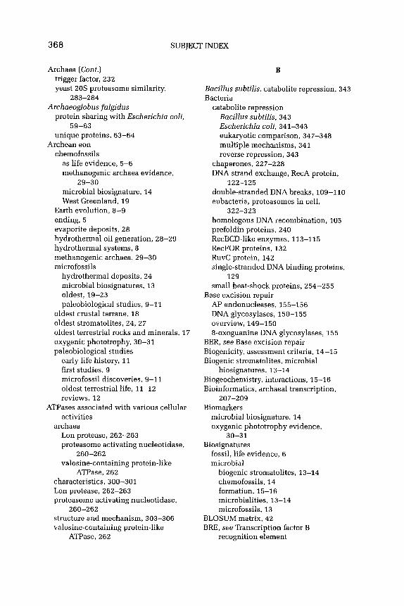
368 SUBJECT INDEX
Archaea (Cont.) trigger factor, 232 yeast 20s proteasome similarity,
283-284 Archaeoglobus fulgidus
protein sharing with Escherichia coli, 59-63
unique proteins, 63-64 Archean eon
chemofossils as life evidence, 5-6 methanogenic archaea evidence,
29-30 microbial biosignature, 14 West Greenland, 19
Earth evolution, 8-Q ending, 5 evaporite deposits, 28 hydrothermal oil generation, 28-29 hydrothermal systems, 8 methanogenic archaea, 29-30 microfossils
hydrothermal deposits, 24 microbial biosignatures, 13 oldest, IQ-23 paleobiological studies, Q-11
oldest crustal terrane, 18 oldest stromatolites, 24, 27 oldest terrestrial rocks and minerals, 17 oxygenic phototrophy, 30-31 paleobiological studies
early life history, 11 first studies, 9 microfossil discoveries, Q-11 oldest terrestrial life, 11-12 reviews, 12
ATPases associated with various cellular activities
archaea Lon protease, 262-263 proteasome activating nucleotidase,
260-262 valosine-containing protein-like
ATPase, 262 characteristics, 300-301 Lon protease, 262-263 proteasome activating nucleotidase,
260-262 structure and mechanism, 303-306 valosine-containing protein-like
ATPase, 262
B
Bacillus subtilis, catabolite repression, 343 Bacteria
catabolite repression Bacillus subtilis, 343 Escherichia coli, 341-343 eukaryotic comparison, 347-348 multiple mechanisms, 341 reverse repression, 343
chaperones, 227-228 DNA strand exchange, RecA protein,
122-125 double-stranded DNA breaks, 109-110 eubacteria, proteasomes in cell,
322-323 homologous DNA recombination, 105 prefoldin proteins, 240 RecBCD-like enzymes, 113-115 RecFOR proteins, 132 RuvC protein, 142 single-stranded DNA binding proteins,
129 small heat-shock proteins, 254-255
Base excision repair AP endonucleases, 155-156 DNA glycosylases, 150-155 overview, 149-150 8-oxoguanine DNA glycosylases, 155
BER, see Base excision repair Biogenicity, assessment criteria, X4-15 Biogenic stromatolites, microbial
biosignatures, 13-14 Biogeochemistry, interactions, 15-16 Bioinformatics, archaeal transcription,
207-209 Biomarkers
microbial biosignature, 14 oxygenic phototrophy evidence,
30-31 Biosignatures
fossil, life evidence, 6 microbial
biogenic stromatolites, 13-14 chemofossils, 14 formation, 15-16 microbialities, 13-14 microfossils, 13
BLOSUM matrix, 42 BRE, see Transcription factor B
recognition element

SUBJECT INDEX 369
C
Carbohydrate transport systems, 361-362
Catabolite repression archaea
carbohydrate transport systems, 361-362
cyclic AMP function, 362 extreme environment adaptation,
348-349 gene regulation, 350 glucose metabolism, 359-361 transcription apparatus,
349-350 bacteria
Bacillus subtilis, 343 Escherichia coli, 341-343 multiple mechanisms, 341 reverse repression, 343
bacterial-eukaryotic comparison, 347-348
eukaryotes animals, 346 multiple mechanisms, 341 plants, 345-346 yeast, 344-345
overview, 339-340 Sulfolobus solfataricus
cr-amylase, 354-355 gene regulatory system, 355-357 a-glucosidase, 352-353 fi-glycosidase, 353-354 mutations, 357-359
Catalysts, protein folding peptidyl-prolyl isomerase, 265-266 protein disulfide isomerase, 266
CCT, see Group II chaperones Cells
Hsp70 role in processes, 234-237 proteasomes
archaea, 323-324 eubacteria, 322-323 eukaryotes, 318-322
CF-2, proteasome inhibition, 309 Chaperonins
group I properties, 247 structure and function, 249-251
group II conformational cycle, 252-253
polypeptide binding site, 253-254 properties, 247-249
overview, 246-247 Chemical fossils, see Chemofossils Chemical inhibitors, proteasomes,
298-299 Chemofossils
as life evidence, 5-6 methanogenic archaea evidence, 29-30 microbial biosignature, 14 West Greenland, Archean eon, 19
Cochaperones, proteasome degradation pathways, 317-318
Complexity, genomic, molecular chaperones, 229-230
Crustal terrane, oldest in Archean eon, 18
Cyclic AMP archaea, 362 yeast catabolite repression, 344-345
D
DARWIN, see Data Analysis and Retrieval with Indexed Nucleotide-Peptide Sequences
Data Analysis and Retrieval with Indexed Nucleotide-Peptide Sequences, homology detection on genome scale, 42
Degradation, proteasomes ubiquitin-dependent pathways,
310-314 ubiquitin-independent pathways,
314-318 Deposits, Archean eon, 24 DNA
archaeal nucleosomes length, 79-80 positioning, 81 template transcription, 81-82 topology, 80-81
archaeal transcription, 205-206 contacting residues, archaeal histones,
91-92 eukaryal nucleosomes, 77-79
DNA alkyltransferases, direct DNA damage reversal, 147-149
DNA annealing proteins, DNA strand exchange, 132-133

370 SUBJECT INDEX
DNA breaks bacteria, 109-110 eukarya, 110 overview, 106-109
DNA damage DNA alkyltransferases, 147-149 photolyase, 147
DNA glycosylases mismatch glycosylases, 153-155 overview, 150-152 8-oxoguanine DNA glycosylases, 155 uracil type, 152-153
DNA packaging, basic problem, 76-77
DNA recombination archaea
DNA break generation, 106-109 DNA strand exchange, 121-127 DNA strand exchange proteins,
131-137 Holliday-junction cleaving enzymes,
137-144 homologous, 103-106,111-121 repair overview, 144-146 single-stranded DNA binding
proteins, 127-131 homologous, DNA break end processing
archaeal proteins, 117, 121 archaeal Sgsl helicase, 116 bacterial RecBCD-like enzymes,
113-115 Escherichia coli RecQ helicase, 115 eukaryal proteins, 116-117 eukaryal Sgsl helicase, 115 overview, 111-113
DNA repair pathways base excision repair, 149-156 direct DNA damage reversal, 147-149 flap endonuclease protein homologues,
160 mismatch repair, 158-160 mutagenesis, 160-161 nucleotide excision repair, 156-158 overview, 146-147,161-162 translesion DNA synthesis, 160-161
DNA strand exchange archaea, RadA protein, 126-127 associated proteins
DNA annealing proteins, 132-133
overview, 131
Rad51 and RadA protein paralogues, 133-136
Rad54 proteins, 136-137 bacterial, RecA protein, 122-125 eukaryal, Rad51 protein, 125-126
DNA synthesis, translesion, 160-161 Double-stranded DNA breaks
archaea, 116-117,121 bacteria, 109-110,113-115 eukarya, 110,115-117 overview, 111-113
Downstream promoter element, 179 DPE, see Downstream promoter element DSBs, see Double-stranded DNA breaks
E
Earth evolution during Archean eon, 8-9 as volcanically active planet, 7-8
Ecosystems, modern as ancient analogs, 16-17
Elongation, archaeal transcription, 191-193
Energy-dependent proteases, 324-325 Environments, extreme
archaeal catabolite repression, 348-349 protein folding, 266-268
Enzymes Holliday-junction cleaving enzymes,
137-144 RecBCD-like enzymes, 113-115
Escherichia coli ancestral proteins, 66 catabolite repression, 341-343 paralogous proteins, 51-57 protein sharing with Archaeoglobus
fulgidus, 59-63 RecQ helicase, 115 single-stranded DNA binding proteins,
129 unique proteins, 63-64
Eubacteria, proteasomes in cell, 322-323 Eukarya
catabolite repression, 341, 344-348 chaperones, 228-229 DNA breaks, 110,115-117 DNA end processing genes, 116-117 DNA recombination, 105, 115-117 DNA strand exchange, 125-126

SUBJECT INDEX 371
histones, 77-79 molecular chaperones, 228-229,
240-241 mRNA, 320-322 nucleosome DNA, 77-79 nucleosomes, 77-79 prefoldin, 238,241-244 prefoldin-group II chaperone
concurrence, 240-241 proteasomes, 285288,318-322 Rad52 protein, 132-133 Rad55-57 complex, 133-134 RPA complex, 129-130 Sgsl helicase, 115 single-stranded DNA binding proteins,
129-130 small heat-shock proteins, 254-255 ubiquitin-dependent proteasome
degradation, 310-311 ubiquitin-independent proteasome
degradation, 314 Evaporite deposits, Archean eon, 28 Evolution
Earth during Archean eon, 8-9 genome and proteins, 43-44 histones, 96 paralogous proteins module, 47-50
Evolutionary biology, genomics era, 39-40 Experimental models, archaeal histones
and nucleosomes, 85-86 Extreme ecosystems, as ancient ecosystem
analogs, 16-17 Extremophiles, protein folding
adaptation, 266-267 molecular chaperone requirements,
267-268 oxidative stress, 268
F
Flap endonuclease proteins, homologues, DNA repair pathways, 160
Fossil biosignatures, as life evidence, 6 Fossils, Archean eon
chemofossils as life evidence, 5-6 methanogenic archaea evidence,
29-30 microbial biosignature, 14 West Greenland, 19
microfossils hydrothermal deposits, 24 microbial biosignatures, 13 oldest, 19-23 paleobiological studies, 9-11
G
Genes catabolite repression, 350, 355-357 divergence
genome and protein evolution, 43-44 genomes, 44-47
duplication, genome and protein evolution, 43-44
transcription differential nucleosome formation,
206 DNA structure, 205-206 global regulators, 206-207 haloarchaeal initiation factor
multiplication, 203-204 modified initiation factors, 204 overview, 203 specific regulation, 209-212 TBP interacting proteins, 204-205
Genomes complexity, molecular chaperones,
229-230 evolution, gene duplication and
divergence, 43-44 gene divergence, 44-47 homology detection, 41-43
Genomics, evolutionary biology, 39-40 Glucose, metabolism in archaea, 359-361 ol-Glucosidase, Sulfolobus solfataricus
catabolite repression, 352-353 /3-Glycosidase, Sulfolobus solfataricus
catabolite repression, 353-354 GroEL, 20s proteasome similarity, 282 GroEL-GroES, structure and function,
249-251 Group II chaperones, prefoldin
concurrence, 240-241
H
Haloarchaea, initiation factor multiplication, 203-204

372 SUBJECT INDEX
Heat-shock proteins Hsp70,234-237 Hsp90,263 HsplOO-Clp family, 259-260 small, see Small heat-shock proteins
Histones archaea
as experimental models, 85-86 natural variants, 93-94 overview, 83-85 structure details, 85 transcription role, 201-202
archaea, Methanothermus fervidus variants
conserved structure, 92 dimer-dimer interaction, 92-93 dimer hydrophobic core
substitutions, 89-90 dimer structural stability, 90 DNA-contacting residues, 91-92 overview, 86-89
eukaryal nucleosomes, 77-79 evolution, 96 nonhistone histone fold-containing
proteins, 94-96 Holliday-junction cleaving enzymes, DNA
recombination, 137-144 Homologous DNA recombination, break
end processing archaeal proteins, 117, 121 archaeal Sgsl helicase, 116 bacterial RecBCD-like enzymes,
113-115 eukaryal proteins, 116-117 eukaryal Sgsl helicase, 115 overview, 11 l-l 13
Homology, detection on genome scale, 41-43
Hsp, see Heat-shock proteins HTH domain, paralogous proteins, 57 Human, catabolite repression, 346 Hydrocarbon droplets, generation during
Archean eon, 28 Hydrolases, 205 proteasomes as, 290-291 Hydrolysis, peptide bonds, 20s
proteasomes, 288-294 Hydrothermal deposits, Archean eon
microfossils, 24 Hydrothermal systems
Archean eon, 8,28-29 life origination, 6-7
I
Immunoglobulin, chaperones, 258 Initiator element, 178-179 Isotopes
evaporite deposit evidence, 28 microbial biosignature, 14
K
Kinetics, catabolite repression-regulated genes, 356-357
Kromberg Formation, Archean eon microfossils, 21-22
L
Last universal common ancestor, paralogy, 40
Lon protease, properties, 262-263 LUCA, see Last universal common
ancestor
M
Maltose transport systems, Sulfolobus shibatae, 361-362
Messenger RNA, eukaryal proteasome levels, 320-322
Metabolic pathways, Archean eon evaporite deposits, 28
Methanogenic archaea, chemofossil evidence, 29-30
Methanothermus fervidus histones, 85 histone variants
conserved structure, 92 dimer-dimer interaction, 92-93 dimer hydrophobic core
substitutions, 89-90 dimer structural stability, 90 DNA-contacting residues,
91-92 overview, 86-89
Microbial biosignatures biogenic stromatolites, 13-14 chemofossils, 14 formation, 15-16

microbialities, 13-14 microfossils, 13
Microfossils, Archean eon hydrothermal deposits, 24 microbial biosignatures, 13 oldest microfossil, 19-23 paleobiological studies, 9-l 1
Mismatch glycosylases, 153-155 Mismatch repair, DNA repair pathways,
158-160 Models
histones and nucleosomes, 85-86
prefoldin structure, 243-244 Molecular chaperones
AAA protein activity, 306-308 archaea
assisted protein folding, 231 chaperone families, 222-223 characteristics, 227 definition, 220-221 principle of action, 221-222 ribosome-assisted chaperones,
231-233 bacterial chaperones, 227-228 domain distribution, 223 eukaryotic
group II chaperones, prefoldin concurrence, 240-241
overview, 228-229 genomic complexity, 229-230 GroEL, 20s proteasome similarity, 282 heat-shock proteins, see Heat-shock
proteins proteasome degradation pathways,
317-318 protein folding in extremophiles,
267-268 MREll protein, 116-117, 121 Mutagenesis, DNA repair pathways,
160-161 Mutations, catabolite repression-regulated
genes, 357-359
NBS1 protein, DNA end processing, 116-117
NER, see Nucleotide excision repair Ntn hydrolases, see Amino-terminal
hydrolases Nucleosomes
archaea description, 79 differential formation, 206 as experimental models, 85-86 transcription role, 201-202
archaea, DNA length, 79-80 nucleosome positioning, 81 template transcription, 81-82 topology, 80-81
eukaryal nucleosomes, 77-79 Nucleotide excision repair, DNA repair
pathways, 156-158
0
Oil, hydrothermal generation during Archean eon, 28-29
Open reading frames, genome and protein evolution, 43-44
ORFs, see Open reading frames Orthology
basic concepts, 40-41 gene divergence in genomes, 44-47 gene duplication and divergence,
43-44 homology detection on genome scale,
41-43 Oxidative stress, protein folding in
extremophiles, 268 8-Oxoguanine DNA glycosylases, 155 Oxygenic phototrophy, Archean eon,
30-31
P
N
NAC, see Nascent polypeptide-assisted complex
Nascent polypeptide-assisted complex, 232-233
Paleobiology, Archean eon early life history, 11 first studies, 9 information source, 5-6 microfossil discoveries, 9-11 oldest terrestrial life, 11-12 reviews, 12
SUBJECT INDEX 373

374 SUBJECT INDEX
P A M score gene divergence, 46 homology detection on genome scale, 42 paralogous proteins, 55
PAN, see Proteasome activating nucleotidase
Paralogous proteins ancestral, min imum number, 64-66 Archaeoglobus fulgidus unique, 63-64 Escherichia coli-Archaeoglobus
fulgidus sharing, 59-63 Escherichia coli unique, 51-57, 63-64 module, 47-50 Rad51 and RadA proteins, 133-136 relationships, 57-59
Paralogy basic concepts, 40-41 gene divergence in genomes, 44-47 gene duplication and divergence,
43-44 homology detection on genome scale,
41-43 PDI, see Protein disulfide isomerase Peptide aldehydes, proteasome inhibition,
299 Peptides, bond hydrolysis, 20s
proteasomes, 288-294 Peptidyl-prolyl isomerase
archaeal molecular chaperones, 221-222
classes, 265-266 PFD, see Prefoldin Pharmacological inhibitors, proteasomes,
298-299 Phosphorylation, eukaryal proteasome
subunits, 319-320 Photolyase, direct DNA damage reversal,
147 Pilbara Craton, Archean eon microfossils,
19-21 Plants, catabolite repression, 345-346 Polarity, archaeal transcription,
188-190 Polypeptides
binding site in group II chaperonins, 253-254
hydrolysis by proteasomes, 292-293 PPI, see Peptidyl-prolyl isomerase Prefoldin
archaea, 238, 242-244 bacteria, 240
discovery, 237-238 eukaryote, 241-244 group II chaperone concurrence,
240-241 molecular mechanism, 245-246
Preinitiation complex, archaeal transcription, 188-190
Prokaryotes proteasomes, 286, 289 ubiquitin-dependent proteasome
degradation, 3 13 ubiquitin-independent proteasome
degradation, 314-318 Promoter elements, archaeal transcription
elements, 179 initiator element, 178-179 overview, 174 -175 TATA box, 175-177 transcription factor B recognition
element, 177-178 Protease-associated regulators
energy-dependent AAA protein chaperone activities,
306-308 AAA protein structure and
mechanism, 303-306 AAA+ superfamily, 300-301 archaea, 324-325 overview, 299-300 proteasome-associated AAA proteins,
301-303,308 non-AAA+ regulators, 308-310
Proteases, Lon protease, 262-263 Proteasome activating nucleotidase,
260-262 Proteasome-associated AAA proteins
characteristics, 301-303 nonproteolytic roles, 308
Proteasomes archaea
in cell, 323-324 characteristics, 323-324 energy-dependent proteases, 324-325 ubiquitin-dependent degradation
pathway, 310-314 ubiquitin-independent degradation
pathway, 314-318 archaea, 20s proteasome
active sites, 285 additional protein binding, 284-285 assembly and processing, 294-298
SUBJECT INDEX 375
chemical inhibitors, 298-299 composition, 281-282 distribution, 285-288 GroEL similarity, 282 non-AAA’ regulators, 308-310 peptide bond hydrolysis mechanism,
288-294 subunit distribution, 285-288 Thermoplasma acidophilum, 282 yeast-archaea similarity, 283-284 yeast similarity, 283-284
eubacteria, 322-323 eukaryotes
overview, 318-319 protein and mRNA levels, 320-322 subcellular localization, 319 subunit phosphorylation, 319-320
Protein disulfide isomerase molecular chaperones, 221-222 protein folding catalysis, 266
Protein folding catalysts, 265-266 cotranslational folding, 231 de ROVO folding, Hsp70 role,
234-237 extremophiles, 266-268 future work, 268-269 in vitro conditions, 230-231 molecular chaperone-assisted folding,
231 Proteins
ancestral proteins, 64-67 DNA annealing proteins, 332-133 DNA end processing, 116-117 DNA strand exchange, 131-137 Flap endonuclease proteins, 160 gene duplication and divergence,
43-44 MREll protein, 116-117, 121 NBS1 protein, 116-117 nonhistone histone fold-containing
proteins, 94-96 paralogous, see Paralogous proteins present-day vs. ancestral proteins,
66-67 proteasome-associated AAA proteins,
301-303, 308 proteasome levels, 320-322 RAD50,116-117,121 RadSl, 125-126 Rad51 paralogues, 133-136
Rad52,132-133 Rad54,136-137 RadA, 126-127 RadA paralogues, 133-136 RecA, 122-125 RecFOR, 132 RuvC, 142 single-stranded DNA binding proteins,
129-131 sp011,110-111 TATA box binding protein,
180-183 TATA box binding protein interacting
protein, 204-205 ubiquitin-domain proteins, 312 ubiquitin-like modifier proteins, 312 XRS2,116-117
Protein translocation Set-dependent system, 264 %+-dependent translocation system,
264-265 Pyrococcusfuriosus, glucose metabolism,
360
R
RAD50 protein, 116-117.121 Rad51 protein, 125-126 Rad51 protein paralogues, 133-136 Rad52 protein, 132-133 Rad54 protein, 136-137 Rad55-57 complex, 133-134 RadA protein, 126-127 RadA protein paralogues, 133-136 RecA protein, 122-125 RecBCD-like enzymes, 113-115 RecFOR proteins, 132 RecQ helicase, 115 Ribosome-assisted molecular chaperones,
231-233 RNA, messenger, eukaryal proteasome
levels, 320-322 RNA polymerase, archaeal transcription
composition, 194-197 structure and mechanism, 198-201 subunit structures. 197-198
Rock record, first accumulation, 3-5 RPA complex, eukaryotes, 129-130 RuvC protein, Holliday-junction cleaving
enzymes, 142

376 SUBJECT INDEX
Sac-dependent protein translocation system, 264
SELEX technology, nucleosome positioning, 81
Sgsl h&case archaea, 116 eukaryotes , 115
Single-stranded DNA binding proteins archaea, 130-131 bacteria, 129 eukaryal RPA complex,
129-130 Small heat-shock proteins
function, 254-256 immunoglobulin fold, 258 structure, 256-258
Spoll protein, 110-111 SRP-dependent protein translocation
system, 264-265 SSB proteins, see Single-stranded DNA
binding proteins Stromatolites, oldest, Archean eon,
24,27 Sucrose, plant catabolite repression,
345-346 Sulfolobus shibatoe, maltose transport
systems, 361-362 Sulfolobus solfataricus
carbohydrate transport systems, 361 catabolite repression
o-amylase, 354-355 gene regulatory system, 355-357 o-glucosidase, 352-353 p-glycosidase, 353-354 mutations, 357-359
characteristics, 351-352
T
TATA box, 175-177 TATA box binding protein, 180-183 TATA box binding protein interacting
protein, 204-205 TBP, see TATA box binding protein Termination, archaeal transcription,
193-194 Terranes, crustal, oldest in Archean eon,
18
Terrestrial life, Archean eon paleobiological search, 11-12
Terrestrial minerals, Archean eon, 17 Terrestrial rocks, Archean eon, 17 TF, see Trigger factor Thermococcus zilligi, glucose metabolism,
359-360 Thermoplasma acidophilum, 20s
proteasomes, 282 Thermoproteus tenax, glucose
metabolism, 360-361 Topology, nucleosomal DNA, 80-81 Transcription
bioinformatic view, 207-209 catabolite repression apparatus,
349-350 elongation, 191-193 gene regulation
differential nucleosome formation, 206
DNA structure, 205-206 global regulators, 206-207 haloarchaeal initiation factor
multiplication, 203-204 modified initiation factors, 204 overview, 203 TBP interacting proteins, 204-205
gene-specific regulation, 209-212 histones, 201-202 nucleosomes, 201-202 nucleosome templates, 81-82 polarity, 188-190 preinitiation, 191 preinitiation complex formation,
188-190 promoter elements
elements, 179 initiator element, 178-179 overview, 174-175 TATA box, 175-177 transcription factor B recognition
element, 177-178 RNA polymerase
composition, 194-197 structure and mechanism, 198-201 subunit structures, 197-198
termination, 193-194 Transcription factor B, 183-187 Transcription factor B recognition
element, 177-178 Transcription initiation factors

Valosine-containing protein-like ATPase, 262
VAT, see Valosine-containing protein-like ATPase
archaea v factors, 187-188 modified versions, 204 overview, 179-180 TATA box binding protein,
180-183 transcription factor B, 183-187
haloarchaea, multiplication, 203-204 Translocation W
See-dependent system, 264 SRP-dependent system, 264-265
Transport systems, carbohydrates, 361-362
Trigger factor, 232
Western Australia, Archean eon microfossils, 19-21
West Greenland, Archean eon chemofossils, 19
u X
XRS2 protein, DNA end processing, 116-117
UbDs, see Ubiquitin-domain proteins Ubiquitin, proteasome degradation
pathways dependent pathways, 310-314 independent pathways, 314-318 Y
Ubiquitin-domain proteins, 312 Ubiquitin-like modifier proteins, 312 UBls, see Ubiquitin-like modifier proteins Unfoldases, proteasome degradation
pathways, 315 Uracil DNA glycosylases, 152-153 Rad54 proteins, 136-137
Yeast catabolite repression, 344-345 proteasome, archaeal similarity,
283-284
SUBJECT INDEX 377

Volume 40
Microbial Cellulases: Protein Architecture, Molecular Properties, and Biosynthesis
Ajay Singh and Kiyoshi Hayashi
Factors Inhibiting and Stimulating Bacterial Growth in Milk: An Historical Perspective
D. K, O’Toole
Challenges in Commercial Biotechnology. Part I. Product, Process, and Market Discovery
Ale5 Prokop
Challenges in Commercial Biotechnology. Part II. Product, Process, and Market Development
Ales’ Prokop
Effects of Genetically Engineered Microorganisms on Microbial Populations and Processes in Natural Habitats
Jack D. Doyle, Guenther Stotzky, Gwendolyn McClung, and Charles W Hendricks
Detection, Isolation, and Stability of Megaplasmic-Encoded Chloroaromatic Herbicide-Degrading Genes within Pseudomonas Species
Douglas J. Cork and Amjad Khalil
INDEX
Volume 41
Microbial Oxidation of Unsaturated Fatty Acids
Ching ‘I: Hou
Improving Productivity of Heterologous Proteins in Recombinant Saccharomyces cerevisiae Fermentations
Amit Vasavada
Manipulations of Catabolic Genes for the Degradation and Detoxification of Xenobiotics
Rup Lal, Sukanya Lal, l? S. Dhanaraj, and D. M. Saxena
Aqueous Two-Phase Extraction for Downstream Processing of Enzymes/Proteins
K. S. M. S. Raghava Rao, N. K. Rastogi, M. K. Gowthaman, and N. G. Karanth
Biotechnological Potentials of Anoxygenic Phototrophic Bacteria. Part I. Production of Single Cell Protein, Vitamins, Ubiquinones, Hormones, and Enzymes and Use in Waste Treatment
Ch. Sasikala and Ch. V Ramana
Biotechnological Potentials of Anoxygenic Phototrophic Bacteria. Part II. Biopolyesters, Biopesticide, Biofuel, and Biofertilizer
Ch. Sasikala and Ch. V Ramana
INDEX
Volume 42
The Insecticidal Proteins of Bacillus thuringiensis
P Ananda Kumar, R. P Sharma, and I? S. Malik
Microbiological Production of Lactic Acid
John H. Litchfield
CONTENTS OF PREVIOUS VOLUMES
379

380 CONTENTS OF PREVIOUS VOLUMES
Biodegradable Polyesters Ch. Sasikala
The Utility of Strains of Morphological Group II Bacillus
Samuel Singer
Phytase Rudy J. Wodzinski and
A. H. J. Ullah
INDEX
Volume 43
Production of Acetic Acid by Clostridium thermoaceticum
Munir Cheryan, Sarad Parekh, Minish Shah, and Kusuma Witjitra
Contact Lenses, Disinfectants, and AcanthamoeboKeratitis
Donald G. Ab?n wxd Manai M. Gabriel
Marine Microorganisms as a Source of New Natural Products
V S. Bernan, M. Greenstein, and W M. Maiese
Stereoselective Biotransformations in Synthesis of Some Pharmaceutical Intermediates
Ramesh N. Pate1
Microbial Xylanolytic Enzyme System: Properties and Applications
Pratima Bajpai
Oleaginous Microorganisms: An Assessment of the Potential
Jacek Leman
INDEX
Volume 44
Biologically Active Fungal Metabolites Cedric Pearce
Old and New Synthetic Capacities of Baker’s Yeast
I? D’Arrigo, G. Pedrocchi-Fantoni, and S. Servi
Investigation of the Carbon- and Sulfur-Oxidizing Capabilities of Microorganisms by Active-Site Modeling
Herbert L. Holland
Microbial Synthesis of D-Ribose: Metabolic Deregulation and Fermentation Process
I! de Wulf and E. f Yandamme
Production and Application of Tannin Acyl Hydrolase: State of the Art
P K. Lefia and B. K. Lonsane
Ethanol Production from Agricultural Biomass Substrates
Rodney J. Bothast and Badal C. Saha
Thermal Processing of Foods, A Retrospective, Part I: Uncertainties in Thermal Processing and Statistical Analysis
M. N. Ramesh, S. G. Prapulla, M. A. Kumar, and M. Mahadevaiah
Thermal Processing of Foods, A Retrospective, Part II: On-Line Methods for Ensuring Commercial Sterility
M. N. Ramesh, M. A. Kumar, S. G. Prapulla, and M. Mahadevaiah
INDEX
Volume 45
One Gene to Whole Pathway: The Role of Norsolorinic Acid in Aflatoxin Research
J. W. Bennett, I?-K. Chang and D. Bhatnagar
Formation of Flavor Compounds in Cheese
I! E Fox and J. M. Wallace

CONTENTS OF PREVIOUS VOLUMES 381
The Role of Microorganisms in Soy Sauce Production
Desmond K. O’Toole
Gene Transfer Among Bacteria in Natural Environments
Xiaoming Yin and G. Stotzky
Breathing Manganese and Iron: Solid-State Respiration
Kenneth H. Nealson and Brenda Little
Enzymatic Deinking Pratima Bajpai
Microbial Production of Docosahexaenoic Acid (DHA, C22:6)
Ajay Singh and Owen l? Word
INDEX
Volume 46
Cumulative Subject index
Volume 47
Seeing Red: The Story of Prodigiosin J. W Bennett and Ronald Bentley
Microbial/Enzymatic Synthesis of Chiral Drug Intermediates
Ramesh N. Pate1
Recent Developments in the Molecular Genetics of the Erythromycin-Producing Organism Saccharopolyspora erythraea
Thomas J. Vanden Boom
Bioactive Products from Streptomyces Vladisalv Behnl
Advances in Phytase Research Edward J. Mullaney, Catherine B. Daly
and Abdul H. J. Ullah
Biotransformation of Unsaturated Fatty Acids of industrial Products
Ching T Hou
Ethanol and Thermotolerance in the Bioconversion of Xylose by Yeasts
Thomas W Jeffries and Yong-Su Jin
Microbial Degradation of the Pesticide Lindane (y-Hexachlorocyclohexane)
Brajesh Kumar Singh, Ramesh Chander Kuhad, Ajay Singh, K. K. Tripathi, and P K. Ghosh
Microbial Production of Oligosaccharides: A Review
S. G. Prapulla, V Subhaprada, and N. G. Karanth
INDEX
Volume 48
Biodegredation of Nitro-Substituted Explosives by White-Rot Fungi: A Mechanistic Approach
Benoit Van Aken and Spiros N. Agathos
Microbial Degredation of Pollutants in Pulp Mill Effluents
Pratima Bajpai
Bioremediation Technologies for Metal-Containing Wastewaters Using Metabolically Active Microorganisms
Thomas Pumpel and Kishorel M. Paknikar
The Role of Microorganisms in Ecological Risk Assessment of Hydrophobic Organic Contaminants in Soils
C. J. A. MacLeod, A. W J. Morriss, and K. ‘I: Semple
The Development of Fungi-A New Concept Introduced By Anton de Bary
Gerhart Drews

382 CONTENTS OF PREVIOUS VOLUMES
Bartolomeo Gosio, 1863-1944: An Appreciation
Ronald Bentley
INDEX
Laboratory Birproduction of Paralytic Shellfish Toxins in Dinoflagellates
Dennis P H. H&h, Dazhi Wang, and Gary H. Chang
Metal Toxicity in Yeasts and the Role of Oxidative Stress
Volume 49
Biodegredation of Explosives Susan J. Rosser, Amrik Basran,
Emma1 R. Travis, Christopher E. French, and Neil C. Bruce
S. I/: Avery
Foodbourne Microbial Pathogens and the Food Research Institute
M. EJlin Doyle and Michael W Pariza
Alexander Flemin and the Discovery of Penicillin
Biodiversity of Acidophilic Prokaryotes Kevin B. Hallberg and D. Barrie
Johnson
J. IN Bennett and King-Thorn Chung
INDEX