review thalassemia in southeast asia: problems · pdf filereview thalassemia in southeast...
TRANSCRIPT

REVIEW
THALASSEMIA IN SOUTHEAST ASIA: PROBLEMS AND STRATEGY FOR PREVENTION AND CONTROL
Suthat Fucharoen and Pranee Winichagoon
Thalassemia Center, Division of Hematology, Department of Medicine, Faculty of Medicine Siriraj Hospital and Institute of Sciences and Technology for Development, Mahidol University, Bangkok 10700, Thailand.
Abstract. In Southeast Asia a-thalassemia, ~-thalassemia, hemoglobin (Hb) E and Hb Constant Spring are prevalent. The gene frequencies of a -thalassemia reach 30-40'X, in Northern Thailand and Laos. J3-Thalassemia gene frequencies vary between I and 9%. Hb E is the hallmark of Southeast Asia attaining a frequency of 50-60% at the junction of Thailand, Laos, and Cambodia. Hb Constant Spring gene frequencies vary between I and 8%. These abnormal genes in different combinations lead to over 60 different thalassemia syndromes. The four major thalassemic diseases are Hb Bart's hydrops fetalis (homozygous a -thalassemia I), homozygous J3 -thalassemia, ~-thalassemialHb E and Hb H diseases. The molecular basis of most of these abnormal genes have been recently described. Therefore, it is possible to set a strategy for prevention and control of thalassemia which includes population screening for heterozygotes, genetic counseling and fetal diagnosis with selective abortion of affected pregnancies.
INTRODUCTION Hemoglobinopathies are the most common genetic disorders among the people living in South
Southeast Asia consists of 10 countries with a east Asia. a -Thalassemia, J3-thalassemia, hemototal population of about 400 million. The ethnic globin (Hb) E and Hb Constant Spring (CS) are origins of people living in these countries are very prevalent. The gene frequencies of f3 -thalassemia heterogeneous. The Mon-Khmer and Tai language reach 30-40'1<, in Northern Thailand and Laos. speaking people occupy Thailand, Laos, Cambodia f3 -Thalassemia gene frequencies vary between I and some parts ofVietnam, Myanmar and Malaysia. and 9%. Hb E is the hallmark of Southeast Asia, The west includes the Burmese (Tibeto-Burman) attaining a gene frequency of S(}{,()'/'o at the junction and the northeast the Vietnamese (Austro-Asia of Thailand, Laos, and Cambodia. Hb Constant tic). The Malayopolynesians (Austronesian) live Spring frequencies vary between I and 8% (Wasi, in Malaysia, Indonesia, Brunei, the Philippines 1983; Fucharoen and Winichagoon, 1987). These and a number of Pacific island nations. Chinese and abnormal genes in different combinations lead to Indians are relative new-comers, spread throughout over 60 thalassemia syndromes (Wasi et ai, 1969). the region. The two major a-thalassemic diseases are Hb
Bart's hydrops fetalis (homozygous a -thalassemia The major public health problems until now,
I) and Hb H disease. Homozygous /3-thalassemia in this region, were infectious diseases and malnuand f3 -thalassemialHb E are major /3 -thalassemic
trition. During these last few years a number of syndromes in this region. countries in Southeast Asia such as Singapore,
Malaysia and Thailand have enjoyed good success in economic growth. The health care of people in these countries has been improved and the number ALPHA THALASSEMIA of infant deaths from infection and malnutrition has decreased. Genetic disorders such as thalassemia The molecular defects of a-thalassemias are and other hemoglobinopathies have gradually heterogeneous (Higgs et ai, 1989). a -Thalassemia emerged as a major residual cause of infant and in Southeast Asia is most often due to a -globin childhood mortality and morbidity. gene deletion (Lie Injo et al. 1982; Winichagoon et
Vol 23 No 4 December 1992 647

SOUTHEAST ASEAN 1 TROP MED PUBLIC HEALTH
ai, 1984; Fischel-Ghodsian et ai, 1988), The severe form of a-thalassemia, a-thalassemia I, involves a deletion of the duplicated a-globin genes whereas the milder form, a -thalassemia 2, has one a -globin gene left functioning on the chromosome. Hemoglobin Constant Spring (CS) is a variant with elongated a -globin chains. Mutation at the termination codon of the a 2-globin gene results in an unstable mRNA and only small amounts of Hb CS are produced. Thus Hb CS has an a-thalassemia 2-like effect.
Hemoglobin Bart's hydrops fetalis
There is no a-globin chain production in homozygous a -thalassemia I resulting in the most serious form of thalassemia disease, Hb Bart's hydrops fetalis. Hemoglobin electrophoresis shows large amounts of Hb Bart's ( Y 4) and about 10-15(1., Hb Portland ( S2 Y 2) without Hb A. The fetus dies in utero or soon after birth because Hb Bart's does not release O2 to the tissues. The affected fetuses are hydropic with severe growth retardation, and abnormal development of vital organs such as brain and lung contributes to the severe morbidity that makes the condition incompatible with life. Maternal complications such as toxemia of pregnancy have been observed in almost all pregnancies (Vaeusorn et ai, submitted). Ultrasonography provides unambiguous detection of Hb Bart's hydrops fetalis at 18-20 weeks of pregnancy (Kanokpongsakdi et ai, 1990). DNA diagnosis from chorionic villi or amniotic fluid fibroblasts can detect Hb Bart's hydrops fetalic as early as 8-10 weeks gestation (Fucharoen et ai, 1991 a). Therapeutic abortion is suggested for the fetus diagnosed as having this disease.
Hb H disease
Two common genotypes lead to the occurrence of Hb H disease in Southeast Asia, ie a-thalassemia 1/ a-thalassemia 2 and a -thalassemia IIHb CS (Fucharoen et ai, 1988a). Both genotypes have similar clinical manifestations although the latter is more anemic. Usually the patients have no symptoms and, if they are in a steady state, do not need treatment. Correct diagnosis and understanding are necessary, otherwise it causes anxiety and mishandling. At birth the cord blood of the babies with Hb H disease contains about 25% Hb Bart's. In adults the hemoglobin phenotype is A + H, and Hb H constitutes 5-15% of the total hemoglobin. After mixing a drop of blood with methylene blue or
brilliant cresyl blue for an hour, multiple intraerythrocytic inclusion bodies are detected. In general, physical development of the patient is normal and there is no thalassemic facies. Only mild anemia and jaundice may be noticeable. However, the patients may develop severe anemia and jaundice after acute infection. There is a sudden fall of hemoglobin levels with associated symptoms of acute infection. Proper management of hemolytic crisis is important. Blood transfusion should be given along with treatment for infection and body temperature should be normalized as quickly as possible to reduce induction of Hb H precipitation occurring as the result of fever.
Hb AE Bart's and Hb EF Bart's diseases
This is the result of complex gene interactions of a- and J3-thalassemias. The former occurs as a result of inheritance of three specific genes, a -thalassemia 1/ a-thalassemia 2 + Hb E or a -thalassemia IIHb CS + Hb E. The symptoms are usually similar to Hb H disease. The typical characteristic of this disease is the hemoglobin phenotype of A + E + Bart's in which Hb E constitutes 13-15% of total hemoglobin. Approximately 5-6% of RBC can be demonstrated to contain Hb H inclusions, although Hb H is too low to be demonstrated by hemoglobin electrophoresis (Thonglairoam et ai, 1989).
EF Bart's disease is an uncommon form of thalassemia intermedia resulting from the co-inheritance of the abnormal a - and J3-globin genes. DNA mapping and hemoglobin electrophoresis indicate that there are four genotypes, involving 5 abnormal globin genes, responsible for this thalassemia syndromes (Fucharoen et ai, 1988b). Hemoglobin electrophoresis revealed the typical phenotypes ofE + F + Bart's in which Hb E constitutes about 85%. The majority of the patients had Hb CS + E + F + Bart's derived from a-thalassemia IIHb CS + homozygous Hb E.
BETA THALASSEMIA
/3 -Thalassemias are very heterogeneous, both in the molecular defects and the clinical manifestations (Weatherall et ai, 1981; Wainscoat et ai, 1982, 1983). In Southeast Asia J3"-thalassemia far exceeds J3 + -thalassemia, and I:f-thalassemial Hb E disease is much more common than homo-
Vol 23 No 4 December 1992 648

THALASSEMIA PREVENTION AND CONTROL
CENTRAL CHINA
(Sichuan )
Codons 11 / ~2 30%
Codon 17 30%
-29 ATA 19%
IVS U-654 11%
-28 ATA
BURMA IVS 1-5 29%
IVS 1-1 Codons 41 / 42
619 hl'del. 2200 Codons 8 '9 20"0
IVS 1-1 14'0 MALAYSlA C"dolls 41 42 12"0 IVS 1-5
Codon 19 Codons 41 i 42
IVS 1-1
IVS U-654
SOUTH CHINA
(Guangxi)
Codons 41 / 42
Codon 17
Codons 71172 -28 ATA
15%
12%
7% 7'}
46%
-28 ATA
Unknown
INDONESIA
IVS 1-5
HONG KONG
Codons ~1/42 48%
IVS ll-654 22%
Codon 17 10%
-28 ATA 8% Unknown 12%
43% 14% 10%
6% 13%
MELANESlA
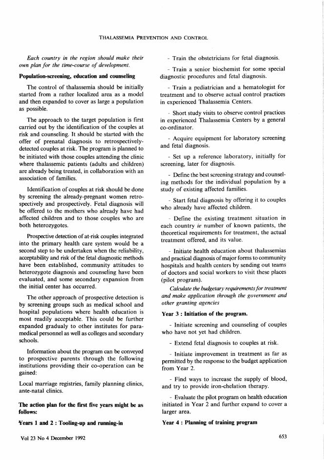
Fig I-Summary of the common 13 -thalassemia mutations in Southeast Asian countries. There are specific patterns of common .B -thalassemia genes in each region. The common /3 -thalassemia genes among the Thai and the Chinese are similar. The mutations among the Burmese (Myanmar) are a partial mixture of the Indian and the Chineselfhai patterns. The Malaysian and Indonesian patterns reflect Chinese, Indian and Malayo-Polynesian
ethnic origins.
zygous ~ -thalassemia. (3 -Thalassemia in Southeast Asia may result from different molecular mechanisms, most of which are base substitutions or small deletions or insertions of one or two nucleotides (Fucharoen Sp et ai, 1989; Laig et ai, 1989; Lie Injo et ai, 1989; Petmitr et ai, 1989; Yang et ai, 1989; Thein et ai, 1990; Winichagoon et ai, 1990). Moreover, (3 -thalassemia with the same genetic defect may reside on the chromosome with different DNA polymorph isms. However, it has been found that l3-thalassemia mutations are relatively population specific, ie each ethnic group has its own set of common mutants (Fig I). Hemoglobin E occurs from a mutation at position 26 of the (3 -globin chain (glu --> Iys). The abnormal gene also results in reduced amounts of f3E-mRNA and in synthesis of 13 E-globin chains (Traeger et ai, 1982). Therefore, Hb E has a mild 13 + -thalassemia phenotype. Homozygous Hb E is asymptomatic. There is no
evidence of anemia and jaundice, and hepatosplenomegaly is not observed in the majority of cases. Homozygous l3-thalassemia is a severe disease and most of the patients die in the pediatric age group. I3-Thalassemia/Hb E has a wide spectrum of severity; the hemoglobin levels range between 3 and 13 g/dl (Winichagoon et ai, 1985; Fucharoen et ai, 1988c). The patients with a very low hemoglobin levels have defective physical development, thalassemic facies, jaundice and hepatosplenomegaly. Most of the patients receive minimal treatment because of the poor resources in these countries. Thus the pathophysiology of the patients differs from the treated homozygous l3-thalassemia cases in developed countries.
Excess a -globin chains causes ineffective erythropoiesis which leads to anemia and increased hemolysis leads to jaundice. Severe anemia results
Vol 23 No 4 December 1992 649

SOUTHEAST ASEAN J TROP MED PUBLIC HEALTH
in massive erythropoiesis which leads to several consequences such as defective physical development and thalassemic facies resulting from the expansion of the bone cavity. Extramedullary hemopoiesis leads to enlarged liver and spleen. The massive erythropoiesis also induces increased iron absorption from the intestine which results in iron overload (Pootrakul et ai, 1981, 1988). Degenerative changes followed iron overload can cause pathophysiology such as cardiac failure, hepatic fibrosis, and other complications as occur in other thalassemia major cases.
Severe thalassemic patients may develop hypersplenism in which splenectomy must be considered. However, many complications are observed among splenectomized cases. Severe infections are major causes of death in splenectomized patients. The causative organisms may differ in children and adult patients and the pattern of infections may differ among different populations. Splenectomized patients have frequently been found to have low arterial oxygen saturation or hypoxemia. Thromboembolism in the pulmonary arteries is frequently detected in adult patients at autopsy. Hypoxemia may be related to platelet aggregation in the pulmonary circulation and treatment with plateletaggregation inhibitors such as aspirin is beneficial to patients with reversible hypoxemia (Sonakul et ai, 1980; Fucharoen et ai, 1981).
At present most children with major thalassemic diseases such as homozygous f3 -thalassemia and f3 -thalassemialHb E born in the villages die undiagnosed, or if they are diagnosed, they are sent home from the local health center untreated. This is mainly because of the lack of policy and resources for maintaining children with chronic disease like thalassemia. However, those patients who can afford to attend the hospital with specialists, eg a university hospital, are still treated by a minimal transfusion program and only a few of them receive regular iron chelation. Most of these thalassemia major patients die before the age of ten because of anemia and infection.
STRATEGIES FOR THE PREVENTION AND CONTROL OF THALASSEMIC
DISEASES IN SOUTHEAST ASIA
Prevention and control of thalassemias in the entire population of any country requires a well planned program to establish the epidemiology of
the disorders and education to raise the awareness of genetic risk in the medical profession and the population at large. Accurate diagnosis and advice should be provided to the carriers, the best possible management should be made available to the affected patients. Prevention of further thalassemic offspring in the case of couples at risk can be planned by prenatal diagnosis. This approach is cost-effective, and is proving remarkably successful in reducing the frequency of thalassemia in Mediterranean countries such as Cyprus, Greece and Italy (Kuliev, 1988; Cao and Rosatelli, 1988; Loukopoulos et ai, 1988; Angastiniotis, 1990). In Thailand it has been estimated that the total cost for treating four major thalassemic diseases namely Hb Bart's hydrops fetalis, homozygous IHhalassemia, f3 -thalassemialHb E and Hb H diseases is about (US$) 220 million per year (Parnsatienkul, 1990). But elsewhere the cost of a total prevention program has been demonstrated to be 1I5th to IIlOth of the cost of treating the existing affected patients (Modell and Berdoukas, 1984). Such an approach to controlling the diseases will permit a commitment to provide the best treatment for the thalassemics. Appropriate strategy for the prevention and control of thalassemia includes the following activities:
(1) Assessment of the magnitude of the problems
The magnitude of thalassemia problems in each country can be assessed from the frequency and incidence of the mutant genes, the severity of the illnesses and the concern in the community. Prevalence alone does not always reflect the magnitude of the problems. For example,' a -thalassemia 2 and Hb E which are very common in Southeast Asia, without the co-existence of a-thalassemia I and f3-thalassemia genes do not lead to disease states. Before planning a thalassemia control program in any country, the magnitude of the problems including epidemiologic survey and the natural history of specific disorders should be well characterized.
(2) Strategies for the control of thalassemia in Southeast Asia
Control strategies consist of prevention of new births of thalassemics and proper treatment of the existing patients.
A. Prevention: Prevention of new births of thalassemics can be carried out by genetic counseling in
Vol 23 No 4 December 1992 650

THALASSEMIA PREVENTION AND CONTROL
combination with prenatal diagnosis. The high risk couples should be identified at the primary health care level and be referred to a regional, well equiped center for proper counseling and prenatal diagnosis.
Laboratory diagnosis and screening test for thalassemia
In Southeast Asia thalassemias with iron overload and patients with iron deficiency co-exist in the same population. Thalassemias with iron overload need iron chelation while those with iron deficiency require iron supplementation or fortification. If the two conditions are not clearly discriminated the iron-overloaded thalassemics will be adversely affected. Moreover, a screening test to discriminate thalassemia from iron deficiency anemia and normal individual is needed. A combination of tests including the one tube osmotic fragility, the DCIP (dichlorophenolindophenol) dye test to detect unstable Hb E, and the plasma ferritin screening test developed by Pintar et al (1982) have proved to be appropriate for the discrimination of these conditions (Fucharoen et ai, 1989).
The screening one tube osmotic fragility test using 0.36'1"0 buffered saline is sensitive enough to detect almost all cases of a -thalassemia I and /3 -thalassemia trait while over 96% of normal red cells are hemolysed (Kattamis et ai, 1981). The red cells of patients with thalassemia and iron deficiency anemia are decreased in osmotic fragility. However, in the vicinity of \0 gldl hemoglobin level the red cell osmotic fragility in most cases of iron deficiency anemia is normal or slightly decreased.
The blue dye DCIP can be used to detect unstable hemoglobin, including Hb E which lacks stability because the weak a 1 /31 contact. Hb E molecules dissociate and precipitate upon incubation with the dye at 3rc. No precipitation or cloudy appearance is observed in normal, iron deficiency anemia and thalassemia trait subjects (Kulapongs et ai, 1976).
Recently, detection of thalassemia and certain abnormal hemoglobins such as Hb E has become possible by immunologic technics using specific monoclonal antibodies. Chui et al (1989) demonstrated that the t -globin gene in adults with a-thalassemia 1 (SEA genotype with about 20 kb deletion of the a -gene cluster) is still functioning.
Vol 23 No 4 December 1992
Detection of the minute amount of I,;' -globin chain can be performed by a simple slot blot immunobinding and enzyme-linked immunosorbent assay (ELISA) using specific monoclonal antibody (Fucharoen et ai, 1991 b). Elevated Hb A2 in /3 -thalassemia trait also can be identified using a monoclonal antibody specific for the
I,;' -globin chain (Shyamala et ai, 1991). Quantitative measurement of Hb A2 can be performed by a simple and rapid ELISA technic.
Although some modification and improvement of the laboratory technics are required, operational research is needed to evaluate how such simple technologies can be transferred to real use in rural areas, considering the issues of quality control, supplies, economics and manpower.
Genetic counseling
Genetic counseling is a communication process dealing with diseases associated with the risk of occurrence of a genetic disorders in a family. The main purpose is to help the individual or the family to become familiar with the medical problems. These include the strategy for diagnosis, the clinical course of the disorders and the management which is available. Counseling is a special skill that depends on training and the ability to communicate with people. Since one counseling session is not enough to transfer all the information, certain backup information such as booklets, leaflets and video tapes are needed. Timing of the counseling is also very important. Although pre-marital counseling is preferred, at the moment the counseling is usually performed after the birth of an affected child. Another problem which is also very important is the development of appropriate manpower. Specialists, especially clinical geneticists, are not available in sufficient numbers; the counselors may be selected from nurses or non-medically trained personel and trained for their suitability in each country.
B. Treatment: Treatment for the thalassemia is mostly palliative. Treatment aiming at cure of the disease is under development, with some promising results.
Palliative treatment
The palliative treatment for thalassemia consists of blood transfusion, reduction of iron overload and treatment of other complications.
651

SOUTHEAST ASEAN J TROP MED PUBLIC HEALTH
I. Blood transfusion: At the moment it is not possible to give adequate blood transfusion to thalassemics in all Southeast Asian countries with high prevalence of the disease. Better organization for blood donation and supplies, possibly including some from developed countries are required.
2. Reduction of iron overload: The only available iron chelator in use at present is desferrioxamine (DesferafH ) produced by Ciba-Geigy. It is very effective and highly specific as an iron chelator and is widely used in developed countries. However, DisferaI' is not the ideal iron chelator because the administration needs 10-12 hours subcutaneous infusion, It is also very expensive and beyond the economic possibility of most countries in Southeast Asia to afford. The ideal iron chelator should have the following properties:
- high affinity for ferric iron with high degree of selectivity
- low toxicity - rapid excretion of the iron complex, with no
effect on the redistribution of iron from the storage, less harmful pool, to deposit in the vulnerable organs
- oral route of application - inexpensive
New iron chelators such as a-ketohydroxy pyridine (L1, CP94 etc), pyridoxal isonicotinoyl hydrazone (PIH and its derivatives) are under development with a very promising results. It is expected that new oral iron chela tors which are cheap enough will be available for the thalassemics in a few years.
3. Treatment of other complications: Non- or minimal-transfusion thalassemics in Southeast Asia develop frequent complications such as infections, cardiac failure, pericarditis, hypoxemia, gallstones, abnormal trace elements metabolism. To provide suitable guidelines for management, the epidemiology and pathophysiology of these complications should be well characterized. Some of these problems are not striking or not observed among the thalassemics in developed countries and thus are not much investigated.
Treatment aiming at cure
At the moment there are three main approaches to the cure of thalassemia including bone marrow transplantation, hemoglobin F switching, and
gene transfer. In the future these approaches may lead to development of suitable drugs and technics which can be applied for the cure of thalassemics. This development, of course, will have to take into account economic considerations in relation to the potential usefulness in countries in this region.
RECOMMENDATIONS FOR PLAN OF ACTION
The prevention of genetic diseases at the community level is a new enterprise in health care. It needs the new development of a new structure. Although this must be based at the primary health care level, the support of the health authorities is very important. Recent advances in science and technology make it possible to plan thalassemia control, and for effective control of thalassemia in this region the following action plan is proposed :
(I) Form a national and regional hemoglobinopathy or thalassemia working group to achieve national and regional co-operation on information gathering, research and development of effective approaches to thalassemia. The group may include hematologists, geneticists, internists, pediatricians, obstetricians, nurses, epidemiologists, representatives of a Parents and Patients' Association. Such a group should meet regularly to define problems, policy making and evaluate the protocols, data and progress. The group from each country should exchange information and report annually to the Ministry of Public Health.
(2) Carry out studies on the epidemiology, pathophysiology, natural history, and management of the major thalassemic syndromes in the region.
(3) Develop a simple screening test for the detection of thalassemia carriers. The technic should be simple enough to be suitable for use at the small district hospital level.
(4) Prenatal diagnosis, a necessary "option" for at-risk couples should be established at the beginning of the program. Trained staff should include obstetricians to perform the fetal tissue sampling and scientists to analyse the samples by either DNA or protein technics (Fucharoen et ai, 199Ia).
(5) The attitude of the people in each community to genetic information and genetic risks needs to be evaluated.
Vol 23 No 4 December 1992 652
THALASSEMIA PREVENTION AND CONTROL
Each country in the region should make their own plan for the time-course of development.
Population-screening, education and counseling
The control of thalassemia should be initially started from a rather localized area as a model and then expanded to cover as large a population as possible.
The approach to the target population is first carried out by the identification of the couples at risk and counseling. It should be started with the offer of prenatal diagnosis to retrospectivelydetected couples at risk. The program is planned to
be initiated with those couples attending the clinic where thalassemic patients (adults and children) are already being treated, in collaboration with an association of families.
Identification of couples at risk should be done by screening the already-pregnant women retrospectively and prospectively. Fetal diagnosis will be offered to the mothers who already have had affected children and to those couples who are both heterozygotes.
Prospective detection of at-risk couples integrated into the primary health care system would be a second step to be undertaken when the reliability, acceptability and risk of the fetal diagnostic methods have been established, community attitudes to heterozygote diagnosis and counseling have been evaluated, and some secondary expansion from the initial center has occurred.
The other approach of prospective detection is by screening groups such as medical school and hospital populations where health education is most readily acceptable. This could be further expanded gradualy to other institutes for paramedical personnel as well as colleges and secondary schools.
Information about the program can be conveyed to prospective parents through the following institutions providing their co-operation can be gained:
Local marriage registries, family planning clinics, ante-natal clinics.
The action plan for the first five years might be as follows:
¥ears 1 and 2 : Tooling-up and running-in
Vol 23 No 4 December 1992
- Train the obstetricians for fetal diagnosis.
- Train a senior biochemist for some special diagnostic procedures and fetal diagnosis.
- Train a pediatrician and a hematologist for treatment and to observe actual control practices in experienced Thalassemia Centers.
- Short study visits to observe control practices in experienced Thalassemia Centers by a general co-ordinator.
- Acquire equipment for laboratory screening and fetal diagnosis.
- Set up a reference laboratory, initially for screening, later for diagnosis.
- Define the best screening strategy and counseling methods for the individual population by a study of existing affected families.
- Start fetal diagnosis by offering it to couples who already have affected children.
- Define the existing treatment situation in each country ie number of known patients, the theoretical requirements for treatment, the actual treatment offered, and its value.
- Initiate health education about thalassemias and practical diagnosis ofmajor forms to community hospitals and health centers by sending out teams of doctors and social workers to visit these places (pilot program).
Calculate the budgetary requirements for treatment and make application through the government and other granting agencies
Year 3 : Initiation of the program.
- Initiate screening and counseling of couples who have not yet had children.
- Extend fetal diagnosis to couples at risk.
- Initiate improvement in treatment as far as permitted by the response to the budget application from Year 2.
- Find ways to increase the supply of blood, and try to provide iron-chelation therapy.
- Evaluate the pilot program on health education initiated in Year 2 and further expand to cover a larger area.
Year 4 : Planning of training program
653

SOUTHEAST ASEAN J TROP MED PUBLIC HEALTH
- Define the requirements for teaching, bloodtesting and counseling to primary health care workers, and develop quality control, methods and visual aids.
- Start teaching the laboratory and obstetric methods for fetal diagnosis.
- Achieving improved treatment will be a long term project because there are major problems to be solved. This should be continued.
Year 5 : Implement teaching of all methods, with quality control
The above activities are designed by means of this catalytic effort to demonstrate the feasibility of simultaneous population screening health education, fetal diagnosis and treatment, to be integrated into the basic health services for delivery, as part of primary health care, to all people in the region. For this reason, the phased introduction leads to a teaching program designed eventually to extend thalassemia control through primary health care to the entire country.
ACKNOWLEDGEMENTS
This study was partially supported by EEC grant TS 2. 0131. TH (H), and the US Public Health Research grant HL 34408.
REFERENCES
Angastiniotis M. Cyprus : thalassemia programme. Lancet 1990; 2 : 1119-20.
Cao A, Rosatelli C. Control of f3 -thalassemia ill Sardinia. Birth Defects 1988; 23 (5B) : 395-404.
Chui DHK, Mentzer WC, Patterson M, et al. Human embryonic ~ -globin chains in fetal and newborn blood. Blood 1989; 74 : 1409-14.
Fischel-Ghodsian N, Vickers MA, Seip M, Winichagoon P, Higgs DR. Characterization of two deletions that remove the entire human a·-globin gene complex (_THAI and _FIL). Br J Haematol1988; 70 : 233-8.
Fucharoen S, Youngchaiyud P, Wasi P. Hypoxaemia and the effect of aspirin in thalassemia. Southeast Asian J Trop Med Public Health 1981; 12: 90-3.
Fucharoen S, Winichagoon P. Hemoglobinopathies in Southeast Asia. Hemoglobin 1987; II : 65-8.
Fucharoen S, Winichagoon P, Pootrakul P, Piankijagum A, Wasi P. Differences between two types of Hb H disease, a-thalassemia lIa-thalassemia 2 and a-thalassemia IlHb Constant Spring. Birth Defects 1988a; 23 (5A) : 309-15.
Fucharoen S, Winichagoon P, Thonglairoam V, Wasi P. EF Bart's disease : interaction of the abnormal a -and f3 -globin genes. Eur J Haematol 1988b; 40 : 75-8.
Fucharoen S, Winichagoon P, Pootrakul P, Piankijagum A, Wasi P. Variable severity of Southeast Asian f3-thalassemialHb E diease. Birth Defects I 988c;
23 (5A) : 241-8.
Fucharoen S, Winichagoon P, Thonglairoam V, Siriboon W, Sae-Ngow B. Laboratory diagnosis for thalassemia. Ann Acad Med 1989; 18: 424-30.
Fucharoen S, Winichagoon P, Thonglairoam V. et al. Prenatal diagnosis of thalassemia and hemoglobinopathies in Thailand : Experience from 100 pregnancies. Southeast Asian J Trop Med Public Health 1991a; 22 : 16-29.
Fucharoen S, Gu LH, Huisman THJ, Moore R, Epstein M, Epstein N. Detection of the embryonic \; -{;hain in red cells of adults with a variety of a -globin gene deficiencies. Blood 1991 b; 78 (Suppl I) : 198a.
Fucharoen Sp, Fucharoen G, Sriroongrueng W, et al. Molecular basis of J3-thalassemia in Thailand: analysis of f3-thalassemia mutation using the polymerase chain reaction. Hum Genet 1989; 84 : 41-6.
Higgs DR, Vickers MA, Wilkie AOM, Pretorius 1M, Jarman AP, Weatherall OJ. A review of the molecular genetics of the human a-globin gene cluster. Blood 1989; 73: 1081-104.
Kanokpongsakdi S, Fucharoen S, Vantanasiri C, Thonglairoam V, Winichagoon P, Manassakorn J. Ultraso no graphic method for detection of haemoglobin Bart's hydrops fetalis in the second trimester of pregnancy. Prenatal Diagnosis 1990; 10: 809-13.
Kattamis C, Efremov G, Pootrakul S. Effectiveness of one tube osmotic fragility screening in detecting J3-thalassemia trait. J Met Genet 1981; 18: 266-70.
Kulapongs P, Sanguansermsri T, Mertz G, Tawarat S. Dichlorophenolindophenol (DCIP) precipitation test: a new screening test for Hb E and H. Pediatr Soc Thailand 1976; 15 : 1-7.
Kuliev AM. The WHO control program for hereditary anemia. Birth Defects 1988; 23 (58) : 383-94.
Laig M, Sanguansermsri T, Wiangnon S, Hundrieser J, Pape M, Flatz G. The spectrum of J3-thalassemia mutation in northern and northeastern Thailand.
Vol 23 No 4 December 1992 654

THALASSEMIA PREVENTION AND CONTROL
Hum Genet 1989; 84 : 47-50.
Lie Injo LE, Solai A, Herrera AR, et al. Hb Bart's level in cord blood and deletion of a -globin genes. Blood 1982; 59 : 370-6.
Lie Injo LE, Cai SP, Kan YW. fj-Thalassemia mutations in Indonesia and their linkage to {3 -haplotypes. Am J Hum Genet 1989; 45 : 971-5.
Loukopoulos D, Kaltsoya-Tassiopoulou A, Fessas P. Thalassemia control in Greece. Birth Defects 1988; 23 (5B) : 405-16.
Modell B, Berdoukas V. The Clinical Approach to Thalassemia. London: Grune and Stratton 1984, pp. 275-7.
Parnsatienkul B. Thalassemia. In : Current Situation and Strategic Plan for Prevention and Control of Blood Diseases in Thailand 1989-1990. Bangkok: Num-Aksorn Karnpim, 1990, pp 5-43.
Petmitr S, Wilairat P, Kownkon 1, Winichagoon P, Fucharoen S. Molecular basis of fj -thalassemialHb E disease in Thailand. Biochem Biophys Res Commun 1989; 162: 846-51.
Pintar 1, Skikne BS, Cook lD. A screening test for assessing iron status. Blood 1982; 59 : 110-3.
Pootrakul P, Vongsmasa V, La-ongpanich P, Wasi P. Serum ferritin levels in thalassemias and the effect of splenectomy. Acta Haematol 1981; 66 : 244-50.
Pootrakul P, Huebers HA, Finch CA, Pippard Ml, Cazzola M. Iron metabolism in thalassemia. Birth Defects 1988; 23 (5B) : 3-8.
Shyamala M, Kiefer CR, Moscoso H, Garver FA. Application of a monoclonal antibody specific for the S-chain of hemoglobin A2 in the diagnosis of {3-thalassemia. Am J Hematol 1991; 38 : 214-9.
Sonakul D, Pacharee P, Laohaphand T, Fucharoen S, Wasi P. Pulmonary artery obstruction in thalassemia. Southeast Asian J Trop Med Public Health 1980; II : 516-23.
Thein SL, Winichagoon P, Hesketh C, et al. The molecular basis of {3 -thalassemia in Thailand: Application to prenatal diagnosis. Am J Hum Genet 1990; 47 : 369-75.
Thonglairoam V, Winichagoon P, Fucharoen S, et al.
Hemoglobin Constant Spring in Bangkok: Molecular
screening by selective enzymatic amplification of the a 2-globin gene. Am J Hematol 1991; 38 : 277-80.
Traeger 1, Winichagoon P, Wood WG. Instability of {3 E-messenger RNA during erythroid cell maturation in hemoglobin E homozygotes. J Clin Invest
1982; 69 : 1050-3.
Wainscoat lS, Old 1M, Weatherall Dl, Orkin SH. The molecular basis for the clinical diversity of {3 -thalassemia in Cypriots. Lancet 1982; I : 1235-7.
Wainscoat lS, Kanavakis E, Wood WG, et at. Thalassemia intermedia in Cyprus: the interactions of aand {3 -thalassemia. Br J Haematol 1983; 53 : 411-6.
Wasi P, NaNakorn S, Pootrakul S, et al. Alpha-and beta-thalassemia in Thailand. Ann NY Acad Sci 1969; 165 : 60-82.
Wasi P. Hemoglobinopathies in Southeast Asia. In : Bowman lE, ed. Distribution and Evolution of Hemoglobin and Globin Loci. New York : Elsevier 1983; pp 179-203.
Weatherall Dl. Pressly L, Wood WG, Higgs DR, Clegg lB. Molecular basis of mild forms of homozygous beta-thalassemia. Lancet 1981; I : 527-9.
WHO Working Group. Community control of hereditary anaemias. Bull WHO 1983; 61 : 63-80.
Winichagoon P, Higgs DR, Goodbourn SEY, Clegg JB, Weatherall Dl, Wasi P. The molecular basis of a -thalassemia in Thailand. EMBO J 1984: 3 : 1813-8.
Winichagoon P. Fucharoen S, Weatherall Dl, Wasi P. Concomitant inheritance of a-thalassemia in {3-thalassemialHbE. Am J Hematol 1985: 20 :
217-22.
Winichagoon P, Fucharoen S, Thonglairoam V, Tanapotiwirut V, Wasi P. fj -thalassemia in Thailand. Ann NY Acad Sci 1990; 612 : 31-42.
Yang KG, Kutlar F, George E, et at. Molecular characterization of {3 -globin gene mutations in Malay patients with HbE fj -thalassemia and thalassaemia major. Br J Haematol 1989; 72 : 73-80.
Vol 23 No 4 December 1992 655