research article reconstruction of cochlea based on micro
TRANSCRIPT

Research ArticleReconstruction of Cochlea Based on Micro-CT and HistologicalImages of the Human Inner Ear
Christos Bellos,1 George Rigas,2 Ioannis F. Spiridon,2 Athanasios Bibas,3,4
Dimitra Iliopoulou,1 Frank Böhnke,5 Dimitrios Koutsouris,1 and Dimitrios I. Fotiadis2
1 Institute of Communications and Computer Systems (ICCS), National Technical University of Athens (NTUA),9 Iroon Polytechniou Street, 15773 Zografou, Greece
2 Unit of Medical Technology and Intelligent Information Systems, Department of Materials Science and Engineering,University of Ioannina, 45110 Ioannina, Greece
3 First Department of Otolaryngology-Head & Neck Surgery, University of Athens, Ippokrateio Hospital,Vas. Sofias Avenue, 11527 Athens, Greece
4UCL Ear Institute, 332 Grays Inn Road, London WC1X 8EE, UK5Department of Otorhinolaryngology, Technical University of Munich, Arcisstraße 21, 80333 Munich, Germany
Correspondence should be addressed to Dimitrios I. Fotiadis; [email protected]
Received 9 May 2014; Accepted 1 June 2014; Published 3 August 2014
Academic Editor: Nenad Filipovic
Copyright © 2014 Christos Bellos et al.This is an open access article distributed under the Creative Commons Attribution License,which permits unrestricted use, distribution, and reproduction in any medium, provided the original work is properly cited.
The study of the normal function and pathology of the inner ear has unique difficulties as it is inaccessible during life and, so,conventional techniques of pathologic studies such as biopsy and surgical excision are not feasible, without further impairingfunction. Mathematical modelling is therefore particularly attractive as a tool in researching the cochlea and its pathology.The firststep towards efficient mathematical modelling is the reconstruction of an accurate three dimensional (3D) model of the cochleathat will be presented in this paper.The high quality of the histological images is being exploited in order to extract several sectionsof the cochlea that are not visible on the micro-CT (mCT) images (i.e., scala media, spiral ligament, and organ of Corti) as wellas other important sections (i.e., basilar membrane, Reissner membrane, scala vestibule, and scala tympani). The reconstructedmodel is being projected in the centerline of the coiled cochlea, extracted from mCT images, and represented in the 3D space.The reconstruction activities are part of the SIFEM project, which will result in the delivery of an infrastructure, semanticallyinterlinking various tools and libraries (i.e., segmentation, reconstruction, and visualization tools) with the clinical knowledge,which is represented by existing data, towards the delivery of a robust multiscale model of the inner ear.
1. Introduction
The number of people with hearing impairment is risingmainly due to a growing global population and longerlife expectancies. Understanding the exact pathophysiolog-ical consequences and mechanisms through which diversecausative factors give rise to hearing impairment in humansrequires a thorough understanding of the normal functionof the cochlea. Despite significant progress, more work isneeded to develop novel approaches to restore hearing [1].Insight into the pathologic basis of ear disease can be obtainedonly by postmortem studies of the cochlea and by developingcredible animal models. Therefore, finite element models
can serve as a powerful platform to study the structure-function relationship in normal and pathological ears andgive insights into the planning of novel surgical proceduresfor the rehabilitation of sensorineural hearing loss.
In order to proceed with the efficient finite elementmodelling of the active mechanisms of the cochlea, it isessential to reconstruct an accurate three dimensional (3D)model. At the present time, the two main data sources usedare histological sections and micro-CT (mCT) images. Rauet al. [2] provide the most comprehensive review on the useof different techniques for the 3D reconstruction of temporalbone images.
Hindawi Publishing CorporationBioMed Research InternationalVolume 2014, Article ID 485783, 7 pageshttp://dx.doi.org/10.1155/2014/485783

2 BioMed Research International
The generation and processing of high resolution com-puted tomography (CT) images in vivo allows the expositionof anatomical structures with amaximum resolution of about0.5mm, which is not sufficient for the presentation of thefine structures within the cochlea. Regarding the utilizationof histological images in the 3D reconstruction of cochlea,different slicing preparation techniques have been used sofar for the 3D reconstruction of the cochlea, includinghistological sections, serial unstained celloidin sections, andtissue block sections [3, 4]. The main advantage of usinghistology images is its superiority in visualizing soft tissueand individual cellular elements, which are not resolvedby micro-CT techniques. The main disadvantage of usinghistology images is the distorted reconstructions due to theanisotropicity of the acquired images, as well as fixation andstaining artifacts. Newer preparation techniques have over-come many of these limitations resulting in more accurate3D reconstruction by applying feature-based autoregistrationalgorithms [2].
Shibata and Nagano [5] and later Vogel [6] had afirst attempt in middle and inner ear reconstruction usingmCT but did not include visualization of the membranouslabyrinth, since it was not resolved. The cochlear partitionwas also not reconstructed as a separate object for furthercalculations in both cases. Poznyakovskiy et al. managedto visualize the soft tissues in the cochlea of a guinea pig,using mCT, by further staining the specimen with osmiumtetroxide (OsO
4) [7]. They presented a reconstruction of
the scala media, but the cochlear partition and the basilarmembrane (BM) could not be distinguished and recon-structed. However, Poznyakovskiy et al., recently, presentedan algorithm for cochlea segmentation [8], which resultedin the reconstruction of scala tympani. In another approachShibata et al. tried to visualize the soft tissues in human fetalcochleae using mCT and could show Reissner’s membraneand the spiral ganglion [9]. Furthermore, a mCT study wasconducted to demonstrate the elevation of the cochlear lumen[10], where a part of the scala tympani and the scala vestibuliwas segmented and represented [11].
Coregistration of micro-CT and histological images maybe a way forward, as it will be able to combine advantagesfrombothmodalities. Attempts of coregistration have alreadybeen published for other anatomical regions [12, 13].
2. Materials and Methods
2.1. Histological Images/Micro-CT Scans. Twenty-six slicesof high quality histological images were provided by theUniversity College London Ear Institute (UCL EI).
The mCT images that were used (1452 frames in DICOMformat) in the reconstruction were provided by the Depart-ment of Otolaryngology of the Technical University ofMunich (TUM-Med). The mCT scans were made with apixel size of 12mm on the charge-coupled device (CCD)area detector using a cone-beam technology with a 5mmfocal spot X-ray tube (voltage 70 kV, target current 200mA).The geometrical magnification was 2.034 and thus a spatialresolution of 5.9mm was obtained. For the reconstruction of3D geometrical objects the slice thickness was also 5.9mm
and the overall isotropic resolution was 5.9mm. Tominimizenoise effects, a long integration time of 4 s was chosen, whichyielded a scan time of 36 h for the specimen. The resultingprojected data were reconstructed and images with 3400 ×3400 pixels/slice were created. The 3590 slices/specimenproduced a large amount of data, that is, 60 Gbyte.
2.2. Annotation of Histological Images/mCT Images. Anno-tation is the generation of regions connected with regard tocontent by integration according to special criteria describinghomogeneity. It is a precondition for surface generation andquantitative determination of volumes. In medical applica-tions, annotation is often performed manually with clinicalexperts outlining structure contours image by image usingpointing devices. This process is time-consuming, and theresults often suffer from intra- or interobserver variabil-ity. Such limitations are addressed by computerized tools,which, by performing (semi)automatic annotation ofmedicalimages, limit user interference and reduce the computationalcost. The main challenge in the development of annotationtools is the achievement of sufficient robustness over imagevariability due to differences in morphology (healthy andimpaired anatomic structures) and/or image artefacts (indis-tinct or disconnected boundaries).
Points of different structures are annotated manually andcorresponding curves are extracted, as depicted in Figure 1.The annotation of the images includes scala vestibuli, scalatympani, scala media, basilar membrane (BM), osseousspiral lamina, organ of Corti, Reissner’s membrane, tectorialmembrane, inner and outer hair cells, stria vascularis, tunnelof Corti, inner and outer pillar cells, and the subtectorialspace.
2.3. 3D Segmentation Methodology. Segmentation dealswith locating and delineating the boundaries of different(sub)structures of interest and is an essential step towardsbuilding 3D models from medical images. Although thesegmentation is generally effortless and swift for the humanvisual system, it can become a highly complex process andconsiderable challenge for algorithm development. Thepurpose of 3D segmentation, which is presented in thispaper, is the cochlea geometry reconstruction from sets oftwo dimensional (2D) images corresponding to successivecross-sectional slices of the cochlea. The reconstructedgeometry will be used as an input for simulations to generatethe mesh and solve the FE problem.
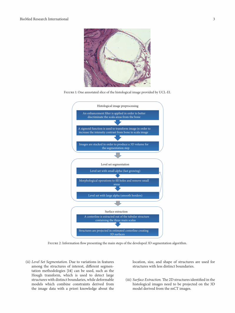
A hierarchical segmentation approach is followed, devel-oping a hybrid segmentation algorithm, whose steps arepresented in Figure 2, consisting of the following.
(i) Histological Image Preprocessing. Large-size structuresare first identified and details on smaller structuresare gradually obtained, while image enhancementand contrast enhancement filters are applied. At eachscale, the image is cropped after segmentation andlimited to the relevant structures in an attempt toreduce the computational cost andminimize the pos-sibility to detect unwanted structures at subsequentscales.
BioMed Research International 3
Figure 1: One annotated slice of the histological image provided by UCL-EI.
An enhancement filter is applied in order to better discriminate the scala areas from the bone
Images are stacked in order to produce a 3D volume for the segmentation step
Level set segmentation
A sigmoid function is used to transform image in order to increase the intensity contrast from bone to scala image
Level set with small alpha (fast growing)
Morphological operations to fill holes and remove small areas
Level set with large alpha (smooth borders)
containing the three main scalas
Surface extraction A centerline is extracted out of the tubular structure
Structures are projected in estimated centerline creating 3D surfaces
Histological image preprocessing
Figure 2: Information flow presenting the main steps of the developed 3D segmentation algorithm.
(ii) Level Set Segmentation. Due to variations in featuresamong the structures of interest, different segmen-tation methodologies [14] can be used, such as theHough transform, which is used to detect largestructures with distinct boundaries, while deformablemodels which combine constraints derived fromthe image data with a priori knowledge about the
location, size, and shape of structures are used forstructures with less distinct boundaries.
(iii) Surface Extraction.The2D structures identified in thehistological images need to be projected on the 3Dmodel derived from the mCT images.

4 BioMed Research International
450
400
350
300
250
200
1500 50 100 15 0 200 25 0 300 350 400 450
Figure 3: Cloud point corresponding to the two main scalas.
300
200
100
0
−100250
200150
10050
0−50
−1000
100
200
300
400
500
Figure 4: The initial model used for centerline estimation.
Image segmentation as described above is repeated forthe whole set of images. Subsequently, the segmented imagesare registered through the application of a least-square fittingalgorithm, which aims at the minimization of the totalvolume, to the detected structures. At this point, the distancebetween successive slices must be known.
3. Results
The final reconstructed model exploits the advantages ofthe histological images (i.e., high quality) as well as theadvantages of the mCT images (many frames that provide anaccurate 3D overview of the coiled cochlea) by projecting the2D structures identified in the histological images on the 3Dmodel derived from themCT images.This projection processincludes the following two steps.
(1) A centerline is extracted of the tubular structurecontaining the three main scalas.
(2) Structures are projected in estimated centerline creat-ing 3D surfaces.
For the extraction of the centerline we used samples fromthe two main scalas.The samples from the two scalas create acloud point (Figure 3).
Then the method of [15] is used for estimating thecenterline of the cloud point.The fittingmethod, presented in[15], takes as input an original Gaussian mixture model andgiven a new sample dataset (derived cloud point) estimatesthe new means and the transformation of the model usingglobal and local (per mixture) affine transformations. Usingthis method the geometry of the initial model is maintainedin the final model. As an initial model, mirrored sphericalGaussian distributions corresponding to the two scalas areplaced across a helix centerline (Figure 4). The final modelgives a set of points corresponding to the centerlines of thetwo scalae as depicted in Figure 5. The average of the twocenterlines gives the centerline of cochlea.
The structures annotated in the histological images needto be projected on the estimated centerline. Given thecontrol points corresponding to a b-spline interpolation isperformed on the centerline and N equal distant points areextracted. Furthermore, areas of three different regions onboth mCT and histological images were calculated. Scalingwas estimated across centerline frommaximum tominimumscale using linear interpolation (Figure 6).
Given a centerline point, the K points (x, y) of each 2Dstructure are projected on the 3D space using the specificpoint as point of reference, the derivative of the centerlineas normal vector, and the scaling estimated previously.

BioMed Research International 5
Figure 5:The fitting result.The blue lines connect both the centers of theGaussian distributions between consecutivemixtures correspondingto the same scala as well as the centers of the mirrored Gaussian distributions corresponding to the two main scalas.
Figure 6: Mapping of a slice of a histological image to a frame of a mCT image.
250
200
150
100
50
0380 340 300 260 220 180 50 150 250 350 450
−50
Figure 7: The projection of 2D structures on centerline plane.

6 BioMed Research International
Current triangles: 123,900
Selected triangles: 0
X
Y
Z
Figure 8: The reconstructed 3D model of the coiled cochlea.
Current triangles: 74,340
Selected triangles: 0X
Y
Z
(a)
Current triangles: 8,260
Selected triangles: 0X
Y
Z
(b)
Figure 9: The cochlear partition (a) and the osseous spiral lamina (b).
Repeating this projection for every a 3D curve (x, y, and z)is created, as depicted in Figure 7.
Finally the 3D curves aremerged in a surface correspond-ing to each structure.
The results of the hierarchical segmentation and thedeveloped hybrid segmentation algorithm are depictedin Figure 8, while Figure 9 presents a slice in the 3Dmodel depicting the cochlear partition (vibrating part)(Figure 9(a)) and the osseous spiral lamina (nonvibratingpart) (Figure 9(b)).
4. Discussion
The presented 3D reconstruction study of the cochlea fromregistration of mCT and histological images provides ananatomical model of the human cochlea, including severalparts that are not visible on mCT (scala media, spiralligament, and organ of Corti) enhancing the accuracy ofthe 3D model and going a step further towards the efficientstudying of a micromechanical model.
The presented reconstruction methodology exploits theadvantages of the mCT and histological images and throughthe registration process eliminates their drawbacks at thesegmentation process. The mCT images offer a superior𝑧-axis resolution but fail to resolve important soft tissue
structures and individual microarchitectural elements (i.e.,spiral ligament and organ of corti). On the other hand,histology images are able to resolve in more detail individualcellular structures, but reconstruction is inferior, as only alimited number of images are used. Histological prepara-tions may also suffer from extreme deformations, tears, anddestructions.
As a future step in the analysis, instead of using a linearscaling, the structures will be fitted to mCT slices perpen-dicular to centerline. Also, in the presented work, cochleastructures curves were extracted from a unique histologicalslice. As a step forward, there will be used curves extractedfrom different slices on different sites on the centerline andan interpolation between those curves will be performed inorder to produce a more accurate reconstruction.
5. Conclusions
This new reliable 3D cochlear model demonstrates the basisfor further numerical simulations using mathematical pro-cedures, such as the finite element or finite volume method.The development of accurate geometric 3D FE cochlearmodels will provide a unique opportunity to simulate bothnormal physiology and cochlear pathology and correlate it tohistopathological findings in a wide variety of ear diseases.

BioMed Research International 7
Additionally itmay be used in innovation of novel therapeuticapproaches in managing sensorineural hearing loss. Thesecalculations remain themost promising approaches to under-stand physiological processes and their pathology, whichcannot be investigated completely with existing measuringtechniques. Although there are studies in the literature thathave reconstructed pathological cochleae from CT, MRI, orhistological data [12], there are no geometric FE reconstruc-tions of the pathological cochlea for FE modeling purposes.
To this end, the SIFEM project [16] could eventuallyassist the management of hearing loss. Perhaps one of themost promising and useful aspects of using FEM is theplanning of surgical procedures and their predicted effectsby employing what-if scenarios. Such scenarios may help inboth innovation of surgical techniques and the better designof auditory implants.
Conflict of Interests
The authors declare that there is no conflict of interestsregarding the publication of this paper.
Acknowledgment
This work is partly funded by the European Commissionthrough ICT Project FP7-ICT-2011.5.2-600933 http://www.sifem-project.eu/.
References
[1] S. N. Merchant, M. J. McKenna, J. C. Adams et al., “Humantemporal bone consortium for research resource enhancement,”Journal of the Association for Research in Otolaryngology, vol. 9,no. 1, pp. 1–4, 2008.
[2] T. S. Rau, W. Wurfel, T. Lenarz, and O. Majdani, “Three-dimensional histological specimen preparation for accurateimaging and spatial reconstruction of the middle and innerear,” International Journal of Computer Assisted Radiology andSurgery, vol. 8, no. 4, pp. 481–509, 2013.
[3] S. F. Li, T. Y. Zhang, and Z. M. Wang, “An approach for precisethree-dimensional modeling of the human inner ear,” ORL;Journal for Oto-Rhino-Laryngology and Its Related Specialties,vol. 68, no. 5, pp. 302–310, 2006.
[4] H.Wang, S. N. Merchant, andM. S. Sorensen, “A downloadablethree-dimensional virtualmodel of the visible ear,”ORL, vol. 69,no. 2, pp. 63–67, 2007.
[5] T. Shibata and T. Nagano, “Applying very high resolutionmicrofocus X-ray CT and 3-D reconstruction to the humanauditory apparatus,” Nature Medicine, vol. 2, no. 8, pp. 933–935,1996.
[6] U. Vogel, “New approach for 3D imaging and geometry model-ing of the human inner ear,” journal for Oto-Rhino-Laryngologyand Its Related Specialties, vol. 61, no. 5, pp. 259–267, 1999.
[7] A. A. Poznyakovskiy, T. Zahnert, Y. Kalaidzidis et al., “Thecreation of geometric three-dimensionalmodels of the inner earbased on micro computer tomography data,” Hearing Research,vol. 243, no. 1-2, pp. 95–104, 2008.
[8] A. A. Poznyakovskiy, T. Zahnert, Y. Kalaidzidis et al., “Asegmentation method to obtain a complete geometry model of
the hearing organ,” Hearing Research, vol. 282, no. 1-2, pp. 25–34, 2011.
[9] T. Shibata, S. Matsumoto, T. Agishi, and T. Nagano, “Visu-alization of Reissner membrane and the spiral ganglion inhuman fetal cochlea by micro-computed tomography,” TheAmerican Journal of Otolaryngology—Head and Neck Medicineand Surgery, vol. 30, no. 2, pp. 112–120, 2009.
[10] B. M. Verbist, L. Ferrarini, J. J. Briaire et al., “Anatomicconsiderations of cochlear morphology and its implicationsfor insertion trauma in cochlear implant surgery,” Otology andNeurotology, vol. 30, no. 4, pp. 471–477, 2009.
[11] A. Lareida, F. Beckmann, A. Schrott-Fischer, R. Glueckert, W.Freysinger, and B. Muller, “High-resolution X-ray tomographyof the human inner ear: synchrotron radiation-based study ofnerve fibre bundles, membranes and ganglion cells,” Journal ofMicroscopy, vol. 234, no. 1, pp. 95–102, 2009.
[12] M. Seise, T. Alhonnoro, and M. Kolesnik, “Interactive reg-istration of 2D histology and 3D CT data for assessmentof radiofrequency ablation treatment,” Journal of PathologyInformatics, vol. 2, article S9, 2011.
[13] G. Sengle, S. F. Tufa, L. Y. Sakai, M. A. Zulliger, and D. R. Keene,“A correlative method for imaging identical regions of samplesbymicro-CT, lightmicroscopy, and electronmicroscopy: Imag-ing adipose tissue in a model system,” Journal of Histochemistryand Cytochemistry, vol. 61, no. 4, pp. 263–271, 2013.
[14] T. F. Chan and L. A. Vese, “Active contours without edges,” IEEETransactions on Image Processing, vol. 10, no. 2, pp. 266–277,2001.
[15] G. Rigas, C. Nikou, Y. Goletsis, and D. I. Fotiadis, “Hierarchicalsimilarity transformations between Gaussian mixtures,” IEEETransactions on Neural Networks and Learning Systems, vol. 24,no. 11, pp. 1824–1835, 2013.
[16] C. Bellos, A. Bibas, D. Kikidis et al., “SIFEM project: semanticinfostructure interlinking an open source finite element tooland libraries with a model repository for the multi-scalemodelling of the inner-ear,” in Proceedings of the 13th IEEEInternational Conference on BioInformatics and BioEngineering,IEEE BIBE, 2013.

Submit your manuscripts athttp://www.hindawi.com
Stem CellsInternational
Hindawi Publishing Corporationhttp://www.hindawi.com Volume 2014
Hindawi Publishing Corporationhttp://www.hindawi.com Volume 2014
MEDIATORSINFLAMMATION
of
Hindawi Publishing Corporationhttp://www.hindawi.com Volume 2014
Behavioural Neurology
EndocrinologyInternational Journal of
Hindawi Publishing Corporationhttp://www.hindawi.com Volume 2014
Hindawi Publishing Corporationhttp://www.hindawi.com Volume 2014
Disease Markers
Hindawi Publishing Corporationhttp://www.hindawi.com Volume 2014
BioMed Research International
OncologyJournal of
Hindawi Publishing Corporationhttp://www.hindawi.com Volume 2014
Hindawi Publishing Corporationhttp://www.hindawi.com Volume 2014
Oxidative Medicine and Cellular Longevity
Hindawi Publishing Corporationhttp://www.hindawi.com Volume 2014
PPAR Research
The Scientific World JournalHindawi Publishing Corporation http://www.hindawi.com Volume 2014
Immunology ResearchHindawi Publishing Corporationhttp://www.hindawi.com Volume 2014
Journal of
ObesityJournal of
Hindawi Publishing Corporationhttp://www.hindawi.com Volume 2014
Hindawi Publishing Corporationhttp://www.hindawi.com Volume 2014
Computational and Mathematical Methods in Medicine
OphthalmologyJournal of
Hindawi Publishing Corporationhttp://www.hindawi.com Volume 2014
Diabetes ResearchJournal of
Hindawi Publishing Corporationhttp://www.hindawi.com Volume 2014
Hindawi Publishing Corporationhttp://www.hindawi.com Volume 2014
Research and TreatmentAIDS
Hindawi Publishing Corporationhttp://www.hindawi.com Volume 2014
Gastroenterology Research and Practice
Hindawi Publishing Corporationhttp://www.hindawi.com Volume 2014
Parkinson’s Disease
Evidence-Based Complementary and Alternative Medicine
Volume 2014Hindawi Publishing Corporationhttp://www.hindawi.com