effect of deferiprone or deferoxamine on right ventricular function in thalassemia major patients...
TRANSCRIPT

RESEARCH Open Access
Effect of deferiprone or deferoxamine on rightventricular function in thalassemia major patientswith myocardial iron overloadGillian C Smith1,2†, Francisco Alpendurada1†, John Paul Carpenter1,2, Mohammed H Alam1,2, Vasili Berdoukas3,Markissia Karagiorga4, Vasili Ladis4, Antonio Piga5, Athanassios Aessopos6, Efstathios D Gotsis7, Mark A Tanner8,Mark A Westwood9, Renzo Galanello10, Michael Roughton11 and Dudley J Pennell1,2*
Abstract
Background: Thalassaemia major (TM) patients need regular blood transfusions that lead to accumulation of ironand death from heart failure. Deferiprone has been reported to be superior to deferoxamine for the removal ofcardiac iron and improvement in left ventricular (LV) function but little is known of their relative effects on theright ventricle (RV), which is being increasingly recognised as an important prognostic factor in cardiomyopathy.Therefore data from a prospective randomised controlled trial (RCT) comparing these chelators was retrospectivelyanalysed to assess the RV responses to these drugs.
Methods: In the RCT, 61 TM patients were randomised to receive either deferiprone or deferoxaminemonotherapy, and CMR scans for T2* and cardiac function were obtained. Data were re-analysed for RV volumesand function at baseline, and after 6 and 12 months of treatment.
Results: From baseline to 12 months, deferiprone reduced RV end systolic volume (ESV) from 37.7 to 34.2 mL (p =0.014), whilst RV ejection fraction (EF) increased from 69.6 to 72.2% (p = 0.001). This was associated with a 27% increasein T2* (p < 0.001) and 3.1% increase in LVEF (p < 0.001). By contrast, deferoxamine showed no change in RVESV (38.1 to39.1 mL, p = 0.38), or RVEF (70.0 to 69.9%, p = 0.93) whereas the T2* increased by 13% (p < 0.001), but with no changein LVEF (0.32%; p = 0.66). Analysis of between drugs treatment effects, showed significant improvements favouringdeferiprone with a mean effect on RVESV of -1.82 mL (p = 0.013) and 1.16% for RVEF (p = 0.008). Using regressionanalysis the improvement in RVEF at 12 months was shown to be greater in patients with lower baseline EF values (p <0.001), with a significant difference in RVEF of 3.5% favouring deferiprone over deferoxamine (p = 0.012).
Conclusion: In this retrospective analysis of a prospective RCT, deferiprone monotherapy was superior todeferoxamine for improvement in RVEF and end-systolic volume. This improvement in the RV volumes andfunction may contribute to the improved cardiac outcomes seen with deferiprone.
IntroductionBlood transfusions are standard therapy for patients withb-thalassaemia major (TM) and prevent death in child-hood, but although clinical status and short term survivalimprove, each unit of blood contains about 200-250 mgof iron which the body cannot eliminate, which leads tolong term iron accumulation. Patients treated only with
blood transfusions may die in the second and third dec-ades of life from the complications of iron overload, inparticular heart failure [1,2]. Myocyte damage is relatedto the production of reactive oxygen species (ROS)formed as levels of labile iron rise, which cause oxidativedamage to membranes and mitochondrial respiratorychain enzyme dysfunction [3,4]. Chelation therapy canreduce tissue iron levels and the incidence of cardiaccomplications, but patients at risk need to be accuratelyprofiled for appropriate treatment. The cardiovascularmagnetic resonance (CMR) relaxation parameter T2* is
* Correspondence: [email protected]† Contributed equally1CMR Unit, Royal Brompton & Harefield NHS Foundation Trust, SydneyStreet, London SW3 6NP, UKFull list of author information is available at the end of the article
Smith et al. Journal of Cardiovascular Magnetic Resonance 2011, 13:34http://www.jcmr-online.com/content/13/1/34
© 2011 Smith et al; licensee BioMed Central Ltd. This is an Open Access article distributed under the terms of the Creative CommonsAttribution License (http://creativecommons.org/licenses/by/2.0), which permits unrestricted use, distribution, and reproduction inany medium, provided the original work is properly cited.

sensitive to storage tissue iron in haemosiderin becauseof the creation of field inhomogeneities by iron particles,and the clinical adoption of this technique is now wide-spread as a mainstay of cardiac iron overload assessmentand treatment,[5-7] with important capability to predictfuture cardiac events,[8] and evidence of significant bene-ficial effects on cardiac mortality [9].Deferoxamine was the first iron chelating agent for clini-
cal use and became standard therapy in the 1970s. It is alarge positively charged lipophobic molecule, is poorlyabsorbed by the digestive system and has a short plasmahalf life [10,11]. It is therefore administered subcuta-neously using a portable syringe system usually overnighttypically 5 times per week. This therapy can be very pro-blematic with poor compliance, and a number of factorsresult in long-term cardiac iron accumulation with its use[12]. The second clinical iron chelator was deferiprone,which is a much smaller neutrally charged lipophilic mole-cule which allows good gastrointestinal absorption andcellular access [10,11]. The plasma half life is longer allow-ing oral administration with three doses per day. Directcomparison trials show that deferiprone has greater effi-cacy than deferoxamine for reducing myocardial iron load-ing and improving left ventricular (LV) systolic function[13,14]. However, there is a paucity of data related to theeffects of these chelators on the right ventricle (RV), whichis known to be an important independent predictor of out-come in dilated cardiomyopathy,[15] and ischaemic heartdisease [16-18]. Recent papers have established the normalranges for RV volumetric parameters for non-iron loadedTM patients and shown a significant relation between T2*and RV ejection fraction (RVEF), including a small percen-tage of patients with impaired RVEF but normal LVEF[19,20]. In order to identify and compare the effects ofdeferiprone and deferoxamine, we reanalyzed the CMRimages for the LA16 trial, which was a randomized con-trolled trial (RCT) comparing the 2 drugs [14]. Ourhypothesis was that deferiprone would improve RV func-tion more than deferoxamine.
MethodsThe LA16 RCT consisted of 61 regularly transfusedpatients with TM from 4 centres in Greece and Italy [14].All patients were previously treated with subcutaneousdeferoxamine monotherapy. Inclusion criteria included aT2* between 8 and 20 ms and LVEF greater than 56%based on the lower normal limit for non-anaemic sub-jects from previously published data [21]. No patient hadheart failure symptoms. Deferiprone was allocated to 29patients (actual dose 92 mg/kg daily) whilst 32 patientswere allocated to continue with deferoxamine therapy(dose of 43 mg/kg/day overnight for an average of 5.7days per week). Written informed consent was obtainedaccording to local ethics committee approval.
Iron loading and cardiac function was assessed usingCMR. The T2* sequences were installed at the localCMR facilities, Athens (GE CVi) and Cagliari (GE Signa).The technique was validated by scanning phantoms ofknown T2* and testing intra-site reproducibility by scan-ning 5 patients twice at the local centre. The samepatients were scanned at the reference site in London(Siemens Sonata) for inter-site reproducibility. A coeffi-cient of variation (CV) ≤ 15% was defined as acceptable.Site inter-study variability was 2.4% for Cagliari and 3.5%for Athens. Comparison with the reference site yieldedCVs of 1.6% and 9.7% respectively. Volumetric data wereacquired using a steady state free precession sequence(FIESTA). A set of contiguous short axis cines wereacquired to give complete coverage of both ventricles.Care was taken to place the basal slice parallel to theatrioventricular groove. Slice thickness was 8 mm with aspacing of 10 mm. No patient had a history of, and noCMR scan showed any features of pulmonary hyperten-sion (normal pulmonary artery size, no right ventricularhypertrophy, no systolic septal flattening). Patients werescanned between 3 and 10 days post transfusion at base-line, 6 months and 12 months after entering the trial. T2*values and LV volumetric data were assessed previouslyusing a CMR viewing and analysis software packageCMRtools (Cardiovascular imaging solutions, London,UK) [14]. LV function was assessed in the RCT using anearly version of the analysis package in which LVvolumes are quantified manually using direct surface pla-nimetry, and therefore for consistency we elected to usethe same version of the software to analyze the RV thuseliminating the need for right sided valve tracking. Carewas taken to include blood volume below the pulmonaryvalve. Basal regions with thin, non trabeculated musclewere considered atrial and were excluded. Papillary mus-cles were also excluded from the blood pool [22].Although local blinding to the treatment arm was notpossible due to the nature of drug administration (oralfor deferiprone and subcutaneous for deferoxamine), allremote scan analysis performed at the core-lab in Lon-don was fully blinded to treatment. Study treatment wasunblinded on completion of LV and T2* analysis. For theanalysis of the RV volumetric data, all data sets wereanonymized and analyzed in random order using thesame analysis package by experienced operators blindedto treatment arm and LV response. To assess reproduci-bility, data-sets with an improvement in RVEF ≥ 5% at 12months were reprocessed blindly.
Statistical analysisContinuous variables were compared using a paired t-test.T2* values were log transformed and changes expressed asgeometric mean with coefficient of variation. Betweengroups comparison of drug effects were assessed using a
Smith et al. Journal of Cardiovascular Magnetic Resonance 2011, 13:34http://www.jcmr-online.com/content/13/1/34
Page 2 of 7

repeated measurement ANOVA. Statistical significancewas set at p < 0.05. To quantify reproducibility the coeffi-cient of variance (CV) was calculated.
ResultsFull data on the LA16 study have been publishedincluding the patient demographics,[14] but the impor-tant trial summary findings are repeated here. In thedeferiprone group 27 patients completed the study; 2patients withdrew due to adverse events (elevated hepa-tic enzymes, in one case probably due to cytomegalo-virus). In the deferoxamine group, 29 patientscompleted; 1 patient withdrew secondary to a reductionin LV function and 2 for personal reasons. The patientgroups were well matched at baseline for cardiac T2*,LV volumes and function and RV volumes and function(table 1). Baseline RVEF was within the normal refer-ence range for thalassaemia patients,[19] except for onepatient in the deferoxamine group (RVEF 1% below thenormal range). Patient compliance was similar for bothgroups. Myocardial T2* improved by 18% at 6 months(p < 0.001) and 27% at 12 months (p < 0.001) in thedeferiprone arm. The LVEF rose by 2.0% from 69.7% atbaseline to 71.7% at 6 months (p < 0.001) and by 3.1%to 72.7 at 12 months (p < 0.001). With deferoxaminetherapy, T2* improved by 9% at 6 months (p = 0.003)and by 13% at 12 months (p < 0.001) but LVEF wasunchanged being 68.4% at baseline and 68.7 at 6 months(+0.52%, p = 0.42) and 68.5 at 12 months (+0.32%, p =0.66).
In the current analysis, the RV mean volumetric andT2* values are detailed in table 2. To summarise, in thedeferiprone arm RV end-diastolic volume (EDV) wasstable, RV end-systolic volume (ESV) decreased signifi-cantly from 37.7 to 34.2 mL at 12 months (p=0.014);and RVEF increased from 69.6% to 72.2% (p = 0.001).For the patients on deferoxamine therapy, the changesin RV parameters from baseline to 12 months showedno significant difference. Analysis of between drugstreatment effects using a repeated measurementANOVA (table 3) showed significant differences favour-ing deferiprone for the reduction of RV ESV (p=0.014 at12 months, Figure 1) and improvement in RVEF (p =0.008 at 12 months, Figure 2). Non significant differ-ences between drugs were found for RV EDV. Withregression analysis, the change in RVEF was found to beinversely related to the baseline EF (p < 0.001) with asignificant difference between drugs favouring deferi-prone by a mean of 3.5% (95% CI 0.8 to 6.3%; p =0.012). The reduction in RVESV over 12 months wasalso related to the baseline ESV value with borderlinesignificance (p = 0.051), and there was a significant dif-ference between drugs favouring deferiprone by a meanof 4.5 mL more than patients on deferoxamine (95% CI1.2 to 7.8 mL; p = 0.009). Therefore the patients benefit-ting most from deferiprone treatment are those with thelower baseline values of RVEF. The CV for intra-obser-ver study RVEF measurement was 2.4% at baseline and2.0% at 12 months. There was no relation betweenchange in RVEF and change in LVEF (r = 0.3, p = 0.9).
DiscussionRV volumetric and functional parameters have been dif-ficult to measure using conventional imaging techniquesdue to the irregular geometry of the RV chamber, thesize and quantity of the RV trabeculae, and the proxi-mity of the RV to the chest wall which impairs echocar-diographic assessment. CMR suffers less from thesedrawbacks because of its inherent 3D nature and highblood to myocardium contrast and is therefore consid-ered to be the most accurate and reproducible techniquefor assessing RV volumes and EF [23,24]. Attention tocorrect definition of the basal slice during acquisitionand subsequent analysis is however pivotal. Theimproved confidence of measuring RV volumes andfunction from CMR and other techniques has assistedthe understanding the importance of the RV in cardiacdisease. RVEF is an important predictor of outcome indilated cardiomyopathy, which is both independent ofand incremental to LV EF [15]. The predictive value ofRV function has also been shown in congenital heartdisease,[25-27] chronic systolic dysfunction,[28] andischemic heart failure,[16,17,29,30] with RVEF beingshown to be an independent predictor of outcome
Table 1 Baseline values for cardiac volume and functionparameters
Deferiprone Deferoxamine p
No. Patients randomized 28 32
Age 25.1 ± 3.8 26.6 ± 4.7 0.33
Male sex (%) 15 (52) 16 (50) 0.99
Weight (kg) 57.7 ± 7.9 60.6 ± 13.2 0.30
Cardiac parameters
Myocardial T2* (ms) 13.0 (32%) 13.3 (30%) 0.77
LVEDV (mL) 134 ± 32 132 ± 23 0.81
LVESV (mL) 43 ± 14 41 ± 13 0.51
LVEF (%) 69.7 ± 5.4 68.4 ± 4.9 0.34
RVEDV (mL) 122.5 ± 24.9 124.7 ± 27.7 0.75
RVESV (mL) 37.7 ± 11.7 38.1 ± 12.6 0.90
RVEF (%) 69.6 ± 5.2 70.0 ± 5.8 0.79
Biochemical markers
Liver iron concentration (μg/L) 6.16 ± 6.0 6.32 ± 5.8 0.92
Serum ferritin (μg/L) 1791 ± 1029 2795 ± 2441 0.039
Haematology
Transfusional iron (mL/kg/year) 152 ± 43.4 144 ± 44.4 0.53
Haemoglobin level (g/L) 105 ± 12.0 113 ± 11.9 0.023
Values are mean ± SD except for T2* where values are geometric mean withCV.
Smith et al. Journal of Cardiovascular Magnetic Resonance 2011, 13:34http://www.jcmr-online.com/content/13/1/34
Page 3 of 7

[16,17]. Accordingly, the effects of myocardial iron load-ing on RV function may be important in thalassaemiapatients.In the current study, we found a significant improve-
ment in RVEF (increase) and RVESV (reduction) withdeferiprone therapy. These improvements parallel thepreviously reported LV response [14]. There was no sig-nificant increase in RVEDV suggesting loading condi-tions did not play an important role. A flat RV responsewas seen in the deferoxamine group, which again mir-rors LV behaviour. The between groups analysis showedsuperiority for deferiprone over deferoxamine for boththe reduction in RVESV and the increase in RVEF. Themagnitude of improvement in RVEF and reduction inRVESV were greater for patients with a higher ESV andlower EF at baseline. Interestingly neither LVEF norRVEF improved significantly in the deferoxamine groupdespite the improvement in T2*. The cause for this dif-ference in functional response is not fully understood,but the explanation may lie in the additional effects ofdeferiprone on restoring normal cardiac mitochondrialfunction,[31] possibly through effects on reducing reac-tive oxygen species [32].There is little other data relating RV function changes
with the iron chelators, but a recently published abstract
relating to a longitudinal trial of the efficacy of defera-sirox in myocardial siderosis,[33] showed a significantimprovement in myocardial iron levels with an improve-ment in RVEF at 1 year, but no change in LV functionat 1,[34] 2,[35] and 3 [36] years of follow up. The signif-icance of this discrepancy between RV and LV responseto deferasirox is not currently clear, though it is possiblethat the RV response is an early signal of myocardialiron clearance as LV compliance and filling pressureimproves.
LimitationsData acquisition for this study was originally designed toassess the change in T2* and LV functional parametersin response to therapy. Therefore no RV long axisimages were obtained to construct 3-dimentional mod-els for volumetric analysis, but the requirement for thiswas removed by using direct manual planimetry forquantitative analysis of RV volumes. Pulmonary arterialpressure was not systematically measured using echocar-diography of the tricuspid regurgitant jet, but here wasno CMR evidence of raised pulmonary artery pressurein our patients, and pulmonary hypertension is rare inwell treated thalassaemia major [37]. Direct RV mea-surement of T2* would have been interesting in thispopulation to compare with changes in RV volumes andfunction, however, it is challenging to measure T2* inthe thin wall of the RV and this was not attempted inthe randomized controlled trial.
ConclusionsThis study has shown that RVESV decreased and RVEFimproved with deferiprone monotherapy and this bene-ficial response was superior to deferoxamine. RV volu-metric and function parameters have in the past beenneglected when reporting the efficacy of iron chelators
Table 2 RV volumetric parameters at baseline, 6 and 12 months (mean ± SD) in the 2 treatment arms
Baseline 6 Months 12 Months p
Deferiprone
T2*, ms 13.0 (32) 15.4 (38) 16.5 (38) < 0.001
RVEDV, mL 122.5 ± 24.9 123.2 ± 26.0 121.3 ± 24.9 0.61
RVESV, mL 37.7 ± 11.7 35.9 ± 11.7 34.2 ± 11.3 0.009
RVSV, mL 84.7 ± 16.5 87.3 ± 16.5 87.1 ± 17.0 0.16
RVEF, % 69.6 ± 5.2 71.4 ± 4.7 72.2 ± 5.3 0.001
Deferoxamine
T2*, ms 13.3 (30) 14.4 (37) 15 (39) <0.001
RVEDV, mL 124.7 ± 27.7 124.4 ± 26.2 128 ± 32.1 0.17
RVESV, mL 38.1 ± 12.6 37.2 ± 12.5 39.1 ± 13.0 0.38
RVSV, mL 86.7 ± 18.0 87.0 ± 15.5 88.9 ± 21.3 0.25
RVEF, % 70.0 ± 5.8 70.8 ± 5.2 69.9 ± 4.6 0.93
The T2* values show the geometric mean and CV. The p value reflects the change from baseline to 12 months.
Table 3 Between drug effect on RV volumetricparameters showing a significant difference in RV ESVand RVEF favouring deferiprone.
Treatment effect
Deferiprone-Deferoxamine
Mean 95% CI p
EDV -1.21 -4.44 2.03 0.47
ESV -1.82 -3.27 -0.37 0.014
SV 0.64 -2.02 3.31 0.64
EF 1.16 0.30 2.01 0.008
Smith et al. Journal of Cardiovascular Magnetic Resonance 2011, 13:34http://www.jcmr-online.com/content/13/1/34
Page 4 of 7
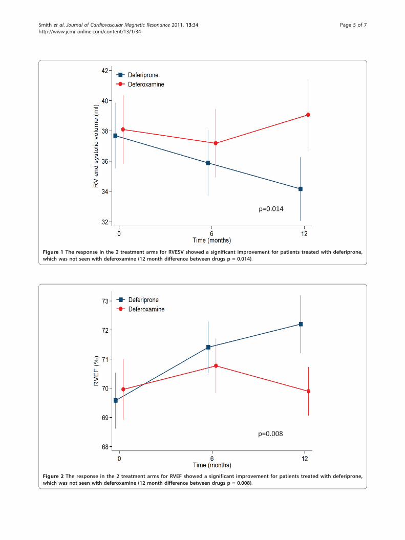
p=0.008
Figure 2 The response in the 2 treatment arms for RVEF showed a significant improvement for patients treated with deferiprone,which was not seen with deferoxamine (12 month difference between drugs p = 0.008).
p=0.014
Figure 1 The response in the 2 treatment arms for RVESV showed a significant improvement for patients treated with deferiprone,which was not seen with deferoxamine (12 month difference between drugs p = 0.014).
Smith et al. Journal of Cardiovascular Magnetic Resonance 2011, 13:34http://www.jcmr-online.com/content/13/1/34
Page 5 of 7

for myocardial iron overload, and may have independentprognostic importance, as they do in other cardiac con-ditions with impaired cardiac function.
Acknowledgements and FundingThis work was supported by the National Institutes for Health ResearchCardiovascular Biomedical Research Unit of Royal Brompton Hospital andImperial College. The original randomized controlled trial (LA16) wassponsored by ApoPharma, but no company funding was received for thisstudy in which the imaging data was reanalyzed.
Author details1CMR Unit, Royal Brompton & Harefield NHS Foundation Trust, SydneyStreet, London SW3 6NP, UK. 2National Heart & Lung Institute, ImperialCollege London, Guy Scadding Building, Dovehouse Street, London SW36LY, UK. 3Division of Haematology/Oncology, Children’s Hospital, 4650Sunset Boulevard, Los Angeles, CA 90027, USA. 4Thalassaemia Unit, AghiaSophia Children’s Hospital, Thivon & Papadiamantopoulou, Goudi, Athens115 27, Greece. 5Division of Paediatrics and thalassaemia centre, Departmentof Clinical and Biological Sciences, University of Torino, S. Luigi GonzagaHospital, Regione Gonzole 10, Orbassano 10043, Torino, Italy. 6FirstDepartment of Internal Medicine, University of Athens Medical School, LaikoHospital, 17 Agiou Thoma Street, Athens 115 27, Greece. 7Department ofMagnetic Resonance, Institute Euromedica-Encephalos, 3 Rizariou Street,Halandri, Athens 152 33, Greece. 8Department of Cardiology, St Richard’sHospital, Western Sussex Hospitals NHS Trust, Chichester, West Sussex, PO196SE UK. 9Department of Cardiology, The London Chest Hospital, BonnerRoad, London E2 9JX, UK. 10Department of Biomedical Science andBiotechnology, University of Cagliari, Ospedale Regionale per leMicrocitemie. Via Edward Jenner, 09121 Cagliari, Italy. 11UCL Cancer TrialsCentre, University College London, 90 Tottenham Court Road, London W1T4TJ, UK.
Authors’ contributionsGCS and FA participated in study design, data analysis and interpretationand manuscript drafting. JPC and MHA performed additional data analysis.VB, MK, VL, AP, AA, EDG, MAT and MAW Served as investigators on theoriginal trial. MR performed statistical analysis. DJP conceived of the studyand was responsible for the final manuscript draft. All authors read andapproved the final manuscript.
Competing interestsGCS is a consultant to Novartis and has received honoraria from ApoPharma.FA has received honoraria from Novartis. JPC has received speaker’shonoraria from Novartis, Swedish Orphan and ApoPharma. VB is a consultantfor ApoPharma. VL has received grant support, consulting fees and lecturefees from ApoPharma and Novartis. AP has received honoraria and researchfunding from Novartis. RG has served on speakers’ bureaus and receivedresearch grants from Novartis and ApoPharma. DJP is a consultant to andhas served on advisory boards and speakers’ bureaus for Novartis,ApoPharma, and Siemens; has received research funding from Novartis; andis a director and stockholder for Cardiovascular Imaging Solutions.
Received: 10 February 2011 Accepted: 6 July 2011Published: 6 July 2011
References1. Olivieri NF, Nathan DG, MacMillan JH, Wayne AS, Liu PP, McGee A,
Martin M, Koren G, Cohen AR: Survival in medically treated patients withhomozygous beta thalassaemia. N Engl J Med 1994, 331:574-8.
2. Modell B, Khan M, Darlison M: Survival in beta thalassaemia major in theUK: Data from the UK Thalassaemia Register. Lancet 2000, 355:2051-2.
3. Rachmilewitz EA, Weizer-Stern O, Adamsky K, Amariglio N, Rechavi G,Breda L, Rivella S, Cabantchik ZI: Role of iron in inducing oxidative stressin thalassemia: Can it be prevented by inhibition of absorption and byantioxidants? Ann N Y Acad Sci 2005, 1054:118-23.
4. Breuer W, Hershko C, Cabantchik ZI: The importance of non-transferrinbound iron in disorders of iron metabolism. Transfus Sci 2000, 3:185-92.
5. Anderson LJ, Holden S, Davis B, Prescott E, Charrier CC, Bunce NH,Firmin DN, Wonke B, Porter J, Walker JM, Pennell DJ: Cardiovascular T2-star(T2*) magnetic resonance for the early diagnosis of myocardial ironoverload. Eur Heart J 2001, 22:2171-9.
6. Wood JC, Otto-Duessel M, Aguilar M, Nick H, Nelson MD, Coates TD,Pollack H, Moats R: Cardiac iron determines cardiac T2*, T2, and T1 in thegerbil model of iron cardiomyopathy. Circulation 2005, 112:535-43.
7. Cohen AR, Galanello R, Pennell DJ, Cunningham MJ, Vichinsky E:Thalassemia. Hematology Am Soc Hematol Educ Program 2004, 14-34.
8. Kirk P, Roughton M, Porter JB, Walker JM, Tanner MA, Patel J, Wu D,Taylor J, Westwood MA, Anderson LJ, Pennell DJ: Cardiac T2* magneticresonance for prediction of cardiac complications in thalassemia major.Circulation 2009, 120:1961-8.
9. Modell B, Khan M, Darlison M, Westwood MA, Ingram D, Pennell DJ:Improved survival of thalassaemia major in the UK and relation to T2*cardiovascular magnetic resonance. J Cardiovasc Magn Reson 2008, 10:42.
10. Porter JB: Practical management of iron overload. Br J Haematol 2001,115:239-52.
11. Kushner JP, Porter JP, Olivieri NF: Secondary iron overload. Hematology2001, 47-61.
12. Tanner MA, Galanello R, Dessi C, Westwood MA, Smith GC, Nair SV,Anderson LJ, Walker JM, Pennell DJ: Myocardial iron loading in patientswith thalassemia major on deferoxamine chelation. J Cardiovasc MagnReson 2006, 8:543-7.
13. Anderson LJ, Holden S, Davis B, Prescott E, Charrier CC, Bunce NH,Firmin DN, Wonke B, Porter J, Walker JM, Pennell DJ: Cardiovascular T2-star(T2*) magnetic resonance for the early diagnosis of myocardial ironoverload. Eur Heart J 2001, 22:2171-9.
14. Pennell DJ, Berdoukas V, Karagiorga M, Ladis V, Piga A, Aessopos A,Gotsis ED, Tanner MA, Smith GC, Westwood MA, Wonke B, Galanello R:Randomized controlled trial of deferiprone or deferoxamine in beta-thalassemia major patients with asymptomatic myocardial siderosis.Blood 2006, 107:3738-44.
15. Juillière Y, Barbier G, Feldmann L, Grentzinger A, Danchin N, Cherrier F:Additional predictive value of both left and right ventricular ejectionfractions on long-term survival in idiopathic dilated cardiomyopathy. EurHeart J 1997, 18:276-80.
16. de Groote P, Millaire A, Foucher-Hossein C, Nugue O, Marchandise X,Ducloux G, Lablanche JM: Right ventricular ejection fraction is anindependent predictor of survival in patients with moderate heartfailure. J Am Coll Cardiol 1998, 32:948-54.
17. Larose E, Ganz P, Reynolds HG, Dorbala S, Di Carli MF, Brown KA,Kwong RY: Right ventricular dysfunction assessed by cardiovascularmagnetic resonance imaging predicts poor prognosis late aftermyocardial infarction. J Am Coll Cardiol 2007, 49:855-62.
18. Miszalski-Jamka T, Klimeczek P, Tomala M, Krupiński M, Zawadowski G,Noelting J, Lada M, Sip K, Banyś R, Mazur W, Kereiakes DJ, Zmudka K,Pasowicz M: Extent of RV dysfunction and myocardial infarction assessedby CMR are independent outcome predictors early after STEMI treatedwith primary angioplasty. JACC Cardiovasc Imaging 2010, 12:1237-46.
19. Carpenter JP, Alpendurada F, Deac M, Maceira A, Garbowski M, Kirk P,Walker JM, Porter JB, Shah F, Banya W, He T, Smith GC, Pennell DJ: Rightventricular volumes and function in thalassemia major patients in theabsence of myocardial iron overload. J Cardiovasc Magn Reson 2010,12:24.
20. Alpendurada F, Carpenter JP, Deac M, Kirk P, Walker JM, Porter JB, Banya W,He T, Smith GC, Pennell DJ: Relation of myocardial T2* to right ventricularfunction in thalassaemia major. Eur Heart J 2010, 31:1648-54.
21. Lorenz CH, Walker ES, Morgan VL, Klein SS, Graham TP: Normal human rightand left ventricular mass, systolic function and gender differences by cinemagnetic resonance imaging. J Cardiovasc Magn Reson 1999, 1:7-21.
22. Hudsmith LE, Petersen SE, Francis JM, Robson MD, Neubauer SJ: Normalhuman left and right ventricular and left atrial dimensions using steadystate free precession magnetic resonance imaging. J Cardiovasc MagnReson 2005, 7:775-82.
23. Grothues F, Smith GC, Moon JC, Bellenger NG, Collins P, Klein HU,Pennell DJ: Comparison of interstudy reproducibility of cardiovascularmagnetic resonance with two-dimensional echocardiography in normalsubjects and in patients with heart failure or left ventricularhypertrophy. Am J Cardiol 2002, 90:29-34.
Smith et al. Journal of Cardiovascular Magnetic Resonance 2011, 13:34http://www.jcmr-online.com/content/13/1/34
Page 6 of 7

24. Grothues F, Moon JC, Bellenger NG, Smith GS, Klein HU, Pennell DJ:Interstudy reproducibility of right ventricular volumes, function, andmass with cardiovascular magnetic resonance. Am Heart J 2004,147:218-33.
25. Roos-Hesselink JW, Meijboom FJ, Spitaels SE, van Domburg R, van Rijen EH,Utens EM, McGhie J, Bos E, Bogers AJ, Simoons ML: Decline in ventricularfunction and clinical condition after Mustard repair for transposition ofthe great arteries (a prospective study of 22-29 years). Eur Heart J 2004,25:1264-70.
26. Graham TP Jr, Bernard YD, Mellen BG, Celermajer D, Baumgartner H,Cetta F, Connolly HM, Davidson WR, Dellborg M, Foster E, Gersony WM,Gessner IH, Hurwitz RA, Kaemmerer H, Kugler JD, Murphy DJ, Noonan JA,Morris C, Perloff JK, Sanders SP, Sutherland JL: Long-term outcome incongenitally corrected transposition of the great arteries: a multi-institutional study. J Am Coll Cardiol 2000, 36:255-61.
27. Gatzoulis MA, Clark AL, Cullen S, Newman CG, Redington AN: Rightventricular diastolic function 15 to 35 years after repair of tetralogy ofFallot. Restrictive physiology predicts superior exercise performance.Circulation 1995, 91:1775-81.
28. Meyer P, Filippatos GS, Ahmed MI, Iskandrian AE, Bittner V, Gilbert J,Perry GJ, White M, Aban IB, Mujib M, Dell’Italia LJ, Ahmed A: Effects of rightventricular ejection fraction on outcomes in chronic systolic heartfailure. Circulation 2010, 121:252-8.
29. Di Salvo TG, Mathier M, Semigran MJ, Dec GW: Preserved right ventricularejection fraction predicts exercise capacity and survival in advancedheart failure. J Am Coll Cardiol 1995, 25:1143-53.
30. Ghio S, Gavazzi A, Campana C, Inserra C, Klersy C, Sebastiani R, Arbustini E,Recusani F, Tavazzi L: Independent and additive prognostic value of rightventricular systolic function and pulmonary artery pressure in patientswith chronic heart failure. J Am Coll Cardiol 2001, 37:183-8.
31. Kakhlon O, Manning H, Breuer W, Melamed-Book N, Lu C, Cortopassi G,Munnich A, Cabantchik ZI: Cell functions impaired by frataxin deficiencyare restored by drug-mediated iron relocation. Blood 2008, 112:5219-27.
32. Kontoghiorghes GJ, Efstathiou A, Kleanthous M, Michaelides Y, Kolnagou A:Risk/benefit assessment, advantages over other drugs and targetingmethods in the use of deferiprone as a pharmaceutical antioxidant iniron loading and non iron loading conditions. Hemoglobin 2009,33:386-97.
33. Smith G, Pennell DJ, Porter JB, Cappellini MD, Chan LL, El-Beshlawy A,Aydinok Y, Ibrahim H, Li CK, Viprakasit V, Elalfy M, Kattamis A, Habr D,Domokos G, Hmissi A, Taher A: Improvement in right ventricular functionfollowing 1 year of deferasirox therapy in patients with β-thalassemia.Blood 2009, 114:abstract 5106.
34. Pennell DJ, Porter JB, Cappellini MD, El-Beshlawy A, Chan LL, Aydinok Y,Elalfy MS, Sutcharitchan P, Li CK, Ibrahim H, Viprakasit V, Kattamis A,Smith G, Habr D, Domokos G, Roubert B, Taher A: Efficacy of deferasirox inreducing and preventing cardiac iron overload in beta-thalassemia.Blood 2010, 115:2364-71.
35. Pennell DJ, Porter JB, Cappellini MD, Chan LL, El-Beshlawy A, Aydinok Y,Ibrahim H, Li CK, Viprakasit V, Elalfy MS, Kattamis A, Smith G, Habr D,Domokos G, Roubert B, Taher A: Continued improvement in myocardialT2* over 2 years of deferasirox therapy in beta-thalassemia majorpatients with cardiac iron overload. Haematologica 2011, 96:48-54.
36. Pennell DJ, Porter JB, Cappellini MD, Chan LL, El-Beshlawy A, Aydinok Y,Ibrahim H, Li CK, Viprakasit V, Elalfy MS, Kattamis A, Smith G, Habr D,Domokos G, Roubert B, Taher A: Continued improvement andnormalization of myocardial T2* in patients with β thalassemia majortreated with Deferasirox (Exjade®) for up to 3 years. Blood 2010, 116:abstract 4276.
37. Aessopos A, Farmakis D, Hatziliami A, Fragodimitri C, Karabatsos F, Joussef J,Mitilineou E, Diamanti-Kandaraki E, Meletis J, Karagiorga M: Cardiac statusin well treated patients with thalassemia major. Eur J Haematol 2004,73:359-66.
doi:10.1186/1532-429X-13-34Cite this article as: Smith et al.: Effect of deferiprone or deferoxamineon right ventricular function in thalassemia major patients withmyocardial iron overload. Journal of Cardiovascular Magnetic Resonance2011 13:34.
Submit your next manuscript to BioMed Centraland take full advantage of:
• Convenient online submission
• Thorough peer review
• No space constraints or color figure charges
• Immediate publication on acceptance
• Inclusion in PubMed, CAS, Scopus and Google Scholar
• Research which is freely available for redistribution
Submit your manuscript at www.biomedcentral.com/submit
Smith et al. Journal of Cardiovascular Magnetic Resonance 2011, 13:34http://www.jcmr-online.com/content/13/1/34
Page 7 of 7