transdermal deferoxamine prevents pressure-induced ... · transdermal deferoxamine prevents...
TRANSCRIPT

Transdermal deferoxamine prevents pressure-induceddiabetic ulcersDominik Duschera,1, Evgenios Neofytoua,1, Victor W. Wongb, Zeshaan N. Maana, Robert C. Rennerta,Mohammed Inayathullahc, Michael Januszyka, Melanie Rodriguesa, Andrey V. Malkovskiyc, Arnetha J. Whitmorea,Graham G. Walmsleya, Michael G. Galveza, Alexander J. Whittama, Michael Brownleed, Jayakumar Rajadasc,2,and Geoffrey C. Gurtnera,2
aHagey Laboratory for Pediatric Regenerative Medicine, Division of Plastic and Reconstructive Surgery, Department of Surgery, Stanford University School ofMedicine, Stanford, CA 94305; bDepartment of Plastic Surgery, Johns Hopkins University School of Medicine, Baltimore, MD 21201; cBiomaterials andAdvanced Drug Delivery Center, Stanford University School of Medicine, Stanford, CA 94305; and dDiabetes Research Center, Albert Einstein College ofMedicine, New York, NY 10461
Edited* by Bruce D. Hammock, University of California, Davis, CA, and approved November 26, 2014 (received for review July 16, 2014)
There is a high mortality in patients with diabetes and severepressure ulcers. For example, chronic pressure sores of the heelsoften lead to limb loss in diabetic patients. A major factor un-derlying this is reduced neovascularization caused by impairedactivity of the transcription factor hypoxia inducible factor-1 alpha(HIF-1α). In diabetes, HIF-1α function is compromised by a high glu-cose-induced and reactive oxygen species-mediated modification ofits coactivator p300, leading to impaired HIF-1α transactivation. Weexamined whether local enhancement of HIF-1α activity would im-prove diabetic wound healing and minimize the severity of diabeticulcers. To improve HIF-1α activity we designed a transdermal drugdelivery system (TDDS) containing the FDA-approved small mole-cule deferoxamine (DFO), an iron chelator that increases HIF-1αtransactivation in diabetes by preventing iron-catalyzed reactiveoxygen stress. Applying this TDDS to a pressure-induced ulcermodel in diabetic mice, we found that transdermal delivery ofDFO significantly improved wound healing. Unexpectedly, prophy-lactic application of this transdermal delivery system also preventeddiabetic ulcer formation. DFO-treated wounds demonstrated in-creased collagen density, improved neovascularization, and reduc-tion of free radical formation, leading to decreased cell death. Thesefindings suggest that transdermal delivery of DFO provides a tar-geted means to both prevent ulcer formation and accelerate dia-betic wound healing with the potential for rapid clinical translation.
wound healing | diabetes | drug delivery | small molecule | angiogenesis
Diabetes mellitus affects over 25 million people in the UnitedStates (1, 2) and costs nearly $250 billion per year (3). Chronic
diabetic wounds and decubiti are important long-term sequalae ofboth diabetes mellitus types 1 and 2 (4). There is a high mortality indiabetic patients who develop decubiti (5–7), and owing to pro-longed disability and the high rates of recurrence these woundsrepresent an especially severe complication of diabetes (8). This isfurther underscored by the fact that diabetic nonhealing wounds arethe leading cause of nontraumatic amputations in the United States(3, 9–11). As such, there is a clear need for new approaches to ef-fectively manage and treat diabetic ulcers.The propensity for wound development in diabetes is associated
with a reduced capacity for ischemia-driven neovascularization (12,13). Hypoxia inducible factor-1 (HIF-1), which consists of a highlyregulated α-subunit and a constitutively expressed β-subunit, is acritical transcriptional regulator of the normal cellular responseto hypoxia, promoting progenitor cell recruitment, proliferation,survival, and neovascularization (14, 15). In nondiabetics, hypoxiacauses stabilization of HIF-1α protein by preventing the normalrapid proteasomal degradation of HIF-1α. It does this by inhibitingthe prolyl hydroxylases (PHDs), which hydroxylate specific prolylresidues on HIF-1α. Without proline hydroxylation HIF-1α is notbound by the von Hippel–Lindau E3 ubiquitin ligase complex andis able to act as a transcription factor for expression of genes critical
to vasculogenesis and wound healing (16). The HIF-1α–HIF-1βheterodimer binds to the hypoxia responsive element of oxygen-sensitive genes, including VEGF (14, 15). In diabetes, HIF-1αfunction is compromised by a high glucose-induced and reactiveoxygen species-mediated modification of its coactivator p300,leading to impaired HIF-1α transactivation (17, 18).Our laboratory has previously demonstrated that deferox-
amine (DFO), an FDA-approved iron-chelating agent currentlyin clinical use for the treatment of hemochromatosis (19), cor-rects impaired HIF-1α–mediated transactivation in diabetes bypreventing iron-catalyzed reactive oxygen stress. Moreover, DFOreduction of free radical formation decreased cell death (20, 21).Collectively, these effects promote wound healing and decreasetissue necrosis in the setting of diabetes (22, 23). Although sys-temic delivery of DFO is not a viable therapeutic option fordiabetic patients owing to potential toxicity and short plasmahalf-life (24), local transdermal drug delivery systems (TDDS)would be very effective for clinical use.
Significance
Diabetes is the leading cause of nontraumatic amputations.There are no effective therapies to prevent diabetic ulcer for-mation and only modestly effective technologies to help withtheir healing. To enhance diabetic wound healing we designeda transdermal delivery system containing the FDA-approvedsmall molecule deferoxamine, an iron chelator that increasesdefective hypoxia inducible factor-1 alpha transactivation in di-abetes by preventing iron-catalyzed reactive oxygen stress. Thissystem overcomes the challenge of delivering hydrophilic mol-ecules through the normally impermeable stratum corneum andboth prevents diabetic ulcer formation and improves the healingof existing diabetic wounds. This represents a prophylacticpharmacological agent to prevent ulcer formation that is rapidlytranslatable into the clinic and has the potential to ultimatelytransform the care and prevention of diabetic complications.
Author contributions: D.D., E.N., V.W.W., M.I., M.B., J.R., and G.C.G. designed research;D.D., E.N., V.W.W., Z.N.M., R.C.R., M.I., A.V.M., M.G.G., and A. J. Whittam performedresearch; J.R. and G.C.G. contributed new reagents/analytic tools; D.D., E.N., V.W.W.,Z.N.M., R.C.R., M.I., M.J., M.R., A.V.M., A. J. Whitmore, G.G.W., M.G.G., A. J. Whittam, J.R.,and G.C.G. analyzed data; and D.D., M.B., and G.C.G. wrote the paper.
Conflict of interest statement: G.C.G., J.R., E.N., and M.G.G. are listed on the followingpatent assigned to Stanford University: Topical and Transdermal Delivery of HIF-1 Modulatorsto Prevent and Treat Chronic Wounds (20100092546).
*This Direct Submission article had a prearranged editor.1D.D. and E.N. contributed equally to this work.2To whom correspondence may be addressed. Email: [email protected] or [email protected].
This article contains supporting information online at www.pnas.org/lookup/suppl/doi:10.1073/pnas.1413445112/-/DCSupplemental.
94–99 | PNAS | January 6, 2015 | vol. 112 | no. 1 www.pnas.org/cgi/doi/10.1073/pnas.1413445112

Transdermal delivery of DFO is complicated by its relativelyhigh atomic mass and hydrophilicity (25). This prevents it frompenetrating the lipophilic outermost layer of the skin, the stratumcorneum, without modification. We therefore developed amatrix-type TDDS that encapsulates DFO with nonionic surfac-tants and polymers for delivery enhancement (26, 27). Dispersedwithin a release-controlling polymer matrix, the polar DFO mol-ecules are enclosed by reverse micelles, which permit delivery ofDFO through the hydrophobic stratum corneum (28, 29). Apply-ing this technology to a murine model of diabetic pressure sores wewere able to prevent ulcer formation and improve diabetic woundhealing through reduction of hyperglycemia-induced oxidativestress, which impairs HIF-1α transcriptional complex formation.
ResultsDevelopment of a TDDS for DFO. To overcome the challenges oftransdermal delivery, DFO was formulated into a monolithicpolymer matrix-type TDDS (Fig. 1). This approach combinesreverse micelle encapsulation of DFO by nonionic surfactants(29, 30) with dispersion in a degradable slow-release matrix (31),which allows for the targeted delivery of DFO molecules to thedermis (26, 32). Specifically, DFO migrates from the TDDS tothe skin following application, as demonstrated by SEM (Fig.2A). Once through the hydrophobic stratum corneum the reversemicelles can then disintegrate in the more hydrophilic, aqueousenvironment of the dermis.To confirm the morphology of the DFO-encapsulating reverse
micelles and to analyze their structural composition, atomic forcemicroscopy (AFM) and Raman spectroscopy imaging of chemicalfunctionalities were performed (Fig. 2 B–E). As expected, AFManalysis showed several topographically similar large objects witha spheroidal shape (Fig. 2B). Moreover, AFM phase imaging,which is sensitive to local sample stiffness, visualized objects in themiddle of every spheroid with a stiffness much higher than that ofthe surrounding shell, representing encapsulated DFO molecules(Fig. 2C). On Raman spectroscopy, doughnut-shaped Raman mapsof lipids with the overall shape of the micelle shell were detected(Fig. 2 B and D), whereas the DFO signal correlated with stiffclusters in AFM phase imaging (Fig. 2 C and E). Together, thesedata indicate successful micellar encapsulation of DFO particles.
DFO Release and Permeation Studies in Vitro and in Vivo. We nextevaluated the release and permeation abilities of the TDDScontaining 1% DFO in vitro and in vivo (Fig. 2 F–H). Over 14 hof incubation in buffer solution under continuous shaking thecumulative amount of drug released by the TDDS gradually in-creased (n = 3), highlighting the ability of the TDDS to providesustained delivery of DFO (Fig. 2F). To determine the dermalpenetration of DFO delivered by the TDDS, in vitro skin per-meation studies were performed (n = 3) using a Franz diffusioncell (33, 34). TDDS application to excised full-thickness humanskin demonstrated penetration of DFO into the deep dermis
within 24 h (Fig. 2G). To test the importance of the micellardelivery of DFO, the TDDS was compared with an otherwiseidentical formulation containing the established universal solventDMSO (35) instead of the reverse micelle-forming surfactants.DMSO allowed for solubilization of the hydrophilic DFO in thehydrophobic chloroform, even in the absence of the surfactantmolecules. Its use resulted in a homogenous mixture of allconstituents and prevented any component precipitation. How-ever, despite similar in vitro release profiles (Fig. S1), minimalDFO was delivered into the dermis by the TDDS with the al-tered formulation, confirming the importance of reverse micelleencapsulation for successful transdermal delivery (Fig. 2G). Forboth formulations no DFO could be detected in the receptorbuffer of the Franz diffusion cell, consistent with the TDDS’sacting as an effective localized delivery system.To further investigate skin permeation of DFO delivered by
the TDDS in vivo (n = 3) we assessed the efficacy of two dif-ferently dosed TDDSs in uninjured diabetic mice. Using HIF-1αup-regulation as a surrogate for effective DFO delivery we foundthat transdermal DFO treatment resulted in a marginally increased
Fig. 1. Development of a transdermal drug de-livery system for DFO. DFO aggregates with PVP andsurfactants to form reverse micelles (RMs). RMs aredispersed in the polymer ethyl cellulose. After re-lease from the polymer matrix the RMs enter thestratum corneum and disintegrate. PVP dissolvesand DFO is delivered to the dermis.
Fig. 2. Encapsulation and controlled release of DFO by a TDDS. (A) SEMimages of the TDDS at time 0 (Left) and 48 h post skin application (Right).Porous structure remains within the polymer after the drug is released tomurine skin (Right). (Scale bars, 100 and 20 μm.) (B) AFM showing the to-pography of formed RM. (C) AFM phase imaging demonstrating DFO particlesinside the RM. (D) Raman spectroscopy showing the lipid shell of the RM. (E)Raman imaging specific for DFO. (Scale bars, 2 μm.) (F) DFO TDDS delivery dem-onstrated a sustained drug release in vitro (n = 3). (G) In vitro penetration profileshowing the concentration and location of DFO in full-thickness human skin after24 h TDDS application (n = 3). (H) Application of different TDDS formulations onthe intact skin of diabetic mice revealed an increase in HIF-1α stabilization ina dose-dependent manner. n = 3. All values represent mean ± SEM; *P < 0.05.
Duscher et al. PNAS | January 6, 2015 | vol. 112 | no. 1 | 95
BIOCH
EMISTR
YEN
GINEE
RING

HIF-1α transcriptional activation at 0.5% and a significant increaseof HIF-1α at 1%DFO (P < 0.05) (Fig. 2H). These data support theefficacy of the TDDS as a local delivery method for DFO. TDDSswith 1% DFO were used for all further in vivo experiments.
DFO Transdermal Treatment Enhances Wound Healing in DiabeticMice. We adapted an established pressure-induced ulcer model(36) for use in diabetic mice (db/db leptin receptor-deficient).Pressure was applied intermittently by placing a ceramic magneton both sides of a fold of dorsal skin (Fig. S2 A and B), with 6-hischemia (magnets on)/reperfusion (magnets off) cycles resultingin the most consistent ulcer size and healing kinetics (Fig. S2C).With this protocol, skin ulcers with a thick eschar became ap-parent after 7 d, and the wounds completely healed by day 35(Fig. S2D). No deaths, infections, or other complications oc-curred, and in subsequent experiments all diabetic ulcers wereinduced with 6-h ischemia/reperfusion intervals.To examine the efficacy of transdermal DFO application in
diabetic wounds we applied either DFO TDDS or vehicle con-trols onto pressure-induced ulcers on the dorsum of diabeticmice. Transdermal treatment was begun 24 h after the last is-chemia/reperfusion cycle and the TDDS was changed every 48 huntil complete ulcer healing (Fig. S3A). TDDS delivery of DFOresulted in significantly accelerated healing (Fig. 3 A and B).Complete resurfacing of ulcers occurred by 27 d in DFO-treatedmice versus 39 d in untreated mice (P < 0.01, Fig. 3B).
Transdermal DFO Delivery Increases VEGF Expression. One of themain characteristics of the ischemic environment of diabeticwounds is impaired VEGF production, which directly com-promises neovascularization necessary for proper wound healing(18, 37). Therefore, we evaluated whether sustained DFO de-livery increased VEGF expression in diabetic ulcers. Followingapplication of DFO TDDS to fully developed diabetic ulcers(Fig. S3B) we assessed VEGF protein levels after 24 and 48 hand observed significantly increased VEGF expression at bothtime points (Fig. 3C, P < 0.01). Furthermore, consistent with itsefficacy as a local drug delivery system, VEGF up-regulation waslimited to the treated area, with adjacent and distant skin beingunaffected (Fig. 3D, P < 0.01).
DFO TDDS Treatment Enhances Neovascularization and Dermal Thickness.To further evaluate the positive effects of DFO TDDS treatment onulcer healing, histological samples were taken upon completewound closure. Healed DFO-treated diabetic ulcers exhibited sig-nificantly increased neovascularization compared with the vehiclecontrol group, demonstrated by increased CD31 immonostaining(greater than threefold, P < 0.01, Fig. 4 A and B). Further histo-logical examination of the healed wounds showed that DFO TDDStreatment also significantly improved the dermal thickness of healeddiabetic ulcers, visualized as increased picrosirius red staining onpolarized light images (greater than threefold, P < 0.01, Fig. 4 Cand D). These data indicate that DFO not only accelerates woundclosure by increasing neovascularization but also effectivelyimproves the quality of the healed skin.
Localized DFO Treatment Effectively Prevents Diabetic Ulcer Formation.To investigate the prophylactic efficacy of DFO we pretreated thedorsal skin of diabetic mice for 48 h with a DFO TDDS, followedby removal of the TDDS and ulcer induction as described above(Fig. S3C). Macroscopic monitoring of ulcer formation showedthat skin pretreatment resulted in prevention of ulcer formationand skin necrosis compared with untreated controls (Fig. 5 A andB, P < 0.01). Histologic analysis confirmed loss of epithelial in-tegrity, destruction of dermal architecture, and a profound in-flammatory response in controls compared with the minimal tissuedestruction observed in DFO TDDS-treated skin (Fig. 5C).
DFO TDDS Attenuates Tissue Necrosis by Decreasing Apoptosis andReactive Oxygen Species Stress. Previous evidence suggests that ap-optosis contributes significantly to cell death following ischemia/reperfusion injury (38, 39). To investigate whether DFO treatmentattenuates these apoptotic effects we performed analysis of proteinlevels of the apoptotic markers cleaved caspase 3 and Bax in DFOpretreated and control wounds. DFO-pretreated mice showed a sig-nificant reduction of both apoptotic markers. (Fig. 6 A–C, P < 0.05).In ischemic tissues, DFO is known to reduce levels of reactive
oxygen species (ROS), which play a major role in diabetic ulcerpathogenesis and persistence (40, 41). Therefore, we evaluatedthe influence of transdermal DFO treatment on superoxide levelsusing DHE immunofluorescent staining (42). We found thattransdermal delivery of DFO resulted in a dramatic decrease ofROS accumulation, consistent with the observed reduction ofapoptosis, skin necrosis, and ulcer formation (Fig. 6D).
Fig. 3. DFO TDDS improves healing of diabetic ulcers. (A) Full-thicknessulcer wounds of diabetic mice treated with a transdermal DFO TDDS for-mulation or vehicle control (n = 10). TDDS were replaced every 48 h. (B)Wound-healing kinetics (wound area as a function of time). Wound closureoccurred significantly faster at day 27 in the DFO-treated group versus day39 in the vehicle-treated controls (n = 10). (C) VEGF protein levels in ulcers ofdiabetic mice after transdermal DFO treatment for 1 and 2 d, respectively(n = 3). (D) Evaluation of VEGF protein expression in skin directly underneaththe TDDS, adjacent to it, and 5 mm distant (n = 3). *P < 0.01.
Fig. 4. Localized DFO treatment enhances neovascularization and dermalthickness. (A) Upon complete healing, immunohistochemistry was performed forthe capillary endothelial cell marker CD31 (red). Increased vascularity was seen intransdermal DFO-treated diabetic mice. Blue indicates DAPI staining (Scale bars,10 μm.) (B) Quantification of CD31-positive pixels per high-power field (HPF) (n =10). (C) Dermal thickness of completely healed wounds was assessed by polar-ized light microscopy after picrosirius red staining. (Scale bars, 10 μm.) (D)Quantification of picrosirius red-positive pixels per HPF (n = 10). *P < 0.01.
96 | www.pnas.org/cgi/doi/10.1073/pnas.1413445112 Duscher et al.

In summary, these findings support the feasibility of using atransdermal DFO delivery system to address impaired diabeticwound healing. We demonstrate the ability to modulate establishedbiologic pathways, which are impaired in diabetic ulcer healing, andeffectively augment tissue repair and restoration. Further, by pro-phylactically preloading skin with DFO we demonstrate the abilityto prevent pressure ulcer formation in a diabetic wound model.
DiscussionIn this paper we describe the development of a highly effectiveTDDS for the treatment and prevention of diabetic wounds.Specifically, transdermal DFO delivery was found to effectivelyup-regulate VEGF secretion and accelerate diabetic woundhealing. Moreover, DFO-treated mice exhibited significantlyincreased angiogenesis and dermal thickness as well as reducedapoptosis and ROS formation in a pressure-induced diabeticulcer model. More interestingly, pretreatment with a DFOTDDS effectively prevented ulcer formation in diabetic mice.Because DFO is already FDA- approved, this TDDS has thepotential for rapid translation into clinical application for theprevention and management of diabetic ulcers.There are currently no available pharmacologic agents for the
prevention of wound development and only one available to ac-celerate healing in existing wounds (becaplermin, PDGF-BB) (43).Unfortunately, an increased cancer risk has been reported inpatients treated with becaplermin, and it is not widely used for thisand other reasons (44). Surprisingly, simple pressure offloadingremains one of the mainstays of both treatment and preventionof diabetic decubiti and ulcers, but the compliance with theseapproaches in the long term remains low (45). Several other tech-nologies such as silicone-coated foam and hydrocolloids have beenused in attempts to reduce the risk of ulcer formation, but nonehave demonstrated significant efficacy (46). There is thus an emi-nent need for effective pharmacological approaches to address thetremendous health-care burden of chronic diabetic wounds.The therapy of a chronic disease such as a diabetic nonhealing
wound requires repeated treatment administration. In the case ofa substance with a short biological half-life such as DFO, the drugwould have to be administered within short intervals up to severaltimes daily. Frequent wound dressing changes have been shown toresult in increased pain, irritation, and infection risk (47). To reduce
application frequency and thereby increase patient safety andcomfort as well as drug efficacy, formulations with sustained drugdelivery are ideal. Moreover, this approach could easily be adoptedbecause both diabetic foot ulcers and pressure sores occur in verystereotypic locations and are in general 1–10 cm2 in size. Currentclinical care for these wounds includes placing of adhesive sheets(i.e., duoderm) to “cushion” these areas (48), so the application ofa therapeutic patch would not be a significant departure fromcurrent practice.Oxidative stress plays a pivotal role in the development of diabetes
complications. The metabolic abnormalities of diabetes cause mito-chondrial superoxide overproduction, resulting in defective angio-genesis in response to ischemia (49). The underlying mechanism ofdiabetes-induced superoxide-mediated impairments in neovascu-larization is a dysfunction of HIF-1α (17, 18). A molecular option totherapeutically modulate HIF-1α activity is the iron chelator DFO(50–52). DFO has a crucial advantage over drugs, which purely up-regulate HIF-1α levels, such as the PHD inhibitor dimethylox-alylglycine, in that it also has a direct antioxidant effect and is capableof reducing the oxidative stress associated with ischemia (53). Inkeeping with this mechanism, DFO has been found to play a pro-tective role during hypoxic preconditioning in brain (51) and hearttissue (54) as well as in cutaneous ischemic preconditioning (23).Consistent with its predicted therapeutic potential we have pre-
viously demonstrated the efficacy of topical administration of DFOin healing diabetic wounds (18). However, in order for DFO to beused for ulcer prevention it needs to penetrate the unwoundedstratum corneum, a difficult process owing to the innate properties ofthe skin. The stratum corneum is composed of nonliving corneocytesand a mixture of lipids organized in bilayers (55). The drug transportacross the stratum corneum barrier is limited by the structural andsolubility requirements for solution and diffusion within the lipidbilayers (55). Transdermal drug delivery can be classified into twocategories: passive delivery and active, physically enhanced delivery(56). Passive delivery approaches include ointments, creams, gels,and transdermal patches. The drugs suitable for passive delivery areusually hydrophobic and have a molecular weight <500 Da, allowingthem to pass through the hydrophobic stratum corneum (57). Un-fortunately, DFO has a molecular weight of 560 Da and consists ofthree hydrophilic bidentate oxygen-containing groups, which reducesits lipid solubility (58). Collectively, these physical properties preventit from penetrating intact stratum corneum.Therefore, to use DFO to prevent and treat diabetic ulcers in “at
risk” patients the development of a novel drug delivery platform was
Fig. 5. Transdermal DFO treatment prevents ulcer formation in diabetic mice.(A) Representative photographs of skin after ulcer induction in diabetic micepretreated with either DFO or control TDDS. No severe ulcer formation in theDFO-treated group. (B) Quantification of control and DFO-treated necrotic area(n = 10). (C) Representative histological H&E-stained tissue sections showing ulcerformation in the vehicle control group (n = 10). *P < 0.01. (Scale bars, 10 μm.)
Fig. 6. DFO TDDS attenuates tissue necrosis by decreasing apoptosis andROS stress. (A) Western blot analysis of Cleaved Caspase-3 (Cl Casp.3) and Baxproteins. DFO-pretreated mice show a significant reduction of both apo-ptotic markers. (B and C) Quantification of Western blot (n = 3). (D) DHEimmunofluorescent stain for oxidative stress reveals decreased ROS accu-mulation (red) in DFO-treated wounds (n = 3). *P < 0.05.
Duscher et al. PNAS | January 6, 2015 | vol. 112 | no. 1 | 97
BIOCH
EMISTR
YEN
GINEE
RING
necessary. Recent innovative approaches for transdermal drug de-livery include both chemical and physical enhancement (55). Moreaggressive chemical enhancers such as sulphoxides or alcohols im-prove the delivery efficiency for hydrophilic molecules but areknown to cause skin irritation and erythema (59). Active physicalenhancers include iontophoresis (60), sonophoresis (61), electro-poration (62), or more invasive approaches such as microneedles(63), thermal ablation (64), or skin abrasion (65), but owing todevice complexity and high cost they are still experimental (57). Aless invasive, passive method of augmenting stratum corneum per-meation involves the use of nonionic surfactants (59). Tween 80 andSpan 20 are widely regarded as safe (59), and chemical enhancedmatrix-type transdermal patches are a very cost-effective way todeliver molecules across the skin barrier (57), making them an at-tractive approach with high clinical translatability.Nanoscale vehicles have undergone rapid development in recent
years and include liposomes, micelles, and reverse micelles (66).Reverse micelles consist of a hydrophilic core containing the hy-drophilic drug and a hydrophobic outer shell, which combined withtheir small particle size makes them an ideal vehicle for penetratingthe hydrophobic stratum corneum (67, 68). The penetration of theskin barrier is dependent on multiple factors, including particle po-larity and size. Nanosized particles are more likely to enter moredeeply into the skin than larger ones. Zheng et al. (69) have shownthat gold cores coated with highly oriented, covalently immobilized,spherical nucleic acid nanoparticle conjugates of siRNA completelypenetrate keratinocytes in vitro, as well as mouse skin and humanepidermis only hours after application. If nanoparticle or micro-particle movement were based strictly on molecular weight, then thiswould not be possible. Presumably the particles in that report movethrough skin via unclear mechanisms not entirely restrained byStokes–Einstein principles, as would be the case in the present study.One particular feature of our approach is the specific target-
ing of well-studied molecular pathways in diabetic pathophysi-ology to not only treat but also prevent ulcer formation. Our datasuggest that preconditioning potential areas of ischemia/reper-fusion can prevent ulcer formation in susceptible tissues. Thesefindings have immense clinical importance, because the pre-vention of ulcer formation via a simple, topical TDDS wouldsignificantly reduce patient morbidity and health-care costs as-sociated with chronic diabetic wounds.
ConclusionDysregulation of the HIF-1α–VEGF axis owing to ROS stress hasbeen identified as a central problem in diabetic wound healing. Wedescribe the formulation of a biodegradable polymeric TDDS thatallows for efficient delivery of DFO to diabetic wounds. Transdermaldelivery of DFOwas found to prevent diabetic ulcer formation whenused prophylactically and to decrease tissue necrosis and improvewound healing in preexisting ulcers by decreasing oxidative stress.DFO application reduced cellular apoptosis and tissue destructionwhile increasing VEGF expression and neovascularization. Thisrepresents, to our knowledge, the first description of pharmacolog-ical prevention of diabetic wounds. Given the status of DFO as anFDA-approved molecule in clinical use for over three decades, itstransdermal application would be an effective and translatable ad-dition to the armamentarium for chronic wound treatment.
Materials and MethodsSee SI Materials and Methods for further information.
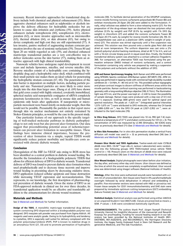
Design of the TDDS. A monolithic matrix-type transdermal drug deliverysystem containing DFO dispersed within a biodegradable polymer wasdesigned. DFO mesylate salt powder was purchased from Sigma-Aldrich. Allreagents used were analytic grade. Owing to its hydrophilicity and tendencyto crystallize, DFO is especially well suited for delivery complexed with thepolymer polyvinylpyrrolidone (PVP). PVP is known to stabilize drugs inan amorphous form (27, 33) and to promote permeation of hydrophilic
molecules (59). To facilitate dermal penetration of the DFO/PVP complexes,reverse micelle-forming nonionic surfactants polysorbate 80 (Tween 80) andsorbitan monolaurete 20 (Span 20) (26) were added to the formulation. Fi-nally, ethyl cellulose was added to form a slow-releasing matrix (27). For thepreparation of the drug-release layer we dissolved the two polymers ethylcellulose (3.5% by weight) and PVP (0.5% by weight) with 1% DFO (byweight) in chloroform (27) and added the nonionic surfactants Tween 80and Span 20 (1% each, by weight) for reverse micelle formation (26, 59). Di-n-butylphthalate was used as a plasticizer (30% weight-in-weight of poly-mers) (27). The solution was stirred vigorously until a fine suspension wasachieved. This solution was then poured onto a sterile glass Petri dish anddried at room temperature. The uniform dispersion was cast onto a 2%(wt/vol) polyvinyl alcohol backing membrane, dried at 40 °C for 6 h, and cutwith a 16-mm circular biopsy punch in equal-sized discs. Finally, the finishedtransdermal delivery system was attached to a contact adhesive (Tegaderm;3M). For comparison, an alternative TDDS was formulated using the per-meation enhancer DMSO instead of nonionic surfactants, and a controlformulation containing only vehicle was prepared by making a suspensionof the polymers and surfactants without the addition of DFO.
AFM and Raman Spectroscopy Imaging. Both Raman and AFM were performedusing NTEGRA Spectra combined AFM-Raman system (NT-MDT) (70). AFM im-aging was performed in tapping mode with commercial high-durability roundedcantilevers (k = 5.4 N/m, R ∼40 nm) at 0.7 Hz. This provided surface topographyand phase-contrast images to discern stiffness of different areas within themicelle particles. Raman confocal scanning was performed in backscatteringgeometry with a long-workingMitutoyo objective (100, 0.7 N.A.). The illuminationlight was 473 nm, and the power was kept at ∼2 mW to lower the possibilityof sample damage. Raman maps were produced with a step size of 0.5 mmand 1-s exposure. Gratings (600 g/mm) were used for optimal signal andspectral resolution. The peaks at ∼1,625 cm−1 (integrated spectral intensities1,575–1,675 cm−1) were attributed to DFO molecules, whereas the CH bandsat 2,800–3,050 cm−1, less the DFO CH peak at 2,927–2,952 cm−1, were at-tributed to the lipid molecules (70).
In Vitro Drug Release. DFO TDDS was placed into 10 mL PBS (pH 7.4) main-tained at a temperature of 37 °C and shaken continuously for 14 h (n = 3). Theconcentration of DFO was measured by LC-MS (Shimadzu; AB SCIEX) everyhour as previously described (71) (See SI Materials and Methods for details).
In Vitro Skin Permeation. For in vitro skin permeation studies a vertical Franzdiffusion cell model was used (n = 3) as previously described (34) (see SIMaterials and Methods for details).
Pressure Ulcer Model and TDDS Application. Twelve-week-old male C57BL/6db/db mice (BKS. CG-M+/+Lepr<db>/J; Jackson Laboratories) were random-ized into the following groups: DFO TDDS-treated versus vehicle TDDScontrol (n = 10). Pressure ulcers on the dorsum of db/db mice were inducedas previously described (36) (see SI Materials and Methods for details).
Ulcer Wound Analysis. Digital photographs were taken before ulcer initiation,the day after, and every other day until closure. Ulcer closure was defined asthe time at which the wound was completely re-epithelialized. Ulcer woundarea was determined using ImageJ software (National Institutes of Health).
Histology. After the mice were euthanized wounds were harvested with a 2-mm rim of unwounded skin. Skin tissues were fixed in 4% paraformaldehydeovernight followed by serial dehydration in ethanol and embedding inparaffin. Five-micrometer sections were stained with H&E or picrosirius red.Frozen tissue samples for CD31 immunohistochemistry and DHE stain wereprepared by immediate optimum cutting temperature (OCT) embedding(Sakura Finetek) (see SI Materials and Methods for details).
Statistical Analysis. Statistical analysis was performed using either ANOVAor an unpaired Student t test (MATLAB). Values are presented as means ±SEM. P values < 0.05 were considered statistically significant.
ACKNOWLEDGMENTS. The authors thank Prof. Jürgen Stampfl (TechnicalUniversity Vienna) for insightful discussion of the manuscript and RevanthKosaraju for proofreading. Funding for wound healing research in our lab-oratory has been provided by the National Institutes of Health (R01-DK074095, R01-AG025016, and R03-DK094521), the Harrington DiscoveryInstitute, the Hagey Family Endowed Fund in Stem Cell Research and Re-generative Medicine, and The Oak Foundation.
98 | www.pnas.org/cgi/doi/10.1073/pnas.1413445112 Duscher et al.

1. Blonde L (2011) Improving care for patients with type 2 diabetes: Applying man-agement guidelines and algorithms, and a review of new evidence for incretin agentsand lifestyle intervention. Am J Manag Care 17(Suppl 14):S368–S376.
2. Golden SH (2011) Emerging therapeutic approaches for the management of diabetesmellitus and macrovascular complications. Am J Cardiol 108(3, Suppl):59B–67B.
3. American Diabetes Association (2013) Economic costs of diabetes in the U.S. in 2012.Diabetes Care 36(4):1033–1046.
4. Jan YK, Shen S, Foreman RD, Ennis WJ (2013) Skin blood flow response to locallyapplied mechanical and thermal stresses in the diabetic foot. Microvasc Res 89:40–46.
5. Brem H, Tomic-Canic M (2007) Cellular and molecular basis of wound healing in di-abetes. J Clin Invest 117(5):1219–1222.
6. Brem H, Lyder C (2004) Protocol for the successful treatment of pressure ulcers. Am JSurg 188(1A Suppl):9–17.
7. Blakytny R, Jude E (2006) The molecular biology of chronic wounds and delayedhealing in diabetes. Diabet Med 23(6):594–608.
8. Wukich DK, et al. (2013) Inpatient management of diabetic foot disorders: a clinicalguide. Diabetes Care 36(9):2862–2871.
9. Barshes NR, et al. (2013) The system of care for the diabetic foot: objectives, out-comes, and opportunities. Diabet Foot Ankle, 10.3402/dfa.v4i0.21847.
10. Ziegler-Graham K, MacKenzie EJ, Ephraim PL, Travison TG, Brookmeyer R (2008) Es-timating the prevalence of limb loss in the United States: 2005 to 2050. Arch Phys MedRehabil 89(3):422–429.
11. Faglia E, Clerici G, Caminiti M, Curci V, Somalvico F (2013) Influence of osteomyelitislocation in the foot of diabetic patients with transtibial amputation. Foot Ankle Int34(2):222–227.
12. Tepper OM, et al. (2002) Human endothelial progenitor cells from type II diabeticsexhibit impaired proliferation, adhesion, and incorporation into vascular structures.Circulation 106(22):2781–2786.
13. Capla JM, et al. (2007) Diabetes impairs endothelial progenitor cell-mediated bloodvessel formation in response to hypoxia. Plast Reconstr Surg 119(1):59–70.
14. Ceradini DJ, et al. (2004) Progenitor cell trafficking is regulated by hypoxic gradientsthrough HIF-1 induction of SDF-1. Nat Med 10(8):858–864.
15. Sarkar K, Fox-Talbot K, Steenbergen C, Bosch-Marcé M, Semenza GL (2009) Adeno-viral transfer of HIF-1alpha enhances vascular responses to critical limb ischemia indiabetic mice. Proc Natl Acad Sci USA 106(44):18769–18774.
16. Bader HL, Hsu T (2012) Systemic VHL gene functions and the VHL disease. FEBS Lett586(11):1562–1569.
17. Thangarajah H, et al. (2010) HIF-1alpha dysfunction in diabetes. Cell Cycle 9(1):75–79.18. Thangarajah H, et al. (2009) The molecular basis for impaired hypoxia-induced VEGF
expression in diabetic tissues. Proc Natl Acad Sci USA 106(32):13505–13510.19. Andrews NC (1999) Disorders of iron metabolism. N Engl J Med 341(26):1986–1995.20. Barnham KJ, Masters CL, Bush AI (2004) Neurodegenerative diseases and oxidative
stress. Nat Rev Drug Discov 3(3):205–214.21. Bergeron RJ, et al. (2003) Methoxylation of desazadesferrithiocin analogues: En-
hanced iron clearing efficiency. J Med Chem 46(8):1470–1477.22. Botusan IR, et al. (2008) Stabilization of HIF-1alpha is critical to improve wound
healing in diabetic mice. Proc Natl Acad Sci USA 105(49):19426–19431.23. Sundin BM, et al. (2000) The role of allopurinol and deferoxamine in preventing
pressure ulcers in pigs. Plast Reconstr Surg 105(4):1408–1421.24. Hom DB, Goding GS, Jr, Price JA, Pernell KJ, Maisel RH (2000) The effects of conju-
gated deferoxamine in porcine skin flaps. Head Neck 22(6):579–584.25. Miller MJ (1989) Syntheses and therapeutic potential of hydroxamic acid based
siderophores and analogs. Chem Rev 89(7):1563–1579.26. Som I, Bhatia K, Yasir M (2012) Status of surfactants as penetration enhancers in
transdermal drug delivery. J Pharm Bioallied Sci 4(1):2–9.27. Kandavilli S, Nair V, Panchagnula R (2002) Polymers in transdermal drug delivery
systems. Pharm Tech 26(5):62–81.28. Müller-Goymann CC (2004) Physicochemical characterization of colloidal drug de-
livery systems such as reverse micelles, vesicles, liquid crystals and nanoparticles fortopical administration. Eur J Pharm Biopharm 58(2):343–356.
29. Jones M-C, Gao H, Leroux J-C (2008) Reverse polymeric micelles for pharmaceuticalapplications. J Control Release 132(3):208–215.
30. Bhatnagar S, Vyas SP (1994) Organogel-based system for transdermal delivery ofpropranolol. J Microencapsul 11(4):431–438.
31. Gupta R, Mukherjee B (2003) Development and in vitro evaluation of diltiazem hy-drochloride transdermal patches based on povidone-ethylcellulose matrices. DrugDev Ind Pharm 29(1):1–7.
32. Gabiga H, Cal K, Janicki S (2000) Effect of penetration enhancers on isosorbide dini-trate penetration through rat skin from a transdermal therapeutic system. Int J Pharm199(1):1–6.
33. Rao PR, Diwan PV (1998) Formulation and in vitro evaluation of polymeric films ofdiltiazem hydrochloride and indomethacin for transdermal administration. Drug DevInd Pharm 24(4):327–336.
34. Franz TJ (1975) Percutaneous absorption on the relevance of in vitro data. J InvestDermatol 64(3):190–195.
35. Anigbogu AN, Williams AC, Barry BW, Edwards HG (1995) Fourier transform Ramanspectroscopy of interactions between the penetration enhancer dimethyl sulfoxideand human stratum corneum. Int J Pharm 125(2):265–282.
36. Stadler I, Zhang RY, Oskoui P, Whittaker MS, Lanzafame RJ (2004) Development of
a simple, noninvasive, clinically relevant model of pressure ulcers in the mouse. J In-
vest Surg 17(4):221–227.37. Martin A, Komada MR, Sane DC (2003) Abnormal angiogenesis in diabetes mellitus.
Med Res Rev 23(2):117–145.38. Eefting F, et al. (2004) Role of apoptosis in reperfusion injury. Cardiovasc Res 61(3):
414–426.39. Mustoe T (2004) Understanding chronic wounds: A unifying hypothesis on their
pathogenesis and implications for therapy. Am J Surg 187(5A):65S–70S.40. Chan W, et al. (2012) Effect of iron chelation on myocardial infarct size and oxidative
stress in ST-elevation-myocardial infarction. Circ Cardiovasc Interv 5(2):270–278.41. Schäfer M, Werner S (2008) Oxidative stress in normal and impaired wound repair.
Pharmacol Res 58(2):165–171.42. Sreejayan N, Ren J (2007) Vascular Biology Protocols (Humana, Totowa, NJ), p xi.43. Nagai MK, Embil JM (2002) Becaplermin: Recombinant platelet derived growth factor,
a new treatment for healing diabetic foot ulcers. Expert Opin Biol Ther 2(2):211–218.44. Papanas N, Maltezos E (2010) Benefit-risk assessment of becaplermin in the treatment
of diabetic foot ulcers. Drug Saf 33(6):455–461.45. Knowles EA, et al. (2002) Offloading diabetic foot wounds using the scotchcast boot:
A retrospective study. Ostomy Wound Manage 48(9):50–53.46. Dumville JC, Deshpande S, O’Meara S, Speak K (2011) Foam dressings for healing
diabetic foot ulcers. Cochrane Database Syst Rev (9):CD009111.47. Price PE, et al. (2008) Dressing-related pain in patients with chronic wounds: An in-
ternational patient perspective. Int Wound J 5(2):159–171.48. Dumville JC, Deshpande S, O’Meara S, Speak K (2013) Hydrocolloid dressings for
healing diabetic foot ulcers. Cochrane Database Syst Rev 8:CD009099.49. Giacco F, Brownlee M (2010) Oxidative stress and diabetic complications. Circ Res
107(9):1058–1070.50. Brownlee M (2001) Biochemistry and molecular cell biology of diabetic complications.
Nature 414(6865):813–820.51. Prass K, et al. (2002) Desferrioxamine induces delayed tolerance against cerebral is-
chemia in vivo and in vitro. J Cereb Blood Flow Metab 22(5):520–525.52. Zhu Y, Zhang L, Gidday JM (2008) Deferroxamine preconditioning promotes long-
lasting retinal ischemic tolerance. J Ocul Pharmacol Ther 24(6):527–535.53. Hartley A, Davies M, Rice-Evans C (1990) Desferrioxamine as a lipid chain-breaking
antioxidant in sickle erythrocyte membranes. FEBS Lett 264(1):145–148.54. Dendorfer A, et al. (2005) Deferoxamine induces prolonged cardiac preconditioning
via accumulation of oxygen radicals. Free Radic Biol Med 38(1):117–124.55. Prausnitz MR, Langer R (2008) Transdermal drug delivery. Nat Biotechnol 26(11):
1261–1268.56. Brown MB, Martin GP, Jones SA, Akomeah FK (2006) Dermal and transdermal drug
delivery systems: Current and future prospects. Drug Deliv 13(3):175–187.57. Prausnitz MR, Mitragotri S, Langer R (2004) Current status and future potential of
transdermal drug delivery. Nat Rev Drug Discov 3(2):115–124.58. Ihnat PM, Vennerstrom JL, Robinson DH (2000) Synthesis and solution properties of
deferoxamine amides. J Pharm Sci 89(12):1525–1536.59. Williams AC, Barry BW (2012) Penetration enhancers. Adv Drug Deliv Rev 64(Suppl):
128–137.60. Kalia YN, Naik A, Garrison J, Guy RH (2004) Iontophoretic drug delivery. Adv Drug
Deliv Rev 56(5):619–658.61. Machet L, Boucaud A (2002) Phonophoresis: Efficiency, mechanisms and skin toler-
ance. Int J Pharm 243(1-2):1–15.62. Denet AR, Vanbever R, Préat V (2004) Skin electroporation for transdermal and
topical delivery. Adv Drug Deliv Rev 56(5):659–674.63. Sun W, et al. (2013) Polyvinylpyrrolidone microneedles enable delivery of intact
proteins for diagnostic and therapeutic applications. Acta Biomater 9(8):7767–7774.64. Levin G, et al. (2005) Transdermal delivery of human growth hormone through RF-
microchannels. Pharm Res 22(4):550–555.65. Herndon TO, Gonzalez S, Gowrishankar TR, Anderson RR, Weaver JC (2004) Trans-
dermal microconduits by microscission for drug delivery and sample acquisition. BMC
Med 2:12.66. Pegoraro C, MacNeil S, Battaglia G (2012) Transdermal drug delivery: From micro to
nano. Nanoscale 4(6):1881–1894.67. Yang Y, Bugno J, Hong S (2013) Nanoscale polymeric penetration enhancers in topical
drug delivery. Polym Chem.68. Jones M-C, Leroux J-C (2010) Reverse micelles from amphiphilic branched polymers.
Soft Matter 6(23):5850–5859.69. Zheng D, et al. (2012) Topical delivery of siRNA-based spherical nucleic acid nano-
particle conjugates for gene regulation. Proc Natl Acad Sci USA 109(30):11975–11980.70. Jiang X, et al. (2014) Promotion of airway anastomotic microvascular regeneration
and alleviation of airway ischemia by deferoxamine nanoparticles. Biomaterials 35(2):
803–813.71. Cermanova J, et al. (2014) Deferoxamine but not dexrazoxane alleviates liver injury
induced by endotoxemia in rats. Shock 42(4):372–379.
Duscher et al. PNAS | January 6, 2015 | vol. 112 | no. 1 | 99
BIOCH
EMISTR
YEN
GINEE
RING