outcomes of childhood hemangiomas treated with the pulsed-dye
TRANSCRIPT
Outcomes of Childhood Hemangiomas Treated with thePulsed-Dye Laser with Dynamic Cooling: A RetrospectiveChart Analysis
CARINA RIZZO, MD,� LORI BRIGHTMAN, MD,y ANNE M. CHAPAS, MD,�y ELIZABETH K. HALE, MD,�y
JULIE L. CANTATORE-FRANCIS, MD,� LEONARD J. BERNSTEIN, MD,y AND ROY G. GERONEMUS, MD�y
BACKGROUND Laser treatment of childhood hemangiomas remains controversial. Previous studieshave used outdated technology, resulting in a potential overrepresentation of adverse outcomes.
OBJECTIVE To evaluate outcomes of hemangiomas treated with the most current laser technology.
METHODS A retrospective chart analysis of 90 patients with a median age of 3.0 months and a total of105 hemangiomas were enrolled over a 2.5-year period. All were treated with the 595-nm long-pulsepulsed-dye laser (LP-PDL) with dynamic epidermal cooling at 2- to 8-week intervals depending on thestage of growth. Exclusion criteria were previous laser, surgical, or corticosteroid treatment. Threereviewers assessed outcomes.
RESULTS Near-complete or complete clearance in color were achieved for 85 (81%) and in thickness for67 (64%) hemangiomas. There was no scarring or atrophy. Ulceration occurred in one case and resolvedduring treatment. Hyperpigmentation and hypopigmentation occurred in 4% and 14% of hemangiomas,respectively.
CONCLUSION Early treatment of childhood hemangiomas with the 595-nm LP-PDL with dynamic cool-ing may reduce the proliferative phase and result in excellent rates of clearing and few adverse events.
Dr. Geronemus serves on the Advisory Board for Candela Corporation.
Hemangiomas are vascular tumors affecting up
to 5% to 10% of infants. The lesions are
present at birth or develop shortly after and are
characterized by a sequence of rapid proliferation,
stabilization, and slow involution. Conservative
management is often initially favored because of the
potential for spontaneous involution, although the
behavior of individual hemangiomas is heteroge-
neous, making it difficult to predict outcomes
reliably. Approximately 20% of hemangiomas result
in pain, bleeding, ulceration, infection, or functional
impairment with vision, feeding, or breathing
necessitating medical or surgical treatment.1
Involution is incomplete by the start of school in
50% of children, and even after spontaneous
involution, 15% to 40% of children are left with
skin changes, including pigmentary alteration,
telangiectasia, and atrophy, as well as fibro-fatty
residuum often necessitating surgical correction.2,3
Treatment of early hemangiomas with laser therapy
may hasten the involutional process and improve
rates of complete clinical clearance, but published
studies have not shown consistent benefits, and laser
treatment of hemangiomas has remained controver-
sial. This is due in part to a lag in the use and eval-
uation of the most advanced laser technology,
resulting in an overreporting of adverse outcomes
due to thermally induced epidermal damage.
The long-pulse pulsed-dye laser (LP-PDL), with a
wavelength of 595 nm and the addition of dynamic
epidermal cooling, has become the standard of care
for treatment of port wine stains. This modality has
& 2009 by the American Society for Dermatologic Surgery, Inc. � Published by Wiley Periodicals, Inc. �ISSN: 1076-0512 � Dermatol Surg 2009;35:1–8 � DOI: 10.1111/j.1524-4725.2009.01356.x
1
�New York University Skin and Cancer Unit, New York, New York; yLaser & Skin Surgery Center of New York,New York, New York
proven effective in the treatment of ulcerated hem-
angiomas,4 and the addition of epidermal cooling
has been demonstrated to decrease the frequency of
adverse events when used in combination with long
pulse widths,5 although to our knowledge, no pub-
lished outcomes are available evaluating the appli-
cation of this updated technology at standard pulse
widths for the treatment of uncomplicated infantile
hemangiomas. This retrospective chart analysis was
undertaken to assess the rate of clearing and adverse
events including atrophy, scarring, ulceration, and
pigmentary changes of early hemangiomas treated
with the 595-nm-wavelength LP-PDL with dynamic
cooling.
Patients and Methods
The records of all consecutive hemangioma patients
treated with LP-PDL for superficial or mixed
superficial and deep hemangiomas in a laser surgery
clinic specializing in the treatment of vascular
lesions from January 2005 to September 2007 were
screened against eligibility criteria. Inclusion
required the treatment of a superficial or mixed
hemangioma with the 595-nm LP-PDL with
dynamic cooling. Exclusion criteria were previous
laser or surgical intervention or previous or con-
comitant treatment with intralesional or systemic
corticosteroids. One patient was excluded because of
the poor quality of photographic documentation.
The parents of all patients gave written informed
consent for photography and participation in
clinical research before inclusion, and the Essex
institutional review board, Essex, New Jersey,
approved the study protocol.
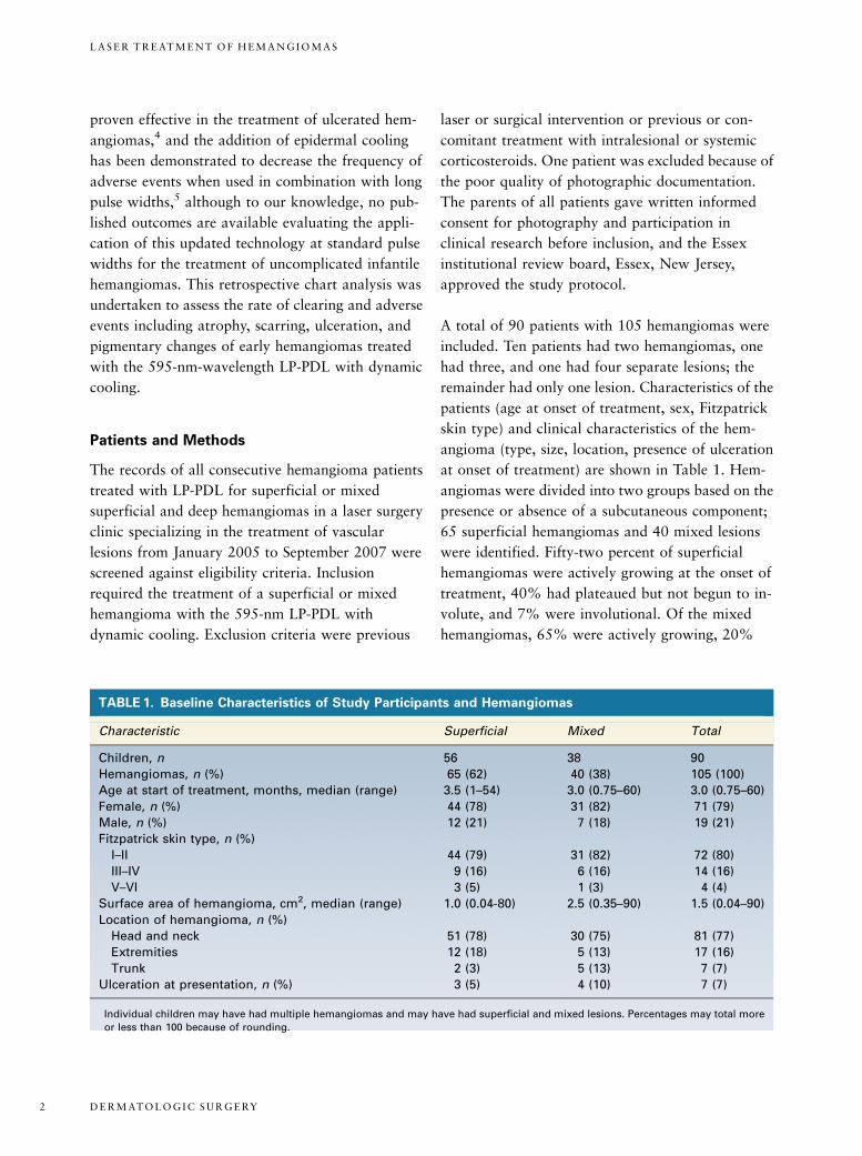
A total of 90 patients with 105 hemangiomas were
included. Ten patients had two hemangiomas, one
had three, and one had four separate lesions; the
remainder had only one lesion. Characteristics of the
patients (age at onset of treatment, sex, Fitzpatrick
skin type) and clinical characteristics of the hem-
angioma (type, size, location, presence of ulceration
at onset of treatment) are shown in Table 1. Hem-
angiomas were divided into two groups based on the
presence or absence of a subcutaneous component;
65 superficial hemangiomas and 40 mixed lesions
were identified. Fifty-two percent of superficial
hemangiomas were actively growing at the onset of
treatment, 40% had plateaued but not begun to in-
volute, and 7% were involutional. Of the mixed
hemangiomas, 65% were actively growing, 20%
TABLE 1. Baseline Characteristics of Study Participants and Hemangiomas
Characteristic Superficial Mixed Total
Children, n 56� 38� 90�
Hemangiomas, n (%) 65 (62) 40 (38) 105 (100)
Age at start of treatment, months, median (range) 3.5 (1–54) 3.0 (0.75–60) 3.0 (0.75–60)
Female, n (%) 44 (78) 31 (82) 71 (79)
Male, n (%) 12 (21) 7 (18) 19 (21)
Fitzpatrick skin type, n (%)
I–II 44 (79) 31 (82) 72 (80)
III–IV 9 (16) 6 (16) 14 (16)
V–VI 3 (5) 1 (3) 4 (4)
Surface area of hemangioma, cm2, median (range) 1.0 (0.04-80) 2.5 (0.35–90) 1.5 (0.04–90)
Location of hemangioma, n (%)
Head and neck 51 (78) 30 (75) 81 (77)
Extremities 12 (18) 5 (13) 17 (16)
Trunk 2 (3) 5 (13) 7 (7)
Ulceration at presentation, n (%) 3 (5) 4 (10) 7 (7)
�Individual children may have had multiple hemangiomas and may have had superficial and mixed lesions. Percentages may total more
or less than 100 because of rounding.
D E R M AT O L O G I C S U R G E RY2
L A S E R T R E AT M E N T O F H E M A N G I O M A S

plateaued, and 11% were involutional. No hem-
angiomas had completely involuted before the start
of treatment. Ulceration was present at the time of
initial evaluation before the initiation of laser
therapy for three superficial and four mixed
hemangiomas.
A 595-nm LP-PDL (V-Beam Perfecta, Candela Corp,
Wayland, MA) associated with a dynamic skin
cooling system was used for all treatments. A spot
size of 7 or 10 mm was used with an average energy
fluence of 11.5 J/cm2 (range 7.5–14.0 J/cm2) or
8.6 J/cm2 (range 6.2–11.5 J/cm2), respectively.
Energy fluence varied according to patient and
hemangioma characteristics, including age, skin
type, location, lesion thickness, and response to
previous treatments. Pulse duration was 0.45 or
1.5 ms depending on the response to prior treatments
and anticipated size of target vessels. Dynamic
cooling device spray with a duration of 30 ms was
applied before each laser pulse, followed by a 30-ms
post-laser pulse delay. Treatments were performed
with a spot overlap of 0% to 20% as needed to
achieve purpura of the entire lesion. Darker skin
types were treated with less spot overlap to minimize
the risk of epidermal damage. Almost all hem-
angiomas were treated without anesthesia, with
topical anesthetic applied before treatment of larger
lesions only for patients aged 1 year and older. For
the treatment of lesions near the orbit, metal eye
shields were applied after corneal anesthesia with
tetricaine 0.7% solution eye drops. Parents were
instructed to avoid trauma and sun exposure to
treated areas and to apply hydrophilic ointment if
crusting developed.
Treatments were repeated at 2- to 8-week intervals,
depending on patient compliance with the treatment
schedule and stage of growth; actively growing hem-
angiomas were treated at 2-week intervals to help
minimize regrowth between treatment sessions,6
whereas lesions that had plateaued were treated at
longer intervals of 4 to 6 weeks. Treatments were
continued until the lesion cleared or stopped
responding or the parent discontinued treatment.
Standardized clinical photographs were taken on the
date of initial evaluation and before each treatment.
Three reviewers (AMC, EKH and JLC) with spe-
cialties in dermatologic laser surgery or pediatric
dermatology independently assessed initial, inter-
mediate, and final treatment photographs. No stan-
dardized or validated method exists for outcomes
measurement of hemangiomas. To facilitate com-
parison, we chose a rating method similar to that
employed in other hemangioma studies. Improve-
ment in color and lesion thickness were graded as
none (0%), minimal (1–24%), fair (25–49%), mod-
erate (50–75%), near complete (76–99%) or com-
plete (100%) resolution. Average values were
calculated when reviewer assessment differed. The
presence or absence of scarring, atrophy, pigmentary
changes, and ulceration were documented when at
least two of three independent reviewers identified
them. The unpaired Student t-test was used to an-
alyze the relationship between age at start of treat-
ment, number of laser treatments, and duration of
follow-up and primary and secondary outcomes.
Results
Ninety patients with 105 hemangiomas treated with
the LP-PDL were evaluated. Mean duration of fol-
low-up was 9.3 months (range 1–72 months): 8.1
months for superficial and 11.4 months for mixed
hemangiomas. The mean number of treat-
ments7 standard deviation was 6.77 4.5:
5.874.2 for superficial and 8.074.7 for mixed
hemangiomas. The number of laser treatments
per child is shown in Figure 1; overall, 28% of
participants required three or fewer treatments, 72%
required seven or fewer treatments, and 11%
underwent more than 12 treatments.
Table 2 shows the improvement in color and thick-
ness at the conclusion of treatment. Fifteen of 65
(23%) superficial hemangiomas demonstrated com-
plete clearance in color, and 38 (58%) demonstrated
near-complete clearance; only two (3%) demon-
strated minimal clearance. Of 40 mixed superficial
and deep hemangiomas, six (15%) demonstrated
3 5 :* * : 2 0 0 9 3
R I Z Z O E T A L

complete clearance of color, 26 (65%) demonstrated
near-complete clearance, and three (8%) had mini-
mal change. Altogether, 81% of hemangiomas
demonstrated better than 75% clearance of color.
Improvement in the thickness of hemangiomas was
separately assessed. Of 65 superficial hemangiomas,
13 (20%) demonstrated no residual thickness, and
38 (58%) demonstrated near-complete resolution of
thickness at the conclusion of treatment. Only two
(3%) superficial hemangiomas demonstrated mini-
mal change in thickness. Of 40 mixed superficial and
deep hemangiomas, only three (8%) demonstrated
complete resolution of thickness, and 13 (33%)
demonstrated near-complete clearance. No im-
provement was noted in one lesion, and 11 (28%)
had minimal change in thickness. Overall, 64% of
hemangiomas demonstrated near-complete or com-
plete resolution in thickness. Representative cases of
complete and near-complete clearance of color and
thickness of superficial and mixed hemangiomas are
shown in Figures 2 and 3, respectively. Age at start of
treatment, lesion size, duration of follow-up, and
number of laser treatments were not significantly
correlated with clearance rates (data not shown).
Adverse events are shown in Table 3. There were no
cases of atrophy or hypertrophic scarring. There was
only one case of minor ulceration that developed
after the onset of treatment and thus potentially
could be a result of laser therapy. Ulceration was
present at initial evaluation, before the onset of
treatment, for three superficial and four mixed
hemangiomas. All ulcers resolved without scarring
during the course of treatment and did not require
treatment to be stopped or delayed. On the date of
final treatment, hyperpigmentation was noted in
four (6%) patients with superficial hemangiomas.
TABLE 2. Primary Outcome Measures at the Completion of Treatment with the Long-Pulse Pulsed-Dye
Laser with Dynamic Cooling
Improvement
n (%)
Superficial (n = 65) Mixed (n = 40) Total (n = 105)
Color
Complete (100%) 15 (23) 6 (15) 21 (20)
Near complete (75–99%) 38 (58) 26 (65) 64 (61)
Moderate (50–74%) 9 (14) 3 (8) 12 (11)
Fair (25–49%) 1 (2) 2 (5) 3 (3)
Minimal (1–25) 2 (3) 3 (8) 5 (5)
None (0%) 0 (0) 0 (0) 0 (0)
Thickness
Complete (100%) 13 (20) 3 (8) 16 (15)
Near complete (75–99%) 38 (58) 13 (33) 51 (49)
Moderate (50–74%) 10 (15) 4 (10) 14 (13)
Fair (25–49%) 2 (3) 8 (20) 10 (10)
Minimal (1–25) 2 (3) 11 (28) 13 (12)
None (0%) 0 (0) 1 (3) 1 (1)
Three independent reviewers evaluated cases, and averages were used in cases in which reviewer assessment differed. Percentages may
total more or less than 100 because of rounding.
Figure 1. Number of laser treatments per hemangioma.
D E R M AT O L O G I C S U R G E RY4
L A S E R T R E AT M E N T O F H E M A N G I O M A S

There were no cases of hyperpigmentation in pa-
tients with mixed lesions. Hypopigmentation was
present on the date of final treatment in 10 (15%)
and five (13%) patients with superficial and mixed
hemangiomas, respectively Dyspigmentation was
not correlated with Fitzpatrick skin type. The de-
velopment of hypopigmentation was significantly
associated with a greater number of laser treatments,
with a mean of 9.175.5 in the group with hypo-
pigmentation, compared with 6.374.2 in those
without (p = .03). Because these treatments were
administered at 2- to 6-week intervals, these patients
were necessarily followed for a longer duration of
time: an average of 17.37 18.6 months in the group
with hypopigmentation, compared with 8.07 8.9
months in the group without (po.003). Hyperpig-
mentation, in contrast, was associated with fewer
treatments over a shorter duration of time: an aver-
age of 4.271.0 treatments over 2.77 1.0 months in
the group with hyperpigmentation, compared with
6.874.6 treatments over 9.67 11.3 months in the
group without hyperpigmentation, which did not
reach statistical significance (p = .27 for number of
treatments, p = .23 for duration of follow-up).
Because clinical photographs were not taken after
the date of final treatment, we were not able to
determine the persistence of these pigmentary
changes.
Discussion
It has been suggested that early treatment of infantile
hemangiomas with the LP-PDL hastens involution
and resolution.7,8 This treatment modality has
shown exceptional success and safety in the treat-
ment of port wine stains and has rapidly become
first-line treatment of these lesions, but optimization
of hemangioma treatment has proven more chal-
lenging, and laser intervention remains controver-
sial. This is largely because of the distinct
characteristics of hemangiomas, which are rapidly
proliferating tumors of densely packed small vessels
with little interspersed dermis and significant depth,
presenting a challenge for optimal laser targeting. It
is also due in part to the unpredictable behavior of
individual hemangiomas making it difficult to reli-
ably assign outcomes to treatment rather than the
natural history of the tumor.
The goal of laser treatment of hemangiomas is to
maximize vascular damage while minimizing non-
specific injury to interspersed dermis and overlying
epidermis. The combination of pulse duration, spot
size, and wavelength determines the effect of laser
treatment on cutaneous blood vessels. Earlier pulsed-
dye lasers (PDLs) had a wavelength of 585 nm, cor-
responding to an absorption peak of hemoglobin.
More recently, LP-PDLs with wavelengths of 595 nm
have enabled greater depth of vascular injury while
maintaining chromophore selectivity. Previous stud-
ies employing the 585-nm PDL with a pulse width of
0.45 ms or less have shown disappointing results in
the treatment of hemangiomas with a subcutaneous
component.9–12 The LP-PDL with dynamic cooling
employing a 595-nm wavelength and pulse widths of
up to 1.5 ms allows better targeting of the deeper
component of hemangiomas.
Figure 2. Representative examples of superficial hem-angiomas at initial evaluation (left), and demonstratingcomplete (A) or near complete clearance (B) at the conclu-sion of treatment with the LP-PDL with dynamic cooling(right) after 2 (A) and 4 (B) treatments, respectively.
3 5 :* * : 2 0 0 9 5
R I Z Z O E T A L

In 2002, Batta and colleagues published the first
randomized controlled trial comparing no interven-
tion with early treatment with the 585-nm PDL
without epidermal cooling for superficial childhood
hemangiomas.7 The study showed that early treat-
ment was significantly more likely to result in com-
plete clearance of hemangiomas at 1 year of age
(30% of those treated with early PDL vs only 5% of
the control group, po.001), but the authors also
reported high rates of adverse outcomes, including
skin atrophy (28% of the treatment group, 8% of
controls) and hypopigmentation (45% of the treat-
Figure 3. Representative examples of mixed superficial and deep hemangiomas at initial evaluation (left), after 6 treatments(center), and demonstrating near complete clearance at the conclusion of treatment with the LP-PDL with dynamic cooling(right) after a total of 7 treatments (A) and 10 treatments (B), respectively.
TABLE 3. Documented Adverse Events
Adverse Event
n (%)
Superficial (n = 65) Mixed (n = 40) Total (n = 105)
Hyperpigmentation 4 (6) 0 (0) 4 (4)
Hypopigmentation 10 (15) 5 (13) 15 (14)
Ulceration 1 (2) 0 (0) 1 (1)
Atrophy 0 (0) 0 (0) 0 (0)
Hypertrophic scarring 0 (0) 0 (0) 0 (0)
There were no cases of atrophy or hypertrophic scarring. Ulceration reflects onset after the initiation of laser therapy.
D E R M AT O L O G I C S U R G E RY6
L A S E R T R E AT M E N T O F H E M A N G I O M A S
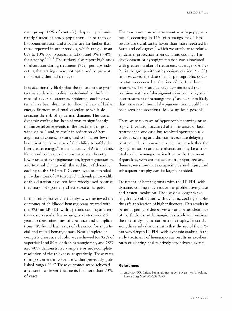
ment group, 15% of controls), despite a predomi-
nantly Caucasian study population. These rates of
hypopigmentation and atrophy are far higher than
those reported in other studies, which ranged from
0% to 10% for hypopigmentation and 0% to 4%
for atrophy.9,10,13 The authors also report high rates
of ulceration during treatment (7%), perhaps indi-
cating that settings were not optimized to prevent
nonspecific thermal damage.
It is additionally likely that the failure to use pro-
tective epidermal cooling contributed to the high
rates of adverse outcomes. Epidermal cooling sys-
tems have been designed to allow delivery of higher
energy fluences to dermal vasculature while de-
creasing the risk of epidermal damage. The use of
dynamic cooling has been shown to significantly
minimize adverse events in the treatment of port
wine stains14 and to result in reduction of hem-
angioma thickness, texture, and color after fewer
laser treatments because of the ability to safely de-
liver greater energy.4 In a small study of Asian infants,
Kono and colleagues demonstrated significantly
lower rates of hypopigmentation, hyperpigmentation,
and textural change with the addition of dynamic
cooling to the 595-nm PDL employed at extended
pulse durations of 10 to 20 ms,5 although pulse widths
of this duration have not been widely used because
they may not optimally affect vascular targets.
In this retrospective chart analysis, we reviewed the
outcomes of childhood hemangiomas treated with
the 595-nm LP-PDL with dynamic cooling at a ter-
tiary care vascular lesion surgery center over 2.5
years to determine rates of clearance and complica-
tions. We found high rates of clearance for superfi-
cial and mixed hemangiomas. Near-complete or
complete clearance of color was achieved for 82% of
superficial and 80% of deep hemangiomas, and 78%
and 40% demonstrated complete or near-complete
resolution of the thickness, respectively. These rates
of improvement in color are within previously pub-
lished ranges.7,9,10 These outcomes were achieved
after seven or fewer treatments for more than 70%
of cases.
The most common adverse event was hypopigmen-
tation, occurring in 14% of hemangiomas. These
results are significantly lower than those reported by
Batta and colleagues,7 which we attribute to relative
epidermal protection from dynamic cooling. The
development of hypopigmentation was associated
with greater number of treatments (average of 6.3 vs
9.1 in the group without hypopigmentation, p = .03).
In most cases, the date of final photographic docu-
mentation occurred at the time of the final laser
treatment. Prior studies have demonstrated the
transient nature of dyspigmentation occurring after
laser treatment of hemangiomas;9 as such, it is likely
that some resolution of dyspigmentation would have
been seen had additional follow-up been possible.
There were no cases of hypertrophic scarring or at-
rophy. Ulceration occurred after the onset of laser
treatment in one case but resolved spontaneously
without scarring and did not necessitate delaying
treatment. It is impossible to determine whether the
dyspigmentation and rare ulceration may be attrib-
uted to the hemangioma itself or to the treatment.
Regardless, with careful selection of spot size and
fluence, we show that nonspecific dermal injury and
subsequent atrophy can be largely avoided.
Treatment of hemangiomas with the LP-PDL with
dynamic cooling may reduce the proliferative phase
and hasten involution. The use of a longer wave-
length in combination with dynamic cooling enables
the safe application of higher fluences. This results in
better targeting of deeper vessels and better clearance
of the thickness of hemangiomas while minimizing
the risk of dyspigmentation and atrophy. In conclu-
sion, this study demonstrates that the use of the 595-
nm-wavelength LP-PDL with dynamic cooling in the
early treatment of hemangiomas results in excellent
rates of clearing and relatively few adverse events.
References
1. Anderson RR. Infant hemangiomas: a controversy worth solving.
Lasers Surg Med 2006;38:92–3.
3 5 :* * : 2 0 0 9 7
R I Z Z O E T A L

2. Drolet BA, Esterly NB, Frieden IJ. Hemangiomas in children. N
Engl J Med 1999;341:173–81.
3. Enjolras O, Riche MC, Merland JJ, et al. Management of alarm-
ing hemangiomas in infancy: a review of 25 cases. Pediatrics
1990;85:491–8.
4. Chang CJ, Kelly KM, Nelson JS. Cryogen spray cooling and
pulsed dye laser treatment of cutaneous hemangiomas. Ann Plast
Surg 2001;46:577–83.
5. Kono T, Sakurai H, Groff WF, et al. Comparison study of a tra-
ditional pulsed dye laser versus a long-pulsed dye laser in the
treatment of early childhood hemangiomas. Lasers Surg Med
2006;38:112–5.
6. Dinehart SM, Kincannon J, Geronemus R. Hemangiomas: eval-
uation and treatment. Dermatol Surg 2001;27:475–85.
7. Batta K, Goodyear HM, Moss C, et al. Randomised controlled
study of early pulsed dye laser treatment of uncomplicated child-
hood haemangiomas: results of a 1-year analysis. Lancet
2002;360:521–7.
8. Garden JM, Bakus AD, Paller AS. Treatment of cutaneous hem-
angiomas by the flashlamp-pumped pulsed dye laser: prospective
analysis. J Pediatr 1992;120:555–60.
9. Hohenleutner S, Badur-Ganter E, Landthaler M, et al. Long-term
results in the treatment of childhood hemangioma with the
flashlamp-pumped pulsed dye laser: an evaluation of 617 cases.
Lasers Surg Med 2001;28:273–7.
10. Poetke M, Philipp C, Berlien HP. Flashlamp-pumped pulsed dye
laser for hemangiomas in infancy: treatment of superficial vs
mixed hemangiomas. Arch Dermatol 2000;136:628–32.
11. Ashinoff R, Geronemus RG. Failure of the flashlamp-pumped
pulsed dye laser to prevent progression to deep hemangioma.
Pediatr Dermatol 1993;10:77–80.
12. Ashinoff R, Geronemus RG. Capillary hemangiomas and treat-
ment with the flash lamp-pumped pulsed dye laser. Arch Dermatol
1991;127:202–5.
13. Raulin C, Greve B. Retrospective clinical comparison of hem-
angioma treatment by flashlamp-pumped (585 nm) and fre-
quency-doubled Nd:YAG (532 nm) lasers. Lasers Surg Med
2001;28:40–3.
14. Waldorf HA, Alster TS, McMillan K, et al. Effect of dynamic
cooling on 585-nm pulsed dye laser treatment of port-wine stain
birthmarks. Dermatol Surg 1997;23:657–62.
Address correspondence and reprint requests to: Roy G.Geronemus, MD, Laser & Skin Surgery Center of NewYork and New York University Skin and Cancer Unit,Director, Laser & Skin Surgery Center of New York, 317East 34th Street, New York, NY 10016, or e-mail:[email protected]
D E R M AT O L O G I C S U R G E RY8
L A S E R T R E AT M E N T O F H E M A N G I O M A S