http:// oy. g-protein coupled receptor
TRANSCRIPT

http://www.youtube.com/watch?v=0Tp7KTf2koY

G-Protein Coupled Receptor

Q. What is the G-Protein Coupled Receptor? 5P
What does G mean?? (NOT; giant, golf, google...)Any examples??

GPCRs are 1. transmembrane receptors that sense molecules outside
the cell and activate inside signal transduction pathways.
2. What is wrong with this definition? 5P

Bacteriorhodopsin is a Paradigm for Membrane Proteins with 7 Helical Segments
Its seven transmembrane segments are connected by short loops.
Note the light-absorbing retinal bound to a lysine residue.
CHAPTER 9. MEMBRANES
Q. Why (only) a helix in the membrane? 5P
1.Schiff base?2.Biological function of bacteriorhodopsin?
Q. Then, why is the chromophore (Schiff base) of bacteriorhodopsin inside of the membrane?2P

• Cell-cell and• Cell-environment
Communication
• GPCR signaling!

GPCR signaling: responding to the outside world
•Cells interact with their environment by interpreting extracellular signals via proteins that span their plasma membrane called receptors
•Receptors are comprised of extracellular and intracellular domains
•The extracellular domain relays information about the outside world to the intracellular domain
•The intracellular domain then interacts with other intracellular signaling proteins
•These intracellular signaling proteins further relay the message to one or more effector proteins (molecules)
•Effector proteins (molecules) mediate the appropriate response

Class A Rhodopsin like (19) Amine (7) Peptide (32) Hormone protein (5) (Rhod)opsin (9) Olfactory (290) Prostanoid (3) Nucleotide-like (2) Cannabinoid (2) Platelet activating factor Gonadotropin-releasing hormone (4) Thyrotropin-releasing hormone and Secretagogue (3) Melatonin (2) Viral (4) Lysosphingolipid and LPA (EDG) (12) Leukotriene B4 receptor (2) Ecdysis triggering hormone receptor Nicotinic acid (niacin) receptor CAPA Class A Orphan/other (14) Class B Secretin like (34) Class C Metabotropic glutamate/pheromone (8) cAMP receptors
~1000 GPCRs

Buck, L. and Axel, R. (1991) Cell, vol. 65, 175-187.

The Nobel Prize in Physiology or Medicine 2004Richard Axel, Linda B. Buck
"for their discoveries of odorant receptors and the organization of the olfactory system"

Specificity in GPCRs• The extracellular domain determines ligand
specificity • The cytoplasmic domain determines G protein
specificity • Together, these two
domains link a particular hormone to a particular signaling pathway

Receiving the Signal: G-protein Coupled Receptors •GPCRs are an important and ubiquitous class of eukaryotic receptors (>1000 in humans)
•The extracellular domain connects to the intracellular domain through 7 transmembrane spans
•The intracellular domain is coupled to a heterotrimeric G-protein
•The heterotrimeric g-protein is composed of 3 subunits: G, G, and G
•When the G subunit is bound to GDP it is “OFF”; when it is bound to GTP it is “ON”
•When the extracellular domain binds to the signal molecule, it causes a conformational change relayed through the transmembrane spans to the intracellular domain
•The conformational change relayed to the intracellular domain causes the G subunit to release GDP and bind to GTP thereby activating both the G and G/G subunits

a & g subunits have covalently attached lipid anchors that bind a G-protein to the plasma membrane cytosolic surface.
Adenylate Cyclase (AC) is a transmembrane protein, with cytosolic domains forming the catalytic site.
AC
hormone signal outside GPCR plasma membrane
GTP GDP ATP cAMP + PP i
cytosol
GDP GTP
The asubunit of a G-protein (Ga) binds GTP, & can hydrolyze it to GDP + Pi.

Palczewski, et al., 2000. Crystal structure of Rhodopsin
Science 289:739-745.

Specificity in GPCRs• The extracellular domain determines ligand
specificity • The cytoplasmic domain determines G protein
specificity • Together, these two
domains link a particular hormone to a particular signaling pathway

G proteins and cAMP

G protein-coupled receptors and their effectors
• Many different cell-surface receptors are coupled to trimeric G proteins– So called because they actually consist of three
subunits.– α, β, and γ– We’ll talk about “monomeric” G proteins later.
• Ligand binding activates the receptor, …which activates the G protein, …which activates an effector enzyme …to generate an intracellular second messenger
• Note: G proteins can either stimulate (Gs) or inhibit (Gi) effector enzymes

The most common second messengers
• Ca2+ calcium is the most common!• IP3 inositol triphosphate• DAG diacylglycerol• NO· nitric oxide• cAMP cyclic AMP

cAMP cycle: GPCR->Gs->adenylyl cyclase->cAMP
Cyclic AMP phosphodiesterase breaks down cAMP to 5’-AMP
2P
2P

Second messenger functions• Second messengers are small molecules that
convey the message from the receptor to the cell interior.
• They provide for: – Amplification - many second messengers are
generated per signalling event– Diffusion - most are small and diffusible, so they
can go where the signaling molecule cannot.

GPCR-associated signalingGPCR-associated signaling
Blood Pressure is tightly controlled by several signaling systems. Many of them involve regulation of cell functions through G protein-coupled receptors (GPCRs). This slide shows a Gq protein-associated pathway involved in the regulation of cytosolic Ca2+.

VASCULAR SMOOTH MUSCLE
AM
P c
GM
P c
PKA
PKG
MUSCULARRELAXATION
VASODILATION CROSS-TALK
Endothelium-dependent
REGULATION OF BLOOD PRESSURE
AC
AM
P c
PKA
G proteins

How Oleic Acid in Olive Oil Reduces Blood Pressure?
(take home question, no points)

Inositol triphosphate (IP3)
• Another second messenger
• Generated by the effector enzyme phospholipase C
• PLC cleaves phosphatidyl inositol bis-phosphate (PIP2) in the membrane into IP3 and diacyl glycerol (DAG)
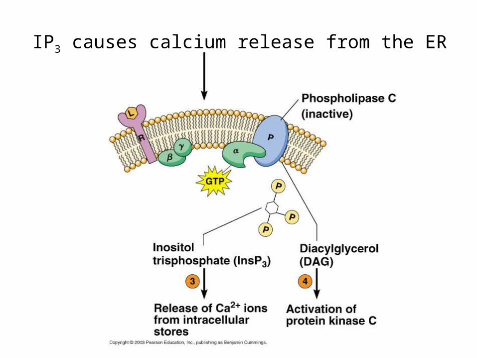
IP3 causes calcium release from the ER

Summary

Fukuhara, et al. 2000. Signaling from G protien-coupled receptors to the nucleus. From: signaling networks and cell cycle control: The molecular basis of cancer and other diseases, Ed. JS Gutkind, Humana Press, NJ.
CROSSTALK?

Maribissen & Gutkind, 2001. G-protein coupled receptors and signaling networks: emerging paradigms. Trend Pharm. Sci. 22:368-376.
Luttrel, et al., 1999. Regulation of tyrosine kinase cacades by G protein coupled receptors. Curr. Opin.Cell Biol. 11:177-183.
Schonberg, T, et al., 1999. Structural basis of G protein-coupled receptor function. Mol. Cell. Endocrin. 151:181-193.
Hamm, H. 1998. The many faces of G protein signaling. JBC 273:669-672.
Ji et al., 1998. G protein coupled receptors I. Diversity of receptor-ligand interactions. JBC 273:17299-17302.
Gether and Koblikas, 1998. G protein coupled receptors: II. Mechansim of agonist actiavtion. JBC 273:17979-17982.
Lefkowitz, RJ. 1998. G protein coupled receptors III: New roles for receptor kinases and b-arrestins in receptor signaling and desensitization. JBC 273:18677.
Gutkind, S. 1998. The pathways connecting G protien coupled receptors to the nucleus through divergent mitogen-activated protein kinase cascades. JBC 273:1839.
Fukuhara et al., 2000. Signaling from G p receptors to the nucleus, text.

QuickTime™ and aTIFF (Uncompressed) decompressor
are needed to see this picture.
QuickTime™ and aTIFF (Uncompressed) decompressor
are needed to see this picture.
Batho/Lumi/Meta I
Metarhodopsin IIRhodopsin 1hv
Transducin
GDP
GTP
Phosphodiesterase
cGMP
GMP+H+
105
plasma membrane
cGMP-gated cation channel
Na+, Ca2+
GTP
GTP
Guanylylcyclase
The photoreceptor is hyperpolarized
QuickTime™ and aTIFF (Uncompressed) decompressor
are needed to see this picture.
Rhdopsin kinasePhosphorylated metarhodopsin II
Arrestin
+
Rhodopsin phosphatase
Metarhodopsin II