the significance of g protein-coupled receptor crystallography for drug...
TRANSCRIPT
ASSOCIATE EDITOR: DIANNE M. PEREZ
The Significance of G Protein-Coupled ReceptorCrystallography for Drug Discovery
John A. Salon, David T. Lodowski, and Krzysztof Palczewski
Department of Molecular Structure, Amgen Incorporated, Thousand Oaks, California (J.A.S.); and Department of Pharmacology, Schoolof Medicine, Case Western Reserve University, Cleveland, Ohio (D.T.L., K.P.)
Abstract . . . . . . . . . . . . . . . . . . . . . . . . . . . . . . . . . . . . . . . . . . . . . . . . . . . . . . . . . . . . . . . . . . . . . . . . . . . . . . . 902I. Introduction. . . . . . . . . . . . . . . . . . . . . . . . . . . . . . . . . . . . . . . . . . . . . . . . . . . . . . . . . . . . . . . . . . . . . . . . . . . . 903
II. Current challenges and opportunities in G protein-coupled receptor drug discovery. . . . . . . . . . . . 903A. Overview of the current G protein-coupled receptor drug discovery process . . . . . . . . . . . . . . . . 903
1. Therapeutic relevance . . . . . . . . . . . . . . . . . . . . . . . . . . . . . . . . . . . . . . . . . . . . . . . . . . . . . . . . . . . . . 9032. Molecular properties . . . . . . . . . . . . . . . . . . . . . . . . . . . . . . . . . . . . . . . . . . . . . . . . . . . . . . . . . . . . . . 9043. G Protein-coupled receptor drug compendium . . . . . . . . . . . . . . . . . . . . . . . . . . . . . . . . . . . . . . . . . 9044. The drug discovery process . . . . . . . . . . . . . . . . . . . . . . . . . . . . . . . . . . . . . . . . . . . . . . . . . . . . . . . . 904
B. G protein-coupled receptor molecular biology challenges . . . . . . . . . . . . . . . . . . . . . . . . . . . . . . . . . 9071. Expansion of G protein-coupled receptor drug targets into class B, C, (D and E)
families . . . . . . . . . . . . . . . . . . . . . . . . . . . . . . . . . . . . . . . . . . . . . . . . . . . . . . . . . . . . . . . . . . . . . . . . . . 9072. Special cases . . . . . . . . . . . . . . . . . . . . . . . . . . . . . . . . . . . . . . . . . . . . . . . . . . . . . . . . . . . . . . . . . . . . . 908
a. Olfactory receptors . . . . . . . . . . . . . . . . . . . . . . . . . . . . . . . . . . . . . . . . . . . . . . . . . . . . . . . . . . . . . 908b. Orphan receptors . . . . . . . . . . . . . . . . . . . . . . . . . . . . . . . . . . . . . . . . . . . . . . . . . . . . . . . . . . . . . . 909c. Receptor isoforms . . . . . . . . . . . . . . . . . . . . . . . . . . . . . . . . . . . . . . . . . . . . . . . . . . . . . . . . . . . . . . 909
3. In vitro reconstitution of monomeric receptors . . . . . . . . . . . . . . . . . . . . . . . . . . . . . . . . . . . . . . . 909C. Increasingly complex pharmacology . . . . . . . . . . . . . . . . . . . . . . . . . . . . . . . . . . . . . . . . . . . . . . . . . . . 909
1. Allostery . . . . . . . . . . . . . . . . . . . . . . . . . . . . . . . . . . . . . . . . . . . . . . . . . . . . . . . . . . . . . . . . . . . . . . . . . 9092. Receptor oligomerization . . . . . . . . . . . . . . . . . . . . . . . . . . . . . . . . . . . . . . . . . . . . . . . . . . . . . . . . . . 9103. Ligand-biased (ligand-selective) signaling . . . . . . . . . . . . . . . . . . . . . . . . . . . . . . . . . . . . . . . . . . . 9124. Constitutive activity . . . . . . . . . . . . . . . . . . . . . . . . . . . . . . . . . . . . . . . . . . . . . . . . . . . . . . . . . . . . . . 912
D. Assay development . . . . . . . . . . . . . . . . . . . . . . . . . . . . . . . . . . . . . . . . . . . . . . . . . . . . . . . . . . . . . . . . . . 9121. The genomic tool box. . . . . . . . . . . . . . . . . . . . . . . . . . . . . . . . . . . . . . . . . . . . . . . . . . . . . . . . . . . . . . 9122. Screening efficiency . . . . . . . . . . . . . . . . . . . . . . . . . . . . . . . . . . . . . . . . . . . . . . . . . . . . . . . . . . . . . . . 9133. Screening mode. . . . . . . . . . . . . . . . . . . . . . . . . . . . . . . . . . . . . . . . . . . . . . . . . . . . . . . . . . . . . . . . . . . 9134. Integrative assays . . . . . . . . . . . . . . . . . . . . . . . . . . . . . . . . . . . . . . . . . . . . . . . . . . . . . . . . . . . . . . . . 9135. New generation biochemical and biophysical assays . . . . . . . . . . . . . . . . . . . . . . . . . . . . . . . . . . 913
E. Limitations of current compound libraries. . . . . . . . . . . . . . . . . . . . . . . . . . . . . . . . . . . . . . . . . . . . . . 9141. Chemical space versus biological space. . . . . . . . . . . . . . . . . . . . . . . . . . . . . . . . . . . . . . . . . . . . . . 9142. Future expansion of chemical screening libraries. . . . . . . . . . . . . . . . . . . . . . . . . . . . . . . . . . . . . 914
III. History of G protein-coupled receptor structural clarification . . . . . . . . . . . . . . . . . . . . . . . . . . . . . . . . 914A. Functional genomics reveals the G protein-coupled receptor superfamily . . . . . . . . . . . . . . . . . . 914B. Electron microscopy of rhodopsin provides a conceptual prototype of G protein-coupled
receptor structure . . . . . . . . . . . . . . . . . . . . . . . . . . . . . . . . . . . . . . . . . . . . . . . . . . . . . . . . . . . . . . . . . . . 915C. Comparative modeling suggests structure-function experiments . . . . . . . . . . . . . . . . . . . . . . . . . . 916D. Structure-function campaigns identify hotspots: common G protein-coupled receptor
functional moieties, trigger mechanisms, and long-awaited high-resolution three-dimensional structures . . . . . . . . . . . . . . . . . . . . . . . . . . . . . . . . . . . . . . . . . . . . . . . . . . . . . . . . . . . . . . . 917
Address correspondence to: Dr. Krzysztof Palczewski, Department of Pharmacology, School of Medicine, Case Western Reserve University,10900 Euclid Ave, Cleveland, Ohio 44106�4965. E-mail: [email protected]
All authors contributed equally to this work.This article is available online at http://pharmrev.aspetjournals.org.doi:10.1124/pr.110.003350.
0031-6997/11/6304-901–937$25.00PHARMACOLOGICAL REVIEWS Vol. 63, No. 4Copyright © 2011 by The American Society for Pharmacology and Experimental Therapeutics 3350/3708070Pharmacol Rev 63:901–937, 2011 Printed in U.S.A.
901
by guest on May 30, 2018
Dow
nloaded from
1. The D(E)RY motif within G protein-coupled receptors . . . . . . . . . . . . . . . . . . . . . . . . . . . . . . . . 9182. The NPxxYx(5,6)F motif within G protein-coupled receptors . . . . . . . . . . . . . . . . . . . . . . . . . 9193. Conservation of water and water-binding sites within the transmembrane domain . . . . . . 9194. Ligand-binding domains of G protein-coupled receptors . . . . . . . . . . . . . . . . . . . . . . . . . . . . . . . 919
IV. Advances in G protein-coupled receptor structural determination . . . . . . . . . . . . . . . . . . . . . . . . . . . . 921A. G protein-coupled receptor construct design and expression . . . . . . . . . . . . . . . . . . . . . . . . . . . . . . 921B. Solubilization and purification of G protein-coupled receptor constructs . . . . . . . . . . . . . . . . . . . 921C. Predicting G protein-coupled receptor construct “crystallizability” . . . . . . . . . . . . . . . . . . . . . . . . 922
1. Assays. . . . . . . . . . . . . . . . . . . . . . . . . . . . . . . . . . . . . . . . . . . . . . . . . . . . . . . . . . . . . . . . . . . . . . . . . . . 9222. Stabilization of G protein-coupled receptors with membrane mimetics during
crystallization . . . . . . . . . . . . . . . . . . . . . . . . . . . . . . . . . . . . . . . . . . . . . . . . . . . . . . . . . . . . . . . . . . . . 9223. Expanding soluble domains of G protein-coupled receptors using nanobodies and
antibody Fv fragments . . . . . . . . . . . . . . . . . . . . . . . . . . . . . . . . . . . . . . . . . . . . . . . . . . . . . . . . . . . . 923D. Recent G protein-coupled receptor successes. . . . . . . . . . . . . . . . . . . . . . . . . . . . . . . . . . . . . . . . . . . . 923
1. Rhodopsin and related structures . . . . . . . . . . . . . . . . . . . . . . . . . . . . . . . . . . . . . . . . . . . . . . . . . . 9232. Reconciling activated structures of (rhod)opsin with biochemistry and biophysics . . . . . . . 9233. Adrenergic receptor structures . . . . . . . . . . . . . . . . . . . . . . . . . . . . . . . . . . . . . . . . . . . . . . . . . . . . . 9254. A2a-adenosine receptor structure . . . . . . . . . . . . . . . . . . . . . . . . . . . . . . . . . . . . . . . . . . . . . . . . . . . . . 9265. C-x-c chemokine receptor type 4 structures . . . . . . . . . . . . . . . . . . . . . . . . . . . . . . . . . . . . . . . . . . 9266. Dopamine D3 receptor structure. . . . . . . . . . . . . . . . . . . . . . . . . . . . . . . . . . . . . . . . . . . . . . . . . . . . 9267. Structures of G protein-coupled receptor extracellular domains. . . . . . . . . . . . . . . . . . . . . . . . 926
E. The need for more G protein-coupled receptor structural work . . . . . . . . . . . . . . . . . . . . . . . . . . . 927V. Ramifications for G protein-coupled receptor drug discovery. . . . . . . . . . . . . . . . . . . . . . . . . . . . . . . . . 927
A. Brief history of de novo approaches. . . . . . . . . . . . . . . . . . . . . . . . . . . . . . . . . . . . . . . . . . . . . . . . . . . . 9271. Examples of success. . . . . . . . . . . . . . . . . . . . . . . . . . . . . . . . . . . . . . . . . . . . . . . . . . . . . . . . . . . . . . . 9272. Strengths and weaknesses . . . . . . . . . . . . . . . . . . . . . . . . . . . . . . . . . . . . . . . . . . . . . . . . . . . . . . . . . 928
B. Future directions for integral membrane proteins in drug discovery . . . . . . . . . . . . . . . . . . . . . . 9281. The current industry climate for G protein-coupled receptor drug discovery. . . . . . . . . . . . . 9282. Requirements for the optimal use of structural studies . . . . . . . . . . . . . . . . . . . . . . . . . . . . . . . 929
VI. Unresolved issues and concluding remarks . . . . . . . . . . . . . . . . . . . . . . . . . . . . . . . . . . . . . . . . . . . . . . . . 929A. Caveats about static crystal structures . . . . . . . . . . . . . . . . . . . . . . . . . . . . . . . . . . . . . . . . . . . . . . . . 929B. Partnering with computational methods . . . . . . . . . . . . . . . . . . . . . . . . . . . . . . . . . . . . . . . . . . . . . . . 929C. Rate of structure determination. . . . . . . . . . . . . . . . . . . . . . . . . . . . . . . . . . . . . . . . . . . . . . . . . . . . . . . 930D. Structure versus function . . . . . . . . . . . . . . . . . . . . . . . . . . . . . . . . . . . . . . . . . . . . . . . . . . . . . . . . . . . . 930E. Cocrystallization with ancillary protein(s) . . . . . . . . . . . . . . . . . . . . . . . . . . . . . . . . . . . . . . . . . . . . . . 930F. Impact upon compound libraries . . . . . . . . . . . . . . . . . . . . . . . . . . . . . . . . . . . . . . . . . . . . . . . . . . . . . . 930G. Synergy with biologics . . . . . . . . . . . . . . . . . . . . . . . . . . . . . . . . . . . . . . . . . . . . . . . . . . . . . . . . . . . . . . . 931H. Other synergies . . . . . . . . . . . . . . . . . . . . . . . . . . . . . . . . . . . . . . . . . . . . . . . . . . . . . . . . . . . . . . . . . . . . . 931I. Selection of therapeutically validated targets . . . . . . . . . . . . . . . . . . . . . . . . . . . . . . . . . . . . . . . . . . . 931
Note added in proof . . . . . . . . . . . . . . . . . . . . . . . . . . . . . . . . . . . . . . . . . . . . . . . . . . . . . . . . . . . . . . . . . . . . . 931Acknowledgments. . . . . . . . . . . . . . . . . . . . . . . . . . . . . . . . . . . . . . . . . . . . . . . . . . . . . . . . . . . . . . . . . . . . . . . 932References . . . . . . . . . . . . . . . . . . . . . . . . . . . . . . . . . . . . . . . . . . . . . . . . . . . . . . . . . . . . . . . . . . . . . . . . . . . . . 932
Abstract——Crucial as molecular sensors for manyvital physiological processes, seven-transmembranedomain G protein-coupled receptors (GPCRs) com-prise the largest family of proteins targeted by drugdiscovery. Together with structures of the prototyp-ical GPCR rhodopsin, solved structures of other li-ganded GPCRs promise to provide insights into thestructural basis of the superfamily’s biochemicalfunctions and assist in the development of new ther-apeutic modalities and drugs. One of the greatesttechnical and theoretical challenges to elucidatingand exploiting structure-function relationships inthese systems is the emerging concept of GPCR con-formational flexibility and its cause-effect relation-
ship for receptor-receptor and receptor-effector in-teractions. Such conformational changes can besubtle and triggered by relatively small binding en-ergy effects, leading to full or partial efficacy in theactivation or inactivation of the receptor system atlarge. Pharmacological dogma generally dictatesthat these changes manifest themselves through ki-netic modulation of the receptor’s G protein part-ners. Atomic resolution information derived fromincreasingly available receptor structures providesan entree to the understanding of these events andpractically applying it to drug design. Supported bystructure-activity relationship information arisingfrom empirical screening, a unified structural model
902 SALON ET AL.
of GPCR activation/inactivation promises to bothaccelerate drug discovery in this field and improveour fundamental understanding of structure-based
drug design in general. This review discusses funda-mental problems that persist in drug design andGPCR structural determination.
I. Introduction
The biological and medical importance of G protein-coupled receptors (GPCRs1) is well established and ex-tensively documented. The breadth of GPCR distribu-tion across nearly all of the body’s organs and tissuesand the cellular role GPCRs play as signal transducersmake GPCRs key regulatory elements in a broad rangeof normal and pathological processes. Thus, GPCRs havebeen and will continue to be an important focus for drugdiscovery (Drews, 2000; Ma and Zemmel, 2002).
Over the past decade, the pursuit of GPCRs as targetsfor drug discovery campaigns has benefited greatly fromthe development and adoption of high-throughput ap-proaches to their pharmacological assay and medicinalchemistry. Availability of these tools in conjunction witha genomically complete GPCR target palette has effec-tively enabled researchers to rapidly screen GPCRs ofspecific therapeutic interest and quickly elaborate uponpotential leads during the ensuing drug developmentprocess, thus sparking a renaissance in GPCR pharma-cology. The emergence of new types of ligand-receptor-effector relationships, including positive and negativeallosterism, inverse agonism, multimeric receptor phar-macology, and ligand biased signaling (discussed in sec-tion II) has widened our perspective beyond simple, two-state (on/off) receptor models and suggests entirely newmechanistic avenues for therapeutic intervention. Suchan expanded scope of options is both enticing and vexingfrom a drug discovery point of view, a paradox onlymagnified by our limited structural insights into themolecular mechanics of the GPCR superfamily.
For nearly 30 years we have probed GPCRs throughlaborious mutagenesis and assay procedures in an at-tempt to distinguish the residues that are functionallyimportant and then fitting our findings into hypotheticalmodels of their structure. Despite the significant toildevoted to these efforts and the consolidated lists ofresidues cataloged as important for binding and/or func-tion, the utility of the findings were inevitably limited bytheir individualized nature. The goal of achieving ahighly resolved and practical understanding of specifi-
cally “druggable” receptor sites remains elusive, and theiterative structure-function process has proven too slowand laborious to proactively guide drug discovery. Newdevelopments in the area of X-ray crystallography sug-gest that the structural veil has now lifted and that weare on the threshold of a new era for GPCR drug discov-ery. Reports have been rapidly emerging about the suc-cessful crystallization and structural determination byX-ray diffraction methods of GPCRs, including the �2-adrenergic (Cherezov et al., 2007; Hanson et al., 2008;Rasmussen et al., 2011; Rosenbaum et al., 2011), �1-adrenergic (Warne et al., 2008, 2011), A2a-adenosine(Jaakola et al., 2008; Xu et al., 2011), chemokine C-X-Cchemokine receptor type 4 (CXCR4) (Wu et al., 2010),and dopamine D3 receptors (Chien et al., 2010). Demon-stration of high-resolution structures for multiple recep-tors and the accelerated rate at which they are appear-ing suggests that we have bypassed some of theroadblocks historically associated with crystallizingmembers of this integral membrane protein family. Ap-plication of such methods to the GPCR superfamilypromises to illuminate at atomic resolution just howthese important membrane proteins work and, in sodoing, significantly change the tactics of our empiricaldrug discovery process.
Given the likelihood that structures for more GPCRswill be forthcoming, rather than attempt to dissect dif-ferences in the currently available structures, we willdescribe the commonalities of successful strategies froma technical perspective and indicate how they can beimplemented to best benefit future GPCR drug discoveryefforts.
II. Current Challenges and Opportunities in GProtein-Coupled Receptor Drug Discovery
A. Overview of the Current G Protein-Coupled Receptordrug Discovery Process
1. Therapeutic Relevance. The medicinal importanceof GPCRs can be partially appreciated by consideringtheir location and function within the cell. The physicallocation and disposition of GPCRs spanning the cell’splasma membrane connect extra- and intracellular en-vironments, providing a direct mechanism for the trans-duction of extracellular messages into intracellular re-sponses. In this way and together with their transmittersand effectors, GPCR systems function to modulate a broadspectrum of cellular phenomena dictated by the needs ofthe tissues and organs they serve. Common biological ac-tions attributed to GPCRs include but are not limited tothe following: modulation of neuronal firing, regulation ofion transport across the plasma membrane and within
1Abbreviations: ADME, Absorption, distribution, metabolism, andexcretion; AFM, atomic force microscopy; CXCR, C-X-C chemokinereceptor; FDA, US Food and Drug Administration; GPCR, G protein-coupled receptor; Gt, photoreceptor G protein (transducin); H,transmembrane helix; HTS, high-throughput screening; IT1t, 6,6-dimethyl-5,6-dihydroimidazo[2,1-b][1,3]thiazol-3-yl)methyl N,N�-di-cyclohexylimidothiocarbamate; LCP, lipidic cubic phase; MD, moleculardynamics; PD, pharmacodynamics; PDB, Protein Data Bank; PK, phar-macokinetics; QSAR, quantitative structure-activity relationship; R&D,research and development; RMSD, root-mean-square deviation; SAR,structure-activity relationship; SBDD, structure-based drug design;SPR, surface plasmon resonance; TM, transmembrane.
GPCRs AND DRUG DISCOVERY 903
intracellular organelles, modulation of homeostasis, con-trol of cell division/proliferation, and modification of cellmorphology. When any of these fundamental processes goawry, the results can lead to acute or chronic human dis-ease, a partial listing of which includes cardiovasculardisease (�1-adrenergic receptor) (Drake et al., 2006),asthma (�2-adrenergic receptor) (Kawakami et al., 2004),and strokes and cerebral hypoperfusion (A2a-adenosine re-ceptor) (Chen et al., 2007a; Duan et al., 2009). Other dis-ease states directly linked to mutations in GPCRs includeretinitis pigmentosa (rhodopsin), female infertility (follicle-stimulating hormone receptor), nephrogenic diabetes in-sipidus (vasopressin receptor), familial exudative vitreo-retinopathy (frizzled receptors), and dominant andrecessive obesity (melanocortin receptors) (for review, seeInsel et al., 2007).
2. Molecular Properties. Although the details ofGPCR signaling in aggregate are complex, the basictenets that describe the initial interaction of the recep-tor with its proximal partner, the G protein heterotri-meric complex, are straightforward. Upon adoption of an“active” conformation (most simply envisioned as theresult of agonist binding), the intracellular domains of aGPCR interact with a membrane-associated GDP-charged G protein heterotrimeric complex (G���) (Old-ham and Hamm, 2008). This heterotrimeric complexthen undergoes GTP/GDP exchange with subsequentdissociation of G� and G�� subunits that in turn inter-act with specific downstream intracellular effector sys-tems (Fig. 1A). Activation of multiple heterocomplexesas well as G� cycling through active and inactive con-figurations via a GTP hydrolysis cycle provides immedi-ate amplification and temporal regulation of the initialreceptor-ligand signaling event (Leskov et al., 2000;Heck and Hofmann, 2001; Minke and Cook, 2002; Bhan-dawat et al., 2005). In due course, through the process ofdesensitization, the active conformation of the receptoris blocked and signaling is attenuated by agonist disso-ciation and/or deactivation through interaction with�-arrestins in response to activation-specific phosphor-ylation by G protein-coupled receptor kinases and/orinternalization (Hall, 2000; Bockaert et al., 2003). Theimmediate activities of these effector systems fall intofour main categories: stimulation of cAMP production,inhibition of cAMP production, stimulation of phospho-lipase C with subsequent mobilization of intracellularCa2�, and activation of plasma membrane proton flux.These phenomena are controlled by which class of G�subunit is activated. There are at least 16 human G�subunits, 5 G� subunits, and 11 G� subunits (Milliganand Kostenis, 2006) (Fig. 2). In addition to G�-controlledevents, the G�� subunits also can regulate their owneffectors, including additional forms of adenylate cyclaseas well as ion channels. The ramifications of signalingcomplexity implicit in the full range of combinatorialpermutations within the heterotrimeric complex itselfhave yet to be fully examined (Jastrzebska et al., 2010).
Possibly because they share a relatively limited down-stream effector ensemble, GPCRs themselves are simi-lar with respect to their gross architectural topologies.Embedded within this structural homogeneity is thecapability of receptor subtypes to distinguish chemicalsubtleties across and within structurally diverse trans-mitter and hormone families (Fig. 1B). This remarkablemolecular recognition property combined with thebreadth of specific pathophysiological conditions relatedto each receptor’s tissue distribution make the GPCRsuperfamily a treasure trove of proven and potentialdrug discovery targets. In addition, plasma membrane-spanning GPCRs display their most important regula-tory sites at or near the extracellular surface, makingthese sites readily available to circulating adminis-tered drugs. Such unfettered access mitigates manydrug development concerns such as drug transportthrough the plasma membrane and intracellular drugmetabolism.
3. G Protein-Coupled Receptor Drug Compendium. Asa biological target class, drugs of the GPCR superfamilyare well represented in our current pharmacopeia (Kroezeet al., 2003; Bjenning et al., 2004; Doggrell, 2004; Dahl andSylte, 2005), with an estimated 30 to 50% of marketeddrugs acting directly on GPCRs or through GPCR-associ-ated mechanisms (Flower, 1999; Robas et al., 2003). Al-though the most obvious GPCR modulators constitutesome 26% of the top sellers and netted some $23.5 billion(9% of the global market share) of drug sales in the year2000, these �150 marketed drugs surprisingly target only�20 of the �750 known GPCR subtypes (Vassilatis et al.,2003; Overington et al., 2006). Moreover, as of 2008, 5 ofthe top 15 generic drugs and 7 (Milligan, 2009) of the top15 prescription drugs targeted GPCRs (McGrath et al.,2010). Of those, only the leukotriene drugs are “new”GPCR targets. This paradox does not imply that the bulkof the GPCR superfamily is therapeutically unimportantor “undruggable.” Instead, it reflects the comparativelylong period needed to bring drugs to market relative to theshort recent period during which postgenomic target ac-cess and high-throughput methods have been available. Itfollows that many drugs now in or emerging from thepharmaceutical pipeline have been enabled largely be-cause of GPCR systems discovered or investigated in thepast decade or so.
4. The Drug Discovery Process. The research anddevelopment (R&D) process is lengthy and expensive,especially during the later clinical stages (Figs. 3 and 4).Two previous estimates of the costs of bringing a newdrug to market range from $1.3 to $1.76 billion becauseof the adverse impact of program attrition, which con-tinues well into the later stages of the developmentprocess (DiMasi et al., 2003; Paul et al., 2010). Suchcosts underscore the weight placed upon pursuing onlywell validated pathophysiological mechanisms and dis-ease markets that are capable of delivering a return onthe R&D investment. Of additional importance is the
904 SALON ET AL.
target-centric modus operandi of the R&D process. To-gether, these factors dictate that during the earlieststages of drug discovery, when therapeutic paradigmsare being envisioned and refined, an over-riding re-search goal is the identification, characterization andvalidation of the association between specific moleculartargets and specific disease states. Although thegenomic catalog of GPCRs is complete, discussion as tothe number and identity of “druggable” targets is stillactive. Basic approaches employed to explore the issueinclude 1) tissue expression profiling (including compar-isons between healthy and disease states); 2) searchingfor regulation of transcripts and gene copies in diseasemodels through microarray, quantitative polymerase
chain reaction, and genomic deep sequencing analyses;3) establishing gene-disease linkage through chromo-some mapping and phenotypic analysis of transgenicanimals; and 4) conducting in vivo/in vitro pharmacolog-ical studies with target-active prelead molecules whenavailable. Absorption, distribution, metabolism, and ex-cretion (ADME) and pharmacokinetic (PK) studies areincreasingly being employed early on in the drug discov-ery process to assist in the interpretation of ongoing andfuture in vivo experimentation. If a GPCR drug targetsurvives this initial validation gauntlet, it then proceedsto primary drug screening, where small moleculesand/or biological compounds are tested as dictated bythe therapeutic paradigm.
NH2CHO
CHO
CHO
CHO
CHOCHO
CHO
CHO
CHOCHO
CHO
CHOCa++, Glutamate
Ligands ~MW (Da)/~Size (Å)B A
(extra) 3LE2LE1LE
XP
CC
monoamines
101/1.0
P
(intra) 4LI3LI2LI1LI
DRY
C W X
NPXX
X
Y
monoamines,nucleo�des, lipids
102/5.0
P
COOH
Gγ
Gβ Gα
GDP
pep�des
103/20
GTPmasked sites
103/10
GDP
Pi
effectorac�va�on
effectorac�va�on GαGγ
```Gβ
glycoproteins
104/50ac�va�onGTP
104/50
C
PP
C
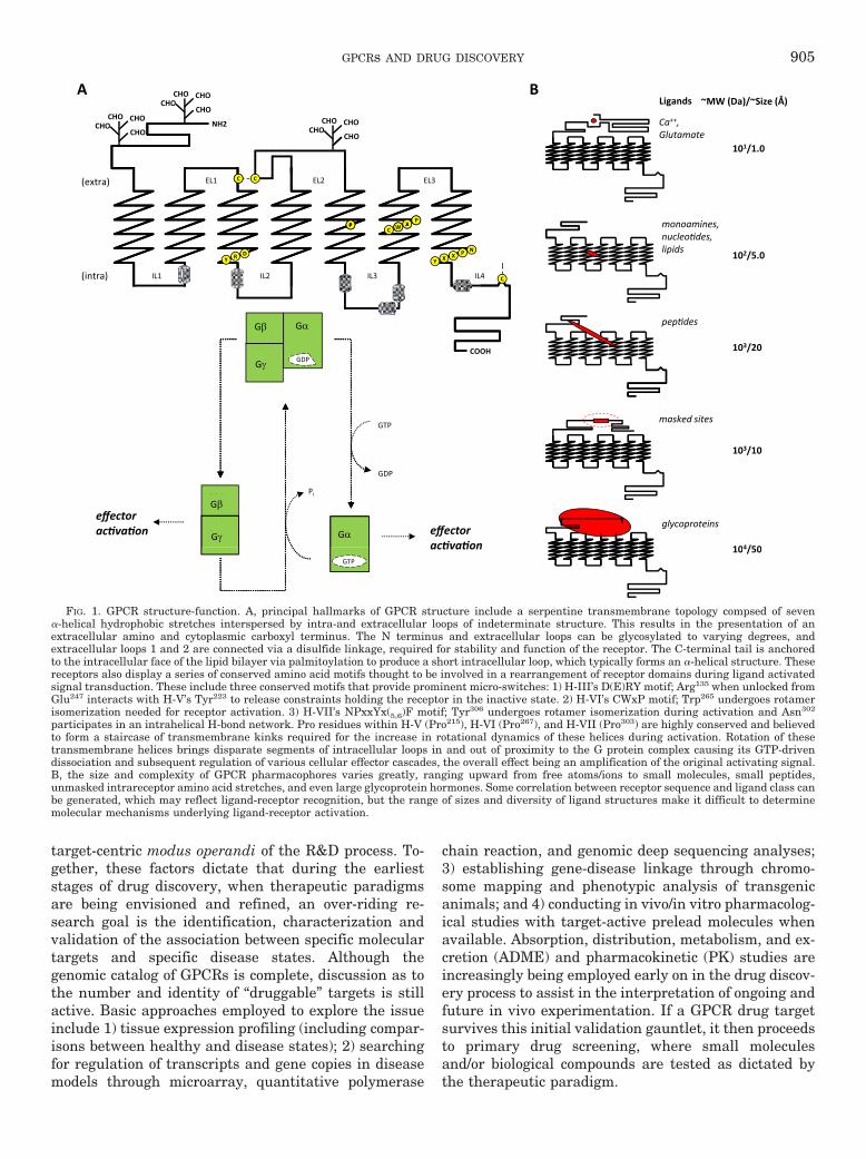
FIG. 1. GPCR structure-function. A, principal hallmarks of GPCR structure include a serpentine transmembrane topology compsed of seven�-helical hydrophobic stretches interspersed by intra-and extracellular loops of indeterminate structure. This results in the presentation of anextracellular amino and cytoplasmic carboxyl terminus. The N terminus and extracellular loops can be glycosylated to varying degrees, andextracellular loops 1 and 2 are connected via a disulfide linkage, required for stability and function of the receptor. The C-terminal tail is anchoredto the intracellular face of the lipid bilayer via palmitoylation to produce a short intracellular loop, which typically forms an �-helical structure. Thesereceptors also display a series of conserved amino acid motifs thought to be involved in a rearrangement of receptor domains during ligand activatedsignal transduction. These include three conserved motifs that provide prominent micro-switches: 1) H-III’s D(E)RY motif; Arg135 when unlocked fromGlu247 interacts with H-V’s Tyr223 to release constraints holding the receptor in the inactive state. 2) H-VI’s CWxP motif; Trp265 undergoes rotamerisomerization needed for receptor activation. 3) H-VII’s NPxxYx(5,6)F motif; Tyr306 undergoes rotamer isomerization during activation and Asn302
participates in an intrahelical H-bond network. Pro residues within H-V (Pro215), H-VI (Pro267), and H-VII (Pro303) are highly conserved and believedto form a staircase of transmembrane kinks required for the increase in rotational dynamics of these helices during activation. Rotation of thesetransmembrane helices brings disparate segments of intracellular loops in and out of proximity to the G protein complex causing its GTP-drivendissociation and subsequent regulation of various cellular effector cascades, the overall effect being an amplification of the original activating signal.B, the size and complexity of GPCR pharmacophores varies greatly, ranging upward from free atoms/ions to small molecules, small peptides,unmasked intrareceptor amino acid stretches, and even large glycoprotein hormones. Some correlation between receptor sequence and ligand class canbe generated, which may reflect ligand-receptor recognition, but the range of sizes and diversity of ligand structures make it difficult to determinemolecular mechanisms underlying ligand-receptor activation.
GPCRs AND DRUG DISCOVERY 905
Once the target is validated, the screening processbegins in earnest and typically employs cell-based as-says in a high-throughput screening (HTS) platform tointerrogate corporate compound libraries consisting ofhundreds of thousands to millions of separate chemicalentities. These assays are typically engineered to behighly specific for a single disease-linked receptor. Pri-mary hits arising from the screen are subsequently con-firmed and evaluated for potency at their primary tar-gets. These activity results then are considered togetherwith the hit structure in making the decision to advancethe primary hit into the lead development process.Structural properties of hits are also considered from theperspective of the “intellectual freedom to operate”needed to devise patentable chemical analogs of theprototype. Generic problems arising from reactive moi-eties that can render the compound toxic through irre-versible covalent attack on either the target itself orother vital cellular proteins are usually apparent at thispoint and revealed through simple inspection of the com-pound’s physicochemical properties. Clearly, narrowingthe intrinsic specificity for a GPCR target must be aninitial priority. Accordingly, characterization and man-agement of drug cross-reactivity is an integral part ofthe hit-to-lead process that occurs immediately after hit
discovery. Conceptually this can be organized accordingto site and gravity of action. Deliberations usually beginby considering cross-reactivity at sites associated withknown health hazards. For example, cross-reactivity of adiverse group of drugs with the human ether-a-go-go-related gene potassium channel can result in suddendeath caused by long QT syndrome (Sanguinetti andTristani-Firouzi, 2006). The potential for such problemsis typically discovered early for a given lead series by invitro testing across a generalized panel of side-effecttargets. Additional benefits can result from incorporat-ing chemical properties that restrict drug distribution,as in the case of adverse CNS effects, for example, bykeeping the compound from crossing the blood-brainbarrier. Next in line are limitations imposed by a com-pound’s pharmacokinetics involving its route(s) of ad-ministration, absorption from an administered site, dis-tribution throughout the body as it gains access to thetarget tissue(s), metabolism and bioavailability via theliver’s P450 redox and P-glycoprotein systems, and ulti-mate elimination of the parent drug with its metabo-lites. Poor ADME properties are the root cause of failurefor �40% of drug candidates during clinical trials, sothese parameters are extensively evaluated throughoutlead development. Pharmacodynamic (PD) properties
Elements Ac�ons
Orthosteric-Bitopic-Allosteric
Ligands (?)
Monomer-DimerHomo-Hetero
Receptors (>750)
G (2) Gi (9) G (5) G12/13 (2)
Flexible G protein Interac�on
G alpha proteins (18) αβ
γα
β
γα
β
γα
β
γ
Ligand Dependant Pathway Signaling
Gs (2) Gi (9) Gq (5) G12/13 (2)
posi�ve
modulator (-) (+)
Direc�onal Ac�vityVariable EfficacyPotency Modula�on
Proximal Effects (4)
effec
t
neutral
inverse
par�al full
[ligand]
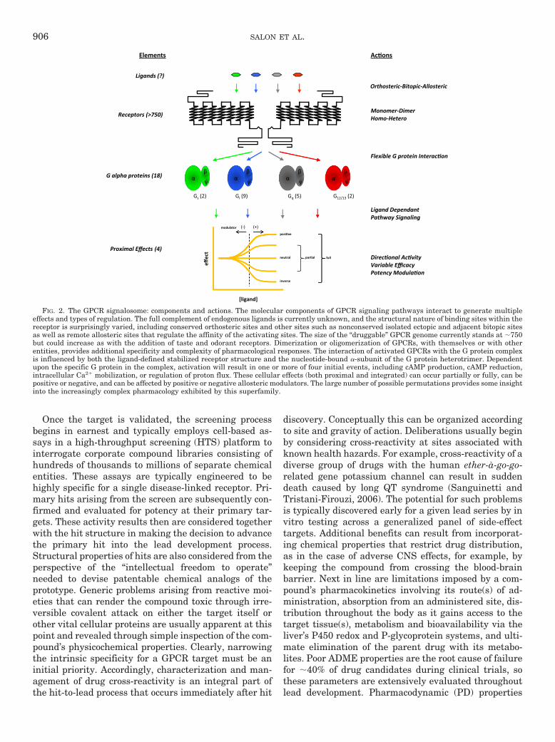
FIG. 2. The GPCR signalosome: components and actions. The molecular components of GPCR signaling pathways interact to generate multipleeffects and types of regulation. The full complement of endogenous ligands is currently unknown, and the structural nature of binding sites within thereceptor is surprisingly varied, including conserved orthosteric sites and other sites such as nonconserved isolated ectopic and adjacent bitopic sitesas well as remote allosteric sites that regulate the affinity of the activating sites. The size of the “druggable” GPCR genome currently stands at �750but could increase as with the addition of taste and odorant receptors. Dimerization or oligomerization of GPCRs, with themselves or with otherentities, provides additional specificity and complexity of pharmacological responses. The interaction of activated GPCRs with the G protein complexis influenced by both the ligand-defined stabilized receptor structure and the nucleotide-bound �-subunit of the G protein heterotrimer. Dependentupon the specific G protein in the complex, activation will result in one or more of four initial events, including cAMP production, cAMP reduction,intracellular Ca2� mobilization, or regulation of proton flux. These cellular effects (both proximal and integrated) can occur partially or fully, can bepositive or negative, and can be affected by positive or negative allosteric modulators. The large number of possible permutations provides some insightinto the increasingly complex pharmacology exhibited by this superfamily.
906 SALON ET AL.
associated with the mechanism of action and involvinginteraction, number of lead compounds, and efficacy at thetarget in vivo are also of great concern and can help definethe margins of risk versus safety as a therapeutic window.
In this fashion, chemical analogs of the initial screen-ing hits are synthesized and tested to improve efficacy,selectivity, and potency and to diminish ADME/PK lia-bilities in a trial-and-error process based upon the pro-jected therapeutic window of activity. Thus, during thejourney from lead compound to drug candidate, eachprimary hit spawns hundreds to thousands of closelyrelated derivatives; some are an improvement over theprototype, some are not, and all are deposited back intothe corporate library for future use. Because the processis target-centric, the content of these libraries reflectsthe history of prior campaigns.
Iterative enhancements to technical tool boxes (e.g.,assay methods or synthetic chemical processes) usuallyprogress as well. Periodic assessments of and adjust-ments to the content and quality of libraries at large arealso made, culling compounds with broad toxicity ordegraded content. Such enhancements contribute to in-cremental improvement of drug discovery, most oftenfavorably affecting operational efficiencies rather thanfundamental biological or chemical characteristics of the
platform. An underlying, but not always explicit, objec-tive of this diligence is lowering the threshold needed toenable primary screens. Although a goal of the process isefficiency (i.e., handling more campaigns in a given timeand resource frame), the process remains largely empir-ical, so the outcome is biased by the assay design andpre-existing library content.
The prospect of enabling structure-based drug design(SBDD) for GPCRs suggests that this empirical para-digm can be improved by enhancing or supplanting con-ventional HTS methods with new approaches that in-clude defining and/or supplementing the content of thecompound collection tested.
B. G Protein-Coupled Receptor MolecularBiology Challenges
1. Expansion of G Protein-Coupled Receptor Drug Tar-gets into Class B, C, (D and E) Families. AlthoughGPCRs couple to G proteins, these receptors are alsoreferred to as seven-transmembrane receptors, reflect-ing their seven-transmembrane-embedded helices andadditional signaling mechanisms independent of G pro-teins (Pierce et al., 2002; Lefkowitz, 2004). The numberof GPCRs in the human genome is estimated at �750separate genes. These can be categorized into five major
DISCOVERY DEVELOPMENT
6-7 yr3-6 yr 0.5-2 yr
ClinicalPreclinical FDADiscovery
Lead Discovery• Understand the disease effects, causes, and
molecular pathways• Iden�fy target and validate disease linkage• Evaluate feasibility of pursuing the target
DISCOVERY DEVELOPMENTClinical trials
• IND submission: describe structure, MOA, side effects, and research plan for clinical trials. FDA/IRB review of risks to par�cipants, monitoring and repor�ng procedures.
• Phase I (2-100 healthy volunteers): safety range, metabolism.(“druggability”)
• Find lead and op�mize for target selec�vityPreclinical
• Evaluate PK, ADME, and Tox• Op�mize lead for safety and efficacy (clear
for human tes�ng)• Establish formula�on and manufacture for
• Phase II (100-500 afflicted volunteers): effec�veness, side effects, MOA, safe/effec�ve dose regimes.
• Phase III (1000-5000 afflicted volunteers): sta�s�cally significant safety and benefit. Safety over �me.
FDA Review• NDA applica�on: findings and analysis of clinical trials, proposed labeling and
manufacture plan• Establish formula�on and manufacture for clinical tests
manufacture plan• Large scale manufacture and launch: customize produc�on and distribu�on
process. Marke�ng.• Ongoing surveillance: long term effects on large pa�ent popula�on
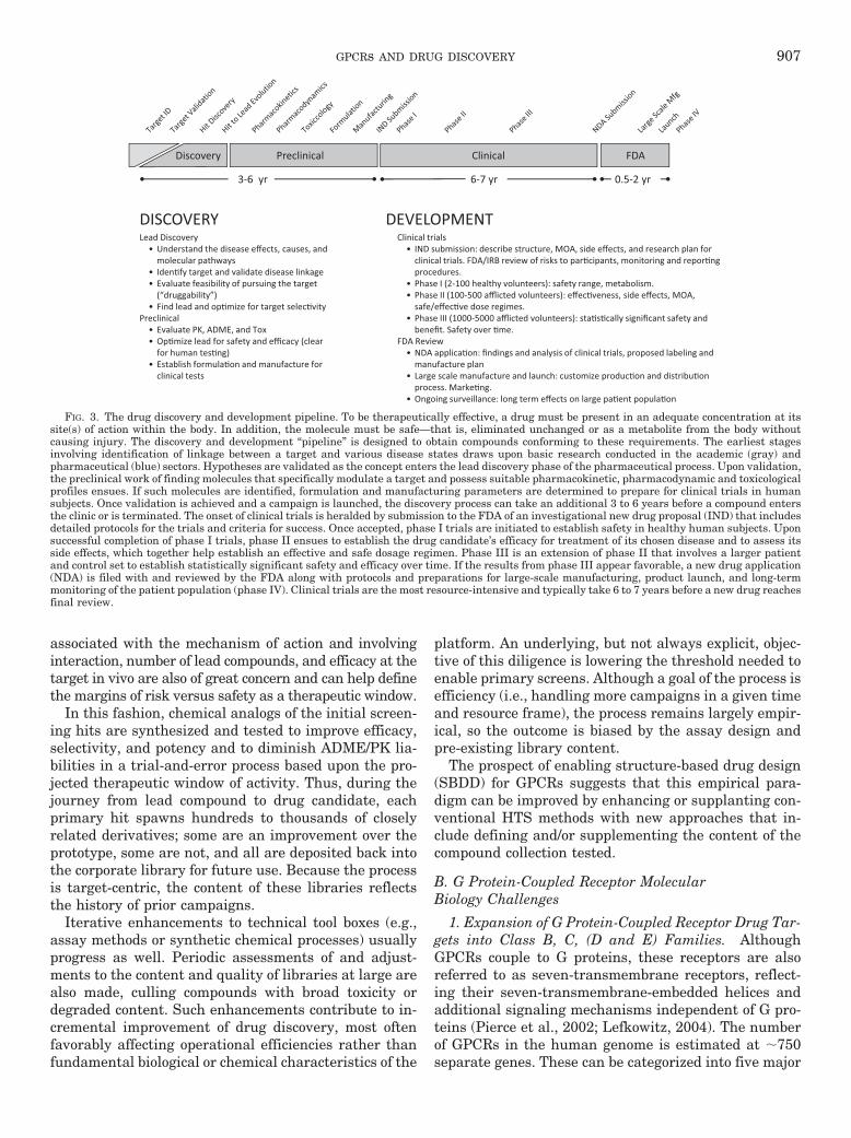
FIG. 3. The drug discovery and development pipeline. To be therapeutically effective, a drug must be present in an adequate concentration at itssite(s) of action within the body. In addition, the molecule must be safe—that is, eliminated unchanged or as a metabolite from the body withoutcausing injury. The discovery and development “pipeline” is designed to obtain compounds conforming to these requirements. The earliest stagesinvolving identification of linkage between a target and various disease states draws upon basic research conducted in the academic (gray) andpharmaceutical (blue) sectors. Hypotheses are validated as the concept enters the lead discovery phase of the pharmaceutical process. Upon validation,the preclinical work of finding molecules that specifically modulate a target and possess suitable pharmacokinetic, pharmacodynamic and toxicologicalprofiles ensues. If such molecules are identified, formulation and manufacturing parameters are determined to prepare for clinical trials in humansubjects. Once validation is achieved and a campaign is launched, the discovery process can take an additional 3 to 6 years before a compound entersthe clinic or is terminated. The onset of clinical trials is heralded by submission to the FDA of an investigational new drug proposal (IND) that includesdetailed protocols for the trials and criteria for success. Once accepted, phase I trials are initiated to establish safety in healthy human subjects. Uponsuccessful completion of phase I trials, phase II ensues to establish the drug candidate’s efficacy for treatment of its chosen disease and to assess itsside effects, which together help establish an effective and safe dosage regimen. Phase III is an extension of phase II that involves a larger patientand control set to establish statistically significant safety and efficacy over time. If the results from phase III appear favorable, a new drug application(NDA) is filed with and reviewed by the FDA along with protocols and preparations for large-scale manufacturing, product launch, and long-termmonitoring of the patient population (phase IV). Clinical trials are the most resource-intensive and typically take 6 to 7 years before a new drug reachesfinal review.
GPCRs AND DRUG DISCOVERY 907
classes by comparisons of sequence and/or chemicalstructure of the ligand (Kobilka, 2007). At the primarystructural level, homology among the superfamily is bestobserved at a limited number of conserved motifs thatprobably play similar functional roles (Mirzadegan etal., 2003). The three primary categories are as follows:class A (rhodopsin family), containing �700 members;class B (calcitonin family), containing �15 members;and class C (the metabotropic glutamate group), con-taining 15 members. Two ancillary categories consist ofclass D (adhesion family), containing 24 members, andclass E (frizzled family), with 24 members. Class A in-cludes those GPCRs activated by biogenic amines, cat-echolamines, glycoproteins, peptides, lipids, and nucle-otides; class B contains GPCRs activated by calcitonin,calcitonin gene-related peptide, and secretin; and classC includes GPCRs activated by glutamate, GABA andCa2�. Alternative categorizations have been proposed bythe International Union of Pharmacology Committee onReceptor Nomenclature and Drug Classification basedupon predicted structures, pharmacology and roles inphysiology and pathology (Foord et al., 2005) (see alsohttp://www.iuphar.org/nciuphar_arti.html; http://www.iuphar-db.org).
By virtue of their historical prevalence and relativeease of accessibility, class A receptors are best repre-sented within the drug market and development pipe-line. Class B and C GPCRs lag considerably behindbecause of challenges associated with their expressionand pharmacological study, such as their larger overalland/or N-terminal sizes that tax expression systems andimpede appropriate receptor-effector stoichiometry inscreening assays. The physicochemical nature of class Band C GPCR ligands also makes assay developmentmore problematic, because inclusion of these unstable,sticky, sometimes ubiquitous molecules is a typical pre-requisite for proper control and interpretation of HTSresults. Class D and E GPCR therapeutics are not yetrepresented in the market but, despite their few mem-bers, will likely find important uses, especially thoseinvolved in taste and adhesion.
2. Special Cases. Scattered throughout branches ofthe GPCR superfamily are groups of receptors and de-rivatives that present special cases. These include puta-tive olfactory receptors, orphan receptors, and receptorisoforms.
a. Olfactory Receptors. Approximately half of the�750 receptors that detect exogenous transmitters
1,000,0001,000
10,000
100,000ds
1
,
100
$ M
)
100
1,000
Com
poun
d
10
1
Cost
2(
10
1
2 4 6 8 10 12 14 yr
ClinicalPreclinical FDADiscovery
1 Compounds: Compounds being explored at a given point for one hypothe�cal campaign leading to one successful drug 2 Costs: Cumula�ve costs for a single successful drug campaign
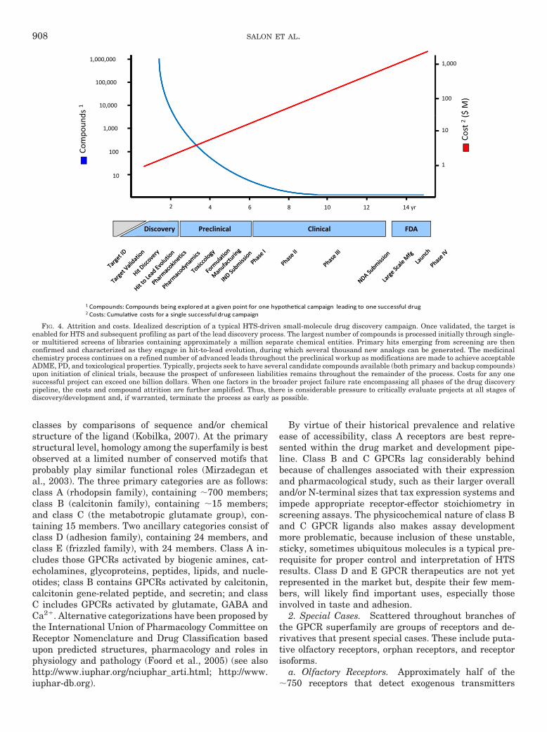
FIG. 4. Attrition and costs. Idealized description of a typical HTS-driven small-molecule drug discovery campaign. Once validated, the target isenabled for HTS and subsequent profiling as part of the lead discovery process. The largest number of compounds is processed initially through single-or multitiered screens of libraries containing approximately a million separate chemical entities. Primary hits emerging from screening are thenconfirmed and characterized as they engage in hit-to-lead evolution, during which several thousand new analogs can be generated. The medicinalchemistry process continues on a refined number of advanced leads throughout the preclinical workup as modifications are made to achieve acceptableADME, PD, and toxicological properties. Typically, projects seek to have several candidate compounds available (both primary and backup compounds)upon initiation of clinical trials, because the prospect of unforeseen liabilities remains throughout the remainder of the process. Costs for any onesuccessful project can exceed one billion dollars. When one factors in the broader project failure rate encompassing all phases of the drug discoverypipeline, the costs and compound attrition are further amplified. Thus, there is considerable pressure to critically evaluate projects at all stages ofdiscovery/development and, if warranted, terminate the process as early as possible.
908 SALON ET AL.
(Fuchs et al., 2001; Glusman et al., 2001; Takeda et al.,2002; Venter et al., 2001; Zozulya et al., 2001), are ded-icated to olfaction. These receptors have typically beenconsidered therapeutically unimportant, but this assess-ment may prove to be untenable. Along with taste re-ceptors, volatile odorant receptors form an importantsensory collective that modulates a host of deeply rootedanimal behaviors such as feeding, mating, and memoryformation. Unfortunately, these receptors harbor theirown molecular eccentricities in terms of expression re-quirements and poorly understood biochemistry, whichat this time makes assay development and drug screen-ing especially problematic.
b. Orphan Receptors. The enabling of orphan recep-tors for drug discovery is a lingering problem com-pounded by difficulties inherent in creating screeningassays without the availability of suitable pharmacolog-ical controls and the laborious task of biochemicallyseeking, purifying, and characterizing endogenous bio-logically active compounds (Howard et al., 2001). Al-though the latter is not a strict prerequisite for screen-ing, it is a necessary component of the target validationprocess. Computational and/or molecular biological ap-proaches to endogenous ligand discovery can also beemployed but these depend upon the accuracy of theirpredictive methods along with a commitment to exhaus-tively screen large panels of orphan receptors againstthousands of synthetic designs/compounds (Shemesh etal., 2008). Depending upon the assays employed, it maybe impossible to ensure that a screen will report a validpositive hit. Also related to the orphan receptor problemis the need to understand the transmitter universe andits biochemical regulation more completely (i.e., to iden-tify post-translational processing and/or metabolictransformation of previously known transmitter sub-stances into different biologically active endogenousligands).
c. Receptor Isoforms. Receptor isoforms are not un-common in the GPCR superfamily, but their functionalimportance and impact on GPCR-mediated signaling ispoorly understood. These alternate forms arise throughalternative splicing and variations in gene loci, includ-ing DNA insertion, deletions, and single nucleotide poly-morphisms that can alter expression and/or function.Such variations can contribute subtly or dramatically tothe onset or progression of disease and its responsive-ness to therapeutics. At least 38 receptor subtypesspanning all five major GPCR families harbor thesemodifications. Although the structural and functionaleccentricities of many isoforms have been characterized,their utility for drug discovery remains to be proven.Attempts to correlate specific isoform expression withindividual pathophysiology may yet prove valuable byassisting in the design of clinical trials and perhapsunveiling novel interconnections between specific GPCRsystems and physiological processes in the general pop-
ulation (Rohrer and Kobilka, 1998; Tang and Insel,2005).
3. In Vitro Reconstitution of Monomeric Receptors. Alarge body of work demonstrates that a monomeric re-ceptor is sufficient to activate G protein (Meyer et al.,2006; Bayburt et al., 2007, 2011; Ernst et al., 2007;Whorton et al., 2008). However, functional activation bya monomeric receptor does not preclude the GPCR dimerfrom being the functional unit. The utilization of hetero-and homodimerization by the GPCR signaling processprovides exquisite levels of sophistication needed to fine-tune the activated state. The combinatorial expansionavailable to the GPCR signaling repertoire through theformation of heterodimers and higher order oligomerspresents yet another level of complexity that must beaddressed in current drug design.
C. Increasingly Complex Pharmacology
Most drugs targeting GPCRs either directly activate(agonists) or inhibit the activation (antagonists) of thesereceptors by their endogenous ligands through stericcompetition at the receptors’ highly conserved or-thosteric ligand-binding sites. A legacy of our early con-cepts of receptor activation, this orthosteric perspectivehas resulted in the preponderance of competitive dis-placement assays that have until recently dominateddrug discovery. The past decade, however, has revealedthat GPCRs and their activation mechanisms are muchmore versatile and complex than previously imagined.Agonism and (silent) antagonism has given way to pos-itive, neutral, and inverse agonism, varying degrees ofefficacy, and positive/negative modulatory effects on or-thosteric potency. Simple two-state (on/off) models ofreceptor activity have been superseded by more complexequilibria encompassing ectopic ligands, multiple recep-tor configurations and conformations, positive versusinverse agonism and the varying degrees of efficacy thateach of these different ensembles produces (Fig. 5). Thetrend is clear. We must advance beyond purely or-thosteric settings to take full advantage of the entireGPCR milieu and pharmacological repertoire.
1. Allostery. The blossoming complexity of GPCRpharmacology is perhaps nowhere better exemplified orformalized as in the area of allosteric action (Gao andJacobson, 2006). Simple two-state models borrowed fromclassic mass-action chemical equilibria have since beenextended to those that include G protein influences asdescribed by complex cubic ternary complexes (Hall,2000) and, more recently, to ternary and quaternarycomplexes that embody allosteric modulation of or-thosteric ligand action (Fig. 5) (Bridges and Lindsley,2008). It is said that allosteric phenomena provide “tex-ture” to GPCR pharmacology by modifying the affinityor signal imparted by the receptor concomitant withbinding of the orthosteric ligand (Leach et al., 2007). Inrevisiting old and validating new targets, the pharma-ceutical industry has taken these new kinetic insights to
GPCRs AND DRUG DISCOVERY 909

heart while fine tuning our therapeutic paradigms tomodulate the biological tone of endogenous ligands(Christopoulos and Kenakin, 2002; May et al., 2007;Keov et al., 2011). In addition to adjusting the naturaltone and tempo of endogenous GPCR ligands, allostericdrugs can more readily achieve selectivity by actingoutside a highly conserved orthosteric cavity (Kenakin,2007; Raddatz et al., 2007).
Even as allostery is now well accepted and its valuebecomes increasingly more evident, there remain majorchallenges to its implementation in drug discovery (Mayet al., 2010). The ability to suitably engineer and tuneassay platforms to detect and quantify allosteric effectsis not yet routine. Furthermore, because of inherentdifficulties in fitting complex mass-action models to ex-perimental data, dissection and tracking of particularcooperativity factors relevant to a therapeutic paradigmduring a structure-activity relationship (SAR) campaigncan be impractical. To include allosteric effects in drugdiscovery, an operational approach is needed that seeksa middle ground wherein both mechanistic and empiri-cal parameters are merged into a model that is a com-promise between the thermodynamic ideal and the real-ity of the biological system in which it operates (Keov et
al., 2011). The challenges of effectively implementingsuch platforms are significant.
Current descriptions employed in assay design do notexplicitly accommodate more interactions than alloste-ric and orthosteric ligand binding on a single receptor-Gprotein complex. Moreover, the complexity escalatesonce other dimensions of GPCR pharmacology and reg-ulation, such as ligand-directed signaling and receptoroligomerization, are included (Smith and Milligan,2010). Allosteric modulators can produce complex effectsthat further complicate their use as therapeutics.Whereas in some cases these modulators may alter thetarget’s binding affinity for endogenous or exogenousagonist ligands, this property may not necessarily resultin greater therapeutic efficacy, because they may elicit acontradictory physiological effect at an off-target tissueor receptor or through changes in ligand affinity forindividual receptor subtypes (May et al., 2010). Struc-tural insights into the biochemical conformations thatunderlie these specific functional states could suggestentirely novel drug designs and guide lead developmenttoward the most relevant pathophysiological pathways.
2. Receptor Oligomerization. Homo- and heterooli-gomerization of GPCRs is now well accepted, and the
FIG. 5. Evolution of pharmacological complexity. Kinetic models of GPCR action have evolved from simple two-state models reflecting classicmass-driven chemical equilibrium principles (a) to more complex cubic ternary complexes (Weiss et al., 1996a,b,c; Hall, 2000) (b) that incorporate Gprotein interactions with both active and inactive receptor isomers and finally to a quaternary complex of GPCR allosterism (Christopoulos andKenakin, 2002) (c), which includes an additional modulatory ligand that can cooperatively affect the binding of the orthosteric ligand and thesubsequent functional interaction of the receptor with its preferred G protein partner. Principle kinetic constants (L, Kx) for each transition pair withvarious cooperativity factors (�, �, �, �, �, �, �, �, , , �,…) describe particular steps. Discovery of additional biochemical phenomena such as receptorhomo-/heterodimerization and the possibility that a given receptor can interact with multiple G protein partners suggests that even these extendedallosteric models are insufficient to fully describe the behavior of GPCRs.
910 SALON ET AL.
functional impact of these types of interactions has beenconvincingly documented in a variety of systems (Dal-rymple et al., 2008). Although the increasing number ofinstances in which this phenomenon can be demon-strated suggests it is the norm rather than an exception,more work is needed to define its occurrence in nativesettings and define the ligand-receptor-effector stoichi-ometry along with the possibility of half-site behaviorand/or cross-receptor effects of antagonist, agonist, andallosteric ligands (Fig. 2). The possibility that ho-modimerization is indeed a native state does not contra-dict the long-standing empirical observation that mostGPCRs reliably produce well behaved activity when het-erologously expressed. However, it cannot be assumedwithout empirical evidence that a heterologous systemequates to physiological state of the receptors. The pros-pect of heterodimerization as a prerequisite for functionis, on the other hand, even more intriguing, because itraises the possibility of exploiting combinatorial degreesof specificity greater than can be afforded by eitherGPCR partner individually. Furthermore, the actualitythat GPCRs function as dimers and that this intermo-lecular association likely occurs at the receptor’s non-conserved helical periphery (Han et al., 2009) opens thepossibility of designing novel drugs that act at theseunique interfaces.
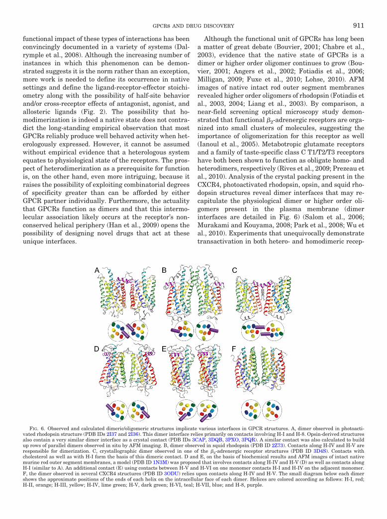
Although the functional unit of GPCRs has long beena matter of great debate (Bouvier, 2001; Chabre et al.,2003), evidence that the native state of GPCRs is adimer or higher order oligomer continues to grow (Bou-vier, 2001; Angers et al., 2002; Fotiadis et al., 2006;Milligan, 2009; Fuxe et al., 2010; Lohse, 2010). AFMimages of native intact rod outer segment membranesrevealed higher order oligomers of rhodopsin (Fotiadis etal., 2003, 2004; Liang et al., 2003). By comparison, anear-field screening optical microscopy study demon-strated that functional �2-adrenergic receptors are orga-nized into small clusters of molecules, suggesting theimportance of oligomerization for this receptor as well(Ianoul et al., 2005). Metabotropic glutamate receptorsand a family of taste-specific class C T1/T2/T3 receptorshave both been shown to function as obligate homo- andheterodimers, respectively (Rives et al., 2009; Prezeau etal., 2010). Analysis of the crystal packing present in theCXCR4, photoactivated rhodopsin, opsin, and squid rho-dopsin structures reveal dimer interfaces that may re-capitulate the physiological dimer or higher order oli-gomers present in the plasma membrane (dimerinterfaces are detailed in Fig. 6) (Salom et al., 2006;Murakami and Kouyama, 2008; Park et al., 2008; Wu etal., 2010). Experiments that unequivocally demonstratetransactivation in both hetero- and homodimeric recep-
FIG. 6. Observed and calculated dimeric/oligomeric structures implicate various interfaces in GPCR structures. A, dimer observed in photoacti-vated rhodopsin structure (PDB IDs 2I37 and 2I36). This dimer interface relies primarily on contacts involving H-I and H-8. Opsin-derived structuresalso contain a very similar dimer interface as a crystal contact (PDB IDs 3CAP, 3DQB, 3PXO, 3PQR). A similar contact was also calculated to buildup rows of parallel dimers observed in situ by AFM imaging. B, dimer observed in squid rhodopsin (PDB ID 2Z73). Contacts along H-IV and H-V areresponsible for dimerization. C, crystallographic dimer observed in one of the �2-adrenergic receptor structures (PDB ID 3D4S). Contacts withcholesterol as well as with H-I form the basis of this dimeric contact. D and E, on the basis of biochemical results and AFM images of intact nativemurine rod outer segment membranes, a model (PDB ID 1N3M) was proposed that involves contacts along H-IV and H-V (D) as well as contacts alongH-I (similar to A). An additional contact (E) using contacts between H-V and H-VI on one monomer contacts H-I and H-IV on the adjacent monomer.F, the dimer observed in several CXCR4 structures (PDB ID 3ODU) relies upon contacts along H-IV and H-V. The small diagram below each dimershows the approximate positions of the ends of each helix on the intracellular face of each dimer. Helices are colored according as follows: H-I, red;H-II, orange; H-III, yellow; H-IV, lime green; H-V, dark green; H-VI, teal; H-VII, blue; and H-8, purple.
GPCRs AND DRUG DISCOVERY 911
tor pairs further support this notion (Salahpour et al.,2004; Terrillon and Bouvier, 2004; Waldhoer et al., 2005;Rivero-Muller et al., 2010). In heterodimers of signaling-deficient and ligand binding-deficient luteinizing hormonereceptor, G protein activation is still observed, arguing forroles for each component monomer during the activationprocess (Rivero-Muller et al., 2010). The utilization ofhetero- and homodimerization by the GPCR signalingprocess provides additional means of regulating the ac-tivated state. The combinatorial expansion available tothe GPCR signaling repertoire through the formation ofheterodimers and higher order oligomers in vivo pres-ents yet another level of complexity that must be ad-dressed in current drug design. The functional conse-quence of dimerization could be unique to each receptoror could be a common feature necessary (e.g., for intra-cellular trafficking or signaling for all GPCRs), but morework is needed to explain this phenomenon in physio-logical context.
3. Ligand-Biased (Ligand-Selective) Signaling. Thehistorical notion that any given receptor subtype is pre-ordained to act through one and only one receptor-de-fined G� subtype-linked effector system has graduallyyielded to the concept of agonist-receptor trafficking(i.e., the ability of a specific agonist to activate a selectsubset of the many possible signaling paths available toa given receptor-G protein system) (Kenakin, 2001; Hoff-mann et al., 2008; Rajagopal et al., 2010; Vaidehi andKenakin, 2010). Examples include the characterizationof distinct signaling profiles for �1- and �2-adrenergicreceptor ligands in the activation of adenylyl cyclase andmitogen-activated protein kinase pathways (Galandrinand Bouvier, 2006) and the opposing G�i and G�s effectsof selective ligands for the �2-adrenergic system (Easonet al., 1992).
Such observations pervaded the assay engineeringcommunity for years and were long viewed as artifacts ofheterologously overexpressed receptors in cell hosts withlimited or imbalanced G protein complements. But thisphenomenon is now understood to be an integral facet ofGPCR behavior, termed “pluridimensional efficacy,” andan early kinetic formulation of the effect stands ready tobe integrated into the expanding thermodynamic de-scription of the GPCR system at large (Kenakin, 2010).From a practical point of view, the availability of natu-rally occurring pluridimensional effects suggests itshould be possible to fine-tune the therapeutic action ofa GPCR drug beyond the simple margins traditionallydictated by the target’s tissue distribution, by also tak-ing advantage of the selective activation allowedthrough the selective downstream activation (Bosier andHermans, 2007; Conn et al., 2009). This ligand-selectivesignaling phenomenon underscores the complexity ofGPCR signaling that must be addressed to effectivelydesign GPCR therapeutics.
4. Constitutive Activity. Originally viewed by manyas an artifact of assay conditions, constitutive activity
can now also be regarded as a physiologically importantequilibrium extreme of the native spectrum of GPCRconformations (Lefkowitz et al., 1993; Costa and Cotec-chia, 2005). Most easily considered in a mass-actioncontext as an elevation in the amount of R* (activatedstate of the GPCR) available to interact with its effec-tors, the phenomenon can be experimentally generatedby either changing the R-R* equilibrium through recep-tor mutagenesis or by increasing the total receptor pop-ulation, R�R*, and hence elevating the amount of R*through overexpression. This can be observed in Lebercongenital amaurosis, where defects in chromophore re-generation or delivery to opsin result in low-grade acti-vation of the visual signaling pathway through the con-stitutive activity of the apoprotein opsin, which leads tothe development of the disease state (Woodruff et al.,2003). When �2-adrenergic receptor is overexpressed inmouse heart, much higher basal rates of activation areobserved, which eventually results in cardiomyopathy(Liggett et al., 2000).
The use of functional assays engineered to displayelevated basal (constitutive) activity revealed that manydrugs previously deemed to be simple (silent) antago-nists did in fact possess intrinsic activity and led to theirsubsequent recategorization as inverse agonists. Theclear in vivo efficacy that these inverse agonist drugsdisplay suggests that the accompanying systemic toneresulting from this constitutive receptor activity cancomprise a general feature of important GPCR-relatedbiological feedback circuits. Although examples exist ofa therapeutic preference of inverse agonists over neutralantagonists, as for example H3 inverse agonists for cog-nition (Schwartz et al., 2003; Arrang et al., 2007), theexistence of constitutive receptor action as a prevalent invivo phenomenon in either normal or pathophysiologicalstates remains to be sufficiently cataloged (de Ligt et al.,2000; Milligan, 2003; Schwartz et al., 2003).
D. Assay Development
In working toward populating an assay tool box forany given target-focused drug discovery campaign, weface a practical dilemma: to know too little about manythings or to know too much about a few. It is noteworthythat technology has helped to both mitigate and propa-gate this problem.
1. The Genomic Tool Box. Closure of the genomicroster for the GPCR superfamily and others along withthe establishment of a full catalog of correspondingclones has theoretically provided the building blocks forall assays needed to attack the therapeutic genome. Inaddition to the target proper, additional componentssuch as assorted signaling proteins and cell lines thathost engineered designs are now readily available. Withthe exception of certain GPCR subclasses (e.g., olfactoryGPCRs and splicing variants), expression of GPCR pro-teins and their effectors for functional assessment isnow relatively routine, although the details of such ex-
912 SALON ET AL.
pression are important and can compromise discovery,for example by inadvertently limiting or biasing phar-macological results.
2. Screening Efficiency. Over the past decade, thetime and cost needed to execute large-scale GPCRscreens has significantly diminished. Developments inHTS technologies have translated into time and costsavings and provided a variety of ultra-high-throughputassay options ranging from labeled and label-free que-ries of bimolecular interactions to image-based mea-sures of multiparameter cell-based events. Integrationof HTS detection platforms with robotics and informat-ics systems for materials handling and data capture/analysis together with visualization tools for data min-ing and report sharing has made execution of millioncompound screens and inspection of the results a timely,straightforward, and efficient process.
Resolution of the information content in such assayswill vary depending upon how it is to be employed andthe scope/precision required. Output typically rangesfrom 50 to 250,000 data points per week and may com-prise assessment of hundreds of thousands of com-pounds against a single target by single point concen-tration (yes/no) activity measurements, as occurs duringa primary screen, or assessment of hundreds of com-pounds in a concentration-dependent fashion againstmultiple targets, as occurs during ensuing SAR leaddevelopment. In these and other instances, archiving,management, and ready availability of such data consti-tute an important informatics estate for ongoing andfuture drug discovery programs.
3. Screening Mode. The past decade has seen a shiftaway from traditional competitive displacement bindingassays toward cell-based functional assays. This shiftwas encouraged by the need to probe more deeply intothe functional activity of compounds at the outset, im-mediately distinguishing between agonist and antago-nist hits. The switch was also encouraged by the prob-lems inherent in introduction of labels into theorthosteric probes needed for competition binding as-says and is, of late, vindicated by the increasing need toentertain nonorthosteric binding sites during the pri-mary screening process.
To this end, first-generation functional assays em-ployed measurements of second-messenger levels of anyone of the canonical major GPCR pathways: cAMP in-duction for G�s, inhibition of forskolin induced cAMP forG�i, and phosphatidylinositol bisphosphate induction(or associated Ca2� levels) by phospholipase C-inducedG�q pathways. The G�q pathway is important to note,because when it was discovered that G�15 and G�16subunits could promiscuously interact with manyGPCRs to elicit G�q-mediated second messengers, theidea of constructing generic platforms became obviousand widely employed (Simon et al., 1991; Offermannsand Simon, 1995; Stables et al., 1997; Zhu et al., 2008).This movement was further fueled by the availability of
the industry’s first truly high-throughput cell-basedplatform for Ca2� fluorescence measurements, the fluo-rometric imaging plate reader. Additional tactics alongthese lines also took advantage of forced couplingthrough the use of G�q chimeras fused to G�s and G�iadaptors. Reporter cell lines incorporating cassettes ofresponse elements sensitive to Ca2� or cAMP levels alsobecame popular and were often used in conjunction witha promiscuous G protein cell host (Knight and Grigliatti,2004). In all cases and in the interest of platform effi-ciency, the measured events were purposefully designedto be the same regardless of the GPCR studied and werevalidated based on preconceptions of signaling built intothe assay. Thus, these assays reported what and onlywhat the researchers were expecting to see; namely,they fail to address the possibility of ligand-biased sig-naling or the subtleties of efficacy arising from allostericeffects. The emerging complexity of GPCR pharmacol-ogy and its potential relevance for the specific regulationof pathophysiological conditions now suggests that suchtactics were ill advised. More recent efforts to secureuniversal results rely more upon preserving the occur-rence of the full spectrum of native downstream GPCRevents [e.g., �-arrestins (Rajagopal et al., 2010)].
4. Integrative Assays. Perhaps the earliest examplesof integrative assays were ex vivo-based organ baths. Itis thanks to their usage that some of the first GPCRtransmitter substances were discovered (Rapport et al.,1948a,b). The concept of approaching pharmacologicalassays holistically, preserving more by modifying less,can be seen in modern “integrative” assays, which havebeen developed to mitigate the problems inherent inengineered artificiality and provide a panoramic windowinto the complex “signalosome.” Such cell-based meth-ods include the microphysiometric measurement of cellmetabolism (Salon and Owicki, 1995), the electricalmeasurement of changes in cell impedance (Verdonk etal., 2006; Peters and Scott, 2009) and the plasmon-baseddetection of cell mass redistribution (Fang and Ferrie,2008; Fang et al., 2008; Lee et al., 2008). Such assays area two-edged sword, providing a broad-based detectionsystem but little to no information about the specificsignaling pathways that are activated. Determination ofthe latter requires subsequent and sometimes nontrivialexperimental dissection of the receptor-dependent phe-nomena (Kenakin, 2011).
5. New Generation Biochemical and Biophysical As-says. Reappreciation of bimolecular interaction assays,whether composed of conventional radio- or fluoroliganddisplacement assays or newer label-free systems, prom-ises to further simplify the identification/quantificationof compound-receptor interactions through direct mea-surement of the immediate drug-target binding event.The nature of these assays makes them less affected bythe practical problems of orchestrating and executingcell-based assays that, as stated above, suffer from cellviability issues, artifactual signaling introduced by cell
GPCRs AND DRUG DISCOVERY 913
engineering and the myriad downstream events that canconfound their interpretation. Bimolecular interactionassays, on the other hand, provide the simplest and mostdirect route to the affinity number most sought duringthe early stages of drug development.
A particularly promising category of bimolecular as-say that is prevalent in the drug discovery communityare plasmon-based methods that have the additionaladvantage of being label-free. Conventional surfaceplasmon resonance (SPR) has been applied to detergentsolubilized as well as purified GPCRs immobilized on asensor chip to qualitatively detect ligand interactionsand quantitatively assess the kinetics of their on and offrates (Navratilova et al., 2005). In addition to providingthe kinetics of binding, SPR methods hold promise asrapid screening assays for GPCR constructs in X-raycrystallization trials and biophysical mapping of recep-tors as part of structure-function campaigns (Tollin etal., 2003; Hruby and Tollin, 2007). Alternatively, real-time plasmon waveguide resonance spectroscopy can beuseful as both a kinetic tool and a means of predictingthe pharmacological action of ligands. The PWR methodoffers increased sensitivity over traditional SPR by de-tecting plasmon shifts in two dimensions; it can reportmass rearrangements associated with the conformationalplasticity of the GPCR system that correlate with pharma-cological activity (Devanathan et al., 2004; Alves et al.,2005; Hruby and Tollin, 2007; Georgieva et al., 2008).
E. Limitations of Current Compound Libraries
1. Chemical Space versus Biological Space. Theproblem of comprehensively spanning all “druggable”chemical space for GPCR drug discovery, at least in anorthosteric setting, is confounded by the structural di-versity of the endogenous transmitters. These activatorsrange from small entities such as Ca2�, to chemicallysimple classic transmitters, to molecules with complexsecondary structure such as peptide transmitters andglycohormones. From a drug design perspective, compet-ing with the more complex ligands is especially challeng-ing, because one must contend with combinatoriallylarger numbers of chemical interactions that involveboth recognition and activity.
In addition, the distribution of content in corporate druglibraries across biological space is likely uneven. As forGPCRs, these collections have evolved through prior drugdiscovery campaigns directed largely toward the or-thosteric modulation of class A targets. Because most cor-porations have been interested in the same targets, thereis likely significant overlap across these corporate collec-tions. Additional requirements that molecular contentmust possess in vivo drug-like qualities, as predicted forexample by Lipinski rules (Lipinski et al., 2001) or asexperimentally measured by PK and ADME, and the me-dicinal chemical “toolbox” employed for expansion of initialhits (Meanwell, 2011) also acts to confine content to iso-lated clusters of “well behaved druggable” space.
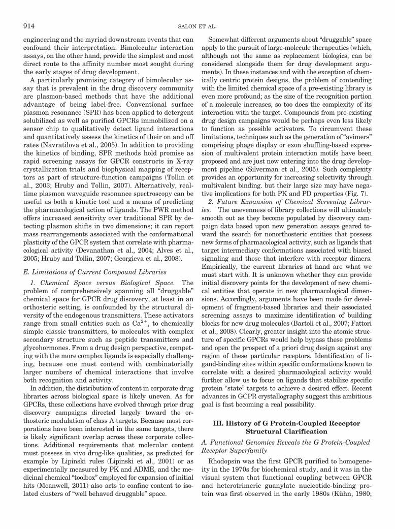
Somewhat different arguments about “druggable” spaceapply to the pursuit of large-molecule therapeutics (which,although not the same as replacement biologics, can beconsidered alongside them for drug development argu-ments). In these instances and with the exception of chem-ically centric protein designs, the problem of contendingwith the limited chemical space of a pre-existing library iseven more profound; as the size of the recognition portionof a molecule increases, so too does the complexity of itsinteraction with the target. Compounds from pre-existingdrug design campaigns would be perhaps even less likelyto function as possible activators. To circumvent theselimitations, techniques such as the generation of “avimers”comprising phage display or exon shuffling-based expres-sion of multivalent protein interaction motifs have beenproposed and are just now entering into the drug develop-ment pipeline (Silverman et al., 2005). Such complexityprovides an opportunity for increasing selectivity throughmultivalent binding. but their large size may have nega-tive implications for both PK and PD properties (Fig. 7).
2. Future Expansion of Chemical Screening Librar-ies. The unevenness of library collections will ultimatelysmooth out as they become populated by discovery cam-paign data based upon new generation assays geared to-ward the search for nonorthosteric entities that possessnew forms of pharmacological activity, such as ligands thattarget intermediary conformations associated with biasedsignaling and those that interfere with receptor dimers.Empirically, the current libraries at hand are what wemust start with. It is unknown whether they can provideinitial discovery points for the development of new chemi-cal entities that operate in new pharmacological dimen-sions. Accordingly, arguments have been made for devel-opment of fragment-based libraries and their associatedscreening assays to maximize identification of buildingblocks for new drug molecules (Bartoli et al., 2007; Fattoriet al., 2008). Clearly, greater insight into the atomic struc-ture of specific GPCRs would help bypass these problemsand open the prospect of a priori drug design against anyregion of these particular receptors. Identification of li-gand-binding sites within specific conformations known tocorrelate with a desired pharmacological activity wouldfurther allow us to focus on ligands that stabilize specificprotein “state” targets to achieve a desired effect. Recentadvances in GCPR crystallography suggest this ambitiousgoal is fast becoming a real possibility.
III. History of G Protein-Coupled ReceptorStructural Clarification
A. Functional Genomics Reveals the G Protein-CoupledReceptor Superfamily
Rhodopsin was the first GPCR purified to homogene-ity in the 1970s for biochemical study, and it was in thevisual system that functional coupling between GPCRand heterotrimeric guanylate nucleotide-binding pro-tein was first observed in the early 1980s (Kuhn, 1980;
914 SALON ET AL.
Fung et al., 1981). The determination of the amino acidsequence of rhodopsin by Hargrave et al. (1983) andOvchinnikov et al. (1983) was a significant achievementand provided the starting point for models of its mem-brane topology. Shortly after this work, Nathans andHogness (1983) used at-that-time novel molecular bio-logical techniques to clone all human visual pigmentsthat later permitted insights into the genetic basis ofpathologic conditions in color vision (Nathans et al.,1986a,b). Cloning of rhodopsin and extrapolation of itsputative molecular model to other GPCRs provided theprerequisites for understanding ligand binding and Gprotein interaction (Filipek et al., 2003b). This advancealso provided early insights into G protein signaling andits ubiquitous presence in cells and tissues (Bitensky etal., 1984; Dixon et al., 1986). Analogous work performedby Dixon et al. (1986) and Fargin et al. (1988) employedthe partial �2-adrenergic receptor protein sequence todesign degenerate PCR primer sequences to clone andsequence the full-length �2-adrenergic receptor as wellas the first orphan receptor (later identified as the5-HT1A receptor). This and subsequent work suggestedthe existence of a new family of integral membrane
proteins, all of which shared signaling pathways relyingupon heterotrimeric G proteins and having in common aseven transmembrane �-helical architecture (Gilman,1987; Baldwin, 1993, 1994; Baldwin et al., 1997; Filipeket al., 2003b). This homologous structure conceptspawned a decade of homology and expression-cloningefforts, during which many of our initial molecular bio-logical discoveries for the GPCR superfamily were made.
B. Electron Microscopy of Rhodopsin Provides aConceptual Prototype of G Protein-CoupledReceptor Structure
The archaeal proton pump bacteriorhodopsin was ini-tially hypothesized to be an analog of rhodopsin beforethe latter’s three-dimensional structural determinationby electron crystallography. Both proteins feature a co-valently bound retinal chromophore linked through aSchiff base to a Lys residue, exhibit a light-dependentchange in spectral absorption, and possess a seven-transmembrane helix architecture. Protein and DNAsequencing of rhodopsin (Hargrave et al., 1983; Nathansand Hogness, 1983, 1984; Ovchinnikov et al., 1983) andthe �2-adrenergic receptor (Dixon et al., 1986) together
ds
10000
proteins
an�bodies
avimers
chem
ical
bon
d
1000
100
ll l l
pep�des
pep�bodiesdr
ug-t
arge
t c
10
100
small molecules
Drug mw
Chemical En��es
(small organic molecules, pep�des, oligonucleo�des, ....)
+ Access all intra- and extracellular targets (including CNS)
Biologics
(cytokines, hormones, soluble receptors, binding, an�bodies…)
-- Access only circula�ng and cell surface targets in the periphery
+ Formulated as tablets, suspensions, drops, inhalants, injectables, patches, creams, etc.
+ Administered daily/weekly.
- Intrinsically less specific, increasing possibility of side effects
+ Rela�vely easy to synthesize, package, and distribute
- Formulated as injectable
+ Administered daily/weekly/monthly.
+ Intrinsically more specific, increasing level of tolerance
- Difficult to manufacture, package, and distribute
+ Simpler (more rapid) discovery and development process - Complex discovery and development process
- Expensive process
p ( p ) y p p
-- Extremely expensive process
FIG. 7. Therapeutic modalities. Drug molecules can be broadly categorized as either small molecules or biologics, with development and FDAreview after new chemical entity and biologic license application guidelines, respectively. Although GPCR drug discovery has historically beendominated by small molecule programs, this predominance is changing in part because of the increasing awareness that target receptors can havemuch larger endogenous ligands (e.g., glycohormones, viral entry proteins) and in part because of methods that merge antibody or other scaffolds withchemical and peptide moieties to provide robust delivery vehicles for active small molecule warheads. An application of this method is the newlydeveloped “avimer” technologies, which are antibody mimetics that present multiple binding epitopes. SBDD methods and infrastructure benefit fromboth modalities by identifying novel sites accessible to small-molecule binding and providing purified GPCR proteins stabilized in specific conforma-tions to capture large molecule drug candidates through more empiric screening methods.
GPCRs AND DRUG DISCOVERY 915
with hydropathy plots facilitated construction of two-dimensional topology models (Argos et al., 1982; Har-grave et al., 1983; Ovchinnikov et al., 1983; Dixon et al.,1986) similar to those generated for bacteriorhodopsin.Electron crystallographic studies of two-dimensionalcrystals of bovine, amphibian, and invertebrate rho-dopsins demonstrated that the arrangement of helices inrhodopsin differed substantially from those in bacterio-rhodopsin (Schertler et al., 1993; Krebs et al., 1998;Davies et al., 2001). These results bolstered the idea thatrhodopsin, and not bacteriorhodopsin, would serve as amodel for all GPCRs, leading to its adoption for modelinghelical arrangements within the transmembrane bundleof GPCRs (Baldwin, 1993, 1994; Baldwin et al., 1997;Unger et al., 1997). These structural results were fur-ther advanced by mutagenesis and biochemical studiesallowing spacial assignment of post-translational modi-fications such as disulfide bonds (Karnik et al., 1988;Karnik and Khorana, 1990), palmitoylation (Ovchin-nikov et al., 1988; Karnik et al., 1993), and phosphory-lation (Palczewski et al., 1991; Ohguro et al., 1993, 1996,1998). These biochemical studies together with specificmutations led to a clear demarcation of the border be-tween loops and transmembrane regions (for review, seeMenon et al., 2001; Sakmar et al., 2002; Filipek et al.,2003a; Hubbell et al., 2003; Palczewski, 2006). For morethan 20 years, rhodopsin has served as the protypicalGPCR for study owing to its relative ease of purificationand the wealth of structural and biochemical informa-tion available.
C. Comparative Modeling SuggestsStructure-Function Experiments
Elucidation of the first vertebrate and invertebrateGPCR structures (Palczewski et al., 2000; Li et al., 2004;Okada et al., 2004; Murakami and Kouyama, 2008; Shi-mamura et al., 2008; Stenkamp, 2008) opened the way toprobe ligand-binding sites and assess structure-functionrelationships for these receptors (Lu et al., 2002; Hub-bell et al., 2003; Park et al., 2004). The overall similarityin helical packing is expected to extend to other GPCRswhose structures have yet to be determined (Lodowskiand Palczewski, 2009; Lodowski et al., 2009; Mustafiand Palczewski, 2009). Given the assumption that allGPCRs share grossly similar membrane structures, it isa reasonable expectation that this data can be used inconjunction with current three-dimensional templatesto derive distinctive homology models.
Currently available structures of rhodopsin and otherGPCRs thus present workable templates for producinghomology models of the remaining GPCRs (Zhang et al.,2006; Michino et al., 2009). However, because of theirlow overall sequence homology, the imprecision of algo-rithms used to predict GPCR overall structure, and thelocation of small-molecule-binding sites within theirtransmembrane domains, this approach has not yetevolved sufficiently to produce models accurate enough
to reliably predict pharmacophore sites. Even small dif-ferences in exact helical arrangements can greatly affectthe predicted binding site for small-molecule ligands.Moreover, it should be considered that bovine rhodopsinremains the only truly “native” ligand-bound GPCRstructure determined. The protein engineering in allcurrent nonrhodopsin GPCR structures can disrupt andobscure the spatial relationships of important regula-tory and functional motifs, such as changes in cytoplas-mic loop three, which occur upon agonist activation orthe breaking of the D(E)RY “ionic” lock (Dror et al.,2009). A related concern is the criteria used to assess theaccuracy of existing models in the absence of high-reso-lution structures. For the many GPCRs, the low overallsequence identity between templates complicates accu-rate alignments and predictive demarcation of ligand-binding sites. Such approaches amount to pseudo-abinitio structural prediction (Schlyer and Horuk, 2006)and despite widespread usage, these methods have gen-erally failed to provide models accurate enough for ther-apeutic ligand design (Deupi et al., 2007).
The recent agonist-bound GPCR structures providelittle structural reason why particular compounds func-tion as full agonists, partial agonists, or even antago-nists, nor do they show consistent structural transfor-mations that accompany the activation of a particularGPCR. The agonist and partial agonist bound �1-adren-ergic receptor structures (Warne et al., 2011) as well asthe tethered agonist-�2-adrenergic receptor structure(Rosenbaum et al., 2011) exhibit only small-scalechanges compared with their antagonist-bound counter-parts, consistent with conclusions on inactive state ofreceptors stated in section B above. Energy for such atransformation is typically between 8 and 12 kcal/mol,with only a small fraction of this energy provided by theligand binding events per se. Because only small confor-mational changes are supported by the energetics of thisprocess, structural differences between the activatedstate before G protein coupling and its inactivated statemust be minimal, further emphasizing the high preci-sion needed in a model if it is to have any utility. Therecent �1-adrenergic agonist structure, the camelidantibody-agonist-�2-adrenergic receptor structure, theagonist bound A2a-adenosine receptor structure as wellas the constitutively active rhodopsin and all-trans-ret-inal regenerated opsin structures provide additionalsnapshots as to the dynamic remodeling, which maybecome possible after the release of molecular restraintsthat accompany GPCR activation occurs (Choe et al.,2011; Rasmussen et al., 2011; Standfuss et al., 2011; Xuet al., 2011). Upon examination of the antibody-agonist-�2-adrenergic receptor structure, larger-scale displace-ments of helices H-V and H-VI are evident comparedwith the small-scale displacements in the �1-adrenergicreceptor structures. It remains unclear as to how todissect the relative influences on the observed struc-tures of the antibody, crystal contacts/conditions, ago-
916 SALON ET AL.
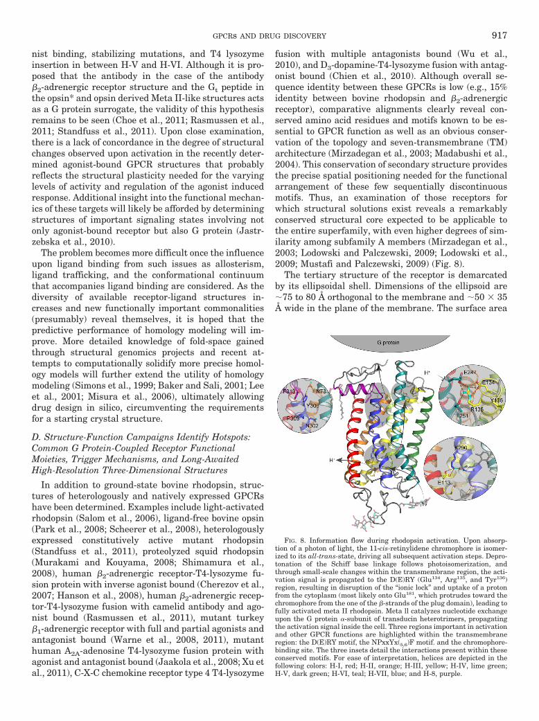
nist binding, stabilizing mutations, and T4 lysozymeinsertion in between H-V and H-VI. Although it is pro-posed that the antibody in the case of the antibody�2-adrenergic receptor structure and the Gt peptide inthe opsin* and opsin derived Meta II-like structures actsas a G protein surrogate, the validity of this hypothesisremains to be seen (Choe et al., 2011; Rasmussen et al.,2011; Standfuss et al., 2011). Upon close examination,there is a lack of concordance in the degree of structuralchanges observed upon activation in the recently deter-mined agonist-bound GPCR structures that probablyreflects the structural plasticity needed for the varyinglevels of activity and regulation of the agonist inducedresponse. Additional insight into the functional mechan-ics of these targets will likely be afforded by determiningstructures of important signaling states involving notonly agonist-bound receptor but also G protein (Jastr-zebska et al., 2010).
The problem becomes more difficult once the influenceupon ligand binding from such issues as allosterism,ligand trafficking, and the conformational continuumthat accompanies ligand binding are considered. As thediversity of available receptor-ligand structures in-creases and new functionally important commonalities(presumably) reveal themselves, it is hoped that thepredictive performance of homology modeling will im-prove. More detailed knowledge of fold-space gainedthrough structural genomics projects and recent at-tempts to computationally solidify more precise homol-ogy models will further extend the utility of homologymodeling (Simons et al., 1999; Baker and Sali, 2001; Leeet al., 2001; Misura et al., 2006), ultimately allowingdrug design in silico, circumventing the requirementsfor a starting crystal structure.
D. Structure-Function Campaigns Identify Hotspots:Common G Protein-Coupled Receptor FunctionalMoieties, Trigger Mechanisms, and Long-AwaitedHigh-Resolution Three-Dimensional Structures
In addition to ground-state bovine rhodopsin, struc-tures of heterologously and natively expressed GPCRshave been determined. Examples include light-activatedrhodopsin (Salom et al., 2006), ligand-free bovine opsin(Park et al., 2008; Scheerer et al., 2008), heterologouslyexpressed constitutively active mutant rhodopsin(Standfuss et al., 2011), proteolyzed squid rhodopsin(Murakami and Kouyama, 2008; Shimamura et al.,2008), human �2-adrenergic receptor-T4-lysozyme fu-sion protein with inverse agonist bound (Cherezov et al.,2007; Hanson et al., 2008), human �2-adrenergic recep-tor-T4-lysozyme fusion with camelid antibody and ago-nist bound (Rasmussen et al., 2011), mutant turkey�1-adrenergic receptor with full and partial agonists andantagonist bound (Warne et al., 2008, 2011), mutanthuman A2A-adenosine T4-lysozyme fusion protein withagonist and antagonist bound (Jaakola et al., 2008; Xu etal., 2011), C-X-C chemokine receptor type 4 T4-lysozyme
fusion with multiple antagonists bound (Wu et al.,2010), and D3-dopamine-T4-lysozyme fusion with antag-onist bound (Chien et al., 2010). Although overall se-quence identity between these GPCRs is low (e.g., 15%identity between bovine rhodopsin and �2-adrenergicreceptor), comparative alignments clearly reveal con-served amino acid residues and motifs known to be es-sential to GPCR function as well as an obvious conser-vation of the topology and seven-transmembrane (TM)architecture (Mirzadegan et al., 2003; Madabushi et al.,2004). This conservation of secondary structure providesthe precise spatial positioning needed for the functionalarrangement of these few sequentially discontinuousmotifs. Thus, an examination of those receptors forwhich structural solutions exist reveals a remarkablyconserved structural core expected to be applicable tothe entire superfamily, with even higher degrees of sim-ilarity among subfamily A members (Mirzadegan et al.,2003; Lodowski and Palczewski, 2009; Lodowski et al.,2009; Mustafi and Palczewski, 2009) (Fig. 8).
The tertiary structure of the receptor is demarcatedby its ellipsoidal shell. Dimensions of the ellipsoid are�75 to 80 Å orthogonal to the membrane and �50 � 35Å wide in the plane of the membrane. The surface area
FIG. 8. Information flow during rhodopsin activation. Upon absorp-tion of a photon of light, the 11-cis-retinylidene chromophore is isomer-ized to its all-trans-state, driving all subsequent activation steps. Depro-tonation of the Schiff base linkage follows photoisomerization, andthrough small-scale changes within the transmembrane region, the acti-vation signal is propagated to the D(E)RY (Glu134, Arg135, and Tyr136)region, resulting in disruption of the “ionic lock” and uptake of a protonfrom the cytoplasm (most likely onto Glu181, which protrudes toward thechromophore from the one of the �-strands of the plug domain), leading tofully activated meta II rhodopsin. Meta ll catalyzes nucleotide exchangeupon the G protein �-subunit of transducin heterotrimers, propagatingthe activation signal inside the cell. Three regions important in activationand other GPCR functions are highlighted within the transmembraneregion: the D(E)RY motif, the NPxxYx(5,6)F motif. and the chromophore-binding site. The three insets detail the interactions present within theseconserved motifs. For ease of interpretation, helices are depicted in thefollowing colors: H-I, red; H-II, orange; H-III, yellow; H-IV, lime green;H-V, dark green; H-VI, teal; H-VII, blue; and H-8, purple.
GPCRs AND DRUG DISCOVERY 917
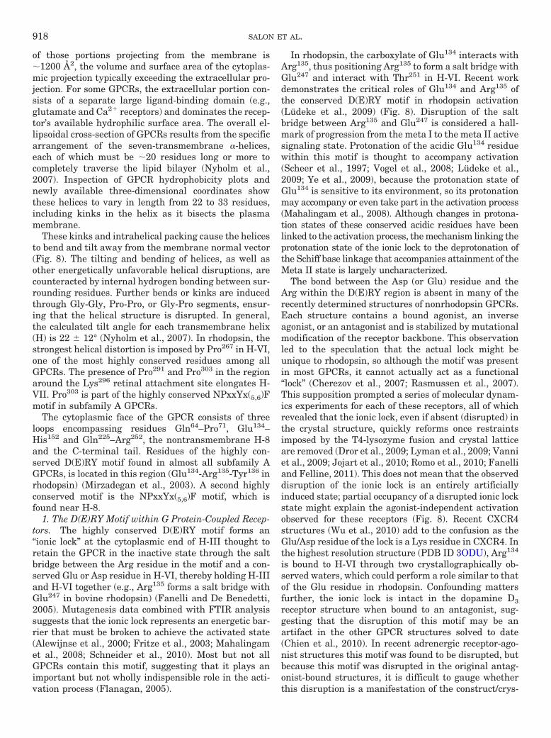
of those portions projecting from the membrane is�1200 Å2, the volume and surface area of the cytoplas-mic projection typically exceeding the extracellular pro-jection. For some GPCRs, the extracellular portion con-sists of a separate large ligand-binding domain (e.g.,glutamate and Ca2� receptors) and dominates the recep-tor’s available hydrophilic surface area. The overall el-lipsoidal cross-section of GPCRs results from the specificarrangement of the seven-transmembrane �-helices,each of which must be �20 residues long or more tocompletely traverse the lipid bilayer (Nyholm et al.,2007). Inspection of GPCR hydrophobicity plots andnewly available three-dimensional coordinates showthese helices to vary in length from 22 to 33 residues,including kinks in the helix as it bisects the plasmamembrane.
These kinks and intrahelical packing cause the helicesto bend and tilt away from the membrane normal vector(Fig. 8). The tilting and bending of helices, as well asother energetically unfavorable helical disruptions, arecounteracted by internal hydrogen bonding between sur-rounding residues. Further bends or kinks are inducedthrough Gly-Gly, Pro-Pro, or Gly-Pro segments, ensur-ing that the helical structure is disrupted. In general,the calculated tilt angle for each transmembrane helix(H) is 22 � 12° (Nyholm et al., 2007). In rhodopsin, thestrongest helical distortion is imposed by Pro267 in H-VI,one of the most highly conserved residues among allGPCRs. The presence of Pro291 and Pro303 in the regionaround the Lys296 retinal attachment site elongates H-VII. Pro303 is part of the highly conserved NPxxYx(5,6)Fmotif in subfamily A GPCRs.
The cytoplasmic face of the GPCR consists of threeloops encompassing residues Gln64–Pro71, Glu134–His152 and Gln225–Arg252, the nontransmembrane H-8and the C-terminal tail. Residues of the highly con-served D(E)RY motif found in almost all subfamily AGPCRs, is located in this region (Glu134-Arg135-Tyr136 inrhodopsin) (Mirzadegan et al., 2003). A second highlyconserved motif is the NPxxYx(5,6)F motif, which isfound near H-8.
1. The D(E)RY Motif within G Protein-Coupled Recep-tors. The highly conserved D(E)RY motif forms an“ionic lock” at the cytoplasmic end of H-III thought toretain the GPCR in the inactive state through the saltbridge between the Arg residue in the motif and a con-served Glu or Asp residue in H-VI, thereby holding H-IIIand H-VI together (e.g., Arg135 forms a salt bridge withGlu247 in bovine rhodopsin) (Fanelli and De Benedetti,2005). Mutagenesis data combined with FTIR analysissuggests that the ionic lock represents an energetic bar-rier that must be broken to achieve the activated state(Alewijnse et al., 2000; Fritze et al., 2003; Mahalingamet al., 2008; Schneider et al., 2010). Most but not allGPCRs contain this motif, suggesting that it plays animportant but not wholly indispensible role in the acti-vation process (Flanagan, 2005).
In rhodopsin, the carboxylate of Glu134 interacts withArg135, thus positioning Arg135 to form a salt bridge withGlu247 and interact with Thr251 in H-VI. Recent workdemonstrates the critical roles of Glu134 and Arg135 ofthe conserved D(E)RY motif in rhodopsin activation(Ludeke et al., 2009) (Fig. 8). Disruption of the saltbridge between Arg135 and Glu247 is considered a hall-mark of progression from the meta I to the meta II activesignaling state. Protonation of the acidic Glu134 residuewithin this motif is thought to accompany activation(Scheer et al., 1997; Vogel et al., 2008; Ludeke et al.,2009; Ye et al., 2009), because the protonation state ofGlu134 is sensitive to its environment, so its protonationmay accompany or even take part in the activation process(Mahalingam et al., 2008). Although changes in protona-tion states of these conserved acidic residues have beenlinked to the activation process, the mechanism linking theprotonation state of the ionic lock to the deprotonation ofthe Schiff base linkage that accompanies attainment of theMeta II state is largely uncharacterized.
The bond between the Asp (or Glu) residue and theArg within the D(E)RY region is absent in many of therecently determined structures of nonrhodopsin GPCRs.Each structure contains a bound agonist, an inverseagonist, or an antagonist and is stabilized by mutationalmodification of the receptor backbone. This observationled to the speculation that the actual lock might beunique to rhodopsin, so although the motif was presentin most GPCRs, it cannot actually act as a functional“lock” (Cherezov et al., 2007; Rasmussen et al., 2007).This supposition prompted a series of molecular dynam-ics experiments for each of these receptors, all of whichrevealed that the ionic lock, even if absent (disrupted) inthe crystal structure, quickly reforms once restraintsimposed by the T4-lysozyme fusion and crystal latticeare removed (Dror et al., 2009; Lyman et al., 2009; Vanniet al., 2009; Jojart et al., 2010; Romo et al., 2010; Fanelliand Felline, 2011). This does not mean that the observeddisruption of the ionic lock is an entirely artificiallyinduced state; partial occupancy of a disrupted ionic lockstate might explain the agonist-independent activationobserved for these receptors (Fig. 8). Recent CXCR4structures (Wu et al., 2010) add to the confusion as theGlu/Asp residue of the lock is a Lys residue in CXCR4. Inthe highest resolution structure (PDB ID 3ODU), Arg134
is bound to H-VI through two crystallographically ob-served waters, which could perform a role similar to thatof the Glu residue in rhodopsin. Confounding mattersfurther, the ionic lock is intact in the dopamine D3receptor structure when bound to an antagonist, sug-gesting that the disruption of this motif may be anartifact in the other GPCR structures solved to date(Chien et al., 2010). In recent adrenergic receptor-ago-nist structures this motif was found to be disrupted, butbecause this motif was disrupted in the original antag-onist-bound structures, it is difficult to gauge whetherthis disruption is a manifestation of the construct/crys-
918 SALON ET AL.
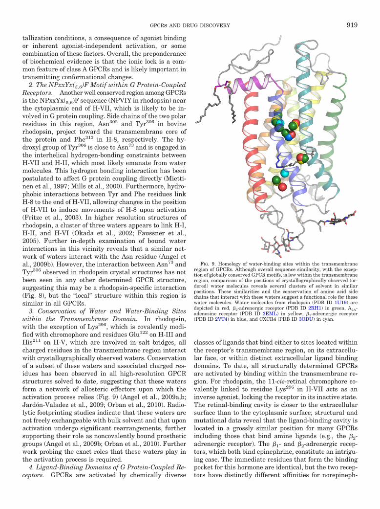
tallization conditions, a consequence of agonist bindingor inherent agonist-independent activation, or somecombination of these factors. Overall, the preponderanceof biochemical evidence is that the ionic lock is a com-mon feature of class A GPCRs and is likely important intransmitting conformational changes.
2. The NPxxYx(5,6)F Motif within G Protein-CoupledReceptors. Another well conserved region among GPCRsis the NPxxYx(5,6)F sequence (NPVIY in rhodopsin) nearthe cytoplasmic end of H-VII, which is likely to be in-volved in G protein coupling. Side chains of the two polarresidues in this region, Asn302 and Tyr306 in bovinerhodopsin, project toward the transmembrane core ofthe protein and Phe313 in H-8, respectively. The hy-droxyl group of Tyr306 is close to Asn73 and is engaged inthe interhelical hydrogen-bonding constraints betweenH-VII and H-II, which most likely emanate from watermolecules. This hydrogen bonding interaction has beenpostulated to affect G protein coupling directly (Mietti-nen et al., 1997; Mills et al., 2000). Furthermore, hydro-phobic interactions between Tyr and Phe residues linkH-8 to the end of H-VII, allowing changes in the positionof H-VII to induce movements of H-8 upon activation(Fritze et al., 2003). In higher resolution structures ofrhodopsin, a cluster of three waters appears to link H-I,H-II, and H-VI (Okada et al., 2002; Faussner et al.,2005). Further in-depth examination of bound waterinteractions in this vicinity reveals that a similar net-work of waters interact with the Asn residue (Angel etal., 2009b). However, the interaction between Asn73 andTyr306 observed in rhodopsin crystal structures has notbeen seen in any other determined GPCR structure,suggesting this may be a rhodopsin-specific interaction(Fig. 8), but the “local” structure within this region issimilar in all GPCRs.
3. Conservation of Water and Water-Binding Siteswithin the Transmembrane Domain. In rhodopsin,with the exception of Lys296, which is covalently modi-fied with chromophore and residues Glu122 on H-III andHis211 on H-V, which are involved in salt bridges, allcharged residues in the transmembrane region interactwith crystallographically observed waters. Conservationof a subset of these waters and associated charged res-idues has been observed in all high-resolution GPCRstructures solved to date, suggesting that these watersform a network of allosteric effectors upon which theactivation process relies (Fig. 9) (Angel et al., 2009a,b;Jardon-Valadez et al., 2009; Orban et al., 2010). Radio-lytic footprinting studies indicate that these waters arenot freely exchangeable with bulk solvent and that uponactivation undergo significant rearrangements, furthersupporting their role as noncovalently bound prostheticgroups (Angel et al., 2009b; Orban et al., 2010). Furtherwork probing the exact roles that these waters play inthe activation process is required.
4. Ligand-Binding Domains of G Protein-Coupled Re-ceptors. GPCRs are activated by chemically diverse
classes of ligands that bind either to sites located withinthe receptor’s transmembrane region, on its extracellu-lar face, or within distinct extracellular ligand bindingdomains. To date, all structurally determined GPCRsare activated by binding within the transmembrane re-gion. For rhodopsin, the 11-cis-retinal chromophore co-valently linked to residue Lys296 in H-VII acts as aninverse agonist, locking the receptor in its inactive state.The retinal-binding cavity is closer to the extracellularsurface than to the cytoplasmic surface; structural andmutational data reveal that the ligand-binding cavity islocated in a grossly similar position for many GPCRsincluding those that bind amine ligands (e.g., the �2-adrenergic receptor). The �1- and �2-adrenergic recep-tors, which both bind epinephrine, constitute an intrigu-ing case. The immediate residues that form the bindingpocket for this hormone are identical, but the two recep-tors have distinctly different affinities for norepineph-
FIG. 9. Homology of water-binding sites within the transmembraneregion of GPCRs. Although overall sequence similarity, with the excep-tion of globally conserved GPCR motifs, is low within the transmembraneregion, comparison of the positions of crystallographically observed (or-dered) water molecules reveals several clusters of solvent in similarpositions. These similarities and the conservation of amino acid sidechains that interact with these waters suggest a functional role for thesewater molecules. Water molecules from rhodopsin (PDB ID 1U19) aredepicted in red, �2-adrenergic receptor (PDB ID 2RH1) in green, A2A-adenosine receptor (PDB ID 3EML) in yellow, �1-adrenergic receptor(PDB ID 2VT4) in blue, and CXCR4 (PDB ID 3ODU) in cyan.
GPCRs AND DRUG DISCOVERY 919
rine, suggesting that residues that do not directly inter-act with the ligand-binding site play a role in ligandselectivity (Fig. 10). The amino-terminal region and ex-tracellular loops of rhodopsin form a plug covering theretinal-binding cavity and contacting the chromophore,shielding the retinal binding pocket from the extracel-lular milieu, a feature unique to rhodopsin. Because thelarge majority of GPCRs must bind ligand suppliedthrough systemic circulation, this plug is absent and is alikely site for ligand entry.
For many GPCRs that recognize amino acid (e.g.,metabotropic glutamate receptor), peptide (e.g., vasoac-tive intestinal peptide 1 receptor) or large protein hor-mones (e.g., thyroid stimulating hormone receptor), theligand-binding site is located on a separate domain lo-cated in the extracellular space. Although several struc-tures of these isolated domains have been determined incomplex with their physiological ligands as well as withinhibitors, the mechanism by which such ligand bindingis coupled to activation of the receptor remains unclear(Kunishima et al., 2000; Fan and Hendrickson, 2005;Parthier et al., 2007; Pioszak et al., 2008, 2010; ter Haaret al., 2010). Although the mechanism by which theseGPCRs with large extracellular domains transfer theiractivation signal to the 7-TM portion of the GPCR hasnot been fully elucidated, some conclusions can bedrawn. Experiments performed with metabotropic glu-tamate receptors mutated to have only one functionalligand binding domain indicate that agonist activates
both protamers with equal efficiency (Brock et al., 2007).Because these disparate ligand-binding domains arelinked to the 7-TM core via a single �-helix and thesereceptors are constitutively dimeric, it is likely that theactivation signal is imparted through a mechanism thatrelies upon intersubunit rearrangement(s) to drive thechanges in the 7-TM domain necessary to activate Gprotein.
Considering the transmembrane architecture that allGPCRs are expected to share, the conservation of motifswithin the transmembrane domain, the variability ofstructural rearrangements that are observed in agonistbound GPCR structures, the conservation of orderedsolvent binding sites within the transmembrane region,and likely mechanism of activation for class B and CGPCRs containing large extracellular ligand binding do-mains, it becomes possible to propose the following con-clusions about the activation process: 1) Rather than asingle distinct activated state that occurs upon bindingof agonist, there exist multiple conformations of theprotein backbone upon binding agonist, only a subset ofwhich are capable of activating G protein. 2) Binding ofagonist must act through a mechanism that begins notwith large movements of helices but with the release ofconstraints that hold the GPCR in an inactive conforma-tion. 3) A network of ordered solvent molecules plays anintegral role in transmission of the activation signalthrough the transmembrane region. Despite the amaz-ing level of progress in the determination of GPCR struc-
FIG. 10. Ligand-binding sites within selected GPCR structures. For these sites, all polar contacts within 3.4 Å of each other are indicated by browndashed lines and nonpolar side chains are also shown when they do not obscure the ligand. In some cases, portions of the transmembrane helices areremoved for clarity. A, chromophore of rhodopsin (PDB ID 1U19) is a tethered ligand (through a Schiff-base linkage); the only polar contact in theinactive state is to the counter ion, Glu113. B, binding of the inverse agonist carazolol to the �2-adrenergic receptor (PDB ID 2RH1). C, antagonistcyanopindolol bound to the �1-adrenergic receptor (PDB ID 2VT4). D, binding of the A2a-adenosine receptor (PDB ID 3EML) to the antagonistZM241385 implicates both solvent mediated interactions as well as direct interactions with amino acid side chains in ligand binding. E, structure ofCXCR4 bound to the antagonist It1t (PDB ID 3ODU) uses both solvent and direct interactions to define the ligand-binding pocket. F, D3 dopaminereceptor (PDB ID 3PBL) bound to the antagonist eticlopride.
920 SALON ET AL.
tures containing agonist compounds, more structuralwork is still needed. To fully understand the activationprocess, we still need the structures of intact class B andC GPCRs in addition to crystal structures that captureproductive signaling states of the receptor in complexwith G protein and agonist compounds (see Note Addedin Proof).
IV. Advances in G Protein-Coupled ReceptorStructural Determination
A. G Protein-Coupled Receptor Construct Designand Expression
The first hurdle in GPCR structural determinationcontinues to be the production and purification of ade-quate amounts of homogenous, properly folded, and fullyfunctional protein. Purification from native tissue, as-suming the protein of interest is present in sufficientquantity, can provide fundamental advantages, such aspresenting normal post-translational modifications thatinfluence the folding of biologically relevant conforma-tions. That said, only bovine rhodopsin has successfullybeen crystallized from its native source and with anintact sequence. All other GPCR structures thus fardetermined have required significant protein (re)engi-neering for stabilization. Cell-free expression, Esche-richia coli, insect, yeast, and various mammalian cellline systems have been used with various success ratesfor GPCR expression. E. coli expression systems havebeen employed to produce heterogeneous preparations ofneurotensin and olfactory receptors (Grisshammer,2006, 2009; White and Grisshammer, 2007; Song et al.,2009). Yeast Saccharomyces cerevisiae and Schizosac-charomyces pombe expression systems have been used toexpress large quantities of A2a-adenosine receptor withligand-binding characteristics near those of wild type(Niebauer et al., 2004; Niebauer and Robinson, 2006;O’Malley et al., 2009). Production through cell-free ex-pression systems has proven useful for structural deter-minations of both soluble and membrane protein targets(Chen et al., 2007b), but large-scale use of these tech-niques for GPCR expression have yet to yield a GPCRstructure (Klammt et al., 2007a,b; Junge et al., 2008,2010). A variety of mammalian cell-based protocols us-ing transfected and virally infected cells to expressGPCRs for biochemical as well as structural determina-tion studies have been published (Sen et al., 2003;Shukla et al., 2006a,b), but rhodopsin mutants ex-pressed in transfected COS-1 cells constitute the onlysuccess for this expression methodology (Standfuss etal., 2007, 2011; Stenkamp, 2008). The most efficient andsuccessful method to date, apart from native expression,has been the expression of truncated/stabilized/engi-neered GPCR constructs in baculovirus-infected insectcell hosts (Cherezov et al., 2007; Rosenbaum et al., 2007;Roth et al., 2008; Warne et al., 2008, 2009; Tate andSchertler, 2009). It is, however, entirely possible that
adequate quantities of native GPCRs needed for struc-ture determination may be produced through novel ex-pression methods that may require further development(Zhang et al., 2005; Li et al., 2007; Salom et al., 2008).
It should be noted that the expense, equipment, andexpertise needed for cloning and producing the qualityand quantity of functional protein needed to begin crys-tallization trials, let alone to achieve a structural deter-mination, often exceed the means of many academiclaboratories. Large-scale protein production via out-sourcing (e.g., Bio-Xtal consortium) has offered re-searchers a way to access large quantities of membraneprotein without the need to invest in the associated in-frastructure needed for their production. However, for rea-sons likely associated with the need to experimentally ex-plore and control conformational flexibility, such contractservices have yet to prove broadly useful for successfulGPCR crystallography.
B. Solubilization and Purification of GProtein-Coupled Receptor Constructs
Once a suitable GPCR expression system has beenoptimized, the task of establishing a production pipelinefor the purification of tens of milligrams of protein to thenearly homogeneous quality needed for structural deter-mination must be accomplished. Because no standard-ized recipe/procedure is currently available for extract-ing a GPCR or any other integral membrane proteinfrom its native environment and retaining its pharma-cologically relevant conformation(s), a trial-and-errorprocess must be undertaken. On the basis of the severalsuccessful crystallization campaigns reported to date,several general guidelines have emerged: 1) ligand ortag affinity purification of the target protein seems suit-able for the isolation process. 2) Detergent selection andconcentration together with their effects upon isolatedreceptor stability, homogeneity, and monodispersitymust be considered in addition to detergent effects onligand binding and receptor/G protein activation. Con-comitant purification and screening of various deter-gents together with appropriate stability testing canprovide a high-throughput mechanism to attack the em-pirical challenge of GPCR purification (Vergis et al.,2010). 3) Strategies that stabilize the GPCR are impor-tant. Purification in the presence of high-affinity antag-onist compounds can assist in retaining activity andhomogeneity through stabilization of the GPCR in aninactive state. A major complication in the crystalliza-tion of GPCRs is that the conformational flexibility in-tegral to their function promotes inherently unstablepurified receptor proteins, thus complicating the goal ofgenerating uniform ordered crystal lattices. Indeed, het-erologously expressed GPCR structures have all re-quired at least point mutations or truncations, and mosthave required the presence of antagonist or inverse ag-onist compounds to restrain conformational flexibilityand allow crystallization. Determination of agonist-
GPCRs AND DRUG DISCOVERY 921

bound GPCR structures also continues to rely upon mu-tational or other stabilization of the energetic minimaassociated with the activated agonist bound states orfortuitous stabilization via crystal contacts or binding/insertion of entire proteins or domains.
C. Predicting G Protein-Coupled ReceptorConstruct “Crystallizability”
Because of inherent difficulties in crystallization, avariety of predictors and screening methods have beenproposed to determine the suitability of particular inte-gral membrane protein constructs for crystallization.These include measurements of self-interaction andthermal denaturation to assess protein stability as apreliminary estimate of crystallizability. For all thesepredictors, it is assumed that by achieving more stableprotein solutions and constructs, the likelihood of crys-tallization is increased. Although this hypothesis maygenerally hold, there are exceptions to the need formonodisperse, homogenous protein for crystallization(Mileni et al., 2010).
1. Assays. Kawate and Gouaux (2006) used gel fil-tration chromatography of membrane protein-fluores-cent protein fusions in various solubilizing detergents toscreen for appropriate detergents based on the degree ofprotein aggregation and monodispersity. Stability ofmembrane proteins has also been assayed with the“thermo-fluor” stability assay, wherein thermal dena-turation of protein in a solution of fluorescent dye ismeasured. Denaturation of the protein exposes hydro-phobic core residues resulting in altered fluorescence(Ericsson et al., 2006; Vedadi et al., 2006). Lipidic cubicphase-melting temperature (LCP-tm) methodology al-lows thermo-fluor type measurements in solid lipid cubicphase crystallization matrices (Liu et al., 2010). Non-fluorescent methods for measuring thermal stabilitythat rely upon residual ligand binding activity can alsoprovide complementary data in the case of GPCRs, be-cause incomplete denaturation can have profound ef-fects upon ligand binding (Tate and Schertler, 2009;Warne et al., 2009), with the caveat that ligand bindingis not necessarily a definitive measurement of receptoractivity.
Light scattering data have also been proposed to pro-vide predictive information for crystallization of solubleproteins. Constructs having narrow unimodal size dis-tributions are much more likely to yield diffracting crys-tals; only a narrow range of values for the second os-motic virial coefficient or B value were found to becompatible with protein crystallization (D’Arcy, 1994;George and Wilson, 1994). Static light scattering studiesof the E. coli integral membrane protein OmpF sug-gested that these values are predictive for membraneproteins as well (Hitscherich et al., 2000, 2001). The Bvalue conflates a variety of factors such as temperature,ionic strength, and pH and estimates the extent of self-interaction of a protein in solution. B values positively
correlate with increases in protein solubility. Self-inter-action chromatography represents an easily usablemethod for determining B values. For example, iterativerounds of self-interaction chromatography and crystal-lization yielded significantly improved Allochromatiumvinosum reaction center-1 crystals (Gabrielsen et al.,2010). Although the isolated cases in which virial coef-ficients have proven useful for improving crystal quality,the broad utility of the second virial coefficient as apredictor of GPCR or other membrane protein crystal-lizability remains to be determined.
2. Stabilization of G Protein-Coupled Receptors withMembrane Mimetics during Crystallization. Althoughrecent progress in structural determination of GPCRshas been remarkable, it is still difficult to make gener-alizations about optimal crystallization conditions. De-tails available for the currently limited number of suc-cessful campaigns, however, do provide clues as to howcrystallization trials of GPCRs should to be conducted.Viscous lipidic cubic phases (LCP) composed of monoo-lein were first used in crystallizing the light responsivearchaeal proton pump bacteriorhodopsin (Gouaux, 1998;Pebay-Peyroula et al., 2000; Nollert et al., 2001) andhave been employed since to crystallize several othermembrane proteins, including the �2-adrenergic, A2a-adenosine, CXCR4, and D3 dopamine receptors (Rosen-baum et al., 2007; Hanson et al., 2008; Jaakola et al.,2008; Warne et al., 2008; Chien et al., 2010; Wacker etal., 2010; Wu et al., 2010). Reconstitution of membraneproteins into this matrix from the detergent-solubilizedstate provides stabilization with a more “membrane-like” lipid environment. The dehydration of the cubicphase by precipitant drives formation of a lamellarphase from which protein crystals grow (Cherezov et al.,2002).
Other crystallization approaches for GPCRs also havemet with some success. Rhodopsin/opsin and squid rho-dopsin were grown from detergent-solubilized proteinwith ammonium sulfate as a precipitant, and the �1-adrenergic receptor was also grown from alkyl-gluco-side/maltoside-solubilized protein with a low molecularweight polyethylene glycol (Okada et al., 2004; Salom etal., 2006; Murakami and Kouyama, 2008; Shimamura etal., 2008; Warne et al., 2008). Although it has not beenextensively employed in the crystallization of GPCRs,reconstitution into lipid/detergent discs or bicelles hasbeen used to generate high-resolution diffracting crys-tals of several integral membrane proteins and presum-ably results in a more membrane-like environment thanLCPs (Faham and Bowie, 2002). Dimyristoylphosphati-dylcholine bicelles were used successfully for the low-resolution structural determination of the antibody Fv-�2-adrenergic receptor complex solved by Rosenbaum etal. (2007) and Bokoch et al. (2010). Another formulationof the LCP/bicelle methodology, namely the lipidicsponge phase, has also been proposed as a crystalliza-tion matrix. This approach met with success in deter-
922 SALON ET AL.
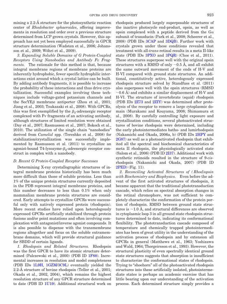
mining a 2.2-Å structure for the photosynthetic reactioncenter of Rhodobacter sphaeroides, offering improve-ments in resolution and order over a previous structuredetermined from LCP grown crystals. However, this ap-proach has not yet been employed successfully in GPCRstructure determination (Wadsten et al., 2006; Johans-son et al., 2009; Wohri et al., 2009).
3. Expanding Soluble Domains of G Protein-CoupledReceptors Using Nanobodies and Antibody Fv Frag-ments. The rationale for this method is that, becauseintegral membrane regions of membrane proteins areinherently hydrophobic, fewer specific hydrophilic inter-actions exist around which a crystal lattice can be built.By adding antibody fragments, it is possible to increasethe probability of these interactions and thus drive crys-tallization. Successful examples involving these tech-niques include voltage-gated potassium channels andthe SecYE� membrane antiporter (Zhou et al., 2001;Jiang et al., 2003; Tsukazaki et al., 2008). With GPCRs,this was first exemplified by the �2-adrenergic receptorcomplexed with Fv fragments of an activating antibody,although structures of limited resolution were obtained(Day et al., 2007; Rasmussen et al., 2007; Bokoch et al.,2010). The utilization of the single chain “nanobodies”derived from Camelid spp. (Tereshko et al., 2008) forstabilization/crystallization was successfully imple-mented by Rasmussen et al. (2011) to crystallize anagonist-bound T4-lysozyme-�2-adrenergic receptor con-struct in complex with a nanobody.
D. Recent G Protein-Coupled Receptor Successes
Determining X-ray crystallographic structures of in-tegral membrane proteins historically has been muchmore difficult than those of soluble proteins. Less than1% of the unique protein structures currently depositedin the PDB represent integral membrane proteins, andthis number decreases to less than 0.1% when onlymammalian membrane protein structures are consid-ered. Early attempts to crystallize GPCRs were success-ful only with natively expressed protein (rhodopsin).More recent studies have relied upon heterologouslyexpressed GPCRs artificially stabilized through proteinfusions and/or point mutations and often involving com-plexation with antagonist/inverse agonist compounds. Itis also possible to dispense with the transmembraneregions altogether and focus on the soluble extramem-brane domains, which will provide structural guidancefor SBDD of certain ligands.
1. Rhodopsin and Related Structures. Rhodopsinwas the first GPCR to have its atomic structure deter-mined (Palczewski et al., 2000) (PDB ID 1F88). Incre-mental increases in resolution and model completeness(PDB IDs 1L9H, 1GZM/3C9L) eventually yielded the2.2-Å structure of bovine rhodopsin (Teller et al., 2001;Okada et al., 2002, 2004), which remains the highestresolution structure of any GPCR structure determinedto date (PDB ID 1U19). Additional structural work on
rhodopsin produced largely superposable structures ofthe inactive photocycle end-product, opsin, as well asopsin complexed with a peptide derived from the G�subunit of transducin (Park et al., 2008; Scheerer et al.,2008) (PDB IDs 3CAP and 3DQB). Further work withcrystals grown under these conditions revealed thattreatment with all-trans-retinal results in a meta II-likestate (PDB IDs 3PXO and 3PQR) (Choe et al., 2011).These structures superpose well with the original opsinstructures with a RMSD of only �0.5 Å, and all exhibitthe same outward movement of the ends of H-V andH-VI compared with ground state structures. An addi-tional, constitutively active, heterologously expressedrhodopsin structure solved by Standfuss et al. (2011)also superposes well with the opsin structures (RMSD�0.6 Å) and exhibits a similar displacement of H-V andH-VI. The structure of invertebrate (squid) rhodopsin(PDB IDs 2Z73 and 2ZIY) was determined after prote-olysis of the receptor to remove a large cytoplasmic do-main (Murakami and Kouyama, 2008; Shimamura etal., 2008). By carefully controlling light exposure andcrystallization conditions, several photoactivated struc-tures of bovine rhodopsin were determined, includingthe early photointermediates batho- and lumirhodopsin(Nakamichi and Okada, 2006a, b) (PDB IDs 2HPY and2G87) as well as a photoactivated rhodopsin that exhib-ited all the spectral and biochemical characteristics ofmeta II rhodopsin, the physiologically activated state(Salom et al., 2006) (PDB ID 2I37). Additional work withsynthetic retinoids resulted in the structure of 9-cis-rhodopsin (Nakamichi and Okada, 2007) (PDB ID2PED) (Fig. 11).
2. Reconciling Activated Structures of ( Rhod)opsinwith Biochemistry and Biophysics. Even before the ad-vent of the first activated structures of rhodopsin, itbecame apparent that the traditional phototransductioncascade, which relies on spectral absorption changes inthe retinal chromophore, was not sufficient to com-pletely characterize the conformation of the protein por-tion of rhodopsin. RMSD between ground state struc-tures is �1.0 Å, and structural differences are observedin cytoplasmic loop 3 in all ground state rhodopsin struc-tures determined to date, indicating its conformationalflexibility. The phototransduction cascade composed oftemperature and chemically trapped photointermedi-ates has been of great utility in the understanding of theactivation process of rhodopsin and by extension allGPCRs in general (Matthews et al., 1963; Yoshizawaand Wald, 1964; Thorgeirsson et al., 1993). However, thestructural plasticity of even spectrally identical groundstate structures suggests that absorption is insufficientto characterize the conformational states of rhodopsin.Trying to “shoehorn” all of the photoactivated rhodopsinstructures into these artificially isolated, photointerme-diate states is perhaps an academic exercise that haslittle bearing upon our understanding of the activationprocess. Each determined structure simply provides a
GPCRs AND DRUG DISCOVERY 923
structural snapshot along the continuum of conforma-tional states that rhodopsin occupies after photoactiva-tion. There is no requirement that the structure hasprogressed through any particular intermediate state.
EPR spin-label experiments using heterologously ex-pressed, spin-labeled rhodopsin were interpreted to sug-gest that major conformational alterations on the orderof 15 to 20 Å occur within the transmembrane domainupon transition of rhodopsin from an inactive to anactive conformation (Farrens et al., 1996; Hubbell et al.,2003). Such large-scale alterations within the helicalbundle have been called into question after isolation ofvarious photointermediate states of rhodopsin that dis-play characteristic absorbance maxima (Matthews et al.,1963; Yoshizawa and Wald, 1964; Thorgeirsson et al.,1993) More recent EPR studies have revised these initialestimates for conformational alterations to �6- to 10-Ådisplacements (Altenbach et al., 2008; Hornak et al.,2010). Furthermore, the chemical differences betweenthe Meta I and Meta II states simply involve the changesin protonation state, and each of these states is in equi-librium. Were large scale movements of entire helices
before G protein binding involved, it is thermodynami-cally unlikely that the equilibrium between the statescould be potentiated by a simple protonation/deprotona-tion event(s). This line of reasoning can be extended,taking into the changes observed in retinal dynamicsthat occur upon photoactivation and the multiple acti-vated states that this implies (only a subset of which arecapable of activating G protein) (Struts et al., 2011).
The activated rhodopsin structures determined todate can be grouped into two broad categories: thosederived from photoactivated rhodopsin (PDB ID 2I37)and those determined from the end product of the pho-totransduction cascade, opsin (PDB IDs 3PXO, 3PQR,3CAP, 3DQB, and 2X72) (Park et al., 2008; Scheerer etal., 2008; Choe et al., 2011; Standfuss et al., 2011).Although the PDB ID 2I37 structure exhibits small-scale changes in the conformation of cytoplasmic loopsand the ends of H-V and H-VI (Salom et al., 2006), thechanges in these regions of opsin-derived structures areof a larger magnitude. It is important to consider that allopsin-derived protein structures begin with material atthe end product of the phototransduction cascade and
FIG. 11. Structural coverage along the continuum of states that comprise the activation of rhodopsin. The structural plasticity necessary fortransmission of the activation signal from receptor to G protein is evident even in the ground state structures of rhodopsin; although all three groupsof structures are grossly similar, each structure captures distinctly different conformers of the opsin backbone although each is spectroscopicallyidentical and each exhibits little to no activity toward G protein. Although slight differences are scattered throughout the structure, the majordifferences observed are in the third cytoplasmic loop (C-III), the loop connecting H-V and H-VI, the ends of which are proposed to undergo structuralrearrangement upon activation. The early photointermediate structures (denoted in pink), which are spectroscopically distinct from both ground stateand later photointermediates superpose well with only the PDB ID 1U19 (P41) structures, again with major differences observed only in the C-III loopcompared with PDB IDs 2I35/2I36 (P3112) or PDB IDs 3C9L/1GZM (P64) structures. The structure of PDB ID 2I37 (which was the first crystalstructure to exhibit the characteristic absorbance at 360 nm indicative of deprotonation of the Schiff base linking the chromophore to Lys296, ahallmark of attaining the activated state) demonstrated only small- to medium-scale shifts in structure that were confined to the C-II and C-III loops,rather than the large-scale rigid body movements proposed by earlier studies. The observed structural changes could best be explained as being dueto a loss of constraint within the transmembrane region, resulting in altered protein backbone dynamics. Because ground state crystals in the sameunit cell and space group (PDB ID 2I36) were available, the direct comparison with photoactivated rhodopsin (PDB ID 2I37) revealed structuralchanges that accompanied photoactivation apart from structural changes due to differences in crystallization conditions/unit cell contacts. Thesecrystals were capable of returning to the ground state upon storage in the dark but were incapable of surviving treatment with hydroxylamine toremove chromophore. From the other end of the spectrum, the Ernst group has to great success used the phototransduction cascade end product, opsin,as a structural target and starting point for probing the activation of rhodopsin. The initial crystal structure of opsin (PDB ID 3CAP) and a followingstructure opsin with a peptide derived from the C terminus of the � subunit of transducin (Gt) (PDB ID 3DQB), although mostly colorless, containdensity within the chromophore active site that may correspond to precipitant, buffer, detergent, or hydrolyzed chromophore when composite omitmaps are calculated from the deposited data, which was fortuitous as the instability of opsin state in the detergent solubilized state would furthercomplicate crystallization. The PDB ID 3CAP and PDB ID 3DQB structures both exhibited larger movements of H-V and H-VI (and the connectingC-III loop) than observed in the PDB ID 2I37 structure. The structure of opsin in the Gt peptide bound structure was postulated by the authors to bethe conformation of opsin in its G protein-interacting state. By treating these very same crystals with all-trans-retinal (PDB IDs 3PXO and 3PQR),the authors were able to obtain crystals that were spectrally indistinguishable from the PDB ID 2I37 crystals. Standfuss et al. (2011) were also ableobtain an additional crystal form of photoactivated rhodopsin from heterologously expressed, constitutively active opsin “regenerated” with all-trans-retinal (PDB ID 2X72). All of the opsin derived structures are identical (with the exception of the observed chromophore) within the precision ofstructures determined at this resolution (RMSD � 0.4 Å). These crystals were incapable of being pushed to the ground state by reconstitution with11-cis-retinal. Recent solid-state NMR studies suggest the existence of multiple conformational states after photoactivation with only a subsetcompetent to transduce the signal to G protein (Struts et al., 2011). These multiple activated states (highlighted with a blue-gray box in the figure)underlie a fundamental structural disconnect between the two groups of activated state structures. It will only be through direct structural observationof the complex or complexes between rhodopsin and Gt that the precise nature of the interactions between activated rhodopsin and G protein will beobserved.
924 SALON ET AL.
neither addition of the Gt peptide nor addition of chro-mophore has an appreciable change upon the structure(RMSD for the transmembrane region of all opsin struc-tures is � 0.4 Å). When these observations and struc-tures are coupled with the retinal dynamics observedupon photoactivation by solid state NMR (Struts et al.,2011), it becomes clear that spectrophotometric analysisand these structures are not sufficient to fully describethe activation process. Measurements of the displace-ment of cytoplasmic loop three, H-V and H-VI observedin opsin-derived structures upon activation agree quitewell with the later EPR-based estimates, with the ca-veat that these measurements were performed with het-erologously expressed opsin mutants, which were thenregenerated. Although the PDB 2I37 crystals could rei-somerize their chromophore when removed from light,reforming the ground state, the opsin-derived crystalsare incapable of being regenerated with 11-cis-retinal toform the ground state. Furthermore, treatment of thephotoactivated (PDB ID 2I37) crystals with hydroxyl-amine to form opsin resulted in a loss of diffractionsimilar to the loss of diffraction seen in the opsin crystalstreated with 11-cis-retinal chromophore (O. Ernst, per-sonal communication). This hints at a fundamental dis-connect between the PDB ID 2I37 photoactivated struc-ture and the opsin-derived structures, because it is notpossible to take either class of crystals to the oppositeendpoint of the phototransduction cascade. Thus, nei-ther class of crystal structures is sufficient to describethe dynamic and conformational state(s) of the receptorthat pass the activation signal from the chromophorepocket to the cytoplasmic face where G protein bindingand activation occurs.
Comparisons of agonist-bound nonrhodopsin GPCRsall exhibit some movement of the H-VI and to a lesserextent H-V, but the movement is smaller in magnitudethan that observed between ground-state rhodopsin andopsin-derived structures. Were the activation process torely upon release of internal restraints that lead to aprofound change in receptor dynamics, these sorts ofvariations can be explained. The influences that crystal-lization conditions, crystal contacts, and thermostabiliz-ing mutations could simply remove energetic barrierspresent in the wild-type receptor, thus exhibiting alarger degree of structural plasticity needed to fully bindand activate G protein (summarized in Fig. 11).
Comparative analysis of all high-resolution structuresof rhodopsin and other GPCRs reveals a subset of crys-tallographically observed waters found in similar posi-tions within the transmembrane bundle (Angel et al.,2009a). These “homologous” waters and their interac-tions with highly conserved and functionally importantresidues such as the D(E)RY and NPxxYx(5,6)F motifsthought to play crucial roles in receptor activation (Mir-zadegan et al., 2003) suggests that these waters arefunctionally important or even essential to the mecha-nism of activation (Angel et al., 2009a,b; Jardon-Valadez
et al., 2009) (Fig. 9). Furthermore, these internal watersdo not freely exchange with bulk solvent in ground-staterhodopsin, meta II, or opsin states, further supportingthe notion that these waters are noncovalent cofactorsintegral to the activation process. This however does notimply that the transmembrane region is completely im-pervious to bulk solvent. Indeed, the Schiff base can bedeuterated in unactivated rhodopsin placed in D2O(Deng et al., 1994), and the water used for chromophorehydrolysis is derived from bulk solvent (Jastrzebska etal., 2011), suggesting that only a subset of these solventmolecules are tightly bound. Further work is needed toelucidate the exact role(s) of these ordered waters in theGPCR activation process.
3. Adrenergic Receptor Structures. In 2007, the re-port of the X-ray structure for the human �2-adrenengicreceptor marked a breakthrough in the structural studyof liganded GPCRs. The structure was solved by twodifferent techniques (Rasmussen et al., 2007; Rosen-baum et al., 2007). In one approach, an antibody Fvfragment–�2-adrenergic receptor complex was gener-ated that yielded low-order diffracting crystals with awell-defined Fv entity but a considerably less ordered�2-adrenergic receptor portion (PDB IDs 2R4R and2R4S). In a second approach, since used to determine themajority of nonrhodopsin GPCR structures to date, a T4lysozyme fusion was introduced within the third cyto-plasmic loop of heterologously expressed �2-adrenergicreceptor to aid crystallization in a modified lipid cubicphase matrix and resulted in high-order diffracting crys-tals (Cherezov et al., 2008; Hanson et al., 2008; Roth etal., 2008). In addition to the high-resolution 2.4-Å cara-zolol-bound structure (PDB ID 2RH1), a series of adren-ergic receptor structures have been solved that con-tained a variety of antagonist and inverse agonistcompounds (Hanson et al., 2008; Roth et al., 2008;Wacker et al., 2010) (PDB IDs 3D4S, 3NYA, 3NY9, and3NY8). These structures revealed a common bindingmode and contact residues for such compounds, suggest-ing further avenues for structure-based drug design us-ing these structures as restraints.
The crystallization of the turkey �1-adrenergic recep-tor involved a series of mutations/truncations designedonce again to constrain the flexibility of the purifiedreceptor and facilitate the formation of the crystallinelattice. The effects of individual modifications were pre-liminarily assessed by thermal denaturation analysisand when combined as an ensemble into to a new con-struct produced diffracting crystals (Tate and Schertler,2009; Warne et al., 2009). Cocrystallization, as has beentrue with all nonopsin GPCR templates determined todate, with an antagonist (cyanopindolol, in this case)was needed to obtain crystals, which diffracted to 2.7 Å(Warne et al., 2008). In this case, standard crystalliza-tion methodology (i.e., vapor diffusion rather than LCPmethods) was employed. Comparison of structures re-
GPCRs AND DRUG DISCOVERY 925
vealed similar binding modes of antagonist to both �1-and �2-adrenergic receptors (Lodowski et al., 2009).
Crystallographic structure determination of both �1and �2 adrenergic receptors has continued with the so-lution of several full and partial agonist structures usingvariations upon the above crystallization schema.Warne et al. (2011) recently published a series of five fulland partial agonist-bound structures of thermostabi-lized turkey �1-adrenergic receptor that exhibit onlysmall-scale structural changes compared with the struc-ture of turkey �1-adrenergic receptor with antagonistbound. These changes are consistent with the scale ofstructural changes observed in the low-resolution pho-toactivated rhodopsin structure (Warne et al., 2011).Rasmussen et al. (2011) and Rosenbaum et al. (2011)have also recently solved two agonist bound structuresof their T4-lysozyme inserted human �2 adrenergic re-ceptor by two different crystallization schemes but withconflicting results. Although both structures use T4 ly-sozyme-inserted mutant protein, one structure uses acovalently attached ligand and shows very little changeupon agonist binding compared with their antagonistbound counterparts (Rosenbaum et al., 2011). The otheruses a complex with a camelid nanobody and exhibitsstructural changes on the scale of those seen in opsincompared with ground-state rhodopsin (Rasmussen etal., 2011).
4. A2a-Adenosine Receptor Structure. Again, a T4-lysozyme chimera coupled with the antagonist 4-(2-(7-amino-2-(furan-2-yl)-[1,2,4]triazolo[1,5-a][1,3,5]triazin-5-ylamino)ethyl)phenol (ZM241385) and use of a cholesterol-saturatedLCP crystallization method yielded crystals of the A2a-adenosine receptor refracting to 2.6 Å (Jaakola et al., 2008)(PDB ID 3EML). As expected and in agreement with allother GPCRs determined to date, the binding site for thisorthosterically competitive ligand was located within thetransmembrane region, grossly overlapping binding sitesdetermined for adrenergic receptor ligands and the rho-dopsin chromophore. More recently, an agonist boundstructure of a T4-lysozyme A2a-adenosine receptor struc-ture was determined (Xu et al., 2011). Compared with theantagonist bound structure, this agonist-bound structureshows smaller changes in the conformation of H-V andH-VI than seen in opsin-derived structures.
5. C-X-C Chemokine Receptor Type 4 Structures. TheStevens group determined the structure of the chemo-kine GPCR, CXCR4, also as a T4 lysozyme chimera incomplex with the small molecular antagonist IT1t (6,6-dimethyl-5,6-dihydroimidazo[2,1-b][1,3]thiazol-3-yl)methyl N,N�-dicyclohexylimidothiocarbamate as well aswith a cyclic peptide antagonist (CVX15), (Wu et al.,2010) (PDB IDs 3ODU, 3OE9, 3OE6, 3OE8, and 3OEO).This receptor is of great importance to human health,because it is involved in both HIV viral entry and cancermetastasis. It is noteworthy that these structures com-pletely lack the amphipathic helix H-8 present in allother GPCR structures determined to date that func-
tions in receptor activation. All CXCR4 crystal struc-tures determined to date exhibit a crystallographicdimer that is quite similar to that seen in squid rhodop-sin (PDB ID 2Z73) as well as in models of rhodopsinoligomerization derived from atomic force microscopydata (PDB ID 1N3M) (Fig. 6).
6. Dopamine D3 Receptor Structure. The T4-ly-sozyme fusion technique was also used to obtain a crys-tal structure of the D3 dopamine receptor in complexwith the D2/D3 selective antagonist eticlopride (R-22)(Chien et al., 2010). Because dopamine signaling is in-volved in cognition and emotion, structures of the dopa-mine receptor can outline possible structure-based drugdesign targets for modulation of these activities. Al-though in many other of the T4 lysozyme fusion con-structs, the ionic lock was found to be disrupted, here itwas found to be intact, suggesting that this crystal struc-ture more fully recapitulates the inactive state of thereceptor.
7. Structures of G Protein-Coupled Receptor Extracel-lular Domains. In contrast to GPCRs that have theirligand-binding domains situated within the transmem-brane region, many GPCRs contain large extracellularligand-binding domains. These extracellular domainscan bind the entire size range of ligands seen in allclasses of GPCRs, from single ions in the case of theCa2� receptors to entire proteins in the case of thefollicle-stimulating hormone receptor (Figs. 1 and 7).Crystallization of these domains ex situ can provideinformation regarding ligand recognition. Several sub-types of metabotropic glutamate receptor extracellularligand-binding domains have been studied (Kunishimaet al., 2000; Muto et al., 2007) (PDB IDs 3HSY, 3MQ4,3LMK, 3FUZ, 2E4U, 1LSR, 1EWK, and at least 15 oth-ers) and have defined ligand binding modes and local-ized structural changes that occur upon both agonistand antagonist binding. All of these glutamate GPCRsare obligate dimers. The distance observed between eachmonomer fits well with the spacing observed for rhodop-sin oligomers within rod outer segment membranes (Fo-tiadis et al., 2003, 2004) and with dimers observed inrhodopsin, opsin, and CXCR4 structures (Salom et al.,2006; Lodowski et al., 2007; Park et al., 2008; Scheereret al., 2008; Wu et al., 2010), providing further evidencefor the physiological existence of GPCRs as dimers orhigher order oligomers (Fig. 6).
Ligand-binding domains from GPCRs that recognizelarger soluble protein/peptide hormones have also beendetermined. These include follicle-stimulating hormonecomplexed with the follicle-stimulating hormone recep-tor (Fan and Hendrickson, 2005) (PDB ID 1XWD), thy-roid-stimulating hormone receptor in complex with anactivating antibody (Sanders et al., 2007) (PDB ID3GO4), parathyroid hormone receptor complexed withparathyroid hormone (Pioszak et al., 2010) (PDB ID3L2J), calcitonin gene-related peptide receptor (ter Haaret al., 2010) (PDB IDs 3N7P, 3N7R, and 3N7S), cortico-
926 SALON ET AL.
tropin-releasing factor receptor 1 (Pioszak et al., 2008)(PDB IDs 3EHS and 3EHT), and frizzled 8 receptor(Dann et al., 2001) (PDB ID 1IJY). What is immediatelyobvious upon examination of these isolated ligand-bind-ing domains is that although they share little to nostructural homology there must be some shared mecha-nism by which these disparate domains can be linked tothe transmembrane portion of the receptor. This raisesthe concern that because the structures for these do-mains have been solved without the contextual con-straints of the GPCR functional core, usage of suchligand-binding domain structures, although promisingfrom a SBDD standpoint, should be approached with theadditional caveat that the modes of ligand binding ob-served may not fully recapitulate that seen in intactreceptor.
E. The Need for More G Protein-Coupled ReceptorStructural Work
With many distinct GPCR structures now available tous, there is a good body of information for delineatingcommon features present within the transmembraneregion of class A GPCRs that give some insight into theirligand binding and possibly their activation. Furtherstructures will strengthen confidence in such commonfeatures but could reveal a subclass of GPCRs withdifferent modes of ligand binding within the TM region.However, certain approaches to structure determinationof GPCRs will be particularly helpful for increasing ourunderstanding of how ligands bind GPCRs, how thisbinding event is coupled to receptor activation, and howthese activated receptors consequently catalyze G pro-tein activation. First, a greater diversity of GPCR tem-plates, especially those of class B and C receptors, isneeded. Second, continued characterization of agonist-bound structures will help define determinants of endog-enous ligand binding and the structural changes thataccompany GPCR activation. This information would beparticularly valuable in the rationale design of thera-peutic ligands and allosteric modulative compounds.Third, and most obviously, structures of agonist-boundGPCRs in complex with their intact appropriate hetero-trimeric G proteins provide the best chance to capturethe structural changes that enable G protein activation.(Please see Note Added in Proof for recent structuralwork on GPCR-G protein complexes.)
V. Ramifications for G Protein-Coupled ReceptorDrug Discovery
A. Brief History of De Novo Approaches
1. Examples of Success. Most drugs in our collectivemedicine chests have been and still are discovered byserendipitous methods (Kubinyi, 1999). Contemporarymedicines have emerged from broad screens of large andcomplex collections of chemical or biological substancesthat, though individually diverse in origin and content,
share en mass an important trait—a low percentage hitrate. Cursory inspection of compound and program at-trition common to the pharmaceutical sector under-scores the long odds faced during drug discovery. Still,the content and effectiveness of our pharmacopeia is atestament to the use of empirically based screening. Nodoubt it is foolish to depend solely on serendipity, but wecannot discount it either. Accordingly, our use of randomscreening will not likely disappear, nor should it, be-cause it continues to be a proven approach for the dis-covery of novel therapeutics.
Yet for those with a more calculating disposition, thepursuit of new drugs through de novo design presents acompelling logic. Benefiting from advances in biophysicsand computational methods, this discipline has steadilyadvanced over the past 30 years and produced scores ofgroundbreaking therapeutics. These approaches can bebroadly categorized into ligand- and structure-basedmethods depending upon the origin of the blueprint thatguides the actual synthesis of molecules. Ligand-basedapproaches are based on quantitative structure-activityrelationship (QSAR) studies of pre-existing moleculesknown to specifically interact with a target system ofinterest. These data are distilled to generate an ideal-ized pharmacophore model that is then used to designnew candidates for synthesis. Structure-based methods,on the other hand, rely upon a high-resolution map of atarget’s putative ligand-binding sites, to provide a mir-ror-image blueprint for the a priori design (or modifica-tion) of entirely novel molecules. Obviously, these twomethods are complementary. Both have had their shareof success, and both are used throughout the pharma-ceutical industry along with computer-aided docking,visualization strategies, and empirically driven assayplatforms, often deriving QSAR information from datagenerated by high-throughput screens.
An early success for the ligand-based approach wasthe design of highly selective small molecule antagonistsagainst integrin receptors for the treatment of thrombo-sis, cancer and osteoporosis. This work used conforma-tionally constrained cyclic peptides bearing a similarsmall binding motif to inform the design of small mole-cule leads that had remarkably high selectivity forclosely related integrin receptor subtypes (Engleman etal., 1996; Samanen, 1996; Samanen et al., 1996; Keenanet al., 1997).
Perhaps the most often cited early example of a suc-cessful “rationally designed” drug is the small moleculecaptopril, an angiotensin-converting enzyme inhibitorfor the treatment of hypertension. Its design benefitedfrom a prior decade’s QSAR study of the inhibitory ac-tion of bradykinin potentiating factor on the conversionof angiotensin as well as from insights gained from thethree-dimensional structure of the closely related en-zyme carboxypeptidase A (Harrold, 2008). Other earlyexamples of purely structure-based approaches are the
GPCRs AND DRUG DISCOVERY 927
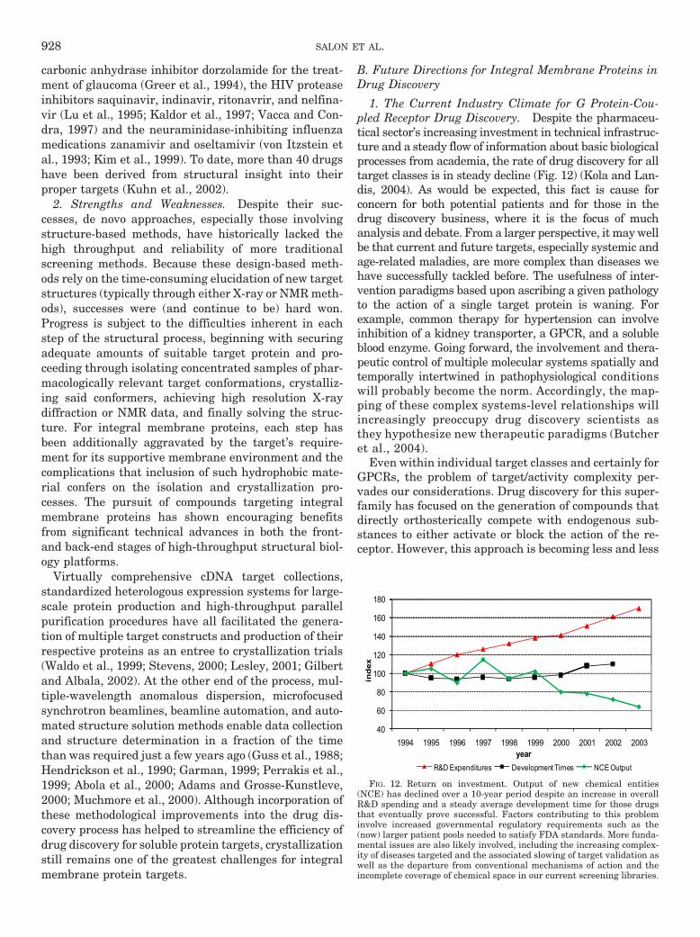
carbonic anhydrase inhibitor dorzolamide for the treat-ment of glaucoma (Greer et al., 1994), the HIV proteaseinhibitors saquinavir, indinavir, ritonavrir, and nelfina-vir (Lu et al., 1995; Kaldor et al., 1997; Vacca and Con-dra, 1997) and the neuraminidase-inhibiting influenzamedications zanamivir and oseltamivir (von Itzstein etal., 1993; Kim et al., 1999). To date, more than 40 drugshave been derived from structural insight into theirproper targets (Kuhn et al., 2002).
2. Strengths and Weaknesses. Despite their suc-cesses, de novo approaches, especially those involvingstructure-based methods, have historically lacked thehigh throughput and reliability of more traditionalscreening methods. Because these design-based meth-ods rely on the time-consuming elucidation of new targetstructures (typically through either X-ray or NMR meth-ods), successes were (and continue to be) hard won.Progress is subject to the difficulties inherent in eachstep of the structural process, beginning with securingadequate amounts of suitable target protein and pro-ceeding through isolating concentrated samples of phar-macologically relevant target conformations, crystalliz-ing said conformers, achieving high resolution X-raydiffraction or NMR data, and finally solving the struc-ture. For integral membrane proteins, each step hasbeen additionally aggravated by the target’s require-ment for its supportive membrane environment and thecomplications that inclusion of such hydrophobic mate-rial confers on the isolation and crystallization pro-cesses. The pursuit of compounds targeting integralmembrane proteins has shown encouraging benefitsfrom significant technical advances in both the front-and back-end stages of high-throughput structural biol-ogy platforms.
Virtually comprehensive cDNA target collections,standardized heterologous expression systems for large-scale protein production and high-throughput parallelpurification procedures have all facilitated the genera-tion of multiple target constructs and production of theirrespective proteins as an entree to crystallization trials(Waldo et al., 1999; Stevens, 2000; Lesley, 2001; Gilbertand Albala, 2002). At the other end of the process, mul-tiple-wavelength anomalous dispersion, microfocusedsynchrotron beamlines, beamline automation, and auto-mated structure solution methods enable data collectionand structure determination in a fraction of the timethan was required just a few years ago (Guss et al., 1988;Hendrickson et al., 1990; Garman, 1999; Perrakis et al.,1999; Abola et al., 2000; Adams and Grosse-Kunstleve,2000; Muchmore et al., 2000). Although incorporation ofthese methodological improvements into the drug dis-covery process has helped to streamline the efficiency ofdrug discovery for soluble protein targets, crystallizationstill remains one of the greatest challenges for integralmembrane protein targets.
B. Future Directions for Integral Membrane Proteins inDrug Discovery
1. The Current Industry Climate for G Protein-Cou-pled Receptor Drug Discovery. Despite the pharmaceu-tical sector’s increasing investment in technical infrastruc-ture and a steady flow of information about basic biologicalprocesses from academia, the rate of drug discovery for alltarget classes is in steady decline (Fig. 12) (Kola and Lan-dis, 2004). As would be expected, this fact is cause forconcern for both potential patients and for those in thedrug discovery business, where it is the focus of muchanalysis and debate. From a larger perspective, it may wellbe that current and future targets, especially systemic andage-related maladies, are more complex than diseases wehave successfully tackled before. The usefulness of inter-vention paradigms based upon ascribing a given pathologyto the action of a single target protein is waning. Forexample, common therapy for hypertension can involveinhibition of a kidney transporter, a GPCR, and a solubleblood enzyme. Going forward, the involvement and thera-peutic control of multiple molecular systems spatially andtemporally intertwined in pathophysiological conditionswill probably become the norm. Accordingly, the map-ping of these complex systems-level relationships willincreasingly preoccupy drug discovery scientists asthey hypothesize new therapeutic paradigms (Butcheret al., 2004).
Even within individual target classes and certainly forGPCRs, the problem of target/activity complexity per-vades our considerations. Drug discovery for this super-family has focused on the generation of compounds thatdirectly orthosterically compete with endogenous sub-stances to either activate or block the action of the re-ceptor. However, this approach is becoming less and less
160
180
120
140
160
ex
60
80
100
inde
40
60
1994 1995 1996 1997 1998 1999 2000 2001 2002 2003year
R&D Expenditures Development Times NCE Output
FIG. 12. Return on investment. Output of new chemical entities(NCE) has declined over a 10-year period despite an increase in overallR&D spending and a steady average development time for those drugsthat eventually prove successful. Factors contributing to this probleminvolve increased governmental regulatory requirements such as the(now) larger patient pools needed to satisfy FDA standards. More funda-mental issues are also likely involved, including the increasing complex-ity of diseases targeted and the associated slowing of target validation aswell as the departure from conventional mechanisms of action and theincomplete coverage of chemical space in our current screening libraries.
928 SALON ET AL.
productive as we pursue receptor-ligand systems involv-ing more elaborate recognition and activation relation-ships, such as peptidergic and glycohormone subtypesinvolving protein-protein interactions generally consid-ered intractable for small molecular intervention. Thereis also the problem of building selectivity into com-pounds that operate within the highly conserved agonistsites of closely related receptor subtypes. There is thusan increasing need to consider nonorthosteric modes ofaction as the basis for new types of GPCR therapeutics.An a priori understanding of the three-dimensionalstructure of such sites could both suggest nonobviousmodifications for orthosteric leads and reveal allostericopportunities for the design of entirely novel moleculesand mechanisms of action.
2. Requirements for the Optimal Use of StructuralStudies. To provide timely guidance, structure-baseddrug design must lead the medicinal synthesis curve.That is, it must prospectively suggest new designsrather than limiting itself to retrospectively explain-ing how old designs work. Understanding how mole-cules already “in-hand” bind and regulate their tar-gets is valuable, of course, but to achieve its maximumpotential, structural information must act as a spring-board to suggest innovative compound designs un-likely to be included in a medicinal chemist’s usualto-do list. Given the speed and rigor with which me-dicinal chemistry and screening now operates, this isindeed an ambitious goal.
A key requisite for progress will be the ability torapidly solve apo-protein crystals and ligand-protein co-crystals, ideally before commitment and expenditure ofsubstantial medicinal chemistry resources. Given theinternal competition of projects for medicinal chemistrysupport, such insights would be welcome and could beexploited to make the synthesis aspects of drug develop-ment more efficient. Initially, tactical arguments fortargeting specific GPCRs for structural determinationare likely to be reminiscent of debates about the value ofmounting HTS campaigns for targets with speculativetherapeutic validity. Such deliberations eventuallyeased as efficiencies in enabling and executing HTSimproved, the ratio of information return over a smallerexperimental investment increased, and the prospect ofsecuring chemical tools to assist in the validation andprioritization of target programs became better ac-cepted. Likewise, one can expect some push back in aproposed mainstream pursuit of GPCR structures untilthey can be more routinely and easily solved. At thatpoint the expectation that comparative structuresshould be part of a program, even at an early projectstage, will likely become the norm and further encour-age a priori generation and selection of drug designstargeting unconventional binding sites and mechanismsof action.
VI. Unresolved Issues and Concluding Remarks
The study of GPCR pharmacology has been the sub-ject of structure-function conjecture since its inceptionand has long awaited the type of intuitive insights thatonly high-resolution atomic coordinates can provide. Theaccelerating success in generating such structures ob-served over the past few years suggests that we can nowbegin to discern the molecular basis of GPCR function.Although these breakthroughs have been impressiveand the promise of their utility is great, a variety ofhurdles remain that must be overcome as we reduce theart of GPCR structure determination to the practice ofdrug discovery.
A. Caveats about Static Crystal Structures
The increasing availability of GPCRs structures willundoubtedly provide insights into the structural basis oftheir molecular function. It is important however, toappreciate caveats associated with individual struc-tures, the most obvious of which is that they representonly a “snapshot” of one of many conformations availableto the receptor. This issue is especially relevant to 3Dcoordinates derived from X-ray crystal structures. Insuch cases, these structures “freeze” at a thermody-namic minimum, which may not fully recapitulate thereceptor’s native membrane environment but rather re-flect restraints imposed by its crystallization matrix.This concern is amplified by the use of modified receptorconstructs and/or ligands to stabilize receptors for crys-tallization. Use of crystal structures derived from spe-cifically stabilized ligand-receptor complexes can predis-pose the resulting drug designs to those ligand-definedconformations, thus limiting the de novo approach. Fur-thermore, in the case of ligand-induced stabilizationmethods and dependent upon the method employed, in-voking the use of an a priori ligand molecule suggeststhat this area of chemical space has been previouslyprobed, further diminishing the novelty of subsequentdrug designs. Modified receptor protein further removesthe solution from in vivo reality and can increase therisk of guiding design efforts down unfruitful paths. Apartial answer to such conundrums can be to structur-ally solve and compare a variety of discrete conforma-tional solutions to provide a kinescopic view of the pro-tein’s dynamics within its full range of function. Suchmerging and comparison of dynamic states would bene-fit from other forms of structural information such asprovided by solution and solid state NMR and dynamiccomputational modeling. The problem, of course, is tocrystallize such discrete conformations.
B. Partnering with Computational Methods
The accuracy of homology models for GPCRs builtfrom templates that are in turn based upon actual three-dimensional coordinates of a previously solved but dif-ferent GPCR structure becomes increasingly less predic-
GPCRs AND DRUG DISCOVERY 929
tive as the receptors diverge. This observation isespecially applicable to drug design when sub-Ångstromtolerances and “second shell” contacts provide the basisfor selectivity and action. Thus, efforts to improve ho-mology modeling algorithms and merge them with denovo structure assignment methods by, for example,taking into consideration template information, knowl-edge of fold space derived from structural genomics ini-tiatives, and improved ab initio structure predictiontechniques, should ultimately provide us with computer-aided design tools that are both predictive and intui-tively effective. Such methods development will be facil-itated by additional GPCR structure determinations toprovide more points of reference and to validate modelbased predictions across GPCR subtypes, types, andclasses. Applications of static models to in silico screen-ing using compound and fragment-based libraries hasbecome a popular exercise but is limited by currentdocking algorithms that focus on preconceived-bindingdomains of the receptor, effectively eliminating alloste-ric opportunities. Such constraints are largely driven byprocessor limitations that require a practical shrinkageof the possible binding box. Our need to understand andexploit important functional phenomena such as alloste-ric modulation and collateral efficacy inherent in thepurposeful conformational adaptability of GPCRs seemsmore suited to the application of molecular dynamic(MD) simulations inclusive of all of the system’s protein,lipid bilayer and water atoms. Such MD simulations arealso limited by processor resources available and theaccuracies of current atomic force fields and MD algo-rithms. As these factors improve, these tools will nat-urally merge with extant GPCR structural informa-tion to improve drug design that is conformationspecific.
C. Rate of Structure Determination
Structure determination of specific GPCR drug tar-gets runs the risk of being retrospective. To be unequiv-ocally guiding, the process of moving from target selec-tion to structure availability must proceed rapidlyenough to provide nonobvious drug designs that wouldhave been unlikely to be identified through randomscreening or QSAR and SAR methods. The rate of gen-erating new designs from structures compared with thepace of discovering new molecules via comprehensivesynthetic analog campaigns is thus a common point-counterpoint for all de novo versus randomization de-bates. Typically, the rationale for obtaining a structureis more compelling when either chemical starting pointsdo not exist, such as when no hits emerge from a HTS orwhere SAR guided synthesis fails to improve selectivityacross related targets, or other off-target phenomenacannot be avoided. Since the initial structures for rho-dopsin and �2-adrenergic receptor were reported, thepace of new GPCR structural determinations has rap-idly accelerated. If target structures can routinely be
solved within 12 months of project inception, this wouldcompare favorably with the lead time it typically takesto enable and execute a HTS study for a given target.This suggests that as the pace of structural determi-nation increases, structurally informed drug designcan be merged with screening-based hit identificationand QSAR campaigns not only to bootstrap new proj-ects but also to provide guidance for ongoing effortsthat have encountered insurmountable chemistrychallenges.
D. Structure versus Function
Functional (i.e., screening) approaches to GPCR drugdiscovery that seek to exploit a textured (i.e., allosteric)approach to pharmacological action will become increas-ingly powerful provided cell-based assays that are morerefined than the current norm are employed. Such prog-ress will also require that ligands (both controls andscreening compounds) that modulate these new areas ofpharmacological space currently exist in our screeningcollections. The novelty of such space is encompassed byboth the specifics of the target and the specifics of theconformational state that is to be modulated. It seemsreasonable to expect that in some if not most cases, suchspace is poorly populated in our current collections. Denovo approaches on the other hand are not beholden to apriori molecular content but rather require insight intowhether or not the structure (i.e., conformation) understudy is relevant to the disease-defined mechanism ofaction (i.e., target-effector pathway). Both approachescan be synergistically combined, providing a powerfultool for future GPCR-targeted drug discovery.
E. Cocrystallization with Ancillary Protein(s)
As crucial components of GPCR signaling system andequal partners in the kinetics of their action, ancillary Gprotein subunit structures, especially the G� subunit, incomplex with their receptor partners will help shed lighton structural determinants of GPCR activity. More spe-cifically, the use of G proteins to capture pathway-spe-cific conformations could reveal binding opportunitiesassociated with ligand trafficking in either the receptor’sorthosteric domain or elsewhere on more accessible andless conserved regions of the GPCR. Furthermore, struc-tures of protein complexes such as GPCR-G protein,GPCR with receptor activity modifying proteins, GPCR-GPCR dimers can suggest a means of allosteric regula-tion of GPCR function by interfering with heretofore-untargeted protein-protein interactions. The structuresof these GPCR complexes will reveal novel “druggable”sites present for targeting via SBDD.
F. Impact upon Compound Libraries
Clearly, a high resolution view of the internal andsurface landscape of GPCRs should assist in the identi-fication and design of novel pharmacophores and as suchwill constitute an invaluable resource for SBDD. This is
930 SALON ET AL.
especially true for a potentially large number of alloste-ric designs whose corresponding chemical space is mostprobably unoccupied in existing libraries. Without suchguidance, these sectors of pharmacologically active com-pound space will only slowly become populated throughthe creeping expansion of pre-existing, distant, and or-thosterically oriented structural neighborhoods. Thecurrent trend in generation and utilization of target-focused libraries for drug discovery is typically rational-ized by invoking the savings realized in screening asmaller number of compounds with a higher en mass hitrate. This scenario can be true when the focused libraryis tasked with providing structures that act throughmechanisms (and sites) similar to those affected by thepopulated prototypes. But it cannot account for alloste-ric/nonorthosteric sites. Realizing that GPCRs consti-tute the core component of a still poorly understoodallosteric system, the design of screening libraries wouldbenefit from as few assumptions as possible when select-ing chemical structures for its population. In the case ofnovel mechanisms of action and allosteric targets, diver-sity cannot be achieved by reference to prior knowledge.Given the almost infinite number of possible compoundsthat encompass all of chemical space, the efficient pur-suit of diversity would benefit from the reciprocal map-ping of novel target binding sites, sites uniquely pro-vided through structural data. Such spatial leads couldthen be embraced on a manageable scale by focusedlibrary approaches to rationally but more efficiently in-crease performance and diversity.
G. Synergy with Biologics
As regards the generation of very-large-moleculedrugs for GPCRs (e.g., therapeutic antibodies), use ofGPCR structures becomes more complicated because ofthe manner in which these therapeutic molecules bind totargets and the in vivo methods employed for antibodygeneration. Multiple recognition sites spread over a re-ceptor’s surface make structural information enlighten-ing but difficult to employ on a purely synthetic basis.Still, one could envision instances where knowledge ofdisparate epitopes constituting a conformational lockmight be employed to construct an artificial mimetic foruse as an in vivo immunogen or capture reagent. Thefact that such a protein would be stabilized in a partic-ular conformation relevant to the targeted molecularmechanism would be of particular benefit and a signifi-cant advance over current methods.
H. Other Synergies
Availability of purified receptor proteins generated aspart of the drug discovery process will undoubtedly con-stitute a windfall for the enabling of other drug discov-ery tools. It is expected that this protein will also be usedfor additional assays including NMR and fluorescencebased measurements of receptor-ligand dynamics, newgenerations binding assays via measurement of plasmon
resonance and molecular exclusion and various otherproteomic/biophysical approaches to characterize andmap structure-function aspects of the receptor.
I. Selection of Therapeutically Validated Targets
However promising and inherently powerful a struc-ture-based drug design approach may be, its ultimatevalue can only be realized if it can operate within thedrug discovery pipeline at large. As such, the breadth ofapplication of a structural approach will inevitably faceconstraints beyond its scope, perhaps the most impor-tant of which is the identification and validation ofGPCR targets. To these ends, concerted efforts to ex-haustively catalog and manage information about nor-mal versus pathological tissue distribution, knock-in/knock-out phenotypes, chromosomal mapping of diseaselinkages, and generation and correlation of microarrayand gene copy information with disease models haveonly grudgingly given up the secrets embedded in theirdata, perhaps because of the immensity of the task andthe difficulty of formulating therapeutic paradigms fromsuch diverse information. Application of systems analy-ses to these information layers, although still in itsinfancy, promises to elucidate complex causal relation-ships and reveal key regulatory points for therapeuticintervention. Given the emergence of pluridimensionalGPCR potency and the prospect that structure-baseddrug design could be instrumental in designing mole-cules to selectively regulate one of the many interactionsand pathways of a given GPCR, it seems reasonable toexpect that a retrospective analysis of past GPCR pro-grams will ensue. Salvage of failed targets and programsmight then be possible through consideration of thebroader range of molecular phenomena now known tooccur within the GPCR signaling paradigm.
In conclusion, the likelihood that the challenges andopportunities discussed in this review will be met andbested seems reasonable, and the convergence of GPCRstructural methods with therapeutic exploitation of newlyemerging modes of GPCR pharmacology will significantlyand favorably affect the field of GPCR drug discovery.
Note Added in Proof
Since the submission of the manuscript for this article, severalarticles of note regarding structure have been published that shedlight on agonist and antagonist binding as well as the activation ofGPCRs and mechanisms of G protein binding. A structure of aT4L-histamine H1 receptor fusion bound to the subtype-specific antag-onist doxepin [(3Z/E)-3-(dibenzo[b,e]oxepin-11(6H)-ylidene)-N,N-dimethylpropan-1-amine] has been determined, exhibiting the subtle-ties of antagonist specificity (Shimamura et al., 2011). Additional workwith thermostabilized A2a-adenosine receptor in complex with the an-tagonist compounds caffeine and xanthine have further characterizedthe diversity of the binding site (Doré et al., 2011; Xu and Stevens,2011). Further work with these thermostabilized A2a-adenosine recep-tor constructs has produced structures bound to the agonists adenosineand 5�-N-ethylcarboxamido adenosine, revealing the variability ofstructural changes within the transmembrane region compatible withagonist binding, as well as proposing mechanisms by which the bindingof agonist may effect attainment of the activated state or states (Lebon
GPCRs AND DRUG DISCOVERY 931
et al., 2011). A 20-Å molecular envelope calculated from single particleanalysis of negatively stained electron microscopic images of the com-plex between native purified rhodopsin and transducin has been iso-lated, revealing the pentameric structure of the receptor-G proteincomplex and providing a snapshot of the activation complex (Jastrzeb-ska et al., 2011). A structure of an agonist-bound T4L-�2 adrenergicreceptor in complex with a Gs-containing heterotrimer stabilized by acamelid antibody has also been determined that may show some of thestructural interactions between GPCR and G protein necessary forrecognition and nucleotide exchange (Rassmussen et al., 2011).
Acknowledgments
This work was supported by National Institutes of Health Na-tional Eye Institute [Grant EY019718 (to D.T.L.), EY008061,EY009339 (to K.P.)]; the National Institutes of Health NationalInstitute of General Medical Sciences [Grant GM079191] (to K.P.).K.P. is John H. Hord Professor of Pharmacology. We thank Drs.Leslie T. Webster, Jr., and Michael E. Magurie for extremely helpfulcomments on the manuscript.
Authorship Contributions
Wrote or contributed to the writing of the manuscript: Salon,Lodowski, and Palczewski.
ReferencesAbola E, Kuhn P, Earnest T, and Stevens RC (2000) Automation of X-ray crystal-
lography. Nat Struct Biol 7:973–977.Adams PD and Grosse-Kunstleve RW (2000) Recent developments in software for the
automation of crystallographic macromolecular structure determination. CurrOpin Struct Biol 10:564–568.
Alewijnse AE, Timmerman H, Jacobs EH, Smit MJ, Roovers E, Cotecchia S, andLeurs R (2000) The effect of mutations in the DRY motif on the constitutiveactivity and structural instability of the histamine H(2) receptor. Mol Pharmacol57:890–898.
Altenbach C, Kusnetzow AK, Ernst OP, Hofmann KP, and Hubbell WL (2008)High-resolution distance mapping in rhodopsin reveals the pattern of helix move-ment due to activation. Proc Natl Acad Sci USA 105:7439–7444.
Alves ID, Salamon Z, Hruby VJ, and Tollin G (2005) Ligand modulation of lateralsegregation of a G-protein-coupled receptor into lipid microdomains in sphingo-myelin/phosphatidylcholine solid-supported bilayers. Biochemistry 44:9168–9178.
Angel TE, Chance MR, and Palczewski K (2009a) Conserved waters mediate struc-tural and functional activation of family A (rhodopsin-like) G protein-coupledreceptors. Proc Natl Acad Sci USA 106:8555–8560.
Angel TE, Gupta S, Jastrzebska B, Palczewski K, and Chance MR (2009b) Structuralwaters define a functional channel mediating activation of the GPCR, rhodopsin.Proc Natl Acad Sci USA 106:14367–14372.
Angers S, Salahpour A, and Bouvier M (2002) Dimerization: an emerging concept forG protein-coupled receptor ontogeny and function. Annu Rev Pharmacol Toxicol42:409–435.
Argos P, Rao JK, and Hargrave PA (1982) Structural prediction of membrane-boundproteins. Eur J Biochem 128:565–575.
Arrang JM, Morisset S, and Gbahou F (2007) Constitutive activity of the histamineH3 receptor. Trends Pharmacol Sci 28:350–357.
Baker D and Sali A (2001) Protein structure prediction and structural genomics.Science 294:93–96.
Baldwin JM (1993) The probable arrangement of the helices in G protein-coupledreceptors. EMBO J 12:1693–1703.
Baldwin JM (1994) Structure and function of receptors coupled to G proteins. CurrOpin Cell Biol 6:180–190.
Baldwin JM, Schertler GF, and Unger VM (1997) An alpha-carbon template for thetransmembrane helices in the rhodopsin family of G-protein-coupled receptors. JMol Biol 272:144–164.
Bartoli S, Fincham CI, and Fattori D (2007) Fragment-based drug design: combiningphilosophy with technology. Curr Opin Drug Discov Devel 10:422–429.
Bayburt TH, Leitz AJ, Xie G, Oprian DD, and Sligar SG (2007) Transducin activationby nanoscale lipid bilayers containing one and two rhodopsins. J Biol Chem282:14875–14881.
Bayburt TH, Vishnivetskiy SA, McLean MA, Morizumi T, Huang CC, Tesmer JJ,Ernst OP, Sligar SG, and Gurevich VV (2011) Monomeric rhodopsin is sufficientfor normal rhodopsin kinase (GRK1) phosphorylation and arrestin-1 binding.J Biol Chem 286:1420–1428.
Bhandawat V, Reisert J, and Yau KW (2005) Elementary response of olfactoryreceptor neurons to odorants. Science 308:1931–1934.
Bitensky MW, Yamazaki A, Wheeler MA, George JS, and Rasenick MM (1984) Themechanism of activation of light-activated phosphodiesterase and evidence forhomology with hormone-activated adenylate cyclase. Adv Cyclic Nucleotide Pro-tein Phosphorylation Res 17:227–237.
Bjenning C, Al-Shamma H, Thomsen W, Leonard J, and Behan D (2004) G protein-coupled receptors as therapeutic targets for obesity and type 2 diabetes. Curr OpinInvestig Drugs 5:1051–1062.
Bockaert J, Marin P, Dumuis A, and Fagni L (2003) The ‘magic tail’ of G protein-
coupled receptors: an anchorage for functional protein networks. FEBS Lett 546:65–72.
Bokoch MP, Zou Y, Rasmussen SG, Liu CW, Nygaard R, Rosenbaum DM, Fung JJ,Choi HJ, Thian FS, Kobilka TS, Puglisi JD, Weis WI, Pardo L, Prosser RS, MuellerL, and Kobilka BK (2010) Ligand-specific regulation of the extracellular surface ofa G-protein-coupled receptor. Nature 463:108–112.
Bosier B and Hermans E (2007) Versatility of GPCR recognition by drugs: frombiological implications to therapeutic relevance. Trends Pharmacol Sci 28:438–446.
Bouvier M (2001) Oligomerization of G-protein-coupled transmitter receptors. NatRev Neurosci 2:274–286.
Bridges TM and Lindsley CW (2008) G-protein-coupled receptors: from classicalmodes of modulation to allosteric mechanisms. ACS Chem Biol 3:530–541.
Brock C, Oueslati N, Soler S, Boudier L, Rondard P, and Pin JP (2007) Activation ofa dimeric metabotropic glutamate receptor by intersubunit rearrangement. J BiolChem 282:33000–33008.
Butcher EC, Berg EL, and Kunkel EJ (2004) Systems biology in drug discovery. NatBiotechnol 22:1253–1259.
Chabre M, Cone R, and Saibil H (2003) Biophysics: is rhodopsin dimeric in nativeretinal rods? Nature 426:30–31; discussion 31.
Chen JF, Sonsalla PK, Pedata F, Melani A, Domenici MR, Popoli P, Geiger J, LopesLV, and de Mendonca A (2007a) Adenosine A2A receptors and brain injury: broadspectrum of neuroprotection, multifaceted actions and “fine tuning” modulation.Prog Neurobiol 83:310–331.
Chen YJ, Pornillos O, Lieu S, Ma C, Chen AP, and Chang G (2007b) X-ray structureof EmrE supports dual topology model. Proc Natl Acad Sci USA 104:18999–19004.
Cherezov V, Clogston J, Misquitta Y, Abdel-Gawad W, and Caffrey M (2002) Mem-brane protein crystallization in meso: lipid type-tailoring of the cubic phase.Biophys J 83:3393–3407.
Cherezov V, Liu J, Griffith M, Hanson MA, and Stevens RC (2008) LCP-FRAP assayfor pre-screening membrane proteins for in meso crystallization. Cryst Growth Des8:4307–4315.
Cherezov V, Rosenbaum DM, Hanson MA, Rasmussen SG, Thian FS, Kobilka TS,Choi HJ, Kuhn P, Weis WI, Kobilka BK, and Stevens RC (2007) High-resolutioncrystal structure of an engineered human beta2-adrenergic G protein-coupledreceptor. Science 318:1258–1265.
Chien EY, Liu W, Zhao Q, Katritch V, Han GW, Hanson MA, Shi L, Newman AH,Javitch JA, Cherezov V, and Stevens RC (2010) Structure of the human dopamineD3 receptor in complex with a D2/D3 selective antagonist. Science 330:1091–1095.
Choe HW, Kim YJ, Park JH, Morizumi T, Pai EF, Krauss N, Hofmann KP, ScheererP, and Ernst OP (2011) Crystal structure of metarhodopsin II. Nature 471:651–655.
Christopoulos A and Kenakin T (2002) G protein-coupled receptor allosterism andcomplexing. Pharmacol Rev 54:323–374.
Conn PJ, Christopoulos A, and Lindsley CW (2009) Allosteric modulators of GPCRs:a novel approach for the treatment of CNS disorders. Nat Rev Drug Discov8:41–54.
Costa T and Cotecchia S (2005) Historical review: negative efficacy and the consti-tutive activity of G-protein-coupled receptors. Trends Pharmacol Sci 26:618–624.
D’Arcy A (1994) Crystallizing proteins - a rational approach? Acta Crystallogr D BiolCrystallogr 50:469–471.
Dahl SG and Sylte I (2005) Molecular modelling of drug targets: the past, the presentand the future. Basic Clin Pharmacol Toxicol 96:151–155.
Dalrymple MB, Pfleger KD, and Eidne KA (2008) G protein-coupled receptor dimers:functional consequences, disease states and drug targets. Pharmacol Ther 118:359–371.
Dann CE, Hsieh JC, Rattner A, Sharma D, Nathans J, and Leahy DJ (2001) Insightsinto Wnt binding and signalling from the structures of two Frizzled cysteine-richdomains. Nature 412:86–90.
Davies A, Gowen BE, Krebs AM, Schertler GF, and Saibil HR (2001) Three-dimensional structure of an invertebrate rhodopsin and basis for ordered align-ment in the photoreceptor membrane. J Mol Biol 314:455–463.
Day PW, Rasmussen SG, Parnot C, Fung JJ, Masood A, Kobilka TS, Yao XJ, ChoiHJ, Weis WI, Rohrer DK, and Kobilka BK (2007) A monoclonal antibody for Gprotein-coupled receptor crystallography. Nat Methods 4:927–929.
de Ligt RA, Kourounakis AP, and IJzerman AP (2000) Inverse agonism at G protein-coupled receptors: (patho)physiological relevance and implications for drug discov-ery. Br J Pharmacol 130:1–12.
Deng H, Huang L, Callender R, and Ebrey T (1994) Evidence for a bound watermolecule next to the retinal Schiff base in bacteriorhodopsin and rhodopsin: aresonance Raman study of the Schiff base hydrogen/deuterium exchange. BiophysJ 66:1129–1136.
Deupi X, Dolker N, Lopez-Rodríguez ML, Campillo M, Ballesteros JA, and Pardo L(2007) Structural models of class A G protein-coupled receptors as a tool for drugdesign: insights on transmembrane bundle plasticity. Curr Top Med Chem 7:991–998.
Devanathan S, Yao Z, Salamon Z, Kobilka B, and Tollin G (2004) Plasmon-waveguide resonance studies of ligand binding to the human beta 2-adrenergicreceptor. Biochemistry 43:3280–3288.
DiMasi JA, Hansen RW, and Grabowski HG (2003) The price of innovation: newestimates of drug development costs. J Health Econ 22:151–185.
Dixon RA, Kobilka BK, Strader DJ, Benovic JL, Dohlman HG, Frielle T, BolanowskiMA, Bennett CD, Rands E, Diehl RE, Mumford RA, Slater EE, Sigal IS, Caron MG,Lefkowitz RJ, and Strader CD (1986) Cloning of the gene and cDNA for mamma-lian beta-adrenergic receptor and homology with rhodopsin. Nature 321:75–79.
Doggrell SA (2004) New drugs and new targets. Drug News Perspect 17:615–632.Doré AS, Robertson N, Errey JC, Ng I, Hollenstein K, Tehan B, Hurrell E, Bennett
K, Congreve M, Magnani F, et al. (2011) Structure of the adenosine A(2a) receptorin complex with ZM241385 and the xanthines XAC and caffeine. Structure 19:1283–1293.
932 SALON ET AL.
Drake MT, Shenoy SK, and Lefkowitz RJ (2006) Trafficking of G protein-coupledreceptors. Circ Res 99:570–582.
Drews J (2000) Drug discovery: a historical perspective. Science 287:1960–1964.Dror RO, Arlow DH, Borhani DW, Jensen MØ, Piana S, and Shaw DE (2009)
Identification of two distinct inactive conformations of the beta2-adrenergic recep-tor reconciles structural and biochemical observations. Proc Natl Acad Sci USA106:4689–4694.
Duan W, Gui L, Zhou Z, Liu Y, Tian H, Chen JF, and Zheng J (2009) Adenosine A2Areceptor deficiency exacerbates white matter lesions and cognitive deficits inducedby chronic cerebral hypoperfusion in mice. J Neurol Sci 285:39–45.
Eason MG, Kurose H, Holt BD, Raymond JR, and Liggett SB (1992) Simultaneouscoupling of alpha 2-adrenergic receptors to two G-proteins with opposing effects.Subtype-selective coupling of alpha 2C10, alpha 2C4, and alpha 2C2 adrenergicreceptors to Gi and Gs. J Biol Chem 267:15795–15801.
Engleman VW, Kellogg MS, and Rogers TE (1996) Cell adhesion integrins as phar-maceutical targets. Annu Rep Med Chem 31:191–200.
Ericsson UB, Hallberg BM, Detitta GT, Dekker N, and Nordlund P (2006) Thermo-fluor-based high-throughput stability optimization of proteins for structural stud-ies. Anal Biochem 357:289–298.
Ernst OP, Gramse V, Kolbe M, Hofmann KP, and Heck M (2007) Monomeric Gprotein-coupled receptor rhodopsin in solution activates its G protein transducinat the diffusion limit. Proc Natl Acad Sci USA 104:10859–10864.
Faham S and Bowie JU (2002) Bicelle crystallization: a new method for crystallizingmembrane proteins yields a monomeric bacteriorhodopsin structure. J Mol Biol316:1–6.
Fan QR and Hendrickson WA (2005) Structure of human follicle-stimulating hor-mone in complex with its receptor. Nature 433:269–277.
Fanelli F and De Benedetti PG (2005) Computational modeling approaches to struc-ture-function analysis of G protein-coupled receptors. Chem Rev 105:3297–3351.
Fanelli F and Felline A (2011) Dimerization and ligand binding affect the structurenetwork of A(2A) adenosine receptor. Biochim Biophys Acta 1808:1256–1266.
Fang Y and Ferrie AM (2008) Label-free optical biosensor for ligand-directed func-tional selectivity acting on beta(2) adrenoceptor in living cells. FEBS Lett 582:558–564.
Fang Y, Frutos AG, and Verklereen R (2008) Label-free cell-based assays for GPCRscreening. Comb Chem High Throughput Screen 11:357–369.
Fargin A, Raymond JR, Lohse MJ, Kobilka BK, Caron MG, and Lefkowitz RJ (1988)The genomic clone G-21 which resembles a beta-adrenergic-receptor sequenceencodes the 5-HT1A receptor. Nature 335:358–360.
Farrens DL, Altenbach C, Yang K, Hubbell WL, and Khorana HG (1996) Require-ment of rigid-body motion of transmembrane helices for light activation of rhodop-sin. Science 274:768–770.
Fattori D, Squarcia A, and Bartoli S (2008) Fragment-based approach to drug leaddiscovery: overview and advances in various techniques. Drugs R D 9:217–227.
Faussner A, Bauer A, Kalatskaya I, Schussler S, Seidl C, Proud D, and Jochum M(2005) The role of helix 8 and of the cytosolic C-termini in the internalization andsignal transduction of B(1) and B(2) bradykinin receptors. FEBS J 272:129–140.
Filipek S, Stenkamp RE, Teller DC, and Palczewski K (2003a) G protein-coupledreceptor rhodopsin: a prospectus. Annu Rev Physiol 65:851–879.
Filipek S, Teller DC, Palczewski K, and Stenkamp R (2003b) The crystallographicmodel of rhodopsin and its use in studies of other G protein-coupled receptors.Annu Rev Biophys Biomol Struct 32:375–397.
Flanagan CA (2005) A GPCR that is not “DRY”. Mol Pharmacol 68:1–3.Flower DR (1999) Modelling G-protein-coupled receptors for drug design. Biochim
Biophys Acta 1422:207–234.Foord SM, Bonner TI, Neubig RR, Rosser EM, Pin JP, Davenport AP, Spedding M,
and Harmar AJ (2005) International Union of Pharmacology. XLVI. G protein-coupled receptor list. Pharmacol Rev 57:279–288.
Fotiadis D, Jastrzebska B, Philippsen A, Muller DJ, Palczewski K, and Engel A(2006) Structure of the rhodopsin dimer: a working model for G-protein-coupledreceptors. Curr Opin Struct Biol 16:252–259.
Fotiadis D, Liang Y, Filipek S, Saperstein DA, Engel A, and Palczewski K (2003)Atomic-force microscopy: rhodopsin dimers in native disc membranes. Nature421:127–128.
Fotiadis D, Liang Y, Filipek S, Saperstein DA, Engel A, and Palczewski K (2004) TheG protein-coupled receptor rhodopsin in the native membrane. FEBS Lett 564:281–288.
Fritze O, Filipek S, Kuksa V, Palczewski K, Hofmann KP, and Ernst OP (2003) Roleof the conserved NPxxY(x)5,6F motif in the rhodopsin ground state and duringactivation. Proc Natl Acad Sci USA 100:2290–2295.
Fuchs T, Glusman G, Horn-Saban S, Lancet D, and Pilpel Y (2001) The humanolfactory subgenome: from sequence to structure and evolution. Hum Genet 108:1–13.
Fung BK, Hurley JB, and Stryer L (1981) Flow of information in the light-triggeredcyclic nucleotide cascade of vision. Proc Natl Acad Sci USA 78:152–156.
Fuxe K, Marcellino D, Borroto-Escuela DO, Frankowska M, Ferraro L, Guidolin D,Ciruela F, and Agnati LF (2010) The changing world of G protein-coupled recep-tors: from monomers to dimers and receptor mosaics with allosteric receptor-receptor interactions. J Recept Signal Transduct Res 30:272–283.
Gabrielsen M, Nagy LA, DeLucas LJ, and Cogdell RJ (2010) Self-interaction chro-matography as a tool for optimizing conditions for membrane protein crystalliza-tion. Acta Crystallogr D Biol Crystallogr 66:44–50.
Galandrin S and Bouvier M (2006) Distinct signaling profiles of beta1 and beta2adrenergic receptor ligands toward adenylyl cyclase and mitogen-activated proteinkinase reveals the pluridimensionality of efficacy. Mol Pharmacol 70:1575–1584.
Gao ZG and Jacobson KA (2006) Keynote review: allosterism in membrane receptors.Drug Discov Today 11:191–202.
Garman E (1999) Cool data: quantity AND quality. Acta Crystallogr D Biol Crystal-logr 55:1641–1653.
George A and Wilson WW (1994) Predicting protein crystallization from a dilutesolution property. Acta Crystallogr D Biol Crystallogr 50:361–365.
Georgieva T, Devanathan S, Stropova D, Park CK, Salamon Z, Tollin G, Hruby VJ,Roeske WR, Yamamura HI, and Varga E (2008) Unique agonist-bound cannabi-noid CB1 receptor conformations indicate agonist specificity in signaling. EurJ Pharmacol 581:19–29.
Gilbert M and Albala JS (2002) Accelerating code to function: sizing up the proteinproduction line. Curr Opin Chem Biol 6:102–105.
Gilman AG (1987) G proteins: transducers of receptor-generated signals. Annu RevBiochem 56:615–649.
Glusman G, Yanai I, Rubin I, and Lancet D (2001) The complete human olfactorysubgenome. Genome Res 11:685–702.
Gouaux E (1998) It’s not just a phase: crystallization and X-ray structure determi-nation of bacteriorhodopsin in lipidic cubic phases. Structure 6:5–10.
Greer J, Erickson JW, Baldwin JJ, and Varney MD (1994) Application of thethree-dimensional structures of protein target molecules in structure-based drugdesign. J Med Chem 37:1035–1054.
Grisshammer R (2006) Understanding recombinant expression of membrane pro-teins. Curr Opin Biotechnol 17:337–340.
Grisshammer R (2009) Purification of recombinant G-protein-coupled receptors.Methods Enzymol 463:631–645.
Guss JM, Merritt EA, Phizackerley RP, Hedman B, Murata M, Hodgson KO, andFreeman HC (1988) Phase determination by multiple-wavelength X-ray-diffraction: crystal-structure of a basic blue copper protein from cucumbers. Sci-ence 241:806–811.
Hall DA (2000) Modeling the functional effects of allosteric modulators at pharma-cological receptors: an extension of the two-state model of receptor activation. MolPharmacol 58:1412–1423.
Han Y, Moreira IS, Urizar E, Weinstein H, and Javitch JA (2009) Allosteric com-munication between protomers of dopamine class A GPCR dimers modulatesactivation. Nat Chem Biol 5:688–695.
Hanson MA, Cherezov V, Griffith MT, Roth CB, Jaakola VP, Chien EY, Velasquez J,Kuhn P, and Stevens RC (2008) A specific cholesterol binding site is established bythe 2.8 A structure of the human beta2-adrenergic receptor. Structure 16:897–905.
Hargrave PA, McDowell JH, Curtis DR, Wang JK, Juszczak E, Fong SL, Rao JK, andArgos P (1983) The structure of bovine rhodopsin. Biophys Struct Mech 9:235–244.
Heck M and Hofmann KP (2001) Maximal rate and nucleotide dependence of rho-dopsin-catalyzed transducin activation: initial rate analysis based on a doubledisplacement mechanism. J Biol Chem 276:10000–10009.
Hendrickson WA, Horton JR, and LeMaster DM (1990) Selenomethionyl proteinsproduced for analysis by multiwavelength anomalous diffraction (MAD): a vehiclefor direct determination of three-dimensional structure. EMBO J 9:1665–1672.
Hitscherich C Jr, Aseyev V, Wiencek J, and Loll PJ (2001) Effects of PEG ondetergent micelles: implications for the crystallization of integral membrane pro-teins. Acta Crystallogr D Biol Crystallogr 57:1020–1029.
Hitscherich C, Jr., Kaplan J, Allaman M, Wiencek J, and Loll PJ (2000) Static lightscattering studies of OmpF porin: implications for integral membrane proteincrystallization. Protein Sci 9:1559–1566.
Hoffmann C, Zurn A, Bunemann M, and Lohse MJ (2008) Conformational changes inG-protein-coupled receptors-the quest for functionally selective conformations isopen. Br J Pharmacol 153 (Suppl 1):S358–S366.
Hornak V, Ahuja S, Eilers M, Goncalves JA, Sheves M, Reeves PJ, and Smith SO(2010) Light activation of rhodopsin: insights from molecular dynamics simula-tions guided by solid-state NMR distance restraints. J Mol Biol 396:510–527.
Howard AD, McAllister G, Feighner SD, Liu Q, Nargund RP, Van der Ploeg LH, andPatchett AA (2001) Orphan G-protein-coupled receptors and natural ligand dis-covery. Trends Pharmacol Sci 22:132–140.
Hruby VJ and Tollin G (2007) Plasmon-waveguide resonance (PWR) spectroscopy fordirectly viewing rates of GPCR/G-protein interactions and quantifying affinities.Curr Opin Pharmacol 7:507–514.
Hubbell WL, Altenbach C, Hubbell CM, and Khorana HG (2003) Rhodopsin struc-ture, dynamics, and activation: a perspective from crystallography, site-directedspin labeling, sulfhydryl reactivity, and disulfide cross-linking. Adv Protein Chem63:243–290.
Ianoul A, Grant DD, Rouleau Y, Bani-Yaghoub M, Johnston LJ, and Pezacki JP(2005) Imaging nanometer domains of beta-adrenergic receptor complexes on thesurface of cardiac myocytes. Nat Chem Biol 1:196–202.
Insel PA, Tang CM, Hahntow I, and Michel MC (2007) Impact of GPCRs in clinicalmedicine: monogenic diseases, genetic variants and drug targets. Biochim BiophysActa 1768:994–1005.
Jaakola VP, Griffith MT, Hanson MA, Cherezov V, Chien EY, Lane JR, Ijzerman AP,and Stevens RC (2008) The 2.6 angstrom crystal structure of a human A2Aadenosine receptor bound to an antagonist. Science 322:1211–1217.
Jardon-Valadez E, Bondar AN, and Tobias DJ (2009) Dynamics of the internal watermolecules in squid rhodopsin. Biophys J 96:2572–2576.
Jastrzebska B, Palczewski K, and Golczak M (2011) Role of bulk water in hydrolysisof the rhodopsin chromophore. J Biol Chem 286:18930–18937.
Jastrzebska B, Ringler P, Lodowski D, Moiseenkova-Bell V, Golczak M, Müller SA,Palczewski K, and Engel A (2011) Rhodopsin-transducin heteropentamer: three-dimensional structure and biochemical characterization. J Struct Biol doi:10.1016/j.jsb.2011.08.016.
Jastrzebska B, Tsybovsky Y, and Palczewski K (2010) Complexes between photoac-tivated rhodopsin and transducin: progress and questions. Biochem J 428:1–10.
Jiang Y, Lee A, Chen J, Ruta V, Cadene M, Chait BT, and MacKinnon R (2003) X-raystructure of a voltage-dependent K� channel. Nature 423:33–41.
Johansson LC, Wohri AB, Katona G, Engstrom S, and Neutze R (2009) Membraneprotein crystallization from lipidic phases. Curr Opin Struct Biol 19:372–378.
Jojart B, Kiss R, Viskolcz B, Kolossvary I, and Keseru GM (2010) Molecular dynam-ics simulation at high sodium chloride concentration: toward the inactive confor-mation of the human adenosine A2A receptor. J Phys Chem Lett 1:1008–1013.
Junge F, Luh LM, Proverbio D, Schafer B, Abele R, Beyermann M, Dotsch V, andBernhard F (2010) Modulation of G-protein coupled receptor sample quality by
GPCRs AND DRUG DISCOVERY 933
modified cell-free expression protocols: a case study of the human endothelin Areceptor. J Struct Biol 172:94–106.
Junge F, Schneider B, Reckel S, Schwarz D, Dotsch V, and Bernhard F (2008)Large-scale production of functional membrane proteins. Cell Mol Life Sci 65:1729–1755.
Kaldor SW, Kalish VJ, Davies JF, 2nd, Shetty BV, Fritz JE, Appelt K, Burgess JA,Campanale KM, Chirgadze NY, Clawson DK, Dressman BA, Hatch SD, Khalil DA,Kosa MB, Lubbehusen PP, Muesing MA, Patick AK, Reich SH, Su KS, and TatlockJH (1997) Viracept (nelfinavir mesylate, AG1343): a potent, orally bioavailableinhibitor of HIV-1 protease. J Med Chem 40:3979–3985.
Karnik SS and Khorana HG (1990) Assembly of functional rhodopsin requires adisulfide bond between cysteine residues 110 and 187. J Biol Chem 265:17520–17524.
Karnik SS, Ridge KD, Bhattacharya S, and Khorana HG (1993) Palmitoylation ofbovine opsin and its cysteine mutants in COS cells. Proc Natl Acad Sci USA90:40–44.
Karnik SS, Sakmar TP, Chen HB, and Khorana HG (1988) Cysteine residues 110and 187 are essential for the formation of correct structure in bovine rhodopsin.Proc Natl Acad Sci USA 85:8459–8463.
Kawakami N, Miyoshi K, Horio S, and Fukui H (2004) Beta(2)-adrenergic receptor-mediated histamine H(1) receptor down-regulation: another possible advantage ofbeta(2) agonists in asthmatic therapy. J Pharmacol Sci 94:449–458.
Kawate T and Gouaux E (2006) Fluorescence-detection size-exclusion chromatogra-phy for precrystallization screening of integral membrane proteins. Structure14:673–681.
Keenan RM, Miller WH, Kwon C, Ali FE, Callahan JF, Calvo RR, Hwang SM, KoppleKD, Peishoff CE, Samanen JM, Wong AS, Yuan CK, and Huffman WF (1997)Discovery of potent nonpeptide vitronectin receptor (alpha v beta 3) antagonists.J Med Chem 40:2289–2292.
Kenakin T (2001) Inverse, protean, and ligand-selective agonism: matters of receptorconformation. FASEB J 15:598–611.
Kenakin T (2007) Collateral efficacy in drug discovery: taking advantage of the good(allosteric) nature of 7TM receptors. Trends Pharmacol Sci 28:407–415.
Kenakin T (2010) A holistic view of GPCR signaling. Nat Biotechnol 28:928–929.Kenakin T (2011) Functional selectivity and biased receptor signaling. J Pharmacol
Exp Ther 336:296–302.Keov P, Sexton PM, and Christopoulos A (2011) Allosteric modulation of G protein-
coupled receptors: a pharmacological perspective. Neuropharmacology 60:24–35.Kim CU, Chen X, and Mendel DB (1999) Neuraminidase inhibitors as anti-influenza
virus agents. Antivir Chem Chemother 10:141–154.Klammt C, Schwarz D, Dotsch V, and Bernhard F (2007a) Cell-free production of
integral membrane proteins on a preparative scale. Methods Mol Biol 375:57–78.Klammt C, Schwarz D, Eifler N, Engel A, Piehler J, Haase W, Hahn S, Dotsch V, and
Bernhard F (2007b) Cell-free production of G protein-coupled receptors for func-tional and structural studies. J Struct Biol 158:482–493.
Knight PJ and Grigliatti TA (2004) Chimeric G proteins extend the range of insectcell-based functional assays for human G protein-coupled receptors. J ReceptSignal Transduct Res 24:241–256.
Kobilka BK (2007) G protein coupled receptor structure and activation. BiochimBiophys Acta 1768:794–807.
Kola I and Landis J (2004) Can the pharmaceutical industry reduce attrition rates?Nat Rev Drug Discov 3:711–715.
Krebs A, Villa C, Edwards PC, and Schertler GF (1998) Characterisation of animproved two-dimensional p22121 crystal from bovine rhodopsin. J Mol Biol282:991–1003.
Kroeze WK, Sheffler DJ, and Roth BL (2003) G-protein-coupled receptors at a glance.J Cell Sci 116:4867–4869.
Kubinyi H (1999) Chance favors the prepared mind–from serendipity to rationaldrug design. J Recept Signal Transduct Res 19:15–39.
Kuhn H (1980) Light- and GTP-regulated interaction of GTPase and other proteinswith bovine photoreceptor membranes. Nature 283:587–589.
Kuhn P, Wilson K, Patch MG, and Stevens RC (2002) The genesis of high-throughputstructure-based drug discovery using protein crystallography. Curr Opin ChemBiol 6:704–710.
Kunishima N, Shimada Y, Tsuji Y, Sato T, Yamamoto M, Kumasaka T, Nakanishi S,Jingami H, and Morikawa K (2000) Structural basis of glutamate recognition by adimeric metabotropic glutamate receptor. Nature 407:971–977.
Leach K, Sexton PM, and Christopoulos A (2007) Allosteric GPCR modulators:taking advantage of permissive receptor pharmacology. Trends Pharmacol Sci28:382–389.
Lebon G, Warne T, Edwards PC, Bennett K, Langmead CJ, Leslie AG, and Tate CG(2011) Agonist-bound adenosine A2A receptor structures reveal common featuresof GPCR activation. Nature 474:521–525.
Lee MR, Baker D, and Kollman PA (2001) 2.1 and 1.8 A average C(alpha) RMSDstructure predictions on two small proteins, HP-36 and s15. J Am Chem Soc123:1040–1046.
Lee PH, Gao A, van Staden C, Ly J, Salon J, Xu A, Fang Y, and Verkleeren R (2008)Evaluation of dynamic mass redistribution technology for pharmacological studiesof recombinant and endogenously expressed G protein-coupled receptors. AssayDrug Dev Technol 6:83–94.
Lefkowitz RJ (2004) Historical review: a brief history and personal retrospective ofseven-transmembrane receptors. Trends Pharmacol Sci 25:413–422.
Lefkowitz RJ, Cotecchia S, Samama P, and Costa T (1993) Constitutive activity ofreceptors coupled to guanine nucleotide regulatory proteins. Trends Pharmacol Sci14:303–307.
Leskov IB, Klenchin VA, Handy JW, Whitlock GG, Govardovskii VI, Bownds MD,Lamb TD, Pugh EN Jr, and Arshavsky VY (2000) The gain of rod phototransduc-tion: reconciliation of biochemical and electrophysiological measurements. Neuron27:525–537.
Lesley SA (2001) High-throughput proteomics: protein expression and purification inthe postgenomic world. Protein Expr Purif 22:159–164.
Li J, Edwards PC, Burghammer M, Villa C, and Schertler GF (2004) Structure ofbovine rhodopsin in a trigonal crystal form. J Mol Biol 343:1409–1438.
Li N, Salom D, Zhang L, Harris T, Ballesteros JA, Golczak M, Jastrzebska B,Palczewski K, Kurahara C, Juan T, Jordan S, and Salon JA (2007) Heterologousexpression of the adenosine A1 receptor in transgenic mouse retina. Biochemistry46:8350–8359.
Liang Y, Fotiadis D, Filipek S, Saperstein DA, Palczewski K, and Engel A (2003)Organization of the G protein-coupled receptors rhodopsin and opsin in nativemembranes. J Biol Chem 278:21655–21662.
Liggett SB, Tepe NM, Lorenz JN, Canning AM, Jantz TD, Mitarai S, Yatani A, andDorn GW 2nd (2000) Early and delayed consequences of beta(2)-adrenergic recep-tor overexpression in mouse hearts: critical role for expression level. Circulation101:1707–1714.
Lipinski CA, Lombardo F, Dominy BW, and Feeney PJ (2001) Experimental andcomputational approaches to estimate solubility and permeability in drug discov-ery and development settings. Adv Drug Deliv Rev 46:3–26.
Liu W, Hanson MA, Stevens RC, and Cherezov V (2010) LCP-Tm: an assay tomeasure and understand stability of membrane proteins in a membrane environ-ment. Biophys J 98:1539–1548.
Lodowski DT, Angel TE, and Palczewski K (2009) Comparative analysis of GPCRcrystal structures. Photochem Photobiol 85:425–430.
Lodowski DT and Palczewski K (2009) Chemokine receptors and other G protein-coupled receptors. Curr Opin HIV AIDS 4:88–95.
Lodowski DT, Salom D, Le Trong I, Teller DC, Ballesteros JA, Palczewski K, andStenkamp RE (2007) Crystal packing analysis of Rhodopsin crystals. J Struct Biol158:455–462.
Lohse MJ (2010) Dimerization in GPCR mobility and signaling. Curr Opin Pharma-col 10:53–58.
Lu M, Blacklow SC, and Kim PS (1995) A trimeric structural domain of the HIV-1transmembrane glycoprotein. Nat Struct Biol 2:1075–1082.
Lu ZL, Saldanha JW, and Hulme EC (2002) Seven-transmembrane receptors: crys-tals clarify. Trends Pharmacol Sci 23:140–146.
Ludeke S, Mahalingam M, and Vogel R (2009) Rhodopsin activation switches in anative membrane environment. Photochem Photobiol 85:437–441.
Lyman E, Higgs C, Kim B, Lupyan D, Shelley JC, Farid R, and Voth GA (2009) A rolefor a specific cholesterol interaction in stabilizing the Apo configuration of thehuman A(2A) adenosine receptor. Structure 17:1660–1668.
Ma P and Zemmel R (2002) Value of novelty? Nat Rev Drug Discov 1:571–572.Madabushi S, Gross AK, Philippi A, Meng EC, Wensel TG, and Lichtarge O (2004)
Evolutionary trace of G protein-coupled receptors reveals clusters of residues thatdetermine global and class-specific functions. J Biol Chem 279:8126–8132.
Mahalingam M, Martínez-Mayorga K, Brown MF, and Vogel R (2008) Two protona-tion switches control rhodopsin activation in membranes. Proc Natl Acad Sci USA105:17795–17800.
Matthews RG, Hubbard R, Brown PK, and Wald G (1963) Tautomeric forms ofmetarhodopsin. J Gen Physiol 47:215–240.
May LT, Leach K, Sexton PM, and Christopoulos A (2007) Allosteric modulation ofG protein-coupled receptors. Annu Rev Pharmacol Toxicol 47:1–51.
May LT, Self TJ, Briddon SJ, and Hill SJ (2010) The effect of allosteric modulatorson the kinetics of agonist-G protein-coupled receptor interactions in single livingcells. Mol Pharmacol 78:511–523.
McGrath NA, Brichacek M, and Njardarson JT (2010) A graphical journey of inno-vative organic architectures that have improved our lives. J Chem Educ 87:1348–1349.
Meanwell NA (2011) Synopsis of some recent tactical application of bioisosteres indrug design. J Med Chem 54:2529–2591.
Menon ST, Han M, and Sakmar TP (2001) Rhodopsin: structural basis of molecularphysiology. Physiol Rev 81:1659–1688.
Meyer BH, Segura JM, Martinez KL, Hovius R, George N, Johnsson K, and Vogel H(2006) FRET imaging reveals that functional neurokinin-1 receptors are mono-meric and reside in membrane microdomains of live cells. Proc Natl Acad Sci USA103:2138–2143.
Michino M, Abola E, GPCR Dock 2008 participants, Brooks CL 3rd, Dixon JS, MoultJ, and Stevens RC (2009) Community-wide assessment of GPCR structure mod-elling and ligand docking: GPCR Dock 2008. Nat Rev Drug Discov 8:455–463.
Miettinen HM, Mills JS, Gripentrog JM, Dratz EA, Granger BL, and Jesaitis AJ(1997) The ligand binding site of the formyl peptide receptor maps in the trans-membrane region. J Immunol 159:4045–4054.
Mileni M, Kamtekar S, Wood DC, Benson TE, Cravatt BF, and Stevens RC (2010)Crystal structure of fatty acid amide hydrolase bound to the carbamate inhibitorURB597: discovery of a deacylating water molecule and insight into enzymeinactivation. J Mol Biol 400:743–754.
Milligan G (2003) Constitutive activity and inverse agonists of G protein-coupledreceptors: a current perspective. Mol Pharmacol 64:1271–1276.
Milligan G (2009) G protein-coupled receptor hetero-dimerization: contribution topharmacology and function. Br J Pharmacol 158:5–14.
Milligan G and Kostenis E (2006) Heterotrimeric G-proteins: a short history. Br JPharmacol 147:S46–S55.
Mills JS, Miettinen HM, Cummings D, and Jesaitis AJ (2000) Characterization ofthe binding site on the formyl peptide receptor using three receptor mutants andanalogs of Met-Leu-Phe and Met-Met-Trp-Leu-Leu. J Biol Chem 275:39012–39017.
Minke B and Cook B (2002) TRP channel proteins and signal transduction. PhysiolRev 82:429–472.
Mirzadegan T, Benko G, Filipek S, and Palczewski K (2003) Sequence analyses ofG-protein-coupled receptors: similarities to rhodopsin. Biochemistry 42:2759–2767.
Misura KM, Chivian D, Rohl CA, Kim DE, and Baker D (2006) Physically realistichomology models built with ROSETTA can be more accurate than their templates.Proc Natl Acad Sci USA 103:5361–5366.
Muchmore SW, Olson J, Jones R, Pan J, Blum M, Greer J, Merrick SM, Magdalinos
934 SALON ET AL.
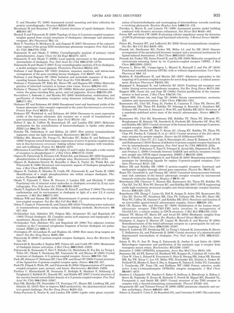
P, and Nienaber VL (2000) Automated crystal mounting and data collection forprotein crystallography. Structure 8:R243–R246.
Murakami M and Kouyama T (2008) Crystal structure of squid rhodopsin. Nature453:363–367.
Mustafi D and Palczewski K (2009) Topology of class A G protein-coupled receptors:insights gained from crystal structures of rhodopsins, adrenergic and adenosinereceptors. Mol Pharmacol 75:1–12.
Muto T, Tsuchiya D, Morikawa K, and Jingami H (2007) Structures of the extracel-lular regions of the group II/III metabotropic glutamate receptors. Proc Natl AcadSci USA 104:3759–3764.
Nakamichi H and Okada T (2006a) Crystallographic analysis of primary visualphotochemistry. Angew Chem Int Ed Engl 45:4270–4273.
Nakamichi H and Okada T (2006b) Local peptide movement in the photoreactionintermediate of rhodopsin. Proc Natl Acad Sci USA 103:12729–12734.
Nakamichi H and Okada T (2007) X-ray crystallographic analysis of 9-cis-rhodopsin,a model analogue visual pigment. Photochem Photobiol 83:232–235.
Nathans J and Hogness DS (1983) Isolation, sequence analysis, and intron-exonarrangement of the gene encoding bovine rhodopsin. Cell 34:807–814.
Nathans J and Hogness DS (1984) Isolation and nucleotide sequence of the geneencoding human rhodopsin. Proc Natl Acad Sci USA 81:4851–4855.
Nathans J, Piantanida TP, Eddy RL, Shows TB, and Hogness DS (1986a) Moleculargenetics of inherited variation in human color vision. Science 232:203–210.
Nathans J, Thomas D, and Hogness DS (1986b) Molecular genetics of human colorvision: the genes encoding blue, green, and red pigments. Science 232:193–202.
Navratilova I, Sodroski J, and Myszka DG (2005) Solubilization, stabilization, andpurification of chemokine receptors using biosensor technology. Anal Biochem339:271–281.
Niebauer RT and Robinson AS (2006) Exceptional total and functional yields of thehuman adenosine (A2a) receptor expressed in the yeast Saccharomyces cerevisiae.Protein Expr Purif 46:204–211.
Niebauer RT, Wedekind A, and Robinson AS (2004) Decreases in yeast expressionyields of the human adenosine A2a receptor are a result of translational orpost-translational events. Protein Expr Purif 37:134–143.
Nollert P, Qiu H, Caffrey M, Rosenbusch JP, and Landau EM (2001) Molecularmechanism for the crystallization of bacteriorhodopsin in lipidic cubic phases.FEBS Lett 504:179–186.
Nyholm TK, Ozdirekcan S, and Killian JA (2007) How protein transmembranesegments sense the lipid environment. Biochemistry 46:1457–1465.
O’Malley MA, Mancini JD, Young CL, McCusker EC, Raden D, and Robinson AS(2009) Progress toward heterologous expression of active G-protein-coupled recep-tors in Saccharomyces cerevisiae: linking cellular stress response with transloca-tion and trafficking. Protein Sci 18:2356–2370.
Offermanns S and Simon MI (1995) G alpha 15 and G alpha 16 couple a wide varietyof receptors to phospholipase C. J Biol Chem 270:15175–15180.
Ohguro H, Palczewski K, Ericsson LH, Walsh KA, and Johnson RS (1993) Sequentialphosphorylation of rhodopsin at multiple sites. Biochemistry 32:5718–5724.
Ohguro H, Rudnicka-Nawrot M, Buczyłko J, Zhao X, Taylor JA, Walsh KA, andPalczewski K (1996) Structural and enzymatic aspects of rhodopsin phosphoryla-tion. J Biol Chem 271:5215–5224.
Ohguro H, Yoshida N, Shindou H, Crabb JW, Palczewski K, and Tsuda M (1998)Identification of a single phosphorylation site within octopus rhodopsin. Pho-tochem Photobiol 68:824–828.
Okada T, Fujiyoshi Y, Silow M, Navarro J, Landau EM, and Shichida Y (2002)Functional role of internal water molecules in rhodopsin revealed by X-ray crys-tallography. Proc Natl Acad Sci USA 99:5982–5987.
Okada T, Sugihara M, Bondar AN, Elstner M, Entel P, and Buss V (2004) The retinalconformation and its environment in rhodopsin in light of a new 2.2 A crystalstructure. J Mol Biol 342:571–583.
Oldham WM and Hamm HE (2008) Heterotrimeric G protein activation by G-pro-tein-coupled receptors. Nat Rev Mol Cell Biol 9:60–71.
Orban T, Gupta S, Palczewski K, and Chance MR (2010) Visualizing water moleculesin transmembrane proteins using radiolytic labeling methods. Biochemistry 49:827–834.
Ovchinnikov IuA, Abdulaev NG, Feıgina MIu, Artamonov ID, and Bogachuk AS(1983) [Visual rhodopsin. III. Complete amino acid sequence and topography in amembrane]. Bioorg Khim 9:1331–1340.
Ovchinnikov YuA, Abdulaev NG, and Bogachuk AS (1988) Two adjacent cysteineresidues in the C-terminal cytoplasmic fragment of bovine rhodopsin are palmi-tylated. FEBS Lett 230:1–5.
Overington JP, Al-Lazikani B, and Hopkins AL (2006) How many drug targets arethere? Nat Rev Drug Discov 5:993–996.
Palczewski K (2006) G protein-coupled receptor rhodopsin. Annu Rev Biochem 75:743–767.
Palczewski K, Buczyłko J, Kaplan MW, Polans AS, and Crabb JW (1991) Mechanismof rhodopsin kinase activation. J Biol Chem 266:12949–12955.
Palczewski K, Kumasaka T, Hori T, Behnke CA, Motoshima H, Fox BA, Le Trong I,Teller DC, Okada T, Stenkamp RE, Yamamoto M, and Miyano M (2000) Crystalstructure of rhodopsin: A G protein-coupled receptor. Science 289:739–745.
Park JH, Scheerer P, Hofmann KP, Choe HW, and Ernst OP (2008) Crystal structureof the ligand-free G-protein-coupled receptor opsin. Nature 454:183–187.
Park PS, Filipek S, Wells JW, and Palczewski K (2004) Oligomerization of G protein-coupled receptors: past, present, and future. Biochemistry 43:15643–15656.
Parthier C, Kleinschmidt M, Neumann P, Rudolph R, Manhart S, Schlenzig D,Fanghanel J, Rahfeld JU, Demuth HU, and Stubbs MT (2007) Crystal structure ofthe incretin-bound extracellular domain of a G protein-coupled receptor. Proc NatlAcad Sci USA 104:13942–13947.
Paul SM, Mytelka DS, Dunwiddie CT, Persinger CC, Munos BH, Lindborg SR, andSchacht AL (2010) How to improve R&D productivity: the pharmaceutical indus-try’s grand challenge. Nat Rev Drug Discov 9:203–214.
Pebay-Peyroula E, Neutze R, and Landau EM (2000) Lipidic cubic phase crystalli-
zation of bacteriorhodopsin and cryotrapping of intermediates: towards resolvinga revolving photocycle. Biochim Biophys Acta 1460:119–132.
Perrakis A, Morris R, and Lamzin VS (1999) Automated protein model buildingcombined with iterative structure refinement. Nat Struct Biol 6:458–463.
Peters MF and Scott CW (2009) Evaluating cellular impedance assays for detectionof GPCR pleiotropic signaling and functional selectivity. J Biomol Screen 14:246–255.
Pierce KL, Premont RT, and Lefkowitz RJ (2002) Seven-transmembrane receptors.Nat Rev Mol Cell Biol 3:639–650.
Pioszak AA, Harikumar KG, Parker NR, Miller LJ, and Xu HE (2010) Dimericarrangement of the parathyroid hormone receptor and a structural mechanism forligand-induced dissociation. J Biol Chem 285:12435–12444.
Pioszak AA, Parker NR, Suino-Powell K, and Xu HE (2008) Molecular recognition ofcorticotropin-releasing factor by its G-protein-coupled receptor CRFR1. J BiolChem 283:32900–32912.
Prezeau L, Rives ML, Comps-Agrar L, Maurel D, Kniazeff J, and Pin JP (2010)Functional crosstalk between GPCRs: with or without oligomerization. Curr OpinPharmacol 10:6–13.
Raddatz R, Schaffhauser H, and Marino MJ (2007) Allosteric approaches to thetargeting of G-protein-coupled receptors for novel drug discovery: a critical assess-ment. Biochem Pharmacol 74:383–391.
Rajagopal S, Rajagopal K, and Lefkowitz RJ (2010) Teaching old receptors newtricks: biasing seven-transmembrane receptors. Nat Rev Drug Discov 9:373–386.
Rapport MM, Green AA, and Page IH (1948a) Partial purification of the vasocon-strictor in beef serum. J Biol Chem 174:735–741.
Rapport MM, Green AA, and Page IH (1948b) Serum vasoconstrictor, serotonin;isolation and characterization. J Biol Chem 176:1243–1251.
Rasmussen SG, Choi HJ, Fung JJ, Pardon E, Casarosa P, Chae PS, Devree BT,Rosenbaum DM, Thian FS, Kobilka TS, Schnapp A, Konetzki I, Sunahara RK,Gellman SH, Pautsch A, Steyaert J, Weis WI, and Kobilka BK (2011) Structure ofa nanobody-stabilized active state of the beta(2) adrenoceptor. Nature 469:175–180.
Rasmussen SG, Choi HJ, Rosenbaum DM, Kobilka TS, Thian FS, Edwards PC,Burghammer M, Ratnala VR, Sanishvili R, Fischetti RF, Schertler GF, Weis WI,and Kobilka BK (2007) Crystal structure of the human beta2 adrenergic G-protein-coupled receptor. Nature 450:383–387.
Rasmussen SG, Devree BT, Zou Y, Kruse AC, Chung KY, Kobilka TS, Thian FS,Chae PS, Pardon E, Calinski D, et al. (2011) Crystal structure of the �(2) adren-ergic receptor-Gs protein complex. Nature doi:10.1038/nature10361.
Rivero-Muller A, Chou YY, Ji I, Lajic S, Hanyaloglu AC, Jonas K, Rahman N, Ji TH,and Huhtaniemi I (2010) Rescue of defective G protein-coupled receptor function invivo by intermolecular cooperation. Proc Natl Acad Sci USA 107:2319–2324.
Rives ML, Vol C, Fukazawa Y, Tinel N, Trinquet E, Ayoub MA, Shigemoto R, Pin JP,and Prezeau L (2009) Crosstalk between GABAB and mGlu1a receptors revealsnew insight into GPCR signal integration. EMBO J 28:2195–2208.
Robas N, O’Reilly M, Katugampola S, and Fidock M (2003) Maximizing serendipity:strategies for identifying ligands for orphan G-protein-coupled receptors. CurrOpin Pharmacol 3:121–126.
Rohrer DK and Kobilka BK (1998) G protein-coupled receptors: functional andmechanistic insights through altered gene expression. Physiol Rev 78:35–52.
Romo TD, Grossfield A, and Pitman MC (2010) Concerted interconversion betweenionic lock substates of the beta(2) adrenergic receptor revealed by microsecondtimescale molecular dynamics. Biophys J 98:76–84.
Rosenbaum DM, Cherezov V, Hanson MA, Rasmussen SG, Thian FS, Kobilka TS,Choi HJ, Yao XJ, Weis WI, Stevens RC, and Kobilka BK (2007) GPCR engineeringyields high-resolution structural insights into beta2-adrenergic receptor function.Science 318:1266–1273.
Rosenbaum DM, Zhang C, Lyons JA, Holl R, Aragao D, Arlow DH, Rasmussen SG,Choi HJ, Devree BT, Sunahara RK, Chae PS, Gellman SH, Dror RO, Shaw DE,Weis WI, Caffrey M, Gmeiner P, and Kobilka BK (2011) Structure and function ofan irreversible agonist-beta(2) adrenoceptor complex. Nature 469:236–240.
Roth CB, Hanson MA, and Stevens RC (2008) Stabilization of the human beta2-adrenergic receptor TM4-TM3-TM5 helix interface by mutagenesis ofGlu122(3.41), a critical residue in GPCR structure. J Mol Biol 376:1305–1319.
Sakmar TP, Menon ST, Marin EP, and Awad ES (2002) Rhodopsin: insights fromrecent structural studies. Annu Rev Biophys Biomol Struct 31:443–484.
Salahpour A, Angers S, Mercier JF, Lagace M, Marullo S, and Bouvier M (2004)Homodimerization of the beta2-adrenergic receptor as a prerequisite for cell sur-face targeting. J Biol Chem 279:33390–33397.
Salom D, Lodowski DT, Stenkamp RE, Le Trong I, Golczak M, Jastrzebska B, HarrisT, Ballesteros JA, and Palczewski K (2006) Crystal structure of a photoactivateddeprotonated intermediate of rhodopsin. Proc Natl Acad Sci USA 103:16123–16128.
Salom D, Wu N, Sun W, Dong Z, Palczewski K, Jordan S, and Salon JA (2008)Heterologous expression and purification of the serotonin type 4 receptor fromtransgenic mouse retina. Biochemistry 47:13296–13307.
Samanen J (1996) GPIIb/IIIa antagonists. Annu Rep Med Chem 31:91–100.Samanen JM, Ali FE, Barton LS, Bondinell WE, Burgess JL, Callahan JF, Calvo RR,
Chen W, Chen L, Erhard K, Feuerstein G, Heys R, Hwang SM, Jakas DR, KeenanRM, Ku TW, Kwon C, Lee CP, Miller WH, Newlander KA, Nichols A, Parker M,Peishoff CE, Rhodes G, Ross S, Shu A, Simpson R, Takata D, Yellin TO, UzsinskasI, Venslavsky JW, Yuan CK, and Huffman WF (1996) Potent, selective, orallyactive 3-oxo-1,4-benzodiazepine GPIIb/IIIa integrin antagonists. J Med Chem39:4867–4870.
Sanders J, Chirgadze DY, Sanders P, Baker S, Sullivan A, Bhardwaja A, Bolton J,Reeve M, Nakatake N, Evans M, Richards T, Powell M, Miguel RN, Blundell TL,Furmaniak J, and Smith BR (2007) Crystal structure of the TSH receptor incomplex with a thyroid-stimulating autoantibody. Thyroid 17:395–410.
Sanguinetti MC and Tristani-Firouzi M (2006) hERG potassium channels and car-diac arrhythmia. Nature 440:463–469.
Scheer A, Fanelli F, Costa T, De Benedetti PG, and Cotecchia S (1997) The activation
GPCRs AND DRUG DISCOVERY 935
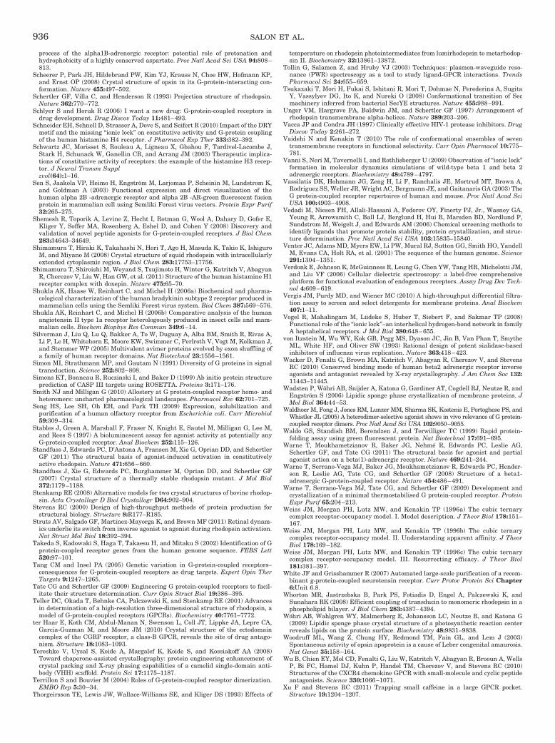
process of the alpha1B-adrenergic receptor: potential role of protonation andhydrophobicity of a highly conserved aspartate. Proc Natl Acad Sci USA 94:808–813.
Scheerer P, Park JH, Hildebrand PW, Kim YJ, Krauss N, Choe HW, Hofmann KP,and Ernst OP (2008) Crystal structure of opsin in its G-protein-interacting con-formation. Nature 455:497–502.
Schertler GF, Villa C, and Henderson R (1993) Projection structure of rhodopsin.Nature 362:770–772.
Schlyer S and Horuk R (2006) I want a new drug: G-protein-coupled receptors indrug development. Drug Discov Today 11:481–493.
Schneider EH, Schnell D, Strasser A, Dove S, and Seifert R (2010) Impact of the DRYmotif and the missing “ionic lock” on constitutive activity and G-protein couplingof the human histamine H4 receptor. J Pharmacol Exp Ther 333:382–392.
Schwartz JC, Morisset S, Rouleau A, Ligneau X, Gbahou F, Tardivel-Lacombe J,Stark H, Schunack W, Ganellin CR, and Arrang JM (2003) Therapeutic implica-tions of constitutive activity of receptors: the example of the histamine H3 recep-tor. J Neural Transm Supplzvol(64):1–16.
Sen S, Jaakola VP, Heimo H, Engstrom M, Larjomaa P, Scheinin M, Lundstrom K,and Goldman A (2003) Functional expression and direct visualization of thehuman alpha 2B -adrenergic receptor and alpha 2B -AR-green fluorescent fusionprotein in mammalian cell using Semliki Forest virus vectors. Protein Expr Purif32:265–275.
Shemesh R, Toporik A, Levine Z, Hecht I, Rotman G, Wool A, Dahary D, Gofer E,Kliger Y, Soffer MA, Rosenberg A, Eshel D, and Cohen Y (2008) Discovery andvalidation of novel peptide agonists for G-protein-coupled receptors. J Biol Chem283:34643–34649.
Shimamura T, Hiraki K, Takahashi N, Hori T, Ago H, Masuda K, Takio K, IshiguroM, and Miyano M (2008) Crystal structure of squid rhodopsin with intracellularlyextended cytoplasmic region. J Biol Chem 283:17753–17756.
Shimamura T, Shiroishi M, Weyand S, Tsujimoto H, Winter G, Katritch V, AbagyanR, Cherezov V, Liu W, Han GW, et al. (2011) Structure of the human histamine H1receptor complex with doxepin. Nature 475:65–70.
Shukla AK, Haase W, Reinhart C, and Michel H (2006a) Biochemical and pharma-cological characterization of the human bradykinin subtype 2 receptor produced inmammalian cells using the Semliki Forest virus system. Biol Chem 387:569–576.
Shukla AK, Reinhart C, and Michel H (2006b) Comparative analysis of the humanangiotensin II type 1a receptor heterologously produced in insect cells and mam-malian cells. Biochem Biophys Res Commun 349:6–14.
Silverman J, Liu Q, Lu Q, Bakker A, To W, Duguay A, Alba BM, Smith R, Rivas A,Li P, Le H, Whitehorn E, Moore KW, Swimmer C, Perlroth V, Vogt M, Kolkman J,and Stemmer WP (2005) Multivalent avimer proteins evolved by exon shuffling ofa family of human receptor domains. Nat Biotechnol 23:1556–1561.
Simon MI, Strathmann MP, and Gautam N (1991) Diversity of G proteins in signaltransduction. Science 252:802–808.
Simons KT, Bonneau R, Ruczinski I, and Baker D (1999) Ab initio protein structureprediction of CASP III targets using ROSETTA. Proteins 3:171–176.
Smith NJ and Milligan G (2010) Allostery at G protein-coupled receptor homo- andheteromers: uncharted pharmacological landscapes. Pharmacol Rev 62:701–725.
Song HS, Lee SH, Oh EH, and Park TH (2009) Expression, solubilization andpurification of a human olfactory receptor from Escherichia coli. Curr Microbiol59:309–314.
Stables J, Green A, Marshall F, Fraser N, Knight E, Sautel M, Milligan G, Lee M,and Rees S (1997) A bioluminescent assay for agonist activity at potentially anyG-protein-coupled receptor. Anal Biochem 252:115–126.
Standfuss J, Edwards PC, D’Antona A, Fransen M, Xie G, Oprian DD, and SchertlerGF (2011) The structural basis of agonist-induced activation in constitutivelyactive rhodopsin. Nature 471:656–660.
Standfuss J, Xie G, Edwards PC, Burghammer M, Oprian DD, and Schertler GF(2007) Crystal structure of a thermally stable rhodopsin mutant. J Mol Biol372:1179–1188.
Stenkamp RE (2008) Alternative models for two crystal structures of bovine rhodop-sin. Acta Crystallogr D Biol Crystallogr D64:902–904.
Stevens RC (2000) Design of high-throughput methods of protein production forstructural biology. Structure 8:R177–R185.
Struts AV, Salgado GF, Martínez-Mayorga K, and Brown MF (2011) Retinal dynam-ics underlie its switch from inverse agonist to agonist during rhodopsin activation.Nat Struct Mol Biol 18:392–394.
Takeda S, Kadowaki S, Haga T, Takaesu H, and Mitaku S (2002) Identification of Gprotein-coupled receptor genes from the human genome sequence. FEBS Lett520:97–101.
Tang CM and Insel PA (2005) Genetic variation in G-protein-coupled receptors–consequences for G-protein-coupled receptors as drug targets. Expert Opin TherTargets 9:1247–1265.
Tate CG and Schertler GF (2009) Engineering G protein-coupled receptors to facil-itate their structure determination. Curr Opin Struct Biol 19:386–395.
Teller DC, Okada T, Behnke CA, Palczewski K, and Stenkamp RE (2001) Advancesin determination of a high-resolution three-dimensional structure of rhodopsin, amodel of G-protein-coupled receptors (GPCRs). Biochemistry 40:7761–7772.
ter Haar E, Koth CM, Abdul-Manan N, Swenson L, Coll JT, Lippke JA, Lepre CA,Garcia-Guzman M, and Moore JM (2010) Crystal structure of the ectodomaincomplex of the CGRP receptor, a class-B GPCR, reveals the site of drug antago-nism. Structure 18:1083–1093.
Tereshko V, Uysal S, Koide A, Margalef K, Koide S, and Kossiakoff AA (2008)Toward chaperone-assisted crystallography: protein engineering enhancement ofcrystal packing and X-ray phasing capabilities of a camelid single-domain anti-body (VHH) scaffold. Protein Sci 17:1175–1187.
Terrillon S and Bouvier M (2004) Roles of G-protein-coupled receptor dimerization.EMBO Rep 5:30–34.
Thorgeirsson TE, Lewis JW, Wallace-Williams SE, and Kliger DS (1993) Effects of
temperature on rhodopsin photointermediates from lumirhodopsin to metarhodop-sin II. Biochemistry 32:13861–13872.
Tollin G, Salamon Z, and Hruby VJ (2003) Techniques: plasmon-waveguide reso-nance (PWR) spectroscopy as a tool to study ligand-GPCR interactions. TrendsPharmacol Sci 24:655–659.
Tsukazaki T, Mori H, Fukai S, Ishitani R, Mori T, Dohmae N, Perederina A, SugitaY, Vassylyev DG, Ito K, and Nureki O (2008) Conformational transition of Secmachinery inferred from bacterial SecYE structures. Nature 455:988–991.
Unger VM, Hargrave PA, Baldwin JM, and Schertler GF (1997) Arrangement ofrhodopsin transmembrane alpha-helices. Nature 389:203–206.
Vacca JP and Condra JH (1997) Clinically effective HIV-1 protease inhibitors. DrugDiscov Today 2:261–272.
Vaidehi N and Kenakin T (2010) The role of conformational ensembles of seventransmembrane receptors in functional selectivity. Curr Opin Pharmacol 10:775–781.
Vanni S, Neri M, Tavernelli I, and Rothlisberger U (2009) Observation of “ionic lock”formation in molecular dynamics simulations of wild-type beta 1 and beta 2adrenergic receptors. Biochemistry 48:4789–4797.
Vassilatis DK, Hohmann JG, Zeng H, Li F, Ranchalis JE, Mortrud MT, Brown A,Rodriguez SS, Weller JR, Wright AC, Bergmann JE, and Gaitanaris GA (2003) TheG protein-coupled receptor repertoires of human and mouse. Proc Natl Acad SciUSA 100:4903–4908.
Vedadi M, Niesen FH, Allali-Hassani A, Fedorov OY, Finerty PJ, Jr., Wasney GA,Yeung R, Arrowsmith C, Ball LJ, Berglund H, Hui R, Marsden BD, Nordlund P,Sundstrom M, Weigelt J, and Edwards AM (2006) Chemical screening methods toidentify ligands that promote protein stability, protein crystallization, and struc-ture determination. Proc Natl Acad Sci USA 103:15835–15840.
Venter JC, Adams MD, Myers EW, Li PW, Mural RJ, Sutton GG, Smith HO, YandellM, Evans CA, Holt RA, et al. (2001) The sequence of the human genome. Science291:1304–1351.
Verdonk E, Johnson K, McGuinness R, Leung G, Chen YW, Tang HR, Michelotti JM,and Liu VF (2006) Cellular dielectric spectroscopy: a label-free comprehensiveplatform for functional evaluation of endogenous receptors. Assay Drug Dev Tech-nol 4:609–619.
Vergis JM, Purdy MD, and Wiener MC (2010) A high-throughput differential filtra-tion assay to screen and select detergents for membrane proteins. Anal Biochem407:1–11.
Vogel R, Mahalingam M, Ludeke S, Huber T, Siebert F, and Sakmar TP (2008)Functional role of the “ionic lock”–an interhelical hydrogen-bond network in familyA heptahelical receptors. J Mol Biol 380:648–655.
von Itzstein M, Wu WY, Kok GB, Pegg MS, Dyason JC, Jin B, Van Phan T, SmytheML, White HF, and Oliver SW (1993) Rational design of potent sialidase-basedinhibitors of influenza virus replication. Nature 363:418–423.
Wacker D, Fenalti G, Brown MA, Katritch V, Abagyan R, Cherezov V, and StevensRC (2010) Conserved binding mode of human beta2 adrenergic receptor inverseagonists and antagonist revealed by X-ray crystallography. J Am Chem Soc 132:11443–11445.
Wadsten P, Wohri AB, Snijder A, Katona G, Gardiner AT, Cogdell RJ, Neutze R, andEngstrom S (2006) Lipidic sponge phase crystallization of membrane proteins. JMol Biol 364:44–53.
Waldhoer M, Fong J, Jones RM, Lunzer MM, Sharma SK, Kostenis E, Portoghese PS, andWhistler JL (2005) A heterodimer-selective agonist shows in vivo relevance of G protein-coupled receptor dimers. Proc Natl Acad Sci USA 102:9050–9055.
Waldo GS, Standish BM, Berendzen J, and Terwilliger TC (1999) Rapid protein-folding assay using green fluorescent protein. Nat Biotechnol 17:691–695.
Warne T, Moukhametzianov R, Baker JG, Nehme R, Edwards PC, Leslie AG,Schertler GF, and Tate CG (2011) The structural basis for agonist and partialagonist action on a beta(1)-adrenergic receptor. Nature 469:241–244.
Warne T, Serrano-Vega MJ, Baker JG, Moukhametzianov R, Edwards PC, Hender-son R, Leslie AG, Tate CG, and Schertler GF (2008) Structure of a beta1-adrenergic G-protein-coupled receptor. Nature 454:486–491.
Warne T, Serrano-Vega MJ, Tate CG, and Schertler GF (2009) Development andcrystallization of a minimal thermostabilised G protein-coupled receptor. ProteinExpr Purif 65:204–213.
Weiss JM, Morgan PH, Lutz MW, and Kenakin TP (1996a) The cubic ternarycomplex receptor-occupancy model. I. Model description. J Theor Biol 178:151–167.
Weiss JM, Morgan PH, Lutz MW, and Kenakin TP (1996b) The cubic ternarycomplex receptor-occupancy model. II. Understanding apparent affinity. J TheorBiol 178:169–182.
Weiss JM, Morgan PH, Lutz MW, and Kenakin TP (1996c) The cubic ternarycomplex receptor-occupancy model. III. Resurrecting efficacy. J Theor Biol181:381–397.
White JF and Grisshammer R (2007) Automated large-scale purification of a recom-binant g-protein-coupled neurotensin receptor. Curr Protoc Protein Sci Chapter6:Unit 6.8.
Whorton MR, Jastrzebska B, Park PS, Fotiadis D, Engel A, Palczewski K, andSunahara RK (2008) Efficient coupling of transducin to monomeric rhodopsin in aphospholipid bilayer. J Biol Chem 283:4387–4394.
Wohri AB, Wahlgren WY, Malmerberg E, Johansson LC, Neutze R, and Katona G(2009) Lipidic sponge phase crystal structure of a photosynthetic reaction centerreveals lipids on the protein surface. Biochemistry 48:9831–9838.
Woodruff ML, Wang Z, Chung HY, Redmond TM, Fain GL, and Lem J (2003)Spontaneous activity of opsin apoprotein is a cause of Leber congenital amaurosis.Nat Genet 35:158–164.
Wu B, Chien EY, Mol CD, Fenalti G, Liu W, Katritch V, Abagyan R, Brooun A, WellsP, Bi FC, Hamel DJ, Kuhn P, Handel TM, Cherezov V, and Stevens RC (2010)Structures of the CXCR4 chemokine GPCR with small-molecule and cyclic peptideantagonists. Science 330:1066–1071.
Xu F and Stevens RC (2011) Trapping small caffeine in a large GPCR pocket.Structure 19:1204–1207.
936 SALON ET AL.

Xu F, Wu H, Katritch V, Han GW, Jacobson KA, Gao ZG, Cherezov V, and StevensRC (2011) Structure of an agonist-bound human A2A adenosine receptor. Science332:322–327.
Ye S, Huber T, Vogel R, and Sakmar TP (2009) FTIR analysis of GPCR activationusing azido probes. Nat Chem Biol 5:397–399.
Yoshizawa T and Wald G (1964) Transformations of squid rhodopsin at low temper-atures. Nature 201:340–345.
Zhang L, Salom D, He J, Okun A, Ballesteros J, Palczewski K, and Li N (2005)Expression of functional G protein-coupled receptors in photoreceptors of trans-genic Xenopus laevis. Biochemistry 44:14509–14518.
Zhang Y, Devries ME, and Skolnick J (2006) Structure modeling of all identifiedG protein-coupled receptors in the human genome. PLoS Comput Biol2:e13.
Zhou Y, Morais-Cabral JH, Kaufman A, and MacKinnon R (2001) Chemistry of ioncoordination and hydration revealed by a K� channel-Fab complex at 2.0 Aresolution. Nature 414:43–48.
Zhu T, Fang LY, and Xie X (2008) Development of a universal high-throughputcalcium assay for G-protein-coupled receptors with promiscuous G-protein Gal-pha15/16. Acta Pharmacol Sin 29:507–516.
Zozulya S, Echeverri F, and Nguyen T (2001) The human olfactory receptor reper-toire. Genome Biol 2:RESEARCH0018.
Harrold M (2008) Angiotensin Converting Enzyme Inhibitors, Antagonists, and Cal-cium Blockers, Lippincott Williams & Wilkins, Philadelphia.
Salon JA and Owicki JC eds (1995) Real-Time Measurements of Receptor Activity;Applications of Microphysiometric Techniques to Receptor Biology. AcademicPress.
GPCRs AND DRUG DISCOVERY 937