pdz protein regulation of g protein coupled receptor...
TRANSCRIPT
1521-0111/88/4/624–639$25.00 http://dx.doi.org/10.1124/mol.115.098509MOLECULAR PHARMACOLOGY Mol Pharmacol 88:624–639, October 2015Copyright ª 2015 by The American Society for Pharmacology and Experimental Therapeutics
MINIREVIEW
PDZ Protein Regulation of G Protein–Coupled ReceptorTrafficking and Signaling Pathways
Henry A. Dunn and Stephen S. G. FergusonJ. Allyn Taylor Centre for Cell Biology, Robarts Research Institute, and the Department of Physiology and Pharmacology,University of Western Ontario, London, Ontario, Canada
Received February 18, 2015; accepted March 25, 2015
ABSTRACTG protein–coupled receptors (GPCRs) contribute to the regulationof every aspect of human physiology and are therapeutic targetsfor the treatment of numerous diseases. As a consequence,understanding the myriad of mechanisms controlling GPCRsignaling and trafficking is essential for the development of newpharmacological strategies for the treatment of human pa-thologies. Of the many GPCR-interacting proteins, postsynapticdensity protein of 95 kilodaltons, disc large, zona occludens-1(PDZ) domain–containing proteins appear most abundant andhave similarly been implicated in disease mechanisms. PDZproteins play an important role in regulating receptor and channelprotein localization within synapses and tight junctions andfunction to scaffold intracellular signaling protein complexes. Inthe current study, we review the known functional interactionsbetween PDZ domain–containing proteins and GPCRs andprovide insight into the potential mechanisms of action. ThesePDZ domain–containing proteins include the membrane-associated guanylate-like kinases [postsynaptic density protein of95 kilodaltons; synapse-associated protein of 97 kilodaltons;postsynaptic density protein of 93 kilodaltons; synapse-associated
protein of 102 kilodaltons; discs, large homolog 5; caspaseactivation and recruitment domain and membrane-associatedguanylate-like kinase domain-containing protein 3; membraneprotein, palmitoylated 3; calcium/calmodulin-dependent serineprotein kinase; membrane-associated guanylate kinase protein(MAGI)-1, MAGI-2, and MAGI-3], Na1/H1 exchanger regulatoryfactor proteins (NHERFs) (NHERF1, NHERF2, PDZ domain–containing kidney protein 1, and PDZ domain–containing kid-ney protein 2), Golgi-associated PDZ proteins (Ga-bindingprotein interacting protein, C-terminus and CFTR-associatedligand), PDZ domain–containing guanine nucleotide exchangefactors (GEFs) 1 and 2, regulator of G protein signaling (RGS)–homology-RhoGEFs (PDZ domain–containing RhoGEF andleukemia-associated RhoGEF), RGS3 and RGS12, spinophilinand neurabin-1, SRC homology 3 domain and multiple ankyrinrepeat domain (Shank) proteins (Shank1, Shank2, and Shank3),partitioning defective proteins 3 and 6, multiple PDZ protein 1,Tamalin, neuronal nitric oxide synthase, syntrophins, proteininteracting with protein kinase C a 1, syntenin-1, and sortingnexin 27.
IntroductionIn the central nervous system, G protein–coupled receptors
(GPCRs) and ion channels are targeted to the membrane ofdendritic postsynaptic terminals in and around a region termed
the postsynaptic density (PSD) (Neubig and Siderovski, 2002;Feng and Zhang, 2009; Magalhaes et al., 2012). Each post-synaptic density is specifically organized, such that dozens tohundreds of receptors are targeted to this specialized mem-brane domain via the interaction of scaffolding proteins withthe receptors. These scaffold proteins contain multiple protein-protein interaction domains that allow them to interact witha multitude of structural and signaling proteins and hold themin close proximity with one another (Feng and Zhang, 2009). Ofthese scaffolding proteins, it is believed that postsynaptic
This work is supported by a Canadian Institutes of Health Research grant[MOP-62738] to S.S.G.F. S.S.G.F. holds a Tier I Canada Research Chair inMolecular Neurobiology and is a Career Investigator of the Heart and StrokeFoundation of Ontario, Canada.
dx.doi.org/10.1124/mol.115.098509.
ABBREVIATIONS: AMPA, a-amino-3-hydroxy-5-methyl-4-isoxazolepropionic acid; AMPAR, AMPA receptor; CAL, conductance regulator–associated ligand; CRF, corticotropin-releasing factor; ERK, extracellular signal-related kinase; GEF, guanine nucleotide exchange factor; GIPC,Ga-binding protein interacting protein carboxyl-terminus; GK, guanylate kinase–like domain; GPCR, G protein–coupled receptor; hIPR, humanprostacyclin receptor; MAGI, membrane-associated guanylate kinase protein; MAGUK, membrane-associated guanylate kinase; mGluR,metabotropic glutamate receptor; MPP, membrane palmitoylated protein; NHERF, Na1/H1 exchanger regulatory factor; NMDA, N-methyl-D-aspartate; NMDAR, NMDA receptor; nNOS, neuronal nitric oxide synthase; mOR, m opioid receptor; Par, partitioning defective protein; PDZ, PSD-95,disc large, zona occludens-1; PH, pleckstrin-homology; PKA, protein kinase A; PKC, protein kinase C; PLC, phospholipase C; PSD, postsynapticdensity; RGS, regulator of G protein signaling; SH3, SRC homology 3; Shank, SRC homology 3 domain and multiple ankyrin repeat domain; SSTR,somatostatin receptor.
624
at ASPE
T Journals on February 25, 2019
molpharm
.aspetjournals.orgD
ownloaded from
density protein of 95 kilodaltons (PSD-95), disc large, zonaoccludens-1 (PDZ) domain–containing proteins are the mostabundant and often provide direct contact with both GPCRsand ion channels at the postsynaptic density (Cheng et al.,2006; Feng and Zhang, 2009). PDZ proteins are not onlyimportant for targeting GPCRs to synapses, but they have animportant role in regulating tight junctions and signalingprotein complexes. In the current review, we will providean overview of the growing understanding of the role ofPDZ domain–containing proteins in the regulation of GPCRsubcellular localization, endocytosis, trafficking, and signaltransduction.
PDZ DomainsPDZ domains are approximately 80–90 amino acid resi-
dues in size and represent the most common protein-proteininteraction domain (Doyle et al., 1996; Feng and Zhang, 2009;Magalhaes et al., 2012). Although there are hundreds ofunique PDZ domain sequences, they all contain a conservedglycine-leucine-glycine-phenylalanine sequence that providesthe domain’s folded, globular, cup-like structure that is ca-pable of recognizing short, finger-like peptides (Harris andLim, 2001). Because of this structure, PDZ domains appearbest suited for binding the distal regions of receptor carboxyl-terminal tails, which are labeled the PDZ-binding motif(Kornau et al., 1995; Niethammer et al., 1996; Harris andLim, 2001; Magalhaes et al., 2012). Interestingly, additionalstudies have identified internal PDZ ligands that, like acarboxyl-terminal tail, project outwardly from the protein(Xu et al., 1998; Christopherson et al., 1999; Hillier et al.,1999; Fouassier et al., 2000; Harris and Lim, 2001; Paascheet al., 2005; Trejo, 2005). In this case, the internal PDZ-binding motif manifests as a sharply folded, finger-likeprojection.
PDZ-Binding MotifsAlthough seemingly imperfect and likely biased against
internal PDZ ligands (reviewed by Trejo, 2005), a simpleclassification system has evolved to identify potential PDZ-binding motifs and helps to predict potential PDZ domain–containing protein interactions (Songyang et al., 1997;Bezprozvanny and Maximov, 2001; Sheng and Sala, 2001;Vaccaro and Dente, 2002). Although there is some delibera-tion over howmany classes of PDZ-binding motifs there are, itis most commonly limited to three classes (Sheng and Sala,2001; Tonikian et al., 2008; Magalhaes et al., 2012). Class IPDZ-binding motifs are the most described class within theliterature and are classified by their final three–amino acidsequence of S/T-x-w, where x indicates any amino acid and w
indicates any hydrophobic amino acid (Songyang et al., 1997;Bezprozvanny and Maximov, 2001; Sheng and Sala, 2001;Vaccaro and Dente, 2002). However, valine, isoleucine, orleucine appear to be the most common of the hydrophobicamino acids that contribute to the formation of a class I PDZ-binding motif (Songyang et al., 1997; Bezprozvanny andMaximov, 2001; Sheng and Sala, 2001; Vaccaro and Dente,2002). Class II and III PDZ-binding motifs are not as wellcharacterized and show slightly more ambiguous sequences,with class II having its final three amino acids as w-x-w, andclass III having C-x-C, where C represents any acidic aminoacid residue (Sheng and Sala, 2001).
GPCR-Interacting PSD-95 Family PDZ Domain–Containing Membrane-Associated Guanylate
Kinase ProteinsPSD-95 (DLG4). PSD-95 contains three PDZ domains, an
SRC homology 3 (SH3) domain, and a guanylate kinase–like(GK) domain (Fig. 1), and is prototypically localized within the
Fig. 1. Molecular topology of protein-protein in-teraction domains found in MAGUK family PDZproteins. CaMKII, Ca2+/calmodulin-dependent ki-nase domain; CARD, caspase activation and re-cruitment domain; CC, coiled-coiled domain; L27,L27 domain.
GPCR Regulation by PDZ Proteins 625
at ASPE
T Journals on February 25, 2019
molpharm
.aspetjournals.orgD
ownloaded from

postsynaptic density (Sampedro et al., 1981; Cho et al., 1992).PSD-95 has been demonstrated to modulate both a-amino-3-hydroxy-5-methyl-4-isoxazolepropionic acid (AMPA) andN-methyl-D-aspartate (NMDA) receptor function as well asa number of GPCRs. With regards to AMPA and NMDAreceptors, it appears that PSD-95 is important for enhancingand/or maintaining these receptors at the synaptic mem-brane, thereby potentiating receptor activation, channel open-ing, receptor-mediated currents, and receptor trafficking(Elias et al., 2006; Elias and Nicoll, 2007). PSD-95 is able toindirectly bind and regulate AMPA receptors via a sharedassociation with transmembrane AMPA receptor–regulatingproteins, such as stargazin (Chen et al., 2000). The b1-adrenergicreceptor (b1AR) is the first GPCR to be reported as aPSD-95–interacting GPCR, and PSD-95 is responsible forantagonizing b1AR endocytosis in response to agonist activa-tion, thereby stabilizing the receptor at the cell surface (Huet al., 2000) (Table 1). Despite the potentiation of b1ARmembrane expression, this interaction appears to have nofunctional consequence on Gas-coupled signaling, as mea-sured by cAMP accumulation (Hu et al., 2000). In contrast,PSD-95 interactions with the serotonin 2A receptor (5-HT2AR)
facilitate Gaq-coupled signaling by the receptor (Xia et al.,2003) (Table 2). PSD-95 has similarly been shown to an-tagonize the agonist-induced endocytosis of 5-HT2AR (Xiaet al., 2003). G protein–coupled receptor kinase 5 phosphor-ylation also disrupts PSD-95 interactions with b1AR, which isconsistent with a PSD-95/b-arrestin competition model (Huet al., 2002). Moreover, the recruitment of b-arrestin2 to5-HT2AR corresponds with the dissociation of PSD-95, suggest-ing competitive binding for 5-HT2AR, with mechanisticimplications for the regulation of endocytosis of PSD-95–associated GPCRs (Schmid and Bohn, 2010). Notably, PSD-95is documented to have an opposing role in 5-HT2CR traf-ficking, where PSD-95 overexpression is suggested to sup-press cell surface receptor expression and promote receptorendocytosis (Gavarini et al., 2006). This decrease in re-ceptor expression at the cell surface is correlated withenhanced desensitization of 5-HT2CR–mediated Ca21 accu-mulation (Gavarini et al., 2006). In PSD-95 null mice,serotonin 2C receptor (5-HT2CR)–mediated cfos induction isimpaired (Abbas et al., 2009). Despite significant sequencehomology, PSD-95 appears to have opposing roles in regulat-ing its trafficking and signaling pathways of 5-HT2AR and
TABLE 1Effect of PDZ proteins on GPCR trafficking
PDZ Protein Trafficking Function GPCR Reference
PSD-95 ↓ Endocytosis b1AR, 5-HT2AR Hu et al., 2000; Xia et al., 2003↑ Recycling D1R Sun et al., 2009↑ Membrane localization GPR30 Akama et al., 2013; Broselid et al., 2014↑ Endocytosis 5-HT2CR, D1R Gavarini et al., 2006; Zhang et al., 2007b
SAP97 ↓ Endocytosis CRFR1, 5-HT2AR Dunn et al., 2013, 2014↑ Recycling b1AR Gardner et al., 2007
SAP102 ↓ Mobility A2A receptor Thurner et al., 2014MPP3 ↑ Membrane localization 5-HT2CR Gavarini et al., 2006MAGI-2 ↓ Endocytosis VPAC1 Gee et al., 2009
↑ Endocytosis b1AR Xu et al., 2001↑ Membrane localization mGluR1a Sugi et al., 2007
NHERF1 ↓ Endocytosis b2AR, TPb Rochdi and Parent, 2003; Wang et al., 2007↑ Recycling b2AR, human k opioid receptor Cao et al., 1999; Li et al., 2002↑ Membrane localization SSTR5, PTH1R Wheeler et al., 2008; Bauch et al., 2014↑ Microvilli localization Serotonin 4A receptor Joubert et al., 2004↑ Cytoskeletal localization Frizzled 4 Wheeler et al., 2011↑ Endocytosis CCR5, platelet-activating factor
receptor, P2Y12RHammad et al., 2010; Dupré et al., 2012;
Nisar et al., 2012PDZK1 ↓ Endocytosis 5-HT2AR Walther et al., 2015
↑ Membrane localization hIPR Turner et al., 2011PDZK2 ↑ Membrane localization hIPR Reid et al., 2012Shank1 ↑ Clustering mGluR5, CL1 Tu et al., 1999; Tobaben et al., 2000Spinophilin ↓ Endocytosis a2AR Brady et al., 2003
↑ Endocytosis mOR Charlton et al., 2008MUPP1 ↑ Membrane localization 5-HT2AR Jones et al., 2009
↑ Tight junction localization SSTR3 Liew et al., 2009Tamalin ↑ Membrane localization mGluR1 Kitano et al., 2002; Sugi et al., 2007
↑ Neurite localization mGluR5 Kitano et al., 2002Syntrophins ↑ Membrane localization a1DAR Chen et al., 2006; Lyssand et al., 2008, 2011PICK1 ↑ Intracellular clustering Prolactin-releasing peptide receptor Lin et al., 2001; Madsen et al., 2012
↓ Recycling Growth hormone–releasinghormone receptor
Katsushima et al., 2013
Syntenin-1 ↑ Membrane localization G protein–coupled receptor 37(endothelin receptor type B-like)
Dunham et al., 2009
SNX27 ↑ Recycling b2AR, b1AR, SSTR5 Lauffer et al., 2010; Temkin et al., 2011;Nakagawa and Asahi, 2013; Bauch et al., 2014
GIPC ↑ Endosome/Golgi localization D2R, dopamine 3 receptor Jeanneteau et al., 2004↑ Trafficking to early endosome LPA1R Varsano et al., 2012↑ Membrane stability Human luteinizing hormone receptor Hirakawa et al., 2003
CAL ↓ Membrane localization b1AR, SSTR5 He et al., 2004; Bauch et al., 2014;Koliwer et al., 2015
↓ Recycling b1AR Koliwer et al., 2015↑ Golgi localization SSTR5 Wente et al., 2005; Bauch et al., 2014
626 Dunn et al.
at ASPE
T Journals on February 25, 2019
molpharm
.aspetjournals.orgD
ownloaded from
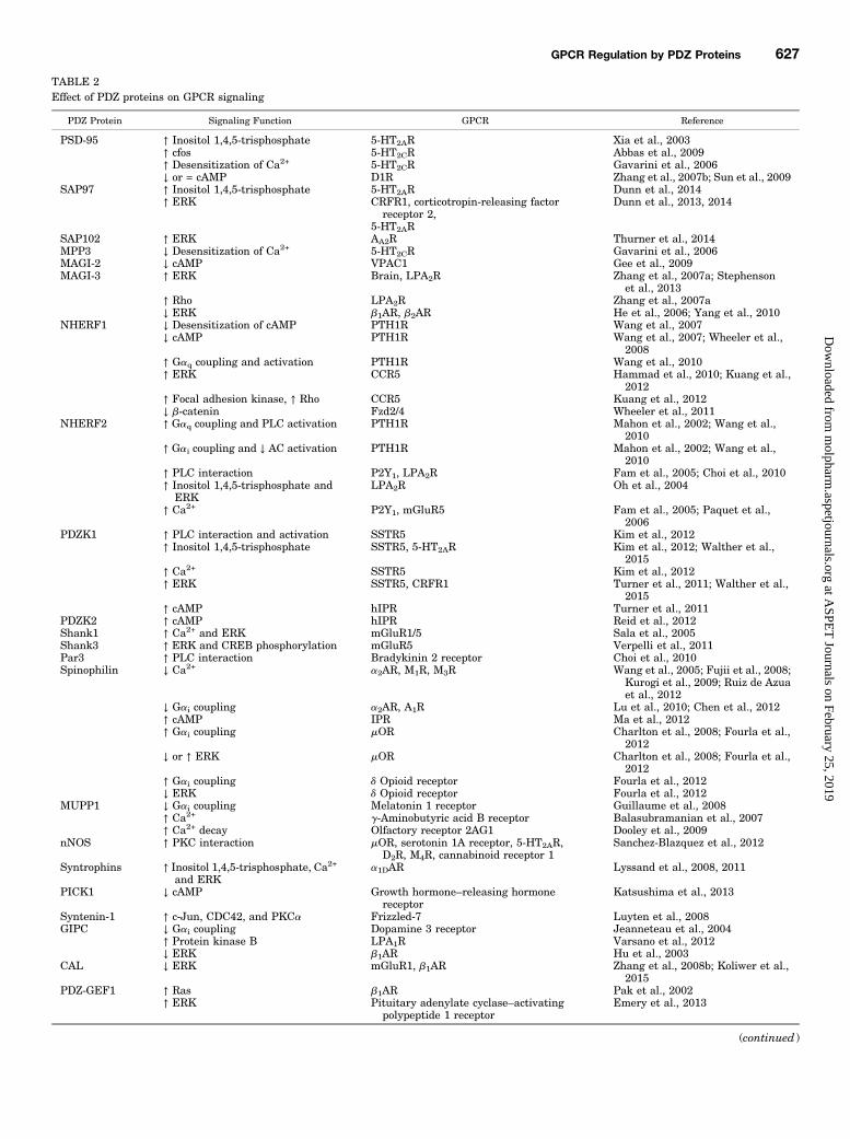
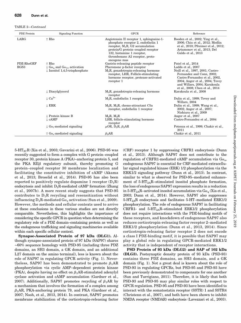
TABLE 2Effect of PDZ proteins on GPCR signaling
PDZ Protein Signaling Function GPCR Reference
PSD-95 ↑ Inositol 1,4,5-trisphosphate 5-HT2AR Xia et al., 2003↑ cfos 5-HT2CR Abbas et al., 2009↑ Desensitization of Ca2+ 5-HT2CR Gavarini et al., 2006↓ or = cAMP D1R Zhang et al., 2007b; Sun et al., 2009
SAP97 ↑ Inositol 1,4,5-trisphosphate 5-HT2AR Dunn et al., 2014↑ ERK CRFR1, corticotropin-releasing factor
receptor 2,5-HT2AR
Dunn et al., 2013, 2014
SAP102 ↑ ERK AA2R Thurner et al., 2014MPP3 ↓ Desensitization of Ca2+ 5-HT2CR Gavarini et al., 2006MAGI-2 ↓ cAMP VPAC1 Gee et al., 2009MAGI-3 ↑ ERK Brain, LPA2R Zhang et al., 2007a; Stephenson
et al., 2013↑ Rho LPA2R Zhang et al., 2007a↓ ERK b1AR, b2AR He et al., 2006; Yang et al., 2010
NHERF1 ↓ Desensitization of cAMP PTH1R Wang et al., 2007↓ cAMP PTH1R Wang et al., 2007; Wheeler et al.,
2008↑ Gaq coupling and activation PTH1R Wang et al., 2010↑ ERK CCR5 Hammad et al., 2010; Kuang et al.,
2012↑ Focal adhesion kinase, ↑ Rho CCR5 Kuang et al., 2012↓ b-catenin Fzd2/4 Wheeler et al., 2011
NHERF2 ↑ Gaq coupling and PLC activation PTH1R Mahon et al., 2002; Wang et al.,2010
↑ Gai coupling and ↓ AC activation PTH1R Mahon et al., 2002; Wang et al.,2010
↑ PLC interaction P2Y1, LPA2R Fam et al., 2005; Choi et al., 2010↑ Inositol 1,4,5-trisphosphate and
ERKLPA2R Oh et al., 2004
↑ Ca2+ P2Y1, mGluR5 Fam et al., 2005; Paquet et al.,2006
PDZK1 ↑ PLC interaction and activation SSTR5 Kim et al., 2012↑ Inositol 1,4,5-trisphosphate SSTR5, 5-HT2AR Kim et al., 2012; Walther et al.,
2015↑ Ca2+ SSTR5 Kim et al., 2012↑ ERK SSTR5, CRFR1 Turner et al., 2011; Walther et al.,
2015↑ cAMP hIPR Turner et al., 2011
PDZK2 ↑ cAMP hIPR Reid et al., 2012Shank1 ↑ Ca2+ and ERK mGluR1/5 Sala et al., 2005Shank3 ↑ ERK and CREB phosphorylation mGluR5 Verpelli et al., 2011Par3 ↑ PLC interaction Bradykinin 2 receptor Choi et al., 2010Spinophilin ↓ Ca2+ a2AR, M1R, M3R Wang et al., 2005; Fujii et al., 2008;
Kurogi et al., 2009; Ruiz de Azuaet al., 2012
↓ Gai coupling a2AR, A1R Lu et al., 2010; Chen et al., 2012↑ cAMP IPR Ma et al., 2012↑ Gai coupling mOR Charlton et al., 2008; Fourla et al.,
2012↓ or ↑ ERK mOR Charlton et al., 2008; Fourla et al.,
2012↑ Gai coupling d Opioid receptor Fourla et al., 2012↓ ERK d Opioid receptor Fourla et al., 2012
MUPP1 ↓ Gai coupling Melatonin 1 receptor Guillaume et al., 2008↑ Ca2+ g-Aminobutyric acid B receptor Balasubramanian et al., 2007↑ Ca2+ decay Olfactory receptor 2AG1 Dooley et al., 2009
nNOS ↑ PKC interaction mOR, serotonin 1A receptor, 5-HT2AR,D2R, M4R, cannabinoid receptor 1
Sanchez-Blazquez et al., 2012
Syntrophins ↑ Inositol 1,4,5-trisphosphate, Ca2+
and ERKa1DAR Lyssand et al., 2008, 2011
PICK1 ↓ cAMP Growth hormone–releasing hormonereceptor
Katsushima et al., 2013
Syntenin-1 ↑ c-Jun, CDC42, and PKCa Frizzled-7 Luyten et al., 2008GIPC ↓ Gai coupling Dopamine 3 receptor Jeanneteau et al., 2004
↑ Protein kinase B LPA1R Varsano et al., 2012↓ ERK b1AR Hu et al., 2003
CAL ↓ ERK mGluR1, b1AR Zhang et al., 2008b; Koliwer et al.,2015
PDZ-GEF1 ↑ Ras b1AR Pak et al., 2002↑ ERK Pituitary adenylate cyclase–activating
polypeptide 1 receptorEmery et al., 2013
(continued )
GPCR Regulation by PDZ Proteins 627
at ASPE
T Journals on February 25, 2019
molpharm
.aspetjournals.orgD
ownloaded from
5-HT2CR (Xia et al., 2003; Gavarini et al., 2006). PSD-95 wasrecently suggested to form a complex with G protein–coupledreceptor 30, protein kinase A (PKA)–anchoring protein 5, andthe PKA RIIb regulatory subunit, thereby promoting Gprotein–coupled receptor 30 membrane localization andfacilitating the constitutive inhibition of cAMP (Akamaet al., 2013; Broselid et al., 2014). PSD-95 has also beenreported to positively regulate dopamine 1 receptor (D1R)endocytosis and inhibit D1R-mediated cAMP formation (Zhanget al., 2007b). A more recent study suggests that PSD-95contributes to D1R recycling and resensitization withoutinfluencing D1R-mediated Gas activation (Sun et al., 2009).However, the methods and cellular contexts used to arriveat these conclusions in these various studies are not directlycomparable. Nevertheless, this highlights the importance ofconsidering the specific GPCR in questionwhen determining theregulatory role of a PDZ domain–containing protein as well asthe endogenous trafficking and signaling machineries availablewithin each specific cellular context.Synapse-Associated Protein of 97 kDa (DLG1). Al-
though synapse-associated protein of 97 kDa (SAP97) shares∼60% sequence homology with PSD-95 (including three PDZdomains, an SH3 domain, a GK domain, and an additionalL27 domain on the amino terminal), less is known about therole of SAP97 in regulating GPCR activity (Fig. 1). Never-theless, SAP97 has been demonstrated to promote b1ARphosphorylation via cyclic AMP–dependent protein kinase(PKA), despite having no effect on b1AR-stimulated adenylylcyclase activation and cAMP accumulation (Gardner et al.,2007). Additionally, SAP97 promotes recycling of b1AR bya mechanism that involves the formation of a complex amongb1AR, PKA-anchoring protein 79, and PKA (Gardner et al.,2007; Nooh, et al., 2013, 2014). In contrast, SAP97 promotesmembrane stabilization of the corticotropin-releasing factor
(CRF) receptor 1 by suppressing CRFR1 endocytosis (Dunnet al., 2013). Although SAP97 does not contribute to theregulation of CRFR1-mediated cAMP accumulation via Gas,endogenous SAP97 is essential for CRF-mediated extracellu-lar signal regulated kinase (ERK) 1/2 phosphorylation via theERK1/2 signaling pathway (Dunn et al., 2013). In contrast,similar to what is observed for PSD-95–mediated enhance-ment of 5-HT2AR–stimulated inositol phosphate formation,the loss of endogenous SAP97 expression results in a reductionin 5-HT2AR–activated inositol accumulation via Gaq (Xia et al.,2003; Dunn et al., 2014). However, SAP97 also suppresses5-HT2AR endocytosis and facilitates 5-HT–mediated ERK1/2phosphorylation. The role of endogenous SAP97 in facilitatingCRFR1- and 5-HT2AR–stimulated ERK1/2 phosphorylationdoes not require interactions with the PDZ-binding motifs ofthese receptors, and knockdown of endogenous SAP97 alsoreduces corticotropin-releasing factor receptor 2–mediatedERK1/2 phosphorylation (Dunn et al., 2013, 2014). Sincecorticotropin-releasing factor receptor 2 does not encodea class I PDZ-binding motif, it is possible that SAP97 mayplay a global role in regulating GPCR-mediated ERK1/2activity that is independent of receptor interactions.PSD Protein of 93 kDa (DLG2) and SAP of 102 kDa
(DLG3). Postsynaptic density protein of 93 kDa (PSD-93)contains three PDZ domains, an SH3 domain, and a GKdomain (Fig. 1). Not a great deal is known about the role ofPSD-93 in regulating GPCRs, but PSD-95 and PSD-93 havebeen previously demonstrated to compensate for one another(Sun and Turrigiano, 2011). Therefore, it is likely that bothPSD-93 and PSD-95 may play similar roles with respect toGPCR regulation. PSD-95 and PSD-93 have been identified tointeract with the somatostatin receptor (SSTR) 1 and SSTR4(Christenn et al., 2007), and both have been shown to inhibitNMDA receptor (NMDAR) endocytosis (Lavezzari et al., 2003).
TABLE 2—Continued
PDZ Protein Signaling Function GPCR Reference
LARG ↑ Rho Angiotensin II receptor 1, sphingosine-1-phosphate receptor 2, endothelin 1receptor, M1R, G2 accumulationprotein/G protein–coupled receptor132, histamine 1 receptor,thromboxane A2 receptor, proto-oncogene mas
Booden et al., 2002; Ying et al.,2006; Chiu et al., 2012; Medlinet al., 2010; Pfreimer et al., 2012;Artamonov et al., 2013; DelGaldo et al., 2013
PDZ-RhoGEF ↑ Rho Gastrin-releasing peptide receptor Patel et al., 2014RGS3 ↓ Gaq and Ga11 activation Pheromone p-factor receptor Ladds et al., 2007
↓ Inositol 1,4,5-trisphosphate M3R, gonadotropin-releasing hormonereceptor, LHR, Follicle-stimulatinghormone receptor, protease-activatedreceptor 1
Neill et al., 1997, 2001; Castro-Fernandez and Conn, 2002;Castro-Fernandez et al., 2002,2004; Anger et al., 2004; Toveyand Willars, 2004; Karakoulaet al., 2008; Chen et al., 2014
↓ Diacylglycerol M3R, gonadotropin-releasing hormonereceptor
Karakoula et al., 2008
↓ Ca2+ M3R, endothelin 1 receptor Dulin et al., 1999; Tovey andWillars, 2004
↓ ERK M2R, M3R, chemo-attractant C5areceptor, endothelin 1 receptor
Dulin et al., 1999; Wang et al.,2002; Anger et al., 2007;Nishiura et al., 2009
↓ Protein kinase B M2R, M3R Anger et al., 2007↓ cAMP LHR, follicle-stimulating hormone
receptorCastro-Fernandez et al., 2004
↓ Gai-mediated signaling mOR, D2R, b2AR Potenza et al., 1999; Chakir et al.,2011
↑ Gas-mediated signaling b2AR Chakir et al., 2011
628 Dunn et al.
at ASPE
T Journals on February 25, 2019
molpharm
.aspetjournals.orgD
ownloaded from
Future studies are needed to examine the role of PSD-93 inthe regulation of GPCR trafficking to determine whether itsfunction overlaps with both PSD-95 and SAP97. Synapse-associated protein of 102 kDA (SAP102) contains threePDZ domains, an SH3 domain, and a GK domain (Fig. 1).SAP102 has been demonstrated to regulate adenosine A2A
receptor mobility and promote A2A receptor–mediated ERKsignaling (Thurner et al., 2014). SAP102 has additionallybeen identified to regulate the trafficking of AMPA and NMDAreceptors. Thus, it is of interest in the future to determinewhether SAP102 plays a role similar to that of other membrane-associated guanylate kinase (MAGUK) proteins in the regula-tion of GPCR activity.Discs, Large Homolog 5. Discs, large homolog 5 (DLG5)
differs from the common topology of the PSD-95 subfamily ofMAGUKs, with the inclusion of an amino-terminal caspaseactivation and recruitment domain, which is similar to thecaspase activation and recruitment domain and membrane-associated guanylate-like kinase domain-containing proteins,and a fourth PDZ domain (de Mendoza et al., 2010) (Fig. 1).CARMA3 has been implicated in facilitating GPCR-inducedactivation of NFkB via lysophosphatidic acid, endothelin-1,and angiotensin II (Scudiero et al., 2014). Although theredoes not appear to be any examples of DLG5 in the directregulation of GPCRs, DLG5 has been implicated in regulatingsynaptogenesis by enhancing the membrane localization ofthe transmembrane protein N-cadherin (Wang et al., 2014).DLG5 has also been demonstrated to scaffold atypical proteinkinase C (PKC) isoforms, and this provides a mechanism bywhich DLG5 contributes to the regulation of GPCR-mediatedsignaling (Nechiporuk et al., 2013).
Other GPCR-Interacting PDZ Domain–Containing MAGUK Proteins
Membrane Palmitoylated Proteins and Calmodulin-Activated Serine/Threonine Kinase (PALS3, LIN-2).Membrane palmitoylated proteins (MPPs) (MPP1/p55, MPP2,MPP3, MPP4, MPP5/PALS1, MPP6/PALS2, and MPP7) areunified by the inclusion of a PDZ domain, an SH3 domain,and a GK domain (Fig. 1). Additionally, all but MPP1 havetwo amino-terminal L27 domains, with MPP5 also includingan amino-terminal coiled-coil domain. MPP1 and MPP2 andMPP5–MPP7 also include a HOOK domain between theirSH3 and GK domains. Although MPP proteins are a rela-tively abundant group of MAGUK proteins, very little isknown about their regulation of GPCR function. MPP3 hasbeen demonstrated to promote the membrane stability of5-HT2CR and prevent receptor desensitization (Gavarini et al.,2006). MPP1 has additionally been implicated in membraneorganization, raft formation, and receptor tyrosine kinase–mediated ERK signaling (Łach et al., 2012; Biernatowskaet al., 2013). Thus, it is plausible that MPPs may generallypromote the membrane organization of integral proteins,including GPCRs.Ca21/calcium/calmodulin-dependent serine protein kinase
(CASK) is very similar in topology to the MPPs, with proteindomains that include a catalytically active Ca21/calmodulin-dependent kinase domain at the amino-terminus followed bytwo L27 domains, a PDZ domain, an SH3 domain, and a GKdomain (te Velthuis et al., 2007; Mukherjee et al., 2008) (Fig. 1).CASK forms a tripartite complex with the PDZ domain
containing Mint1 and Veli proteins, but the role of Mint1 andVeli proteins in the regulation of GPCRs remains undeter-mined (Butz et al., 1998). Like MPP3, CASK has been shownto interact with 5-HT2CR (Bécamel et al., 2002, 2004; Gavariniet al., 2006). Although the functional consequence of thisinteraction on 5-HT2CR trafficking and signaling remains tobe tested, CASK has been implicated in regulating the traf-ficking of NMDAR and AMPA receptor (AMPAR), partly via itsregulation of SAP97 conformation and receptor interactions(Jeyifous et al., 2009; Lin et al., 2013). Interestingly, CASK hasbeen demonstrated to interact with PKA, PKC«, and theregulator of G protein signaling (RGS) 4, which may suggesta role for CASK in regulating GPCR-mediated signaling (Hongand Hsueh, 2006).
Membrane-Associated Guanylate Kinase withInverted Orientation PDZ Protein Family
Membrane-associated guanylate kinase with inverted ori-entation proteins (MAGIs) include three proteins with anamino terminal PDZ domain followed by a GK domain, twotryptophan-tryptophan (WW) domains, and five more PDZdomains (Fig. 1). MAGI proteins differ from other MAGUKproteins in the exclusion of an SH3 domain (Dobrosotskayaet al., 1997). MAGI-1 colocalizes with the brain angiogenesisinhibitor 1 receptor (BAI-1R) at the cell membrane via aninteraction with the receptor carboxyl-terminal tail, andMAGI-3 interacts with BAI-1R to promote ERK phosphory-lation (Shiratsuchi et al., 1998; Stephenson et al., 2013).MAGI-3 promotes ERK and RhoA signaling, which ismediated by lysophosphatidic acid receptor 2 (LPA2R), butantagonizes ERK1/2 activation in response to the activation ofeither b1AR or b2AR (He et al., 2006; Zhang et al., 2007a;Yang et al., 2010). MAGI-2 interacts with b1AR via its firstPDZ domain and functions to promote b1AR endocytosiswithout affecting b1AR-mediated cAMP signaling (Xu et al.,2001). In contrast, MAGI-2 interacts with the vasoactiveintestinal peptide receptor 1 (VPAC1) and functions to bothinhibit VPAC1 endocytosis and suppress VPAC1-mediatedcAMP signaling (Gee et al., 2009). MAGI-2 also promotes thecell surface expression of metabotropic glutamate receptor(mGluR) 1a via its association with the PDZ domain–containingprotein tamalin (Sugi et al., 2007). Thus, similar towhat has beenreported for PSD-95 family PDZ proteins, the MAGI family ofPDZ proteins contributes to the regulation of the endocytosis andcell signaling of a number of GPCRs, but the functional effects ofthese protein interactions have differential effects, depending onthe GPCR studied.
Na1/H1 Exchanger Regulatory Factor Family ofPDZ Proteins
Na1/H1 Exchanger Regulatory Factor 1. Na1/H1
exchanger regulatory factor (NHERF) 1 (ezrin/radixin/moesin–binding protein 50) is a relatively small PDZ domain–containingprotein characterized by two PDZ domains and a carboxyl-terminal ezrin-binding domain (Fig. 2). NHERF1 representsone of the earliest PDZ proteins to be shown to interact witha GPCR (Hall et al., 1998). NHERF1 regulates Na1/H1 ex-change via its interaction with b2AR without altering cAMPsignaling, and has since been demonstrated to regulate a
GPCR Regulation by PDZ Proteins 629
at ASPE
T Journals on February 25, 2019
molpharm
.aspetjournals.orgD
ownloaded from
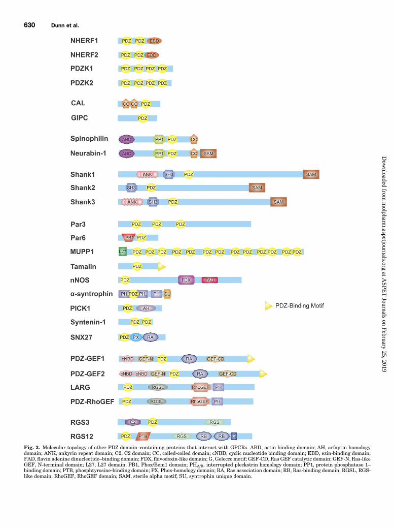
Fig. 2. Molecular topology of other PDZ domain–containing proteins that interact with GPCRs. ABD, actin binding domain; AH, arfaptin homologydomain; ANK, ankyrin repeat domain; C2, C2 domain; CC, coiled-coiled domain; cNBD, cyclic nucleotide binding domain; EBD, ezin-binding domain;FAD, flavin adenine dinucleotide–binding domain; FDX, flavodoxin-like domain; G, Golocco motif; GEF-CD, Ras GEF catalytic domain; GEF-N, Ras-likeGEF, N-terminal domain; L27, L27 domain; PB1, Phox/Bem1 domain; PHA/B, interrupted pleckstrin homology domain; PP1, protein phosphatase 1–binding domain; PTB, phosphtyrosine-binding domain; PX, Phox-homology domain; RA, Ras association domain; RB, Ras-binding domain; RGSL, RGS-like domain; RhoGEF, RhoGEF domain; SAM, sterile alpha motif; SU, syntrophin unique domain.
630 Dunn et al.
at ASPE
T Journals on February 25, 2019
molpharm
.aspetjournals.orgD
ownloaded from
number of GPCRs. NHERF1 regulates the recycling of b2AR,and its binding to the receptor is disrupted byG protein–coupledreceptor kinase phosphorylation of b2AR at serine residue 411(Cao et al., 1999). However, NHERF1 is reported to inhibitrecycling of the parathyroid 1 receptor (PTH1R) (Wang et al.,2007). NHERF1 also inhibits PTH1R desensitization andendocytosis, a function that appears to involve NHERF1-dependent inhibition of b-arrestin2 recruitment to PTH1R(Wang et al., 2007, 2009). NHERF1 expression also enhancesPTH1R-mediated cAMP signaling and couples PTHR1 to theactivation of Gaq (Wang et al., 2007, 2010; Wheeler et al., 2008).NHERF1 expression enhances cell surface expression of thek opioid receptor, inhibiting downregulation and promotingreceptor recycling (Li et al., 2002). In contrast, NHERF1increases thromboxane receptor b cell surface expression byblocking the internalization of the receptor (Rochdi and Parent,2003). An additional mechanism by which NHERF1 mayincrease GPCR membrane targeting is via its competition withthe cystic fibrosis transmembrane conductance regulator–associated ligand (CAL) to antagonize CAL-mediated retentionof GPCRs in the Golgi (Bauch et al., 2014).In contrast to the role of NHERF1 in antagonizing the
endocytosis of PTHR1 and thromboxane receptor b, NHERF1is reported to facilitate the endocytosis of a number of GPCRs.NHERF1 enhances chemokine (C-C motif) receptor 5 (CCR5)endocytosis and b-arrestin1 recruitment, thereby promot-ing the activation of ERK, Rho, and focal adhesion kinasesignaling pathways, as well as potentially contributes toCCR5-mediated HIV-1 entry (Hammad et al., 2010; Kuanget al., 2012). NHERF1 overexpression also rescues theendocytosis of an internalization-defective platelet-activatingfactor receptor and antagonizes platelet-activating factorreceptor–mediated inositol phosphate formation (Dupré et al.,2012). Agonist activation of the purinergic P2Y12 receptorresults in the b-arrestin–dependent recruitment of NHERF1 tothe receptor and promotes the formation of a P2Y12 receptor/NHERF1 complex that does not require PDZ-binding motifinteractions (Nisar et al., 2012). NHERF1 also regulatedfrizzled family receptor activity (Wheeler et al., 2011). Thus,NHERF1 appears to play an integral, but complex, role inregulating the endocytosis and recycling of a variety of differentGPCRs.Na1/H1 Exchanger Regulatory Factor 2. The topology
of Na1/H1 exchanger regulatory factor 2 (NHERF2) is quitesimilar to NHERF1 as it shares a 44% sequence homologywith NHERF1 and contains two PDZ domains and a carboxyl-terminal ezrin-binding domain (Ardura and Friedman, 2011)(Fig. 2). Similar to NHERF1, NHERF2 contributes to theregulation of PTH1R (Mahon et al., 2002; Wang et al.,2010). NHERF2 functions to antagonize PTHR1 couplingto Gas-coupling, while concomitantly promoting the couplingof PTH1R to both the activation of Gaq and Gai (Mahon et al.,2002; Wang et al., 2010). NHERF2 also interacts directly withphospholipase C (PLC) b to enhance P2Y1 receptor–mediatedCa21 signaling (Fam et al., 2005). Similarly, NHERF2 interactswith PLCb3 and the LPA2R, allowing for the formation of aprotein complex that directly links the receptor to PLCb3-mediated inositol phosphate signaling (Oh et al., 2004; Choiet al., 2010). NHERF2 andmGluR5 show overlapping expressionin a mouse brain at postsynaptic neuronal sites and astrocyticprocesses, and NHERF2 prolongs the mGluR5-mediated Ca21
response (Paquet et al., 2006).
PDZ Domain–Containing Kidney Protein 1 (Na1/H1
Exchanger Regulatory Factor 3) and 2 (Na1/H1 Ex-changer Regulatory Factor 4). PDZ domain–containingkidney protein 1 (PDZK1), formerly known as Na1/H1
exchanger regulatory factor 3, differs from NHERF1 andNHERF2 in structural topology by having four PDZ domainsand no carboxyl-terminal ezrin-binding domain (Fig. 2).Nevertheless, PDZK1has been implicated in regulating a subsetof GPCRs. PDZK1 promotes the formation of a complex betweenSSTRs and PLCb3, similar to what is observed for LPA2R (Ohet al., 2004; Choi et al., 2010), thereby facilitating somatostatin-stimulated PLC activation, Ca21 mobilization, and ERK1/2phosphorylation (Kim et al., 2012). PDZK1 also functions toenhance human prostacyclin receptor (hIPR) cell surfacelocalization and cAMP signaling and contributes to endothelialcell migration and angiogenesis (Turner et al., 2011). PDZK1inhibits 5-HT2AR endocytosis, and siRNA knockdown of PDZK1results in reduced 5-HT2AR–mediated inositol phosphateaccumulation, but is not involved in 5-HT2AR–stimulatedERK1/2 phosphorylation (Walther et al., 2015). However,PDZK1 interactions with 5-HT2AR do not appear to be requiredfor its regulation of 5-HT2AR activity. In contrast, althoughPDZK1 does not regulate CRFR1-mediated cAMP accumula-tion, unlike what is observed for 5-HT2AR, PDZK1 facilitatesCRFR1-mediated ERK1/2 phosphorylation. Similar to PDZK1,PDZ domain–containing kidney protein 2 (PDZK2) also has fourPDZ domains and has been shown to regulate hIPR (Reid et al.,2012). Agonist activation of hIPR increases PDZK2 associationand results in PKA- and PKC-mediated phosphorylation ofPDZK2 (Reid et al., 2012). Like PDZK1, PDZK2 also enhanceshIPR cell surface expression and cAMP accumulation (Reidet al., 2012). Taken together, PDZK1 and PDZK2 appear to beimportant for regulating the trafficking of an increasing subsetof GPCRs and may be biased toward increased Gaq signaling,similar to what is observed for both NHERF1 and NHERF2.
PDZ Proteins that Regulate Golgi TraffickingGa-Binding Protein Interacting Protein Carboxyl-
Terminus (TIP-2, Synectin). RGS Ga-binding protein–interacting protein carboxyl-terminus (GIPC) is a PDZ domain–containing protein, with one PDZ domain that is implicated inthe sorting of nascent proteins from the Golgi network (Liu et al.,2001) (Fig. 2). With regards to GPCRs, GIPC has been shown totarget D2R to endosomes and the Golgi apparatus (Jeanneteauet al., 2004). Furthermore, GIPC expression suppresses dopa-mine 3 receptor Gai-coupling and prevents dopamine 3 receptordegradation (Jeanneteau et al., 2004). GIPC also plays a role inregulating both the human luteinizing hormone receptor andLPA1R trafficking (Hirakawa et al., 2003; Varsano et al., 2012).The interaction of GIPC with LPA1R is essential for LPA1Rtrafficking from APPL-positive signaling endosomes to earlyendosome antigen 1–positive early endosomes (Varsano et al.,2012). Additionally, GIPC links LPA1R to the protein kinase Bsignaling pathway, cell proliferation, and cell motility (Varsanoet al., 2012). GIPC also contributes to the suppression of b1AR-mediated ERK activation, but does affect b1AR-stimulated cAMPaccumulation (Hu et al., 2003).CAL (Golgi-Associated Coiled-Coil and PDZ Domain–
Containing Protein, PIST). CAL is also named Golgi-associated coiled-coil and PDZ domain–containing protein dueto its common subcellular localization within the trans-Golgi
GPCR Regulation by PDZ Proteins 631
at ASPE
T Journals on February 25, 2019
molpharm
.aspetjournals.orgD
ownloaded from
network and a structural topology consisting of two coiled-coildomains and one PDZ domain (Fig. 2). CAL is selectivelylocalized to the trans-Golgi network in neurons as well as othercell types and interacts with Rab6a, a small GTPase implicatedin Golgi-related trafficking pathways (Bergbrede et al., 2009;Valente et al., 2010; Chen et al., 2012). CAL reduces plasmamembrane expression and recycling of b1AR, and interfereswith both b1AR-mediated ERK signaling and postendocytoticreceptor degradation via the lysosome (He et al., 2004; Koliweret al., 2015). CAL overexpression retains SSTR5 in the Golgiapparatus, thereby reducing SSTR5 cell surface expression(Wente et al., 2005; Bauch et al., 2014). Additionally, CALcolocalizes with mGluR1a following agonist activation, and itsoverexpression decreases mGluR1a-stimulated ERK signaling(Zhang et al., 2008b). CAL is suggested to regulate mGluR5afunction by increasing the expression of the receptor by amechanism that involves the inhibition of mGluR5a ubiquitina-tion (Cheng et al., 2010). Taken together, it appears CAL couldhave a regulatory role over the subcellular localization of asubset of GPCRs, perhaps by contributing to the post-translationalmodification of nascent and mature proteins that ultimatelyinfluence the sorting and trafficking fate.
Additional GPCR-Interacting PDZ ProteinsSpinophilin (Neurabin-2) and Neurabin-1. Both
spinophilin/neurabin-2 and neurabin-1 contain an amino-terminal actin-binding domain, a protein phosphatase 1g-binding domain, a single PDZ domain, and a coiled-coil domain,with neurabin-1 also containing a carboxyl-terminal sterilealphamotif domain (Kelker et al., 2007) (Fig. 2). Spinophilin hasbeen shown to interact with both D2R and a2-adrenergicreceptor (a2AR) (Smith et al., 1999; Richman et al., 2001; Wangand Limbird, 2002; Brady et al., 2003; Wang et al., 2004).However, these interactions appear to be mediated by the thirdintracellular loop domains of these GPCRs, as opposed tointeractions with PDZ-binding motifs. Spinophilin functions topromote membrane localization and inhibit the endocytosis anddesensitization of a2ARs by competing for b-arrestin2 binding(Wang et al., 2004b). The interaction between spinophilin anda2AR is prevented by PKA-mediated phosphorylation ofspinophilin, which results in increased agonist-stimulateda2AAR endocytosis (Xu et al., 2008). b2AR activation alsostimulates PKA-mediated spinophilin phosphorylation to in-crease a2AAR endocytosis (Cottingham et al., 2013). Conversely,spinophilin appears to promote RGS2-mediated inhibition ofa2AR-evoked Ca21 signaling and RGS2-mediated modulation ofa1-adrenergic receptor–NMDAR crosstalk (Wang et al., 2005;Liu et al., 2006). In spinophilin knockout mice, the a2A-adrenergic receptor (a2AAR) exhibits increased G proteincoupling and sensitized responses to a2AAR agonists (Lu et al.,2010; Cottingham et al., 2012). Both spinophilin and neurabin-1are implicated in the D1R-dependent regulation of AMPAR aswell as long-term depression and potentiation, respectively(Allen et al., 2006). Spinophilin promotes prostacyclin receptorsignaling via Gas and influences both M1 muscarinic acetylcho-line receptor and M3 muscarinic acetylcholine receptor(m3AChR) activity by enhancing RGS8-mediated inhibition ofthe Gaq-coupled signaling (Fujii et al., 2008; Kurogi et al., 2009;Ma et al., 2012). Similarly, spinophilin recruits RGS4 tom3AChR, and like RGS8, RGS4 antagonizes m3AChR inositolphosphate signaling (Ruiz de Azua et al., 2012). Spinophilin also
promotes m-opioid receptor (mOR)–mediated signaling via Gai,but inhibits mOR-mediated ERK activation, while facilitatingmOR endocytosis (Charlton et al., 2008; Fourla et al., 2012).The interaction between spinophilin and opioid receptors
appears to occur via the opioid receptor third intracellular loopand a conserved region of the carboxyl-termini, which isproximal to the seventh transmembrane domain (Fourlaet al., 2012). Interestingly, this region appears to correlate witha small helical region identified in many class A rhodopsin-likeGPCRs as helix 8 (Huynh et al., 2009). This domain is suggestedto run perpendicular to the other seven helical transmembranedomains and is initiated by an N-P-x-x-Y motif (Huynh et al.,2009). In examining the amino acid sequences of class Arhodopsin-like GPCRswith this motif, a possible internal class IPDZ-bindingmotif, as characterized by an S/T-x-wmotif, may bepresent near this region (Trejo, 2005). Furthermore, homolo-gous regions are found within a2ARs and D2R, which alsointeract with spinophilin via the third intracellular loop domain.Notably, a recent study has identified helix 8 of D2R to associatewith the PDZ domain of GIPC (Sensoy and Weinstein, 2015).Future studies could look to investigate whether secondaryinteractions with spinophilin may occur within the a2ARs andD2R carboxyl-terminal/helix 8, and whether these interactionsrequire spinophilin’s PDZ domain.SH3 and Multiple Ankyrin Repeat Domain Proteins.
SH3 and multiple ankyrin repeat domain (Shank) proteins areunified by the inclusion of multiple ankyrin repeat domains, anSH3 domain, a PDZ domain, and a sterile alpha motif domain;however, Shank2 lacks the ankyrin repeats (Fig. 2). Shank1Benhances mGluR1/5-mediated ERK1/2 and Ca21-dependentsignaling, and Shank3 is important for mGluR5-mediatedERK1/2 and cAMP response element-binding protein phos-phorylation and subsequent mGluR5-mediated long-term de-pression (Sala et al., 2005; Verpelli et al., 2011). Furthermore,Shank3 prevents mGluR1-mediated inhibition of NMDAR viaits association with Homer1A (Guo et al., 2004; Bertaso et al.,2010). Similarly, Shank1/3modulates muscarinic acetylcholinereceptor 1– and D2R-mediated inhibition of L-type Ca21
channels via Homer proteins (Olson et al., 2005). With regardsto GPCR trafficking, Shank influences the clustering andsubcellular localization of mGluR5 and calcium-independenta-latrotoxin receptor CIRL/latrophilin 1 (Tu et al., 1999;Tobaben et al., 2000). Interestingly, a Shank/Homer1Acomplex can suppress NMDAR and AMPAR clustering andsurface expression (Sala et al., 2003). Shank1 directly interactswith dynamin-2, which may provide insight into a mechanismof action in preventing GPCR-mediated crosstalk mechanismsand receptor surface expression (Okamoto et al., 2001). Futurestudies could look to investigate the role of Shank proteins inregulating GPCR trafficking and the crosstalk between GPCRsand ion channels.Partitioning Defective Proteins 3 and 6. Partitioning
defective (Par) proteins have been implicated in cellularpolarization, and Par3 and Par6 are PDZ domain–containingmembers of the Par family (Fig. 2) (Macara, 2004). Par3 isimplicated as having a role in synaptogenesis as a conse-quence of its interaction with BAI-1R (Duman et al., 2013).Additionally, Par3 has been shown to increase bradykininreceptor interactions with PLCb1 (Choi et al., 2010). In-terestingly, both Par3 and Par6 interact and catalyze theactivation of PLCb downstream of heterotrimeric G proteinsand form a complex with atypical PKCs (Joberty et al., 2000;
632 Dunn et al.
at ASPE
T Journals on February 25, 2019
molpharm
.aspetjournals.orgD
ownloaded from
Cai et al., 2005). Taken together, these observations suggestthat Par3 and Par6 may contribute to the regulation of GPCR-mediated Gaq signaling as well as feedback receptor de-sensitization by atypical PKCs.Multiple PDZ Protein 1. Multiple PDZ protein 1
(MUPP1) is one of the largest PDZ domain–containing proteinsand is comprised of an amino terminal L27 domain followed by13 PDZ domains (Fig. 2). The interaction of MUPP1 with themelatonin 1 receptor facilitates melatonin 1 receptor Gai-coupling, resulting in the inhibition of adenylyl cyclase activity(Guillaume et al., 2008). MUPP1 has also been shown topromote g-aminobutyric acid B receptor–mediated Ca21 signal-ing, although MUPP1 knockdown prolongs the decay of theodorant receptor olfactory receptor 2AG1–mediated Ca21 re-sponse (Balasubramanian et al., 2007; Dooley et al., 2009). Withregards to GPCR trafficking, MUPP1 increases the cell surfaceexpression of 5-HT2AR (Jones et al., 2009). Additionally,MUPP1promotes the targeting of SSTR3 to tight junctions, therebyinfluencing transepithelial permeability (Liew et al., 2009;Vockel et al., 2010). Given that MUPP1 influences NMDA-dependent AMPA trafficking and clustering, it is likely thatMUPP1 also regulates the trafficking of GPCRs that encodePDZ-binding motifs, thereby contributing to GPCR-dependentregulation of synaptic activity (Krapivinsky et al., 2004).Tamalin (General Receptor for Phosphoinositides-
Associated Scaffold Protein). Tamalin or general receptorfor phosphoinositides-associated scaffold protein encodesa PDZ domain, leucine zipper, and class I PDZ-binding motifon the distal carboxyl-terminal (Kitano et al., 2002, 2003)(Fig. 2). Tamalin promotes the plasma membrane localizationof mGluR1a as well as the neuritic targeting of mGluR5 inhippocampal neurons (Kitano et al., 2002). Tamalin alsointeracts with mGluR2, mGluR3, and the g-aminobutyric acidB2 receptor, but the functional consequence of these inter-actions remains to be determined (Kitano et al., 2002). In theabsence of mGluRs or other potential GPCR binding partners,tamalin displays an autoinhibitory confirmation that iscaused by the interaction between the tamalin PDZ domainand tamalin PDZ-binding motif (Sugi et al., 2007). UponmGluR1a binding to the tamalin PDZ domain, the tamalinPDZ-binding motif is free to associate with MAGI-2 to furtherenhance the membrane localization of mGluR1a (Sugi et al.,2007). PDZ–guanine nucleotide exchange factor (GEF) 1/2also contains PDZ-binding motifs, and future studies couldlook to determine whether they similarly exhibit autoregula-tion (Kuiperij et al., 2003, 2006; Ogawa et al., 2007).Neuronal Nitric Oxide Synthase. Neuronal nitric oxide
synthase (nNOS) contains an amino-terminal PDZ domain,a flavodoxin-like domain, and a flavin adenine dinucleotide–binding domain (Fig. 2). nNOS, in conjunction with RGS17, hasbeen demonstrated to complex withmultiple GPCRs, includingmOR, d opioid receptor, serotonin 1A receptor, 5-HT2AR, a2AR,D1R, D2R, M2 muscarinic acetylcholine receptor, M4 musca-rinic acetylcholine receptor, mGluR2, mGluR5, and cannabi-noid receptor 1 (Sánchez-Blázquez et al., 2012). Activation ofthese receptors leads to the nNOS/NO-dependent recruitmentof PKCg and Raf-1 to many of these GPCRs. nNOS alsofacilitates crosstalk between mOR and NMDAR (Rodríguez-Muñoz et al., 2008; Sánchez-Blázquez et al., 2010; Garzónet al., 2011). Interestingly, nNOS interacts with both PSD-95and PSD-93, and is targeted to the neuromuscular junction viaits interaction with PDZ protein a-syntrophin (Brenman et al.,
1996; Adams et al., 2010). Although this nNOS interaction withPSD-95 is suggested to regulateNMDARactivity (Christophersonet al., 1999), it is yet to be determined whether these PDZ/PDZprotein interactions regulate GPCR function.Syntrophins. a-syntrophin, b1-syntrophin, and b2-syntro-
phin all have an amino-terminal pleckstrin-homology (PH)domain interrupted by a PDZ domain, followed by another PHdomain and a syntrophin unique calmodulin-binding domain(Fig. 2) (Adams et al., 1995; Ahn et al., 1996; Chen et al., 2006).These syntrophins interact with a1DAR and collectively facili-tate the functional expression of the receptor at the membrane,promoting a1DAR-mediated phosphatidylinositol hydrolysis,ERK1/2 phosphorylation, and Ca21 mobilization (Chen et al.,2006; Lyssand et al., 2008, 2010, 2011). Neither g1-syntrophinnor g2-syntrophin comparably bind a1DAR, despite containingone PDZ domain and a PH domain, and their potential role inGPCR regulation remains uncertain (Chen et al., 2006).a-syntrophin can additionally scaffold the PDZ protein nNOSand notably binds Gbg subunits via its PDZ domain (Brenmanet al., 1996; Adams et al., 2010; Zhou et al., 2005).Protein Interacting with Protein Kinase C a 1. The
protein interacting with protein kinase C a 1 (PICK1) encodesone PDZ domain and an arfaptin homology domain/bin/amphiphysin/Rvs domain involved in cell membrane inter-actions (Katsushima et al., 2013) (Fig. 2). PICK1 promotes theintracellular clustering of the prolactin-releasing peptidereceptor, influences plasma membrane expression of thegrowth hormone–releasing hormone receptor, and antago-nizes growth hormone–releasing hormone receptor–mediatedcAMP signaling (Lin et al., 2001; Katsushima et al., 2013).PICK1 regulates PKC phosphorylation of mGluR7a, regulatesthe presynaptic clustering of mGluR7, and mediates stablemGluR7 cell surface expression (Boudin et al., 2000; Devet al., 2000; Suh et al., 2008). mGluR7a knock-in mice lackinga PDZ-binding motif exhibit deficits in hippocampal-dependentspatial memory and are highly susceptible to the convulsantdrugs, and the disruption of the mGluR7a-PICK1 complexinduces epilepsy-like seizures (Bertaso et al., 2008; Zhanget al., 2008a). Taken together, it appears PICK1 may beimportant for regulating the trafficking of a subset of GPCRsand may prove important in regulating GPCR-mediated signal-ing pathways. Notably, PICK1 can both homodimerize andheterodimerize with another PDZ domain–containing protein,syntenin-1 (Staudinger et al., 1997; Koroll et al., 2001).Syntenin-1. Syntenin-1 contains two PDZ domains (Fig. 2)
and has been found to self-associate as well as heterodimerizewith PICK1 and form a complex with mGluR7 (Koroll et al.,2001; Hirbec et al., 2002; Enz andCroci, 2003). Although PICK1regulates mGluR7 phosphorylation, clustering, and membraneexpression, it is not yet clear what role syntenin-1 may play inthis regulation (Boudin et al., 2000; Dev et al., 2000; Suh et al.,2008). Nonetheless, syntenin-1 has been demonstrated toenhance the membrane expression of G protein–coupledreceptor 37 (endothelin receptor type B–like) (Dunham et al.,2009). In regards to signaling, syntenin-1 interactswith frizzled-7and promotes c-Jun phosphorylation, CDC42 activation, andPKCa recruitment to the membrane (Luyten et al., 2008).Syntenin-1 can also heterodimerize with syntenin-2, althoughlittle is known about the role of syntenin-2 in GPCR regulation(Koroll et al., 2001).Sorting Nexin-27. Sorting nexin-27 (SNX27) differs from
other sorting nexins through the inclusion of an amino-terminal
GPCR Regulation by PDZ Proteins 633
at ASPE
T Journals on February 25, 2019
molpharm
.aspetjournals.orgD
ownloaded from
PDZ domain, followed by a Phox homology domain and a Ras-associating domain (Fig. 2). SNX27 interacts with bothserotonin 4A receptor and b2AR in early endosome antigen 1–positive early endosomes (Joubert et al., 2004; Lauffer et al.,2010). Moreover, SNX27 is involved in regulating the recyclingof b2AR, b1AR, and SSTR5, thereby preventing receptordegradation (Lauffer et al., 2010; Temkin et al., 2011;Nakagawa and Asahi, 2013; Bauch et al., 2014). The regulationof b2AR recycling by SNX27 is dependent upon Phox homologydomain–mediated associations with the endosomal membrane(Lauffer et al., 2010). Furthermore, SNX27 interacts with theendosomal WASH complex to target b2AR to the retromertubule for efficient recycling (Temkin et al., 2011). Takentogether, it appears SNX27 is capable of promoting theendosomal sorting and recycling of a subset of GPCRs, a rolethat may be generalizable to several other PDZ motif-encodingGPCRs.PDZ-GEFs (RAPGEFs, CNrasGEF, RA-GEF). PDZ
domain–containing GEFs (PDZ-GEF1 and PDZ-GEF2) sharean approximately 56% sequence homology and include oneor two cyclic nucleotide-binding domains, respectively, anN-terminal Ras GEF domain, a PDZ domain, a Ras-associatingdomain, and aRasGEF catalytic domainwithin theirmolecularstructure (Kuiperij et al., 2003, 2006) (Fig. 2). Similar to thePDZ domain–containing protein tamalin, PDZ-GEF1/2 havealso been reported to contain a class I PDZ-binding motif attheir carboxyl-termini, suggesting a capability for homo/hetero-oligomerization with PDZ domain–containing proteins orperhaps even autoregulatory capability via self-association(Kuiperij et al., 2003, 2006; Ogawa et al., 2007). Our currentunderstanding of PDZ-GEF2 regulation of GPCRs is poor, butPDZ-GEF1 couples b1AR to the activation of Ras (Pak et al.,2002). Furthermore, PDZ-GEF1 is essential for coupling thepituitary adenylate cyclase–activating polypeptide type I re-ceptor to the ERK1/2 signaling pathway and the subsequentactivation of neuritogenesis, with no effect on cAMP accumu-lation (Emery et al., 2013).RGS Proteins with PDZ Domains (PDZ Domain–
Containing RhoGEF, Leukemia-Associated RhoGEF,RGS3, and RGS12). PDZ domain–containing RhoGEF (PDZ-RhoGEF) and leukemia-associated RhoGEF (LARG) aremembers of the RGS homology domain–containing RhoGEFsubfamily and include an amino-terminal PDZ domain, anRGS-homology domain, a RhoGEF domain, and a PH domain(Fig. 2). LARG transduces Gaq/12/13 activation into Rhoactivation via GPCRs, such as the Mas receptor, G2 accumu-lation receptor, muscarinic acetylcholine receptor 1, angioten-sin II receptor 1, sphingosine-1 phosphate receptor 2,histamine H1 receptor, thromboxane A2 receptor, and endo-thelin 1 receptor (Booden et al., 2002; Ying et al., 2006; Medlinet al., 2010; Chiu et al., 2012; Pfreimer et al., 2012; Artamonovet al., 2013; Del Galdo et al., 2013). Similarly, PDZ-RhoGEF isproposed to contribute to gastrin-releasing peptide receptor–mediated activation of the Rho/ROCK pathway via Ga13 (Patelet al., 2014). Finally, both PDZ-RhoGEF and LARG have beenimplicated in sustaining Rho activation following thrombin andlysophosphatidic acid receptor activation (Chikumi et al., 2002;Wang et al., 2004a; Yamada et al., 2005). Interestingly, bothproteins appear capable of homo- and hetero-dimerization(Chikumi et al., 2004).RGS12 contains one PDZ domain, a phosphotyrosine-
binding domain, an RGS domain, two Ras-binding domains,
and a GoLoco motif (Fig. 2). The RGS12 PDZ domain binds tothe interleukin-8 receptor B PDZ-binding motif, but thefunctional consequence of this interaction is not well defined(Snow et al., 1998). Notably, RGS12 has been suggested tocouple D2R to inward rectifier potassium channels Kir3.1/3.2(Oxford and Webb, 2004). Regulator of G protein signaling 3(RGS3) contains a membrane-targeting C2 domain, one PDZdomain, and an RGS domain (Fig. 2). RGS3 has beenidentified to inhibit Gaq- and Gai-mediated signaling byacting as a GTPase-activating protein (Scheschonka et al.,2000). RGS3 antagonizes Gaq/11 signaling via pheromone Pfactor receptor and muscarinic acetylcholine receptor 3 activa-tion, and RGS3 promotes Ca21 oscillatory behavior duringsubmaximal muscarinic acetylcholine receptor 3 activation(Wang et al., 2002; Anger et al., 2004, 2007; Tovey and Willars,2004; Ladds et al., 2007; Karakoula et al., 2008). RGS3 alsoantagonizes follicle-stimulating hormone receptor– andluteinizing hormone receptor–mediated inositol phosphate andcAMP accumulation (Castro-Fernandez et al., 2004). Further-more, RGS3 has been demonstrated to suppress Gai-mediatedsignaling pathways via mOR, muscarinic acetylcholine receptor1, complement C5a receptor, and b2AR, and even promotea Gas bias for b2AR (Potenza et al., 1999; Anger et al., 2007;Nishiura et al., 2009; Chakir et al., 2011). In contrast, RGS3was shown to inhibit gonadotropin-releasing hormonereceptor–stimulated inositol phosphate signaling via Gaq, buthad no effect on cAMP signaling (Neill et al., 1997, 2001;Castro-Fernandez and Conn, 2002; Castro-Fernandez et al.,2002; Karakoula et al., 2008). Interestingly, RGS3 palmitoyla-tion is increased following gonadotropin-releasing hormonereceptor activation (Castro-Fernandez et al., 2002). Curiously,truncated RGS3 isoforms that have been reported to lack theamino-terminal andPDZdomain have also demonstrated a rolein influencing GPCR activity, including sphingosine-1-phosphate receptors 1–3, angiotensin II receptor 1, endothelin1 receptor, gonadotropin-releasing hormone receptor, serotonin1A receptor, and muscarinic acetylcholine receptor 2/3 (Drueyet al., 1996; Castro-Fernandez et al., 2003; Cho et al., 2003;Anger et al., 2004, 2007; Jaén and Doupnik, 2005). Distinguish-ing the role of LARG, PDZ-RhoGEF, RGS3, and RGS12 PDZdomain interactions, as opposed to RGS domain interactionswith heterotrimeric G proteins, in the regulation of GPCRsignaling remains a challenge.
Role of PDZ Proteins in GPCR-RegulatedPhysiology
PSD-95 Family of MAGUK PDZ Proteins. The PDZdomain–containing MAGUK proteins play an essential role inhuman neurophysiology and development. This is demon-strated in mouse knockout studies, where PSD-95 and PSD-93double-knockout mice exhibit severe deficiencies in AMPAcurrents, and SAP97 knockout mice show neonatal lethality(Caruana and Bernstein, 2001; Howard et al., 2010). Ofparticular interest is the observation that PSD-95 is essentialfor hallucinogenic and atypical antipsychotic actions of5-HT2AR and 5-HT2CR (Abbas et al., 2009). In addition tobeing involved in atypical antipsychotic actions (Abbaset al., 2009), PDZ protein interactions with GPCRs also appearto be important in regulating stress and anxiety responses(Magalhaes et al., 2010). Preactivation of the CRFR1 receptorsensitizes 5-HT2AR–stimulated inositol phosphate formation,
634 Dunn et al.
at ASPE
T Journals on February 25, 2019
molpharm
.aspetjournals.orgD
ownloaded from

which is dependent upon intact PDZ-binding motifs in bothreceptors, receptor endocytosis, and recycling (Magalhaes et al.,2010). Furthermore, the phenomenon can be blocked by a Tat-tagged fusion protein corresponding to the last 15 amino acidsof the CRFR1 tail. In addition, pretreatment of mice withsubthreshold doses of CRF into the prefrontal cortex sensitizesmouse anxiety responses to 2,5-dimethoxy-4-iodoamphetaminetreatment (Magalhaes et al., 2010). Thus, it is possible thatPDZ protein interactions may serve as a good pharmaceuticaltarget for the treatment of disease.SAP102 is important during early synaptic development,
and SAP97 appears to be important in SSTR1-mediatedgrowth cone dynamics, as evidenced by colocalization withinthe growth cone (Kim and Sheng, 2004; Elias et al., 2006; Caiet al., 2008). However, this role may not be limited to SAP97and may include additional PDZ domain–containing proteins(Cai et al., 2008). PSD-95 plays a functional role in synapticplasticity and contributes to GPCR-mediated regulation ofboth long-term potentiation and long-term depression (Xu,2011). Notably, SAP97 also modulates the ability to regulateAMPA and NMDA receptors by promoting synaptic traffick-ing of these receptors (Howard et al., 2010). Acute over-expression of SAP97 in hippocampal slice cultures restoredsynaptic transmission in PSD-95/PSD-93 double knockoutmice, and long-term overexpression of SAP97 throughoutdevelopment led to enhancements in synaptic transmission invivo (Howard et al., 2010). This regulation of NMDAR- andAMPAR-mediated synaptic transmission is likely to alsoinvolve a role of GPCRs. PSD-95 is reported to have animportant role in regulating the trafficking dynamics of D1Rin striatal neurons, and this regulatory role may contribute toL-adenosine triphosphate–induced dyskinesia (Porras et al.,2012). Thus, the role of PSD-95 in regulating D1R dynamicsmay be complicated by its ability to disrupt the formation ofD1R/NMDAR complexes, a function that may be potentiallydirectly associated with its role in the regulation of synapticactivity (Zhang et al., 2009). The association of PSD-95 withb1AR allows it to form a complex with NMDAR, and this maycontribute to the regulation of synaptic activity by adrenergicligands (Hu et al., 2000).Other PDZ Proteins. There are a number of other
examples of PDZ proteins regulating GPCR-mediated regu-lation of physiologic functions. In the immune system, it hasbeen found that the interaction of NHERF1 with thecomplement component C3a receptor is required for C3a-mediated mast cell degranulation, NFkB-activation, andchemokine production (Subramanian et al., 2012). CCR5functions as a coreceptor for HIV-1 viral entry into mamma-lian cells by functioning as a cofactor for the entry of the virus(Henrich and Kuritzkes, 2013). NHERF1 interactions withCCR5 function to enhance actin filament rearrangement ofhost cells, a function that is essential to allow postcell entryHIV-1 replication (Hammad et al., 2010; Kuang et al., 2012).PDZK1 interactions with hIPR selectively facilitate hIPR-dependent activation of endothelial migration and vascularangiogenesis in vitro (Turner et al., 2011). MUPP1, the largestof the PDZ domain–containing adaptor proteins, promotes thetargeting of SSTR3 to tight junctions and consequentlyinfluences transepithelial permeability of skin cells (Liewet al., 2009; Vockel et al., 2010). Tamalin plays an importantrole in regulating mGluR signaling, and tamalin knockoutmice exhibit differences in their acute and adaptive responses
to morphine administration (Ogawa et al., 2007). Similarly,nNOS mediates a mechanism of crosstalk between mOR andNMDA receptors to regulate opioid tolerance and analgesia(Rodríguez-Muñoz et al., 2008; Sánchez-Blázquez et al., 2010;Garzón et al., 2011). PICK1 interactions with mGluR7a havebeen shown to be important for presynaptic mGluR7aclustering. mGluR7a knock-in mice lacking a PDZ-bindingmotif exhibit deficits in hippocampal-dependent spatialmemory, and the disruption of the mGluR7a-PICK1 complexinduces epileptic-like seizures (Boudin et al., 2000; Bertasoet al., 2008; Zhang et al., 2008a). a-Syntrophin and b2-syntrophin knockout mice display normal systolic bloodpressure and resting heart rate; however, a double knockoutprevents a1DAR-mediated blood pressure responses andexhibits a distinct hypotonic phenotype at rest, therebydemonstrating the capability for PDZ protein compensation invivo (Lyssand et al., 2008).
Concluding RemarksGPCRs are influential in the regulation of every aspect of
human physiology. Therefore, any advancement in theunderstanding of how they can be regulated could contributeto the design and development of new pharmacologicaltreatment and prevention strategies for a multitude of humandiseases (Bockaert et al., 2010; Heng et al., 2013). Accord-ingly, it is becoming clear that PDZ proteins play animportant role in the regulation of GPCR signaling andtrafficking. Considering it is estimated that 20% of GPCRshave PDZ-binding motifs and over 800 GPCRs have beenidentified in the human genome, it is safe to assume that thisfield is still in its infancy (Fredriksson et al., 2003; Lee andZheng, 2010). Nevertheless, our growing understanding of thefunctional specificities and redundancies in PDZ regulation ofGPCRs may lead to the development of new pharmacologicalcompounds for precise modulation of GPCR activity. Sucha strategy could be pertinent in the pharmacological treat-ment of a multitude of human pathologies, including, but notlimited to, mental illnesses, cystic fibrosis, and osteoporosis(Abbas et al., 2009; Magalhaes et al., 2010; Mahon, 2012;Holcomb et al., 2014).
Authorship Contributions
Wrote or contributed to the writing of the manuscript: Dunn,Ferguson.
References
Abbas AI, Yadav PN, Yao WD, Arbuckle MI, Grant SG, Caron MG, and Roth BL(2009) PSD-95 is essential for hallucinogen and atypical antipsychotic drug actionsat serotonin receptors. J Neurosci 29:7124–7136.
Adams ME, Anderson KN, and Froehner SC (2010) The alpha-syntrophin PH andPDZ domains scaffold acetylcholine receptors, utrophin, and neuronal nitric oxidesynthase at the neuromuscular junction. J Neurosci 30:11004–11010.
Adams ME, Dwyer TM, Dowler LL, White RA, and Froehner SC (1995) Mouse alpha1- and beta 2-syntrophin gene structure, chromosome localization, and homologywith a discs large domain. J Biol Chem 270:25859–25865.
Ahn AH, Freener CA, Gussoni E, Yoshida M, Ozawa E, and Kunkel LM (1996) Thethree human syntrophin genes are expressed in diverse tissues, have distinctchromosomal locations, and each bind to dystrophin and its relatives. J Biol Chem271:2724–2730.
Akama KT, Thompson LI, Milner TA, and McEwen BS (2013) Post-synaptic density-95 (PSD-95) binding capacity of G-protein-coupled receptor 30 (GPR30), an estro-gen receptor that can be identified in hippocampal dendritic spines. J Biol Chem288:6438–6450.
Allen PB, Zachariou V, Svenningsson P, Lepore AC, Centonze D, Costa C, Rossi S,Bender G, Chen G, and Feng J et al. (2006) Distinct roles for spinophilin andneurabin in dopamine-mediated plasticity. Neuroscience 140:897–911.
Anger T, Klintworth N, Stumpf C, Daniel WG, Mende U, and Garlichs CD (2007)RGS protein specificity towards Gq- and Gi/o-mediated ERK 1/2 and Akt activa-tion, in vitro. J Biochem Mol Biol 40:899–910.
GPCR Regulation by PDZ Proteins 635
at ASPE
T Journals on February 25, 2019
molpharm
.aspetjournals.orgD
ownloaded from

Anger T, Zhang W, and Mende U (2004) Differential contribution of GTPase activa-tion and effector antagonism to the inhibitory effect of RGS proteins on Gq-mediatedsignaling in vivo. J Biol Chem 279:3906–3915.
Ardura JA and Friedman PA (2011) Regulation of G protein-coupled receptor func-tion by Na1/H1 exchange regulatory factors. Pharmacol Rev 63:882–900.
Artamonov MV, Momotani K, Stevenson A, Trentham DR, Derewenda U, DerewendaZS, Read PW, Gutkind JS, and Somlyo AV (2013) Agonist-induced Ca21 sensiti-zation in smooth muscle: redundancy of Rho guanine nucleotide exchange factors(RhoGEFs) and response kinetics, a caged compound study. J Biol Chem 288:34030–34040.
Balasubramanian S, Fam SR, and Hall RA (2007) GABAB receptor association with thePDZ scaffold Mupp1 alters receptor stability and function. J Biol Chem 282:4162–4171.
Bauch C, Koliwer J, Buck F, Hönck HH, and Kreienkamp HJ (2014) Subcellularsorting of the G-protein coupled mouse somatostatin receptor 5 by a network ofPDZ-domain containing proteins. PLoS One 9:e88529.
Bécamel C, Alonso G, Galéotti N, Demey E, Jouin P, Ullmer C, Dumuis A, Bockaert J,and Marin P (2002) Synaptic multiprotein complexes associated with 5-HT(2C)receptors: a proteomic approach. EMBO J 21:2332–2342.
Bécamel C, Gavarini S, Chanrion B, Alonso G, Galéotti N, Dumuis A, Bockaert J,and Marin P (2004) The serotonin 5-HT2A and 5-HT2C receptors interact withspecific sets of PDZ proteins. J Biol Chem 279:20257–20266.
Bergbrede T, Chuky N, Schoebel S, Blankenfeldt W, Geyer M, Fuchs E, Goody RS,Barr F, and Alexandrov K (2009) Biophysical analysis of the interaction of Rab6aGTPase with its effector domains. J Biol Chem 284:2628–2635.
Bertaso F, Roussignol G, Worley P, Bockaert J, Fagni L, and Ango F (2010) Homer1a-dependent crosstalk between NMDA and metabotropic glutamate receptors inmouse neurons. PLoS One 5:e9755.
Bertaso F, Zhang C, Scheschonka A, de Bock F, Fontanaud P, Marin P, Huganir RL,Betz H, Bockaert J, and Fagni L et al. (2008) PICK1 uncoupling from mGluR7acauses absence-like seizures. Nat Neurosci 11:940–948.
Bezprozvanny I and Maximov A (2001) Classification of PDZ domains. FEBS Lett509:457–462.
Biernatowska A, Podkalicka J, Majkowski M, Hryniewicz-Jankowska A, Augoff K,Kozak K, Korzeniewski J, and Sikorski AF (2013) The role of MPP1/p55 and itspalmitoylation in resting state raft organization in HEL cells. Biochim BiophysActa 1833:1876–1884.
Bockaert J, Perroy J, Bécamel C, Marin P, and Fagni L (2010) GPCR interactingproteins (GIPs) in the nervous system: roles in physiology and pathologies. AnnuRev Pharmacol Toxicol 50:89–109.
Booden MA, Siderovski DP, and Der CJ (2002) Leukemia-associated Rho guaninenucleotide exchange factor promotes G alpha q-coupled activation of RhoA. MolCell Biol 22:4053–4061.
Boudin H, Doan A, Xia J, Shigemoto R, Huganir RL, Worley P, and Craig AM (2000)Presynaptic clustering of mGluR7a requires the PICK1 PDZ domain binding site.Neuron 28:485–497.
Brady AE, Wang Q, Colbran RJ, Allen PB, Greengard P, and Limbird LE (2003)Spinophilin stabilizes cell surface expression of alpha 2B-adrenergic receptors.J Biol Chem 278:32405–32412.
Brenman JE, Chao DS, Gee SH, McGee AW, Craven SE, Santillano DR, Wu Z, HuangF, Xia H, and Peters MF et al. (1996) Interaction of nitric oxide synthase with thepostsynaptic density protein PSD-95 and alpha1-syntrophin mediated by PDZdomains. Cell 84:757–767.
Broselid S, Berg KA, Chavera TA, Kahn R, Clarke WP, Olde B, and Leeb-LundbergLM (2014) G protein-coupled receptor 30 (GPR30) forms a plasma membranecomplex with membrane-associated guanylate kinases (MAGUKs) and proteinkinase A-anchoring protein 5 (AKAP5) that constitutively inhibits cAMP pro-duction. J Biol Chem 289:22117–22127.
Butz S, Okamoto M, and Südhof TC (1998) A tripartite protein complex with the po-tential to couple synaptic vesicle exocytosis to cell adhesion in brain. Cell 94:773–782.
Cai C, Li H, Kangasniemi A, Pihlajamaa T, Von Ossowski L, Kerkelä K, Schulz S,Rivera C, and Keinänen K (2008) Somatostatin receptor subtype 1 is a PDZ ligandfor synapse-associated protein 97 and a potential regulator of growth cone dy-namics. Neuroscience 157:833–843.
Cai Y, Stafford LJ, Bryan BA, Mitchell D, and Liu M (2005) G-protein-activatedphospholipase C-beta, new partners for cell polarity proteins Par3 and Par6.Oncogene 24:4293–4300.
Cao TT, Deacon HW, Reczek D, Bretscher A, and von Zastrow M (1999) A kinase-regulated PDZ-domain interaction controls endocytic sorting of the beta2-adrenergic receptor. Nature 401:286–290.
Caruana G and Bernstein A (2001) Craniofacial dysmorphogenesis including cleftpalate in mice with an insertional mutation in the discs large gene. Mol Cell Biol21:1475–1483.
Castro-Fernández C, Brothers SP, and Michael Conn P (2003) A Galphas mutation(D229S) differentially effects gonadotropin-releasing hormone receptor regulationby RGS10, RGS3 and RGS3T. Mol Cell Endocrinol 200:119–126.
Castro-Fernández C and Conn PM (2002) Regulation of the gonadotropin-releasinghormone receptor (GnRHR) by RGS proteins: role of the GnRHR carboxyl-terminus.Mol Cell Endocrinol 191:149–156.
Castro-Fernández C, Janovick JA, Brothers SP, Fisher RA, Ji TH, and Conn PM(2002) Regulation of RGS3 and RGS10 palmitoylation by GnRH. Endocrinology143:1310–1317.
Castro-Fernández C, Maya-Núñez G, and Méndez JP (2004) Regulation of follicle-stimulating and luteinizing hormone receptor signaling by. Endocrine 25:49–54.
Chakir K, Depry C, Dimaano VL, Zhu WZ, Vanderheyden M, Bartunek J, Abraham TP,Tomaselli GF, Liu SB, Xiang YK, Zhang M, Takimoto E, Dulin N, Xiao RP, Zhang J,and Kass DA (2011) Galphas-biased beta2-adrenergic receptor signaling from re-storing synchronous contraction in the failing heart. Sci Transl Med 3:100ra88.
Charlton JJ, Allen PB, Psifogeorgou K, Chakravarty S, Gomes I, Neve RL, Devi LA,Greengard P, Nestler EJ, and Zachariou V (2008) Multiple actions of spinophilinregulate mu opioid receptor function. Neuron 58:238–247.
Chen A, Gössling EK, Witkowski L, Bhindi A, Bauch C, Roussy G, Sarret P,Kreienkamp HJ, and Stroh T (2012) Regional and subcellular distribution of thereceptor-targeting protein PIST in the rat central nervous system. J Comp Neurol520:889–913.
Chen B, Siderovski DP, Neubig RR, Lawson MA, and Trejo J (2014) Regulation ofprotease-activated receptor 1 signaling by the adaptor protein complex 2 and R4subfamily of regulator of G protein signaling proteins. J Biol Chem 289:1580–1591.
Chen L, Chetkovich DM, Petralia RS, Sweeney NT, Kawasaki Y, Wenthold RJ, BredtDS, and Nicoll RA (2000) Stargazin regulates synaptic targeting of AMPA recep-tors by two distinct mechanisms. Nature 408:936–943.
Chen Z, Hague C, Hall RA, and Minneman KP (2006) Syntrophins regulate alpha1D-adrenergic receptors through a PDZ domain-mediated interaction. J Biol Chem281:12414–12420.
Cheng D, Hoogenraad CC, Rush J, Ramm E, Schlager MA, Duong DM, Xu P,Wijayawardana SR, Hanfelt J, and Nakagawa T et al. (2006) Relative and absolutequantification of postsynaptic density proteome isolated from rat forebrain andcerebellum. Mol Cell Proteomics 5:1158–1170.
Cheng S, Zhang J, Zhu P, Ma Y, Xiong Y, Sun L, Xu J, Zhang H, and He J (2010) ThePDZ domain protein CAL interacts with mGluR5a and modulates receptor ex-pression. J Neurochem 112:588–598.
Chikumi H, Barac A, Behbahani B, Gao Y, Teramoto H, Zheng Y, and Gutkind JS(2004) Homo- and hetero-oligomerization of PDZ-RhoGEF, LARG and p115Rho-GEF by their C-terminal region regulates their in vivo Rho GEF activity andtransforming potential. Oncogene 23:233–240.
Chikumi H, Fukuhara S, and Gutkind JS (2002) Regulation of G protein-linkedguanine nucleotide exchange factors for Rho, PDZ-RhoGEF, and LARG by tyrosinephosphorylation: evidence of a role for focal adhesion kinase. J Biol Chem 277:12463–12473.
Chiu WC, Juang JM, Chang SN, Wu CK, Tsai CT, Tseng YZ, and Chiang FT (2012)Angiotensin II regulates the LARG/RhoA/MYPT1 axis in rat vascular smoothmuscle in vitro. Acta Pharmacol Sin 33:1502–1510.
Cho H, Harrison K, Schwartz O, and Kehrl JH (2003) The aorta and heart differ-entially express RGS (regulators of G-protein signalling) proteins that selectivelyregulate sphingosine 1-phosphate, angiotensin II and endothelin-1 signalling.Biochem J 371:973–980.
Cho KO, Hunt CA, and Kennedy MB (1992) The rat brain postsynaptic densityfraction contains a homolog of the Drosophila discs-large tumor suppressor protein.Neuron 9:929–942.
Choi JW, Lim S, Oh YS, Kim EK, Kim SH, Kim YH, Heo K, Kim J, Kim JK, and YangYR et al. (2010) Subtype-specific role of phospholipase C-beta in bradykinin andLPA signaling through differential binding of different PDZ scaffold proteins. CellSignal 22:1153–1161.
Christenn M, Kindler S, Schulz S, Buck F, Richter D, and Kreienkamp HJ (2007)Interaction of brain somatostatin receptors with the PDZ domains of PSD-95.FEBS Lett 581:5173–5177.
Christopherson KS, Hillier BJ, Lim WA, and Bredt DS (1999) PSD-95 assemblesa ternary complex with the N-methyl-D-aspartic acid receptor and a bivalentneuronal NO synthase PDZ domain. J Biol Chem 274:27467–27473.
Cottingham C, Li X, and Wang Q (2012) Noradrenergic antidepressant responses todesipramine in vivo are reciprocally regulated by arrestin3 and spinophilin. Neu-ropharmacology 62:2354–2362.
Cottingham C, Lu R, Jiao K, and Wang Q (2013) Cross-talk from b-adrenergicreceptors modulates a2A-adrenergic receptor endocytosis in sympathetic neuronsvia protein kinase A and spinophilin. J Biol Chem 288:29193–29205.
de Mendoza A, Suga H, and Ruiz-Trillo I (2010) Evolution of the MAGUK proteingene family in premetazoan lineages. BMC Evol Biol 10:93.
Del Galdo S, Vettel C, Heringdorf DM, and Wieland T (2013) The activation of RhoCin vascular endothelial cells is required for the S1P receptor type 2-induced in-hibition of angiogenesis. Cell Signal 25:2478–2484.
Dev KK, Nakajima Y, Kitano J, Braithwaite SP, Henley JM, and Nakanishi S (2000)PICK1 interacts with and regulates PKC phosphorylation of mGLUR7. J Neurosci20:7252–7257.
Dobrosotskaya I, Guy RK, and James GL (1997) MAGI-1, a membrane-associatedguanylate kinase with a unique arrangement of protein-protein interactiondomains. J Biol Chem 272:31589–31597.
Dooley R, Baumgart S, Rasche S, Hatt H, and Neuhaus EM (2009) Olfactory receptorsignaling is regulated by the post-synaptic density 95, Drosophila discs large, zona-occludens 1 (PDZ) scaffold multi-PDZ domain protein 1. FEBS J 276:7279–7290.
Doyle DA, Lee A, Lewis J, Kim E, Sheng M, and MacKinnon R (1996) Crystalstructures of a complexed and peptide-free membrane protein-binding domain:molecular basis of peptide recognition by PDZ. Cell 85:1067–1076.
Druey KM, Blumer KJ, Kang VH, and Kehrl JH (1996) Inhibition of G-protein-mediated MAP kinase activation by a new mammalian gene family. Nature 379:742–746.
Dulin NO, Sorokin A, Reed E, Elliott S, Kehrl JH, and DunnMJ (1999) RGS3 inhibitsG protein-mediated signaling via translocation to the membrane and binding toGalpha11. Mol Cell Biol 19:714–723.
Duman JG, Tzeng CP, Tu YK, Munjal T, Schwechter B, Ho TS, and Tolias KF (2013)The adhesion-GPCR BAI1 regulates synaptogenesis by controlling the recruitmentof the Par3/Tiam1 polarity complex to synaptic sites. J Neurosci 33:6964–6978.
Dunham JH, Meyer RC, Garcia EL, and Hall RA (2009) GPR37 surface expressionenhancement via N-terminal truncation or protein-protein interactions. Bio-chemistry 48:10286–10297.
Dunn HA, Walther C, Godin CM, Hall RA, and Ferguson SS (2013) Role of SAP97protein in the regulation of corticotropin-releasing factor receptor 1 endocytosisand extracellular signal-regulated kinase 1/2 signaling. J Biol Chem 288:15023–15034.
Dunn HA, Walther C, Yuan GY, Caetano FA, Godin CM, and Ferguson SS (2014)Role of SAP97 in the regulation of 5-HT2AR endocytosis and signaling. MolPharmacol 86:275–283.
636 Dunn et al.
at ASPE
T Journals on February 25, 2019
molpharm
.aspetjournals.orgD
ownloaded from
Dupré DJ, Rola-Pleszczynski M, and Stankova J (2012) Rescue of internalization-defective platelet-activating factor receptor function by EBP50/NHERF1. J CellCommun Signal 6:205–216.
Elias GM, Funke L, Stein V, Grant SG, Bredt DS, and Nicoll RA (2006) Synapse-specific and developmentally regulated targeting of AMPA receptors by a family ofMAGUK scaffolding proteins. Neuron 52:307–320.
Elias GM and Nicoll RA (2007) Synaptic trafficking of glutamate receptors byMAGUK scaffolding proteins. Trends Cell Biol 17:343–352.
Emery AC, Eiden MV, Mustafa T, and Eiden LE (2013) Rapgef2 connects GPCR-mediated cAMP signals to ERK activation in neuronal and endocrine cells. SciSignal 6:ra51.
Enz R and Croci C (2003) Different binding motifs in metabotropic glutamate re-ceptor type 7b for filamin A, protein phosphatase 1C, protein interacting withprotein kinase C (PICK) 1 and syntenin allow the formation of multimeric proteincomplexes. Biochem J 372:183–191.
Fam SR, Paquet M, Castleberry AM, Oller H, Lee CJ, Traynelis SF, Smith Y, YunCC, and Hall RA (2005) P2Y1 receptor signaling is controlled by interaction withthe PDZ scaffold NHERF-2. Proc Natl Acad Sci USA 102:8042–8047.
Feng W and Zhang M (2009) Organization and dynamics of PDZ-domain-relatedsupramodules in the postsynaptic density. Nat Rev Neurosci 10:87–99.
Fouassier L, Yun CC, Fitz JG, and Doctor RB (2000) Evidence for ezrin-radixin-moesin-binding phosphoprotein 50 (EBP50) self-association through PDZ-PDZinteractions. J Biol Chem 275:25039–25045.
Fourla DD, Papakonstantinou MP, Vrana SM, and Georgoussi Z (2012) Selectiveinteractions of spinophilin with the C-terminal domains of the d- and m-opioidreceptors and G proteins differentially modulate opioid receptor signaling. CellSignal 24:2315–2328.
Fredriksson R, Lagerström MC, Lundin LG, and Schiöth HB (2003) The G-protein-coupled receptors in the human genome form five main families. Phylogeneticanalysis, paralogon groups, and fingerprints. Mol Pharmacol 63:1256–1272.
Fujii S, Yamazoe G, Itoh M, Kubo Y, and Saitoh O (2008) Spinophilin inhibits thebinding of RGS8 to M1-mAChR but enhances the regulatory function of RGS8.Biochem Biophys Res Commun 377:200–204.
Gardner LA, Naren AP, and Bahouth SW (2007) Assembly of an SAP97-AKAP79-cAMP-dependent protein kinase scaffold at the type 1 PSD-95/DLG/ZO1 motif ofthe human beta(1)-adrenergic receptor generates a receptosome involved in re-ceptor recycling and networking. J Biol Chem 282:5085–5099.
Garzón J, Rodríguez-Muñoz M, Vicente-Sánchez A, Bailón C, Martínez-Murillo R,and Sánchez-Blázquez P (2011) RGSZ2 binds to the neural nitric oxide synthasePDZ domain to regulate mu-opioid receptor-mediated potentiation of the N-methyl-D-aspartate receptor-calmodulin-dependent protein kinase II pathway. AntioxidRedox Signal 15:873–887.
Gavarini S, Bécamel C, Altier C, Lory P, Poncet J, Wijnholds J, Bockaert J,and Marin P (2006) Opposite effects of PSD-95 and MPP3 PDZ proteins on sero-tonin 5-hydroxytryptamine2C receptor desensitization and membrane stability.Mol Biol Cell 17:4619–4631.
Gee HY, Kim YW, Jo MJ, Namkung W, Kim JY, Park HW, Kim KS, Kim H, Baba A,Yang J, Kim E, Kim KH, and Lee MG (2009) Synaptic scaffolding molecule binds toand regulates vasoactive intestinal polypeptide type-1 receptor in epithelial cells.Gastroenterology 137:607–617.
Guillaume JL, Daulat AM, Maurice P, Levoye A, Migaud M, Brydon L, Malpaux B,Borg-Capra C, and Jockers R (2008) The PDZ protein mupp1 promotes Gi couplingand signaling of the Mt1 melatonin receptor. J Biol Chem 283:16762–16771.
Guo W, Wei F, Zou S, Robbins MT, Sugiyo S, Ikeda T, Tu JC, Worley PF, Dubner R,and Ren K (2004) Group I metabotropic glutamate receptor NMDA receptor cou-pling and signaling cascade mediate spinal dorsal horn NMDA receptor 2B tyro-sine phosphorylation associated with inflammatory hyperalgesia. J Neurosci 24:9161–9173.
Hall RA, Premont RT, Chow CW, Blitzer JT, Pitcher JA, Claing A, Stoffel RH, BarakLS, Shenolikar S, and Weinman EJ et al. (1998) The beta2-adrenergic receptorinteracts with the Na1/H1-exchanger regulatory factor to control Na1/H1 ex-change. Nature 392:626–630.
Hammad MM, Kuang YQ, Yan R, Allen H, and Dupré DJ (2010) Na1/H1 exchangerregulatory factor-1 is involved in chemokine receptor homodimer CCR5 in-ternalization and signal transduction but does not affect CXCR4 homodimer orCXCR4-CCR5 heterodimer. J Biol Chem 285:34653–34664.
Harris BZ and Lim WA (2001) Mechanism and role of PDZ domains in signalingcomplex assembly. J Cell Sci 114:3219–3231.
He J, Bellini M, Inuzuka H, Xu J, Xiong Y, Yang X, Castleberry AM, and Hall RA(2006) Proteomic analysis of beta1-adrenergic receptor interactions with PDZscaffold proteins. J Biol Chem 281:2820–2827.
He J, Bellini M, Xu J, Castleberry AM, and Hall RA (2004) Interaction with cysticfibrosis transmembrane conductance regulator-associated ligand (CAL) inhibitsbeta1-adrenergic receptor surface expression. J Biol Chem 279:50190–50196.
Heng BC, Aubel D, and Fussenegger M (2013) An overview of the diverse roles ofG-protein coupled receptors (GPCRs) in the pathophysiology of various humandiseases. Biotechnol Adv 31:1676–1694.
Henrich TJ and Kuritzkes DR (2013) HIV-1 entry inhibitors: recent development andclinical use. Curr Opin Virol 3:51–57.
Hillier BJ, Christopherson KS, Prehoda KE, Bredt DS, and Lim WA (1999) Un-expected modes of PDZ domain scaffolding revealed by structure of nNOS-syntrophin complex. Science 284:812–815.
Hirakawa T, Galet C, Kishi M, and Ascoli M (2003) GIPC binds to the humanlutropin receptor (hLHR) through an unusual PDZ domain binding motif, and itregulates the sorting of the internalized human choriogonadotropin and the den-sity of cell surface hLHR. J Biol Chem 278:49348–49357.
Hirbec H, Perestenko O, Nishimune A, Meyer G, Nakanishi S, Henley JM, and DevKK (2002) The PDZ proteins PICK1, GRIP, and syntenin bind multiple gluta-mate receptor subtypes. Analysis of PDZ binding motifs. J Biol Chem 277:15221–15224.
Holcomb J, Spellmon N, Trescott L, Sun F, Li C, and Yang Z (2014) PDZ structureand implication in selective drug design against cystic fibrosis. Curr Drug TargetsDOI: 10.1053/j.gastro.2009.01.065 [published ahead of print].
Hong CJ and Hsueh YP (2006) CASK associates with glutamate receptor interactingprotein and signaling molecules. Biochem Biophys Res Commun 351:771–776.
Howard MA, Elias GM, Elias LA, Swat W, and Nicoll RA (2010) The role of SAP97 insynaptic glutamate receptor dynamics. Proc Natl Acad Sci USA 107:3805–3810.
Hu LA, Chen W, Martin NP, Whalen EJ, Premont RT, and Lefkowitz RJ (2003) GIPCinteracts with the beta1-adrenergic receptor and regulates beta1-adrenergicreceptor-mediated ERK activation. J Biol Chem 278:26295–26301.
Hu LA, Chen W, Premont RT, Cong M, and Lefkowitz RJ (2002) G protein-coupledreceptor kinase 5 regulates beta 1-adrenergic receptor association with PSD-95.J Biol Chem 277:1607–1613.
Hu LA, Tang Y, Miller WE, Cong M, Lau AG, Lefkowitz RJ, and Hall RA (2000) Beta1-adrenergic receptor association with PSD-95. Inhibition of receptor in-ternalization and facilitation of beta 1-adrenergic receptor interaction withN-methyl-D-aspartate receptors. J Biol Chem 275:38659–38666.
Huynh J, Thomas WG, Aguilar MI, and Pattenden LK (2009) Role of helix 8 inG protein-coupled receptors based on structure-function studies on the type 1angiotensin receptor. Mol Cell Endocrinol 302:118–127.
Jaén C and Doupnik CA (2005) Neuronal Kir3.1/Kir3.2a channels coupled to sero-tonin 1A and muscarinic m2 receptors are differentially modulated by the “short”RGS3 isoform. Neuropharmacology 49:465–476.
Jeanneteau F, Diaz J, Sokoloff P, and Griffon N (2004) Interactions of GIPC withdopamine D2, D3 but not D4 receptors define a novel mode of regulation ofG protein-coupled receptors. Mol Biol Cell 15:696–705.
Jeyifous O, Waites CL, Specht CG, Fujisawa S, Schubert M, Lin EI, Marshall J, AokiC, de Silva T, and Montgomery JM et al. (2009) SAP97 and CASK mediate sortingof NMDA receptors through a previously unknown secretory pathway. Nat Neu-rosci 12:1011–1019.
Joberty G, Petersen C, Gao L, and Macara IG (2000) The cell-polarity protein Par6links Par3 and atypical protein kinase C to Cdc42. Nat Cell Biol 2:531–539.
Jones KA, Srivastava DP, Allen JA, Strachan RT, Roth BL, and Penzes P (2009)Rapid modulation of spine morphology by the 5-HT2A serotonin receptor throughkalirin-7 signaling. Proc Natl Acad Sci USA 106:19575–19580.
Joubert L, Hanson B, Barthet G, Sebben M, Claeysen S, Hong W, Marin P, DumuisA, and Bockaert J (2004) New sorting nexin (SNX27) and NHERF specificallyinteract with the 5-HT4a receptor splice variant: roles in receptor targeting. J CellSci 117:5367–5379.
Karakoula A, Tovey SC, Brighton PJ, and Willars GB (2008) Lack of receptor-selective effects of either RGS2, RGS3 or RGS4 on muscarinic M3- andgonadotropin-releasing hormone receptor-mediated signalling through G alphaq/11. Eur J Pharmacol 587:16–24.
Katsushima Y, Sato T, Yamada C, Ito M, Suzuki Y, Ogawa E, Sukegawa I, SukegawaJ, Fukunaga K, and Yanagisawa T (2013) Interaction of PICK1 with C-terminus ofgrowth hormone-releasing hormone receptor (GHRHR) modulates trafficking andsignal transduction of human GHRHR. J Pharmacol Sci 122:193–204.
Kelker MS, Dancheck B, Ju T, Kessler RP, Hudak J, Nairn AC, and Peti W (2007)Structural basis for spinophilin-neurabin receptor interaction. Biochemistry 46:2333–2344.
Kim E and Sheng M (2004) PDZ domain proteins of synapses. Nat Rev Neurosci 5:771–781.
Kim JK, Kwon O, Kim J, Kim EK, Park HK, Lee JE, Kim KL, Choi JW, Lim S,and Seok H et al. (2012) PDZ domain-containing 1 (PDZK1) protein regulates phos-pholipase C-b3 (PLC-b3)-specific activation of somatostatin by forming a ternarycomplex with PLC-b3 and somatostatin receptors. J Biol Chem 287:21012–21024.
Kitano J, Kimura K, Yamazaki Y, Soda T, Shigemoto R, Nakajima Y, and NakanishiS (2002) Tamalin, a PDZ domain-containing protein, links a protein complex for-mation of group 1 metabotropic glutamate receptors and the guanine nucleotideexchange factor cytohesins. J Neurosci 22:1280–1289.
Kitano J, Yamazaki Y, Kimura K, Masukado T, Nakajima Y, and Nakanishi S (2003)Tamalin is a scaffold protein that interacts with multiple neuronal proteins indistinct modes of protein-protein association. J Biol Chem 278:14762–14768.
Koliwer J, Park M, Bauch C, von Zastrow M, and Kreienkamp HJ (2015) The Golgi-associated PDZ domain protein PIST/GOPC stabilizes the b1-adrenergic receptorin intracellular compartments after internalization. J Biol Chem 290:6120–6129.
Kornau HC, Schenker LT, Kennedy MB, and Seeburg PH (1995) Domain interactionbetween NMDA receptor subunits and the postsynaptic density protein PSD-95.Science 269:1737–1740.
Koroll M, Rathjen FG, and Volkmer H (2001) The neural cell recognition moleculeneurofascin interacts with syntenin-1 but not with syntenin-2, both of which revealself-associating activity. J Biol Chem 276:10646–10654.
Krapivinsky G, Medina I, Krapivinsky L, Gapon S, and Clapham DE (2004) SynGAP-MUPP1-CaMKII synaptic complexes regulate p38 MAP kinase activity and NMDAreceptor-dependent synaptic AMPA receptor potentiation. Neuron 43:563–574.
Kuang YQ, Pang W, Zheng YT, and Dupré DJ (2012) NHERF1 regulates gp120-induced internalization and signaling by CCR5, and HIV-1 production. Eur JImmunol 42:299–310.
Kuiperij HB, de Rooij J, Rehmann H, van Triest M, Wittinghofer A, Bos JL,and Zwartkruis FJ (2003) Characterisation of PDZ-GEFs, a family of guanine nucleo-tide exchange factors specific for Rap1 and Rap2. Biochim Biophys Acta 1593:141–149.
Kuiperij HB, Rehmann H, and Zwartkruis FJ (2006) Biochemistry of the Rap-specificguanine nucleotide exchange factors PDZ-GEF1 and -2. Methods Enzymol 407:174–186.
Kurogi M, Nagatomo K, Kubo Y, and Saitoh O (2009) Effects of spinophilin on the functionof RGS8 regulating signals from M2 and M3-mAChRs. Neuroreport 20:1134–1139.
Łach A, Grzybek M, Heger E, Korycka J, Wolny M, Kubiak J, Kolondra A,Bogusławska DM, Augoff K, and Majkowski M et al. (2012) Palmitoylation ofMPP1 (membrane-palmitoylated protein 1)/p55 is crucial for lateral membraneorganization in erythroid cells. J Biol Chem 287:18974–18984.
GPCR Regulation by PDZ Proteins 637
at ASPE
T Journals on February 25, 2019
molpharm
.aspetjournals.orgD
ownloaded from
Ladds G, Goddard A, Hill C, Thornton S, and Davey J (2007) Differential effects ofRGS proteins on G alpha(q) and G alpha(11) activity. Cell Signal 19:103–113.
Lauffer BE, Melero C, Temkin P, Lei C, Hong W, Kortemme T, and von Zastrow M(2010) SNX27 mediates PDZ-directed sorting from endosomes to the plasmamembrane. J Cell Biol 190:565–574.
Lavezzari G, McCallum J, Lee R, and Roche KW (2003) Differential binding of theAP-2 adaptor complex and PSD-95 to the C-terminus of the NMDA receptor sub-unit NR2B regulates surface expression. Neuropharmacology 45:729–737.
Lee HJ and Zheng JJ (2010) PDZ domains and their binding partners: structure,specificity, and modification. Cell Commun Signal 8:8.
Li JG, Chen C, and Liu-Chen LY (2002) Ezrin-radixin-moesin-binding phosphopro-tein-50/Na1/H1 exchanger regulatory factor (EBP50/NHERF) blocks U50,488H-induced down-regulation of the human kappa opioid receptor by enhancing itsrecycling rate. J Biol Chem 277:27545–27552.
Liew CW, Vockel M, Glassmeier G, Brandner JM, Fernandez-Ballester GJ, SchwarzJR, Schulz S, Buck F, Serrano L, and Richter D et al. (2009) Interaction of thehuman somatostatin receptor 3 with the multiple PDZ domain protein MUPP1enables somatostatin to control permeability of epithelial tight junctions. FEBSLett 583:49–54.
Lin EI, Jeyifous O, and Green WN (2013) CASK regulates SAP97 conformation andits interactions with AMPA and NMDA receptors. J Neurosci 33:12067–12076.
Lin SH, Arai AC, Wang Z, Nothacker HP, and Civelli O (2001) The carboxyl terminusof the prolactin-releasing peptide receptor interacts with PDZ domain proteinsinvolved in alpha-amino-3-hydroxy-5-methylisoxazole-4-propionic acid receptorclustering. Mol Pharmacol 60:916–923.
Liu TF, Kandala G, and Setaluri V (2001) PDZ domain protein GIPC interacts withthe cytoplasmic tail of melanosomal membrane protein gp75 (tyrosinase-relatedprotein-1). J Biol Chem 276:35768–35777.
Liu W, Yuen EY, Allen PB, Feng J, Greengard P, and Yan Z (2006) Adrenergicmodulation of NMDA receptors in prefrontal cortex is differentially regulated byRGS proteins and spinophilin. Proc Natl Acad Sci USA 103:18338–18343.
Lu R, Chen Y, Cottingham C, Peng N, Jiao K, Limbird LE, Wyss JM, and Wang Q(2010) Enhanced hypotensive, bradycardic, and hypnotic responses to alpha2-adrenergic agonists in spinophilin-null mice are accompanied by increasedG protein coupling to the alpha2A-adrenergic receptor. Mol Pharmacol 78:279–286.
Luyten A, Mortier E, Van Campenhout C, Taelman V, Degeest G, Wuytens G,Lambaerts K, David G, Bellefroid EJ, and Zimmermann P (2008) The postsynapticdensity 95/disc-large/zona occludens protein syntenin directly interacts with friz-zled 7 and supports noncanonical Wnt signaling. Mol Biol Cell 19:1594–1604.
Lyssand JS, DeFino MC, Tang XB, Hertz AL, Feller DB, Wacker JL, Adams ME,and Hague C (2008) Blood pressure is regulated by an alpha1D-adrenergicreceptor/dystrophin signalosome. J Biol Chem 283:18792–18800.
Lyssand JS, Lee KS, DeFino M, Adams ME, and Hague C (2011) Syntrophin isoformsplay specific functional roles in the a1D-adrenergic receptor/DAPC signalosome.Biochem Biophys Res Commun 412:596–601.
Lyssand JS, Whiting JL, Lee KS, Kastl R, Wacker JL, Bruchas MR, Miyatake M,Langeberg LK, Chavkin C, and Scott JD et al. (2010) Alpha-dystrobrevin-1 recruitsalpha-catulin to the alpha1D-adrenergic receptor/dystrophin-associated proteincomplex signalosome. Proc Natl Acad Sci USA 107:21854–21859.
Ma P, Cierniewska A, Signarvic R, Cieslak M, Kong H, Sinnamon AJ, Neubig RR,Newman DK, Stalker TJ, and Brass LF (2012) A newly identified complex of spi-nophilin and the tyrosine phosphatase, SHP-1, modulates platelet activation byregulating G protein-dependent signaling. Blood 119:1935–1945.
Macara IG (2004) Par proteins: partners in polarization. Curr Biol 14:R160–R162.Madsen KL, Thorsen TS, Rahbek-Clemmensen T, Eriksen J, and Gether U (2012) Proteininteracting with C kinase 1 (PICK1) reduces reinsertion rates of interaction partnerssorted to Rab11-dependent slow recycling pathway. J Biol Chem 287:12293–12308.
Magalhaes AC, Dunn H, and Ferguson SS (2012) Regulation of GPCR activity,trafficking and localization by GPCR-interacting proteins. Br J Pharmacol 165:1717–1736.
Magalhaes AC, Holmes KD, Dale LB, Comps-Agrar L, Lee D, Yadav PN, Drysdale L,Poulter MO, Roth BL, and Pin JP et al. (2010) CRF receptor 1 regulates anxietybehavior via sensitization of 5-HT2 receptor signaling. Nat Neurosci 13:622–629.
Mahon MJ (2012) The parathyroid hormone receptorsome and the potential fortherapeutic intervention. Curr Drug Targets 13:116–128.
Mahon MJ, Donowitz M, Yun CC, and Segre GV (2002) Na(1)/H(1 ) exchanger regulatoryfactor 2 directs parathyroid hormone 1 receptor signalling. Nature 417:858–861.
Medlin MD, Staus DP, Dubash AD, Taylor JM, and Mack CP (2010) Sphingosine1-phosphate receptor 2 signals through leukemia-associated RhoGEF (LARG), to pro-mote smooth muscle cell differentiation. Arterioscler Thromb Vasc Biol 30:1779–1786.
Mukherjee K, Sharma M, Urlaub H, Bourenkov GP, Jahn R, Südhof TC, and Wahl MC(2008) CASK functions as a Mg21-independent neurexin kinase. Cell 133:328–339.
Nakagawa T and Asahi M (2013) b1-adrenergic receptor recycles via a membranousorganelle, recycling endosome, by binding with sorting nexin27. J Membr Biol 246:571–579.
Nechiporuk T, Klezovitch O, Nguyen L, and Vasioukhin V (2013) Dlg5 maintainsapical aPKC and regulates progenitor differentiation during lung morphogenesis.Dev Biol 377:375–384.
Neill JD, Duck LW, Sellers JC, Musgrove LC, and Kehrl JH (2001) A regulator ofG protein signaling, RGS3, inhibits gonadotropin-releasing hormone (GnRH)-stimulated luteinizing hormone (LH) secretion. BMC Cell Biol 2:21.
Neill JD, Duck LW, Sellers JC, Musgrove LC, Scheschonka A, Druey KM, and KehrlJH (1997) Potential role for a regulator of G protein signaling (RGS3) ingonadotropin-releasing hormone (GnRH) stimulated desensitization. Endocrinol-ogy 138:843–846.
Neubig RR and Siderovski DP (2002) Regulators of G-protein signalling as newcentral nervous system drug targets. Nat Rev Drug Discov 1:187–197.
Niethammer M, Kim E, and Sheng M (1996) Interaction between the C terminus ofNMDA receptor subunits and multiple members of the PSD-95 family ofmembrane-associated guanylate kinases. J Neurosci 16:2157–2163.
Nisar SP, Cunningham M, Saxena K, Pope RJ, Kelly E, and Mundell SJ (2012)Arrestin scaffolds NHERF1 to the P2Y12 receptor to regulate receptor in-ternalization. J Biol Chem 287:24505–24515.
Nishiura H, Nonaka H, Revollo IS, Semba U, Li Y, Ota Y, Irie A, Harada K, Kehrl JH,and Yamamoto T (2009) Pro- and anti-apoptotic dual functions of the C5a receptor:involvement of regulator of G protein signaling 3 and extracellular signal-regulated kinase. Lab Invest 89:676–694.
Nooh MM, Chumpia MM, Hamilton TB, and Bahouth SW (2014) Sorting of b1-adrenergicreceptors is mediated by pathways that are either dependent on or independent of type IPDZ, protein kinase A (PKA), and SAP97. J Biol Chem 289:2277–2294.
Nooh MM, Naren AP, Kim SJ, Xiang YK, and Bahouth SW (2013) SAP97 controls thetrafficking and resensitization of the beta-1-adrenergic receptor through its PDZ2and I3 domains. PLoS One 8:e63379.
Ogawa M, Miyakawa T, Nakamura K, Kitano J, Furushima K, Kiyonari H,Nakayama R, Nakao K, Moriyoshi K, and Nakanishi S (2007) Altered sensitivitiesto morphine and cocaine in scaffold protein tamalin knockout mice. Proc Natl AcadSci USA 104:14789–14794.
Oh YS, Jo NW, Choi JW, Kim HS, Seo SW, Kang KO, Hwang JI, Heo K, Kim SH,and Kim YH et al. (2004) NHERF2 specifically interacts with LPA2 receptor anddefines the specificity and efficiency of receptor-mediated phospholipase C-beta3activation. Mol Cell Biol 24:5069–5079.
Okamoto PM, Gamby C, Wells D, Fallon J, and Vallee RB (2001) Dynamin isoform-specific interaction with the shank/ProSAP scaffolding proteins of the postsynapticdensity and actin cytoskeleton. J Biol Chem 276:48458–48465.
Olson PA, Tkatch T, Hernandez-Lopez S, Ulrich S, Ilijic E, Mugnaini E, Zhang H,Bezprozvanny I, and Surmeier DJ (2005) G-protein-coupled receptor modulation ofstriatal CaV1.3 L-type Ca21 channels is dependent on a Shank-binding domain.J Neurosci 25:1050–1062.
Oxford GS and Webb CK (2004) GoLoco motif peptides as probes of Galpha subunitspecificity in coupling of G-protein-coupled receptors to ion channels. MethodsEnzymol 390:437–450.
Paasche JD, Attramadal T, Kristiansen K, Oksvold MP, Johansen HK, Huitfeldt HS,Dahl SG, and Attramadal H (2005) Subtype-specific sorting of the ETA endothelinreceptor by a novel endocytic recycling signal for G protein-coupled receptors. MolPharmacol 67:1581–1590.
Pak Y, Pham N, and Rotin D (2002) Direct binding of the beta1 adrenergic receptor tothe cyclic AMP-dependent guanine nucleotide exchange factor CNrasGEF leads toRas activation. Mol Cell Biol 22:7942–7952.
Paquet M, Asay MJ, Fam SR, Inuzuka H, Castleberry AM, Oller H, Smith Y, Yun CC,Traynelis SF, and Hall RA (2006) The PDZ scaffold NHERF-2 interacts withmGluR5 and regulates receptor activity. J Biol Chem 281:29949–29961.
Patel M, Kawano T, Suzuki N, Hamakubo T, Karginov AV, and Kozasa T (2014)Ga13/PDZ-RhoGEF/RhoA signaling is essential for gastrin-releasing peptidereceptor-mediated colon cancer cell migration. Mol Pharmacol 86:252–262.
Pfreimer M, Vatter P, Langer T, Wieland T, Gierschik P, and Moepps B (2012) LARGlinks histamine-H1-receptor-activated Gq to Rho-GTPase-dependent signalingpathways. Cell Signal 24:652–663.
Porras G, Berthet A, Dehay B, Li Q, Ladepeche L, Normand E, Dovero S, Martinez A,Doudnikoff E, and Martin-Négrier ML et al. (2012) PSD-95 expression controlsL-DOPA dyskinesia through dopamine D1 receptor trafficking. J Clin Invest 122:3977–3989.
Potenza MN, Gold SJ, Roby-Shemkowitz A, Lerner MR, and Nestler EJ (1999) Effectsof regulators of G protein-signaling proteins on the functional response of the mu-opioid receptor in a melanophore-based assay. J Pharmacol Exp Ther 291:482–491.
Reid HM, Turner EC, Mulvaney EP, Hyland PB, McLean C, and Kinsella BT (2012)Interaction of the human prostacyclin receptor and the NHERF4 family memberintestinal and kidney enriched PDZ protein (IKEPP). Biochim Biophys Acta 1823:1998–2012.
Richman JG, Brady AE, Wang Q, Hensel JL, Colbran RJ, and Limbird LE (2001)Agonist-regulated Interaction between alpha2-adrenergic receptors and spinophi-lin. J Biol Chem 276:15003–15008.
Rochdi MD and Parent JL (2003) Galphaq-coupled receptor internalization specifi-cally induced by Galphaq signaling. Regulation by EBP50. J Biol Chem 278:17827–17837.
Rodríguez-Muñoz M, de la Torre-Madrid E, Sánchez-Blázquez P, Wang JB,and Garzón J (2008) NMDAR-nNOS generated zinc recruits PKCgamma to theHINT1-RGS17 complex bound to the C terminus of Mu-opioid receptors. CellSignal 20:1855–1864.
Ruiz de Azua I, Nakajima K, Rossi M, Cui Y, Jou W, Gavrilova O, and Wess J (2012)Spinophilin as a novel regulator of M3 muscarinic receptor-mediated insulin re-lease in vitro and in vivo. FASEB J 26:4275–4286.
Sala C, Futai K, Yamamoto K, Worley PF, Hayashi Y, and Sheng M (2003) Inhibitionof dendritic spine morphogenesis and synaptic transmission by activity-inducibleprotein Homer1a. J Neurosci 23:6327–6337.
Sala C, Roussignol G, Meldolesi J, and Fagni L (2005) Key role of the postsynapticdensity scaffold proteins Shank and Homer in the functional architecture of Ca21homeostasis at dendritic spines in hippocampal neurons. J Neurosci 25:4587–4592.
Sampedro MN, Bussineau CM, and Cotman CW (1981) Postsynaptic density anti-gens: preparation and characterization of an antiserum against postsynapticdensities. J Cell Biol 90:675–686.
Sánchez-Blázquez P, Rodríguez-Muñoz M, Bailón C, and Garzón J (2012) GPCRspromote the release of zinc ions mediated by nNOS/NO and the redox transducerRGSZ2 protein. Antioxid Redox Signal 17:1163–1177.
Sánchez-Blázquez P, Rodríguez-Muñoz M, and Garzón J (2010) Mu-opioid receptorstransiently activate the Akt-nNOS pathway to produce sustained potentiation ofPKC-mediated NMDAR-CaMKII signaling. PLoS One 5:e11278.
Scheschonka A, Dessauer CW, Sinnarajah S, Chidiac P, Shi CS, and Kehrl JH (2000)RGS3 is a GTPase-activating protein for g(ialpha) and g(qalpha) and a potent in-hibitor of signaling by GTPase-deficient forms of g(qalpha) and g(11alpha). MolPharmacol 58:719–728.
638 Dunn et al.
at ASPE
T Journals on February 25, 2019
molpharm
.aspetjournals.orgD
ownloaded from
Schmid CL and Bohn LM (2010) Serotonin, but not N-methyltryptamines, activatesthe serotonin 2A receptor via a ß-arrestin2/Src/Akt signaling complex in vivo.J Neurosci 30:13513–13524.
Scudiero I, Vito P, and Stilo R (2014) The three CARMA sisters: so different, sosimilar: a portrait of the three CARMA proteins and their involvement in humandisorders. J Cell Physiol 229:990–997.
Sensoy O and Weinstein H (2015) A mechanistic role of helix 8 in GPCRs: compu-tational modeling of the dopamine D2 receptor interaction with the GIPC1-PDZ-domain. Biochim Biophys Acta 1848:976–983.
Sheng M and Sala C (2001) PDZ domains and the organization of supramolecularcomplexes. Annu Rev Neurosci 24:1–29.
Shiratsuchi T, Futamura M, Oda K, Nishimori H, Nakamura Y, and Tokino T (1998)Cloning and characterization of BAI-associated protein 1: a PDZ domain-containingprotein that interacts with BAI1. Biochem Biophys Res Commun 247:597–604.
Smith FD, Oxford GS, and Milgram SL (1999) Association of the D2 dopamine re-ceptor third cytoplasmic loop with spinophilin, a protein phosphatase-1-interactingprotein. J Biol Chem 274:19894–19900.
Snow BE, Hall RA, Krumins AM, Brothers GM, Bouchard D, Brothers CA, Chung S,Mangion J, Gilman AG, and Lefkowitz RJ et al. (1998) GTPase activating speci-ficity of RGS12 and binding specificity of an alternatively spliced PDZ (PSD-95/Dlg/ZO-1) domain. J Biol Chem 273:17749–17755.
Songyang Z, Fanning AS, Fu C, Xu J, Marfatia SM, Chishti AH, Crompton A, ChanAC, Anderson JM, and Cantley LC (1997) Recognition of unique carboxyl-terminalmotifs by distinct PDZ domains. Science 275:73–77.
Staudinger J, Lu J, and Olson EN (1997) Specific interaction of the PDZ domainprotein PICK1 with the COOH terminus of protein kinase C-alpha. J Biol Chem272:32019–32024.
Stephenson JR, Paavola KJ, Schaefer SA, Kaur B, Van Meir EG, and Hall RA (2013)Brain-specific angiogenesis inhibitor-1 signaling, regulation, and enrichment in thepostsynaptic density. J Biol Chem 288:22248–22256.
Subramanian H, Gupta K, and Ali H (2012) Roles for NHERF1 and NHERF2 on theregulation of C3a receptor signaling in human mast cells. PLoS One 7:e51355.
Sugi T, Oyama T, Muto T, Nakanishi S, Morikawa K, and Jingami H (2007) Crystalstructures of autoinhibitory PDZ domain of Tamalin: implications for metabotropicglutamate receptor trafficking regulation. EMBO J 26:2192–2205.
Suh YH, Pelkey KA, Lavezzari G, Roche PA, Huganir RL, McBain CJ, and Roche KW(2008) Corequirement of PICK1 binding and PKC phosphorylation for stable surfaceexpression of the metabotropic glutamate receptor mGluR7. Neuron 58:736–748.
Sun P, Wang J, Gu W, Cheng W, Jin GZ, Friedman E, Zheng J, and Zhen X (2009)PSD-95 regulates D1 dopamine receptor resensitization, but not receptor-mediatedGs-protein activation. Cell Res 19:612–624.
Sun Q and Turrigiano GG (2011) PSD-95 and PSD-93 play critical but distinct rolesin synaptic scaling up and down. J Neurosci 31:6800–6808.
te Velthuis AJ, Admiraal JF, and Bagowski CP (2007) Molecular evolution of theMAGUK family in metazoan genomes. BMC Evol Biol 7:129.
Temkin P, Lauffer B, Jäger S, Cimermancic P, Krogan NJ, and von Zastrow M (2011)SNX27 mediates retromer tubule entry and endosome-to-plasma membrane traf-ficking of signalling receptors. Nat Cell Biol 13:715–721.
Thurner P, Gsandtner I, Kudlacek O, Choquet D, Nanoff C, Freissmuth M,and Zezula J (2014) A two-state model for the diffusion of the A2A adenosinereceptor in hippocampal neurons: agonist-induced switch to slow mobility ismodified by synapse-associated protein 102 (SAP102). J Biol Chem 289:9263–9274.
Tobaben S, Südhof TC, and Stahl B (2000) The G protein-coupled receptor CL1interacts directly with proteins of the Shank family. J Biol Chem 275:36204–36210.
Tonikian R, Zhang Y, Sazinsky SL, Currell B, Yeh JH, Reva B, Held HA, AppletonBA, Evangelista M, and Wu Y et al. (2008) A specificity map for the PDZ domainfamily. PLoS Biol 6:e239.
Tovey SC and Willars GB (2004) Single-cell imaging of intracellular Ca21 andphospholipase C activity reveals that RGS 2, 3, and 4 differentially regulate sig-naling via the Galphaq/11-linked muscarinic M3 receptor. Mol Pharmacol 66:1453–1464.
Trejo J (2005) Internal PDZ ligands: novel endocytic recycling motifs for G protein-coupled receptors. Mol Pharmacol 67:1388–1390.
Tu JC, Xiao B, Naisbitt S, Yuan JP, Petralia RS, Brakeman P, Doan A, Aakalu VK,Lanahan AA, and Sheng M et al. (1999) Coupling of mGluR/Homer and PSD-95complexes by the Shank family of postsynaptic density proteins. Neuron 23:583–592.
Turner EC, Mulvaney EP, Reid HM, and Kinsella BT (2011) Interaction of the humanprostacyclin receptor with the PDZ adapter protein PDZK1: role in endothelial cellmigration and angiogenesis. Mol Biol Cell 22:2664–2679.
Vaccaro P and Dente L (2002) PDZ domains: troubles in classification. FEBS Lett512:345–349.
Valente C, Polishchuk R, and De Matteis MA (2010) Rab6 and myosin II at thecutting edge of membrane fission. Nat Cell Biol 12:635–638.
Varsano T, Taupin V, Guo L, Baterina OY, Jr, and Farquhar MG (2012) The PDZprotein GIPC regulates trafficking of the LPA1 receptor from APPL signalingendosomes and attenuates the cell’s response to LPA. PLoS One 7:e49227.
Verpelli C, Dvoretskova E, Vicidomini C, Rossi F, Chiappalone M, Schoen M, DiStefano B, Mantegazza R, Broccoli V, and Böckers TM et al. (2011) Importance ofShank3 protein in regulating metabotropic glutamate receptor 5 (mGluR5) ex-pression and signaling at synapses. J Biol Chem 286:34839–34850.
Vockel M, Breitenbach U, Kreienkamp HJ, and Brandner JM (2010) Somatostatinregulates tight junction function and composition in human keratinocytes. ExpDermatol 19:888–894.
Walther C, Caetano FA, Dunn HA, and Ferguson SS (2015) PDZK1/NHERF3 dif-ferentially regulates corticotropin-releasing factor receptor 1 and serotonin 2Areceptor signaling and endocytosis. Cell Signal 27:519–531.
Wang B, Ardura JA, Romero G, Yang Y, Hall RA, and Friedman PA (2010) Na/Hexchanger regulatory factors control parathyroid hormone receptor signaling by
facilitating differential activation of G(alpha) protein subunits. J Biol Chem 285:26976–26986.
Wang B, Bisello A, Yang Y, Romero GG, and Friedman PA (2007) NHERF1 regulatesparathyroid hormone receptor membrane retention without affecting recycling.J Biol Chem 282:36214–36222.
Wang B, Yang Y, Abou-Samra AB, and Friedman PA (2009) NHERF1 regulatesparathyroid hormone receptor desensitization: interference with beta-arrestinbinding. Mol Pharmacol 75:1189–1197.
Wang Q and Limbird LE (2002) Regulated interactions of the alpha 2A adrenergicreceptor with spinophilin, 14-3-3zeta, and arrestin 3. J Biol Chem 277:50589–50596.
Wang Q, Liu M, Kozasa T, Rothstein JD, Sternweis PC, and Neubig RR (2004a)Thrombin and lysophosphatidic acid receptors utilize distinct rhoGEFs in prostatecancer cells. J Biol Chem 279:28831–28834.
Wang Q, Liu M, Mullah B, Siderovski DP, and Neubig RR (2002) Receptor-selectiveeffects of endogenous RGS3 and RGS5 to regulate mitogen-activated protein kinaseactivation in rat vascular smooth muscle cells. J Biol Chem 277:24949–24958.
Wang Q, Zhao J, Brady AE, Feng J, Allen PB, Lefkowitz RJ, Greengard P,and Limbird LE (2004b) Spinophilin blocks arrestin actions in vitro and in vivo atG protein-coupled receptors. Science 304:1940–1944.
Wang SH, Celic I, Choi SY, Riccomagno M, Wang Q, Sun LO, Mitchell SP, Vasiou-khin V, Huganir RL, and Kolodkin AL (2014) Dlg5 regulates dendritic spine for-mation and synaptogenesis by controlling subcellular N-cadherin localization.J Neurosci 34:12745–12761.
Wang X, Zeng W, Soyombo AA, Tang W, Ross EM, Barnes AP, Milgram SL,Penninger JM, Allen PB, and Greengard P et al. (2005) Spinophilin regulatesCa21 signalling by binding the N-terminal domain of RGS2 and the third in-tracellular loop of G-protein-coupled receptors. Nat Cell Biol 7:405–411.
Wente W, Stroh T, Beaudet A, Richter D, and Kreienkamp HJ (2005) Interactionswith PDZ domain proteins PIST/GOPC and PDZK1 regulate intracellular sortingof the somatostatin receptor subtype 5. J Biol Chem 280:32419–32425.
Wheeler D, Garrido JL, Bisello A, Kim YK, Friedman PA, and Romero G (2008)Regulation of parathyroid hormone type 1 receptor dynamics, traffic, and signalingby the Na1/H1 exchanger regulatory factor-1 in rat osteosarcoma ROS 17/2.8cells. Mol Endocrinol 22:1163–1170.
Wheeler DS, Barrick SR, Grubisha MJ, Brufsky AM, Friedman PA, and Romero G(2011) Direct interaction between NHERF1 and frizzled regulates b-catenin sig-naling. Oncogene 30:32–42.
Xia Z, Gray JA, Compton-Toth BA, and Roth BL (2003) A direct interaction of PSD-95with 5-HT2A serotonin receptors regulates receptor trafficking and signal trans-duction. J Biol Chem 278:21901–21908.
Xu J, Chen Y, Lu R, Cottingham C, Jiao K, and Wang Q (2008) Protein kinaseA phosphorylation of spinophilin modulates its interaction with the alpha2A-adrenergic receptor (AR) and alters temporal properties of alpha 2AARinternalization. J Biol Chem 283:14516–14523.
Xu J, Paquet M, Lau AG, Wood JD, Ross CA, and Hall RA (2001) Beta 1-adrenergicreceptor association with the synaptic scaffolding protein membrane-associatedguanylate kinase inverted-2 (MAGI-2). Differential regulation of receptor in-ternalization by MAGI-2 and PSD-95. J Biol Chem 276:41310–41317.
Xu W (2011) PSD-95-like membrane associated guanylate kinases (PSD-MAGUKs)and synaptic plasticity. Curr Opin Neurobiol 21:306–312.
Xu XZ, Choudhury A, Li X, and Montell C (1998) Coordination of an array of sig-naling proteins through homo- and heteromeric interactions between PDZ domainsand target proteins. J Cell Biol 142:545–555.
Yamada T, Ohoka Y, Kogo M, and Inagaki S (2005) Physical and functional inter-actions of the lysophosphatidic acid receptors with PDZ domain-containing Rhoguanine nucleotide exchange factors (RhoGEFs). J Biol Chem 280:19358–19363.
Yang X, Zheng J, Xiong Y, Shen H, Sun L, Huang Y, Sun C, Li Y, and He J (2010)Beta-2 adrenergic receptor mediated ERK activation is regulated by interactionwith MAGI-3. FEBS Lett 584:2207–2212.
Ying Z, Jin L, Palmer T, and Webb RC (2006) Angiotensin II up-regulates theleukemia-associated Rho guanine nucleotide exchange factor (RhoGEF), a regula-tor of G protein signaling domain-containing RhoGEF, in vascular smooth musclecells. Mol Pharmacol 69:932–940.
Zhang CS, Bertaso F, Eulenburg V, Lerner-Natoli M, Herin GA, Bauer L, Bockaert J,Fagni L, Betz H, and Scheschonka A (2008a) Knock-in mice lacking the PDZ-ligandmotif of mGluR7a show impaired PKC-dependent autoinhibition of glutamate re-lease, spatial working memory deficits, and increased susceptibility to pentylene-tetrazol. J Neurosci 28:8604–8614.
Zhang H, Wang D, Sun H, Hall RA, and Yun CC (2007a) MAGI-3 regulates LPA-induced activation of Erk and RhoA. Cell Signal 19:261–268.
Zhang J, Cheng S, Xiong Y, Ma Y, Luo D, Jeromin A, Zhang H, and He J (2008b) Anovel association of mGluR1a with the PDZ scaffold protein CAL modulates re-ceptor activity. FEBS Lett 582:4117–4124.
Zhang J, Vinuela A, Neely MH, Hallett PJ, Grant SG, Miller GM, Isacson O, CaronMG, and Yao WD (2007b) Inhibition of the dopamine D1 receptor signaling by PSD-95.J Biol Chem 282:15778–15789.
Zhang J, Xu TX, Hallett PJ, Watanabe M, Grant SG, Isacson O, and Yao WD (2009)PSD-95 uncouples dopamine-glutamate interaction in the D1/PSD-95/NMDA re-ceptor complex. J Neurosci 29:2948–2960.
Zhou YW, Oak SA, Senogles SE, and Jarrett HW (2005) Laminin-alpha1 globulardomains 3 and 4 induce heterotrimeric G protein binding to alpha-syntrophin’sPDZ domain and alter intracellular Ca21 in muscle. Am J Physiol Cell Physiol288:C377–C388.
Address correspondence to: Dr. Stephen S. G. Ferguson, Robarts ResearchInstitute, University of Western Ontario, 100 Perth Dr., London, Ontario,Canada, N6A 5K8. E-mail: [email protected]
GPCR Regulation by PDZ Proteins 639
at ASPE
T Journals on February 25, 2019
molpharm
.aspetjournals.orgD
ownloaded from