interaction between g-protein-coupled receptor signaling pathways
TRANSCRIPT
Interaction Between G-Protein-Coupled Receptor Signaling Pathways in Saccharomyces cerevisiae
an Honors Project submitted by
Danielle Schlafer 126 Baltusrol Road
Crossville, Tennessee 38558 (931) 200-2289
a BS student in Biochemistry
April 28, 2010
Project Advisor: Dr. Stephen Wright
2010 Danielle Schlafer
Schlafer 1
TABLE OF CONTENTS
• Abstract 2
• Introduction 3
• Materials and Methods 8
• Results 13
• Discussion 21
• Acknowledgements 24
• References 25
Schlafer 2
ABSTRACT
The purpose of this study was to investigate possible interactions between the two G-
protein-coupled receptor (GPCR) signaling pathways in baker’s yeast, Saccharomyces
cerevisiae. S. cerevisiae is an important and widely used model organism in biological research,
and GPCRs are popular drug targets, comprising about one third of all pharmaceuticals. Since S.
cerevisiae possesses only two GPCR-regulated pathways, the mating and glucose-sensing
pathways, it serves as an excellent model for exploring the function of this class of proteins.
Activity of the mating pathway was measured in strains with gene deletions from the glucose-
sensing pathway. Activity of the Ste2 mating pathway was inhibited with deletion of the
glucose-sensing receptor, Gpr1, and/or its G protein, Gpa2, but the Ste3 mating pathway was
not affected. Because the activity of the glucose-sensing pathway is difficult to measure, a
chimeric protein fusing the C-terminus of the glucose-sensing GPCR, Gpr1, to the N-terminus of
the pheromone-sensing G protein, Gpa1, was constructed. The chimera did not provide
effective signal transduction and also yielded petite mutants. From the results it may be implied
that the detection of glucose as an energy and carbon source has a regulatory effect on activation
of the pheromone-sensing mating pathway.
Schlafer 3
INTRODUCTION
Saccharomyces cerevisiae, commonly known as baker’s or brewer’s yeast, is a widely
used model organism for molecular and cell biology research. S. cerevisiae is an easily grown
and manipulated eukaryote with a genome that has been completely sequenced. Its proteins
share many structural characteristics with those in human cells. Among these shared
characteristics are G-protein-coupled receptors (GPCRs), a group of proteins found ―in just about
every organ system‖ in the human body as well as many other types of eukaryotic cells (Filmore,
2004).
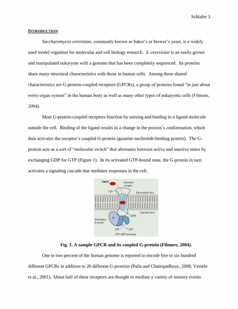
Most G-protein-coupled receptors function by sensing and binding to a ligand molecule
outside the cell. Binding of the ligand results in a change in the protein’s conformation, which
then activates the receptor’s coupled G-protein (guanine nucleotide-binding protein). The G-
protein acts as a sort of ―molecular switch‖ that alternates between active and inactive states by
exchanging GDP for GTP (Figure 1). In its activated GTP-bound state, the G-protein in turn
activates a signaling cascade that mediates responses in the cell.
Fig. 1. A sample GPCR and its coupled G-protein (Filmore, 2004).
One to two percent of the human genome is reported to encode five to six hundred
different GPCRs in addition to 26 different G-proteins (Paila and Chattopadhyay, 2008; Versele
et al., 2001). About half of these receptors are thought to mediate a variety of sensory events

Schlafer 4
including vision, taste, and olfaction (Wise et al., 2004). GPCRs also bind a wide variety of
stimuli including ―light, protons, Ca2+
, odorants, amino acids, nucleotides, proteins, peptides,
steroids, and fatty acids‖ (Xue et al., 2008).
Due to their abundance and variety of functions, GPCRs have become a popular target
for the development of new drug therapies. According to Wise and colleagues (2004),
drugs active at G-protein-coupled receptors (GPCRs) have therapeutic benefit
across a broad spectrum of human diseases as diverse as pain, cognitive dysfunction,
hypertension, peptic ulcers, rhinitis, and asthma. Of the approximately 500 clinically
marketed drugs, greater than 30% are modulators of GPCR function, representing
approximately 9% of global pharmaceutical sales, making GPCRs the most
successful of any target class in terms of drug discovery.
This information illustrates the potential benefit inherent in a more complete understanding of
this group of proteins.
Though humans contain hundreds of types of GPCRs, yeast cells have only two types.
Each of the yeast mating types contains a unique pheromone receptor, Ste2p (mating type
MATa) or Ste3p (mating type MAT -protein Gpa1
(Nakayama et al., 1988). The second receptor is Gpr1p, a glucose-sensing GPCR that associates
with the G-protein Gpa2 (Xue et al., 1998). The smaller variety of GPCRs in S. cerevisiae lends
itself as an excellent model to study GPCR activity and regulation.
Ste2 and the Mating Pathway
Ste2p is a class D (fungal mating pheromone receptor) GPCR with characteristic seven
transmembrane domains, but little sequence similarity to other GPCRs. However, this receptor
demonstrates ligand binding and activation mechanisms similar to other GPCRs and can even
couple to mammalian G-proteins (Brown et al., 2000).
Schlafer 5
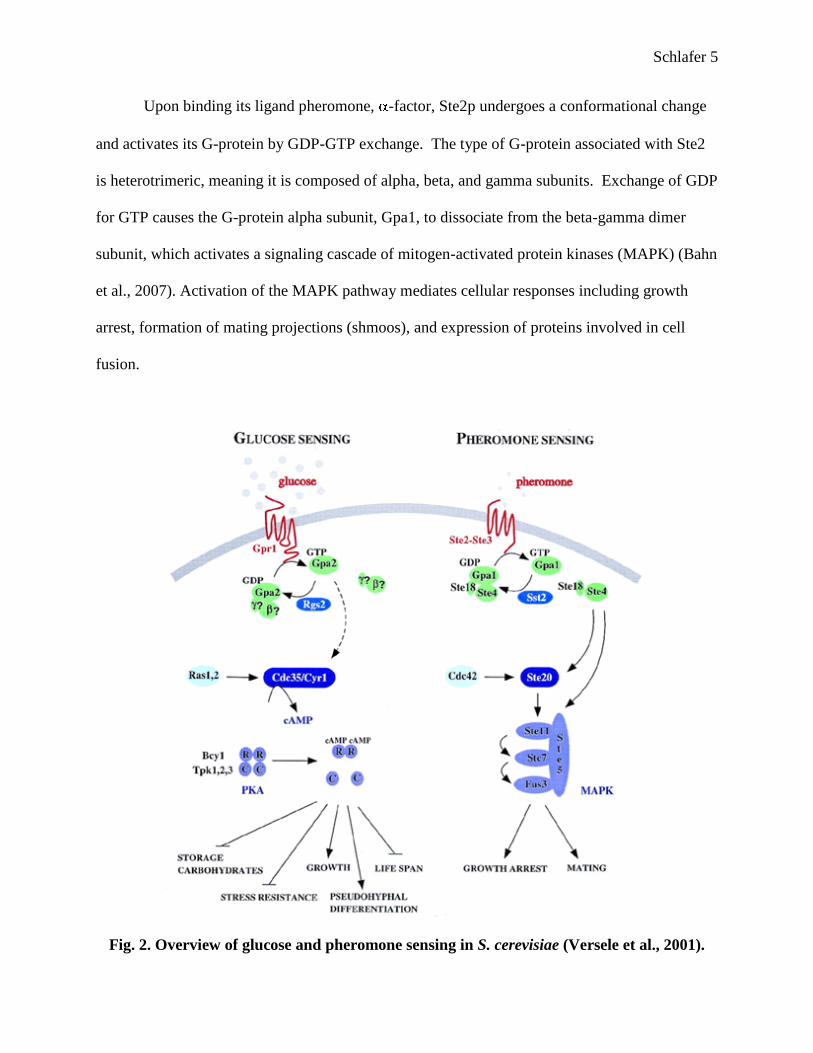
Upon binding its ligand pheromone, -factor, Ste2p undergoes a conformational change
and activates its G-protein by GDP-GTP exchange. The type of G-protein associated with Ste2
is heterotrimeric, meaning it is composed of alpha, beta, and gamma subunits. Exchange of GDP
for GTP causes the G-protein alpha subunit, Gpa1, to dissociate from the beta-gamma dimer
subunit, which activates a signaling cascade of mitogen-activated protein kinases (MAPK) (Bahn
et al., 2007). Activation of the MAPK pathway mediates cellular responses including growth
arrest, formation of mating projections (shmoos), and expression of proteins involved in cell
fusion.
Fig. 2. Overview of glucose and pheromone sensing in S. cerevisiae (Versele et al., 2001).
Schlafer 6
Gpr1 and the Glucose-Sensing Pathway
The relatively recently discovered second GPCR in yeast is the glucose receptor Gpr1
(Kraakman et al., 1999). Gpr1 interacts with its G-protein, Gpa2, which unlike Gpa1, does not
appear to have beta-gamma subunits. Glucose sensing is responsible for cAMP pathway
activation, which converts the cell from utilizing gluconeogenesis to utilizing fermentation for
energy production. This pathway has also ―been shown to regulate morphogenesis,‖ causing
cells to undergo pseudohyphal differentiation (Tamaki, 2007). In addition, it has been shown
that the Gpr1 pathway ―regulates cell size by affecting both growth rate and cell division,‖
allowing cells to adapt to starvation periods by altering metabolic activity (Tamaki et al., 2005;
Rolland et al., 2002).
It has been shown that certain GPCRs do interact with each other and in some cases form
homo- or heterodimers (Milligan, 2001). Ste2 forms a homodimer, but it is unknown whether
Gpr1 forms either homo- or heterodimers (Kim et al., 2009). It has also been shown that the G-
protein of the glucose-sensing pathway, Gpa2, is not capable of functionally coupling to the
mating GPCR Ste2 (Blumer and Thorner, 1990). However, it is unknown whether the G-protein
Gpa1 can couple to the receptor Gpr1.
In preliminary research on the Ste2p pathway conducted in the summer of 2008, an S.
cerevisiae strain lacking Gpr1 (Gpr1 ) exhibited decreased levels of pheromone-induced cell
cycle arrest compared with other mutant and wild type strains. In other words, the cells
continued to grow, possibly indicating decreased activity of the mating pathway. This
unexpected finding raises the question of whether Gpr1 and/or its pathway components are in
some way connected to the Ste2 pathway. As seen in Figure 2, there is currently no known
mechanism by which these two pathways overlap. By exploring these pathways, it is possible to
Schlafer 7
gain more insight into the function of these GPCRs, particularly Gpr1, about which less is
known.
The activity of the Ste2 pathway is relatively easy to monitor and measure, using
different procedures to observe growth arrest, shmoo formation, and mating gene activation,
which are either directly or, through the use of reporter genes, indirectly measurable. However,
the Gpr1 pathway causes cellular responses that are not easily quantified.
In hopes of monitoring the activity of the GPCR pathways, a fused Gpr1-Gpa1 protein
was engineered. This construct would theoretically allow the mating pathway to be activated by
the binding of glucose to Gpr1. Then certain assays could be used to quantify Gpr1’s activity
through the pathways’ response using the chimera protein cells as well as other mutant strains of
S. cerevisiae.

Schlafer 8
MATERIALS AND METHODS
Preparation of plasmids
Plasmid pBUGf containing GPA1 DNA, provided by George Umanah of the University
of Tennessee, was transformed into competent JM109 bacterial cells and grown on selective
media following the E. coli competent cells protocol (Promega). The plasmid was then purified
using the PureYield Plasmid Midiprep kit (Promega).
The same procedure was used to prepare a GPR1 plasmid, using ultra-competent
bacterial cells (Stratagene Solopack Gold) in place of the competent JM109 cells to ensure
higher transformation efficiency.
PCR and Restriction Digest
In order to create a chimeric protein, the genes coding for both units must be attached for
the genes to be expressed simultaneously. The polymerase chain reaction (PCR) uses single-
stranded DNA and primers with specifically designed complementary base sequences to amplify
a certain portion of DNA.
First, primers that included bases from the beginning and end of GPR1 and from each side
of the insertion site in the Gpa1 plasmid were designed and ordered. Primers PRPA-FOR: 5’-
CGACGGATCTAGAACTAGTGGATCCATAATGATAACTGAGGGATTTCCCC-3’ and
prpa-rev: 5’-GCGTACTCACTGTACACCCCATTAATGGTCCATTTCTTAAGAAGGC -3’
were used to perform the polymerase chain reaction on the Gpr1 plasmid (underlined portions
indicate GPR1 sequences). This procedure created multiple copies of the GPR1 gene with
―sticky ends‖ to correlate with the corresponding ends in the GPA1 plasmid for GPR1 insertion.

Schlafer 9
Fig. 3. Restriction map of pBUGf (Gpa1 plasmid), shown with PCR product at the
insertion site.
The GPA1 plasmid was digested with BamHI restriction enzyme, cutting open the
circular DNA at a specific site engineered into the plasmid (see Fig. 3), thus providing an
insertion site for the PCR product. The cut plasmid and the PCR product were then transformed
into TM 5117 (Far1 Gpa1 Ste2 ) S. cerevisiae cells to recombine by in vivo ligation (Gietz
and Woods, 2002).
Western Blot
In order to confirm the phenotype of the fused protein, a Western blot was performed.
Western blotting is a technique used to detect the presence of certain proteins in a sample. Using

Schlafer 10
gel electrophoresis, proteins were separated along an 8-16% Tris-Glycine gel according to size,
and then electro-transferred to Immobilon-P membrane paper. The membrane was blocked in
Tris-buffered saline (TBS) buffer with 5% milk for approximately one hour, after which it was
incubated with anti-FLAG polyclonal rabbit antibody at a dilution of 1:4000 in TBS-Tween for
90 minutes at room temperature. The membrane was then washed twice for five minutes in
TBS/milk, once for five minutes in TBS plus 0.1% Tween (TBST), once for five minutes in
TBS/milk, and briefly rinsed in TBST. Goat anti-rabbit HRP secondary antibody (Biorad) at a
dilution of 1:3000 in TBST was added and incubated for approximately three hours at room
temperature.
During this process, the antibodies bind to specific proteins, allowing the proteins to be
detected. To rinse off unbound secondary antibodies, the membrane was washed three times for
five minutes in TBS, twice for five minutes in TBST, and twice for five minutes in TBS. The
samples were amplified and detected using an Opti-4CN substrate kit (Biorad).
ß-galactosidase Assay
This assay involves using a reporter gene to monitor the activity of the pheromone mating
pathway. FUS1 is a gene activated by the pheromone pathway that causes the yeast cell to
change shape for fusion with another cell. The TM strains are engineered so that FUS1 is
attached to the lacZ gene, which codes for the enzyme beta-galactosidase. Measuring the
amount of product released after beta-galactosidase reacts with certain substrates can determine
the amount of beta-galactosidase produced. The product released gives off measurable
fluorescence proportional to the amount of enzyme present, and thus to the level of FUS1-lacZ
gene induction.
Schlafer 11
Overnight cultures of cells were centrifuged, resuspended in PBS buffer, and incubated at
30°C for one hour. Cells were resuspended in varying concentrations of dextrose solution and
150 l of each were added to a 96-well plate. 30 l of 5% Triton-X 100 50mM PIPES buffer
with the fluorescent substrate fluorescein di-β-D-galactopyranoside (FDG) was added to each
well. The plate was incubated at 37°C for 30 minutes and then read on a SpectraMax Gemini XS
spectrofluorometer.
Mating Assay
Cultures of wild type, Gpr1 , and Gpa2 cells of both mating type MATa (BY4741) and
MAT (BY4742) were grown overnight, centrifuged, washed three times in sterile water, and
resuspended in 6 ml of sterile water. Cells were counted using a hemocytometer. Each possible
combination of MATa and MAT cells was mixed in 100 l YPD media, maintaining a 1:5 ratio
of MATa to MAT cells. Mixtures were incubated at 30°C for five hours. Cells were harvested
by centrifugation, washed three times with sterile water, and resuspended in 2 ml sterile water.
Cells were diluted 1:20 in water, and then 75 l was plated onto MLMK, a selective media.
Plates were incubated for two days at 30°C, and then the number of colonies formed was
counted. From this data, mating efficiency was calculated using the formula:
mating efficiency (%) = (100 x X)/W
where X=the number of colonies formed between mutant strains, and W=the number of colonies
formed between wild type strains.
Schlafer 12
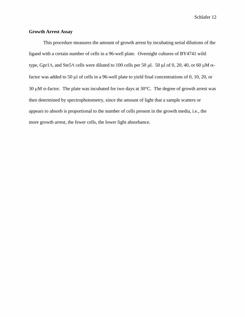
Growth Arrest Assay
This procedure measures the amount of growth arrest by incubating serial dilutions of the
ligand with a certain number of cells in a 96-well plate. Overnight cultures of BY4741 wild
type, Gpr1 , and Ste5 cells were diluted to 100 cells per 50 l. 50 l of 0, 20, 40, or 60 M -
factor was added to 50 l of cells in a 96-well plate to yield final concentrations of 0, 10, 20, or
30 M -factor. The plate was incubated for two days at 30°C. The degree of growth arrest was
then determined by spectrophotometry, since the amount of light that a sample scatters or
appears to absorb is proportional to the number of cells present in the growth media, i.e., the
more growth arrest, the fewer cells, the lower light absorbance.

Schlafer 13
RESULTS
Growth Arrest Assay
When pheromone activates a yeast cell’s mating pathway, one result is arrest of the cell
cycle in G1 phase in preparation for fusion with another cell. Using spectrophotometry, the
growth arrest assay measures the amount of growth arrest that has or has not occurred as a result
of pheromone addition.
After addition of -factor pheromone, Gpr1 cells showed less pheromone-induced
growth arrest than did wild type cells. In fact, absorbance values for the Gpr1 strain were
comparable to values for Ste5 , a sterile strain of S. cerevisiae in which the Ste2 pathway is not
functional (Figure 4). This data indicates that without Gpr1 present, the binding of pheromone
does not activate the Ste2 pathway.
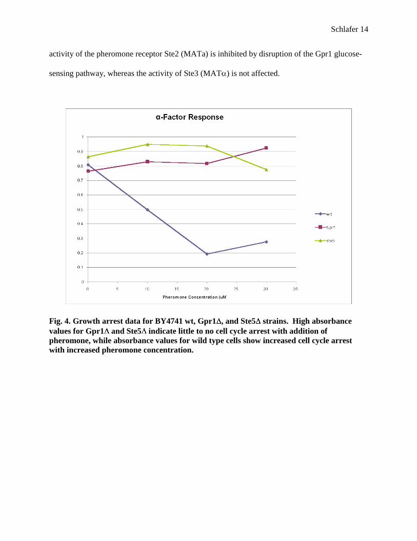
Mating Assay
This assay was used to determine the degree of mating efficiency between the two mating
types, MATa and MAT . Strains with either the glucose-sensing receptor (Gpr1) or its G-
protein (Gpa2) deleted were used to investigate whether disruption of the Gpr1 pathway would
affect mating efficiency.
Results of the mating assay showed no significant difference in mating efficiency
between wild type MATa and either Gpr1 or Gpa2 MAT cells as compared to mating
between both wild type strains (Figure 5). However, the student’s t-test showed a significant
decrease in mating efficiency compared to wild type when either Gpr1 or Gpa2 MATa cells
were crossed with each of the MAT strains. From this information, one can infer that the
Schlafer 14
activity of the pheromone receptor Ste2 (MATa) is inhibited by disruption of the Gpr1 glucose-
sensing pathway, whereas the activity of Ste3 (MAT ) is not affected.
Fig. 4. Growth arrest data for BY4741 wt, Gpr1 , and Ste5 strains. High absorbance
values for Gpr1 and Ste5 indicate little to no cell cycle arrest with addition of
pheromone, while absorbance values for wild type cells show increased cell cycle arrest
with increased pheromone concentration.
Schlafer 15
P=0.000004
*
P=0.00001
*
P=0.00004
*
P=0.00005
*
P=0.0002
*
P=0.0001
*
P=0.71P=0.47
0
20
40
60
80
100
120
140
160
180
200
41wtX42wt
41wtX42Gpr1
41wtX42Gpa2
41Gpr1X42wt
41Gpr1X42Gpr1
41Gpr1X42Gpa2
41Gpa2X42wt
41Gpa2X42Gpr1
41Gpa2X42Gpa2
Mating Combination (MAT a X MAT alpha)
Ma
tin
g E
ffic
ien
cy (
%)
Figure 5. Effect of GPR1 or GPA2 deletion on mating efficiency between BY4741 (MATa)
and BY4742 (MAT ) cells. Asterisks indicate a significant difference in mating efficiency
compared to wild type. (n=3)
Dot Blot, Protein Assay, and Western Blot
After much trial and error with the transformation protocol, transformant colonies were
produced using 6 l of BamHI-digested pBUGf and 2 or 6 l of PCR product, following the
transformation protocol (Gietz and Woods, 2002). Twenty-one distinct colonies were selected,
two from the 6 l / 6 l plate, and 19 from the 2 l / 6 l plate. A dot blot was performed with
anti-FLAG polyclonal rabbit antibody in order to gain a rough idea of the amount of protein
being expressed in each colony (see Figure 6), which was subsequently quantified using the
BioRad protein assay (Figure 7).

Schlafer 16
Fig. 6. Dot blot with transformant colonies. Anti-FLAG antibody showed the presence of
FLAG-tagged proteins in the darker circles.
The samples producing the highest amounts of protein were colony A from the 6 l / 6 l
plate, and colonies B, F, O, Q, and S from the 2 l / 6 l plate. These samples contained
approximately 0.5 to 1 mg of protein per ml of solution (Figure 7b).
a)
BioRad Protein Assay 7/22/09
y = 0.2808x + 0.2189
R2 = 0.9989
0
0.2
0.4
0.6
0.8
1
0 0.5 1 1.5 2 2.5
Amount of protein (mg/ml)
Ab
so
rban
ce (
595 n
m)
b)
Fig. 7. BioRad protein assay BSA standard curve (a) and most concentrated samples (b).
Sample A (bold font) was from the 6 l / 6 l plate, and colonies B, F, O, Q, and S were
from the 2 l / 6 l plate.
Sample Absorbance
(595 nm) Concentration
(mg/ml)
A 0.342857 0.441442308
B 0.467857 0.886599003
F 0.396429 0.632225783
O 0.382143 0.581349715
Q 0.539286 1.140975783
S 0.557143 1.204569088
Schlafer 17
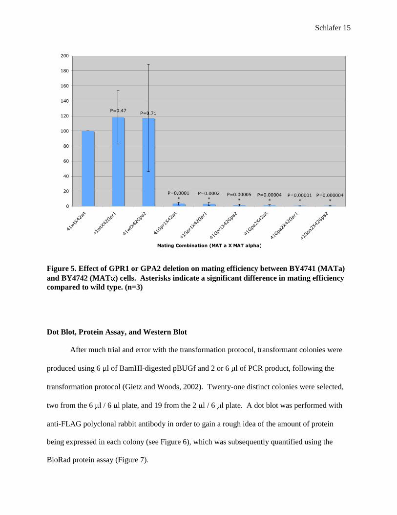
A Western blot was performed using samples from colonies listed in Figure 7. The
chimeric protein, consisting of Gpa1 fused to Gpr1, if present in the cell, would have a molecular
weight of roughly 350 kilodaltons. Samples A and O showed relatively small bands on the blot
corresponding to this high molecular weight (Figure 8; O not shown). Although faint, these
bands suggest that the chimeric protein was successfully inserted and expressed in each strain.
Fig. 8. Western blot. Sample A showed a faint band around 350 Kd.
Petite Mutation
Upon microscopic examination of the transformant cells, they were discovered to be
much smaller than normal yeast, and were in fact mistaken for bacterial contamination for
several days (Figure 9). This phenomenon is known as a petite mutation, which is most often the
result of a mutation in mitochondrial DNA (Goldring et al., 1971). It is hard to say what
specifically caused this mutation, but it could be cause for further study. It is also uncertain how

Schlafer 18
this mutation might have affected the results of experiments involving these cells, due to their
small size and slow growth.
When the petite cells were grown on YPD (nonselective) media, a mixture of normal-
sized and petite cells was observed (Figure 10). This might indicate that the normal-sized cells
had lost the plasmid and reverted to normal size/growth conditions.
Fig. 9. Petite mutation in Gpr1-Gpa1 transformant cells.
Fig. 10. Revertants grown on YPD showed both petite and normal-sized cells.
Schlafer 19
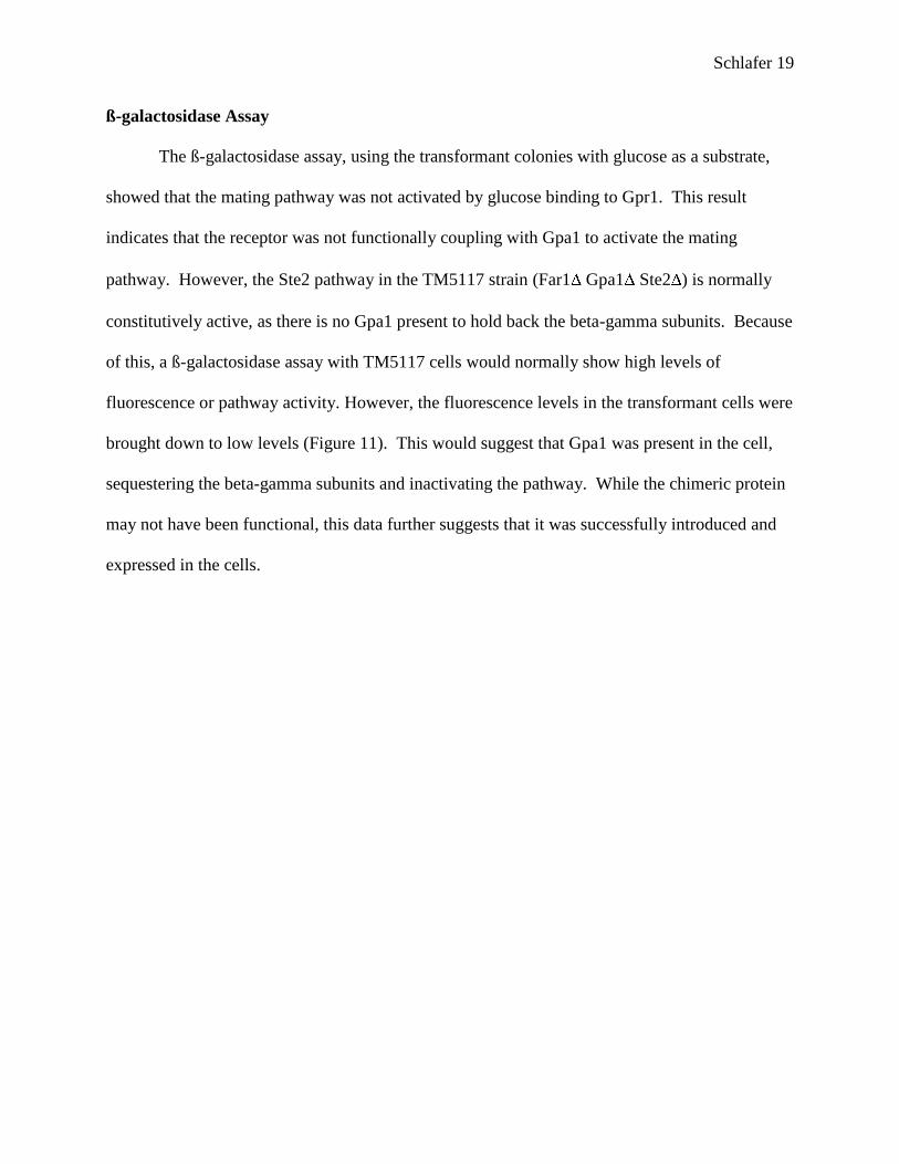
ß-galactosidase Assay
The ß-galactosidase assay, using the transformant colonies with glucose as a substrate,
showed that the mating pathway was not activated by glucose binding to Gpr1. This result
indicates that the receptor was not functionally coupling with Gpa1 to activate the mating
pathway. However, the Ste2 pathway in the TM5117 strain (Far1 Gpa1 Ste2 ) is normally
constitutively active, as there is no Gpa1 present to hold back the beta-gamma subunits. Because
of this, a ß-galactosidase assay with TM5117 cells would normally show high levels of
fluorescence or pathway activity. However, the fluorescence levels in the transformant cells were
brought down to low levels (Figure 11). This would suggest that Gpa1 was present in the cell,
sequestering the beta-gamma subunits and inactivating the pathway. While the chimeric protein
may not have been functional, this data further suggests that it was successfully introduced and
expressed in the cells.
Schlafer 20
Selected Colonies with Glucose
0
2
4
6
8
10
12
14
0 0.5 1 1.5 2 2.5
Glucose Concentration (%)
Flu
orescen
ce
A
F
O
Q
LM23 with alpha-Factor
0
100
200
300
400
500
600
700
800
900
1000
1100
0.00E+
00
2.00E-
06
4.00E-
06
6.00E-
06
8.00E-
06
1.00E-
05
1.20E-
05
Alpha-factor Concentration (mM)
Flu
orescen
ce
Fig. 11. ß-galactosidase Assay. Bottom graph shows regular active (~1000 units) and
inactive (~10 units) levels of fluorescence.

Schlafer 21
DISCUSSION
Gpr1-Gpa1 Chimera Protein
The preliminary focus of this project was the creation and implementation of a chimeric
protein consisting of the glucose-sensing GPCR, Gpr1, and the G subunit of the mating
pathway, Gpa1, in Saccharomyces cerevisiae. The data shows that the chimera was successfully
constructed and inserted into the TM5117 strain (Figures 8 and 11). However, results of the -
galactosidase assay showed that binding of glucose to the Gpr1-Gpa1 protein did not result in
signal transduction to the Ste2 mating pathway (Figure 11). This finding suggests that the C-
terminus of Gpr1 and the N-terminus of Gpa1 do not interact to regulate their respective
pathways. However, this does not rule out other possible interactions between the two pathways.
Petite Mutation
Disruptions in mitochondrial DNA result in a phenomenon known as the petite mutation,
in which cells become markedly smaller than wild type cells (hence the name ―petite‖). Yeasts
such as S. cerevisiae are facultative anaerobes, meaning they can survive with or without the
presence of oxygen. Although aerobic respiration produces more ATP, yeasts prefer alcoholic
fermentation, even in the presence of oxygen, because it enables them to produce energy more
quickly and also produces ethanol, which can inhibit the growth of competing organisms
(Rolland et al., 2002). In yeasts, petite mutants ―are characterized by an inability to utilize
nonfermentable substrates such as glycerol for growth‖ (Goldring et al., 1971).
Many varied factors have been shown to induce petite colonies in yeast including
chemicals such as ethidium bromide, as well as a large number of genes that contribute to the
stability of mitochondrial DNA (Contamine and Picard, 2000). Currently among these genes are
Schlafer 22
a small number of cell division cycle, or CDC, genes. In particular, mutations in CDC8 or
CDC21 genes have been shown to disrupt replication of mitochondrial DNA (Newlon et al.,
1979). While two CDC genes, CDC42 and CDC35, are downstream regulators of the
pheromone and glucose-sensing pathways, respectively, it is unclear whether they could play a
role in mitochondrial DNA stability. According to Contamine and Picard (2000), the effects of
most CDC genes on mitochondrial DNA are ―either quite trivial or still very puzzling,‖ so it is
likely the petite mutations seen in this experiment stemmed from another source.
Perhaps the actual procedure used to create the chimeric protein caused some sort of
mutation leading to the petite phenotype. Transformation procedures have been shown to be
―highly mutagenic,‖ so it is likely that the petite mutation was a residual effect of transforming
the TM5117 strain with the chimeric protein plasmid (Contamine and Picard, 2000). Since petite
mutants grown on nonselective media reverted back to normal size (Figure 10), the petite
mutation might be further linked to the transformation with the fused protein. It is possible that
the normal-sized cells seen here resulted from petite cells losing the fused protein-containing
plasmid when grown in nonselective media.
Effect of Gpr1 Deletion on Ste2 Pathway
Although the Gpr1-Gpa1 chimeric protein appeared not to be functional, results of the
growth arrest and mating assays still reveal the possibility of a link between the pheromone and
glucose-sensing pathways. If some sort of regulatory connection between the two pathways
exists, it is likely further downstream as opposed to occurring between the glucose receptor and
pheromone G subunit, or perhaps also involves the pheromone receptor. If the latter is the
case, data from the mating assay (Figure 5), indicates that only Ste2 is affected by inactivation of
Schlafer 23
the glucose pathway, whereas the pheromone pathway controlled by Ste3 remains fully
functional. A possible explanation might be that Ste3 is has a slightly different structure than
Ste2, which somehow allows it to be immune to any possible cues from the glucose-sensing
pathway.
While the exact mechanism has yet to be elucidated, it follows logically that the cell
would curb ―nonessential‖ activities—such as mating—in times when energy sources are scarce.
It has been shown that when glucose or other fermentable carbon sources are unavailable and the
cell is using non-fermentable carbon sources, genes involved in resistance to stress are highly
active (Rolland, et al. 2002). Perhaps a nonfunctional glucose-sensing pathway, as in Gpr1 or
Gpa2 , induces a stress response in the cell which includes inactivation of the mating pathway.
According to Rolland et al. (2002), the ―dramatic effects of glucose on growth and
metabolism clearly support a hormone-like function for this sugar in yeast cells,‖ the presence or
absence of which regulates many important processes in the cell. Additionally, ―nutrient-sensing
and –signalling mechanisms must have evolved early in evolution and might be at the origin of
the sophisticated hormone- and growth factor-induced signal transduction pathways‖ (Rolland et
al., 2002). Experimental evidence from this study supports such an evolutionary link between
the nutrient-sensing Gpr1 pathway and pheromone-sensing Ste2 pathway. The exact mechanism
of this purported link, however, remains to be discovered.

Schlafer 24
ACKNOWLEDGEMENTS
I would like to thank the Appalachian College Association’s Colonel Lee B. Ledford
Scholars Program for providing a summer research grant, George Umanah for assisting with
plasmids, and the Carson-Newman Biology and Chemistry departments, especially Dr. Stephen
Wright, for support and direction.

Schlafer 25
REFERENCES
Bahn, Y., Xue, C., Idnurm, A., Rutherford, J.C., Heitman, J., and Cardenas, M.E. (2007) Sensing
the environment: lessons from fungi. Nature Reviews Microbiology. 5: 57- 69.
Blumer, K.J. and Thorner, J. (1990) and subunits of a yeast guanine nucleotide-binding
protein are not essential for membrane association of the subunit but are required for
receptor coupling. Proc. Natl. Acad. Sci. USA. 90: 9921-9925.
Brown, A.J., Dyos, S.L., Whiteway, M.S., White, J.H.M., Watson, M.E.A., Marzioch, M., Clare,
J.J., Cousens, D.J., Padden, C., Plumpton, C., Romanos, M.A., and Dowell, S.J. (2000)
Functional coupling of mammalian receptors to the yeast mating pathway using novel
yeast/mammalian G protein -subunit chimeras. Yeast. 16(1): 11-22.
Contamine, V., and Picard, M. (2000) Maintenance and integrity of the mitochondrial genome: a
plethora of nuclear genes in the budding yeast. Microbiology and Molecular Biology
Reviews. 64(2): 281-315.
Filmore, D. (2004) It's a GPCR world. Modern Drug Discovery (American Chemical Society).
2004 (November): 24–28.
Gietz, R.D. and Woods, R.A. (2002) Transformation of yeast by the Liac/SS carrier DNA/PEG
method. Methods in Enzymology. 350: 87-96.
Goldring, E.S., Grossman, L.I., and Marmur, J. (1971) Petite mutation in yeast. Journal of
Bacteriology. 107(1): 377-381.
Kim, H. Lee, B.K., Naider, F., and Becker, J.M. (2009) Identification of specific transmembrane
residues and ligand-induced interface changes involved in homo-dimer formation of a
yeast G protein-coupled receptor. Biochemistry. 48(46): 10976-10987.
Kraakman, L., Lemaire, K., Pingsheng, M., Teunissen, A., Donaton, M.C.V., Van Dijck, P.,
Winderickx, J., de Winde, J.H., and Thevelein, J.M. (1999) Saccharomyces cerevisiae G-
protein-coupled receptor, Gpr1, is specifically required for glucose activation of the
cAMP pathway during the transition to growth on glucose. Molecular Microbiology.
32(5): 1002-1012.
Milligan, G. (2001) Oligomerisation of G-protein-coupled receptors. Journal of Cell Science.
114: 1265-1271.
Nakayama, N., Kaziro, Y., Arai, K., and Matsumoto, K. (1988) Role of STE Genes in the Mating
Factor Signaling Pathway Mediated by GPA1 in Saccharomyces cerevisiae. Molecular
and Cellular Biology. 8(9): 3777-3783.

Schlafer 26
Newlon, C.S., Ludescher, R.D., and Walter, S.K. (1979) Production of petites by cell cycle
mutants of Saccharomyces cerevisiae defective in DNA synthesis. Mol. Gen. Genet. 169:
189-194.
Paila, Y.D., and Chattopadhyay, A. (2008) The function of G-protein coupled receptors and
membrane cholesterol: specific or general interaction? Glycoconj J. 2008 (December)
Rolland, F., Winderickx, J., and Thevelein, J. M. (2002) Glucose-sensing and –signalling
mechanisms in yeast. FEMS Yeast Research. 2(2): 183-201.
Tamaki, H. (2007) Glucose-stimulated cAMP-protein kinase A pathway in yeast Saccharomyces
cerevisiae. Journal of Bioscience and Bioengineering. 104(4): 245-250.
Tamaki, H., Yun, C., Mizutani, T., Tsuzuki, T., Takagi, Y., Shinozaki, M., Kodama, Y.,
Shirahige, and Kumagai, H. (2005) Glucose-dependent cell size is regulated by a G
- 26 -protein-coupled receptor system in yeast Saccharomyces cerevisiae. Genes
to Cells. 10: 193-206.
Versele, M., Lemaire, K., and Thevelein, J.M. (2001) Sex and sugar in yeast: two distinct GPCR
systems. EMBO Reports. 2(7): 574-579.
Wise, A., Jupe, S.C., and Rees, S. (2004) The identification of ligands at orphan G-protein
coupled receptors. Annual Review of Pharmacology and Toxicology. 44:43-66.
Xue, C., Hsueh, Y., and Heitman, J. (2008) Magnificent seven: roles of G protein-coupled
receptors in extracellular sensing in fungi. FEMS Microbiol Rev. 32: 1010-1032.
Xue, Y., Batlle, M., and Hirsch, J.P. (1998) GPR1 encodes a putative G protein-coupled receptor
that associates with the Gpa2p G subunit and functions in a Ras-independent pathway.
The EMBO Journal. 17(7): 1996-2007.