cannabinoid ligand-receptor signaling in the uterus - pnas · cannabinoid ligand-receptor...
TRANSCRIPT

Proc. Natl. Acad. Sci. USAVol. 92, pp. 4332-4336, May 1995Pharmacology
Cannabinoid ligand-receptor signaling in the mouse uterusS. K. DAS*, B. C. PARIA*, I. CHAKRABORTY, AND S. K. DEY
Department of Physiology, University of Kansas Medical Center, Ralph L. Smith Research Center, Kansas City, KS 66160-7338
Communicated by Robert L. Sinsheimer, University of California, Santa Barbara, CA, January 30, 1995 (received for review December 6, 1994)
ABSTRACT Using RNA (Northern) blot hybridizationand reverse transcription-PCR, we demonstrate that thebrain-type cannabinoid receptor (CB1-R) mRNA, but not thespleen-type cannabinoid receptor (CB2-R) mRNA, is ex-pressed in the mouse uterus and that this organ has thecapacity to synthesize the putative endogenous cannabinoidligand, anandamide (arachidonylethanolamide). The psycho-active cannabinoid component of marijuana-A9-tetrahydro-cannabinol (THC)-or anandamide, but not the inactive andnonpsychoactive cannabidiol (CBD), inhibited forskolin-stimulated cyclic AMP formation in the mouse uterus, whichwas prevented by pertussis toxin pretreatment. These resultssuggest that uterine CB1-R is coupled to inhibitory guaninenucleotide-binding protein and is biologically active. Autora-diographic studies identified ligand binding sites ([3H]anan-damide) in the uterine epithelium and stromal cells, suggest-ing that these cells are perhaps the targets for cannabinoidaction. Scatchard analysis of the binding of [3H]WIN 55212-2,another cannabinoid receptor ligand, showed a single class ofhigh-affinity binding sites in the endometrium with an ap-parent Kd of 2.4 nM and Bmax of 5.4 x 10⁹ molecules per mgof protein. The gene encoding lactoferrin is an estrogen-responsive gene in the mouse uterus that was rapidly andtransiently up-regulated by THC, but not by CBD, in ovari-ectomized mice in the absence of ovarian steroids. This effect,unlike that of 17j3-estradiol (E2), was not influenced by a pureantiestrogen, ICI 182780, suggesting that the THC-induceduterine lactoferrin gene expression does not involve estrogenreceptors. We propose that the uterus is a new target forcannabinoid ligand-receptor signaling.
Marijuana has been used for centuries as a psychoactive drug.A9-Tetrahydrocannabinol (THC) is the major psychoactivecomponent of marijuana. Marijuana and its cannabinoid de-rivatives induce a wide spectrum of effects including analgesia,antiemetic, anticonvulsion, attenuation of intraocular pressurein glaucoma, antiinflammation, and immunosuppression (1).One of the major concerns of habitual marijuana smoking orexposure to cannabinoid derivatives is their reported adverseeffects on reproductive functions, including fetal loss, preg-nancy failure, retarded embryonic development, and reducedfertilizing capacity of sperm (reviewed in refs. 2-5). Themechanisms by which these effects of cannabinoids are medi-ated are poorly defined. Furthermore, controversies still pre-vail surrounding the "estrogenic-antiestrogenic" effects ofcannabinoids in uterine biology (2, 6, 7) and their interactionswith uterine estrogen receptors (8, 9). Previous identificationof guanine nucleotide-binding protein (G-protein)-coupledcannabinoid receptors together with the recent cloning ofthese cannabinoid receptors in the brain (CB1-R) and spleen(CB2-R) (10-12) have begun to provide evidence that many ofthe central and peripheral effects of cannabinoids are likely tobe mediated via these receptors. Furthermore, the identifica-tion of a putative endogenous cannabinoid ligand, anandamide(arachidonylethanolamide), in the brain suggests that canna-
The publication costs of this article were defrayed in part by page chargepayment. This article must therefore be hereby marked "advertisement" inaccordance with 18 U.S.C. §1734 solely to indicate this fact.
binoid ligand-receptor signaling may be normally operative inthe central nervous system (13, 14), although the physiologicalsignificance of this signaling pathway is yet to be defined.The CB2-R gene has been shown to be expressed only in the
spleen, while the CB1-R gene is primarily expressed in thebrain (11, 12). The other tissues that have been shown toexpress CB1-R mRNA are the testis, spleen, and peripheralblood leukocytes (15-17). The expression of cannabinoidreceptors in the spleen and leukocyte has been associated withthe antiinflammatory and immunosuppressive roles of canna-binoids (16, 17), while the observation of reduced fertilizingcapacity of sperm exposed to cannabinoid ligands is consistentwith the detection of CB1-R mRNA in the testis and canna-binoid binding sites in the sperm (4, 5, 15). On the other hand,the effects and mode of action of cannabinoids in uterinebiology and pregnancy remain largely undefined and contro-versial in spite of the numerous reports published in this fieldduring the last two decades (2, 3). In the present investigation,we sought to examine whether functional cannabinoid recep-tors are expressed in the uterus and whether this organ has thecapacity to synthesize anandamide. We further examinedwhether THC can influence uterine functions at the molecularlevel by examining the expression of the gene encoding lacto-ferrin (LF), an estrogen-responsive gene in the mouse uterus.
MATERIALS AND METHODSNorthern Blot Hybridization of CB1-R mRNA. Uteri from
CD-1 mice on days 1, 4, and 7 of pregnancy (day 1 = vaginalplug) were used in this study to determine whether thechanging hormonal status of the mother and correspondingchanges in uterine physiology alter the expression of CB1-RmRNA (18). Total RNA from the mouse brain or uteri wasextracted by using a modified guanidine thiocyanate procedure(19). Poly(A)+ RNA was isolated by oligo(dT)-cellulose col-umn chromatography (19). A 1.17-kb fragment encoding97-1271 nucleotides of the rat CB1-R cDNA clone SKR6 wassubcloned into a pGEM vector (11). A cDNA clone of humanfibroblast cytoplasmic p-actin was subcloned into a pGEMvector (20). For Northern hybridization, antisense 32P-labeledcomplementary RNA probes were generated. Probes hadspecific activities of about 2 x 10⁹ dpm/,g. Total RNA orpoly(A)+ RNA was denatured, separated by formaldehyde/agarose gel electrophoresis, transferred to nylon membranes,and UV-cross-linked. Northern blots were prehybridized, hy-bridized, and washed as described (19). Transcripts weredetected by autoradiography. Each blot was reprobed withp-actin to ensure RNA integrity.Reverse Transcription (RT)-PCR Analysis of CB1-R and
CB2-R mRNAs. For RT-PCR analysis, the following primerswere used: 5'-GGAGAACATCCAGTGTGGGG-3' (sense)and 5'-CATTGGGGCTGTCTTTACGG-3' (antisense) for
Abbreviations: CB1-R, brain-type cannabinoid receptor; CB2-R,spleen-type cannabinoid receptor; THC, A9-tetrahydrocannabinol;CBD, cannabidiol; LF, lactoferrin; RT-PCR, reverse transcription-PCR; G protein, guanine nucleotide-binding protein; Gi protein,inhibitory G protein; E2, 17,3-estradiol.*S. K. Das and B.C.P. contributed equally to this work.
4332

Proc. Natl. Acad. Sci USA 92 (1995) 4333
the CB1-R transcript (11) and 5'-CCTGTTGAAGATCGGC-AGCG-3' (sense) and 5'-GGTAGGAGATCAACGCCG-AG-3' (antisense) for the CB2-R transcript (12). The internaloligonucleotides 5'-GGTTCTGGAGAACCTACTGG-3' and5'-TGGGCAGCCTGCTGACT-3' were used for Southernhybridization of the amplified products for CB1-R and CB2-R,respectively. The 3-actin sense and antisense primers designedfrom the mouse p-actin cDNA were 5'-GTGGGCCG-CTCTAGGCACCAA-3' and5'-CTCTTTGATGTCACGCA-CGATTTC-3', respectively (21). An internal oligonucleotide,5'-CCACGGGCATTGTGATGGAC-3', was used for South-ern analysis. RNA from brain, spleen, or uterus was isolated(19, 22). Total RNA (1 ,tg) from each sample was reverse-transcribed by using specific antisense primers. A portion(one-third) of the RT products were subjected to PCR ampli-fication by using the sense and antisense primers in the bufferas described by us (22). PCR cycle parameters were as follows:94°C for 4 min, 55°C for 1.5 min, and 72°C for 2.5 min for thefirst cycle followed by 94°C for 1 min, 55°C for 1.5 min, and72°C for 2.5 min for a total of 35 cycles. One-tenth of the totalamplified product was run on 1.5% agarose gels, stained withethidium bromide, and blotted for Southern hybridization.Experimental and negative control reactions were run simul-taneously.
Analysis ofForskolin-Stimulated cAMP Accumulation. Day1 pregnant uteri were homogenized in 1 ml of 50mM Tris (pH7.5) containing 1 mM EGTA and 0.5 mM 3-isobutyl-l-meth-ylxanthene and centrifuged at 2000 x g for 15 min. Thesupernatant was centrifuged at 100,000 x g for 1 h to obtainmembranes. Membranes were resuspended, and an aliquot(350 ,tg of protein) was incubated in 1 ml of the same buffercontaining .10 t,M GTP, 1 mM ATP, and 3 mM MgSO4 withor without forskolin (1-5 tLM) at 25°C for 10 min to determineits maximal stimulatory concentration. To assess the effects ofagonists, THC (0.1-10 tLM), cannabidiol (CBD; 0.1-10 tM),or anandamide (0.1-10 tLM) was added to the incubationmixture with 5 uLM forskolin. To determine that the effects ofthe agonists were coupled to the inhibitory G protein (Gi),preincubation with pertussis toxin at 5 ng/ml for 5 min wasperformed before adding forskolin and THC. Reactions wereterminated by boiling (5 min) followed by centrifugation. ThecAMP contents of the supernatants were determined byprotein-binding assay (23, 24). Results are means ± SEM offour or five experiments except for the experiments with CBDor pertussis toxin pretreatment, which were carried out twice.Autoradiographic Localization of Ligand-Binding Sites.
Autoradiographic detection was performed with [3H]anand-amide (22). Frozen uterine sections (10 ,im) mounted ontopoly(L-lysine)-coated slides were incubated with 4.5 nM[3H]anandamide (specific activity, 221 Ci/mmol; DuPont/NEN; 1 Ci = 37 GBq) in 50 mM Tris-HCl buffer (pH 7.4)containing 5% bovine serum albumin (BSA) in the absence orpresence of 1000-fold molar excess of unlabeled anandamidefor 4 h at 37°C. After incubation, sections were washed fivetimes in the same buffer containing 1% BSA at 4°C, air-dried,and subjected to autoradiography (22). Autoradiographic ex-posure was for 7 days. Dark-field photomicrographs at x40magnification are shown. White grains indicate the bindingsites.
Radioligand-Binding Studies. Uterine epithelial-stromalcell mixtures were obtained from day 4 pregnant mice by gentlyscraping the endometrium from the myometrium. Membranepreparations and binding studies were performed as described(15). In brief, membranes (150 tpg of protein) were incubatedfor 1 h at 30°C in 1 ml of binding buffer (15) containingtritiated WIN 55212-2 (45.5 Ci/mmol; DuPont/NEN) rangingfrom 0.5 to 5.0 nM. Nonspecific binding was determined byperforming parallel binding reactions in the presence of 1 ,AMunlabeled WIN 55212-2. After incubation, membranes werefiltered on glass fiber filters (G6; Fisher Scientific) and washed
three times with 5 ml of the same buffer (15). Filters were driedand assayed in a 1-scintillation counter (Packard). Nonspecificbinding was subtracted from total binding to yield specificbinding. The binding kinetics were calculated by Scatchardanalysis with ENZFITTER, a nonlinear regression data analysisIBM personal computer program for ligand binding.Enzymatic Synthesis of Anandamide. The enzymatic syn-
thesis of anandamide was determined as described (25-27).Day 1 or day 4 pregnant uteri or brain tissues were homoge-nized in ice-cold TE buffer (10 mM Tris.HCl/1 mM EDTA,pH 7.6) containing 1.5 mM phenylmethylsulfonyl fluoride(PMSF) and were centrifuged at 2000 x g for 15 min. Thesupernatants (500 j,g of protein) were incubated in 1 ml of 0.1M Tris-HCl (pH 9.0) containing 1.5 mM PMSF with 1 ,uCi of[5,6,8,9,11,12,14,15-3H(N)]arachidonic acid (specific activity,221 Ci/mmol; DuPont/NEN) in the presence or absence of 7mM ethanolamine at 37°C for 1 h. The reaction mixture wasextracted with 2 ml of chloroform/methanol, 1:1 (vol/vol). Theorganic phase was removed, dried under N2, and redissolved in10 t1l of 1:1 chloroform:methanol. The samples were spottedonto silica gel-coated plates and run parallel with 3H-labeledstandards. TLC was performed in a solvent system consistingof the organic phase derived from ethyl acetate/hexane/aceticacid/water, 100:50:20:100 (vol/vol), or diethylether/methanol/water, 100:3:0.5 (vol/vol). The plate was sprayed with autora-diographic enhancer (EN3HANCE; DuPont/NEN) and ex-posed to an x-ray film at -70°C for 1-2 days. Reactions runwith heat-treated tissue extracts showed no conversion ofarachidonic acid to anandamide in the presence of ethanol-amine (data not shown).
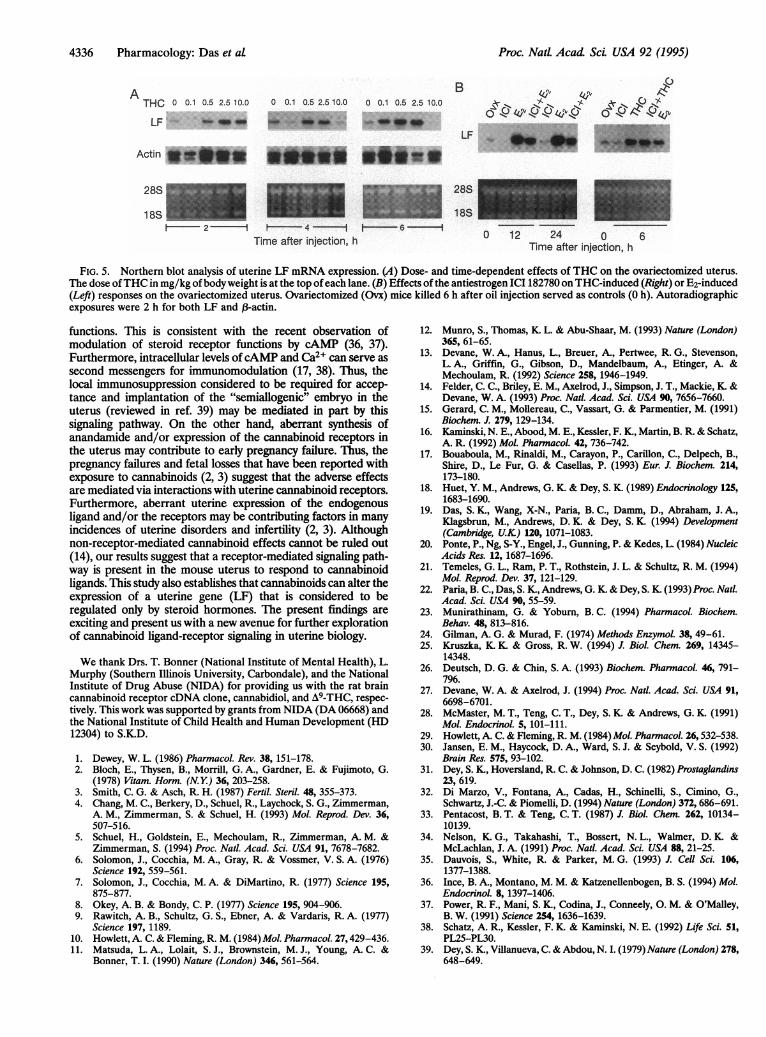
Analysis ofLF Gene Expression. Mice were ovariectomizedwithout regard to their estrous cycle and rested for 2 weeks. Todetermine the acute effects of THC, they were treated with aninjection of various doses of THC and killed at the indicatedtimes. To determine whether the THC effects were mediatedvia uterine estrogen receptors, ovariectomized mice were givenan injection of 17P/-estradiol (E2; 250 ng per mouse) or THC(2.5 mg/kg of body weight) with or without a prior (30 min)injection of a pure antiestrogen, ICI 182780 (25 ,tg per mouse),and were killed at the indicated times. THC, E2, or ICI 182780was dissolved in sesame oil and injected (0.1 ml per mouse)subcutaneously. Ovariectomized mice treated with oil (0.1 mlper mouse) served as controls. Total uterine RNA (2 ,tg) wassubjected to Northern blot hybridization with a mouse LFcomplementary RNA probe as described (28). The same blotwas reprobed with /3-actin probe, and duplicate gels werestained with acridine orange to ensure RNA integrity andequal loading. After hybridization, the blots were subjected toautoradiography and radioimage quantitation by using theradioanalytic image system (AMBIS).
RESULTS AND DISCUSSION
Analysis of Cannabinoid Receptor mRNAs in the Uterus. Asreported (11), Northern blot analysis detected a 6.0-kb tran-script in total brain or pregnant uterine poly(A)+ RNAsamples. The abundance of CB1-RmRNA was markedly lowerin the uterus. However, a predominant 1.2-kb transcript wasdetected in the uterus and showed a higher accumulation ondays 4 and 7 ofpregnancy than that on day 1 (Fig. 1A). Whetherthis smaller transcript is the result of alternate splicing orrepresents a truncated form of the receptor is not known.RT-PCR also detected CB1-R mRNA in the uterus, confirm-ing the results of Northern blot hybridization (Fig. 1B). ThismRNA was detected in the uterus during early pregnancy andin ovariectomized uteri before and after THC treatment (Fig.1B). In contrast, RT-PCR could not detect CB2-R mRNA inthe uterus, although this mRNA was detected in the rat ormouse spleen (Fig. 1C).
Pharmacology: Das et all

Proc. Natl. Acad Sci USA 92 (1995)
1234567
C+,.-1234567.1 234567
bp-182
- 539
FIG. 1. Analysis of cannabinoid receptor transcripts in the uterus.(A) Northern blot analysis of CB1-R. Lanes: 1, total RNA (6.0 jLg)from the whole brain; 2-4, poly(A)+ uterine RNA (10 j,g) from days1, 4, and 7 of pregnancy, respectively. The blot was reprobed withP-actin. Autoradiographic exposures were 2 days for CB1-R and 2 hfor p-actin. (B) Southern analysis of RT-PCR-amplified products ofCB1-R (284 bp) and p-actin (539 bp). Lanes: 1, brain; 2, day 1 pregnantuterus; 3, day 4 pregnant uterus; 4, ovariectomized uterus; 5, THC-treated ovariectomized uterus, 6, brain RNA without the RT reaction,and 7, primer control. After 2 weeks of rest, adult ovariectomized micewere given a subcutaneous injection of sesame oil (0.1 ml) orTHC (2.5mg/kg of body weight in oil) and killed 6 h later. (C) Southern analysisof RT-PCR-amplified products of CB2-R (182 bp) and ,-actin (539bp). Lanes: 1, rat spleen; 2, mouse spleen; 3, day 1 pregnant uterus; 4,day 4 pregnant uterus; 5 and 6, rat and mouse spleen RNA without theRT reaction, respectively; 7, primer control.
Effects of Cannabinoid Agonists on Forskolin-StimulatedcAMP Accumulation in the Uterus. To examine whether thecannabinoid receptor expressed in the uterus, like that in thebrain, was coupled to a G protein (Gi) (11) and biologicallyactive, the effects of cannabinoid ligands (THC or anandam-ide) on forskolin-stimulated cAMP accumulation in uterinemembrane preparations were monitored with or without per-tussis toxin pretreatment. The results show that these ligandsinhibited forskolin-stimulated cAMP accumulation in a dose-dependent manner, and this inhibition was prevented bypertussis toxin pretreatment (Fig. 2A). Anandamide was morepotent than THC in this response. However, as observed forother cannabinoid ligands (29), anandamide was less inhibitoryat higher concentrations. These effects ofTHC or anandamideappear to be specific, since an inactive and nonpsychoactivecannabinoid, CBD, did not alter this response (Fig. 2A).
Analysis of WIN 55212-2 Binding Kinetics in the Endome-trium. Although 60% of the binding in the endometrium, likethat observed in other systems (15), was nonspecific, analysisof the equilibrium binding data (Fig. 2B) indicates a single classof high-affinity binding sites with an apparent Kd of 2.4 nM andBmax of 5.4 x 10⁹ molecules per mg of protein. The Hillcoefficient was calculated to be 1, suggesting a single class ofreceptor sites and an absence of cooperative interactions.Although the binding affinity of Win 55212-2 appears to behigher in the endometrium, the binding capacity is consider-ably lower than that obtained with cerebellar sections (30).
Analysis of Autoradiographic Binding Sites in the Uterus.The possible sites of cannabinoid action in the uterus weredetermined by examining the [3H]anandamide-binding sites byautoradiography in frozen uterine sections from day 1 and day4 pregnant mice. Specific binding sites were primarily noted inuterine epithelial and stromal cells (Fig. 3).
Analysis ofAnandamide Synthesis in the Uterus. The resultsdemonstrate that the uterus, like the brain, has the capacity tosynthesize anandamide in the presence of ethanolamine (Fig.4); about 7% of the [3H]arachidonic acid was converted toanandamide by the brain or uterine homogenates. Similarresults were obtained with the use of two different solvent
._ 600-0
. 500-
E 400.OEa 300-a2
o 200-
100-
*BL FS -7 -6
Agonist, log M-5
3 4 5 6 7
Bound, fmol/mg of proteinFIG. 2. Characterization of cannabinoid receptors in the uterus.
(A) Effects of cannabinoid agonists on forskolin-stimulated cAMPaccumulation in the day 1 pregnant uterus. BL, basal; FS, forskolin-stimulation; PT, pertussis toxin pretreatment. (B) Scatchard analysisof tritiated WIN 55212-2 binding in day 4 pregnant endometrialmembranes. (Inset) Equilibrium binding kinetics. Results shown arethe average of duplicate experiments.
systems in TLC analysis as described in Materials and Methods.Furthermore, as observed for the brain (25), uterine micro-somal preparations exhibited a markedly enhanced synthesiz-ing capacity (data not shown). Of the tissues previouslyexamined (25-27), the brain has been shown to exhibit thehighest anandamide-synthesizing capacity. However, our dataprovide evidence that the anandamide-synthesizing capacity ofthe uterus is comparable to that of the brain. Although theuterus has the capacity to synthesize anandamide, it is not yetestablished whether this synthesis occurs in vivo. Arachidonicacid and ethanolamine are likely to have cooperative effects onanandamide synthesis, suggesting coactivation of phospho-lipase A2 (PLA2) and phospholipase D (PLD) (25, 27). Al-though PLA2 activity is present in the rodent uterus and isregulated by steroid hormones (31), the presence of PLD hasnot yet been examined. It is to be noted, however, that analternative pathway for the formation of anandamide throughphosphodiesterase-mediated cleavage of N-arachidonoyl-phosphatidylethanolamine has recently been described in cul-tured rat neurons (32).
Effects of THC on Uterine LF Gene Expression. To deter-mine whether THC can affect uterine functions at the molec-
B
A
28S--
18S--
bp-284
kb
-6.0
-1.2
A 800-
700--539
Actin
1 2 3 4
I I rn l
4334 Pharmacology: Das et al.
v

Proc. Natl. Acad Sci USA 92 (1995) 4335
ular level, the expression of the gene encoding LF, an estrogen-responsive gene in the mouse uterus (28, 33), was examined byNorthern blot hybridization before and after an injection ofTHC in adult ovariectomized mice. LF is an iron-bindingglycoprotein that has been proposed to be involved in immu-nomodulation and growth promotion (33, 34). THC rapidlyand transiently upregulated the levels of uterine LF mRNA(Fig. 5A). This drug at 0.5, 2.5, or 10 mg/kg of body weightincreased the mRNA levels 2- to 4-fold within 2 h, whereas itdid not influence the mRNA levels at 0.1 mg/kg until 6 h(3.5-fold) after the injection. The maximal induction of LF
Ethanolamine+ - + - +
AA
.....;
t.:,d:~i i: ' *:i'iiiiii. d:: :: ..1. ..:I ....I
Std 1 2 3 4 5 6 Std
FIG. 4. The synthesis of anandamide catalyzed by the brain oruterine homogenates. Lanes: 1 and 2, day 1 pregnant uterus; 3 and 4,day 4 pregnant uterus; 5 and 6, brain homogenates. Standards of[3H]arachidonic acid (AA) and [3H]anandamide (AM) are shown.
FIG. 3. Autoradiographic bind-ing sites of [3H]anandamide in thepregnant uterus. (A and B) Day 1pregnant uterus. (C and D) Day 4pregnant uterus. (B and D) Non-specific binding in the presence ofunlabeled anandamide. LE, lumi-nal epithelium; GE, glandular epi-thelium; S, stroma; M, myome-trium. (x25.)
mRNA levels (-5-fold) by 0.5 or 2.5 mg/kg occurred at 6 h.However, the levels obtained 2 h after an injection of THC at10 mg/kg declined by 4 h. These results suggest that theup-regulation of LF mRNA levels in the uterus by THC istime- and dose-dependent. This THC response was specific,since an inactive cannabinoid, CBD (at 0.5 or 2.5 mg/kg), didnot induce the uterine LF gene (data not shown). The re-sponses of the uterine LF gene to THC are different fromthose observed for E2. As reported previously (28), the majoraccumulation of uterine LF mRNA began 12 h after aninjection of E2. As expected, this E2-mediated response wasattenuated by pretreatment with a pure antiestrogen, ICI182780 (35). In contrast, the THC-mediated effect was notinfluenced by this antiestrogen (Fig. 5B). This suggests thatTHC influenced this uterine gene by a mechanism not involv-ing the estrogen receptor. In addition, THC did not competewith E2 for uterine nuclear estrogen receptors (data notshown), suggesting a different mode of action. The more rapidand transient response of the uterine LF gene to THCtreatment as compared with E2 treatment suggests that THCexerted this effect perhaps via its interaction with cannabinoidreceptors. The presence of CB1-R mRNA in the ovariecto-mized or THC-treated uterus (Fig. 1B) suggests that canna-binoid receptors were available for interaction with the ligands.
CONCLUSIONSince cannabinoid ligand-receptor signaling can modulate ad-enylyl cyclase and Ca2+ channel activities (14), the two importantmembers of the second-messenger system, it could be proposedthat this signaling may be important for modulating uterine
Pharmacology: Das et al
4336 Pharmacology: Das et al
ATHC 0 0.1 0.5 2.5 10.0 0 0.1 0.5 2.510.0
LF - ___» ^d
Actin3lir * /it"
Proc. Natl. Acad. Sci USA 92 (1995)
0 0.1 0.5 2.5 10.0 -
LF*:,- s-i -ri. .X
_~~~~~~~~ _o_,._.,~,o,oo X~~,'~ .
28S
18Sie 4 I -6 1
Time after injection, h 0 12 24 0 6Time after injection, h
FIG. 5. Northern blot analysis of uterine LF mRNA expression. (A) Dose- and time-dependent effects of THC on the ovariectomized uterus.The dose ofTHC in mg/kg ofbodyweight is at the top of each lane. (B) Effects of the antiestrogen ICI 182780 on THC-induced (Right) or E2-induced(Left) responses on the ovariectomized uterus. Ovariectomized (Ovx) mice killed 6 h after oil injection served as controls (0 h). Autoradiographicexposures were 2 h for both LF and ,-actin.
functions. This is consistent with the recent observation ofmodulation of steroid receptor functions by cAMP (36, 37).Furthermore, intracellular levels ofcAMP and Ca2+ can serve assecond messengers for immunomodulation (17, 38). Thus, thelocal immunosuppression considered to be required for accep-tance and implantation of the "semiallogenic" embryo in theuterus (reviewed in ref. 39) may be mediated in part by thissignaling pathway. On the other hand, aberrant synthesis ofanandamide and/or expression of the cannabinoid receptors inthe uterus may contribute to early pregnancy failure. Thus, thepregnancy failures and fetal losses that have been reported withexposure to cannabinoids (2, 3) suggest that the adverse effectsare mediated via interactions with uterine cannabinoid receptors.Furthermore, aberrant uterine expression of the endogenousligand and/or the receptors may be contributing factors in manyincidences of uterine disorders and infertility (2, 3). Althoughnon-receptor-mediated cannabinoid effects cannot be ruled out(14), our results suggest that a receptor-mediated signaling path-way is present in the mouse uterus to respond to cannabinoidligands. This study also establishes that cannabinoids can alter theexpression of a uterine gene (LF) that is considered to beregulated only by steroid hormones. The present findings areexciting and present us with a new avenue for further explorationof cannabinoid ligand-receptor signaling in uterine biology.We thank Drs. T. Bonner (National Institute of Mental Health), L.
Murphy (Southern Illinois University, Carbondale), and the NationalInstitute of Drug Abuse (NIDA) for providing us with the rat braincannabinoid receptor cDNA clone, cannabidiol, and A9-THC, respec-tively. This work was supported by grants from NIDA (DA 06668) andthe National Institute of Child Health and Human Development (HD12304) to S.K.D.
1. Dewey, W. L. (1986) Pharmacol. Rev. 38, 151-178.2. Bloch, E., Thysen, B., Morrill, G. A., Gardner, E. & Fujimoto, G.
(1978) Vitam. Horm. (N.Y.) 36, 203-258.3. Smith, C. G. & Asch, R. H. (1987) Fertil. Steril. 48, 355-373.4. Chang, M. C., Berkery, D., Schuel, R., Laychock, S. G., Zimmerman,
A. M., Zimmerman, S. & Schuel, H. (1993) Mol. Reprod. Dev. 36,507-516.
5. Schuel, H., Goldstein, E., Mechoulam, R., Zimmerman, A. M. &Zimmerman, S. (1994) Proc. Natl. Acad. Sci. USA 91, 7678-7682.
6. Solomon, J., Cocchia, M. A., Gray, R. & Vossmer, V. S. A. (1976)Science 192, 559-561.
7. Solomon, J., Cocchia, M. A. & DiMartino, R. (1977) Science 195,875-877.
8. Okey, A. B. & Bondy, C. P. (1977) Science 195, 904-906.9. Rawitch, A. B., Schultz, G. S., Ebner, A. & Vardaris, R. A. (1977)
Science 197, 1189.10. Howlett, A. C. & Fleming, R. M. (1984) Mol. Pharmacol. 27,429-436.11. Matsuda, L. A., Lolait, S. J., Brownstein, M. J., Young, A. C. &
Bonner, T. I. (1990) Nature (London) 346, 561-564.
12.
13.
14.
15.
16.
17.
18.
19.
20.
21.
22.
23.
24.25.
26.
27.
28.
29.30.
31.
32.
33.
34.
35.
36.
37.
38.
39.
Munro, S., Thomas, K. L. & Abu-Shaar, M. (1993) Nature (London)365, 61-65.Devane, W. A., Hanus, L., Breuer, A., Pertwee, R. G., Stevenson,L.A., Griffin, G., Gibson, D., Mandelbaum, A., Etinger, A. &Mechoulam, R. (1992) Science 258, 1946-1949.Felder, C. C., Briley, E. M., Axelrod, J., Simpson, J. T., Mackie, K. &Devane, W. A. (1993) Proc. Natl. Acad. Sci. USA 90, 7656-7660.Gerard, C. M., Mollereau, C., Vassart, G. & Parmentier, M. (1991)Biochem. J. 279, 129-134.Kaminski, N. E., Abood, M. E., Kessler, F. K., Martin, B. R. & Schatz,A. R. (1992) Mol. Pharmacol. 42, 736-742.Bouaboula, M., Rinaldi, M., Carayon, P., Carillon, C., Delpech, B.,Shire, D., Le Fur, G. & Casellas, P. (1993) Eur. J. Biochem. 214,173-180.Huet, Y. M., Andrews, G. K. & Dey, S. K. (1989) Endocrinology 125,1683-1690.Das, S. K., Wang, X-N., Paria, B. C., Damm, D., Abraham, J. A.,Klagsbrun, M., Andrews, D. K. & Dey, S. K. (1994) Development(Cambridge, U.K) 120, 1071-1083.Ponte, P., Ng, S-Y., Engel, J., Gunning, P. & Kedes, L. (1984) NucleicAcids Res. 12, 1687-1696.Temeles, G. L., Ram, P. T., Rothstein, J. L. & Schultz, R. M. (1994)Mol. Reprod. Dev. 37, 121-129.Paria, B. C., Das, S. K., Andrews, G. K. & Dey, S. K. (1993) Proc. Natl.Acad. Sci. USA 90, 55-59.Munirathinam, G. & Yoburn, B. C. (1994) Pharmacol. Biochem.Behav. 48, 813-816.Gilman, A. G. & Murad, F. (1974) Methods Enzymol. 38, 49-61.Kruszka, K. K. & Gross, R. W. (1994) J. Biol. Chem. 269, 14345-14348.Deutsch, D. G. & Chin, S. A. (1993) Biochem. Pharmacol. 46, 791-796.Devane, W. A. & Axelrod, J. (1994) Proc. Natl. Acad. Sci. USA 91,6698-6701.McMaster, M. T., Teng, C. T., Dey, S. K. & Andrews, G. K. (1991)Mol. Endocrinol. 5, 101-111.Howlett, A. C. & Fleming, R. M. (1984) Mol. Pharmacol. 26,532-538.Jansen, E. M., Haycock, D. A., Ward, S. J. & Seybold, V. S. (1992)Brain Res. 575, 93-102.Dey, S. K., Hoversland, R. C. & Johnson, D. C. (1982) Prostaglandins23, 619.Di Marzo, V., Fontana, A., Cadas, H., Schinelli, S., Cimino, G.,Schwartz, J.-C. & Piomelli, D. (1994) Nature (London) 372, 686-691.Pentacost, B. T. & Teng, C. T. (1987) J. Biol. Chem. 262, 10134-10139.Nelson, K. G., Takahashi, T., Bossert, N. L., Walmer, D. K. &McLachlan, J. A. (1991) Proc. Natl. Acad. Sci. USA 88, 21-25.Dauvois, S., White, R. & Parker, M. G. (1993) J. Cell Sci. 106,1377-1388.Ince, B. A., Montano, M. M. & Katzenellenbogen, B. S. (1994) Mol.Endocrinol. 8, 1397-1406.Power, R. F., Mani, S. K., Codina, J., Conneely, O. M. & O'Malley,B. W. (1991) Science 254, 1636-1639.Schatz, A. R., Kessler, F. K. & Kaminski, N. E. (1992) Life Sci. 51,PL25-PL30.Dey, S. K., Villanueva, C. & Abdou, N. I. (1979) Nature (London) 278,648-649.