reception, transduction, response ligand is the fancy word for signaling molecule. energy can be...
TRANSCRIPT

The Nervous System
Reception, Transduction, Response
Ligand is the fancy word for signaling molecule.
Energy can be in the form of ions
Signal Transduction Pathway

Step 1: Reception…What 3 words must you say?
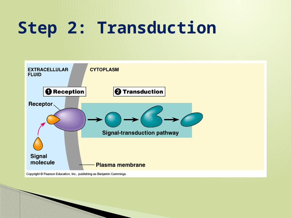
Step 2: Transduction
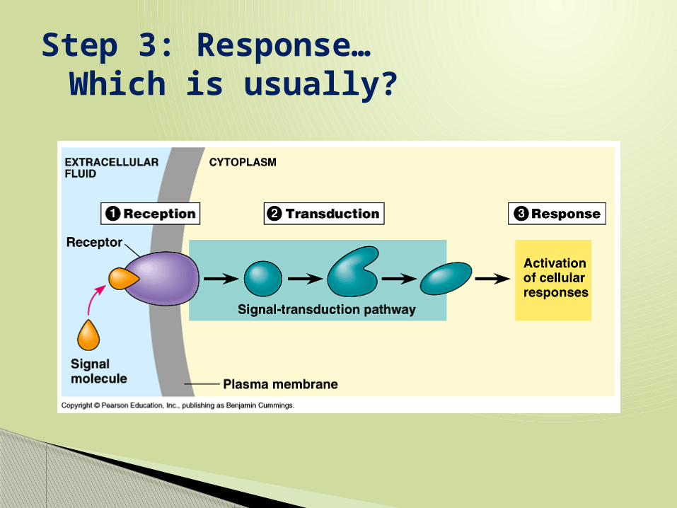
Step 3: Response…Which is usually?
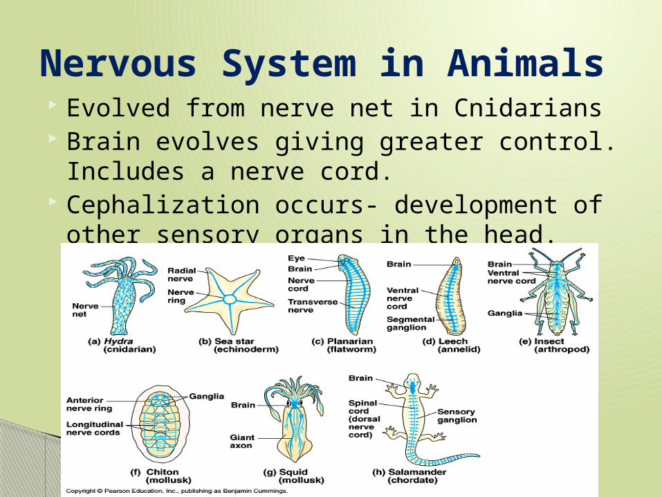
Evolved from nerve net in Cnidarians Brain evolves giving greater control.
Includes a nerve cord. Cephalization occurs- development of other
sensory organs in the head.
Nervous System in Animals

A stimulus is a form of energy like light (electromagnetic) or pressure (mechanical), or sound waves.
Stimulus

Overview of the Nervous System

1. Sensory Input-Sensory receptors receive a stimulus and send it into the brain/ spinal cord.
2. Integration- the CNS integrates/interprets/thinks about the sensory input (stimulus).
3. Motor Output- Impulse sent from the brain to the muscles to respond. Effector cells in muscles and glands will respond.
Peripheral Nervous System-has sensory receptors and motor nerves.
Nervous System in Animals

CNS vs. PNS

Cell Body- receives stimuli from all dendrites, and creates one signal
Dendrites- carry stimuli into the cell body Axon- carries signal away from cell body
and towards next neuron. Myelin Sheath- lipid covering over axon for
insulation. Composed of Schwann cells (PNS)
Synaptic Terminal- end of axon Synapse- gap between neurons or neuron
and effector cell.
Neurons- nerve cells

Neuron structure
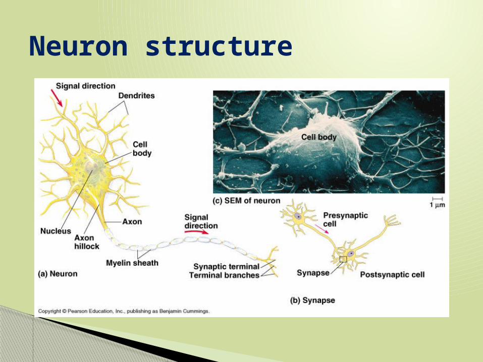
Neuron structure
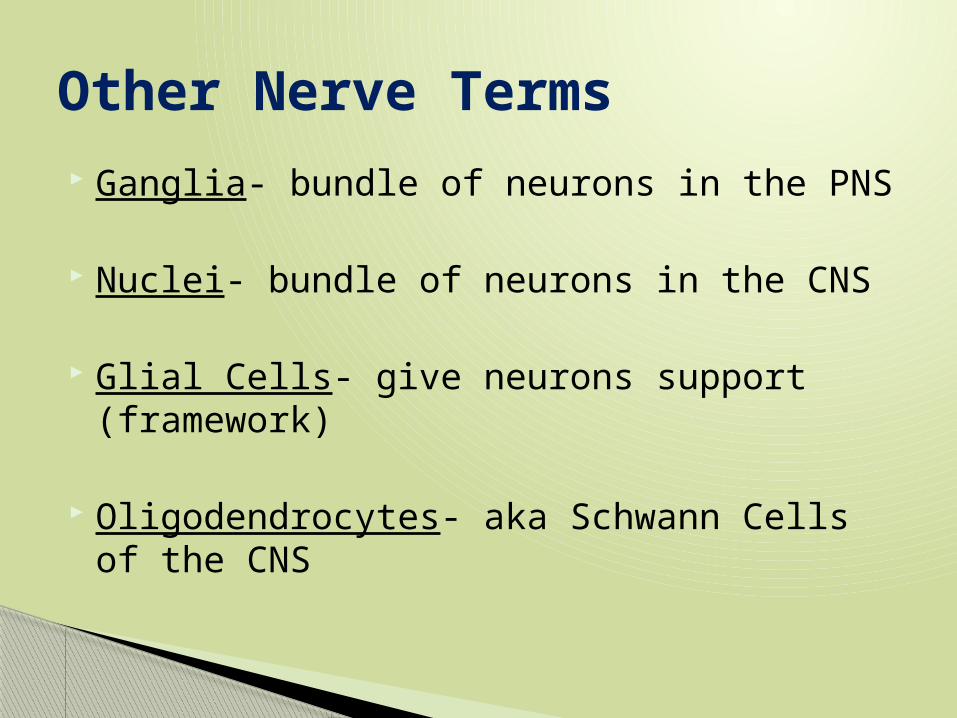
Ganglia- bundle of neurons in the PNS
Nuclei- bundle of neurons in the CNS
Glial Cells- give neurons support (framework)
Oligodendrocytes- aka Schwann Cells of the CNS
Other Nerve Terms

Schwann Cells OR Oligiodendrocytes
Axon Nodes ofRanvier
Schwanncell
Myelin sheathNucleus ofSchwann cell
Schwanncell
Nodes of Ranvier
Layers of myelinAxon
0.1 µm
Multiple Sclerosis- MS- Schwann Cells die in CNS & PNS and causes the signal (electrical current) to burn muscles into permanent contractions.

Data Set Question 1 (U4, D1)

Ions can be considered ___________ Concentration gradients are
________________ and so they can be considered _____________
Active transport requires _______________ . Diagram a cell pump.
Remember…
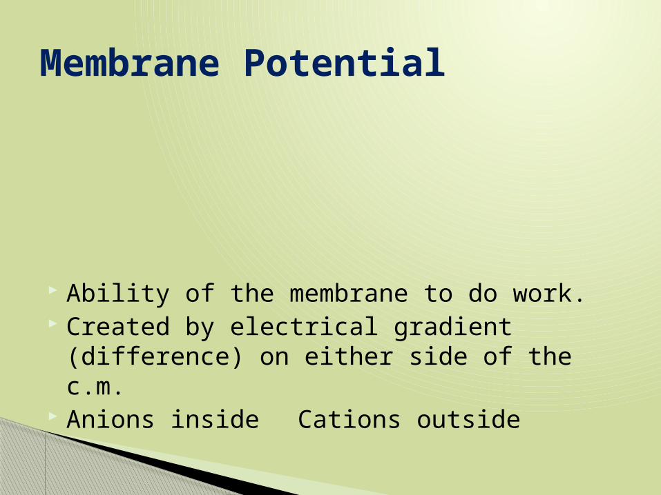
Ability of the membrane to do work. Created by electrical gradient (difference)
on either side of the c.m. Anions inside Cations outside
Membrane Potential
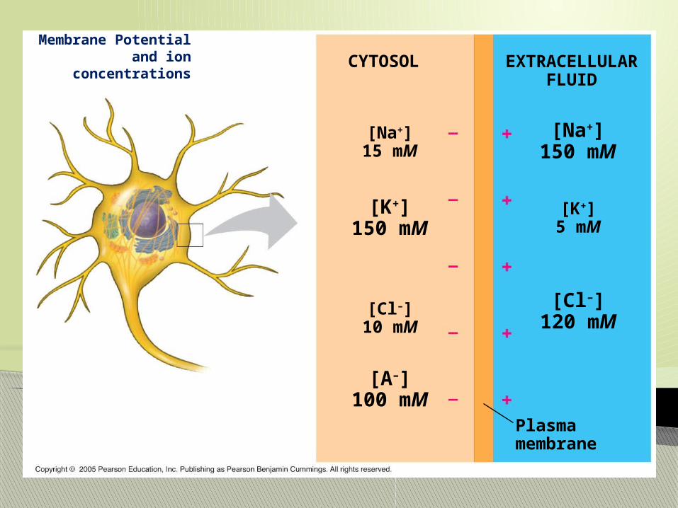
Membrane Potential and ion
concentrationsCYTOSOL EXTRACELLULAR
FLUID
[Na+]15 mM
[K+]150 mM
[A–]100 mM
[Na+]150 mM
[K+]5 mM
[Cl–]120 mM
[Cl–]10 mM
Plasmamembrane
Resting Potential- Unstimulated neuron, need to establish the [gradient]
1. NaK Pump responsible for generating nerve impulse.
◦ NaK Pumps are either ligand gated or voltage gated, which helps create gradient faster.
Resting Potential

Na+/K+ pumps
Cytoplasmic Na+ bonds tothe sodium-potassium pump
CYTOPLASMNa+
[Na+] low[K+] high
Na+
Na+
EXTRACELLULARFLUID
[Na+] high[K+] low
Na+
Na+
Na+
ATP
ADP
P
Na+ binding stimulatesphosphorylation by ATP.
Na+
Na+
Na+
K+
Phosphorylation causesthe protein to change itsconformation, expelling Na+
to the outside.
P
Extracellular K+ bindsto the protein, triggeringrelease of the phosphategroup.
PP
Loss of the phosphaterestores the protein’soriginal conformation.
K+ is released and Na+
sites are receptive again;the cycle repeats.
K+
K+
K+
K+
K+

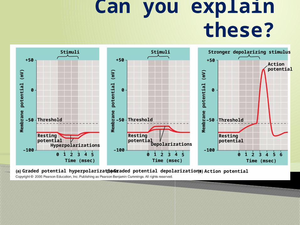
1. Depolarization- destroys membrane potential, Na floods into cell◦ Depolarization is “graded”◦ Threshold potential-minimum Na that must
enter to generate a nerve impulse◦ Action Potential- “Spike” electrical generated
impulse, ana ction will occur
2. Repolarization- neuron pumps out K to try and return to resting potential.
Action Potential Steps
3. Hyperpolarization- the cell will pull in some K to get back to resting potential.◦ Must Hyperpolarize so that the neuron can get
back to resting potential, and to recreate the [gradient]/ polarity
4. Refractory Period- neuron can’t make new impulse
Action Potential Steps
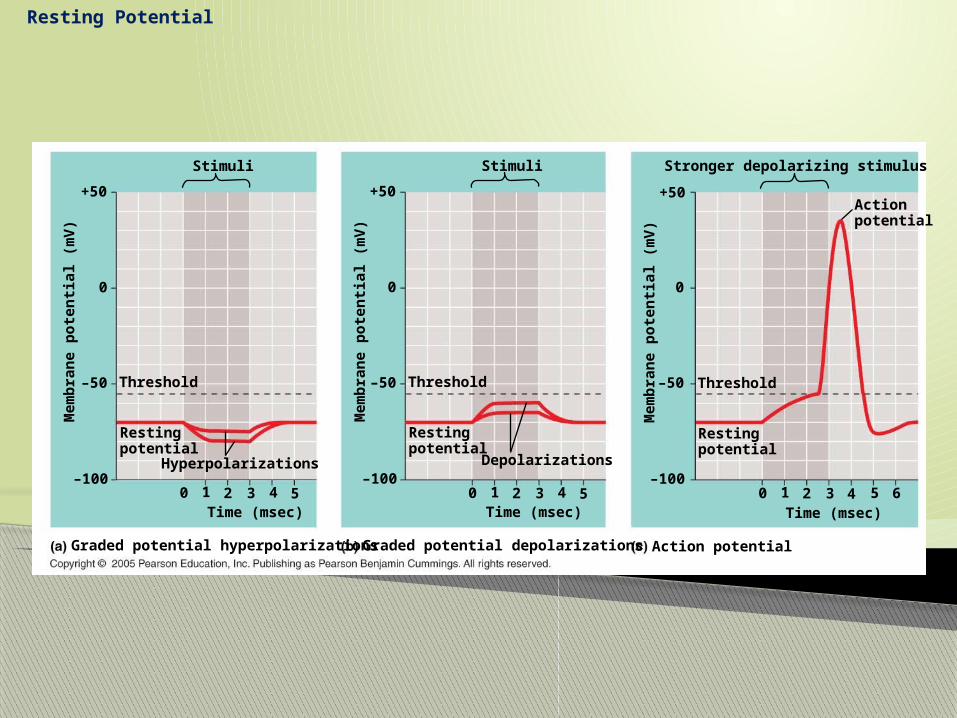
Resting Potential
Hyperpolarizations
Graded potential hyperpolarizations Graded potential depolarizations
5Time (msec)
Restingpotential
43210
Threshold
–100
–50
0
Mem
bra
ne
po
ten
tial
(m
V)
Stimuli
+50
Depolarizations
5Time (msec)
Restingpotential
43210
Threshold
–100
–50
0
Mem
bra
ne
po
ten
tial
(m
V)
Stimuli
+50
Action potential
5Time (msec)
Restingpotential
43210
Threshold
–100
–50
0
Mem
bra
ne
po
ten
tial
(m
V)
Stronger depolarizing stimulus
+50Actionpotential
6
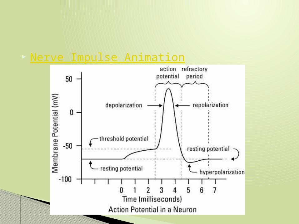
Nerve Impulse Animation
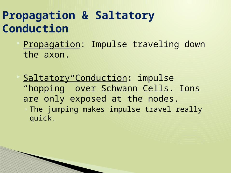
Propagation: Impulse traveling down the axon.
Saltatory Conduction: impulse “hopping” over Schwann Cells. Ions are only exposed at the nodes.◦ The jumping makes impulse travel really quick.
Propagation & Saltatory Conduction
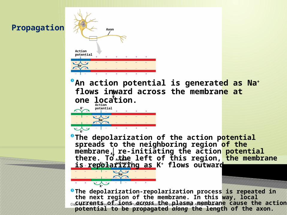
Propagation
An action potential is generated as Na+ flows inward across the membrane at one location.
Na+
Action potential
Axon
Na+
Action potentialK+
The depolarization of the action potential spreads to the neighboring region of the membrane, re-initiating the action potential there. To the left of this region, the membrane is repolarizing as K+ flows outward.
K+
Na+
Action potentialK+
The depolarization-repolarization process is repeated in the next region of the membrane. In this way, local currents of ions across the plasma membrane cause the action potential to be propagated along the length of the axon.
K+

Saltatory Conduction
Cell body
Schwann cell
Depolarized region(node of Ranvier)
Myelinsheath
Axon

1. Sensory Neuron- receive stimulus 2. Interneuron- in CNS (spinal cord) takes
sesory imput and gives signal to motor neuron
3. Motor Neuron- carries energy to effector cell. ( ________/__________)
This is why you don’t think about a reflex, the signal never made it to the brain for integration.
Reflex Arc-Simplest Neural Pathway

Reflex Arc

Data Set Question 2

Diffusion is _________ and uses no ________
Ligands bind to receptor proteins and cause a:
Do you think all ligands cause the same response?
Remember
Where are synapses located? ______&________
Draw 2 neurons & label the synapse

2 Types of Synapses◦ 1. Electrical- direct cell contact, in brain◦ 2. Chemical- most common in animals- requires a
neurotransmitter (chemical ligand)
Synapses and Nerve Impulses
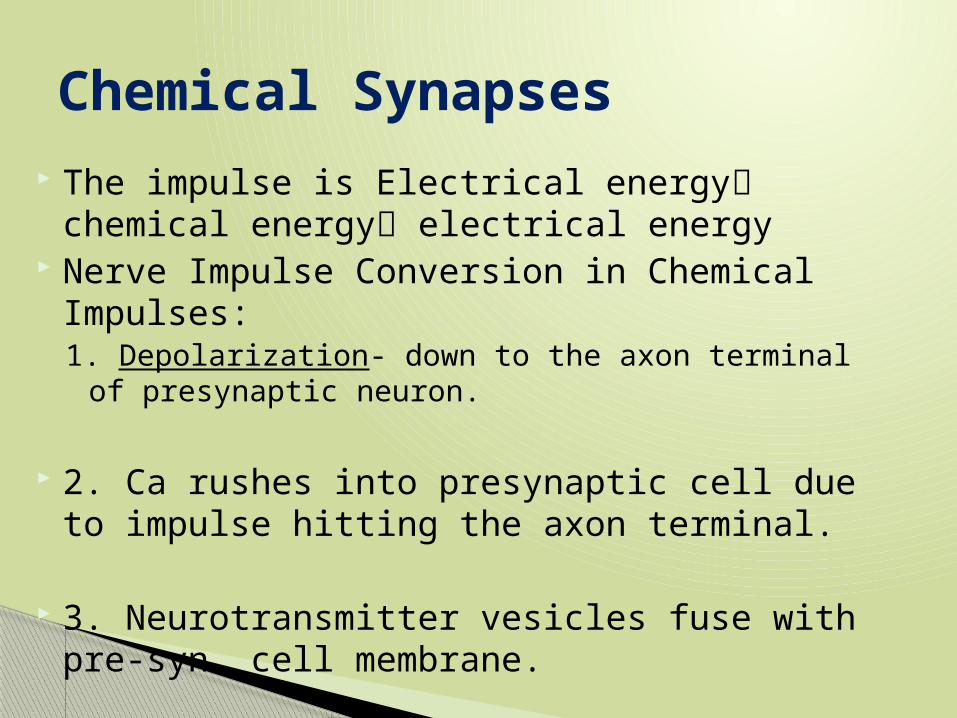
The impulse is Electrical energy chemical energy electrical energy
Nerve Impulse Conversion in Chemical Impulses:1. Depolarization- down to the axon terminal of
presynaptic neuron.
2. Ca rushes into presynaptic cell due to impulse hitting the axon terminal.
3. Neurotransmitter vesicles fuse with pre-syn. cell membrane.
Chemical Synapses

4. Neurotransmitter released into synapse
5. Neurotransmitter binds to receptor protein on post syn cell and causes a CSC
6. Na floods into post syn. Cell and causes depolarization.
Nerve Impulse cont.
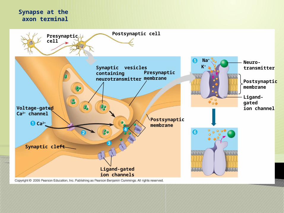
Synapse at the axon terminal
Postsynaptic cellPresynaptic cell
Synaptic vesiclescontainingneurotransmitter
Presynaptic membrane
Voltage-gatedCa2+ channel
Ca2+Postsynaptic membrane
Postsynaptic membrane
Neuro-transmitter
Ligand-gatedion channel
Na+
K+
Ligand-gatedion channels
Synaptic cleft

IPSP & EPSP
Excitatory Post Synaptic Potential- causes Post syn. Cell to do act or keep impulse going
Inhibitory Post Synaptic Potential- causes Post syn. Cell to stop impulse transmission
Summation- adding of all dendrite stimuli to reach threshold potential
EPSP & IPSP
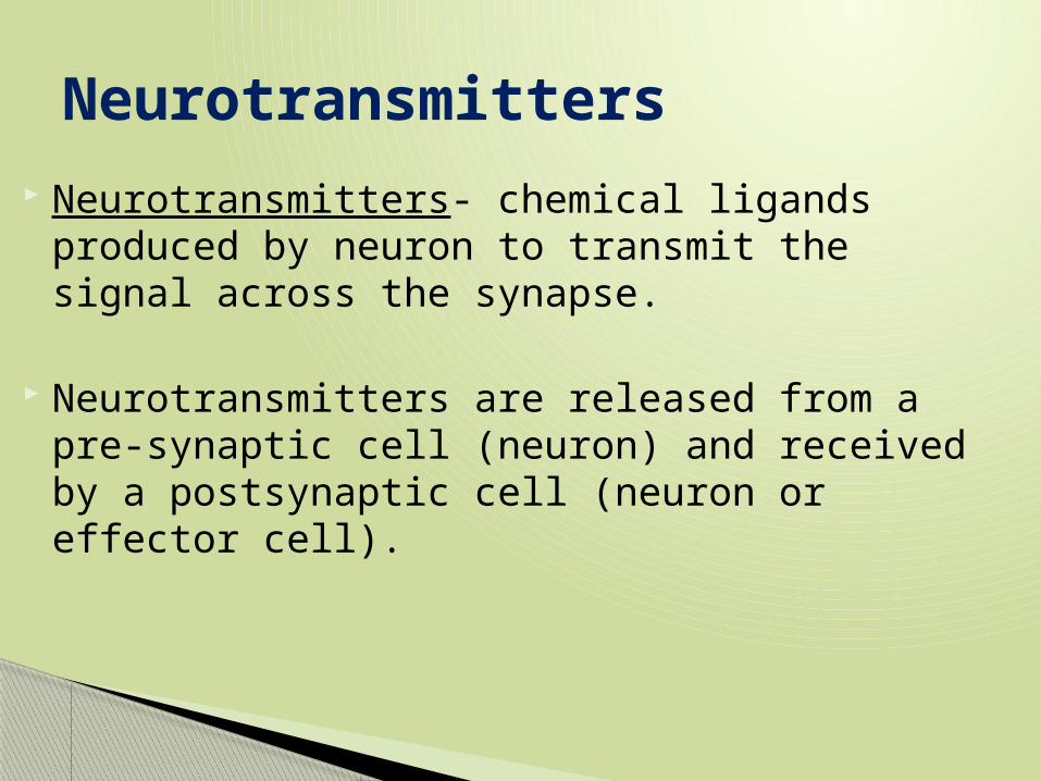
Neurotransmitters- chemical ligands produced by neuron to transmit the signal across the synapse.
Neurotransmitters are released from a pre-synaptic cell (neuron) and received by a postsynaptic cell (neuron or effector cell).
Neurotransmitters

Acetylcholine- (ACh)makes muscles contract in PNS, can be excitatory or inhibitory in CNS.
Cholinesterase breaks down ACh
Biogenic Amines◦ 1 & 2. Epinephrine and norepinephrine- fight
or flight, speeds up body functions◦ 3.Dopamine=happy◦ 4. Serotonin=sleep *both out of whack in
ADD/Schiz
Neurotransmitters

Amino Acids◦ 1. Substance P- relays pain stimulus◦ 2. Endorphins- block Substance P “second wind”
Gases- work by diffusion◦ 1. NO◦ 2. CO
*both inhibit nerve signaling and muscle contractions
Neurotransmitters

Data Set Question 3
Hyperpolarizations
Graded potential hyperpolarizations Graded potential depolarizations
5Time (msec)
Restingpotential
43210
Threshold
–100
–50
0
Mem
bra
ne
po
ten
tial
(m
V)
Stimuli
+50
Depolarizations
5Time (msec)
Restingpotential
43210
Threshold
–100
–50
0
Mem
bra
ne
po
ten
tial
(m
V)
Stimuli
+50
Action potential
5Time (msec)
Restingpotential
43210
Threshold
–100
–50
0
Mem
bra
ne
po
ten
tial
(m
V)
Stronger depolarizing stimulus
+50Actionpotential
6
Can you explain these?

Where do you think a stimulus is converted and amplified in STP?

1. Sensation- action potential is at the brain, and senses a nerve impulse.
2. Perception- integration of sensation by brain
Sense Perception

Special neurons detect stimuli. Stimuli will be detected by their
____________.
Stimuli is defined as _________________ That will cause a _ _ _
Sensory Reception
1. Summation will cause Threshold potential to be reached.
2. Amplification can occur on the way to the CNS.
3. Saltatory conduction is responsible for signal propagation.
4. Integration by CNS for appropriate response.
Sensory Transduction Pathway
Decrease in continuous stimulus coming into the CNS.◦ CLOTHING DETECTED BY BODY, BUT IS NOT
RESPONDING.
Sensory Adaptation

1. Internorepectors- detect internal stimuli-pressure, balance, homeostasis
2. Externoreceptors- external stimuli Mechanoreceptors- detect bend/stretch of
membranes/hairs Nociceptors-detect pain using Substance P Thermoreceptors- detect cold Chemoreceptors-detect cheimicals: osmo-water,
gustatory-taste, olfactory-smell Electromagnetic receptors-detect photo-light,
electro- electrical, magno-magnetic
Types of Sensory Receptors

Stimulus receptorsLighttouch
Pain Cold HairHeat
Hairmovement
Strongpressure
Hypodermis
Nerve Connectivetissue
Dermis
Epidermis
Accomplished by mechanoreceptors in the inner ear.
Hairs bend, mechanoreceptors detect this, cause a depolarization of auditory nerves and create action potential.
Lateral lines in fish Tympanum in insects and amphibians
Sensation of Hearing

Accomplished by mechanoreceptors in the inner ear
Sensation of Balance and Motion

Accomplished by chemoreceptors in the nose (olfactory) and mouth (gustatory).
Or hairs if you’re a bug!
Taste is 80% smell and 20% tase
Five senses of taste:◦ Sweet◦ Sour◦ Bitter◦ Salty◦ Umami
Sensation of Taste
All animals have photoreceptors- detect colors
Some photoreceptors contain photopigments that detect color
Sensation of Sight

Second biggest consumer of ATP Must overcome friction and gravity
Animals move in/on: water, land, air
Locomotion

Muscles provide a pulling force
Motor Unit= muscle and corresponding motor nerve
Muscle Structure & Function

1. ACh attaches to receptor proteins on muscle cell.
2. Depolarization occurs (release of Na)
3. Na causes Ca to be released. Ca is a secondary messenger.
4. Ultimately it causes two different proteins actin and myosin to slide over one another.
Myosin pulls on actin.
Muscle Contraction Steps- Sliding Filament Theory
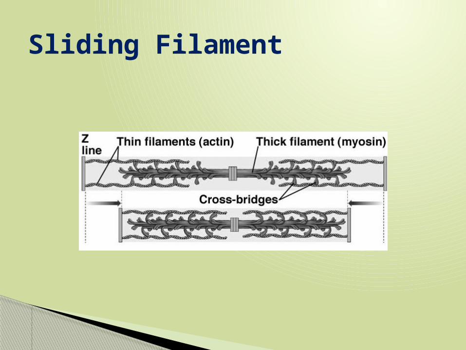
Sliding Filament

Acetylcholinesterase breaks down ACh and actin and myosin slide back to original position.
How do muscles relax?

Synapse and Neurotransmitter