signaling pathways coupled to activation of the kinin b1 receptor
TRANSCRIPT
5
Signaling Pathways Coupled to Activation of the Kinin B1 Receptor
Pamela Ehrenfeld, Carlos D. Figueroa, Kanti D. Bhoola and Carola E. Matus
Laboratory of Cellular Pathology, Institute of Anatomy, Histology and Pathology, Universidad Austral de Chile, Valdivia
Chile
1. Introduction
1.1 The field of kinins and their receptors
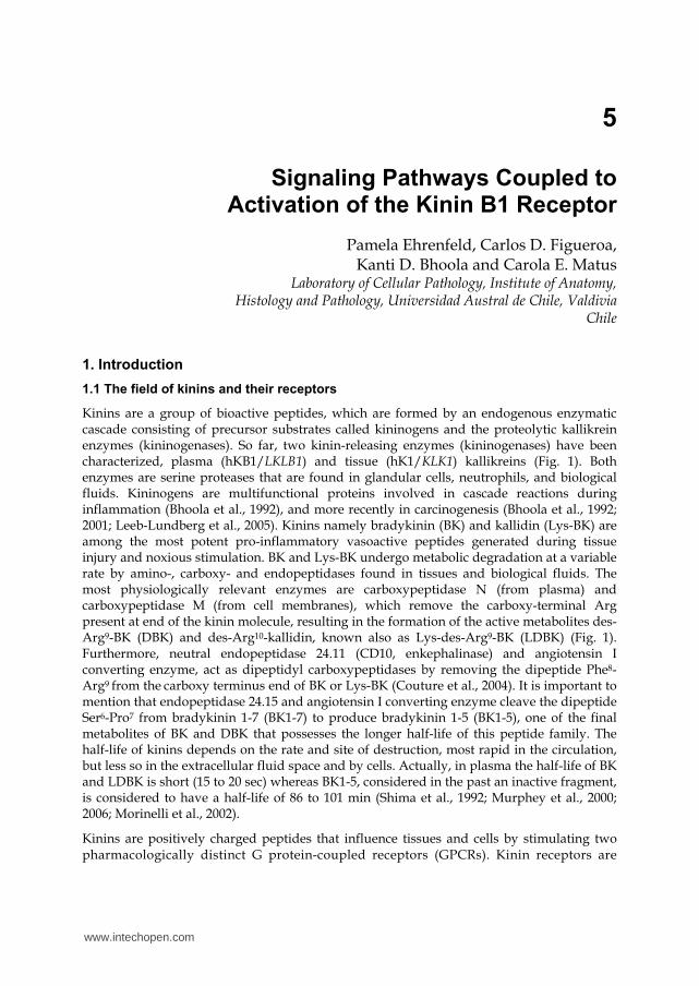
Kinins are a group of bioactive peptides, which are formed by an endogenous enzymatic cascade consisting of precursor substrates called kininogens and the proteolytic kallikrein enzymes (kininogenases). So far, two kinin-releasing enzymes (kininogenases) have been characterized, plasma (hKB1/LKLB1) and tissue (hK1/KLK1) kallikreins (Fig. 1). Both enzymes are serine proteases that are found in glandular cells, neutrophils, and biological fluids. Kininogens are multifunctional proteins involved in cascade reactions during inflammation (Bhoola et al., 1992), and more recently in carcinogenesis (Bhoola et al., 1992; 2001; Leeb-Lundberg et al., 2005). Kinins namely bradykinin (BK) and kallidin (Lys-BK) are among the most potent pro-inflammatory vasoactive peptides generated during tissue injury and noxious stimulation. BK and Lys-BK undergo metabolic degradation at a variable rate by amino-, carboxy- and endopeptidases found in tissues and biological fluids. The most physiologically relevant enzymes are carboxypeptidase N (from plasma) and carboxypeptidase M (from cell membranes), which remove the carboxy-terminal Arg present at end of the kinin molecule, resulting in the formation of the active metabolites des-Arg9-BK (DBK) and des-Arg10-kallidin, known also as Lys-des-Arg9-BK (LDBK) (Fig. 1). Furthermore, neutral endopeptidase 24.11 (CD10, enkephalinase) and angiotensin I converting enzyme, act as dipeptidyl carboxypeptidases by removing the dipeptide Phe8-Arg9 from the carboxy terminus end of BK or Lys-BK (Couture et al., 2004). It is important to mention that endopeptidase 24.15 and angiotensin I converting enzyme cleave the dipeptide Ser6-Pro7 from bradykinin 1-7 (BK1-7) to produce bradykinin 1-5 (BK1-5), one of the final metabolites of BK and DBK that possesses the longer half-life of this peptide family. The half-life of kinins depends on the rate and site of destruction, most rapid in the circulation, but less so in the extracellular fluid space and by cells. Actually, in plasma the half-life of BK and LDBK is short (15 to 20 sec) whereas BK1-5, considered in the past an inactive fragment, is considered to have a half-life of 86 to 101 min (Shima et al., 1992; Murphey et al., 2000; 2006; Morinelli et al., 2002).
Kinins are positively charged peptides that influence tissues and cells by stimulating two pharmacologically distinct G protein-coupled receptors (GPCRs). Kinin receptors are
www.intechopen.com
Advances in Protein Kinases
110
Fig. 1. Major components of the kinin system. The kininogenases plasma and tissue kallikreins hydrolize either high or low molecular weight kininogens to release the kinin domain. Carboxypeptidases remove the Arg9 from bradykinin or Lys-bradykinin to generate B1 receptor agonists.
situated on the plasma membranes of many cell types and are coupled principally to G┙q and G┙i, (Austin et al., 1997; Regoli et al., 2001). These receptors are designated as kinin B1 (B1R) or B2 (B2R) receptors (Fig.1). B2R is a preformed receptor, widely distributed and activated by the parent molecules BK and Lys-BK. B1R is activated by DBK and LDBK, two kinins that lack the Arg9 at the carboxy-terminus of the parental BK or LBK molecule (Bhoola et al., 1992; 2001; Leeb-Lundberg et al., 2005).
B1R is generally expressed at low levels in normal tissues, but is strongly up-regulated during inflammation, tissue injury, cancer, exposure to bacterial endotoxins such as lypopolysaccharide, growth factors (epidermal growth factor and endothelial growth factor) cytokines (principally interleukin-l┚, interleukin-8 and tumor necrosis factor-┙) or even its own agonist, LDBK (Marceau et al., 1998; Yang et al., 2001). For this reason, there is an increase in the number of B1 binding sites in inflamed or carcinogenic tissue. Moreover, an up-regulation of the kinin B1R has been found in several tumors, immune-modulated disorders such as rheumatoid arthritis, transplant rejection, glomerulonephritis and in human fibrotic lung tissue.
Additional differences between both types of kinin receptors reside in the fact that the B2R is internalized and extensively recycled to the cell surface by its agonist, whereas the B1R is up-regulated in inflammatory disorders and cancer, and its expression is controlled by signaling pathways such as stress mitogen-activated protein and nuclear factor kappa B
www.intechopen.com
Signaling Pathways Coupled to Activation of the Kinin B1 Receptor
111
(NF-κB) (Marceau et al., 2002). In fact, sequence analyses show the presence of a transcriptional regulatory site for NF-κB in the promoter region of the B1R (Bachvarov et al., 1996; Ni et al., 1998) a finding that may explain why expression of the B1R gene is tissue and cell specific (Yang et al., 2001).
The different cell types in which expression and activation of kinin receptors has been determined include endothelial, epithelial and neural cells, smooth muscle cells, neutrophils, lymphocytes, monocytes, keratinocytes, chondrocytes and fibroblasts. Depending upon the cell type involved, kinin receptors activate different intracellular signaling pathways, which regulate processes such as, cell proliferation, differentiation, migration, vascular permeability, contraction of smooth muscle, excitation of nerve endings and release of a variety of biologically active secondary mediators. In this chapter, we will focus primarily on the most important signaling pathways triggered after B1R stimulation.
2. Structure of kinin B1 receptor
In general, the information on the structure of kinin receptors has been obtained by chemical
cross-linking experiments, mutagenesis approaches, classical pharmacological assays, and studies with domain-specific anti-receptor antibodies (Prado et al., 2002; Blaukat, 2004). All
these approaches have shown that B1R belongs to the GPCR family and that it is integrated
into the plasma membrane. The amino-terminus end is exposed to the extracellular space and contains (three) consensus sites of the Asn-Xaa-Ser/Thr type for N-glycosilation and the
most of them are clustered on their N-terminal domains. Is noteworthy that the kinin-binding site is located at the amino-terminus end of the third extracellular loop. According
to Kang et al.,(2005) the presence of multiple bands observed for the recombinant expressed human B1R is strongly reminiscent of patterns of the partially glycosylated B2R. However, a
B1R in which the N-terminal domain had been truncated to remove putative N-glycosylation sites migrated as a homogeneous specie (Kang et al., 2005). Furthermore, the
treatment of the hemagglutinin-tagged human B1R, expressed in HEK293 cells, with N-glycosidase F resulted in the conversion of the receptor from a heterogeneous specie
migrating at 35 to 45 kDa to a relatively homogeneous one migrating at 37 kDa. We have observed a similar electrophoretic pattern of the B1R protein in human neutrophils
(Ehrenfeld et al., 2006), breast cancer cells (Molina et al., 2009), endothelial cells and fibroblasts (our group, unpublished data). It is postulated that glycosylation probably
increases the hydrophilicity of the extracellular portions of B1R affecting or regulating ligand affinity, efficient G protein coupling, maturation (folding, stabilization), intracellular
trafficking and receptor oligomerization or receptor degradation (Menke et al., 1994; MacNeil et al., 1995; Leeb-Lundberg et al., 2005). Even more, it has been demonstrated that
kinin-mediated mitogenic signaling and prostate cell growth is blocked by B1 and B2 receptor antagonists indicating that these effects depend on both kinin receptors. These
results provide evidence for the existence of B1R/B2R heterodimers in PC3 prostate cancer cells and demonstrate that antagonism of one receptor interferes with the ability of the
other, possibly at the level of receptor-Gq protein coupling (Barki-Harrington et al., 2003). It is known that B1R can dimerize, oligomerize or heterodimerize with the B2R or other molecule. Actually, it is known that B1R heterodimerizes with membrane carboxypeptidase
M, facilitating receptor signaling via carboxypeptidase M-mediated conversion of
bradykinin or kallidin to a des-[Arg9]kinin. This critical interaction potentiates B1R signaling
www.intechopen.com
Advances in Protein Kinases
112
and uncovers a new mode of GPCR activation by a cell surface peptidase (Zhang et al., 2011). Therefore, it is clear that knowledge of the molecular structure has been important in
determining B1R function.
3. Classical signaling pathways activated by kinin B1 receptor
In general, B1R activates most of the signaling pathways activated by B2R (reviewed by Marceau et al., 1998 and Leeb-Lundberg et al., 2005). Although both kinin receptors are coupled to similar signal transduction pathways, the differences in patterns of signaling are due to different degrees of short-term regulation that include both receptor desensitization and internalization (Leeb-Lundberg et al., 2005). The B2R is stable in the absence of its agonist, but it is rapidly desensitized after ligand stimulation by a mechanism that includes
recruitment of -arrestin 2, internalization in a clathrin-dependent manner and recycling upon agonist treatment (Enquist et al., 2007). The B1R is not phosphorylated either under basal conditions or in response to agonist (Blaukat et al., 1999) and desensitizes slightly upon further stimulation with its agonist (Mathis et al., 1996). Actually, the B1R is constitutively internalized in the absence of agonist via a clathrin-dependent pathway, do
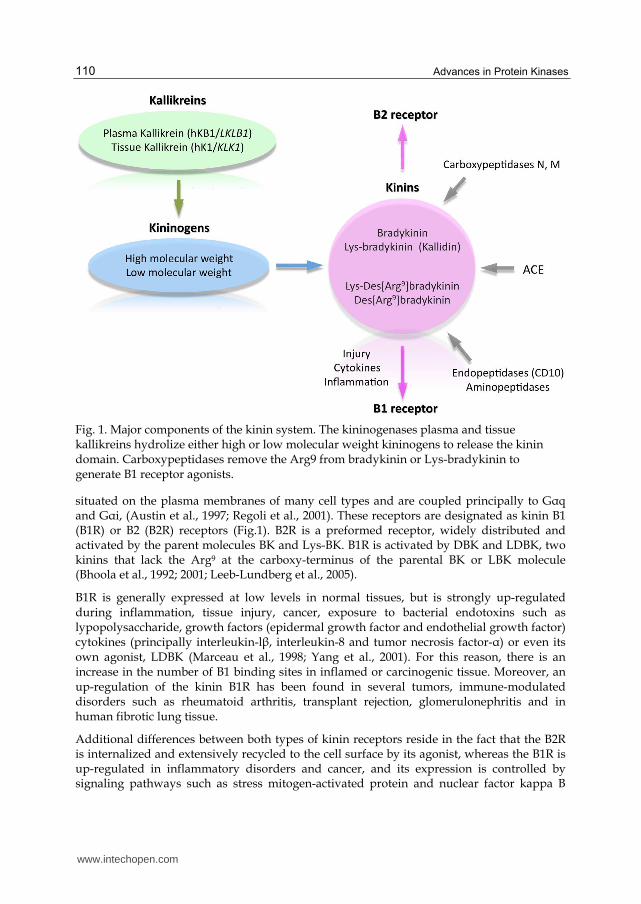
not recruit -arrestin 2, bind G protein-coupled receptor sorting protein and go to lysosomes for degradation. Binding of agonist to the B1R inhibits its constitutive internalization thereby reducing the rate of spontaneous clearance of receptors from the cell membrane and at the same time delaying B1R degradation (Enquist et al., 2007). This behavior is according with B1R up-regulation by cytokines and other inflammatory stimuli that result in a high number of receptors on the cell surface, available for ligand binding (Fig. 2).
Fig. 2. Regulation of B1R levels on the cell membrane. The B1R is constitutively internalized, but the number of B1R molecules on the cell membrane increases after binding of its ligand.
www.intechopen.com
Signaling Pathways Coupled to Activation of the Kinin B1 Receptor
113
It is known that B1R can be coupled to different pathways according to the cell type involved. Signaling of the B1R results in the activation of protein kinase C (PKC) and tyrosine kinase cascades, coordinated with activation of the mitogen-activated protein
kinase (MAPK) pathway and NF-B. B1R activation also stimulates phosphatidilinositol hydrolysis in smooth muscle cells leading to mobilization of intracellular Ca2+, phospholipase C or phospholipase A, and appears to induce biosynthesis and release of prostaglandins (Bhoola et al., 1992; 2001; Leeb-Lundberg et al., 2005). In vascular smooth muscle cells, B1R stimulation produces a significant dependence on extracellular Ca2+ and a transient increase in phosphatidilinositol hydrolysis that is more sustained than that generated by the B2R (Tropea et al., 1993). Nevertheless, in the rat renal cortical collecting duct cells, keratinocytes, breast cancer cells and neutrophils B1R does not induce intracellular Ca2+ mobilization (Ehrenfeld et al., 2006; Matus et al., 2008; Molina et al., 2009)
Another characteristic of B1R is its capacity to regulate the cell cycle. In some cases, B1R is proliferative as seen in fibroblasts, vascular smooth muscle cells, estrogen-sensitive breast cancer cells, and this response involves MAPK activation (Marceau and Tremblay, 1986; Christopher et al., 2001; Molina et al., 2009). In vascular smooth muscle cells, B1R is induced only in response to injury, regulates proliferation by pathways that include activation of cholera toxin-sensitive G┙q, PKC, Src kinase and MAPK. In estrogen-sensitive breast cancer cells, Molina et al.,(2009) showed that nanomolar concentrations of the B1R agonist produced an increase in BrdU incorporation as measured in a proliferation assay. The use of inhibitors of MEK, the kinase which phosphorylates ERK1/2 MAPK, such as PD98059 or UO126, decrease the phosphorylation of ERK1/2 and completely abolished the incorporation of BrdU. At the same time, we demonstrated that this effect was dependent on epidermal growth factor receptor (EGFR) transactivation because the use of AG1478 blunted the activation of this MAPK. In addition, we have observed the proliferative effect of B1R stimulation on MDA-MB-231 estrogen-insensitive breast cancer cells (unpublished data). On the contrary, an antiproliferative effect has been observed in vascular smooth muscle cells, probably due to prolonged activation of MAPK and increased p27Kip1 activity (Dixon et al., 2002).
Studies on the involvement of B1R in cell migration have shown that in primary cultures of arterial smooth muscle cells, activation of B1R inhibited cell migration, an effect that involves activation of PI3K, but not nitric oxide or prostanoid release (Morissette et al., 2006). In contrast, activation of B1R induces the migration of human PC3 prostate cancer cells via activation of focal adhesion kinase (FAK), an important kinase involved in cytoskeletal reorganization and cell migration (Taub et al., 2003). On the other hand, studies performed in human neutrophils show that B1R stimulates leukocyte chemotaxis, though the pathways implicated in this process are still unknown (Ahluwalia & Perretti, 1996; Paegelow et al., 2002; Ehrenfeld et al., 2006). Moreover, it is known that release of the kinin moiety from kininogens sited on the neutrophil surface by enzymatic action of the classical tissue kallikrein (hK1) results in opening of junctions between the endothelial cells, thereby causing plasma extravasation (Stuardo et al., 2004). B1R modulates the release of matrix metalloproteases from both human neutrophils (Ehrenfeld et al., 2009) and breast cancer cells (Ehrenfeld et al., 2011), an event that may contribute to the extracellular matrix remodeling in processes such as inflammation, wound healing and cancer. Part of the effects produced by B1R in neutrophils and cancer cells include activation of ERK1/2 and p38 MAPK and transactivation of EGFR in breast cancer cells, an event that usually results in the activation of MAPK.
www.intechopen.com
Advances in Protein Kinases
114
Phosphorylation of a 125-kDa protein, following stimulation of B1R expressed in human keratinocytes was identified as FAK (Yurko et al., 2001; Matus et al., 2008). The activation of this kinase after stimulation of B1R has also been observed in immortalized HaCaT keratinocytes (unpublished data) and in human PC3 prostate cancer cells (Taub et al., 2003). Moreover, we have shown that treatment of keratinocytes with herbimycin before stimulation with B1R agonists reduces FAK phosphorylation (Matus et al., 2008). Thus, it is likely that B1R activation may contribute to cell motility in several cell types. Another signaling pathway used by B1R includes activation of PKC. Overexposure of human keratinocytes to phorbol 12-myristate 13-acetate (PMA) or the preincubation with GF109203X, a potent and selective inhibitor of PKCs, demonstrated that phosphorylation of the EGFR was greatly reduced, corroborating the involvement of PKC (Matus et al., 2008).
4. Mechanisms of EGFR transactivation by kinin B1 receptor
It is known that various stress factors such as ultraviolet light and ionizing radiation can activate receptor tyrosine kinases (RTKs), like EGFR, in the absence of ligand. Also, activation of GPCRs, like the B1R, can activate RTKs by a mechanism that has been called “transactivation” by Alex Ullrich's group (Daub et al., 1996). They found that several GPCR agonists were able to activate the EGFR that in turn acts as a signal transducer for GPCRs. The EGFR belongs to a family of type I RTKs that comprise four members: EGFR (ErbB1/HER1), ErbB2 (HER2/Neu), ErbB3 (HER3) and ErbB4 (HER4) (reviewed by Liebmann, 2011). Activation of these receptors occurs by dimerization after ligand binding or by high receptor density on the cell membrane. When the ligand binds to its receptor, it induces the formation of homo and heterodimers activating the intrinsic tyrosine kinase domain that results in phosphorylation of specific tyrosine residues forming part of the cytoplasmic receptor tail. These residues serve as docking sites for a variety of signaling molecules (Endoh et al., 2009). Overexpression of EGFR is an indication of poor prognostic in multiple tumor types. EGFR autophosphorylation in sites such as Tyr845 are directly phosphorylated by Src kinase family (Wetzker & Böhmer, 2003) and GPCR agonists induce activation of Src, which directly phosphorylates EGFR by ligand-independent pathways (Biscardi et al., 1999; Tice et al., 1999). Furthermore, phosphorylation of EGFR at Tyr845 in the kinase domain is implicated in stabilizing the activation loop, maintaining the active state enzyme, and providing a binding surface for substrate proteins among which are the MAPK pathways (Cooper & Howell, 1993; Hubbard et al., 1994). EGFR and cytoplasmic tyrosine kinase c-Src cooperate in several cellular functions such as proliferation and apoptosis. Boerner et al.,(2005) showed that c-Src-dependent phosphorylation of Tyr845 of EGFR is required for the DNA synthesis induced by activation of some G protein-coupled receptors in murine fibroblasts and breast cancer cells. The role of Tyr845 in DNA synthesis and cell proliferation was demonstrated by the microinjection of phosphoTyr845-containing peptide in these cells, which was able to ablate EGF-stimulate S-phase entry in both cell systems. This finding suggests that this residue is an important regulator of DNA synthesis induced by mitogens like EGF. Sato et al., (2003) using A431 carcinoma cells demonstrated that the expression of adaptor protein p52shc, or stimulation with EGF or H2O2 leads to phosphorylation of EGFR on Tyr845. Phosphorylation in this residue was inhibited by PP2, but not by AG1478, and is associated with Src activation and phosphorylation of activators of transcription type 3 and 5 (STAT 3/5). This effect was inhibited by introduction of an
www.intechopen.com
Signaling Pathways Coupled to Activation of the Kinin B1 Receptor
115
antibody against phosphorylated Tyr845 or by transfection of a dominant-negative for c-Src into the cells. Moreover, the co-incubation of puried c-Scr and EGFR leads to phosphorylation of Tyr845 in vitro. Altogether, these results demonstrate that c-Src can directly phosphorylate EGFR on Tyr845. Similarly, different cell lines and animal studies have shown that MAPK and signal transducers like STAT-3 are important mediators of EGFR signaling after phosphorylation of different tyrosine residues in liver cells and hepatocellular carcinoma. Interestingly, it has been shown that homogenates of hepatocellular carcinomas present phosphorylation at Tyr845, but no EGFR phosphorylation at Tyr998, Tyr1045 or Tyr1068 (Kannangai et al., 2006). On the other hand, the SH2 domain of PLC┛ binds at phospho-Tyr992, resulting in activation of PLC┛-mediated downstream signaling (Emlet et al., 1997). Phosphorylation of this residue was demonstrated in 38 of the 39 cases of esophageal carcinomas by inmunohistochemistry with an anti-phosphoTyr992-EGFR antibody (Miyawaki et al., 2008). Immunoreactivity to phosphorylated Tyr992 was mainly associated with areas of severe dysplasia and microinvasive foci adjacent to invasive carcinoma, suggesting a role of phosphorylated Tyr992-EGFR in invasion. Moreover, phosphorylation of Tyr992, in responses of ionizing radiation in chinese hamster ovary or A431 cells, is related to activation of PLC┛ and increases when the phosphatase SHP2 is blocked (Sturla et al., 2005).
Phosphorylation of EGFR at Tyr1045 creates a major docking site for c-Cbl, an adaptor protein that leads to receptor ubiquitination and degradation following EGFR activation (Levkowitz, et al.,1999; Ettenberg, et al.,1999). The GRB2 adaptor protein binds activated EGFR at phospho-Tyr1068 (Rojas et al., 1996). Interestingly, Yamauchi et al.,(1998) demonstrated that binding of growth hormone to its receptor, which belongs to the cytokine receptor superfamily, activates Janus kinase tyrosine kinase, STAT proteins and MAPK that regulate expression of c-fos. This activation depends on phosphorylation of Tyr1068 by Janus kinase tyrosine kinase 2, providing docking sites for Grb2 and Shc protein adaptor and activating MAPK and gene expression. Phosphorylated Tyr1148 and Tyr1173 provide a docking site for the Shc scaffold protein and induce MAPK activation (Zwick, et al.,1999). Phosphorylation of these tyrosine residues is a key step for signaling activation and inhibition of intrinsic tyrosine kinase activity by highly potent and selective inhibitors such as AG1478 (tyrphostin), that block receptor phosphorylation and the subsequent signaling pathways triggered by EGFR (Fig. 3).
In several cell types, the EGFR transactivation induced by GPCRs is mediated by the release of EGFR ligands such as heparin-binding EGF-like growth factor (HB-EGF), transforming
growth alpha (TGF-) and/or amphiregulin (Fig. 3). These ligands are generated by activation of the ADAM family (a disintegrin and metalloprotease) of zinc-dependent metalloproteases. Members of the ADAM family like ADAM10, ADAM12 and ADAM17 mediate GPCR-induced EGFR transactivation in different cell types (reviewed by Rozengurt et al., 2007). The mechanism by which members of the ADAM family are activated has been suggested to depend on the GPCR and the cell type involved. Activation can be through reactive oxygen species, PKC and Src, PI3K or ERKs (review by Ohtsu et al., 2006; Lemjabbar-Alaoui er al, 2011; Maretzky et al., 2011; Sun et al., 2010). In addition, different reports indicate that the use of Src inhibitors such as PP2 and GM6001, a broad spectrum metalloprotease inhibitor, partially block the MAPK or Akt pathways (Chen et al., 2011; Cramer et al., 2001; Mugabe et al., 2010; Stirnweiss et al., 2006) suggesting that GPCRs do not necessarily transactivate the EGFR to activate signaling pathways. Through
www.intechopen.com
Advances in Protein Kinases
116
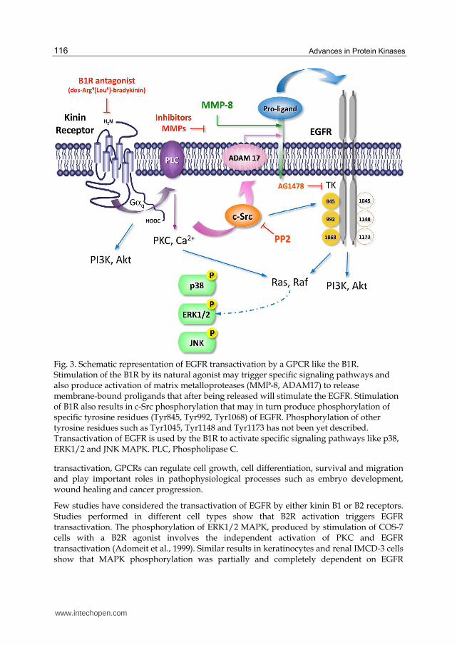
Fig. 3. Schematic representation of EGFR transactivation by a GPCR like the B1R. Stimulation of the B1R by its natural agonist may trigger specific signaling pathways and also produce activation of matrix metalloproteases (MMP-8, ADAM17) to release membrane-bound proligands that after being released will stimulate the EGFR. Stimulation of B1R also results in c-Src phosphorylation that may in turn produce phosphorylation of specific tyrosine residues (Tyr845, Tyr992, Tyr1068) of EGFR. Phosphorylation of other tyrosine residues such as Tyr1045, Tyr1148 and Tyr1173 has not been yet described. Transactivation of EGFR is used by the B1R to activate specific signaling pathways like p38, ERK1/2 and JNK MAPK. PLC, Phospholipase C.
transactivation, GPCRs can regulate cell growth, cell differentiation, survival and migration and play important roles in pathophysiological processes such as embryo development, wound healing and cancer progression.
Few studies have considered the transactivation of EGFR by either kinin B1 or B2 receptors. Studies performed in different cell types show that B2R activation triggers EGFR transactivation. The phosphorylation of ERK1/2 MAPK, produced by stimulation of COS-7 cells with a B2R agonist involves the independent activation of PKC and EGFR transactivation (Adomeit et al., 1999). Similar results in keratinocytes and renal IMCD-3 cells show that MAPK phosphorylation was partially and completely dependent on EGFR
www.intechopen.com
Signaling Pathways Coupled to Activation of the Kinin B1 Receptor
117
transactivation, respectively (Mukhin et al., 2006 and Vidal et al., 2005). It has been shown that bradykinin can produce activation of p60Src and Src-dependent phosphorylation on
Tyr845 of EGFR as well as recruitment of PLC in primary cultures of rat adrenal chromaffin cells and PC12 cells (Hur et al., 2004). Moreover, Yang et al., (2005) demonstrated, using Western blot and [3H]thymidine incorporation, that bradykinin induces proliferation of vascular smooth muscle cells mediated, at least in part, through activation of kinases of the Src family, EGFR transactivation, and PI3K-AKT pathways. Studies directed to elucidate the transactivation mechanism induced by B2R have shown that depending on the cell type, involve ADAM17 or metalloprotease 8 to cleave EGFR ligands (Dey et al., 2010; Methner et al., 2009). Reports regarding these events and the B1R are scarce. Matus et al., (2008) showed that stimulation of the kinin B1R in human keratinocytes produced the phosphorylation of tyrosine residues in a protein with a molecular mass of 170 kDa, that was later identified as EGFR. In these cells, the B1R induces the phosphorylation of Tyr845, Tyr992 and Tyr1068 residues on the EGFR molecule, an effect that was blocked by AG1478, a specific inhibitor of the EGFR tyrosine kinase activity. Similar results were obtained in the HaCaT keratinocyte cell line when EGFR transactivation by the B1R was analyzed (Matus et al., unpublished data).
Studies performed on estrogen-sensitive breast cancer cells strongly suggest that the proliferative effect induced by the B1R depends on the activity of EGFR and subsequent ERK1/2 MAPK phosphorylation (Molina et al., 2009). We have also reported that release of metalloproteases in MCF-7 and MDA-MB-231 breast cancer cells was blocked by AG1478, an observation that was confirmed by transfection of breast cancer cells with the dominant negative EGFR mutant HERCD533 (Ehrenfeld et al., 2011). The transactivation mechanism involved in EGFR activation by B1R agonists has not been elucidated yet, but work performed by our group in human HaCaT keratinocytes, MCF-7 breast cancer cell has visualized that B1R stimulation produces Src phosphorylation, that is blocked by the specific Src inhibitor, PP2 (Figs. 4 and 5). Interestingly, Src phosphorylates specifically the Tyr845 residue present in the active site of EGFR, therefore when we inhibited Src activity, phosphorylation of EGFR in Tyr845 was also inhibited (our group, unpublished data (Fig. 3).
5. Potential usefulness of currently available kinin antagonists and kinase inhibitors in pathological processes
The importance of kinin receptors, especially the B1R, is illustrated by many publications
that demonstrate their involvement in different pathological processes such as cancer, and
especially breast cancer as mentioned early in the text. From a functional point a view, both
B1 and B2 receptors are central players in the aetiology of pain, inflammation and cancer.
Thus, the use of antagonists or inhibitors directed to specific intracellular pathways may be
a useful approach to understand the mechanisms of particular pathological processes and
then to promote them as useful pharmacological agents. Frequently, B2R is associated with
the acute phase of inflammation and noniception, whereas the B1R after its up-regulation by
inflammatory mediators is more relevant during chronic or persistent inflammation. For this
reason, the use of antagonists of both peptidic and non-peptidic nature or blockade of kinase
pathways triggered by activation of kinin receptors may become important clinical tools for
treatment of persistent inflammation, cancer and pain, especially when no other therapy is
available or provides beneficial effects (Campos et al., 2006).
www.intechopen.com
Advances in Protein Kinases
118
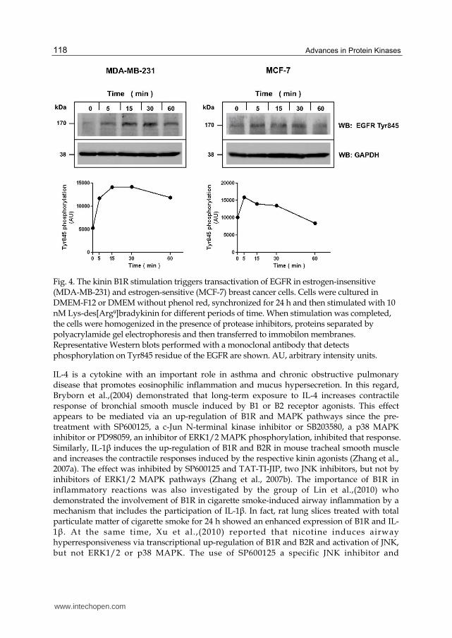
Fig. 4. The kinin B1R stimulation triggers transactivation of EGFR in estrogen-insensitive (MDA-MB-231) and estrogen-sensitive (MCF-7) breast cancer cells. Cells were cultured in DMEM-F12 or DMEM without phenol red, synchronized for 24 h and then stimulated with 10 nM Lys-des[Arg9]bradykinin for different periods of time. When stimulation was completed, the cells were homogenized in the presence of protease inhibitors, proteins separated by polyacrylamide gel electrophoresis and then transferred to immobilon membranes. Representative Western blots performed with a monoclonal antibody that detects phosphorylation on Tyr845 residue of the EGFR are shown. AU, arbitrary intensity units.
IL-4 is a cytokine with an important role in asthma and chronic obstructive pulmonary disease that promotes eosinophilic inflammation and mucus hypersecretion. In this regard, Bryborn et al.,(2004) demonstrated that long-term exposure to IL-4 increases contractile response of bronchial smooth muscle induced by B1 or B2 receptor agonists. This effect appears to be mediated via an up-regulation of B1R and MAPK pathways since the pre-treatment with SP600125, a c-Jun N-terminal kinase inhibitor or SB203580, a p38 MAPK inhibitor or PD98059, an inhibitor of ERK1/2 MAPK phosphorylation, inhibited that response. Similarly, IL-1┚ induces the up-regulation of B1R and B2R in mouse tracheal smooth muscle and increases the contractile responses induced by the respective kinin agonists (Zhang et al., 2007a). The effect was inhibited by SP600125 and TAT-TI-JIP, two JNK inhibitors, but not by inhibitors of ERK1/2 MAPK pathways (Zhang et al., 2007b). The importance of B1R in inflammatory reactions was also investigated by the group of Lin et al.,(2010) who demonstrated the involvement of B1R in cigarette smoke-induced airway inflammation by a mechanism that includes the participation of IL-1┚. In fact, rat lung slices treated with total particulate matter of cigarette smoke for 24 h showed an enhanced expression of B1R and IL-1┚. At the same time, Xu et al.,(2010) reported that nicotine induces airway hyperresponsiveness via transcriptional up-regulation of B1R and B2R and activation of JNK, but not ERK1/2 or p38 MAPK. The use of SP600125 a specific JNK inhibitor and
www.intechopen.com
Signaling Pathways Coupled to Activation of the Kinin B1 Receptor
119
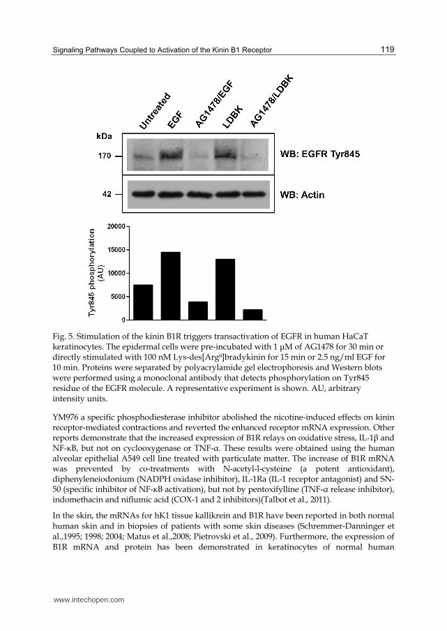
Fig. 5. Stimulation of the kinin B1R triggers transactivation of EGFR in human HaCaT keratinocytes. The epidermal cells were pre-incubated with 1 μM of AG1478 for 30 min or directly stimulated with 100 nM Lys-des[Arg9]bradykinin for 15 min or 2.5 ng/ml EGF for 10 min. Proteins were separated by polyacrylamide gel electrophoresis and Western blots were performed using a monoclonal antibody that detects phosphorylation on Tyr845 residue of the EGFR molecule. A representative experiment is shown. AU, arbitrary intensity units.
YM976 a specific phosphodiesterase inhibitor abolished the nicotine-induced effects on kinin receptor-mediated contractions and reverted the enhanced receptor mRNA expression. Other reports demonstrate that the increased expression of B1R relays on oxidative stress, IL-1┚ and NF-κB, but not on cyclooxygenase or TNF-┙. These results were obtained using the human alveolar epithelial A549 cell line treated with particulate matter. The increase of B1R mRNA was prevented by co-treatments with N-acetyl-l-cysteine (a potent antioxidant), diphenyleneiodonium (NADPH oxidase inhibitor), IL-1Ra (IL-1 receptor antagonist) and SN-50 (specific inhibitor of NF-κB activation), but not by pentoxifylline (TNF-┙ release inhibitor), indomethacin and niflumic acid (COX-1 and 2 inhibitors)(Talbot et al., 2011).
In the skin, the mRNAs for hK1 tissue kallikrein and B1R have been reported in both normal human skin and in biopsies of patients with some skin diseases (Schremmer-Danninger et al.,1995; 1998; 2004; Matus et al.,2008; Pietrovski et al., 2009). Furthermore, the expression of B1R mRNA and protein has been demonstrated in keratinocytes of normal human
www.intechopen.com
Advances in Protein Kinases
120
epidermis, in primary cultures and in the immortalized HaCaT cell line (Matus et al.,2008). Additionally, the expression of B1R in skin may be enhanced when this organ is exposed to many noxious agents present in our environment. In cutaneous models of inflammation developed in mice, peptidic and non-peptidic kinin receptor antagonists have been effective in reducing cutaneous neurogenic inflammation (Pietrovski et al., 2009). Recently, Pietrovski et al.,(2011) demonstrated that both B1R and B2R participate in the hyperproliferative and inflammatory responses of mouse skin suggesting that the use of kinin receptor antagonists may be useful in the treatment of some skin disorders.
Another disease in which the role of B1R has been considered is diabetes. Dias & Couture (2011) used the B1R antagonist, SSR240612 in a rat model of diabetes and insulin resistance, based on the hypothesis that B1R activates signaling pathways that lead to increased oxidative stress and promote insulin resistance (Dias et al., 2010). The use of such an experimental design reversed the effects of glucose on the expression of IL-1┚, TNF-┙, and macrophage CD68 that are linked to the deposition of adipose tissue. Given the association between chronic inflammation and insulin resistance, inhibition of B1R activity may contribute to increase sensitivity to insulin and to prevent obesity. In general, diabetes induces sensory polyneuropathy associated with pain (allodynia and termal hyperalgesia), conditions that are difficult to treat. For this reason, Dias et al.,(2007) tested the non-peptide and orally active B1R antagonist SSR240612 in a rat model of insulin resistance and tactile cold allodynia. Their results are the first to associate the B1R with allodynia, pain that was alleviated by treatment with SSR240612, probably by direct inhibition of B1R affecting the spinal cord and/or afferent sensory nerve excitation. Hawkinson et al.,(2007) probed other B1R antagonist named benzamide B1R antagonist 7-chloro-2-[3-(9-pyridin—4-yl-3,9-diaza-spiro[5.5]undecanecarbonyl)phenyl]-2,3-dihydro-isoindol-1-one (ELN441958). This antagonist reduces in a dose-dependent manner the carrageenan-induced thermal hyperalgesia in a rhesus monkey tail-withdrawal model.
In renal disease, B1R has been associated with tissue inflammation and renal fibrosis. Focal and segmental glomeruloesclerosis is one of the most important causes of end-stage renal failure and up-regulation of B1R in podocyte injury has been demonstrated by Pereira et al.,(2011). Despite this report, there are not additional studies that examine the use of specific antagonists or inhibitors for specific signaling pathways activated by the B1R in this disease. Respect to the participation of the kinin B1R in cancer, only a few reports indicates the importance of interrupting its intracellular signaling; the major efforts have been orientated to the B2R. Thus, many studies have tried to elucidate the importance of B2R in cancer and the consequence of using specific antagonists to reduce cell proliferation, enhance apoptosis or decrease the secretion of metalloproteases (Chan et al., 2002; Stewart et al.,2002; 2003; 2005; Jutras et al., 2010; Wang et al., 2010). Although one of these studies demonstrate that both PGE2 and BK stimulate invasion of head and neck squamous carcinoma cells via EGFR, the treatment of these cells with the B2R antagonist CU201 resulted in growth inhibition (Thomas et al., 2006). The combination of CU201 with the EGFR small-molecule inhibitor erlotinib resulted in additive inhibitory effects on cell growth in the same type of cells in vitro.
6. Conclusion
Despite the high number of reports demonstrating the involvement of B1R in the regulation of various diseases, studies on the use of specific kinin antagonists and/or kinase inhibitors
www.intechopen.com
Signaling Pathways Coupled to Activation of the Kinin B1 Receptor
121
as potential therapeutic drugs has been limited. Therefore, further validation of kinin receptors as important therapeutic targets is crucial. Even though EGFR tyrosine kinase inhibitors such as erlotinib and gefitinib interfere with ATP binding and have been used in clinical trials, inhibiting tyrosine kinase activity and subsequently inhibiting signal
transduction pathways triggered by EGFR such as MAPK, PI3K/Akt, STAT and PLC/PKC. Activation of these pathways also influences cell proliferation, survival and the metastatic potential of tumor cells. Others EGFR tyrosine kinase inhibitors (PD15035, AG1478 and PKI 166) have been tested in animal model as mice with Type 2 Diabetic, mice with high-fat diet-fed or rats with diabetic nephropathy. Results of experiments indicate that these inhibitors restore ischemia-induced neovascularization and blood flow recovery in type 2 diabetic mice, improves glucose tolerance, insulin sensitivity and reduces subclinical inflammation in high-fat diet-fed mice and attenuates the development diabetic nephropathy in rats. (Advani et al., 2011; Choi et al., 2011; Prada et al., 2009).
Despite the success of inhibitors reported in clinical trials, some patients still develop resistance to anti-EGFR therapy. In addition, there are two mutations of EGFR that have been described. Mutations of the exon coding for tyrosine kinase domain (18 to 21) and truncating mutations (exons 2 to 7) that involves signaling pathways. The first group of mutations is involved in the failure of tyrosine kinase inhibitors whereas the second group of mutation leads to resistance of drugs (Colabufo, 2011). However, an alternative approach for overcoming resistance may be to simultaneously target additional pathways such as VEGF/VEGFR, and/or inhibit parallel signaling pathways that complement those activated by EGFR (e.g., mesenchymal-epithelial transition factor and mTOR) (Giaccone & Wang, 2011) and we suggest, by the evidence expose in this chapter, may include the B1R, or B2R. Furthermore, understanding of the intracellular signaling pathways initiated by kinin receptors linked to EGFR and their involvement in the causation and progression of most human neoplasias (e.g., non-small cell lung cancer, colorectal cancer, glioblastoma, breast cancer and squamous cell carcinomas) suggests that these signaling pathways could be used as targets for development of novel drugs that inhibit EGFR activation. Currently there are insufficient studies on the inhibition of EGFR transactivation triggered by B1R to elucidate whether the interaction between EGFR and B1R could provide novel molecular agents for prevention or treatment of human cancer.
7. Acknowledgments
The authors wish to thank the financial support of grants 11090292 (PE) and 1110464 (CDF) from FONDECYT (Chile) and grant 24090029 from CONICYT (CEM).
8. References
Adomeit, A.; Graness, A.; Gross, S.; Seedorf, K.; Wetzker, R. & Liebmann, C. (1999).
Bradykinin B(2) receptor-mediated mitogen-activated protein kinase activation in
COS-7 cells requires dual signaling via both protein kinase C pathway and
epidermal growth factor receptor transactivation. Molecular & Cellular Biology,
Vol.19, Nº8, (August 1999), pp. 5289-1297, ISSN 0270-7306
Advani, A.; Wiggins, KJ.; Cox, AJ.; Zhang, Y.; Gilbert, RE. & Kelly, DJ. (2011). Inhibition of
the epidermal growth factor receptor preserves podocytes and attenuates
www.intechopen.com
Advances in Protein Kinases
122
albuminuria in experimental diabetic nephropathy. Nephrology (Carlton), Vol.16,
Nº6, (August 2011), pp. 573-581, ISSN 1320-5358
Ahluwalia, A. & Perretti, M. (1996). Involvement of bradykinin B1 receptors in the
polymorphonuclear leukocyte accumulation induced by IL-1 beta in vivo in the
mouse. Journal of Immunology, Vol.156, Nº1, (January 1996), pp. 269-274, ISSN 0022-
1767
Austin, CE.; Faussner, A.; Robinson, HE.; Chakravarty, S.; Kyle, DJ.; Bathon, JM. & Proud D.
(1997). Stable expression of the human kinin B1 receptor in Chinese hamster ovary
cells. Characterization of ligand binding and effector pathways. Journal of Biological
Chemistry Vol.272, Nº 17, (April 1997), pp. 11420-11425, ISSN 0021-9258
Bachvarov, DR.; Hess, JF.; Menke, JG.; Larrivée, JF. & Marceau, F. (1996). Structure and
genomic organization of the human B1 receptor gene for kinins (BDKRB1).
Genomics, Vol.33, Nº3, (May 1996), pp. 374-381, ISSN 0888-7543
Barki-Harrington L., Bookout AL., Wang G., Lamb ME., Leeb-Lundberg LM. & Daaka Y.
(2003). Requirement for direct cross-talk between B1 and B2 kinin receptors for the
proliferation of androgen-insensitive prostate cancer PC3 cells. Biochemistry, Vol.2,
Pt2, (April 2003), pp. 581-587, ISSN 0006-2960
Bhoola KD.; Figueroa CD. & Worthy K. (1992). Bioregulation of kinins: kallikreins,
kininogens, and kininases. Pharmacological Reviews, Vol.44, Nº1 (March 1992), pp. 1-
80, ISSN 0031-6997
Bhoola KD.; Ramsaroop, R.; Plendl, J.; Cassim, B.; Dlamini, Z. & Naicker, S. (2001).
Kallikreins and kinin receptor expression in inflammation and cancer. Biological
Chemistry Vol.382, Nº1, (January 2001), pp. 77-89, ISSN 1431-6730
Biscardi, JS.; Maa, MC.; Tice, DA.; Cox, ME.; Leu, TH. & Parsons, SJ. (1999). c-Src-mediated
phosphorylation of the epidermal growth factor receptor on Tyr845 and Tyr1101 is
associated with modulation of receptor function. Journal of Biological Chemistry,
Vol.274, Nº (March 1999), pp. 8335-8343, ISSN 0021-9258
Blaukat A. (2004). Identification of G-protein-coupled receptor phosphorylation sites by 2D
phosphopeptide mapping. Methods in Molecular Biology, Vol. 259, pp. 283-297, ISSN
1064-3745
Boerner, JL.; Biscardi, JS.; Silva, CM. & Parsons, SJ. (2005). Transactivating agonist of the
EGF receptor require Tyr 845 phosphorylation for induction of DNA synthesis.
Molecular Carcinogenics, Vol.44, Nº4, (December 2005), pp. 262-273, ISSN 0899-
1987
Bryborn, M.; Adner, M. & Cardell, LO. (2004). Interleukin-4 increases murine airway
response to kinins, via up-regulation of bradykinin B1-receptors and altered
signalling along mitogen-activated protein kinase pathways. Clinical & Experimental
Allergy, Vol.34, Nº8, (August 2004), pp. 1291-1298, ISSN 0954-7894
Campos, MM.; Leal, PC.; Yunes, RA. & Calixto, JB. (2006). Non-peptide antagonists for kinin
B1 receptors: new insights into their therapeutic potential for the management of
inflammation and pain. Trends in Pharmacological Sciences, Vol.27, Nº12, (December
2006), pp. 646-651, ISSN 0165-6147
Chan, DC.; Gera, L.; Stewart, JM.; Helfrich, B.; Zhao, TL.; Feng, WY.; Chan, KK.; Covey, JM.
& Bunn, PA Jr. (2002). Bradykinin antagonist dimer, CU201, inhibits the growth of
human lung cancer cell lines in vitro and in vivo and produces synergistic growth
www.intechopen.com
Signaling Pathways Coupled to Activation of the Kinin B1 Receptor
123
inhibition in combination with other antitumor agents. Clinical Cancer Research,
Vol.8, Nº5, (May 2002), pp. 1280-1287, ISSN 1078-0432
Chen, H.; Ma, N.; Xia, J.; Liu, J. & Xu, Z. (2011). ┚2-Adrenergic Receptor-induced
Transactivation of EGFR and PDGFR via Src kinase promotes rat cardiomyocytes
survival. Cell Biology International, (September 2011), [Epub ahead of print],
doi:10.1042/CBI20110162, ISSN 1065-6995
Choi, SK.; Galán, M.; Partyka, M.; Trebak, M.; Belmadani, S. & Matrougui, K. (2011). Chronic
inhibition of epidermal growth factor receptor tyrosine kinase and extracellular
signal-regulated kinases 1 and 2 (ERK1/2) augments vascular response to limb
ischemia in type 2 diabetic mice. American Journal of Pathology, (November 2011),
[Epub ahead of print], ISSN 0002-9440
Christopher, J.; Velarde, V. & Jaffa, AA. (2001). Induction of B(1)-kinin receptors in vascular
smooth muscle cells: cellular mechanisms of map kinase activation. Hypertension,
Vol.38, Nº3 Pt2, (September), pp. 602-605, ISSN 0194-911X
Colabufo, NA.; Contino, M.; Niso, M.; Berardi, F.; Leopoldo, M. & Perrone, R. (2011). EGFR
tyrosine kinase inhibitors and multidrug resistance: perspectives. Frontiers in
Bioscience, Vol.16, (January 2011), pp. 1811-1823, ISSN 1093-9946
Cooper, JA. & Howell, B. (1993). The when and how of Src regulation. Cell, Vol.73, Nº6,
(June 1993), pp. 1051-1054, ISSN 0092-8674
Couture, R. & Girolami, JP. (2004) Putative roles of kinin receptors in the therapeutic effects
of angiotensin 1-converting enzyme inhibitors in diabetes mellitus. European Journal
of Pharmacology, Vol.500, Nº1-3, (October 2004), pp. 467-485, ISSN 0014-2999
Cramer, H.; Schmenger, K.; Heinrich, K.; Horstmeyer, A.; Boning, H.; Breit, A.; Piiper, A.;
Lundstrom, K.; Muller-Esterl, W.; Schroeder, C. (2001). Coupling of endothelin
receptors to the ERK/MAP kinase pathway. Roles of palmitoylation and G┙q. Eur J
Biochem, 268:5449-5459.
Daub, H.; Weiss, FU.; Wallasch, C.; Ullrich, A. (1996). Role of transactivation of the EGF
receptor in signalling by G-protein-coupled receptors. Nature. 8; 379 (6565):557-
560.
Dey, M.; Baldys, A.; Sumter, DB.; Göoz, P.; Luttrell, LM.; Raymond, JR.; Göoz, M. (2010).
Bradykinin decreases podocyte permeability through ADAM17-dependent
epidermal growth factor receptor activation and zonula occludens-1
rearrangement. J Pharmacol Exp Ther. 334:775-783.
Dias, JP.; Couture, R. (2011). Blockade of kinin B(1) receptor reverses plasma fatty acids
composition changes and body and tissue fat gain in a rat model of insulin
resistance. Diabetes Obes Metab. 24. doi: 10.1111/j.1463-1326.2011.01521.x. [Epub
ahead of print]
Dias, JP.; Ismael, MA.; Pilon, M.; de Champlain, J.; Ferrari, B.; Carayon, P.; Couture, R.
(2007). The kinin B1 receptor antagonist SSR240612 reverses tactile and cold
allodynia in an experimental rat model of insulin resistance. Br J Pharmacol. 152:280-
287.
Dias, JP.; Talbot, S.; Sénécal, J.; Carayon, P.; Couture, R. (2010). Kinin B1 receptor enhances
the oxidative stress in a rat model of insulin resistance: outcome in hypertension,
allodynia and metabolic complications. PLoS One. ;5:e12622.
www.intechopen.com
Advances in Protein Kinases
124
Dixon, BS.; Evanoff, D.; Fang, WB.; Dennis, MJ. (2002) Bradykinin B1 receptor blocks PDGF-
induced mitogenesis by prolonging ERK activation and increasing p27Kip1. Am J
Physiol Cell Physiol. 283:C193-203.
Ehrenfeld, P.; Conejeros, I.; Pavicic, MF.; Matus, CE.; Gonzalez, CB.; Quest, AF.; Bhoola,
KD.; Poblete, MT.; Burgos, RA.; Figueroa, CD. (2011). Activation of kinin B1
receptor increases the release of metalloproteases-2 and -9 from both estrogen-
sensitive and -insensitive breast cancer cells. Cancer Lett. 301:106-118.
Ehrenfeld, P.; Matus, CE.; Pavicic, F.; Toledo C.; Nualart, F.; Gonzalez, CB.; Burgos, RA.;
Bhoola, KD.; Figueroa, CD. (2009). Kinin B1 receptor activation turns on
exocytosis of matrix metalloprotease-9 and myeloperoxidase in human
neutrophils: involvement of mitogen-activated protein kinase family. J Leukoc
Biol. 86: 1179-1189.
Ehrenfeld, P.; Millan, C.; Matus, CE.; Figueroa, J.; Burgos, R.; Nualart, F.; Bhoola, KD.;
Figueroa, CD. (2006). Activation of kinin B1 receptors induces chemotaxis of human
neutrophils. J Leukoc Biol. 80:117-124.
Endoh, H.; Ishibashi, Y.; Yamaki, Ei.; Yoshida, T.; Yajima, T.; Kimura, H.; Kosaka, T.;
Onozato, R.; Tanaka, S.; Mitsudomi, T.; Kuwano, H. (2009). Immunohistochemical
analysis of phosphorylated epidermal growth factor receptor might provide a
surrogate marker of EGFR mutation. Lung Cancer. 63: 241-246.
Enquist, J.; Skröder, C.; Whistler, JL.; Leeb-Lundberg, LM. (2007). Kinins promote B2 receptor endocytosis and delay constitutive B1 receptor endocytosis. Molecular Pharmacology. Vol 71, Nº2, (February 2007), pp.494-507, ISSN 1521-0111.
Emlet, DR.; Moscatello, DK.; Ludlow, LB.; Wong, AJ. (1997). Subsets of epidermal growth
factor receptors during activation and endocytosis. J Biol Chem. 272:4079-4086.
Ettenberg, SA.; Keane, MM.; Nau, MM.; Frankel, M.; Wang, LM.; Pierce, JH.; Lipkowitz, S.
(1999). cbl-b inhibits epidermal growth factor receptor signaling. Oncogene. 18:1855-
1866.
Giaccone, G.; Wang, Y. (2011). Strategies for overcoming resistance to EGFR family tyrosine
kinase inhibitors. Cancer Treat Rev. 37:456-464.
Hawkinson, JE.; Szoke, BG.; Garofalo, AW.; Hom, DS.; Zhang, H.; Dreyer, M., Fukuda, JY.,
Chen L.; Samant, B.; Simmonds, S.; Zeitz, KP.; Wadsworth, A:, Liao, A.; Chavez,
RA.; Zmolek, W.; Ruslim, L.; Bova, MP.; Holcomb, R.; Butelman, ER.; Ko, MC.;
Malmberg AB. (2007). Pharmacological, pharmacokinetic, and primate analgesic
efficacy profile of the novel bradykinin B1 Receptor antagonist ELN441958. J
Pharmacol Exp Ther. 322:619-630.
Hubbard, SR.; Wei, L.; Ellis, L.; Hendrickson, WA. (1994). Crystal structure of the tyrosine
kinase domain of the human insulin receptor. Nature. ;372:746-754.
Hur, EM.; Park, YS.; Lee, BD.; Jang, IH.; Kim, TD.; Suh, PG.; Ryu, SH.; Kim, KT.
(2004).Sensitization of epidermal growth factor-induced signaling by bradykinin is
mediated by c-Src. Implications for a role of lipid microdomains. J Biol Chem. 279:
5852-5860.
Jutras, S.; Bachvarova, M.; Keita, M.; Bascands, JL.; Mes-Masson, AM.; Stewart, JM.;
Buchvarov, D. (2010). Strong cytotoxic effect of the bradykinin antagonist BKM-570
in ovarian cancer cells-analysis of the molecular mechanism of its antiproliferative
action. FEBS J. 277:5146-5160. doi: 10.1111/j.1742-4658.2010.07928.
www.intechopen.com
Signaling Pathways Coupled to Activation of the Kinin B1 Receptor
125
Kannangai, R.; Sahin, F.; Torbenson, MS. (2006). EGFR is phosphorylated at Tyr 845 in
hepatocellular carcinoma. Mod Pathol. 19: 1456-1461.
Kang, DS.; Gustafsson, C.; Mörgelin, M.; Leeb-Lundberg, LM. (2005). B1 bradykinin receptor
homo-oligomers in receptor cell surface expression and signaling: effects of
receptor fragments. Mol Pharmacol. 67:309-318.
Leeb-Lundberg, LM.; Marceau, F.; Müller-Esterl, W.; Pettibone, DJ.; Zuraw, BL. (2005).
International union of pharmacology. XLV. Classification of the kinin receptor
family: from molecular mechanisms to pathophysiological consequences. Pharmacol
Rev. 57: 27-77.
Lemjabbar-Alaoui, H.; Sidhu, SS.; Mengistab, A.; Gallup, M.; Basbaum, C. (2011).
TACE/ADAM-17 phosphorylation by PKC-epsilon mediates premalignant
changes in tobacco smoke-exposed lung cells. PLoS One. 15;6(3):e17489.
Levkowitz, G.; Waterman, H.; Ettenberg, SA.; Katz, M.; Tsygankov, AY.; Alroy, I.; Lavi, S.;
Iwai, K.; Reiss, Y.; Ciechanover, A.; Lipkowitz, S.; Yarden, Y. (1999). Ubiquitin
ligase activity and tyrosine phosphorylation underlie suppression of growth factor
signaling by c-Cbl/Sli-1. Mol Cell. 4:1029-40.
Liebmann, C. (2011). EGF receptor activation by GPCRs: an universal pathway reveals
different versions. Mol Cell Endocrinol. 331:222-231.
Lin, JC.; Talbot, S.; Lahjouji, K.; Roy, JP.; Sénécal, J.; Couture, R.; Morin, A. (2010).
Mechanism of cigarette smoke-induced kinin B(1) receptor expression in rat
airways. Peptides. 31:1940-1945.
MacNeil, T.; Bierilo, KK.; Menke, JG.; Hess, JF. (1995) Cloning and pharmacological
characterization of a rabbit bradykinin B1 receptor. Biochim Biophys Acta. 1264:223-
228.
Marceau, F.; Bachvarov, DR. (1998). Kinin receptors. Clin Rev Allergy Immunol. 16:385-401.
Marceau, F.; Sabourin, T.; Houle, S.; Fortin, JP.; Petitclerc, E.; Molinaro, G.; Adam A. (2002)
Kinin receptors: functional aspects. Int Immunopharmacol. 2:1729-1739.
Marceau, F.; Tremblay B. (1986) Mitogenic effect of bradykinin and of des-Arg9-bradykinin
on cultured fibroblasts. Life Sci. 39:2351-2358.
Maretzky, T.; Zhou, W.; Huang, XY.; Blobel, CP. (2011). A transforming Src mutant increases
the bioavailability of EGFR ligands via stimulation of the cell-surface
metalloproteinase ADAM17. Oncogene. 3;30(5):611-618.
Mathis, SA.; Criscimagna, NL.; Leeb-Lundberg, LM. (1996). B1 and B2 kinin receptors mediate distinct patterns of intracellular Ca2+ signaling in single cultured vascular smooth muscle cells. Molecular Pharmacology. Vol 50, Nº1, (July 1996), pp. 128-39, ISSN 1521-0111.
Matus, CE.; Ehrenfeld, P.; Pavicic, F.; Sarmiento, JM.; Astroza, A.; Sanchez, T;. Salem, C.;
Concha, M.; Vidal, MA.; Gonzalez, CB.; Figueroa, CD. (2008). Activation of kinin B
receptor triggers differentiation of cultured human keratinocytes. Br J Dermatol.
159:792-803.
Menke, JG.; Borkowski, JA.; Bierilo, KK.; MacNeil, T.; Derrick, AW.; Schneck, KA.; Ransom,
RW.; Strader, CD.; Linemeyer, DL.; Hess, JF. (1994) Expression cloning of a human
B1 bradykinin receptor. J Biol Chem. 269:21583-21586.
Methner, C.; Donat, U.; Felix, SB.; Krieg, T. (2009). Cardioprotection of bradykinin at
reperfusion involves transactivation of the epidermal growth factor receptor via
matrix metalloproteinase-8. Acta Physiol (Oxf). 197:265-271.
www.intechopen.com
Advances in Protein Kinases
126
Miyawaki, M.; Hijiya, N.; Tsukamoto, Y.; Nakada, C.; Kawahara, K.; Moriyama, M. (2008).
Enhanced phosphorylation of the epidermal growth factor receptor at the site of
tyrosine 992 in esophageal carcinomas. APMIS. 116: 1097-1106.
Molina, L.; Matus, CE.; Astroza, A.; Pavicic, F.; Tapia, E.; Toledo, C.; Perez, JA.; Nualart, F.;
Gonzalez, CB.; Burgos, RA.; Figueroa, CD.; Ehrenfeld, P.; Poblete, MT. (2009).
Stimulation of the bradykinin B(1) receptor induces the proliferation of estrogen-
sensitive breast cancer cells and activates the ERK1/2 signaling pathway. Breast
Cancer Res Treat. 118:499-510.
Morinelli, TA.; Meier, GP.; Webb, JG.; Jaffa, AA.; Privitera, PJ.; Margolius, HS. (2002).
Utilization of a radioimmunoassay to detect the generation of Arg-Pro-Pro-Gly-
Phe, a stable endproduct of bradykinin metabolism (from cultured rat aortic
smooth muscle cells exposed to bradykinin). Int Immunopharmacol. 2:1995-2003.
Morissette G, Sabourin T, Adam A, Marceau F. (2006). Inhibition of human and rabbit
arterial smooth muscle cell migration mediated by the kinin B1 receptor: role of
receptor density and released mediators. Can J Physiol Pharmacol, Vol. 84, N°11,
(November 2006), pp1107-1119, ISSN
Mugabe, BE.; Yaghini, FA.; Song, CY.; Buharalioglu, CK.; Waters, CM.; Malik, KU. (2010).
Angiotensin II-induced migration of vascular smooth muscle cells is mediated by
p38 mitogen-activated protein kinase-activated c-Src through spleen tyrosine
kinase and epidermal growth factor receptor transactivation. J Pharmacol Exp Ther.
332:116-124.
Mukhin, YV.; Gooz, M.; Raymond, JR.; Garnovskaya, MN. (2006). Collagenase-2 and -3
mediate epidermal growth factor receptor transactivation by bradykinin B2
receptor in kidney cells. J Pharmacol Exp Ther. 318:1033-1043.
Murphey, LJ.; Hachey, DL.; Oates, JA.; Morrow, JD.; Brown NJ. (2000) Metabolism of
bradykinin In vivo in humans: identification of BK1-5 as a stable plasma peptide
metabolite. J Pharmacol Exp Ther. 294:263-269.
Murphey, LJ.; Malave, HA.; Petro, J.; Biaggioni, I.; Byrne, DW.; Vaughan, DE.; Luther, JM.;
Pretorius, M.; Brown, NJ. (2006) Bradykinin and its metabolite bradykinin 1-5
inhibit thrombin-induced platelet aggregation in humans. J Pharmacol Exp Ther.
318:1287-1292.
Ni, A.; Chai, KX.; Chao, L.; Chao, J. (1998). Molecular cloning and expression of rat
bradykinin B1 receptor. Biochim Biophys Acta. 1442:177-185.
Ohtsu, H.; Dempsey, PJ.; Eguchi, S. (2006). ADAMs as mediators of EGF receptor
transactivation by G protein-coupled receptors. Am J Physiol Cell Physiol. 291(1):C1-
10.
Paegelow, I.; Trzeczak, S.; Böckmann, S.; Vietinghoff, G. (2002) Migratory responses of
polymorphonuclear leukocytes to kinin peptides. Pharmacology. 66:153-161.
Pereira, RL.; Buscariollo, BN.; Corrêa-Costa, M.; Semedo, P.; Oliveira, CD.; Reis, VO.;
Maquigussa, E.; Araújo, RC.; Braga, TT.; Soares, MF.; Moura, IC.; Malheiros, DM.;
Filho, AP.; Keller, AC.; Câmara, NO. (2011). Bradykinin receptor 1 activation
exacerbates experimental focal and segmental glomerulosclerosis. Kidney Int.
79:1217-1227. doi: 10.1038/ki.2011.14. Epub 2011 Mar 16.
Pietrovski, EF.; Otuki, MF.; Regoli, D.; Bader, M.; Pesquero, JB.; Cabrini, DA.; Zampronio,
AR. (2009). The non-peptide kinin receptor antagonists FR 173657 and SSR
www.intechopen.com
Signaling Pathways Coupled to Activation of the Kinin B1 Receptor
127
240612: preclinical evidence for the treatment of skin inflammation. Regul Pept.
152:67-72.
Pietrovski, EF.; Paludo, KS.; Mendes, DA.; Guimarães, Fde S.; Veiga, SS.; Buchi, Dde F.;
Fonseca, RG.; Zampronio, AR.; Bader, M.; Pesquero, JB.; Ferreira, J.; Otuki, MF.;
Cabrini, DA. (2011). B1 and B2 kinin receptor participation in hyperproliferative
and inflammatory skin processes in mice. J Dermatol Sci. 64:23-30.
Prada, PO.; Ropelle, ER.; Mourão, RH.; de Souza, CT.; Pauli, JR.; Cintra, DE.; Schenka, A.;
Rocco, SA.; Rittner, R.; Franchini, KG.; Vassallo, J.; Velloso, LA.; Carvalheira, JB.;
Saad, MJ. (2009). EGFR tyrosine kinase inhibitor (PD153035) improves glucose
tolerance and insulin action in high-fat diet-fed mice. Diabetes. 58:2910-2919.
Prado, GN.; Taylor, L.; Zhou, X.; Ricupero, D.; Mierke, DF.; Polgar, P. (2002) Mechanisms
regulating the expression, self-maintenance, and signaling-function of the
bradykinin B2 and B1 receptors. J Cell Physiol. 193:275-286.
Regoli, D.; Rizzi, A.; Perron, SI.; Gobeil, F Jr. (2001). Classification of kinin receptors. Biol
Chem. 382:31-35.
Rojas, M.; Yao, S.; Lin, YZ. (1996). Controlling epidermal growth factor (EGF)-stimulated
Ras activation in intact cells by a cell-permeable peptide mimicking
phosphorylated EGF receptor. J Biol Chem. 271:27456-27461.
Rozengurt, E. (2007). Mitogenic signaling pathways induced by G protein-coupled
receptors. J Cell Physiol. 213:589-602.
Schremmer-Danninger, E.; Heinz-Erian, P.; Töpfer-Petersen, E.; Roscher, AA. (1995).
Autoradiographic localization and characterization of bradykinin receptors in
human skin. Eur J Pharmacol. 283:207-216.
Sato, K.; Nagao, T.; Iwasaki, T.; Nishihira, Y. & Fukami, Y. (2003). Src-dependent
phosphorylation of the EGF receptor Tyr-845 mediates Stat-p21waf1 pathway in
A431 cells. Genes to cells : devoted to molecular & cellular mechanisms, Vol 8, N°12,
(December 2008), pp. 995-1003, ISSN 1356-9597
Schremmer-Danninger, E.; Heinz-Erian, P.; Töpfer-Petersen E. & Roscher AA. (1995).
Autoradiographic localization and characterization of bradykinin receptors in
human skin. European journal of pharmacology, Vol 283, N° 1-3 (September 1995), pp.
207-216, ISSN 0014-2999
Schremmer-Danninger, E.; Toepfer-Petersen, E.; Fritz H. & Roscher AA. (1998). Bradykinin-
induced tyrosine phosphorylation of proteins in cultured human keratinocytes.
Biological research, Vol 31, N°3, (1998), pp. 189-198, ISSN 0716-9760
Schremmer-Danninger, E.; Naidoo, S.; Neuhof, C.; Valeske, K.; Snyman, C.; Sander, C.;
Bhoola, KD. & Neuhof H. (2004). Visualisation of tissue kallikrein, kininogen and
kinin receptors in human skin following trauma and in dermal diseases. Biological
chemistry, Vol 385, N° 11 , (Nov 2004), pp.1069-1076, ISSN 1431-6730
Shima, C.; Majima, M. & Katori, M. (1992) A stable metabolite, Arg-Pro-Pro-Gly-Phe, of
bradykinin in the degradation pathway in human plasma. Japanese journal of
pharmacology, Vol 60, N°2, (October 1992), pp.111-119, ISSN 0021-5198
Stewart, JM. (2003). Bradykinin antagonists as anti-cancer agents. Current pharmaceutical
design. Vol 9, N°25, (2003), pp.2036-2042, ISSN 1381-6128
Stewart, JM.; Gera, L.; Chan, DC.; Bunn, PA Jr.; York, EJ.; Simkeviciene, V. & Helfrich B.
(2002). Bradykinin-related compounds as new drugs for cancer and inflammation.
www.intechopen.com
Advances in Protein Kinases
128
Canadian journal of physiology and pharmacology, Vol 80, N°25, (April 2002), pp. 275-
280, ISSN 0008-4212
Stewart, JM.; Gera, L.; Chan, DC.; York, EJ.; Simkeviciene, V.; Bunn, PA Jr. & Taraseviciene-
Stewart L. (2005). Combination cancer chemotherapy with one compound:
pluripotent bradykinin antagonists. Peptides, Vol 26, N°8, (August 2005), pp.1288-
1291, ISSN 0196-9781
Stirnweiss, J.; Valkova, C.; Ziesché, E.; Drube, S. & Liebmann, C. (2006). Muscarinic M2
receptors mediate transactivation of EGF receptor through Fyn kinase and without
matrix metalloproteases. Cell Signaling, Vol 18, N° 8, (August), pp. 1338-1349, ISSN
0898-6568
Stuardo, M.; Gonzalez, CB.; Nualart, F.; Boric, M.; Corthorn, J.; Bhoola, KD. & Figueroa, CD.
(2004) Stimulated human neutrophils form biologically active kinin peptides from
high and low molecular weight kininogens. Journal of leukocyte biology, Vol 75, N°4,
(April 2004), pp. 631-640, ISSN 0741-5400
Sturla, LM.; Amorino, G.; Alexander, MS.; Mikkelsen, RB.; Valerie, K. & Schmidt-Ullrichr,
RK. (2005). Requirement of Tyr-992 and Tyr-1173 in phosphorylation of the
epidermal growth factor receptor by ionizing radiation and modulation by SHP2.
The Journal of Biological Chemistry, Vol 280, N° 15 , (April 2005), pp. 14597-14604,
ISSN 0021-9258
Sun, C.; Wu, MH.; Guo, M.; Day, ML.; Lee, ES. & Yuan, SY. (2010). ADAM15 regulates
endothelial permeability and neutrophil migration via Src/ERK1/2 signalling.
Cardiovascular Research, Vol 87, N°2, (2010), pp. 348-355, ISSN 0008-6363
Talbot, S.; Lin, JC.; Lahjouji, K.; Roy, JP.; Sénécal, J.; Morin, A. & Couture, R. (2011). Cigarette
smoke-induced kinin B1 receptor promotes NADPH oxidase activity in cultured
human alveolar epithelial cells. Peptides, Vol 32, N° 7, (May 2011), pp.1447-1456,
ISSN 0196-9781
Taub, JS.; Guo, R.; Leeb-Lundberg, LM.;, Madden JF. & Daaka, Y. (2003). Bradykinin
receptor subtype 1 expression and function in prostate cancer. Cancer Research, Vol
63, N°9, (May 2003), pp. 2037-2041, ISSN 0008-5472
Thomas, SM.; Bhola, NE.; Zhang, Q.; Contrucci, SC.; Wentzel, AL. & Freilino, ML. (2006).
Cross-talk between G protein-coupled receptor and epidermal growth factor
receptor signaling pathways contributes to growth and invasion of head and neck
squamous cell carcinoma. Cancer Research, Vol 66, N°24, ( 2006), pp.11831-11839,
ISSN 0008-5472
Tice, DA.; Biscardi, JS.; Nickles, AL. & Parsons, SJ. (1999). Mechanism of biological synergy
between cellular Src and epidermal growth factor receptor. Proceedings of the
National Academy of Sciences of the United States of America, Vol 96, N°4, (February
1999), pp.1415–1420, ISSN 0027-8424
Tropea, MM.; Gummelt, D.; Herzig, MS. & Leeb-Lundberg, LM. (1993). B1 and B2 kinin
receptors on cultured rabbit superior mesenteric artery smooth muscle cells:
receptor-specific stimulation of inositol phosphate formation and arachidonic
acid release by des-Arg9-bradykinin and bradykinin. The Journal of pharmacology
and experimental therapeutics, Vol 264, N°2, ( February 1993), pp.930-937, ISSN
0022-3565
www.intechopen.com
Signaling Pathways Coupled to Activation of the Kinin B1 Receptor
129
Vidal, MA.; Astroza, A.; Matus, CE.; Ehrenfeld, P.; Pavicic, F.; Sanchez, T.; Salem, C.;
Figueroa, J.; Concha, M.; Gonzalez, CB. & Figueroa, CD. (2005). Kinin B2
receptor-coupled signal transduction in human cultured keratinocytes. The
Journal of investigative dermatology, Vol 124, N° 1, (January 2005), pp.178-186, ISSN
0022-202
Wang, J.; Krishnamoorthi, V.; Wang E.; Yang C.; Baptista, D.; Wu, X.; Liu, M.; Gardner, M.;
Elkins P.; Hines, J. & Liu, P. (2010). LC/MS characterization of impurities and
degradation products of a potent antitumor peptidic dimer, CU201. Journal of
pharmaceutical and biomedical analysis, Vol 51, N°4, (March 2010), pp. 824-833, ISSN
0731-7085
Wetzker, R. & Böhmer, FD. (2003). Transactivation joins multiple tracks to the ERK/MAPK
cascade. Nature reviews. Molecular cell biology, Vol 4, Nº8, (August 2010), pp. 651-657,
ISSN 1471-0072
Xu, CB.; Lei, Y.; Chen, Q.; Pehrson, C.; Larsson, L. & Edvinsson, L. (2010). Cigarette smoke
extracts promote vascular smooth muscle cell proliferation and enhances
contractile responses in the vasculature and airway. Basic and clinical pharmacology
and toxicology, Vol 107, N°6, (December 2010), pp.940-948, ISSN 1742-7835
Yang, CM.; Lin, MI.; Hsieh, HL.; Sun, CC.; Ma, YH. & Hsiao, LD. (2005). Bradykinin-
induced p42/p44 MAPK phosphorylation and cell proliferation via Src, EGF
receptors, and PI3-K/Akt in vascular smooth muscle cells. Journal of cellular
physiology, Vol 203, N°3, ( June 2005), pp. 538-546, ISSN 0021-9541
Yang, X.; Taylor, L.; Yu, J.; Fenton, MJ. &Polgar, P. (2001). Mediator caused induction of a
human bradykinin B1 receptor minigene: participation of c-Jun in the process.
Journal of cellular biochemistry, Vol 82, N°1, (April 2001), pp.163-170, ISSN 0730-
2312
Yamauchi, T.; Ueki, K.; Tobe, K.; Tamemoto, H.; Sekine, N.; Wada, M.; Honjo, M.;
Takahashi, M.; Hirai, H.; Tsushima, T.; Akanuma, Y.; Fujita, T.; Komuro, I.;
Yazaki, Y. & Kadowaki, T. (1998). Growth hormone-induced tyrosine
phosphorylation of EGF receptor as an essential element leading to MAP kinase
activation and gene expression. Endocrine journal, Vol, 45, N°, (April 1998), pp.
S27-31, ISSN 0918-8959
Yurko, MA.; O'Toole, EA. & Woodley, DT. (2001). Phosphorylation of focal adhesion kinase
(pp125(FAK)) is increased in human keratinocytes induced to migrate by
extracellular matrices. Journal of cellular physiology, Vol.188, N°1, (July 2001), pp.24-
32, ISSN 0021-9541
Zhang, Y.; Adner, M. & Cardell, LO. (2007a). IL-1beta-induced transcriptional up-regulation
of bradykinin B1 and B2 receptors in murine airways. American journal of respiratory
cell and molecular biology. Vol.36, N°6, (June 2007), pp.697-705, ISSN 1044-1549
Zhang, Y.; Brovkovych, V.; Brovkovych, S.; Tan, F.; Lee, BS.; Sharma, T. & Skidgel, RA.
(2007b). Dynamic receptor-dependent activation of inducible nitric-oxide synthase
by ERK-mediated phosphorylation of Ser745. Journal Biological Chemistry, Vol. 282,
N°44, (November 2007), pp.32453-32461, ISSN 0021-9258
Zhang, X.; Tan, F.; Brovkovych, V.; Zhang, Y. & Skidgel, RA. (2011). Cross-talk between
carboxyeptidase M and the kinin B1 receptor mediates a new mode of G protein-
www.intechopen.com

Advances in Protein Kinases
130
coupled receptor signaling. Journal Biological Chemistry, Vol. 286, N° 27, (May 2011),
pp.18547-18561, ISSN 0021-9258
Zwick, E.; Hackel, PO.; Prenzel, N.; Ullrich, A. (1999). The EGF receptor as central
transducer of heterologous signalling systems. Trends Pharmacology Science, Vol.20,
N° 10 , (October 1999), pp.408-412, ISSN:0165-6147
www.intechopen.com
Advances in Protein KinasesEdited by Dr. Gabriela Da Silva Xavier
ISBN 978-953-51-0633-3Hard cover, 374 pagesPublisher InTechPublished online 05, June, 2012Published in print edition June, 2012
InTech EuropeUniversity Campus STeP Ri Slavka Krautzeka 83/A 51000 Rijeka, Croatia Phone: +385 (51) 770 447 Fax: +385 (51) 686 166www.intechopen.com
InTech ChinaUnit 405, Office Block, Hotel Equatorial Shanghai No.65, Yan An Road (West), Shanghai, 200040, China
Phone: +86-21-62489820 Fax: +86-21-62489821
Proteins are the work horses of the cell. As regulators of protein function, protein kinases are involved in thecontrol of cellular functions via intricate signalling pathways, allowing for fine tuning of physiological functions.This book is a collaborative effort, with contribution from experts in their respective fields, reflecting the spirit ofcollaboration - across disciplines and borders - that exists in modern science. Here, we review the existingliterature and, on occasions, provide novel data on the function of protein kinases in various systems. We alsodiscuss the implications of these findings in the context of disease, treatment, and drug development.
How to referenceIn order to correctly reference this scholarly work, feel free to copy and paste the following:
Pamela Ehrenfeld, Carlos D. Figueroa, Kanti D. Bhoola and Carola E. Matus (2012). Signaling PathwaysCoupled to Activation of the Kinin B1 Receptor, Advances in Protein Kinases, Dr. Gabriela Da Silva Xavier(Ed.), ISBN: 978-953-51-0633-3, InTech, Available from: http://www.intechopen.com/books/advances-in-protein-kinases/signaling-pathways-coupled-to-activation-of-the-kinin-b1-receptor

© 2012 The Author(s). Licensee IntechOpen. This is an open access articledistributed under the terms of the Creative Commons Attribution 3.0License, which permits unrestricted use, distribution, and reproduction inany medium, provided the original work is properly cited.