signaling pathways is attenuated with dha activation of
TRANSCRIPT

Page 1/29
Aβ25–35 induction of necroptosis throughactivation of the RIPK1/RIPK3 and RIPK1/ERK1/2signaling pathways is attenuated with DHAtreatment in THP-1 monocytesShiqi Yuan
Department of Neurology,The Third A�liated Hospital of Southern Medical Universityhttps://orcid.org/0000-0002-2794-2546
Huan Li Department of Neurology,The Third A�liated Hospital of Southern Medical University
Canhong Yang Department of Neurology,The Third A�liated Hospital of Southern Medical University
Wenyi Xie Department of Neurology,The Third A�liated Hospital of Southern Medical University
Yuanyuan Wang Department of Neurology, The third A�liated of Southern Medical University
Jiafa Zhang Department of Neurology,The Third A�liated Hospital of Southern Medical University
Zibo Cai Department of Neurology,The Third A�liated Hospital of Southern Medical University
Zhenlin Mao Department of Neurology,The Third A�liated Hospital of Southern Medical University
Weibing Xie Judicial Identi�cation Center of Southern Medical University
Tianming Lü ( [email protected] )https://orcid.org/0000-0003-0747-4228
Research
Keywords: Alzheimer's disease, Aβ25–35, THP-1 cells, Docosahexaenoic acid (DHA), Necroptosis, MAPK,NF-kB
Posted Date: December 3rd, 2019
DOI: https://doi.org/10.21203/rs.2.17970/v1

Page 2/29
License: This work is licensed under a Creative Commons Attribution 4.0 International License. Read Full License

Page 3/29
AbstractBackground: Monocytes play a crucial role in Alzheimer's disease (AD), and docosahexaenoic acid (DHA)treatment has a neuroprotective effect for many neurodegenerative diseases. However, mechanisms thatregulate monocyte and Aβ protein interaction in AD and the effects of DHA on monocytes in the contextof AD are not fully understood.
Methods: The experiments were designed to further explore possible mechanisms of interaction betweenmonocytes and Aβ plaques. Another objective of this study was to investigate a potential mechanism forAβ-mediated regulation of necroptosis and the MAPK and NF-kB signaling pathways, as well as howthese pathways might be modulated by DHA in human THP-1 monocytes. We also investigated whetherDHA indirectly suppressed THP-1 cell-mediated neuronal activation. We used cell viability and cytotoxicityassays, �ow cytometry, transwell migration assays, and Western blotting to perform our study.
Results: Our �ndings indicate that Aβ25-35 regulates two aspects of THP-1 cells necroptosis. We alsoobserved that increased resistance to apoptosis in THP-1 cells that is correlated with THP-1 monocytedifferentiation. Our results also indicated that DHA treatment restored migration of THP-1 monocytes thathad been treated with Aβ25–35. Pre-treatment of THP-1 monocytes with DHA effectively inhibited Aβ-induced activation and markedly suppressed protein expression of TNF-α IL-1β and IL-6. We also foundthat THP-1 cell necroptosis was induced by Aβ25–35 through modulation of RIPK1/RIPK3 andphosphorylation status of ERK1/2 and could be attenuated by DHA treatment.
Conclusion: Aβ25–35-mediated induction of necroptosis through activation of the RIPK1/RIPK3 andRIPK1/ ERK1/2 signaling pathways in THP-1 monocytes can be attenuated by DHA treatment, indicatingthat DHA treatment could be a promising new therapy for AD management.
IntroductionAlzheimer’s disease (AD), the leading cause of dementia, encompasses a range of neurologicalsymptoms, including memory loss and cognitive impairment [1]. The neuropathological changes ofAlzheimer disease (AD) consist of abundant amyloid plaques and neuro�brillary tangles, neuropilthreads, and dystrophic neurites containing hyperphosphorylated tau [2, 3]. AD is likely caused at least inpart by an imbalance between amyloid-β (Aβ) protein production and clearance, which leads to Aβaccumulation in the central nervous system (CNS). The main pathways that act to clear excess Aβ areproteolytic degradation, transcytosis across the blood-brain barrier (BBB), and perivascular lymphaticdrainage[4, 5].
Monocytes play a crucial role in AD, given that monocyte-derived perivascular macrophages e�cientlyphagocytose accumulations of Aβ[6]. Aβ accumulation can stimulate the RAGE and PECAM–1 receptorsto promote the migration of monocytes along human brain endothelial cells [7]. Using live intravital two-photon microscopy in triple-transgenic APPswe/PS1+/-/Cx3cr1gfp/+ mice, Michaud et al. demonstratethat patrolling monocytes are attracted to and crawl onto the luminal walls of Aβ-positive veins.
Page 4/29
Furthermore, they report the presence of crawling monocytes carrying Aβ in veins that are able to circulateback into the bloodstream [8]. Compared to monocytes, microglia have a limited ability to degrade Aβplaques, resulting from the low activity levels of several microglial lysosomal enzymes [9]. Patrollingmonocytes may therefore play a more important role in Aβ clearance, and further increasing vascular Aβclearance via patrolling monocytes could have signi�cant impact on AD. Reducing the migration,phagocytosis, or number of mononuclear cells in transgenic AD mice is detrimental, whereas treatmentswith compounds that increase the number and phagocytic activity of mononuclear cells are generallybene�cial[10]. Finding a way to promote effective monocyte-mediated Aβ protein clearance whileavoiding the activation of proin�ammatory and neurotoxic pathways is an attractive approach fortreating AD [11] The experiments in this current study were designed to further explore possiblemechanisms of interaction between monocytes and Aβ protein.
Studies have shown that the neurotoxicity directly or indirectly induced by accumulation of Aβ protein is aprimary cause of neuron injury in AD. Kwon et al. have shown drug treatment can directly ameliorate thetoxic effects Aβ accumulation causes in neurons [12, 13]. Aβ accumulation can also indirectly exert atoxic effect on neurons through targeting microglial cells, so protection of microglial cells is alsoimportant for alleviating AD symptoms [14, 15]. Further studies are necessary to understand themechanism by which Aβ plaques interact with monocytes to exert neurotoxic effects.
Docosahexaenoic acid (DHA) is an essential omega–3 polyunsaturated fatty acid (PUFA) foundpredominantly in marine products. Preclinical and clinical studies have demonstrated that DHA exerts aneuroprotective effect in the context of several neurodegenerative diseases, including AD, resulting mostlyfrom the antioxidant and anti-in�ammatory properties of DHA and its ability to activate several othersignaling pathways[16]. Given that monocytes are involved in the clearance of Aβ plaques, additionalstudies are needed to elucidate a potential mechanism for how DHA might be involved in promotingmonocyte-mediated clearance of Aβ plaques.
Necroptosis is a lytic cell death program de�ned by activation of the receptor interacting protein kinase–1(RIPK1) and RIPK3 to form an oligomeric “necrosome,” which leads to the phosphorylation and activationof the effector protein pseudokinase mixed lineage kinase domain-like (MLKL)[17].This necroptosispathway is implicated in several neurodegenerative diseases [18]. NF-κB plays a key role in modulatingimmune and in�ammatory responses, while mitogen-activated protein kinases (MAPKs) (P38, ERK1/2)are key regulators of a variety of cellular functions including cell survival, apoptosis and cellularresponses to in�ammation[19, 20]. Upon TNF binding to its receptor to form Complex I, RIPK1 caninteract with Complex I, which activates the MAPK signaling cascade and the transcription factor NF-κB[21]. Therefore, we further investigated a possible mechanism for Aβ-mediated regulation of necroptosis,MAPK and NF-kB signaling pathways in monocytes.
The experiments in this study were designed to further explore possible mechanisms by whichmonocytes and Aβ protein might interact. Another objective of this study was to investigate a possiblemechanism for Aβ-mediated regulation of necroptosis, MAPK and NF-kB signaling pathways in vitro in
Page 5/29
human THP–1 monocytes. We also investigated the role of DHA in modulating Aβ-regulated signalingpathways and whether DHA indirectly suppressed THP–1 cell-mediated neuronal activation.
Methods
Materialscis–4,7,10,13,16,19-docosahexaenoic acid DHA was purchased from Sigma-Aldrich (St. Louis, MO, USA).Neurobasal medium, B–27 supplement, and penicillin, DMEM medium, 1640 medium, and fetal bovineserum (FBS) were purchased from Gibco (Grand Island, NY, USA). CCK8, Necrostatin–1 (NEC–1), GSK872were purchased from MedChem Express (New Jersey, USA). Anti-RIPK3 (A5431), MLKL (A5579), TNF(A0277), IL–6(A0286), and IL–1β(A1112) antibodies were purchased from ABclonal (Wuhan, China). P-p44/42 MAPK (ERK1/2), p44/42 MAPK(ERK1/2), NF-κB p65, NF-κB p65, p-p38 MAPK and p38 MAPKwere purchased from Cell Signaling Technology Company (Danvers, MA, USA). GAPDH antibody andHorseradish peroxidase-conjugated secondary antibodies were purchased from Proteintech (Chicago,USA). β-Amyloid (25–35) was purchased from GL Biochem Shanghai China . CD11b-PE 7-AAD, FITCAnnexin V Apoptosis Detection Kit I were purchased from BD Pharmingen San Diego, CA, USA) CytoTox96 Non-Radioactive Cytotoxicity Assay LDH was purchased from Promega Madison USA .
Preparation of Aβ25–35 peptideAβ25–35 was dissolved in sterile distilled water at a concentration of 1 nM and then �ltered through a0.22 μm �lter. Aβ25–35 was aggregated during a 7 day incubation period at 37°C and then stored in a−20°C freezer for later use.
Preparation of DHADHA was dissolved in DMSO at a concentration of 10 nM and then �ltered through a 0.22 μm �lter. ThisDHA solution was stored in a −80°C freezer for later use. The control group was added with the sameconcentration DMSO.
Cell culture conditionsThe human THP–1 cell line was a gift from NanFang Hospital of Southern Medical University and wascultured in RPMI 1640 medium supplemented with 10% FBS at 37°C in a humidi�ed incubator (5% CO2,95% air). The THP–1 cells 10 × 104 cells/ml were pretreated with DHA in 0% FBS for 1, 3, or 5 days.Conditioned media (CM) from each THP–1 plate was then collected and stored at −80°C for later use.
The SY5Y cell line was a gift from The Judicial Identi�cation Center of Southern Medical University andwas cultured in the same growth conditions as the THP–1 cell line. SY5Y cells were treated with Aβ25–

Page 6/29
35 aggregates and the conditioned media (CM) collected from plates of THP–1 cells.
Primary cortical neurons were prepared from embryonic day 16–18 (E16–18) C57BL/6 mice, which werepurchased from the Experimental Animal Center of Southern Medical University. All animal experimentswere approved by the Institutional Animal Care and Use Committee of Southern Medical University inaccordance with the guidelines of the Institutional Animal Care and Use Committee of the NationalInstitutes of Health and the Guide for the Care and Use of Laboratory Animals. The cerebral cortices ofE16–18 embryos were dissected under bacterial conditions and placed in sterile PBS. The cerebellum,hippocampus and medulla were removed and the cortex was isolated; meninges and blood vessels werethen removed. Brain tissue was minced with a razor blade and then digested in 0.25% trypsin at 37°C for6 min. Addition of complete 10% FBS medium was used to terminate digestion. Cell suspensions werethen �ltered using a 70 μm cell �lter. The �ltered cell suspension was collected in a 15ml conical andcentrifuged at 1000 rpm for 5 min. The supernatant was discarded and the cells were seeded onto L-lysine coated plates. Initial administration of a modi�ed neurobasal media (B27 supplement, 1% L-glutamine, 0.05% glutamate, and 1% penicillin/streptomycin) was followed by media changes every otherday using a standard neurobasal media (B27 supplement, 1% L-glutamine, 1% penicillin/streptomycin).After 5 days of culture, primary cortical neurons were stimulated with addition of varying concentrationsof Aβ25–35 or CM obtained from THP–1 monocytes.
Detection of cell viability using cell counting kit–8 (CCK8)Cells for each treatment group were counted and seeded in 96-well plates at 100 μl/well in quintuplicatefor each treatment group. After treatment for the appropriate time, 10 μl of CCK8 were added and theabsorbance at 450 nm was measured using a plate reader.
Detection of cytotoxicity via LDH releaseCytotoxicity was monitored using the CytoTox 96 Non-Radioactive Cytotoxicity Assay (LDH assay) kit,which quanti�es the amount of lactate dehydrogenase (LDH) released from cells into the media. Theassay was performed according to the manufacturer’s protocol. Brie�y, following treatment, the cultureplate was centrifuged at 250 × g in a high-e�ciency multi-purpose centrifuge for 5 min, 50 μl ofsupernatant was pipetted into a new 96-well plate, and LDH activity determined was based onabsorbance readings at 490 nm. The amount of LDH release is used to calculate cytotoxicity. Threereplicates were used for each experiment.
Measure of apoptosis/ necrosis and CD11b expression by �owcytometryFlow cytometry experiments were done using the FITC Annexin V Apoptosis Detection Kit I (BDPharmingen) according to the manufacturer’s protocol. After stimulating the various cell groups, 6-well
Page 7/29
plates of THP–1 cells were centrifuged at 1000 rpm for 5 min twice with gentle PBS wash steps betweenspins. 500 μl of binding solution was then added to gently suspend the cells. After waiting 10 min 20 μlof CD11b- PE, 5 μl of Annexin V-FITC stain, and 5 μl of 7-AAD was added to each well and gently mixed.The plate was then incubated at room temperature (20–25°C) in the dark for 15 min. The cells were thenanalyzed using a �ow cytometer. BD FACSDiva 8.0.1 was used to analyze cell apoptosis/necrosis andCD11b expression.
Transwell migration assaysTHP–1 cells were collected by centrifugation at 1,200 × g for 5 min at room temperature andresuspended in RPMI 1640 was seeded into the upper chamber (3450, 5.0 μm porous polycarbonatemembrane, Corning, USA), and the lower chamber was coated with 600 μl of RPMI 1640 media with 10%FBS. After 12 h of culture, the transwell chamber membranes were cleaned and �xed with 4%paraformaldehyde for 30 min and then stained with 1% crystal violet for 20 min. Cell counting wascarried out using a microscope (100×) and was performed in triplicate.
Western blot analysisThe protein concentration of each sample was determined using a dye-binding protein assay kit(Bioimage, San Diego, USA), and the protein was stored at –20°C for later use. Protein samples weresubjected to sodium dodecyl sulfate polyacrylamide gel electrophoresis (SDS-PAGE), after which theprotein was transferred to a PVDF membrane (Millipore Corporation, Billerica, MA 01821, USA). Themembrane was blocked with 5% bovine serum albumin (Pharmingen San Diego, CA, USA) for 1 h at roomtemperature, followed by overnight incubation in primary antibody at 4°C, followed by incubation with ahorseradish peroxidase-coupled secondary antibody at room temperature for 1 h. Protein bands werevisualized with a chemiluminescence detection kit (Beyotime, Guangzhou, China).
Statistical analysisSpss23.0 software was used for statistical analysis of all data obtained. For experiments comparingmultiple groups, statistical analysis was done using Tukey’s method with one-way ANOVA. Forexperiments comparing two groups, statistical analysis was done using a t test. Data are shown asaverages ± SD, and differences are considered to be signi�cant at p < 0.05.
Results
Aβ25–35 have two aspects of THP–1 cells viabilityTo investigate the effect of stimulating THP–1 monocytes with Aβ25–35, we treated cells with varyingconcentrations of Aβ25–35 for different amounts of time and examined cell viability and cytotoxicity.

Page 8/29
These experiments indicated that THP–1 cells increased viability compared to control when Aβ25–35concentrations were lower than 10 μM. However after seeing a gradual increase in viability up to 10 μMconcentrations, THP–1 cells had decreased viability with prolonged Aβ25–35 stimulation (Fig. 1A–1C).
The CCK8 assay measures cell viability based on intact mitochondrial function, and the LDH releaseassay determines cytotoxicity based on cell membrane integrity. Therefore, to determine if Aβ25–35stimulation has cytotoxic effects on THP–1 monocytes, we measured the LDH content of cellsupernatants under the same conditions used for viability assays (Fig. 2A–2C). The results of the LDHassay followed similar trends to those observed with the CCK8 assay. Therefore, stimulation with Aβ25–35 for a relatively short time or with a low concentration has minimal cytotoxic effects on THP–1 cellsand promotes viability, and Aβ25–35 have two aspects of THP–1 cells viability.
Aβ25–35 treatment in�uences THP–1 cell apoptosis (necrosis) anddifferentiationOne of the most striking features that macrophages acquire as a result of differentiation is increasedresistance to apoptosis[22]. To further explore how Aβ25–35 treatment in�uences THP–1 cells, wewanted to determine whether apoptosis (necrosis) and THP–1 cell differentiation might be involved.Therefore, THP–1 cells were treated with Aβ25–35 for 3 days, and then analyzed by �ow cytometry withAnnexin V-FITC, 7AAD and the antigen CD11b (a surface marker for THP–1 cell differentiation [22] Figure3A shows the gating scheme used: normal cells (Q3), early apoptotic cells (Q4), late apoptotic /necroticcells (Q2), and CD11b PE-A (cell expression after differentiation). Treatment with low concentrations ofAβ25–35 (0–10 μM) signi�cantly protected THP–1 cells from apoptosis (necrosis), but treatment withhigh concentrations (10–40 μM) resulted in increased apoptosis (necrosis) (Fig. 3B). Simultaneously,compared with the control group (0 μM Aβ), CD11b expression increased with Aβ25–35 concentration(Fig. 3C).
Taken together. The increased apoptotic resistance is generated at the same time as the cytotoxic effect.Treated with low concentration of Aβ25–35, the increased resistance to apoptosis is dominated,conversely, the advantage of cytotoxic effect is highlighted. These results indicate Aβ25–35 regulatestwo aspects of THP–1 cells necroptosis, and the increased resistance to apoptosis is correlated withTHP–1 monocyte differentiation.
DHA inhibits Aβ-induced activation of THP–1 cellsTo investigate whether DHA treatment is cytotoxic to THP–1 cells, we repeated the CCK8 viability assaywith THP–1 cells treated with varying concentrations of DHA for 1 d, 3 d and 5 d. This experimentshowed that DHA has a positive effect on THP–1 cell viability at a range of concentrations between0.0625 and 1 μM (Fig. 4).

Page 9/29
To determine how DHA affects viability of Aβ25–35 treated THP–1 cells, THP–1 cells were pre-treatedwith or without various concentrations of DHA in RPMI 1640 with 0% FBS for 24 h followed by treatmentwith 50 μM Aβ25–35 for 3 days. After 3 days, cell viability was analyzed. As shown in Fig. 5, DHApretreatment signi�cantly inhibited the activation of THP–1 cells induced by Aβ25–35 in a concentration-dependent manner.
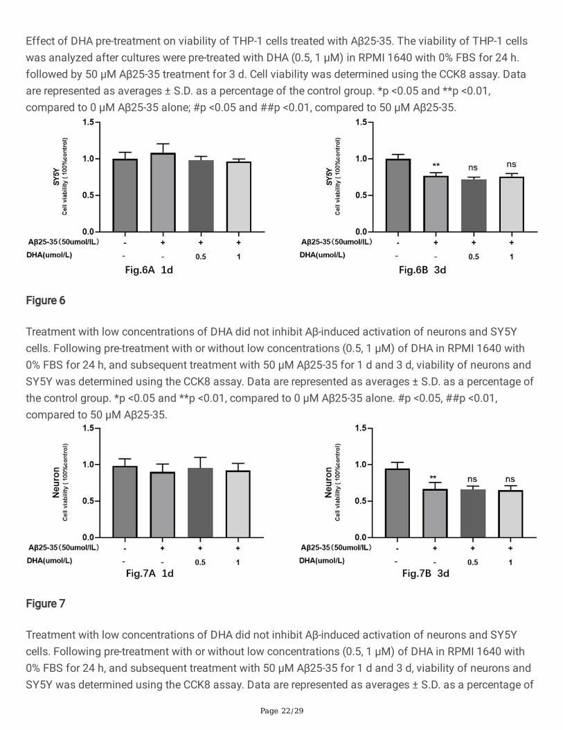
DHA indirectly suppresses THP–1 cell-mediated neuronal activationSubstantial evidence suggests that DHA can directly reduce Aβ-induced activation[13, 23]. We thereforetested whether pre-treatment with low concentrations (0.5, 1 μM) of DHA in RPMI 1640 with 0% FBS for24 h could reduce Aβ25–35-mediated activation of neurons and SY5Y cells. Using the CCK8 assay as areadout, we observed that when treating neurons and SY5Y cells with 50 μM Aβ25–35 alone for 1 d or 3d, viability did not signi�cantly decrease until day 3 (Fig. 6, Fig. 7). Furthermore, low concentrations ofDHA did not inhibit Aβ-induced activation of neurons and SY5Y cells, similar to previous �ndings[23].
We next investigated whether Aβ aggregates could exert their effect on neuronal cells through THP–1cells and how DHA might regulate this process. THP–1 cells were pre-treated with or without variousconcentrations of DHA in RPMI 1640 with 0% FBS for 24 h, followed by 50 μM Aβ25–35 treatment for 3 d(the media was not changed). CM from each THP–1 cell treatment group was collected and used tostimulate neurons for 3 d or 5 d. Using the CCK8 assay as a readout, CM treated with Aβ alone (Aβ-CM)induced neuronal toxicity to a much greater extent than the vehicle control (control CM), while CM fromTHP–1 cells that were pretreated with DHA prior to Aβ treatment (DHA+Aβ-CM) induced signi�cantly lesscytotoxicity in cortical neurons compared with Aβ-CM (Fig. 8B). Interestingly, compared to cells directlytreated with 50 μM Aβ25–35 for 3 days (Fig. 6B), less cytotoxicity was observed in cortical neuronstreated with Aβ-CM for 3 days (Fig. 8A).Taken together, these �ndings indicate that THP–1 cells playedan important role in Aβ clearance and DHA treatment can indirectly suppress THP–1 cell-mediatedneuronal activation.
DHA restored the THP–1 monocytes migration treated with Aβ25–35To investigate whether DHA treatment could modulate the Aβ25–35-induced THP–1 monocytesmigration phenotype THP–1 cells were pre-treated with or without low concentrations (0.5, 1 μM) of DHAin RPMI 1640 with 0% FBS for 24 h, followed by 50 μM Aβ25–35 treatment for 3 days. A transwell systemwas used to test the migratory ability of THP–1 cells collected from different treatment groups. As shownin Fig. 9, the number of migratory THP–1 monocytes after 12h were signi�cantly decreased in Aβ groupcompared to control. In comparison to Aβ group, the number of migratory cells in the Aβ+DHA groupswas signi�cantly higher. Therefore, the results indicate that DHA pre-treatment inhibits the anti-migratoryeffect exerted by Aβ25–35 on THP–1 monocytes.

Page 10/29
DHA suppresses Aβ-induced expression of pro-in�ammatorycytokines in THP–1 cellsTNF-α IL–1β, and IL–6 are the primary pro-in�ammatory cytokines in the in�ammatory responsecascade. To investigate how DHA treatment affects Aβ-induced expression of pro-in�ammatorycytokines, the protein expression levels of TNF-α IL–1β and IL–6 in DHA and Aβ25–35 treated THP–1cells were quanti�ed. TNF-α IL–1β, and IL–6 expression all increased signi�cantly in THP–1 cells aftertreatment with 50 μM Aβ25–35 alone (Fig. 10). However, when THP–1 cells were treated with DHA priorto treatment with Aβ25–35, expression of these pro-in�ammatory cytokines was notably decreasedcompared to 50 μM Aβ25–35 treatment alone. These results demonstrate that DHA treatment in THP–1cells suppresses Aβ-induced protein expression of TNF-α IL–1β and IL–6.
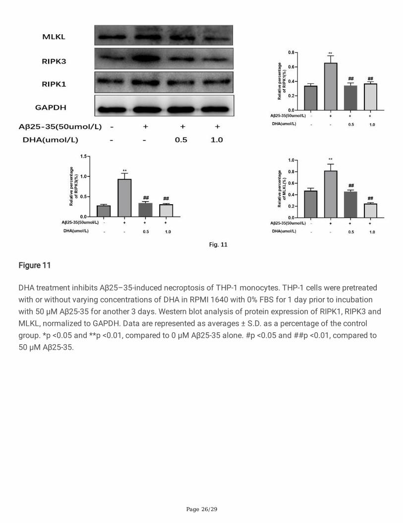
DHA inhibits Aβ25–35-induced necroptosis of THP–1 monocytesWe next investigated whether Aβ-mediated regulation of necroptosis could be modulated by DHA inhuman THP–1 monocytes. Western blotting for key necroptosis proteins revealed that cells pre-treatedwith DHA had lower expression levels of RIPK1, RIPK3, and MLKL compared to cells treated with onlyAβ25–35 (Fig. 11). Therefore, our resulted indicate that DHA treatment inhibits Aβ25–35-inducednecroptosis of THP–1 monocytes Aβ25–35.
DHA attenuates Aβ25–35-induced necroptosis of THP–1 monocytesvia the RIPK1/RIPK3 signaling pathwayNecroptosis is a lytic cell death program de�ned by activation of the receptor interacting protein kinase–1(RIPK1) and RIPK3 to form an oligomeric “necrosome,” which leads to the phosphorylation and activationof the effector pseudokinase mixed lineage kinase domain-like (MLKL). To further investigate whetherDHA could attenuate Aβ25–35-induced necroptosis of THP–1 monocytes via the RIPK1/ RIPK3 signalingpathway, we tested whether NEC–1 (RIPK1 inhibitor[24]) could inhibit Aβ-induced activation of THP–1cells. Viability of THP–1 cells was determined after cultures were pre-treated with or without variousconcentrations of NEC–1 in RPMI 1640 with 0% FBS for 24 h followed by 50 μM Aβ25–35 treatment for3 day. NEC–1 pre-treatment signi�cantly inhibited Aβ25–35-induced activation of THP–1 cells in aconcentration-dependent manner (Fig. 12A). The 10 μM NEC–1 concentration was chosen for allsubsequent experiments.
THP–1 cells were pre-treated with or without NEC–1 and DHA in RPMI 1640 with 0% FBS for 24 hfollowed by 50 μM Aβ25–35 treatment for 3 days. Western blot analysis was then used to determineRIPK3 expression levels. Expression of RIPK3 was suppressed by NEC–1 treatment in the presence orabsence of DHA compared when compared with Aβ25–35 treatment (Fig. 12B-C). This �nding indicatesthat DHA could prevent Aβ-induced necroptosis of THP–1 cells via the RIPK1/RIPK3 signaling pathway.

Page 11/29
DHA suppresses ERK1/2 signaling activated Aβ25–35 but does notaffect p38 or NF-κB/p65 signalingNF-κB signaling plays a key role in immune and in�ammatory responses, while MAPK kinases (p38,ERK1/2) are key regulators of a variety of cellular functions, including cell survival, apoptosis andin�ammation response[19, 20]. We tested whether DHA treatment modulated Aβ-mediated regulation ofMAPK and NF-κB signaling in human THP–1 monocytes. THP–1 cells were pretreated with or withoutvarying concentrations of DHA in RPMI 1640 with 0% for 1 day prior to incubation with 50 μM Aβ25–35for another 3 days. Compared to the 0 μM Aβ25–35 treatment group, the phosphorylation levels of p38and ERK1/2 were signi�cantly increased in the 50 μM Aβ25–35 treatment group (Fig. 13). However,compared to the 50 μM Aβ25–35 treatment group, the phosphorylation level of ERK1/2, but not p38 orp65, was decreased in the Aβ+DHA treatment groups. Taken together, our results show that DHAtreatment suppresses Aβ25–35-induced ERK1/2 signaling in THP–1 cells, but does not affect p38 or NF-κB/p65 signaling.
DHA targets RIPK1 to inhibit ERK1/2 phosphorylation in THP–1 cellstreated with Aβ25–35Increasing evidence indicates that RIPK1 can regulate cell survival via activation of the ERK1/2 signalingpathway[25, 26]. Therefore, THP–1 cells were pre-treated with or without NEC–1 and DHA in RPMI 1640with 0% FBS for 24 h, followed by treatment with 50 μM Aβ25–35 for 3 days. Western blotting was usedto determine the phosphorylation status of ERK1/2. As shown in Fig. 14, phosphorylation of ERK1/2 wassuppressed by the RIPK1 inhibitor NEC–1 in the presence or absence of DHA compared with Aβ25–35treatment. Hence, our results indicate that DHA treatment inhibits ERK1/2 phosphorylation via RIPK1 inTHP–1 cells treated with Aβ25–35.
DiscussionMonocytes play a crucial role in AD, as monocyte-derived perivascular macrophages e�ciently executeAβ phagocytosis[6, 27]. The mechanism connecting brain amyloid accumulation and monocytedegeneration is multifactorial. Deposition of Aβ aggregates in blood vessels can directly damage thevessel wall and allow more monocytes to pass through the parenchyma[28].Toll‐like receptor 2 (TLR2)and Toll‐like receptor 4 (TLR4) were shown to be indirectly involvedin Aβ phagocytosis through theformation of a receptor complex with CD14 and the subsequent activation of monocytes[29]. Aβ -inducedmigration of monocytes across human brain endothelial cells involves both RAGE and PECAM–1[7].Increasing vascular Aβ clearance through the activity of patrolling monocytes could help ameliorate ADsymptoms.In this regard, reduction of the migration, phagocytosis, or number of mononuclear cells intransgenic AD mice is detrimental, whereas compounds that increase their number and phagocyticactivity are generally bene�cial [10]. Oligomeric amyloid-β(1–42) is known to induce THP–1 monocyteadhesion and maturation [30], but these �ndings have not been further expored. Therefore, our

Page 12/29
experiments were designed to further explore possible mechanisms of the interaction between monocytesand Aβ. We demonstrated that Aβ25–35 in�uences THP–1 cell viability, as con�rmed by LDH and CCK8assays. Macrophages are known to become more resistant to apoptosis as a result of differentiation [22,31], Interestingly, by �ow cytometry analysis, we observed that increased apoptotic resistance develops incorrelation with trends in Aβ25–35-mediated THP–1 cell cytotoxicity. When THP–1 cells are treated withlow concentrations of Aβ25–35, we observed increased resistance to apoptosis, conversely, theadvantage of cytotoxic effect is highlighted. Taken together, our results indicate that Aβ25–35 had a dualeffect on THP–1 cell apoptosis (necrosis), and increased THP–1 cell resistance to apoptosis isassociated with THP–1 monocyte differentiation.
Studies have shown that DHA has a neuroprotective effect in many neurodegenerative diseases throughits antioxidant and anti-in�ammatory properties and its ability to activate various cell signalingpathways[16, 32]. Microglial cells and macrophages are key cells in the immune system, and they playimportant roles in CNS repair and regeneration[33]. In addition to their neuroprotective roles, microglialcells and macrophages are also the major producers of proin�ammatory cytokines, such as TNF-α, IL–1β,and IL–6, which can greatly inhibit brain repair and neurogenesis[34]. Here, we found that lowconcentrations of DHA were insu�cient to inhibit Aβ-induced activation of neurons and SY5Y cells butcould indirectly suppress THP–1 cell-mediated neuronal activation effectively. Moreover, we found thatpre-treatment with DHA effectively attenuated Aβ-induced activation of THP–1 monocytes and markedlysuppressed expression of TNF-α, IL–1β, and IL–6. Interestingly, our results also indicated that DHAtreatment restored migration of THP–1 monocytes that had been treated with Aβ25–35.
Necroptosis, a form of regulated necrotic cell death mediated by RIPK1, RIPK3, and MLKL (mixed-lineagekinasedomain-like pseudokinase) [35], can be activated under apoptosis-de�cient conditions [17, 36].Substantial evidence indicates that necroptosis is involved in AD pathogenesis [37, 38]. RIPK1, RIPK3,and MLKL expression were all found to be increased in the brains of individuals with AD individuals aswell as in the brains of AD animal models [39]. Therefore, we investigated whether Aβ aggregates or DHAmediated regulation of necroptosis in human THP–1 monocytes. Our resulted indicate that Aβ25–35treatment induced necroptosis of THP–1 monocytes that could be attenuated with DHA treatment.
NEC–1(necrostatin–1), a speci�c inhibitor of RIPK1, prevents cell necroptosis[40, 41]. Our results showedthat NEC–1 pre-treatment signi�cantly inhibited the activation of THP–1 cells induced by Aβ25–35 in aconcentration-dependent manner. Additionally, expression of RIPK3 was suppressed by NEC–1 treatmentin the presence or absence of DHA compared with Aβ25–35 treatment. This �nding suggests that DHAtreatment could attenuate Aβ25–35-induced necroptosis of THP–1 monocytes via modulation of theRIPK1/RIPK3 signaling pathway.
The mitogen-activated protein kinases (MAPKs) in mammals include c-Jun NH2-terminal kinase (JNK),p38 MAPK, and extracellular signal-regulated kinase (ERK). These enzymes are serine–threonine proteinkinases that regulate various cellular activities including proliferation, differentiation, apoptosis or cellsurvival, in�ammation, and innate immunity[42, 43]. Impaired signaling pathways contribute to the

Page 13/29
pathology of many human diseases, including cancer and neurodegenerative diseases such as AD,Parkinson’s disease, and amyotrophic lateral sclerosis [44, 45]. Subsequent studies have shown that NF-κB is a ubiquitously expressed dimeric transcription factor involved in cellular processes such asin�ammation, adhesion, proliferation, differentiation, apoptosis, and oncogenesis [46]. This family oftranscription factors also plays an important role in nervous system development and function [47]. Weexplored whether Aβ aggregates or DHA regulate the MAPK and NF-κB signaling pathways in humanTHP–1 monocytes. Our �ndings indicate that Aβ25–35 treatment activated the ERK1/2 and p38signaling pathways, but not NF-κB/p65 signaling, while pre-treatment with DHA followed by Aβ25–35treatment suppressed only ERK1/2 signaling.
Several studies have shown that RIPK1 activates MAPKs such as p38, JNK, and ERK during apoptosisfollowing TNF-α treatment[21, 25, 48] and that RIPK1 plays an important role at the crossroads of a cell’sdecision to live or die [49]. Our study revealed that ERK1/2 phosphorylation is suppressed by NEC–1 inthe presence or absence of DHA treatment when compared with Aβ25–35 treatment. Our �ndingsindicate that DHA treatment could inhibit ERK1/2 phosphorylation via RIPK1 in THP–1 cells treated withAβ25–35. Previous work has demonstrated that ERK activation plays a critical role in necroptosis, andthe RIPK1/ERK signaling pathway may present a new therapeutic avenue for treatment of ischemia–reperfusion injury and neurodegenerative diseases where necroptotic cell death is implicated[50].However, the relationship between necroptotic signaling pathways and ERK1/2 is incompletelyunderstood and requires further exploration. Since this study relied on an in vitro model using THP–1cells instead of primary monocytes, additional studies with primary monocytes and an in vivo model areneeded to con�rm and extend our �ndings.
ConclusionOur �ndings indicate that Aβ25–35 regulates two aspects of THP–1 cells necroptosis. We also observedthat increased resistance to apoptosis in THP–1 cells that is correlated with THP–1 monocytedifferentiation. Our results also indicated that DHA treatment restored migration of THP–1 monocytesthat had been treated with Aβ25–35. We found that pre-treatment with DHA effectively inhibited Aβ-induced activation of THP–1 monocytes and markedly suppressed protein expression of TNF-α, IL–1β,and IL–6. DHA treatment can attenuate necroptosis via modulation of the RIPK1/RIPK3 signalingpathway and the phosphorylation status of ERK1/2 in THP–1 monocytes. Our study offers insight intothe anti-in�ammatory and anti-necroptotic properties of DHA. Therefore, our �ndings suggest that DHAmight could have therapeutic potential for use in AD management.
AbbreviationsAD,Alzheimer’s disease;DHA, Docosahexaenoic acid; MAPKs,mitogen-activated protein kinases;NF-κB,nuclear transcription factor-kappa B; TNF-α, tumor necrosis factor-alpha; IL–6, interleukin–6; IL–1β,interleukin–1 beta; iNOS, inducible NO synthase; MLKL, mixed lineage kinase domain-like; NEC–1,necrostatin–1.

Page 14/29
Declarations
AcknowledgementsJudicial Identi�cation Center of Southern Medical University Provides Experimental equipment andtechnical guidance.
FundingThis work was supported by grants from Natural Science Foundation of Guangdong Province (Grant No.2017A030313461) Science and Technology Project of Guangzhou City (201803010010), MedicalScience and Technology Research Fund of Guangdong Province(A201861), Scienti�c Research LaunchProject of Southern Medical University (PY2017N029) and Health Science and Technology Project ofGuangzhou City 20191A010010 .
Availability of data and materialsThe datasets used and/or analyzed during the current study are available from the corresponding authoron reasonable request.
Authors’ contributionsTianming Lü conceived and designed the study, Shiqi Yuan and Huan Li performed the experiments andwrote the paper. Wenyi Xie, Yuanyuan Wang, Jiafa Zhang, Zibo cai, and Zhenlin Mao performed part ofthe experiments. Weibing Xie and Tianming Lü reviewed and edited the manuscript.
Ethics approval and consent to participateNot applicable.
Consent for publicationNot applicable.
Competing interestsThere is no con�ict of interest to declare.
References

Page 15/29
1.Winblad B, Amouyel P, Andrieu S, Ballard C, Brayne C, Brodaty H, Cedazo-Minguez A, Dubois B,Edvardsson D, Feldman H, et al: Defeating Alzheimer’s disease and other dementias: a priority forEuropean science and society. Lancet Neurol 2016, 15:455–532.
2.Serrano-Pozo A, Frosch MP, Masliah E, Hyman BT: Neuropathological Alterations in Alzheimer Disease.Csh Perspect Med 2011, 1:a6189.
3.Querfurth HW: Alzheimer’s Disease (vol 362, pg 329, 2010). New Engl J Med 2011, 364:588.
4.Mawuenyega KG, Sigurdson W, Ovod V, Munsell L, Kasten T, Morris JC, Yarasheski KE, Bateman RJ:Decreased Clearance of CNS -Amyloid in Alzheimer’s Disease. Science 2010, 330:1774.
5.Yang C, Huang X, Huang X, Mai H, Li J, Jiang T, Wang X, Lu T: Aquaporin–4 and Alzheimer’s Disease. JAlzheimers Dis 2016, 52:391–402.
6.Hawkes CA, McLaurin J: Selective targeting of perivascular macrophages for clearance of beta-amyloidin cerebral amyloid angiopathy. Proc Natl Acad Sci U S A 2009, 106:1261–1266.
7.Giri R, Shen Y, Stins M, Du Yan S, Schmidt AM, Stern D, Kim KS, Zlokovic B, Kalra VK: beta-amyloid-induced migration of monocytes across human brain endothelial cells involves RAGE and PECAM–1. AmJ Physiol Cell Physiol 2000, 279:C1772-C1781.
8.Michaud J, Bellavance M, Préfontaine P, Rivest S: Real-Time In Vivo Imaging Reveals the Ability ofMonocytes to Clear Vascular Amyloid Beta. Cell Rep 2013, 5:646–653.
9.Majumdar A, Chung H, Dolios G, Wang R, Asamoah N, Lobel P, Max�eld FR: Degradation of �brillarforms of Alzheimer’s amyloid beta-peptide by macrophages. Neurobiol Aging 2008, 29:707–715.
10.Michaud JP, Halle M, Lampron A, Theriault P, Prefontaine P, Filali M, Tribout-Jover P, Lanteigne AM,Jodoin R, Cluff C, et al: Toll-like receptor 4 stimulation with the detoxi�ed ligand monophosphoryl lipid Aimproves Alzheimer’s disease-related pathology. Proceedings of the National Academy of Sciences 2013,110:1941–1946.
11.Hohs�eld LA, Humpel C: Migration of blood cells to β-amyloid plaques in Alzheimer’s disease. ExpGerontol 2015, 65:8–15.
12.Cheng Y, Li Z, Kardami E, Loh YP: Neuroprotective effects of LMW and HMW FGF2 against amyloidbeta toxicity in primary cultured hippocampal neurons. Neurosci Lett 2016, 632:109–113.
13.Kwon SH, Ma SX, Hwang JY, Lee SY, Jang CG: Involvement of the Nrf2/HO–1 signaling pathway insulfuretin-induced protection against amyloid beta25–35 neurotoxicity. Neuroscience 2015, 304:14–28.
14.Liu N, Zhuang Y, Zhou Z, Zhao J, Chen Q, Zheng J: NF-κB dependent up-regulation of TRPC6 by Aβ inBV–2 microglia cells increases COX–2 expression and contributes to hippocampus neuron damage.

Page 16/29
Neurosci Lett 2017, 651:1–8.
15.Zhao Y, Zeng Y, Wu A, Yu C, Tang Y, Wang X, Xiong R, Chen H, Wu J, Qin D: Lychee Seed FractionInhibits Aβ(1–42)-Induced Neuroin�ammation in BV–2 Cells via NF-κB Signaling Pathway. FrontPharmacol 2018, 9.
16.Belkouch M, Hachem M, Elgot A, Lo Van A, Picq M, Guichardant M, Lagarde M, Bernoud-Hubac N: Thepleiotropic effects of omega–3 docosahexaenoic acid on the hallmarks of Alzheimer’s disease. TheJournal of Nutritional Biochemistry 2016, 38:1–11.
17.Linkermann A, Green DR: Necroptosis. New Engl J Med 2014, 370:455–465.
18.Lin Q, Chen P, Wang W, Lin C, Zhou Y, Yu L, Lin Y, Xu Y, Kang D: RIP1/RIP3/MLKL mediatesdopaminergic neuron necroptosis in a mouse model of Parkinson disease. Laboratory investigation; ajournal of technical methods and pathology 2019.
19.Park S, Hwang J, Jang M, Lee SH, Park J, Han I: A novel caffeic acid–1-piperonylpiperazinehybridization compound HBU–47 inhibits LPS-mediated in�ammation in RAW264.7 macrophage cells. IntImmunopharmacol 2014, 19:60–65.
20.Alexa A, Gógl G, Glatz G, Garai Á, Zeke A, Varga J, Dudás E, Jeszenői N, Bodor A, Hetényi C, Reményi A:Structural assembly of the signaling competent ERK2–RSK1 heterodimeric protein kinase complex.Proceedings of the National Academy of Sciences 2015, 112:2711–2716.
21.Newton K: RIPK1 and RIPK3: critical regulators of in�ammation and cell death. Trends Cell Biol 2015,25:347–353.
22.Busca A, Saxena M, Iqbal S, Angel J, Kumar A: PI3K/Akt regulates survival during differentiation ofhuman macrophages by maintaining NF‐κB‐dependent expression of antiapoptotic Bcl‐xL. J LeukocyteBiol 2014, 96:1011–1022.
23.Zhang Y, Brown RE, Zhang P, Zhao Y, Ju X, Song C: DHA, EPA and their combination at various ratiosdifferently modulated Aβ25–35-induced neurotoxicity in SH-SY5Y cells. Prostaglandins, Leukotrienes andEssential Fatty Acids 2018, 136:85–94.
24.Filipczak PT, Thomas C, Chen W, Salzman A, McDonald JD, Lin Y, Belinsky SA: TSC2 De�ciencyUnmasks a Novel Necrosis Pathway That Is Suppressed by the RIP1/RIP3/MLKL Signaling Cascade.Cancer Res 2016, 76:7130–7139.
25.Najjar M, Saleh D, Zelic M, Nogusa S, Shah S, Tai A, Finger JN, Polykratis A, Gough PJ, Bertin J, et al:RIPK1 and RIPK3 Kinases Promote Cell-Death-Independent In�ammation by Toll-like Receptor 4.Immunity 2016, 45:46–59.

Page 17/29
26.Zhang M, Li J, Geng R, Ge W, Zhou Y, Zhang C, Cheng Y, Geng D: The Inhibition of ERK ActivationMediates the Protection of Necrostatin–1 on Glutamate Toxicity in HT–22 Cells. Neurotox Res 2013,24:64–70.
27.Hallé M, Tribout-Jover P, Lanteigne A, Boulais J, St-Jean JR, Jodoin R, Girouard M, Constantin F,Migneault A, Renaud F, et al: Methods to monitor monocytes-mediated amyloid-beta uptake andphagocytosis in the context of adjuvanted immunotherapies. J Immunol Methods 2015, 424:64–79.
28.Zuroff L, Daley D, Black KL, Koronyo-Hamaoui M: Clearance of cerebral Abeta in Alzheimer’s disease:reassessing the role of microglia and monocytes. Cell Mol Life Sci 2017, 74:2167–2201.
29.Udan ML, Ajit D, Crouse NR, Nichols MR: Toll-like receptors 2 and 4 mediate Abeta(1–42) activation ofthe innate immune response in a human monocytic cell line. J Neurochem 2008, 104:524–533.
30.Crouse NR, Ajit D, Udan MLD, Nichols MR: Oligomeric amyloid-β(1–42) induces THP–1 humanmonocyte adhesion and maturation. Brain Res 2009, 1254:109–119.
31.Gordon S, Taylor PR: Monocyte and macrophage heterogeneity. Nat Rev Immunol 2005, 5:953–964.
32.McLean FH, Campbell FM, Sergi D, Grant C, Morris AC, Hay EA, MacKenzie A, Mayer CD, Langston RF,Williams LM: Early and reversible changes to the hippocampal proteome in mice on a high-fat diet. NutrMetab 2019, 16.
33.Hanisch U, Kettenmann H: Microglia: active sensor and versatile effector cells in the normal andpathologic brain. Nat Neurosci 2007, 10:1387–1394.
34.Xiong X, Liu L, Yang Q: Functions and mechanisms of microglia/macrophages in neuroin�ammationand neurogenesis after stroke. Prog Neurobiol 2016, 142:23–44.
35.de Almagro MC, Vucic D: Necroptosis: Pathway diversity and characteristics. Semin Cell Dev Biol 2015,39:56–62.
36.Shan B, Pan H, Najafov A, Yuan J: Necroptosis in development and diseases. Gene Dev 2018, 32:327–340.
37.Caccamo A, Branca C, Piras IS, Ferreira E, Huentelman MJ, Liang WS, Readhead B, Dudley JT,Spangenberg EE, Green KN, et al: Necroptosis activation in Alzheimer’s disease. Nat Neurosci 2017,20:1236–1246.
38.Yuan J, Amin P, Ofengeim D: Necroptosis and RIPK1-mediated neuroin�ammation in CNS diseases.Nat Rev Neurosci 2019, 20:19–33.
39.Ofengeim D, Mazzitelli S, Ito Y, DeWitt JP, Mi�in L, Zou C, Das S, Adiconis X, Chen H, Zhu H, et al:RIPK1 mediates a disease-associated microglial response in Alzheimer's disease. Proceedings of the

Page 18/29
National Academy of Sciences 2017, 114:E8788-E8797.
40.Kanou T, Ohsumi A, Kim H, Chen M, Bai X, Guan Z, Hwang D, Cypel M, Keshavjee S, Liu M: Inhibition ofregulated necrosis attenuates receptor-interacting protein kinase 1-mediated ischemia-reperfusion injuryafter lung transplantation. J Heart Lung Transplant 2018, 37:1261–1270.
41.Jing L, Song F, Liu Z, Li J, Wu B, Fu Z, Jiang J, Chen Z: MLKL-PITPalpha signaling-mediatednecroptosis contributes to cisplatin-triggered cell death in lung cancer A549 cells. Cancer Lett 2018,414:136–146.
42.Arthur JSC, Ley SC: Mitogen-activated protein kinases in innate immunity. Nat Rev Immunol 2013,13:679–692.
43.Qin S, Yang C, Huang W, Du S, Mai H, Xiao J, Lü T: Sulforaphane attenuates microglia-mediatedneuronal necroptosis through down-regulation of MAPK/NF-κB signaling pathways in LPS-activated BV–2 microglia. Pharmacol Res 2018, 133:218–235.
44.Kim EK, Choi E: Compromised MAPK signaling in human diseases: an update. Arch Toxicol 2015,89:867–882.
45.Xu M, Yan T, Fan K, Wang M, Qi Y, Xiao F, Bi K, Jia Y: Polysaccharide of Schisandra Chinensis Fructusameliorates cognitive decline in a mouse model of Alzheimer’s disease. J Ethnopharmacol 2019,237:354–365.
46.Fao L, Mota SI, Rego AC: Shaping the Nrf2-ARE-related pathways in Alzheimer’s and Parkinson’sdiseases. Ageing Res Rev 2019, 54.
47.Mincheva-Tasheva S, Soler RM: NF-κB Signaling Pathways. The Neuroscientist 2013, 19:175–194.
48.Alvarez SE, Harikumar KB, Hait NC, Allegood J, Strub GM, Kim EY, Maceyka M, Jiang H, Luo C, KordulaT, et al: Sphingosine–1-phosphate is a missing cofactor for the E3 ubiquitin ligase TRAF2. Nature 2010,465:1084–1088.
49.Festjens N, Vanden Berghe T, Cornelis S, Vandenabeele P: RIP1, a kinase on the crossroads of a cell’sdecision to live or die. Cell Death Differ 2007, 14:400–410.
50.Zhang M, Li J, Geng R, Ge W, Zhou Y, Zhang C, Cheng Y, Geng D: The Inhibition of ERK ActivationMediates the Protection of Necrostatin–1 on Glutamate Toxicity in HT–22 Cells. Neurotox Res 2013,24:64–70.
Figures

Page 19/29
Figure 1
THP-1 cell viability following treatment with a concentration gradient of Aβ25-35, with different stimulustimes; cell viability was determined using the CCK8 assay Fig. 1A 1B 1C. All samples compared tocontrol (0 μM Aβ25-35); data are represented as averages ± S.D. * P < 0.05 ** P < 0.01
Figure 2
Cytotoxicity was determined using an LDH release assay, Fig. 2A 2B 2C. All samples compared tocontrol (0 μM Aβ25-35); data are represented as averages ± S.D. * P < 0.05 ** P < 0.01

Page 20/29
Figure 3
THP1 cells activity treated with Aβ25-35 whether the apoptosis (necrosis) and differentiation of THP1cells is involved, after THP1 cells was treated with Aβ25-35 for 3 days, Annexin v-�tc, 7AAD and antigenCD11b (as a surface marker for THP1 cells differentiation were used for �ow cytometry analysis. Fig 3A:Normal cells (Q3), Early apoptotic cells (Q4), Late apoptotic /necrosis cell (Q2), CD11b PE-A (cell

Page 21/29
expression after differentiation). The statistical results shown as Fig 3B 3C. Data are presented as mean± S.D. Compared with Control group (Aβ25-35 0uM), * P < 0.05 ** P < 0.01
Figure 4
Effect of DHA on viability of THP-1 cells. DHA was not cytotoxic to THP-1 cells at concentrations of up to1 μM. THP-1 cells were treated with varying concentrations (0.0625-1 μM) of DHA for 1 d, 3 d, and 5 d.Data are represented as averages ± S.D. as a percentage of the control group, * P < 0.05 ** P < 0.01.
Figure 5
Page 22/29
Effect of DHA pre-treatment on viability of THP-1 cells treated with Aβ25-35. The viability of THP-1 cellswas analyzed after cultures were pre-treated with DHA (0.5, 1 μM) in RPMI 1640 with 0% FBS for 24 h.followed by 50 μM Aβ25-35 treatment for 3 d. Cell viability was determined using the CCK8 assay. Dataare represented as averages ± S.D. as a percentage of the control group. *p <0.05 and **p <0.01,compared to 0 μM Aβ25-35 alone; #p <0.05 and ##p <0.01, compared to 50 μM Aβ25-35.
Figure 6
Treatment with low concentrations of DHA did not inhibit Aβ-induced activation of neurons and SY5Ycells. Following pre-treatment with or without low concentrations (0.5, 1 μM) of DHA in RPMI 1640 with0% FBS for 24 h, and subsequent treatment with 50 μM Aβ25-35 for 1 d and 3 d, viability of neurons andSY5Y was determined using the CCK8 assay. Data are represented as averages ± S.D. as a percentage ofthe control group. *p <0.05 and **p <0.01, compared to 0 μM Aβ25-35 alone. #p <0.05, ##p <0.01,compared to 50 μM Aβ25-35.
Figure 7
Treatment with low concentrations of DHA did not inhibit Aβ-induced activation of neurons and SY5Ycells. Following pre-treatment with or without low concentrations (0.5, 1 μM) of DHA in RPMI 1640 with0% FBS for 24 h, and subsequent treatment with 50 μM Aβ25-35 for 1 d and 3 d, viability of neurons andSY5Y was determined using the CCK8 assay. Data are represented as averages ± S.D. as a percentage of

Page 23/29
the control group. *p <0.05 and **p <0.01, compared to 0 μM Aβ25-35 alone. #p <0.05, ##p <0.01,compared to 50 μM Aβ25-35.
Figure 8
DHA indirectly suppresses THP-1 cell-mediated neuronal activation. THP-1 cells were pre-treated with orwithout various concentrations of DHA in RPMI 1640 with 0% FBS for 24 h, followed by 50 μM Aβ25-35treatment for 3. CM from each THP-1 treatment group was collected and used to stimulate neurons for 3or 5 d. Cell viability was determined using the CCK8 assay. Data are represented as averages ± S.D. as apercentage of the control group. *p <0.05 and **p <0.01, compared to control-CM alone. #p <0.05 and##p <0.01, compared to Aβ-CM.

Page 24/29
Figure 9
DHA pre-treatment inhibits the anti-migratory effect exerted by Aβ25–35 on THP-1 monocytes. THP-1cells were pre-treated with or without low concentrations (0.5, 1 μM) of DHA in RPMI 1640 with 0% FBSfor 24 h, followed by 50 μM Aβ25-35 treatment for 3 days. A transwell system was used to test themigratory ability of THP-1 cells after 12 h from different treatment groups, Cell counting was carried outusing a microscope (100X). Data are represented as averages ± S.D. as a percentage of the control group.*p <0.05 and **p <0.01, compared to 0 μM Aβ25-35 alone. #p <0.05 and ##p <0.01, compared to 50 μMAβ25-35.

Page 25/29
Figure 10
DHA treatment has an inhibitory effect on expression of pro-in�ammatory cytokines in Aβ-stimulatedTHP-1 cells. THP-1 cells were pretreated with or without varying concentrations of DHA in RPMI 1640 with0% FBS for 1 day prior to incubation with 50 μM Aβ25-35 for another 3 days. Western blot analysis ofprotein expression of TNF-α, IL-1β and IL-6, normalized to GAPDH. Data are represented as averages ±S.D. *p <0.05 and **p <0.01, compared to 0 μM Aβ25-35 alone. #p <0.05 and ##p <0.01, compared to 50μM Aβ25-35.
Page 26/29
Figure 11
DHA treatment inhibits Aβ25–35-induced necroptosis of THP-1 monocytes. THP-1 cells were pretreatedwith or without varying concentrations of DHA in RPMI 1640 with 0% FBS for 1 day prior to incubationwith 50 μM Aβ25-35 for another 3 days. Western blot analysis of protein expression of RIPK1, RIPK3 andMLKL, normalized to GAPDH. Data are represented as averages ± S.D. as a percentage of the controlgroup. *p <0.05 and **p <0.01, compared to 0 μM Aβ25-35 alone. #p <0.05 and ##p <0.01, compared to50 μM Aβ25-35.

Page 27/29
Figure 12
DHA treatment attenuates Aβ25–35-induced necroptosis in THP-1 monocytes via the RIPK1/RIPK3signaling pathway. THP-1 cells were pre-treated with or without various concentrations of NEC-1 in RPMI1640 with 0% FBS for 24 h, followed by 50 μM Aβ25-35 stimulation for 3 d; cell viability was determinedusing the CCK8 assay (Fig. 12A). THP-1 cells were pretreated with or without NEC-1 and DHA in RPMI1640 with 0% FBS for 24 h, followed by 50 μM Aβ25-35 stimulation for 3 d; Western blotting analysis wasused to determine expression levels of RIPK3 (normalized to GAPDH) Fig. 12B-C Data are representedas averages ± SD. as a percentage of the control group. *p <0.05 and **p <0.01, compared to 0 μM Aβ25-35 alone. #p <0.05 and ##p <0.01, compared to 50 μM Aβ25-35.

Page 28/29
Figure 13
DHA suppresses the ERK1/2 signaling pathway induced by Aβ25–35 in THP-1 cells, but not the p38 orNF-κB/p65 signaling pathways. THP-1 cells were pretreated with or without varying concentrations ofDHA in RPMI 1640 with 0% FBS for 1 day prior to incubation with 50 μM Aβ25-35 for another 3 days. Thephosphorylation status of ERK1/2 (A), p38 (B), and p65 (C) were determined by Western blot analysis.

Page 29/29
Data are represented as averages ± SD, *p <0.05 and **p <0.01, compared to 0 μM Aβ25-35 alone. #p<0.05 and ##p <0.01, compared to 50 μM Aβ25-35.
Figure 14
DHA treatment inhibits ERK1/2 phosphorylation via RIPK1 in THP-1 cells treated with Aβ25-35. THP-1cells were pre-treated with or without NEC-1 and DHA in RPMI 1640 with 0% FBS for 24 h, followed by 50μM Aβ25-35 treatment for 3 d. Western blotting was used to determine the phosphorylation status ofERK1/2. Data are represented as averages ± SD, *p <0.05 and **p <0.01, compared to 0 μM Aβ25-35alone. #p <0.05 and ##p <0.01, compared to 50 μM Aβ25-35.