distribution and regulation of the g protein- coupled receptor
TRANSCRIPT

20
Distribution and Regulation of the G Protein-Coupled Receptor Gpr88 in the Striatum:
Relevance to Parkinson’s Disease
Renaud Massart1, Pierre Sokoloff3 and Jorge Diaz1,2
1 INSERM UMR-894, Psychiatry and Neurosciences Center, Paris, 2Paris Descartes University, Faculty of Pharmacy / Laboratory
of Neurobiology and Molecular Pharmacology (UMRs894 INSERM), Paris,
3Pierre Fabre Research Institute, Neurology and Psychiatry Department, Castres, France
1. Introduction
The human basal ganglia constitutes a functional neural network located at the base of the
forebrain. It receives most of its afferent inputs through the striatum, the major nucleus of
the basal ganglia accomplishing fast neurotransmitter-mediated operations through
somatotopically organized projections to the principal neuron cell type, the striatal
GABAergic spiny projection neurons. This spiny projection neurons, which make up 95 % of
the neuron population of striatum (Kemp & Powell 1971), receive excitatory glutamatergic
inputs from all areas of the cortex and specific thalamic nuclei (Gerfen & Wilson 1996; Bolam
et al., 2000; Voorn et al., 2004; Doig et al., 2010), and also modulatory dopaminergic inputs
from the substantia nigra pars compacta (Smith & Kieval 2000; Utter. & Basso 2008). Spiny
Projection Neurons include two major subpopulations giving rise to the direct striato-nigral
pathway, and the indirect striato-pallidal pathway which communicates information to the
basal ganglia output structures; the internal segment of the globus pallidus and the
substantia nigra pars reticulata (Smith, Y. & Kieval 2000; Gerfen & Wilson 1996). Although
the two neuron subpopulations are GABAergic, they differ in a number of properties
including the expression of different complements of dopamine, Adenosine, NMDA and
acetylcholine receptor subtypes as well as of peptide content; the direct striato-nigral
pathway neurons coexpress substance P and dynorphin, whereas the indirect striato-
pallidal pathway neurons express enkephalin (Gerfen et al., 1990, 1991; Reiner & Anderson
1990; Gerfen & Wilson 1996; Le Moine & Bloch, 1995).
Based on the fact that striatal medium-spiny neurons are the major input targets and the
major projection neurons of striatum, it is thought that integration of neurotransmission in
these neurons is an important determinant of the functional organization of the striatum.
Thus, changes in neurotransmission on striatal spiny projection neurons have been involved
in the regulation of voluntary movement, behavioral control, cognitive function and reward
mechanisms. For instance, massive spiny projection neuron loss and major dopamine
www.intechopen.com
Mechanisms in Parkinson’s Disease – Models and Treatments
394
deficits in striatum lead to severe motor disorders, such as the excess of involuntary
movements encountered in Huntington’s disease and the rigidity and poverty of
movements that typifies Parkinson’s disease, respectively (Ross et al., 1997; Wolfgang &
Stanley, 2003). Therefore, investigations addressed to characterize new receptor proteins
displaying high densities and potential involvement in neurotransmission mechanisms
within the striatum can provide new insight into the basal ganglia physiology and
pathophysiology and also new clues for therapy of severe motor disorders.
A previous study reported a novel striatum-specific transcript, the strg/Gpr88, encoding an
orphan G protein-coupled receptor of human and rodents (Mizushima et al., 2000). It display
highest sequence homology with 5HT1D and β3 receptors. Since the original description, little
data have been documented on the biological function (s) and the the cellular and subcellular
distribution of the Gpr88 protein. Hence, the Gpr88 endogenous putative ligand, the detailed
Gpr88 protein distribution and GPR88 functional roles are unknown. One approach to gain
functional insights into this novel gene coding for an orphan receptor is the precise analysis of
its spatial and temporal expression to provide information about the neural morphological
substrates supporting Gpr88 functions in the striatal complex.
Hence, the present findings provide in situ hybridization and light-level immunohistochemical
evidence for Gpr88 localization in the rat and monkey striatum and its subcellular distribution
in striatal neurons by using a validated polyclonal antibody specifically recognizing Gpr88,
(Massart et al 2009). We also describe morphological data on the spatiotemporal Gpr88
expression in the developing rat striatum, suggesting that both nigrostriatal and corticostriatal
pathways control its normal striatal pattern of expression. Using treatments with l-DOPA and
dopamine antagonists, in unilateral 6-hydroxydopamine- and cortical ibotenate-lesioned rats,
we further demonstrated that striatal Gpr88 expression is modulated by dopamine- and
glutamate-regulated mechanisms involving trans-synaptic influences of the corticostriatal
pathway input activity.
2. Widespread Gpr88 expression within the striatal complex
Using in situ hybridization and immunohistochemistry approaches, we demonstrated that
Gpr88 mRNA and protein expression are specially abundant within restricted basal
telencephalic structures including the dorsal striatum, nucleus accumbens, and olfactory
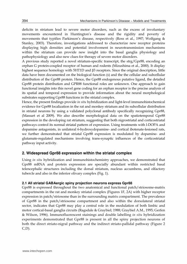
tubercle and also in the inferior olivary complex (Fig. 1).
2.1 All striatal GABAergic spiny projection neurons express Gpr88
Gpr88 is expressed throughout the two anatomical and functional patch/striosome-matrix
compartments in the rat and monkey striatal complex (Figures 1F, 2A) with higher receptor
expression in patch/striosome than in the surrounding matrix compartment. The prevalence
of Gpr88 in the patch/striosome compartment and also within the dorsolateral striatal
sector, indicates that Gpr88 may play a central role in the modulation of both limbic and
motor cortical-basal ganglia circuits (Ragsdale & Graybiel; 1988; Graybiel A.M., 1995; Gerfen
& Wilson, 1996). Immunofluorescent stainings and double labelling in situ hybridization
experiments demonstrated that Gpr88 is present in all the spiny projection neurons of
both the direct striato-nigral pathway and the indirect striato-pallidal pathway (Figure 2
C,D).
www.intechopen.com
Distribution and Regulation of the G Protein-Coupled Receptor Gpr88 in the Striatum: Relevance to Parkinson’s Disease
395
Fig. 1. Gpr88 distribution in the rat and the monkey brains. Both Gpr88 mRNA (A,C) and Gpr88 protein (B,D,E,F) are particularly concentrated throughout the striatum (St), nucleus accumbens (Acb), olfactory tubercle (Tu) and the inferior olive complex (IO) of the rat (A-E). Similar levels and distribution-pattern of Gpr88 immunorreactivity is detected in nucleus caudatus (C), putamen (Pu) and nucleus accumbens (Acb) of the monkey brain (F). Significant levels of Gpr88 are also present with a laminar distribution throughout the neocortex. Arrows in (F), point out small and intense Gpr88 stained areas corresponding to striosome striatal subcompartments. RT-QPCR data from rodents suggest that Gpr88 displays the highest expression levels compared to other known GPCRs of the striatum (Massart et al., 2007, 2008). The pattern of Gpr88 throughout the striatum of adult rats and monkeys is characterized by widespread distribution and regional differences (Figure 1), suggesting a central role of this orphan receptor in the modulation of sensorimotor related informations (Flaherty & Graybiel1994; Voorn et al., 2004). Although Gpr88 is prevalent in the striatal complex, we also detected moderate levels of both Gpr88 transcripts and protein throughout the cerebral neocortex (Figure 1A,B). Both signals display a similar non-homogeneous laminar distribution characterized by higher expression in the upper neocortical layers II-IV than in the lower layers V-VI. No Gpr88 expression was detected in the cortical layer I. Moreover, cortical Gpr88 expression represents about 20% of the GPR88 striatal expression, as assessed by different quantitive approaches including Western blot, immunohistochemistry and in situ hybridization. Double immuno-fluorescent labellings for Gpr88 and different neural cell-type specific markers have demonstrated that Gpr88 is an exclusive neuronal receptor of the brain, being absent from glial cells (Massart et al., 2009).
www.intechopen.com

Mechanisms in Parkinson’s Disease – Models and Treatments
396
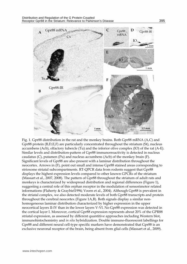
Fig. 2. Gpr88 distribution in the rat dorsal striatum. (A) The immunofluorescent Gpr88 signal is heterogeneously distributed within the dorsal striatum and characterized by its marked concentration in the striatal dorsolateral region and the patches compartments. The inset illustrates putative medium spiny neurons displaying intense Gpr88 labelling on the cell surface along the soma and dendrites. (C, D) double-labelling in situ hybridization indicates that all Substance P (dark-stained cells in C) and enkephalin (dark-stained cells in D) neurons, also express Gpr88 transcripts (detected by silver grain labellings). The distribution of the electron-dense immunoreactive reaction product, reflects the subcellular GPR88 presence in submembranous sites around the perikaryon (arrow-heads in B asterisk and arrows in E). The receptor is concentrated in symmetrical synapses in the cell body (asterisk in E) but also in asymmetrical synapses (asterisk in F) in dendritic spines (Sp). Note the absence of immunolabelling in synaptic contacts of two adjacent nerve terminals (t) in E. (Enk) Enkephalin, (Nu) Cell nucleus, (So) neuronal cell body, (Sp) Substance P in C, (Sp) dendritic spine in F.
Electron microscopic analysis of the Gpr88 immunolabelling in the rat dorsal striatum demonstrated a high proportion of electron dense Gpr88 positive dendritic spines, dendrite shafts and cell bodies (Figure 2 B,E,F) that are characteristic features of GABAergic spiny projection neurons (Somogyi et al., 1982; Bolam et al., 1983). However, no axonal or terminal Gpr88 immunolabeled profiles were observed. Likewise, globus pallidus and substantia nigra pars reticulata, two basal ganglia regions receiving the striato-pallidal and striato-nigral terminals respectively (Surmeier et al., 2007), lack Gpr88 immunoreactivity. All these morphological findings highlight a potential functional role for Gpr88 in synaptic events occurring on somatodendritic compartments and their integration in striato-nigral and striato-pallidal medium spiny neurons. Gpr88 immunoreactivity was often concentrated on discrete postsynaptic sub-membranous sites in a large proportion of asymmetrical (excitatory)
www.intechopen.com

Distribution and Regulation of the G Protein-Coupled Receptor Gpr88 in the Striatum: Relevance to Parkinson’s Disease
397
synapses that generally receive glutamate as neurotransmitter (Bouyer et al., 1984; Bolam et al., 2000) and also on symmetrical (inhibitory) synapses which could be supplied by terminals originating from GABAergic aspiny or cholinergic interneurons or even by intrastriatal GABAergic axon-collaterals from medium-spiny projection neurons. Double immunofluorescent labellings in the same section demonstrated no association between Gpr88 immunoreactivity and tyrosine hydroxylase immunolabelled axon-terminals. In contrast, the Gpr88 immunoreactive signal was often juxtaposed to most vesicular glutamate transporter1 immunoreactive terminals, indicating that Gpr88 is preferentially located on synapses supplied by cortical inputs, rather than by vesicular glutamate transporter2 immunoreactive thalamic inputs contacting medium spiny neurons (Herzog et al., 2001; Kaneko & Fujiyama, 2002; Fremeau et al., 2004). Moreover, electron microscopy analysis demonstrates that Gpr88 immunoreactive signal is often present on the head of spines, where corticostriatal inputs mainly contact the dendritic tree of striatal spiny projection neurons (Bouyer et al., 1984; Dube et al., 1988; Ribak & Roberts 1990; Smith et al., 1994). The preferential subcellular distribution of Gpr88 in striatal asymmetrical synapses of virtually all GABAergic projection neurons suggests a role for Gpr88 in the modulation of medium spiny neurons activity to cortical glutamatergic inputs and a potential role in the regulation of the flow of cortical information through the basal ganglia. Several lines of evidence indicate that cortical excitatory signals are modulated by dopaminergic synaptic contacts located on the neck of spines (Arbuthnott et al., 2000). Gpr88 location at specific synaptic sites, where corticostriatal and nigrostriatal afferents converge, further suggests involvement of Gpr88 in the modulation of both glutamatergic and dopaminergic signals received by the striatal medium spiny neurons.
3. Spatial and temporal Gpr88 expression in the developing rat striatum
To gain functional insights into striatal Gpr88 we have determined the profile of GPR88 expression in the prenatal and postnatal developing striatum of the rat by in situ hybridization and immunohistochemistry. Morphological data indicate that Gpr88 expression emerges with a homogeneous distribution, in the ventrolateral portion of the developing striatum at the embryonic day 16 (E16) of rat development (Figure 3A,B,C), a time when striatal neurons are both morphologically and functionally immature (van der Kooy & Fishell, 1987) and also when the patch-matrix striatal compartments, are not yet differentiated. The homogeneous Gpr88 mRNA distribution becomes heterogeneous when clusters of developing neurons displaying dense Gpr88 expression are seen throughout the dorsal and ventral striatal regions by the fetal stage E19-E20 (Figures 3D,E). Using double immunohistochemistry stained brain sections for Gpr88 and tyrosine hydroxylase, we confirmed that rich Gpr88 small areas strictly match the densely dopamine innervated striatal patch/striosome compartments (Gerfen et al., 1987). Levels of Gpr88 expression in patches compartments increase until the end of the first postnatal week and then decline in the second postnatal week with the ongoing development to eventually reach adult expression levels. Such developmental profile of Gpr88 expression in the prenatal and postnatal rat striatum suggests that the pattern of Gpr88 expression may be under the influence of afferent inputs reaching to the striatal primordia. This idea is based on the fact that the patchy-pattern profile of intense GPR88 expression in developing rat striatum closely matches the reported spatial and temporal development of the nigrostriatal dopamine afferents (Voorn et al., 1988), suggesting that the nigral dopamine inputs influence the patterning of striatal GPR88 expression. Such type of influence by the
www.intechopen.com

Mechanisms in Parkinson’s Disease – Models and Treatments
398
dopamine inputs has been demonstrated for the establishment of the pattern of opiate receptors expression in the embryonic patch compartment (van der Kooy & Fishell, 1992). The cortical projections are the second major afferent input to the striatum that may act in concert with nigral dopamine inputs to guide development of striatal subcompartment phenotypes. For instance, studies in the monkey have shown the patchy distribution of corticostriatal afferents before the day of birth (Goldman-Rakic, 1981). Moreover, organotypic assays involving co-cultures of the striatum with substantia nigra or cortex indicate that afferents from these structures have a prominent influence on the development of striatal patch/matrix compartments (Snyder-Keller & Costantini 1996; Snyder-Keller et al., 2001; Snyder-Keller, 2004). The mutual influence of dopaminergic and glutamatergic pathways within the developing striatum is probably important for the setting up of striatal neurotransmission circuits, as previously shown by dopamine manipulations that influence corticostriatal synaptic configurations (Meshul & Tan, 1994; Meshul et al., 1999; Meshul & Allen, 2000; Avila-Costa et al., 2005). These observations support the idea that cortical glutamatergic inputs and/or dopamine glutamate interactions may exert a control on Gpr88 expression in the developing medium spiny neurons.
Fig. 3. Developmental profile of Gpr88 expression in the rat striatum. Gpr88 mRNA (A, D) and Gpr88-protein (E) expression in the developing striatum. (A) Homogeneous distribution of Gpr88 transcripts in the dorsolateral sector of differentiating striatum at E16. (D) Heterogeneous distribution of Gpr88-mRNA at E19. (F) Clusters of striatal developing neurons displaying dense Gpr88 immunoreactive signal (Gpr88-rich patches) at E20. (Cx) cortex, (St) striatum, (Tu) olfactory tubercle.
www.intechopen.com

Distribution and Regulation of the G Protein-Coupled Receptor Gpr88 in the Striatum: Relevance to Parkinson’s Disease
399
Although dopamine and glutamate afferents are the most likely candidates to modulate Gpr88 expression in developing medium-spiny projection neurons, other factors associated with nigral and cortical inputs may also play an important role in controlling Gpr88 expression within the striatal primordium. For instance, the nigrostriatal and corticostriatal pathways supply the striatum with brain-derived neurotrophic factor (BDNF) (Altar et al., 1997; Seroogy et al., 1994) which has been shown to influence survival, sprouting, and synaptogenesis in different neural systems (Hammond et al., 1999; Alsina et al., 2001; Mamounas et al., 2000). Moreover, studies in mature animals have shown that BDNF has profound effects on neurotransmission, activity-dependent synaptic remodeling, neurogenesis and receptors expression (Altar et al., 1997; Lessmann, 1998; Guillin et al., 2001; Tanaka et al., 2008; Taliaz, 2010). Rather than exclusive effects of either dopamine or glutamate on striatal Gpr88, continuous interplay among afferent signaling systems, including dopamine, glutamate and BDNF, is likely to refine the pattern expression of Gpr88 throughout the period of striatal development. Based on the spatiotemporal profile of GPR88 expression during striatal differentiation, we propose that the early receptor expression is modulated at least in part through a nigrostriatal and corticostriatal pathway dependent mechanisms. In support to the hypothesis of BDNF regulating Gpr88 expression during development, heterozygote BDNF-knockout mice have diminished Gpr88 mRNA levels in both the caude putamen and the shell of the nucleus accumbens (Massart et al., 2005).
4. Modulation of striatal Gpr88 expression by nigrostriatal and corticostriatal pathways in the rat in a model of Parkinson’s disease
The demonstration of the regulation of striatal Gpr88 expression by nigrostriatal dopamine and cortical glutamate inputs was carried out in a rat model of Parkinson’s disease (Schwarting & Huston 1996; Massart et al., 2009). Unilateral lesion of dopamine nigrostriatal pathway, caused by infusion of 6-OHDA in the medial forebrain bundle, produced a decrease in Gpr88 protein and mRNA expressions (Table 1). However, in situ hybridization analysis with double labelling showed that the effects of dopamine depletion were different in the two subpopulations of striatal medium spiny neurons. At the cellular level, 6-OHDA lesion induced a decrease in mRNA expression in striato-pallidal pathway neurons and inversely, a rise in striato-nigral projection neurons, in the dopamine depleted striatum (Table 1). Recently reported data (Heiman et al., 2008; Massart et al., 2009) showed that striatal Gpr88 mRNA expression is twice as high in striato-pallidal output neurons as in striato-nigral output neurons of rodents, the overall lesion-induced Gpr88 downregulation is consistent with the strong decrease in Gpr88 expression occurring in striato-pallidal pathway neurons, not compensated by the limited increase occurring in striato-nigral pathway neurons. These opposed variations are nearly completely reversed by a typical antiparkinsonian treatment with l-DOPA (Table 1). Our finding revealed that D1 receptors, but not D2 receptors, activation exerts a positive influence on Gpr88 expression in the indirect striato-pallidal pathway of the dopamine-depleted hemisphere. On the contrary, D2 receptors stimulation controls Gpr88 expression in the direct striato-nigral pathway. This is rather surprising since D1 and D2 receptors are largely segregated to striatal neurons of the striato-nigral and striato-pallidal pathways, respectively (Gerfen et al., 1990; Le Moine & Bloch, 1995). In fact, in striato-pallidal medium spiny neurons harboring D2 receptors/Enk, in contrast to striato-nigral medium spiny
www.intechopen.com

Mechanisms in Parkinson’s Disease – Models and Treatments
400
Treatment
Gpr88 protein –
immunohistochemistry
Gpr88 mRNA - In situ hybridization
Total mRNA mRNA / SP+ cells
mRNA / ENK+ cells
Intact Lesioned Intact Lesioned Intact Lesioned Intact Lesioned
6-OHDA lesion
Vehicle 107 ± 3.8 91 ± 2.9 * 69 ± 3.2 56 ± 1.4 * 22 ± 1.4
29 ± 1.9 ***
42 ± 2.1 31 ± 1.8
***
L- DOPA
99 ± 5.1 90 ± 4.6 73 ± 4.2 70 ± 3.7 22 ± 1.4 26 ± 1.7 40 ± 0.8 38 ± 1.3
L-DOPA + SCH23390
108 ± 5.9 92 ± 5.6 76 ± 6.8 62 ± 3.2 23 ± 2.7 21 ± 1.6 43 ± 3.3 26 ± 4.3
***
L-DOPA + Haloperidol
100 ± 5.5 92 ± 6.1 74 ± 4.4 75 ± 3.0 37 ± 1.9 *** 26 ± 0.9 40 ± 1.4 39 ± 1.1
lbotenate lesion
Vehicle 107 ± 3 83 ± 3 *** 89 ± 3.8
78 ± 1.9 ***
19 ± 0.5 17 ± 0.5 32 ± 0.9 23 ± 1.2
***
Table 1. Effects on Gpr88 expression of dopamine depletion, induced by unilateral 6-OHDA infusion into the medial forebrain bundle, or of a bilateral lesion of the cortex induced by multiple infusions of ibotenate. All data are expressed as group mean ±SEM. The raw data for 6-OHDA (nigro-striatal) lesion were analysed by two-way ANOVA with lesion and treatment as independent variable and the Bonferroni test for multiple comparisons was applied in post hoc analysis to determine which values were significantly different. For data from ibotenate-induced lesion, the Student’s unpaired two-tailed t-test was used to compare Ibotenate-injected vs. vehicle-injected rats. Alpha level level was set at 0.05. GraphPad 5 software (La Jolla, California, USA) was used to perform statistical analysis . * P < 0.05; **P
< 0.01; ***P < 0.001 vs. intact side (6-OHDA lesion) or intact, vehicle-infused animals (ibotenate lesion). See Massart et al. 2009 for details.
neurons containing D1 receptors/Sp, Gpr88 expression was downregulated by the 6-OHDA lesion and the reversion of this effect by l-DOPA was dependent on D1 receptor stimulation, as indicated by its blockade by the D1 receptor-selective antagonist SCH23390, but not by haloperidol, a D2 receptor-selective antagonist (Table 1). In parallel, in D1 receptor/Sp-expressing striato-nigral neurons, Gpr88 expression was upregulated by the lesion, in contrast to D2 receptor/Enk striato-pallidal output neurons. The reversion of this effect by l-DOPA was dependent of D2 receptors stimulation, as indicated by the absence of effects of SCH23390 (Table 1). Moreover, co-administration of l-DOPA and D2-receptor antagonist haloperidol raised Gpr88 expression in striato-nigral medium spiny neurons of the contralateral hemisphere (See Massart et al 2009; Taymans., 2005). These results suggest that l-DOPA effects on Gpr88, in each of the two medium spiny neuron subsets, are not directly mediated by the respective dopamine receptor subtypes they express, but indirectly by dopamine receptor transmission through a different neurotransmitter afferent input to the medium spiny neurons. In particular l-DOPA and intrastriatal dopamine transmission can act as a neuromodulator of glutamate release in the dopamine depleted striatum (Jonkers et al., 2002; David et al., 2005; Stephens 2005). l-DOPA effects on Gpr88 expression in striato-pallidal pathway neurons are likely regulated through D1 receptor present on the soma and dendrites of excitatory corticostriatal projection neurons, leading to activation of the corticostriatal inputs. In contrast, l-DOPA/D2 receptors stimulation-induced Gpr88 decrease in striato-nigral neurons was probably mediated by reduced glutamate release from corticostriatal inputs by stimulation of presynaptic D2 receptors (Cepeda et al., 2001). Thus, l-DOPA-induced differential changes in Gpr88 levels
www.intechopen.com

Distribution and Regulation of the G Protein-Coupled Receptor Gpr88 in the Striatum: Relevance to Parkinson’s Disease
401
on both striato-nigral and striato-pallidal medium spiny neurons, may be mediated through dopamine-induced influences in corticostriatal glutamatergic neurotransmission mechanisms, as previously suggested for the modulation of other striatal markers expressed in these neurons (Uhl et al., 1988; Salin et al., 1997; Zeng et al., 2000; Blandini et al., 2003; Robelet et al 2004; Carta et al., 2005). In support to the above hypothesis, corticostriatal deafferentation, elicited by ibotenate
infusions, induced a marked Gpr88 mRNA and protein down-regulation in striato-pallidal
neurons without significantly affecting Gpr88 in striato-nigral neurons (Table 1). These data
agree with the involvement of corticostriatal glutamatergic input in the effects of dopamine
depletion induced Gpr88 changes in the striatal medium spiny projections neurons, and
with a greater influence of cortical inputs on Gpr88 expression in the striato-pallidal
pathway neurons.
5. Conclusions
Gpr88 is an important constituent of the basal ganglia, being one of the most abundant
GPCR in this brain region. Although its function is unknown, detailed analysis of its gene
expression in striatal spiny projection neurons suggests that Gpr88 has typical features of a
GPCR in charge of transducing extracellular signals. First, it is expressed at the plasma
membrane of striatal medium-spiny projection neurons, and probably exposed to the
extracellular signals. Second, establishment of Gpr88 expression during development is
concomitant with major dopaminergic and glutamatergic afferences reaching the embryonic
striatum. Third, Gpr88 expression is enriched in the patch/striosome compartment, which
suggests its involvement in the modulation of both limbic and motor cortical-basal ganglia
circuits. Fourth, Gpr88 expression is influenced by modifications of cortical and nigral
inputs to medium spiny neurons occurring in a situation modeling the pronounced loss of
dopamine-producing neurons, occurring in Parkinson’s disease.
Striato-nigral and striato-pallidal pathways neurons play an important role in integrating
circuits of the basal ganglia/basal forebrain and finding on new proteins in these two major
striatal output pathways, may contribute to a better understanding of certain
pathophysiologic states (e.g., movement and psychiatric disorders). Hence, the rich and
selective expression of GPR88 in the two striatal subpopulations medium-spiny projection
neurons, directly receiving dopaminergic and glutamatergic inputs provides an anatomical
basis for potential therapeutic applications, particularly in the striatum where modulation of
glutamatergic and dopaminergic functions have important consequences for Parkinson’s
disease and its treatment.
6. References
Altar, C. A.; Cai, N.; Bliven, T.; Juhasz, M.; Conner, J. M.; Acheson, A. L.; Lindsay, R. M. & Wiegand, S. J. (1997) Anterograde transport of brain-derived neurotrophic factor and its role in the brain. Nature, Vol. 389, No. 6653, (February 1998), pp. 856-860, ISSN 0028-0836
Alsina, B.; Vu, T. & Cohen-Cory S. 2001. Visualizing synapse formation in arborizing optic axons in vivo: dynamics and modulation by BDNF. Nat Neurosci., Vol. 4, No. 11, (October 2001), pp. 1093-1101, ISSN 1097-6256
www.intechopen.com

Mechanisms in Parkinson’s Disease – Models and Treatments
402
Arbuthnott, G. W.; Ingham, C. A. & Wickens, J. R. (2000). Dopamine and synaptic plasticity in the neostriatum. J Anat., Vol. 196, No.4 , (August 2000), pp. 587-596 ISSN 0021-8782
Avila-Costa, M. R.; Colın-Barenque, L.; Aley-Medina, P.; Valdez, A. L.; Librado, J. L.; Martinez, E. F. & Fortoul, T. I. (2005). Bilateral increase of perforated synapses after unilateral dopamine depletion. Int. J. Neurosci., Vol. 115, No. 1, (March 2005), pp. 79–86, ISSN 0020-7454
Blandini, F.; Fancellu, R.; Orzi, F.; Conti, G.; Greco, R.; Tassorelli, C. & Nappi G. (2003). Selective stimulation of striatal dopamine receptors of the D1 or D2-class causes opposite changes of fos expression in the rat cerebral cortex. Eur. J. Neurosci., Vol. 17, No. 4, (February 2003), pp. 763-70, ISSN 0953-816X
Bolam, J. P.; Somogyi, P.; Takagi, H.; Fodor, I. & Smith, A. D. (1983). Localisation of substance P-like immunoreactivity in neurons and nerve terminals in the neostriatum of the rat: a correlated light and electron microscopic study. J. Neurocytol., Vol. 12, No. 2, (April 1983), pp. 325-344, ISSN 0300-4864
Bolam, J. P.; Hanley, J. J.; Booth, P. A. C. & Bevan, M. D. (2000). Synaptic organisation of the basal ganglia. J. Anat., Vol. 196, No. 4, (August 2000), pp. 527-542, ISSN 0021-8782
Bouyer, J. J.; Park D. H.; Joh, T. H. & Pickel V. M. (1984). Chemical and structural analysis of the relation between cortical inputs and tyrosine hydroxylase-containing terminals in rat neostriatum. Brain Research, Vol. 302, No. 2, (June 1984), pp. 267-275, ISSN 0006-8993
Carta, A. R.; Tronci, E.; Pinna, A. & Morelli M. (2005). Different responsiveness of striatonigral and striatopallidal neurons to L-DOPA after a subchronic intermittent L-DOPA treatment. Eur. J. Neurosci., Vol. 21, No. 5, (April 2005), pp. 1196-204, ISSN 0953-816X
Cepeda, C.; Hurst, R. S.; Altemus, K. L.; Flores-Hernandez, J.; Calvert. C. R.; Jokel, E. S.; Grandy, D. K.; Low, M. J.; Rubinstein, M.; Ariano, M. A. & Levine, M. S. (2001). Facilitated glutamatergic transmission in the striatum of D2 dopamine receptor-deficient mice. J. Neurophysiol., Vol. 85, No. 2, (February 2001), pp. 659–670, ISSN 0022-3077
David, H. N.; Ansseau, M. & Abraini, J. H. (2005). Dopamine–glutamate reciprocal modulation of release and motor responses in the rat caudate–putamen and nucleus accumbens of “intact” animals, Brain Res. Rev., Vol. 50, No. 2, (November 2005), pp. 336–360, ISBN/ISSN (no mentioned)
Doig, N. M.; Moss, J. & Bolam, J. P. (2010 ). Cortical and Thalamic Innervation of Direct and Indirect Pathway Medium-Sized Spiny Neurons in Mouse. J. Neurosci., Vol. 30, No. 44, (November 2010), pp.14610 –14618, ISSN 1529-2401 (Electronic), ISSN 0270-6474 (Linking)
Dube, L.; Smith, A. D. & Bolam, J. P. (1988) Identification of synaptic terminals of thalamic or cortical origin in contact with distinct medium-size spiny neurons in the rat neostriatum. J. Comp. Neurol., Vol. 267, No. 4, (January 1988), pp. 455-471, ISSN 0021-9967
Flaherty, A. W. & Graybiel, A. M. (1994). Input-output organization of the sensorimotor striatum in the squirrel monkey. J. Neurosci., Vol. 14, No. 2, (February 1994), pp. 599-610, ISSN 0270-6474
Fremeau, R. T.; Kam, K.; Qureshi, T.; Johnson, J.; Copenhagen, D. R.; Storm-Mathisen, J.; Chaudhry, F. A.; Nicoll, R. A. & Edwards, R. H. (2004). Vesicular glutamate transporters 1 and 2 target to functionally distinct synaptic release sites. Science, Vol. 304, No. 5678, (May 2004), pp. 1815-1819, ISSN 1095-9203 (Electronic), ISSN 0036-8075 (Linking)
www.intechopen.com

Distribution and Regulation of the G Protein-Coupled Receptor Gpr88 in the Striatum: Relevance to Parkinson’s Disease
403
Fujiyama, F.; Unzai, T.; Nakamura, K.; Nomura, S. & Kaneko, T. (2006). Difference in organization of corticostriatal and thalamostriatal synapses between patch and matrix compartments of rat neostriatum. Eur. J. Neurosci., Vol. 24, No. 10, (December 2006), pp. 2813–2824, ISSN 0953-816X
Gerfen, C. R.; Herkenham, M. & Thibault J. (1987). The neostriatal mosaic: II. Patch- and matrix-directed mesostriatal dopaminergic and non-dopaminergic systems. J Neurosci., Vol. 7, No. 12, (December 1987), pp. 3915-3934, ISSN 0270-6474
Gerfen, C. R.; Engber, T. M.; Mahan, L. C.; Susel, Z.; Chase, T. N.; Monsma, F. J. & Sibley, D. R. (1990). D1 and D2 dopamine receptor-regulated gene expression of striatonigral and striatopallidal neurons. Science, Vol. 250, No. 4986, (December 1990), pp. 1429-1432, ISSN 0036-8075
Gerfen, C. R.; McGinty, J. F. & Young, W. S. (1991). Dopamine differentially regulates dynorphin, substance P, and enkephalin expression in striatal neurons: in situ hybridisation histochemical analysis. J. Neurosci., Vol. 11, No. 4, (April 1991), pp. 1016-1031, ISSN 0270-6474
Gerfen, C. R. & Wilson, C. (1996). The basal ganglia. In: Handbook of chemical neuroanatomy. Integrated systems of the CNS, part III. Vol. 12, Swanson, L. W., Björklund, A. & Hökfelt, T. (eds), pp. 371-468, Elsevier, ISBN 0-444-82451-0, Amsterdam, Netherlands
Gerfen, C. R. (2000). Molecular effects of dopamine on striatal-projection pathways. Trends Neurosci., Vol. 23, No. 10 Suppl, (October 2000), pp. 64-70, ISSN 0166-2236
Goldman-Rakic P. S. (1981). Prenatal formation of cortical input and development of cytoarchitectonic compartments in the neostriatum of the rhesus monkey. J Neurosci., Vol. 1, No. 7, (September 1981), pp. 721-735, ISSN 0270-6474
Graybiel, A. M. (1995). The basal ganglia. Trends Neurosci., Vol. 18, No. 2, (February 1995), pp. 60-62, ISSN 0166-2236
Guillin, O.; Diaz, J.; Carroll, P.; Griffon, N.; Schwartz, J.-C. & Sokoloff, P. (2001). BDNF controls dopamine D3 receptor expression and triggers behavioral sensitisation. Nature, Vol. 411, No. 6833, (May 2001), pp. 86-89, ISSN 0028-0836
Hammond, E. N.; Tetzlaff, W.; Mestres, P. & Giehl, K. M. (1999). BDNF, but not NT-3, promotes long-term survival of axotomized adult rat corticospinal neurons in vivo. Neuroreport, Vol. 10, No. 12, (November 1999), pp. 2671-2675, ISSN 0959-4965
Heiman, M.; Schaefer, A.; Gong, S.; Peterson, J. D.; Day, M.; Ramsey, K. E.; Suarez-Farinas, M.; Schwarz, C.; Stephan, D. A.; Surmeier, D. J.; Greengard, P. & Heintz, N. (2008). A Translational Profiling Approach for the Molecular Characterization of CNS Cell Types. Cell, Vol. 135, No. 4, (November 2008), pp. 738–748, ISSN 1097-4172 (Electronic), ISSN 0092-8674 (Linking)
Herzog, E.; Bellenchi, G. C.; Gras, C.; Bernard, V.; Ravassard, P.; Bedet, C.; Gasnier, B.; Giros, B. & El Mestikawy, S. (2001). The existence of a second vesicular glutamate transporter specifies subpopulations of glutamatergic neurons. J. Neurosci., Vol. 21, No. 22, (November 2001), pp. RC181, ISSN 1529-2401 (Electronic), ISSN 0270-6474 (Linking)
Jonkers, N.; Sarre, S.; Ebinger, G. & Michotte, Y. (2002). MK801 suppresses the L-DOPA-induced increase of glutamate in striatum of hemi-Parkinson rats. Brain Res., Vol. 926, No. 1-2, (January 2002), pp. 149–155, ISSN 0006-8993
Kaneko, T. & Fujiyama, F. (2002). Complementary distribution of vesicular glutamate transporters in the central nervous system. Neurosci. Res., Vol. 42, No. 4, (May 2002), pp. 243-250, ISSN 0168-0102
www.intechopen.com

Mechanisms in Parkinson’s Disease – Models and Treatments
404
Kemp, J. M. & Powell, T.P. (1971). The structure of the caudate nucleus of the cat: light and electron microscopy. Philos. Trans. R. Soc. Lond. B. Biol. Sci., Vol. 262, No. 845, (September 1971), pp. 383-401, ISSN 0962-8436
Le Moine, C. & Bloch, B. (1995). D1 and D2 dopamine receptor gene expression in the rat striatum: sensitive cRNA probes demonstrate prominent segregation of D1 and D2 mRNAs in distinct neuronal populations of the dorsal and ventral striatum. J. Comp. Neurol., Vol. 355, No. 3, (May 1995), pp. 418-426, ISSN 0021-9967
Lessmann, V. (1998). Neurotrophin-dependent modulation of glutamatergic synaptic transmission in the mammalian CNS. Gen Pharmacol., Vol. 31, No. 5, (November 1998), pp. 667-74, ISSN 0306-3623
Mamounas, L. A.; Altar, C. A.; Blue, M. E.; Kaplan, D. R.; Tessarollo, L. & Lyons, W. E. (2000). BDNF promotes the regenerative sprouting, but not survival, of injured serotonergic axons in the adult rat brain. J Neurosci., Vol. 20, No. 2, (January 2000), pp. 771-782, ISSN 1529-2401 (Electronic), ISSN 0270-6474 (Linking)
Massart, R.; Diaz, J.; Griffon, N.; Vernier, P. & Sokoloff, P. (1995). Regional and cellular expression pattern of the striatum-specific orphan G protein-coupled receptor Gpr88. 7e Colloque de la Société des Neurosciences, France, May 2005
Massart, R.; Guilloux, J. P; Mignon, V.; Lauressergues, E.; Cussac, D.; Sokoloff, P. & Diaz, J. (2007). Complex regulation of the orphan G protein-coupled receptor GPR88 in the unilaterally 6-OHDA lesioned rat model of Parkinson’s disease. 37th Annual Meeting of Neuroscience, San-Diego, USA, November 2007
Massart, R.; Guilloux, J. P.; Mignon, V.; Lauressergues, E.; Cussac, D.; Sokoloff, P. & Diaz, J. (2008) Striatal synaptic and cortical nuclear GPR88, a promising target for psychiatric and movement disorders. ECNP Workshop, Nice, France, Mars 2008
Massart, R.; Guilloux, J. P.; Mignon, V.; Sokoloff, P. & Diaz, J. (2009). Striatal Gpr88 expression is confined to the whole projection neuron population and is regulated by dopaminergic and glutamatergic afferents. Eur J Neurosci., Vol. 30, No. 3, (August 2009), pp. 397-414, ISSN 1460-9568 (Electronic), ISSN 0953-816X (Linking)
Meshul, C. K. & Tan, S. E. (1994). Haloperidol-induced morphological alterations are associated with changes in calcium/calmodulin kinase II activity and glutamate immunoreactivity. Synapse., Vol. 18, No. 3, (November 1994), pp. 205-217, ISSN 0887-4476
Meshul, C. K.; Emre, N.; Nakamura, C. M.; Allen, C.; Donohue, M. K. & Buckman, J. F. (1999). Time-dependent changes in striatal glutamate synapses following a 6-hydroxydopamine lesion. Neuroscience., Vol. 88, No. 1, (March 1999), pp. 1–16, ISSN 0306-4522
Meshul, C. K. & Allen, C. (2000). Haloperidol reverses the changes in striatal glutamatergic immunolabeling following a 6-OHDA lesion. Synapse, Vol. 36, No. 2, (May 2000), pp. 129-142, ISSN 0887-4476
Mizushima, K.; Miyamoto, Y.; Tsukahara, F.; Hirai, M.; Sakaki, Y. & Ito, T. (2000). A novel G-protein-coupled receptor gene expressed in striatum. Genomics, Vol. 69, No. 3, (November 2000), pp. 314-321, ISSN 0888-7543
Ragsdale, C. W. & Graybiel, A. M. (1988). Fibers from the basolateral nucleus of the amygdala selectively innervate striosomes in the caudate nucleus of the cat. J. Comp. Neurol., Vol. 269, No. 4, (March 1988), pp. 506-522, ISSN 0021-9967
www.intechopen.com

Distribution and Regulation of the G Protein-Coupled Receptor Gpr88 in the Striatum: Relevance to Parkinson’s Disease
405
Reiner, A. & Anderson, K. D. (1990). The patterns of neurotransmitter and neuropeptide co-occurrence among striatal projection neurons: conclusions based on recent findings. Brain Res Rev., Vol. 15, No. 3, (September 1990), pp. 251-65, ISBN/ISSN (no mentioned)
Ribak, C. E. & Roberts, R. C. (1990). GABAergic synapses in the brain identified with antisera to GABA and its synthesizing enzyme, glutamate decarboxylase. Journal of Electron Microscopy Technique, Vol. 15, No. 1, (May 1990), pp. 34-48, ISSN 0741-0581
Robelet, S.; Melon, C.; Guillet, B.; Salin, P. & Kerkerian-Le Goff, L. (2004). Chronic l-DOPA treatment increases Extracellular glutamate levels and GLT1 expression in the basal ganglia in a rat model of Parkinson’s disease. Eur. J. Neurosci., Vol. 20, No. 5, (September 2004), pp. 1255–1266, ISSN 0953-816X
Ross, C. A.; Becher, M. W.; Colomer, V.; Engelender, S.; Wood J. D. & Sharp A. H. (1997). Huntington’s disease and dentatorubral- allidoluysian atrophy: proteins, pathogenesis and pathology. Brain Pathol., Vol. 7, No. 3, (July 1997), pp. 1003–1016, ISSN 1015-6305
Salin, P.; Dziewczapolski, G.; Gershanik, O. S.; Nieoullon, A. & Raisman-Vozari, R. (1997). Differential regional effects of long-term L-DOPA treatment on preproenkephalin and preprotachykinin gene expression in the striatum of 6-hydroxydopamine-lesioned rat. Brain Res.Mol. Brain Res., Vol. 47, No. 1-2, (July 1997), pp. 311-321, ISSN 0169-328X
Schwarting, R. K. W. & Huston, J. P. (1996). Unilateral 6-hydroxydopamine lesions of meso-striatal dopamine neurons and their physiological sequelae. Prog. Neurobiol., Vol. 49, No. 3, (June 1996), pp. 215-266, ISSN 0301-0082
Seroogy, K. B.; Lundgren, K. H.; Tran, T. M.; Guthrie, K. M.; Isackson, P. J. & Gall, C. M. (1994). Dopaminergic neurons in rat ventral midbrain express brain-derived neurotrophic factor and neurotrophin-3 mRNAs. J Comp Neurol., Vol. 342, No. 3, (April 1994), pp. 321-334, ISSN 0021-9967
Smith, Y.; Bennett, B. D.; Bolam, J. P.; Parent, A. & Sadikot, A. F. (1994). Synaptic relationships between dopaminergic ,fferents and cortical or thalamic input in the sensorimotor territory of the striatum in monkey. J. Comp. Neurol., Vol. 344, No. 1, (June 1994), pp. 1-19, ISSN 0021-9967
Smith, Y. & Kieval, J. Z. (2000) Anatomy of the dopamine system in the basal ganglia. Trends Neurosci., 23, No. 10 Suppl, (October 2000), pp. 28-33, ISSN 0166-2236
Snyder-Keller, A. & Costantini, L. C. (1996). Glutamate receptor subtypes localize to patches in the developing striatum. Brain Res Dev Brain Res., Vol. 94, No. 2, (July 1996), pp. 246-250, ISSN 0165-3806
Snyder-Keller, A.; Costantini, L. C. & Graber, D. J. (2001). Development of striatal patch/matrix organization in organotypic co-cultures of perinatal striatum, cortex and substantia nigra. Neuroscience, Vol. 103, No. 1, (April 2001), pp. 97-109, ISSN 0306-4522
Snyder-Keller, A. (2004). Pattern of corticostriatal innervation in organotypic cocultures is dependent on the age of the cortical tissue. Exp Neurol., Vol. 185, No. 2, (January 2004), pp. 262-271, ISSN 0014-4886
Somogyi, P.; Priestley, J. V.; Cuello, A. C.; Smith, A. D. & Takagi, H. (1982). Synaptic connections of enkephalin-immunoreactive nerve terminals in the neostriatum: a correlated light and electron microscopic study. J. Neurocytol., Vol. 11, No. 5, (October 1982), pp. 779-807, ISSN 0300-4864
Stephens, B.; Mueller, A. J.; Shering, A. F.; Hood, S. H.; Taggart, P.; Arbuthnott, G. W.; Bell, J. E.; Kilford, L.; Kingsbury, A. E.; Daniel, S. E. & Ingham, C.A. (2005). Evidence of a
www.intechopen.com

Mechanisms in Parkinson’s Disease – Models and Treatments
406
breakdown of corticostriatal connections in Parkinson's disease. Neuroscience, Vol. 132, No. 3, (April 2005), pp. 741-754, ISSN 0306-4522
Surmeier, D. J.; Ding, J.; Day, M.; Wang, Z. & Shen, W. (2007). D1 and D2 dopamine-receptor modulation of striatal glutamatergic signaling in striatal medium spiny neurons. Trends Neurosci., Vol. 30, No.5 , (May 2007), pp. 228-235, ISSN 0166-2236
Taliaz, D .; Stall, N .; Dar, D. E. & Zangen. A. (2010). Knockdown of brain-derived neurotrophic factor in specific brain sites precipitates behaviors associated with depression and reduces neurogenesis. Mol Psychiatry., Vol. 15, No. 1, (July 2009), pp. 80-92, ISSN 1476-5578 (Electronic), ISSN 1359-4184 (Linking)
Tanaka J.; Horiike Y.; Matsuzaki M.; Miyazaki, T.; Ellis-Davies G. C. & Kasai H. (2008). Protein synthesis and neurotrophin-dependent structural plasticity of single dendritic spines. Science, Vol. 319, No. 5870, (March 2008), pp. 1683-87, ISSN 1095-9203 (Electronic), 0036-8075 (Linking)
Taymans, J. M.; Kia, H. K.; Groenewegen, H. J.; Leysen, J.E. & Langlois, X. (2005). Bilateral control of brain activity by dopamine D1 receptors : evidence from induction patterns of regulator of G protein signalling 2 and c-fos mRNA in D1-challenged hemiparkinsonian rats. Neuroscience, Vol. 134, No. 2, (June 2005), pp. 643-56, ISSN 0306-4522
Uhl, G. R.; Navia, B. & Douglas, J. (1988). Differential expression of preproenkephalin and preprodynorphin mRNAs in striatal neurons: high levels of preproenkephalin expression depend on cerebral cortical afferents. J. Neurosci., Vol. 8, No. 12, (December 1988), pp. 4755–4764, ISSN 0270-6474
Utter, A. A. & Basso, M. A. (2008). The basal ganglia: An overview of circuits and function. Neuroscience and Biobehavioral Reviews. Vol. 32, No. 3, (January 2008), pp. 333–342, ISSN 0149-7634
van der Kooy, D. & Fishell, G. (1987). Neuronal birthdate underlies the development of striatal compartments. Brain Res., Vol. 401, No. 1, (January 1987), pp. 155-161, ISSN 0006-8993
van der Kooy, D. & Fishell, G. (1992). Embryonic lesions of the substantia nigra prevent the patchy expression of opiate receptors, but not the segregation of patch and matrix compartment neurons, in the developing rat striatum. Brain Res Dev Brain Res., Vol. 66, No. 1, (March 1992), pp. 614-623, ISSN 0165-3806
Voorn, P.; Kalsbeek, A.; Jorritsma-Byham, B. & Groenewegen, H. J. (1988). The pre- and postnatal development of the dopaminergic cell groups in the ventral mesencephalon and the dopaminergic innervation of the striatum of the rat. Neuroscience., Vol. 25, No. 3, (June 1988), pp. 857-887, ISSN 0306-4522
Voorn, P.; Vanderschuren, L. J.; Groenewegen, H. J.; Robbins, T.W. & Pennartz, C. M. A. (2004). Putting a spin on the dorsal-ventral divide of the striatum. Trends Neurosci., Vol.27, No. 8, (August 2004), pp. 468-474, ISSN 0166-2236
Wolfgang, H. O. & Stanley, F. (2003) . Parkinsonism. In: Movement Disorders. Neurological Disorders: Course and Treatment, Second Edition, pp. 1021-1079, Elsevier Science, ISBN 978-0-12-125831-3, USA
Zeng, B. Y.; Pearce, R. K.; MacKenzie, G. M. & Jenner P. (2000). Alterations in preproenkephalin and adenosine-2a receptor mRNA, but not preprotachikinin mRNA correlate with occurrence of dyskinesia in normal monkeys chronically treated with L-DOPA. Eur. J. Neurosci., Vol. 12, No. 3, (April 2000), pp. 1096-1104, ISSN 0953-816X
www.intechopen.com

Mechanisms in Parkinson's Disease - Models and TreatmentsEdited by Dr. Juliana Dushanova
ISBN 978-953-307-876-2Hard cover, 582 pagesPublisher InTechPublished online 08, February, 2012Published in print edition February, 2012
InTech EuropeUniversity Campus STeP Ri Slavka Krautzeka 83/A 51000 Rijeka, Croatia Phone: +385 (51) 770 447 Fax: +385 (51) 686 166www.intechopen.com
InTech ChinaUnit 405, Office Block, Hotel Equatorial Shanghai No.65, Yan An Road (West), Shanghai, 200040, China
Phone: +86-21-62489820 Fax: +86-21-62489821
Parkinson's disease (PD) results primarily from the death of dopaminergic neurons in the substantia nigra.Current PD medications treat symptoms; none halt or retard dopaminergic neuron degeneration. The mainobstacle to developing neuroprotective therapies is a limited understanding of the key molecular mechanismsthat provoke neurodegeneration. The discovery of PD genes has led to the hypothesis that misfolding ofproteins and dysfunction of the ubiquitin-proteasome pathway are pivotal to PD pathogenesis. Previouslyimplicated culprits in PD neurodegeneration, mitochondrial dysfunction, and oxidative stress may also act inpart by causing the accumulation of misfolded proteins, in addition to producing other deleterious events indopaminergic neurons. Neurotoxin-based models have been important in elucidating the molecular cascade ofcell death in dopaminergic neurons. PD models based on the manipulation of PD genes should prove valuablein elucidating important aspects of the disease, such as selective vulnerability of substantia nigra dopaminergicneurons to the degenerative process.
How to referenceIn order to correctly reference this scholarly work, feel free to copy and paste the following:
Renaud Massart, Pierre Sokoloff and Jorge Diaz (2012). Distribution and Regulation of the G Protein- CoupledReceptor Gpr88 in the Striatum: Relevance to Parkinson’s Disease, Mechanisms in Parkinson's Disease -Models and Treatments, Dr. Juliana Dushanova (Ed.), ISBN: 978-953-307-876-2, InTech, Available from:http://www.intechopen.com/books/mechanisms-in-parkinson-s-disease-models-and-treatments/distribution-and-regulation-of-the-g-protein-coupled-receptor-gpr88-in-striatum

© 2012 The Author(s). Licensee IntechOpen. This is an open access articledistributed under the terms of the Creative Commons Attribution 3.0License, which permits unrestricted use, distribution, and reproduction inany medium, provided the original work is properly cited.