endocrine skin breast adipose tissue cartilage...
TRANSCRIPT
Endocrine
Skin
Breast
Adipose Tissue
Cartilage
Bone
! ! !ENDOCRINE SYSTEM!
-Endocrinology is the study of hormones, their receptors and the intracellular signaling pathways they invoke. !-The endocrine system is composed of ductless glands that produce hormones. !-Hormones are regulatory chemicals that are secreted into the bloodstream. Hormones induce a change in cellular metabolism. !
-Distinct endocrine organs are scattered throughout the body.!
-In addition to the classical endocrine organs, many other cells in the body secrete hormones. Myocytes in the atria of the heart and scattered epithelial cells in the stomach and intestine are examples of what is sometimes called the "diffuse" endocrine system.!
http://www.westmont.edu/~tanowitz/Lectures/EndocrinePhys.html!
http://arbl.cvmbs.colostate.edu/hbooks/pathphys/endocrine/basics/overview.html!
http://users.rcn.com/jkimball.ma.ultranet/BiologyPages/
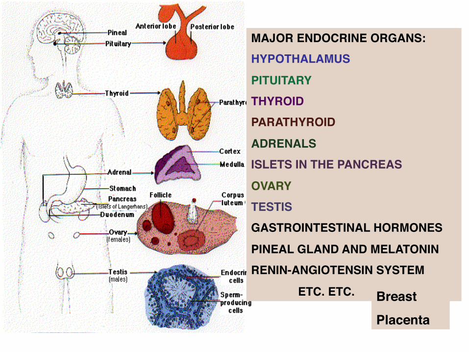
MAJOR ENDOCRINE ORGANS:!HYPOTHALAMUS!
PITUITARY!THYROID!PARATHYROID!
ADRENALS!ISLETS IN THE PANCREAS!
OVARY!TESTIS!GASTROINTESTINAL HORMONES!
PINEAL GLAND AND MELATONIN!RENIN-ANGIOTENSIN SYSTEM!
!ETC. ETC.! Breast!Placenta
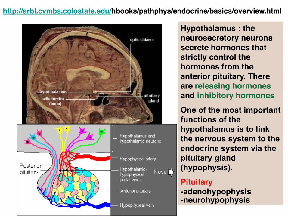
http://arbl.cvmbs.colostate.edu/hbooks/pathphys/endocrine/basics/overview.html
Hypothalamus : the neurosecretory neurons secrete hormones that strictly control the hormones from the anterior pituitary. There are releasing hormones and inhibitory hormones!One of the most important functions of the hypothalamus is to link the nervous system to the endocrine system via the pituitary gland (hypophysis).!Pituitary !-adenohypophysis !-neurohypophysis!
The Hypothalamus is responsible for certain metabolic processes and other activities of the Autonomic Nervous System.
It synthesizes and secretes neurohormones, often called hypothalamic-releasing hormones, and these in turn stimulate or inhibit the secretion of pituitary hormones.
The Hypothalamus controls Blood pressure, Body temperature, hunger, thirst, fatigue, anger, and circadian cycles.
The hypothalamus controls body weight and appetite, but it is not entirely clear how. Sensory inputs, including taste, smell, and gut distension, all tell the hypothalamus if we are hungry, full, or smelling a steak. Yet it is mysterious how we are able to vary our eating habits day to day and yet maintain about the same weight (sometimes despite all efforts to the contrary!) -- leptin gene
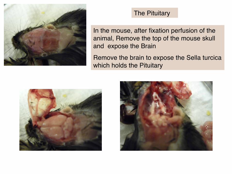
In the mouse, after fixation perfusion of the animal, Remove the top of the mouse skull and expose the Brain!
Remove the brain to expose the Sella turcica which holds the Pituitary!
The Pituitary!
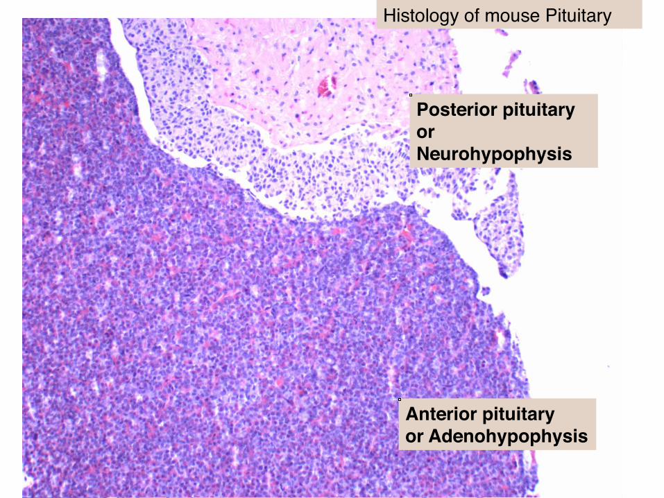
Anterior pituitary !or Adenohypophysis!
Posterior pituitary or Neurohypophysis!
Histology of mouse Pituitary!
Neurohypophysis: - is made up of unmyelinated axons from hypothalamic neurosecretory neurons. About 100,000 axons participate in this process to form the posterior pituitary. In addition to axons, the neurohypophysis contains glial cells and other poorly-defined cells called called pituicytes. !-secretes !-oxytocin : principal actions : !
!--stimulating contractions of the uterus at the time of birth !!--stimulating release of milk when the baby begins to suckle!
- antidiuretic hormone: also known as arginine vasopressin. ADH acts on the collecting ducts of the kidney to facilitate the reabsorption of water into the blood. This it acts to reduce the volume of urine formed (giving it its name of antidiuretic hormone)!!
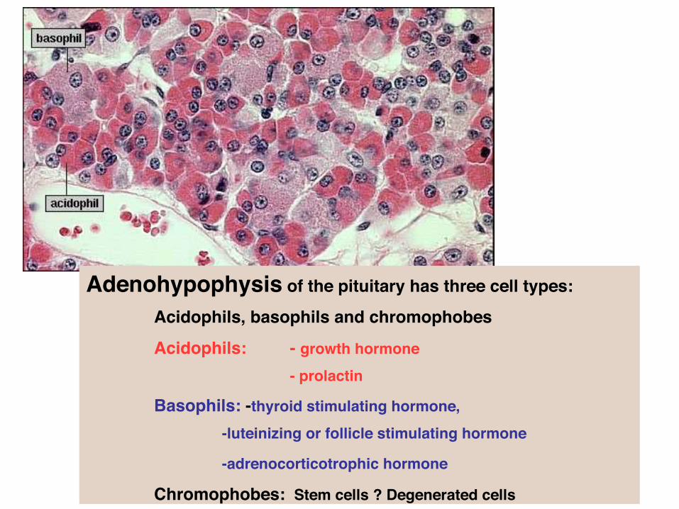
Adenohypophysis of the pituitary has three cell types:!
!Acidophils, basophils and chromophobes!
!Acidophils: !- growth hormone !
! ! !- prolactin!!Basophils: -thyroid stimulating hormone, !
! !-luteinizing or follicle stimulating hormone !
! !-adrenocorticotrophic hormone !
!Chromophobes: Stem cells ? Degenerated cells

Thyroid and Parathyroid! These are found in sections of the mouse trachea!
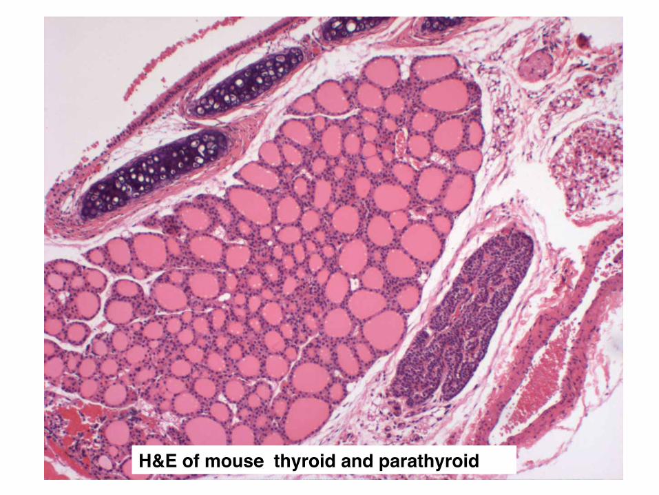
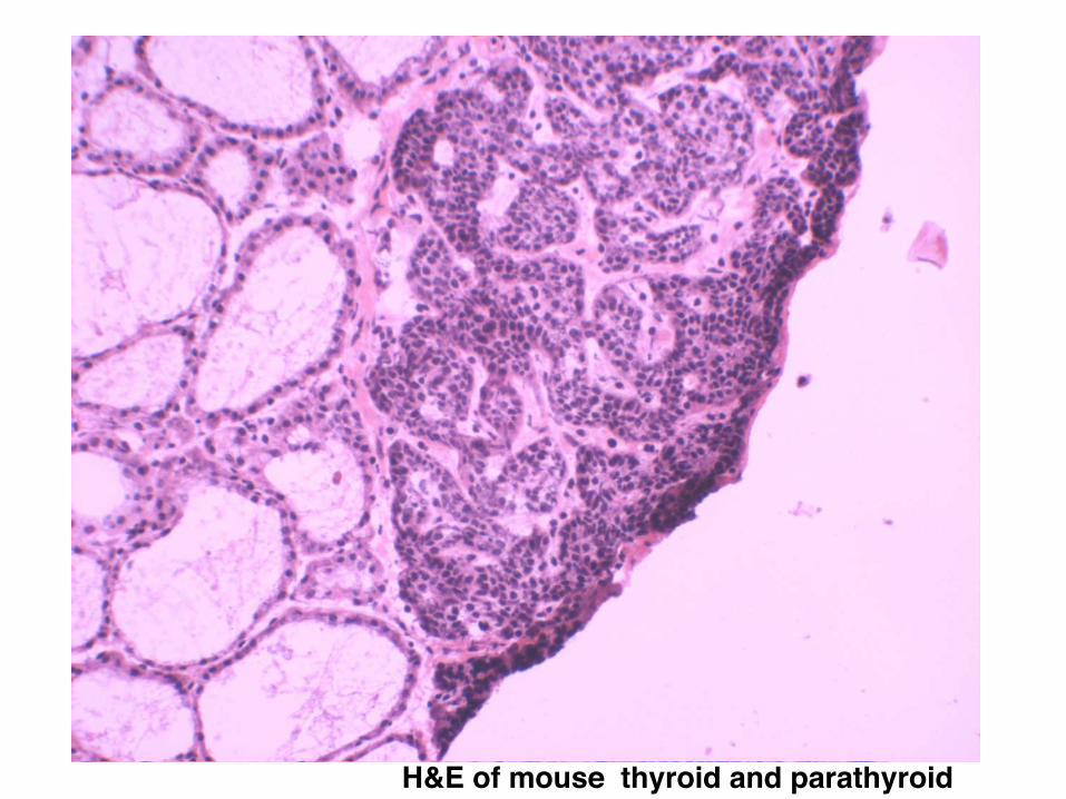
H&E of mouse thyroid and parathyroid!

THYROID epithelial cells - the cells responsible for synthesis of thyroid hormones - are arranged in spheres called thyroid follicles. Follicles are filled with colloid, a proteinaceous depot of thyroid hormone precursor. In the low (left) and high-magnification (right) images of thyroid, follicles are cut in cross section at different levels, appearing as roughly circular forms of varying size. In standard histologic preparations such as these, colloid stains pink. In addition to thyroid epithelial cells, the thyroid gland houses one other important endocrine cell. Nestled in spaces between thyroid follicles are parafollicular or C cells, which secrete the hormone calcitonin.
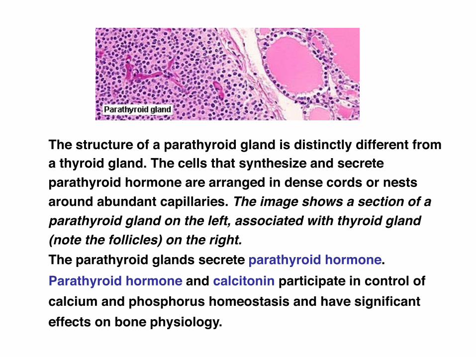
The structure of a parathyroid gland is distinctly different from a thyroid gland. The cells that synthesize and secrete parathyroid hormone are arranged in dense cords or nests around abundant capillaries. The image shows a section of a parathyroid gland on the left, associated with thyroid gland (note the follicles) on the right. The parathyroid glands secrete parathyroid hormone. !Parathyroid hormone and calcitonin participate in control of calcium and phosphorus homeostasis and have significant effects on bone physiology.
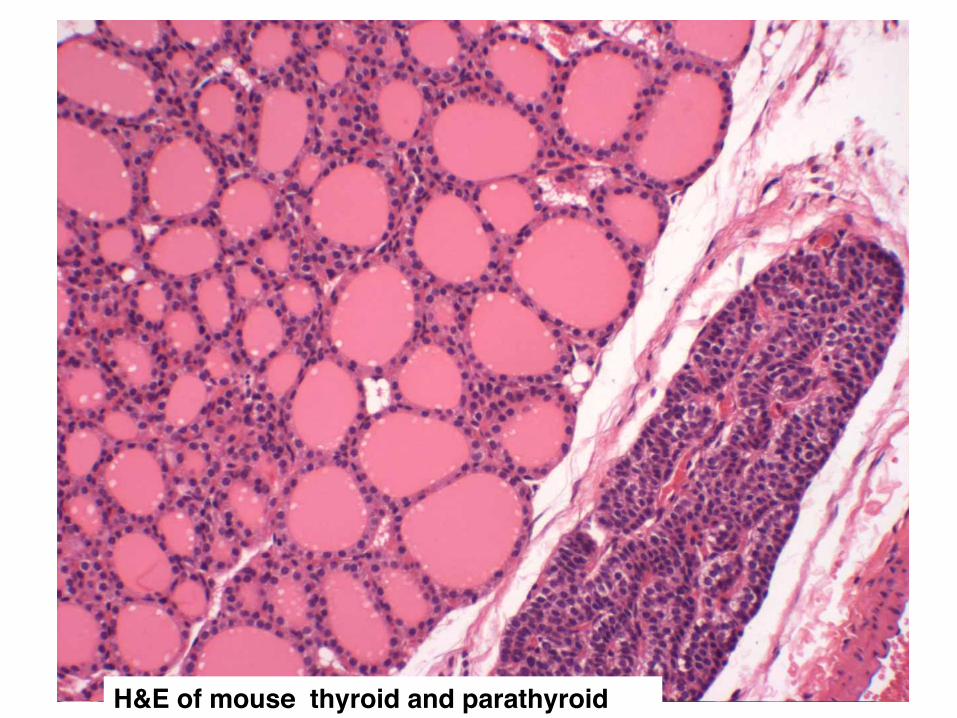
H&E of mouse thyroid and parathyroid!
H&E of mouse thyroid and parathyroid!
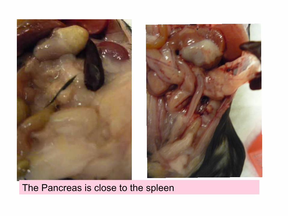
The Pancreas is close to the spleen


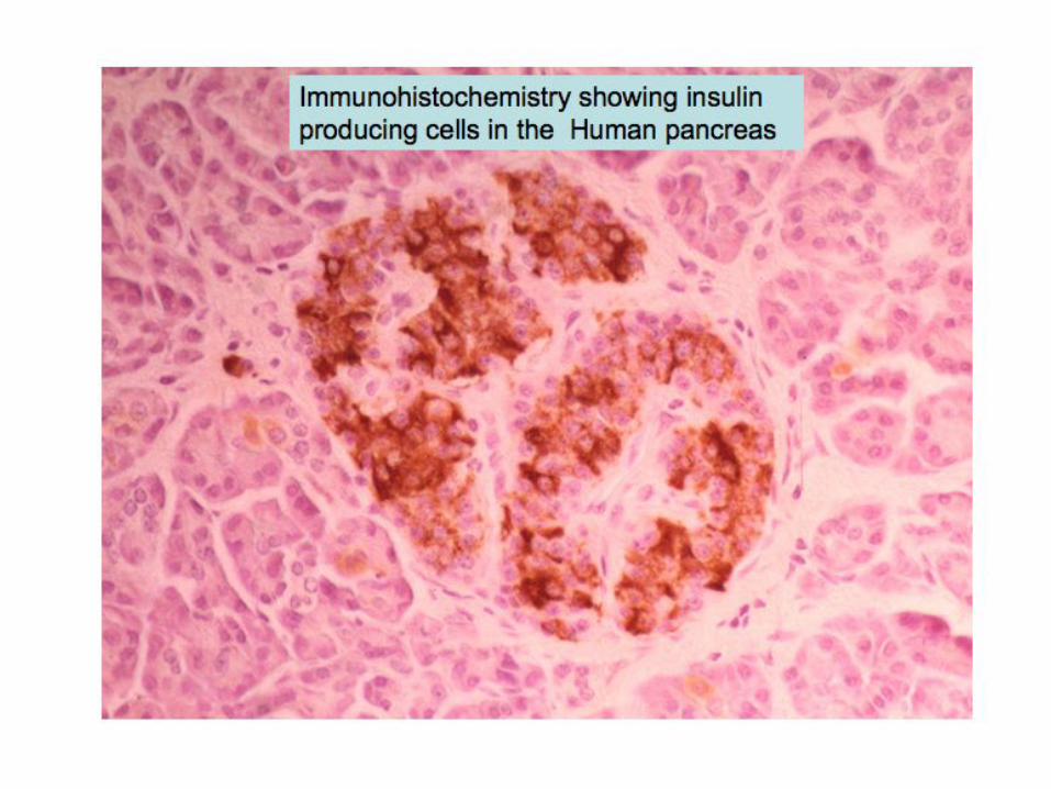
Pancreatic islets of Langerhans house three major cell types, each of which produces a different endocrine product: !!*Alpha cells (A cells) secrete the hormone glucagon.!• Beta cells (B cells) produce insulin and are the most abundant of the islet cells.!• Delta cells (D cells) secrete the hormone somatostatin, which is also produced by a number of other endocrine cells in the body!
Aside from the insulin, glucagon and somatostatin, a number of other "minor" hormones have been identified as products of pancreatic islets cells ( pancreatic polypeptide)
Islets are richly vascularized, allowing their secreted hormones ready access to the circulation. Although islets comprise only 1-2% of the mass of the pancreas, they receive about 10 to 15% of the pancreatic blood flow. Additionally, they are innervated by parasympathetic and sympathetic neurons, and nervous signals clearly modulate secretion of insulin and glucagon.

Adrenals or Supra-renals!

Adrenal from a Male mouse and from a Female mouse
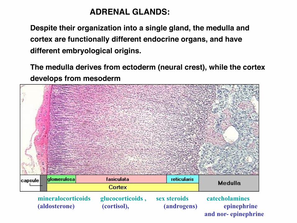
! !ADRENAL GLANDS:!
Despite their organization into a single gland, the medulla and cortex are functionally different endocrine organs, and have different embryological origins. !
The medulla derives from ectoderm (neural crest), while the cortex develops from mesoderm
mineralocorticoids glucocorticoids , sex steroids catecholamines (aldosterone) (cortisol), (androgens) epinephrine
and nor- epinephrine
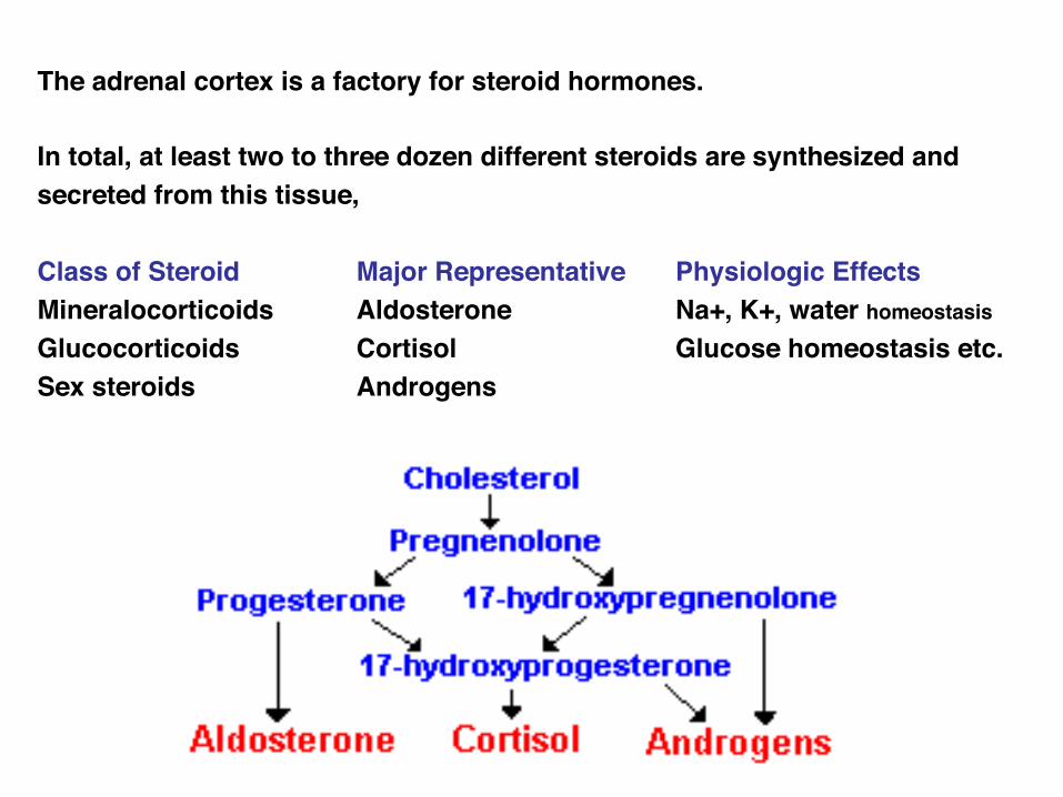
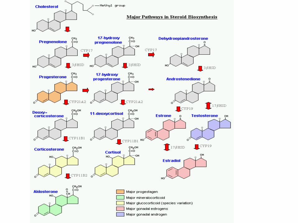
The adrenal cortex is a factory for steroid hormones. !!In total, at least two to three dozen different steroids are synthesized and secreted from this tissue, !!Class of Steroid ! !Major Representative !Physiologic Effects!Mineralocorticoids !Aldosterone ! !Na+, K+, water homeostasis!Glucocorticoids ! !Cortisol! ! !Glucose homeostasis etc.!Sex steroids ! !Androgens
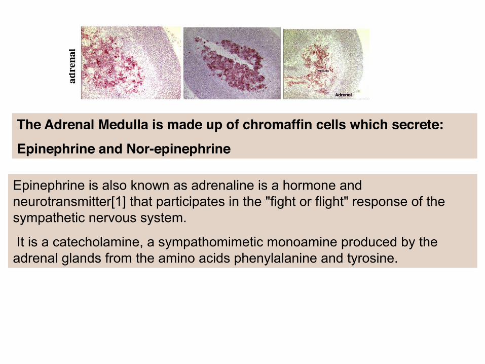
The Adrenal Medulla is made up of chromaffin cells which secrete:!Epinephrine and Nor-epinephrine
Epinephrine is also known as adrenaline is a hormone and neurotransmitter[1] that participates in the "fight or flight" response of the sympathetic nervous system.
It is a catecholamine, a sympathomimetic monoamine produced by the adrenal glands from the amino acids phenylalanine and tyrosine.
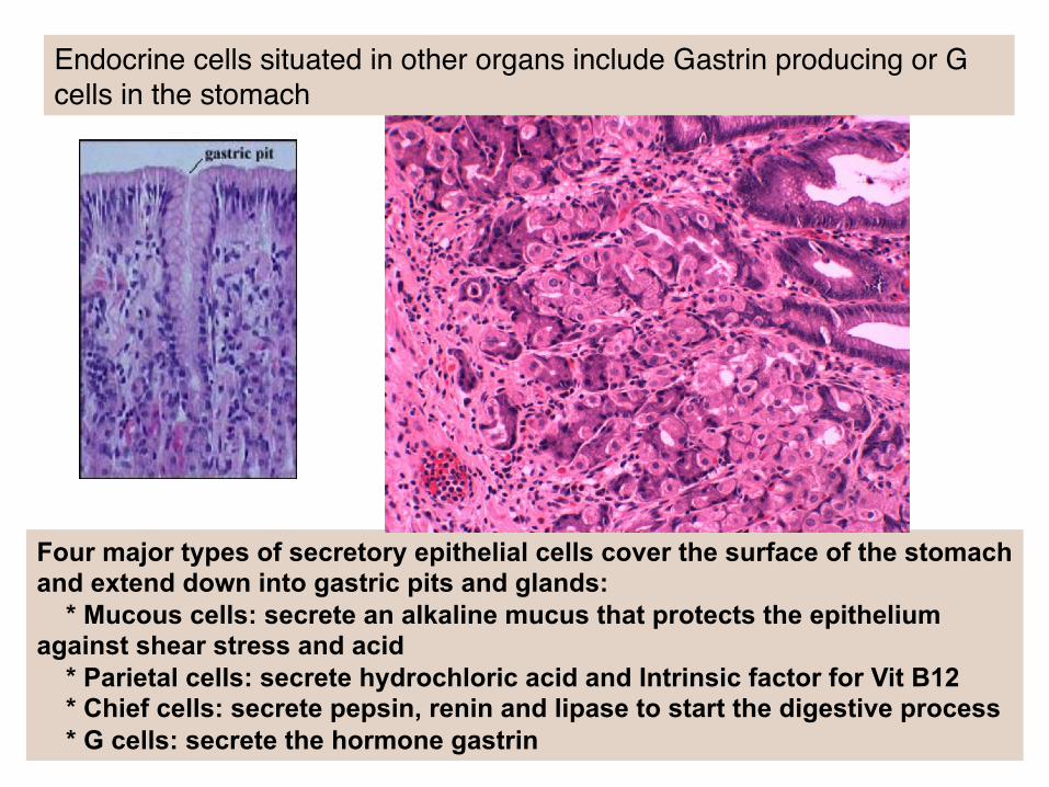
Four major types of secretory epithelial cells cover the surface of the stomach and extend down into gastric pits and glands: * Mucous cells: secrete an alkaline mucus that protects the epithelium against shear stress and acid * Parietal cells: secrete hydrochloric acid and Intrinsic factor for Vit B12 * Chief cells: secrete pepsin, renin and lipase to start the digestive process * G cells: secrete the hormone gastrin!
Endocrine cells situated in other organs include Gastrin producing or G cells in the stomach!

Endocrine cells situated in other organs include those in the intestine !

MAJOR ENDOCRINE ORGANS:!HYPOTHALAMUS!
PITUITARY!THYROID!PARATHYROID!
ADRENALS!ISLETS IN THE PANCREAS!
OVARY!TESTIS!GASTROINTESTINAL HORMONES!
PINEAL GLAND AND MELATONIN!RENIN-ANGIOTENSIN SYSTEM!
!ETC. ETC.! Breast!Placenta
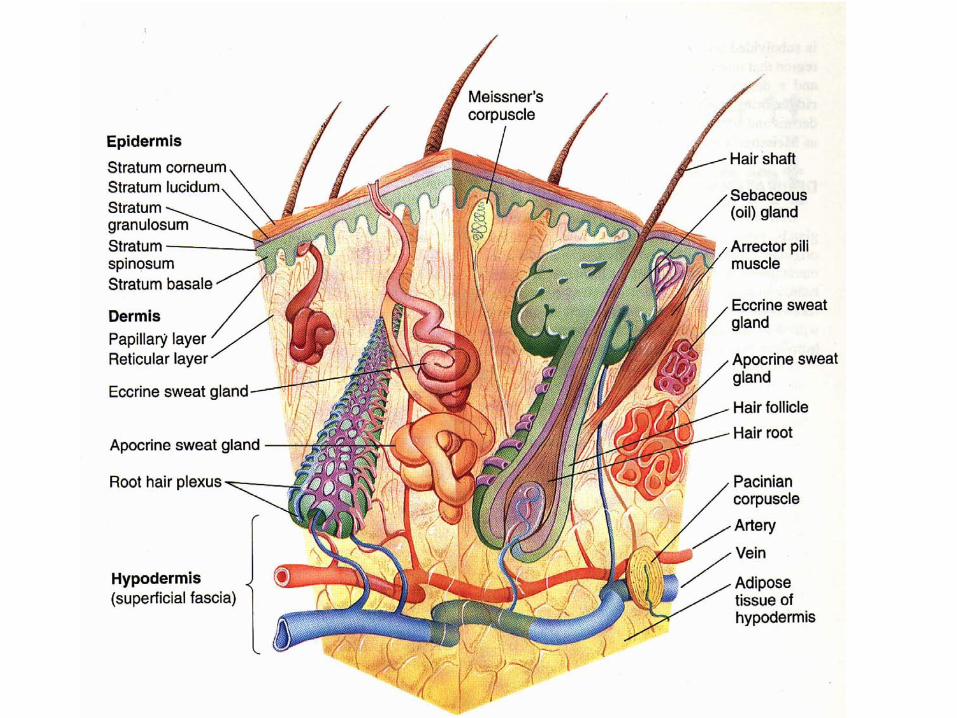

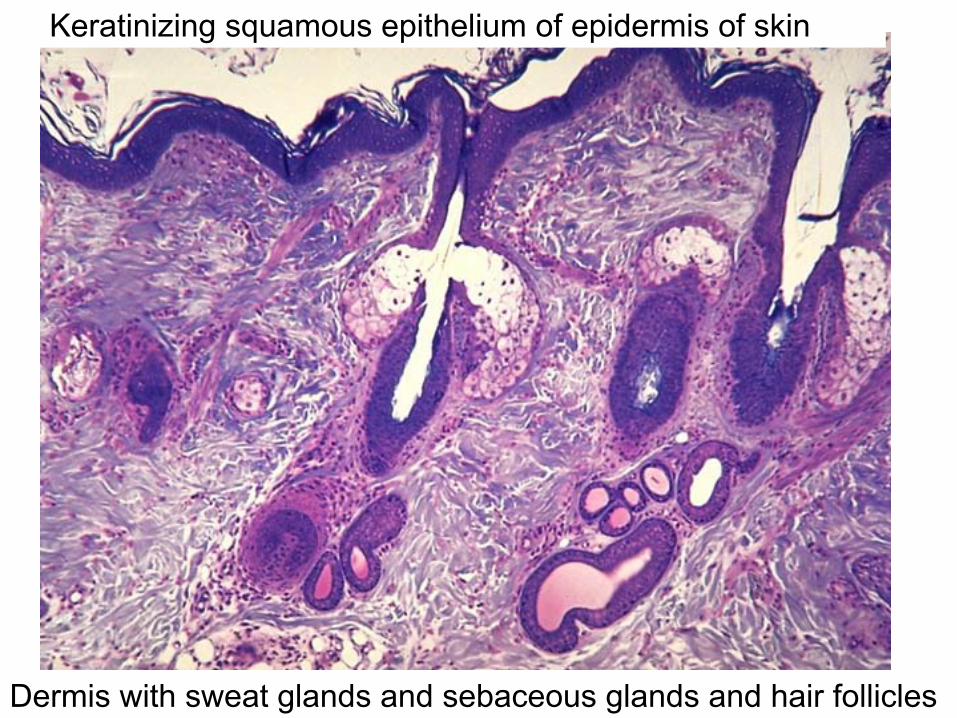
Keratinizing squamous epithelium of epidermis of skin
Dermis with sweat glands and sebaceous glands and hair follicles
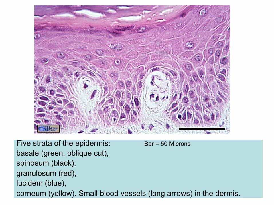
Five strata of the epidermis: Bar = 50 Microns basale (green, oblique cut), spinosum (black), granulosum (red), lucidem (blue), corneum (yellow). Small blood vessels (long arrows) in the dermis.
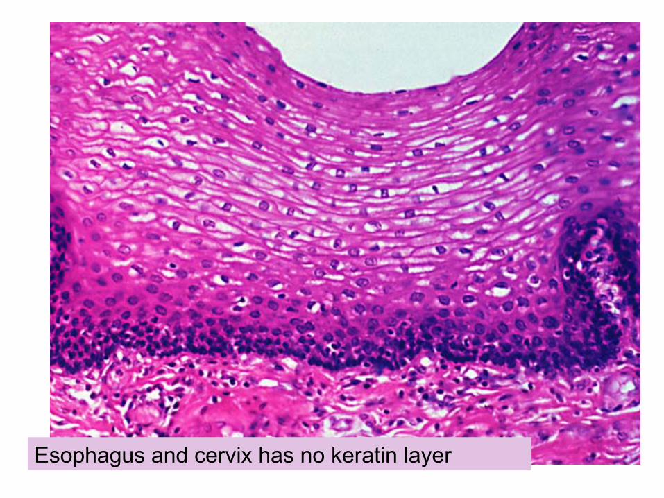
Esophagus and cervix has no keratin layer
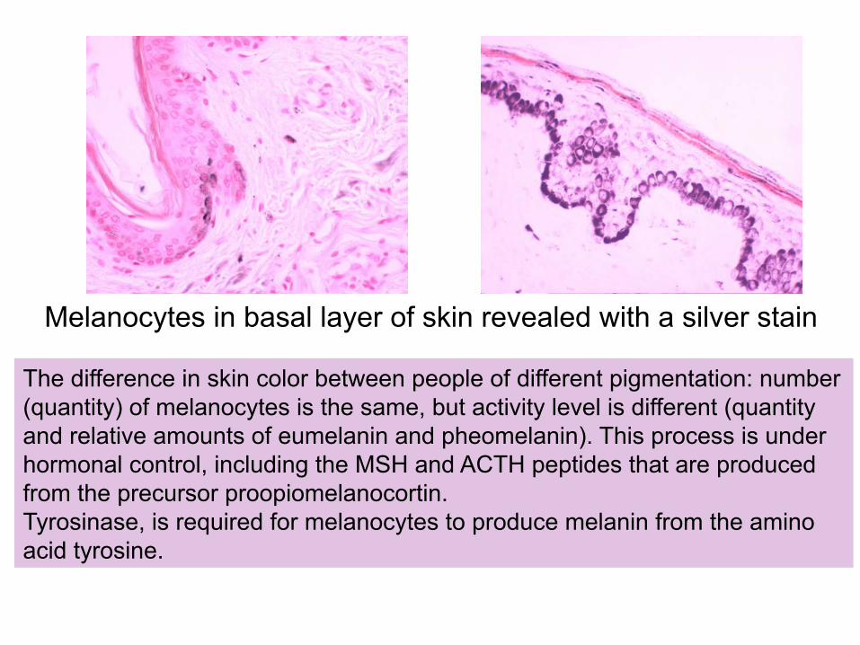
Melanocytes in basal layer of skin revealed with a silver stain
The difference in skin color between people of different pigmentation: number (quantity) of melanocytes is the same, but activity level is different (quantity and relative amounts of eumelanin and pheomelanin). This process is under hormonal control, including the MSH and ACTH peptides that are produced from the precursor proopiomelanocortin. Tyrosinase, is required for melanocytes to produce melanin from the amino acid tyrosine.
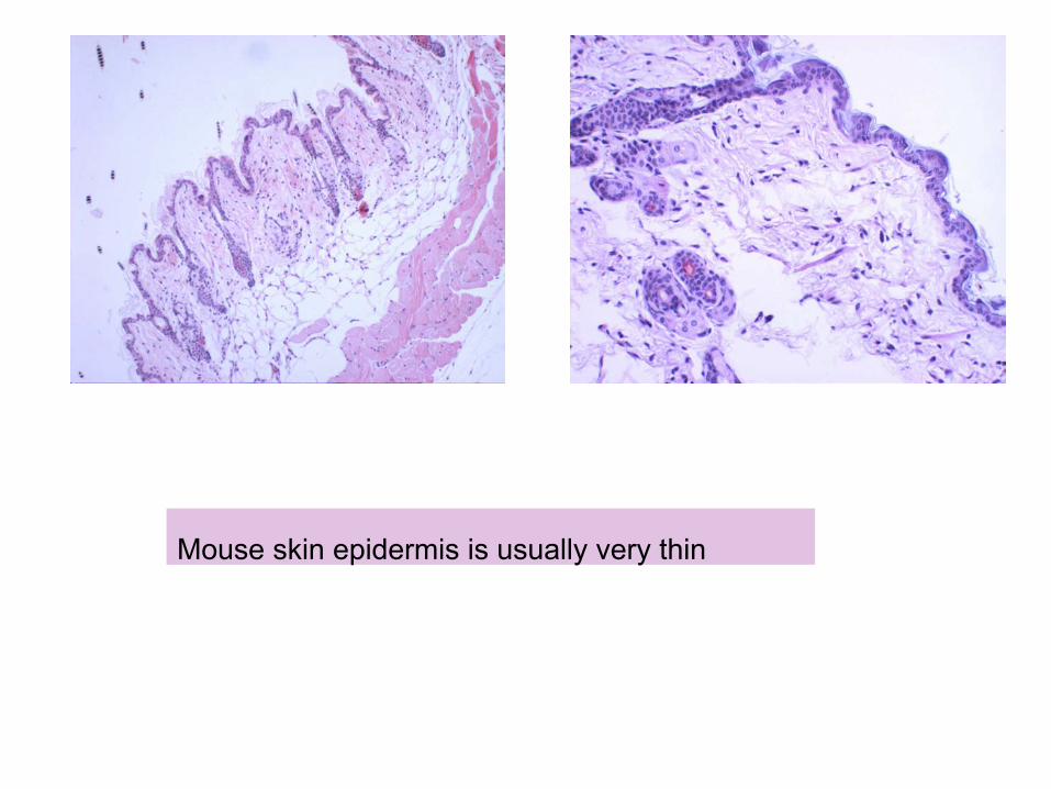
Mouse skin epidermis is usually very thin
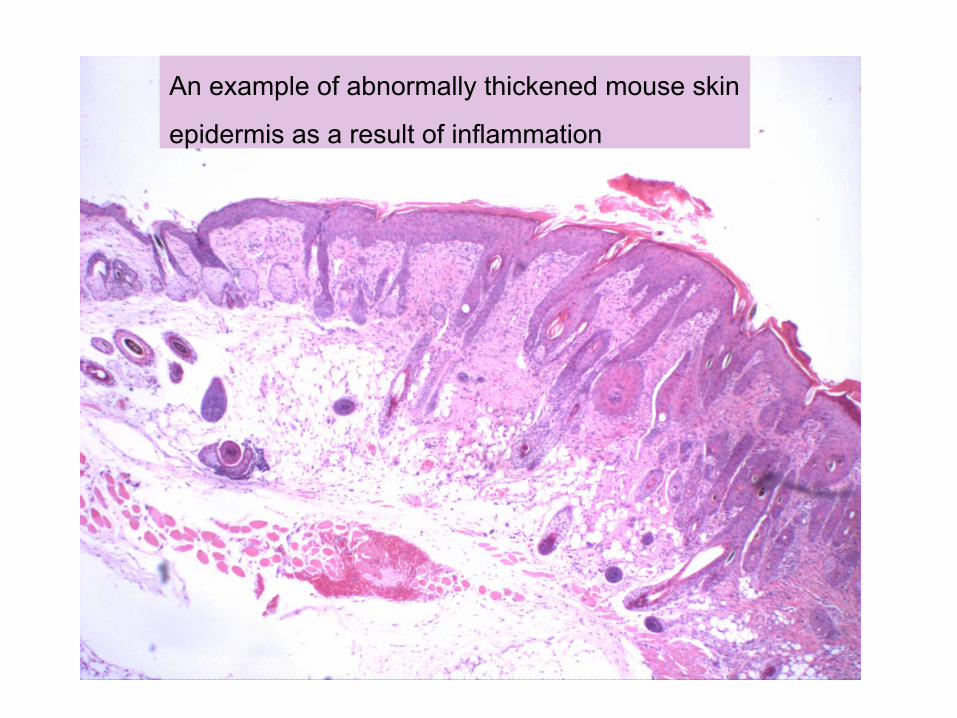
An example of abnormally thickened mouse skin
epidermis as a result of inflammation
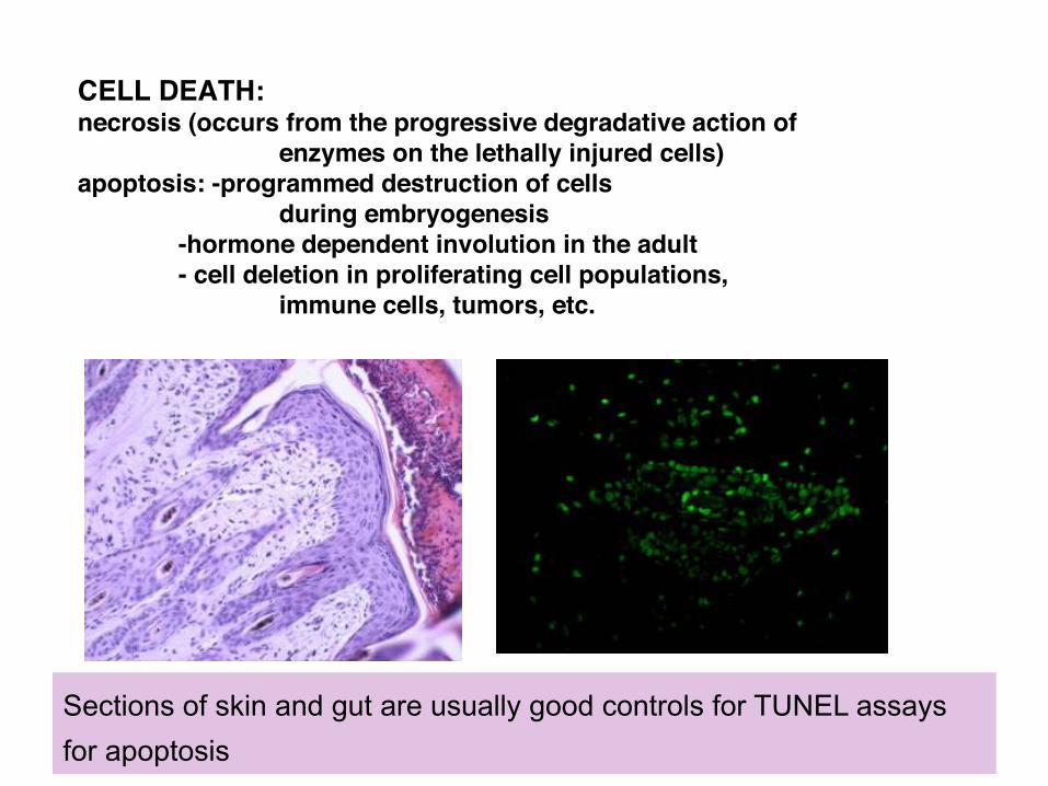
CELL DEATH:necrosis (occurs from the progressive degradative action of
enzymes on the lethally injured cells)apoptosis: -programmed destruction of cells
during embryogenesis-hormone dependent involution in the adult- cell deletion in proliferating cell populations,
immune cells, tumors, etc.
Sections of skin and gut are usually good controls for TUNEL assays for apoptosis
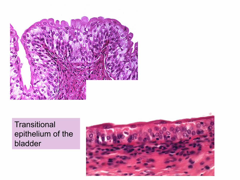
Transitional epithelium of the bladder

Bladder epithelium: Transitional NOT Stratified Squamous!
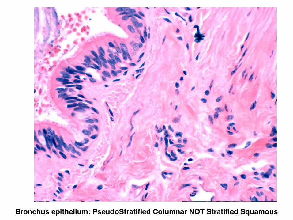
Bronchus epithelium: PseudoStratified Columnar NOT Stratified Squamous!
What are the cell / tissue types you will see in the skin?
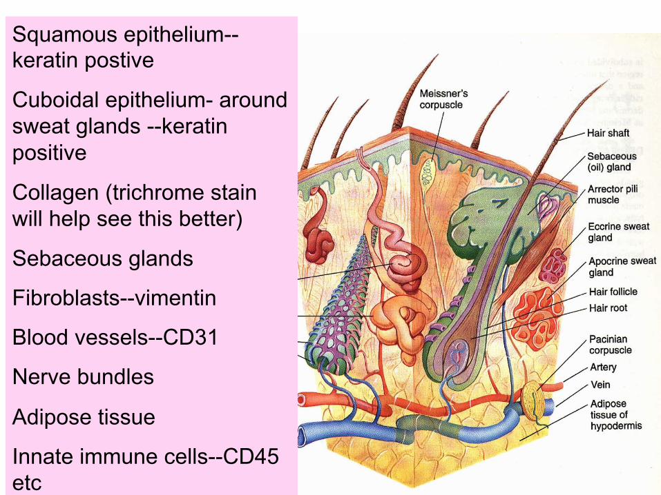
Squamous epithelium--keratin postive
Cuboidal epithelium- around sweat glands --keratin positive
Collagen (trichrome stain will help see this better)
Sebaceous glands
Fibroblasts--vimentin
Blood vessels--CD31
Nerve bundles
Adipose tissue
Innate immune cells--CD45 etc
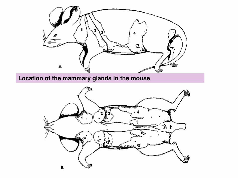
Location of the mammary glands in the mouse!
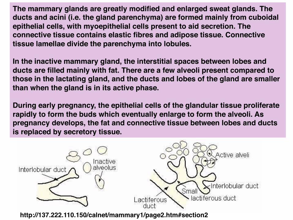
The mammary glands are greatly modified and enlarged sweat glands. The ducts and acini (i.e. the gland parenchyma) are formed mainly from cuboidal epithelial cells, with myoepithelial cells present to aid secretion. The connective tissue contains elastic fibres and adipose tissue. Connective tissue lamellae divide the parenchyma into lobules. !!In the inactive mammary gland, the interstitial spaces between lobes and ducts are filled mainly with fat. There are a few alveoli present compared to those in the lactating gland, and the ducts and lobes of the gland are smaller than when the gland is in its active phase. !!During early pregnancy, the epithelial cells of the glandular tissue proliferate rapidly to form the buds which eventually enlarge to form the alveoli. As pregnancy develops, the fat and connective tissue between lobes and ducts is replaced by secretory tissue.
http://137.222.110.150/calnet/mammary1/page2.htm#section2!
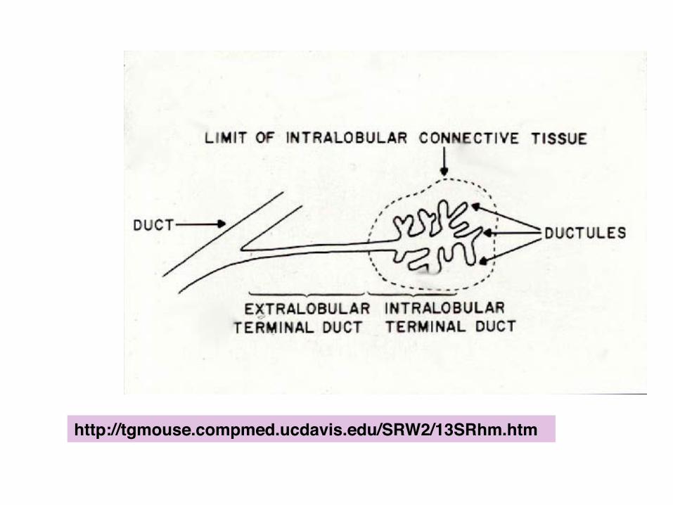
http://tgmouse.compmed.ucdavis.edu/SRW2/13SRhm.htm
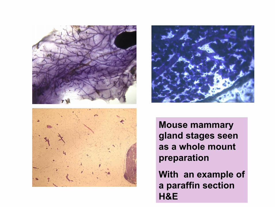
Mouse mammary gland stages seen as a whole mount preparation
With an example of a paraffin section H&E!
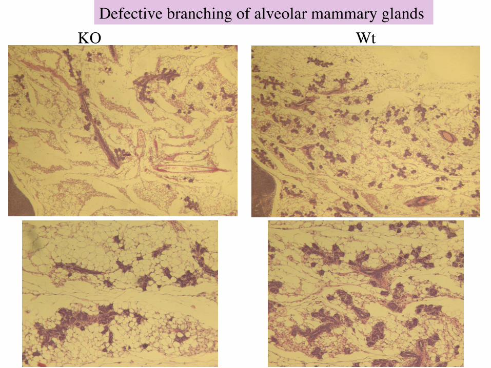
Defective branching of alveolar mammary glands KO Wt
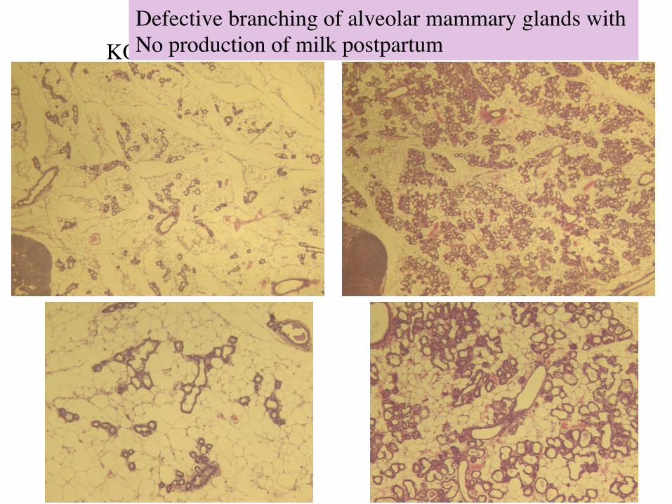
Day 1 post partum (pictures 279/280 and 283/284) KO Wt
Defective branching of alveolar mammary glands with No production of milk postpartum

An H&E of a human Breast
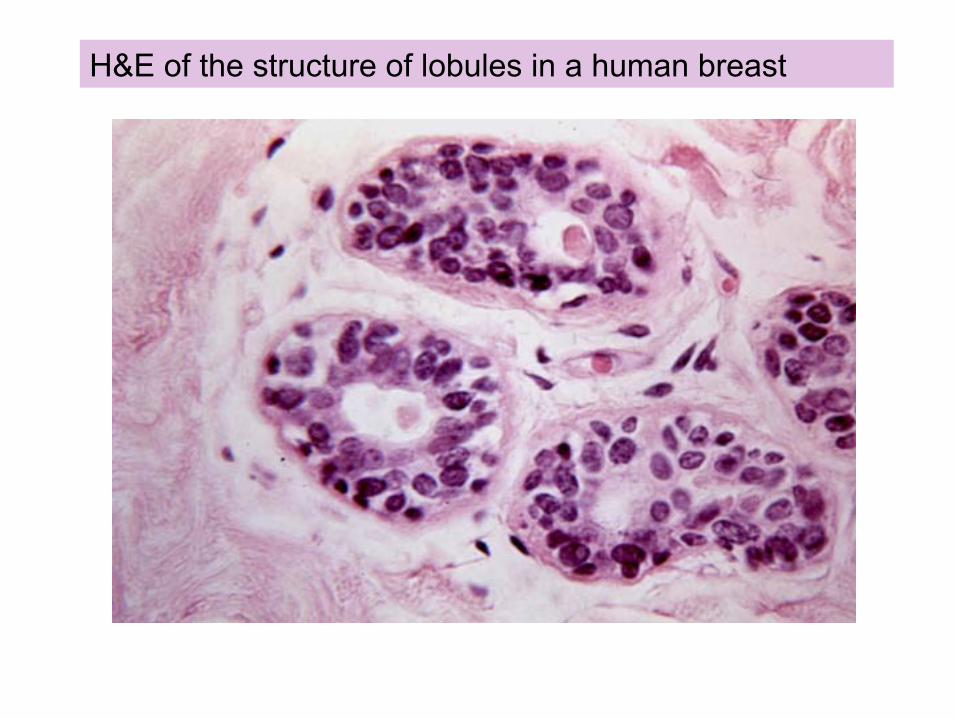
H&E of the structure of lobules in a human breast
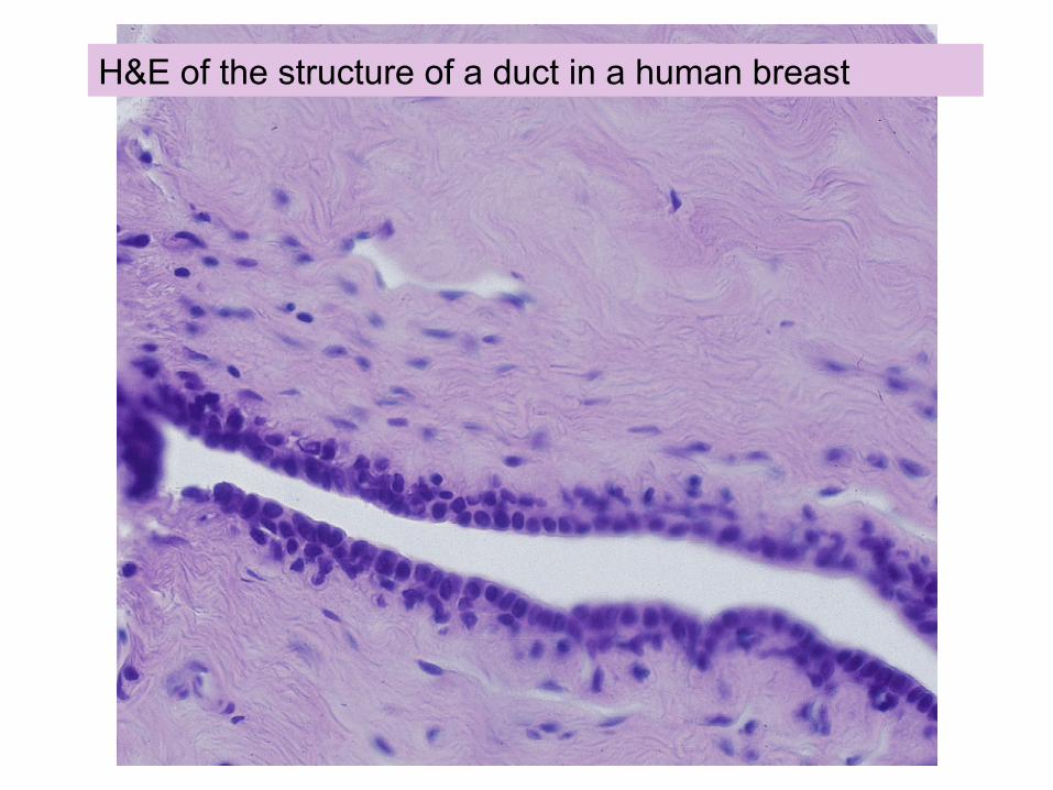
H&E of the structure of a duct in a human breast
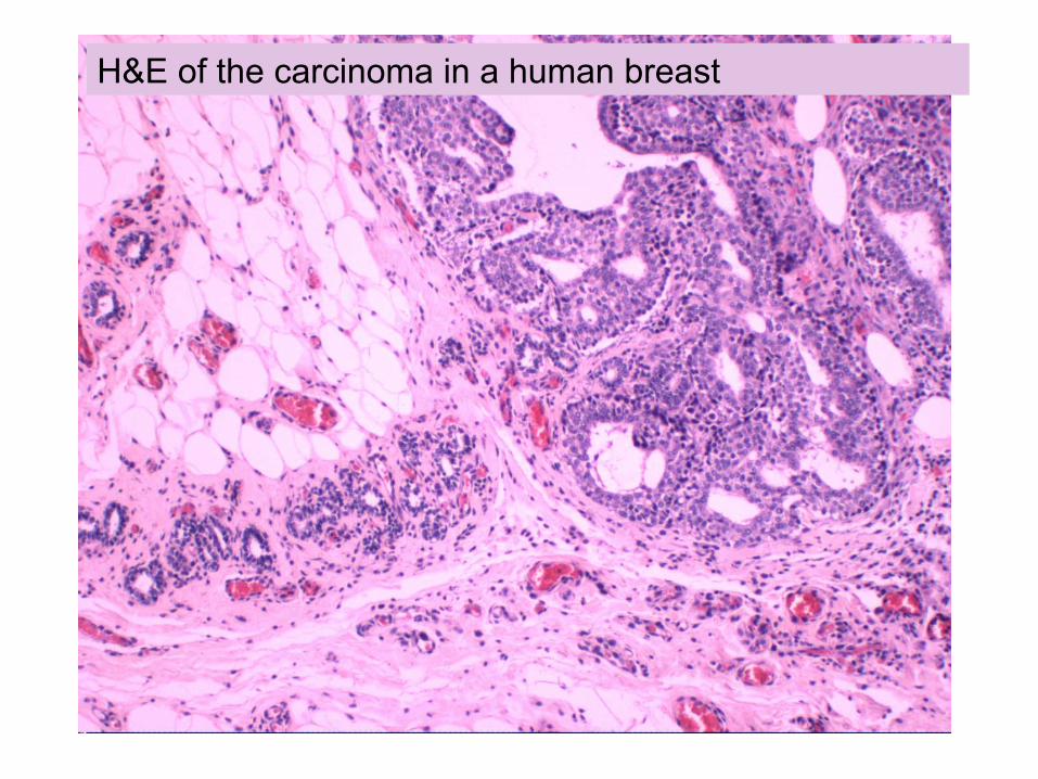
H&E of the carcinoma in a human breast
What are the cell / tissue types you will see in breast tissue?
Cuboidal cells--keratin positive
Adipocytes
Blood vessels
Fibroblasts
Nerve fibers
Innate immune cells

Adipose tissues!
White Adipose Tissue (WAT): Adult adipose tissue contains lipids that dissolve during processing into paraffin. WAT is unilocular and appears like empty vacuoles on H&E Brown Adipose Tissue (BAT): abundant in the embryo, in the interscapular area. BAT is multilocular and serves as a source of heat in hybernating animals. The cells contain abundant mitochondria and thus appear pink on H&E.
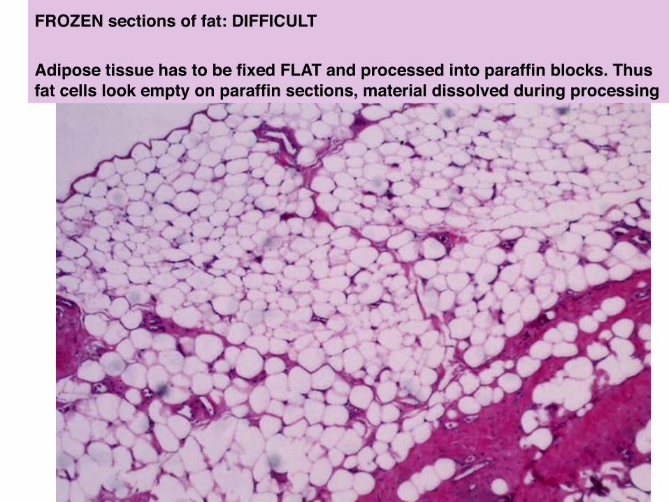
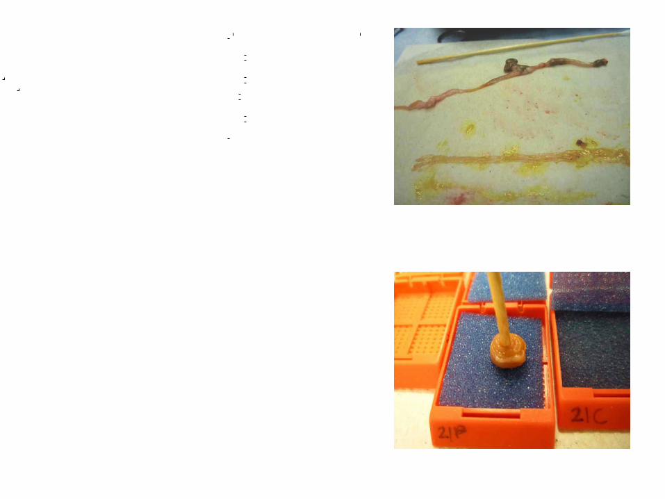
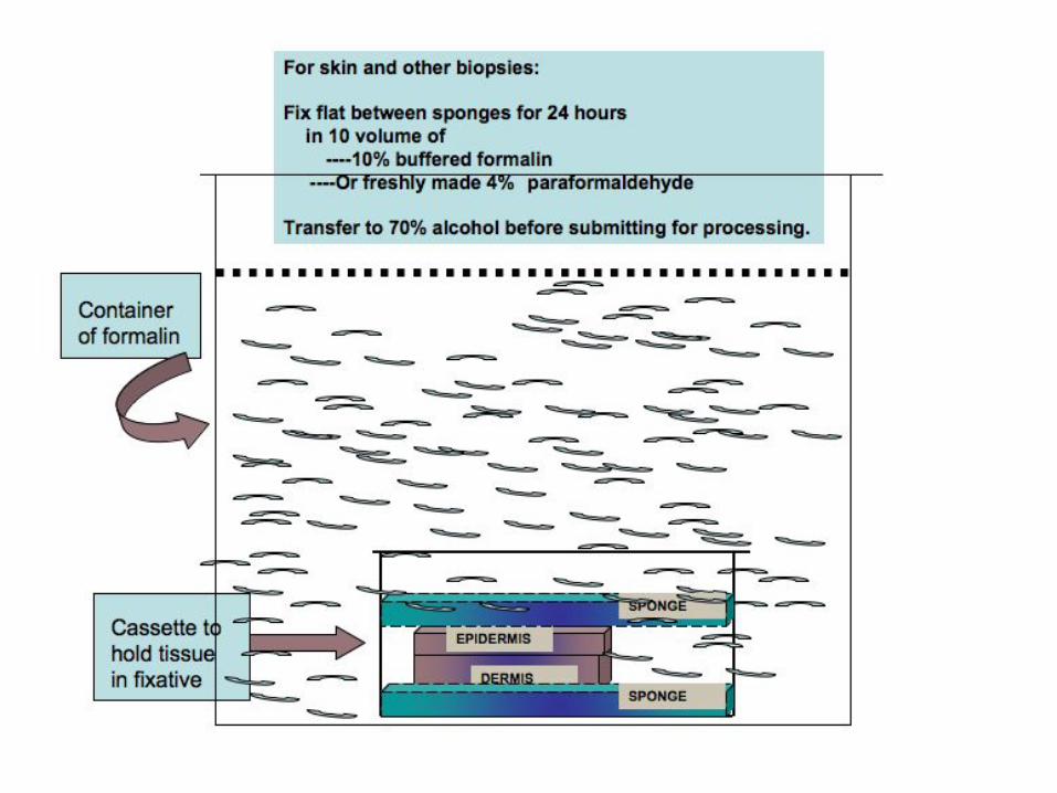
FROZEN sections of fat: DIFFICULT!!Adipose tissue has to be fixed FLAT and processed into paraffin blocks. Thus fat cells look empty on paraffin sections, material dissolved during processing!
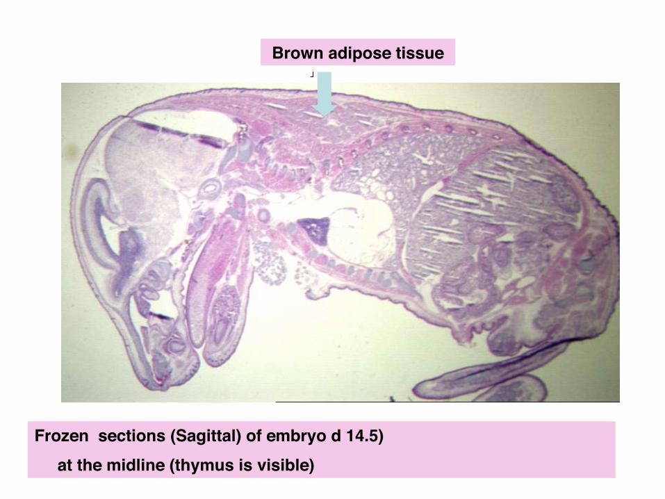
Frozen sections (Sagittal) of embryo d 14.5) ! at the midline (thymus is visible)
Brown adipose tissue !
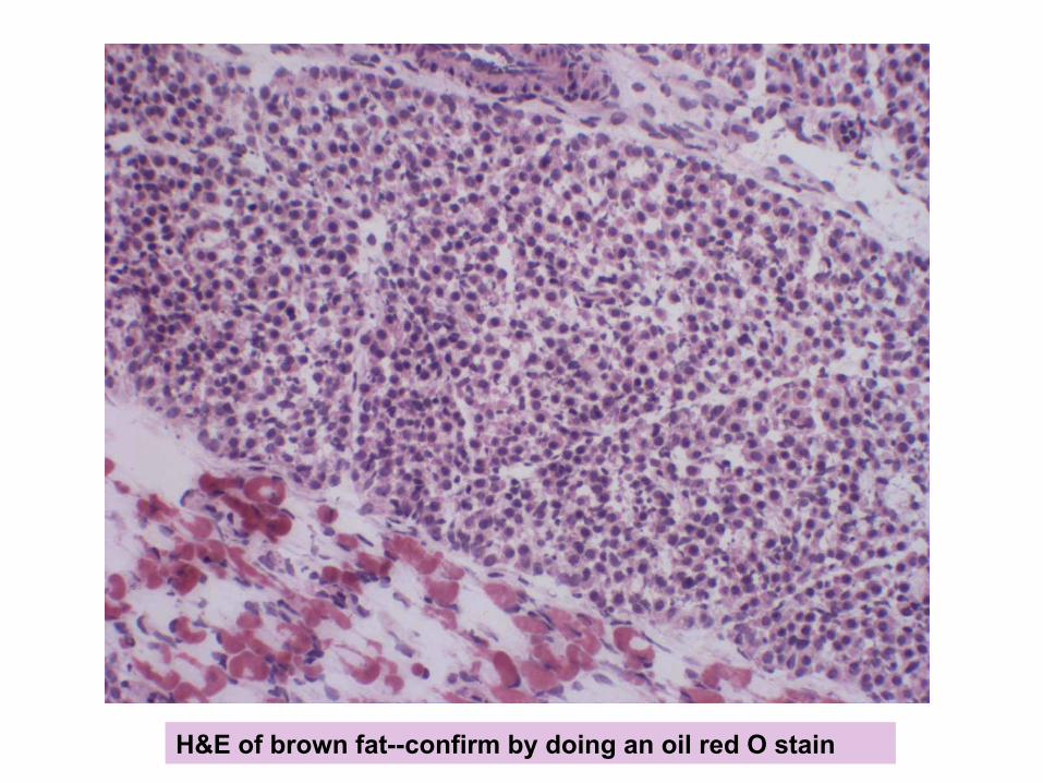
H&E of brown fat--confirm by doing an oil red O stain!

H&E of brown fat in a mutant mouse, show that is deficient--confirm by doing an oil red O stain!
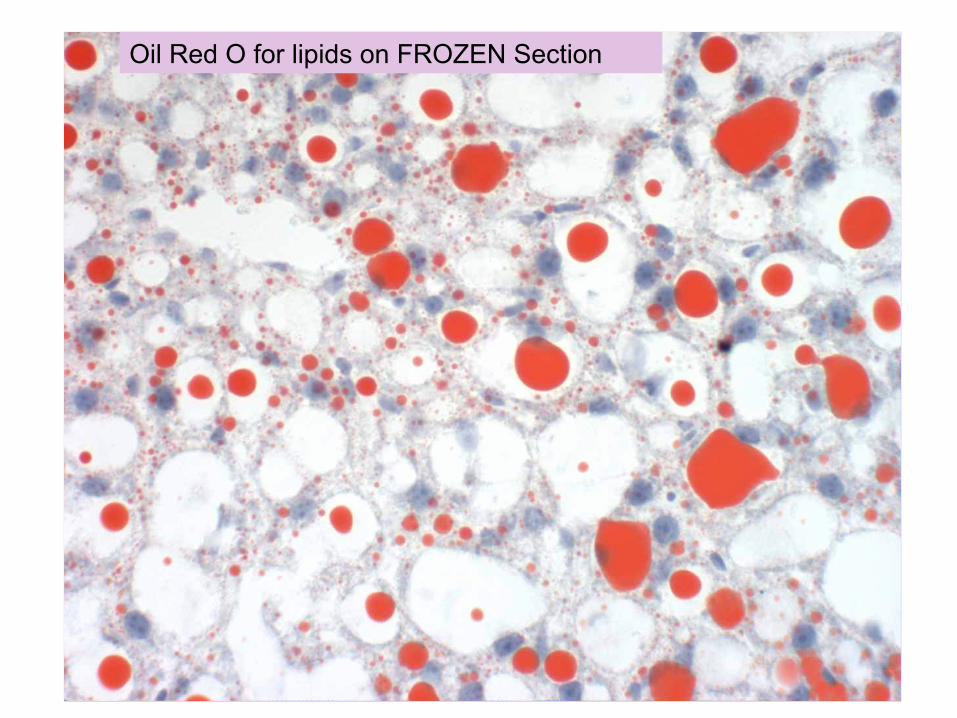
Oil Red O for lipids on FROZEN Section
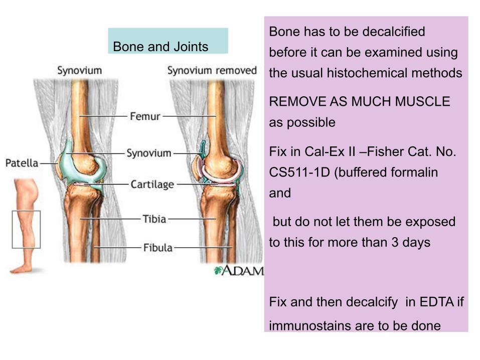
Bone and Joints Bone has to be decalcified before it can be examined using the usual histochemical methods
REMOVE AS MUCH MUSCLE as possible
Fix in Cal-Ex II –Fisher Cat. No. CS511-1D (buffered formalin and
but do not let them be exposed to this for more than 3 days
Fix and then decalcify in EDTA if
immunostains are to be done
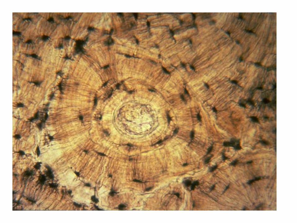
Cartilage is a type of dense connective tissue . It is composed of cells called chondrocytes which are dispersed in a firm gel-like ground substance, called the matrix.
Cartilage is avascular (contains no blood vessels) and nutrients are diffused through the matrix. !The main purpose of cartilage is to provide a framework upon which bone deposition could begin.
Another important purpose of cartilage is to provide smooth surfaces for the movement of articulating bones.
Hyaline Cartilage is the most abundant type of cartilage.
Elastic cartilage
Fibrocartilage
The matrix of cartilage acts as a barrier, preventing the entry of lymphocytes or diffusion of immunoglobulins . This property allows for the transplantation of cartilage from one individual to another without fear of tissue rejection.
Bioengineering techniques are being developed to generate new cartilage, using a cellular "scaffolding" material and cultured cells to grow artificial cartilage.
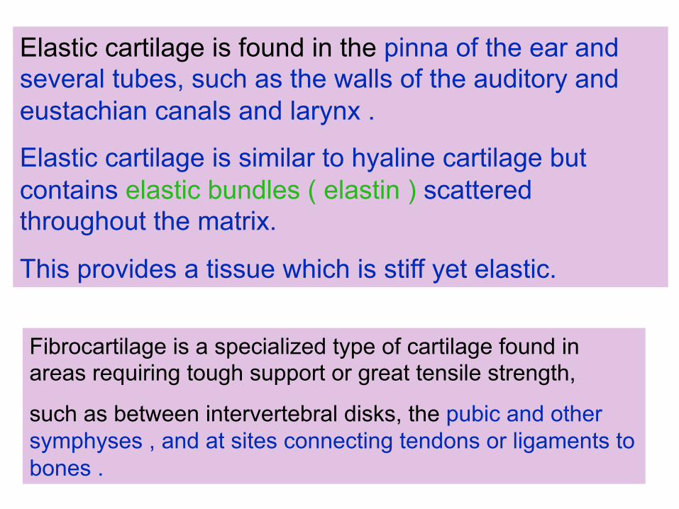
Elastic cartilage is found in the pinna of the ear and several tubes, such as the walls of the auditory and eustachian canals and larynx .
Elastic cartilage is similar to hyaline cartilage but contains elastic bundles ( elastin ) scattered throughout the matrix.
This provides a tissue which is stiff yet elastic. !
Fibrocartilage is a specialized type of cartilage found in areas requiring tough support or great tensile strength,
such as between intervertebral disks, the pubic and other symphyses , and at sites connecting tendons or ligaments to bones .
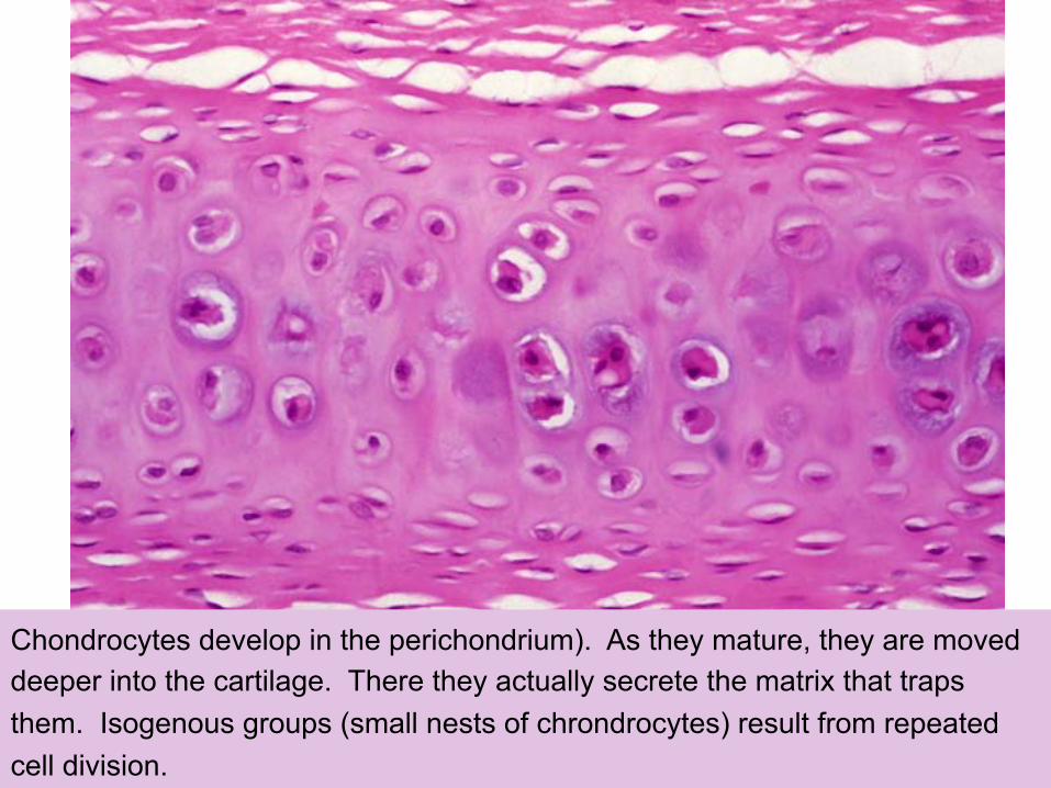
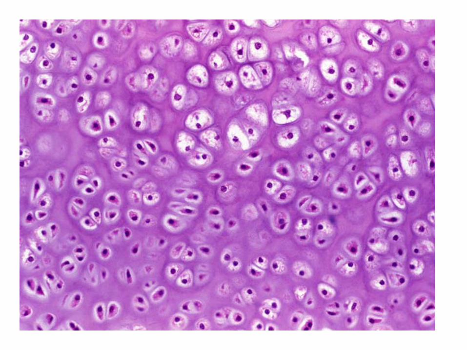
Chondrocytes develop in the perichondrium). As they mature, they are moved deeper into the cartilage. There they actually secrete the matrix that traps them. Isogenous groups (small nests of chrondrocytes) result from repeated cell division.
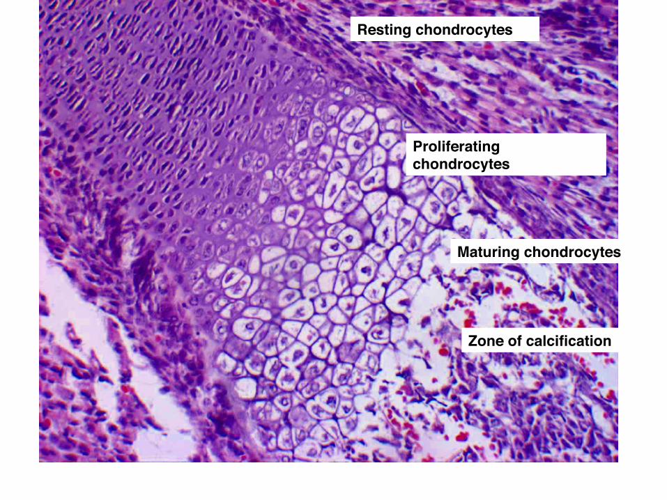
Proliferating chondrocytes!
Resting chondrocytes!
Maturing chondrocytes!
Zone of calcification!
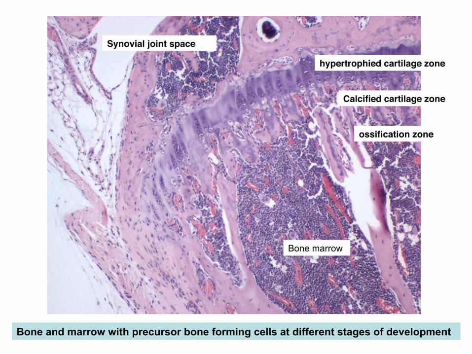
Calcified cartilage zone!
hypertrophied cartilage zone!
ossification zone!
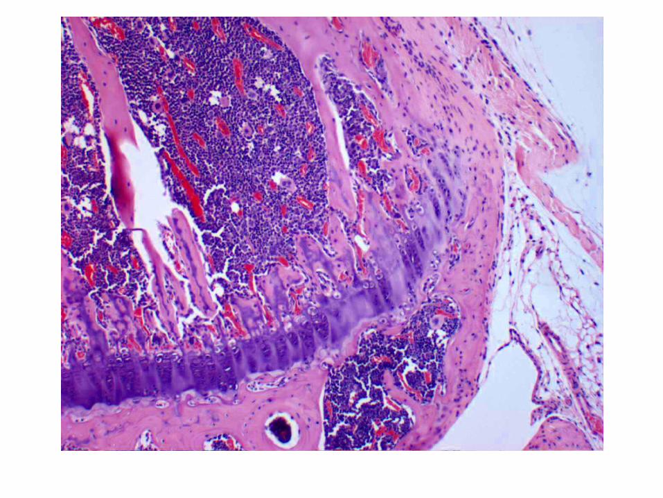
Bone and marrow with precursor bone forming cells at different stages of development!
Synovial joint space!
Bone marrow

Osteoblasts (build bone) are found lining bony spicules!
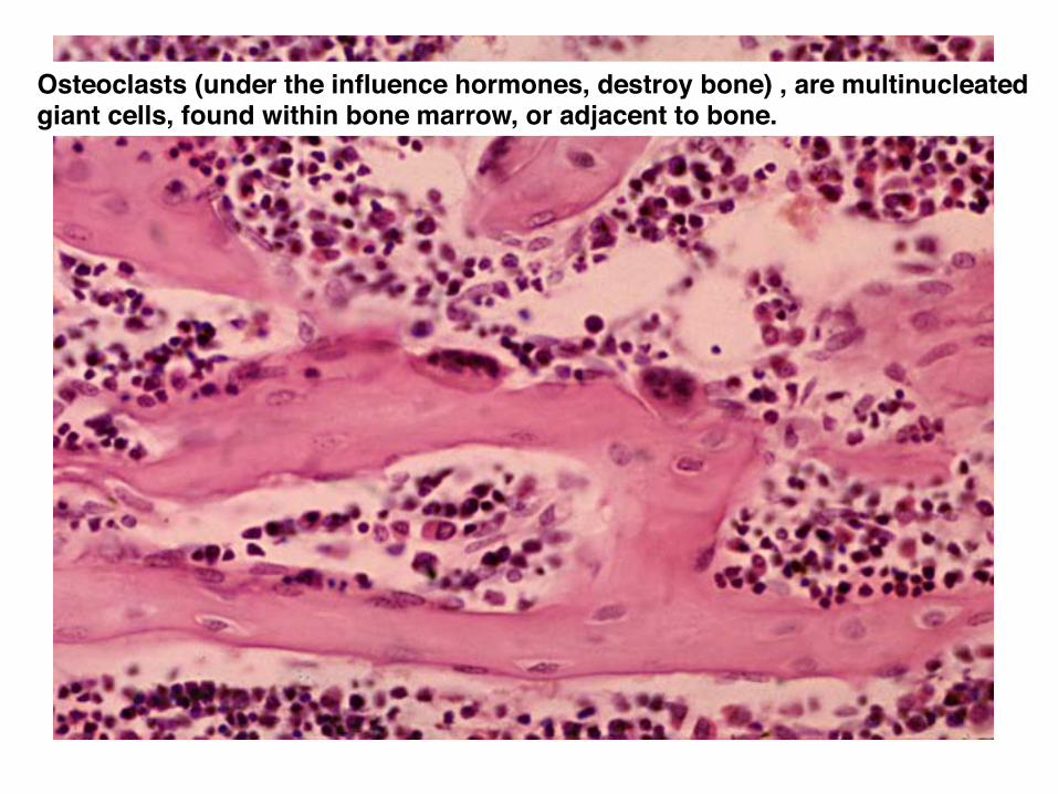
Osteoclasts (under the influence hormones, destroy bone) , are multinucleated giant cells, found within bone marrow, or adjacent to bone.!
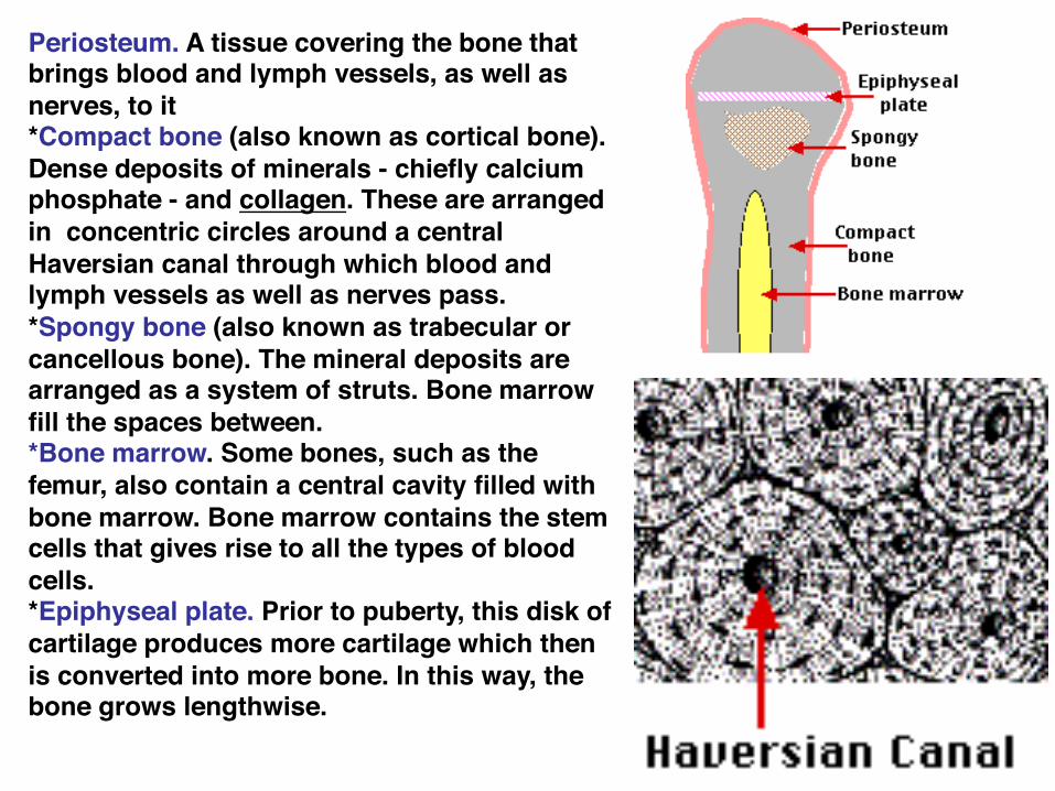
Periosteum. A tissue covering the bone that brings blood and lymph vessels, as well as nerves, to it !*Compact bone (also known as cortical bone). Dense deposits of minerals - chiefly calcium phosphate - and collagen. These are arranged in concentric circles around a central Haversian canal through which blood and lymph vessels as well as nerves pass. !*Spongy bone (also known as trabecular or cancellous bone). The mineral deposits are arranged as a system of struts. Bone marrow fill the spaces between. !*Bone marrow. Some bones, such as the femur, also contain a central cavity filled with bone marrow. Bone marrow contains the stem cells that gives rise to all the types of blood cells. !*Epiphyseal plate. Prior to puberty, this disk of cartilage produces more cartilage which then is converted into more bone. In this way, the bone grows lengthwise. !

Histo-chemistry methods
Examples: Alizarin Red and Alcian blue on cleared embryos to examine bone and cartilage carefully
Safranin-O may also be used
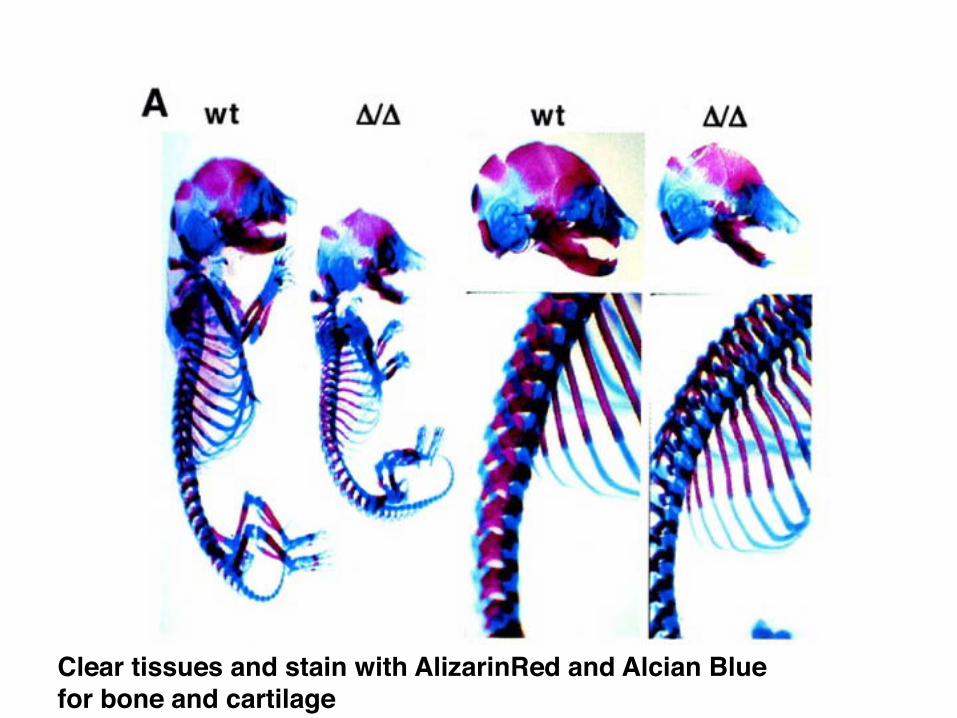
Clear tissues and stain with AlizarinRed and Alcian Blue for bone and cartilage!
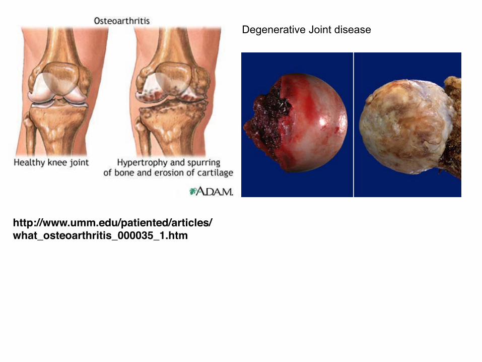
http://www.umm.edu/patiented/articles/what_osteoarthritis_000035_1.htm
Degenerative Joint disease
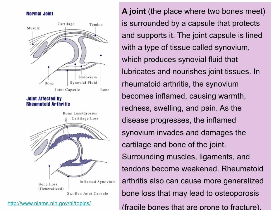
A joint (the place where two bones meet) is surrounded by a capsule that protects and supports it. The joint capsule is lined with a type of tissue called synovium, which produces synovial fluid that lubricates and nourishes joint tissues. In rheumatoid arthritis, the synovium becomes inflamed, causing warmth, redness, swelling, and pain. As the disease progresses, the inflamed synovium invades and damages the cartilage and bone of the joint. Surrounding muscles, ligaments, and tendons become weakened. Rheumatoid arthritis also can cause more generalized bone loss that may lead to osteoporosis
(fragile bones that are prone to fracture). http://www.niams.nih.gov/hi/topics/