23-1 chapter 23 the endocrine system endocrine and nervous systems work together endocrine system...
TRANSCRIPT
23-1
Chapter 23The Endocrine System
• Endocrine and nervous systems work together
• Endocrine system– hormones released into the bloodstream travel
throughout the body– results may take hours, but last longer
• Nervous system– certain parts release hormones into blood– rest releases neurotransmitters excite or inhibit nerve,
muscle & gland cells– results in milliseconds, brief duration of effects
23-2
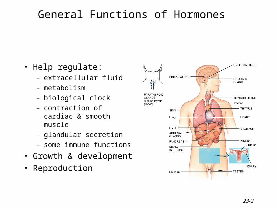
General Functions of Hormones
• Help regulate:– extracellular fluid
– metabolism
– biological clock
– contraction of cardiac & smooth muscle
– glandular secretion
– some immune functions
• Growth & development
• Reproduction
23-3
Endocrine Glands Defined
• Exocrine glands– secrete products into ducts which empty into body
cavities or body surface– sweat, oil, mucous, & digestive glands
• Endocrine glands– secrete products (hormones) into bloodstream– pituitary, thyroid, parathyroid, adrenal, pineal– other organs secrete hormones as a 2nd function
• hypothalamus, thymus, pancreas,ovaries,testes, kidneys, stomach, liver, small intestine, skin, heart & placenta

23-4
Hormone Receptors
• Hormones only affect target cells with specific membrane proteins called receptors
23-5
Role of Hormone Receptors
• Constantly being synthesized & broken down
• A range of 2000-100,000 receptors / target cell
• Down-regulation– excess hormone, produces a decrease in number of
receptors• receptors undergo endocytosis and are degraded
– decreases sensitivity of target cell to hormone
• Up-regulation– deficiency of hormone, produces an increase in the
number of receptors– target tissue more sensitive to the hormone
23-6
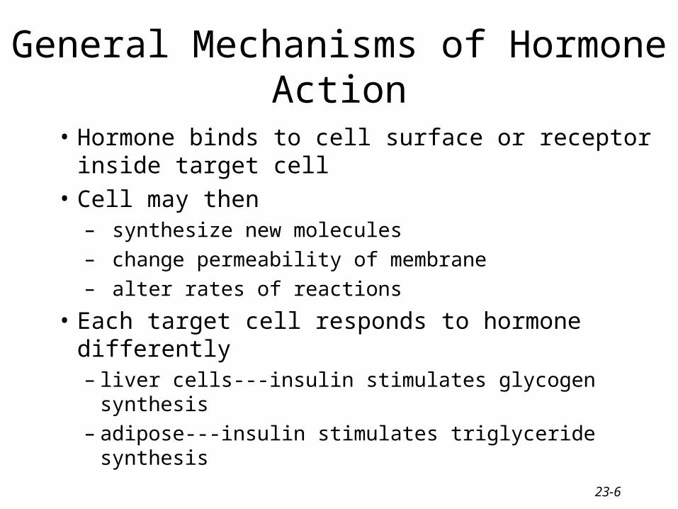
General Mechanisms of Hormone Action
• Hormone binds to cell surface or receptor inside target cell
• Cell may then– synthesize new molecules– change permeability of membrane– alter rates of reactions
• Each target cell responds to hormone differently– liver cells---insulin stimulates glycogen synthesis– adipose---insulin stimulates triglyceride synthesis
23-7
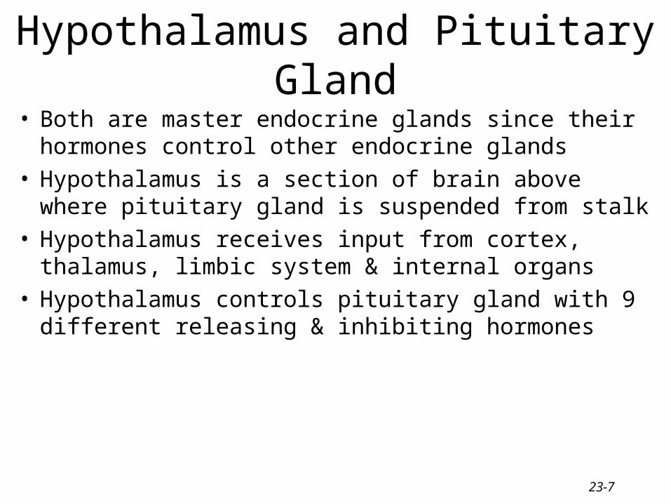
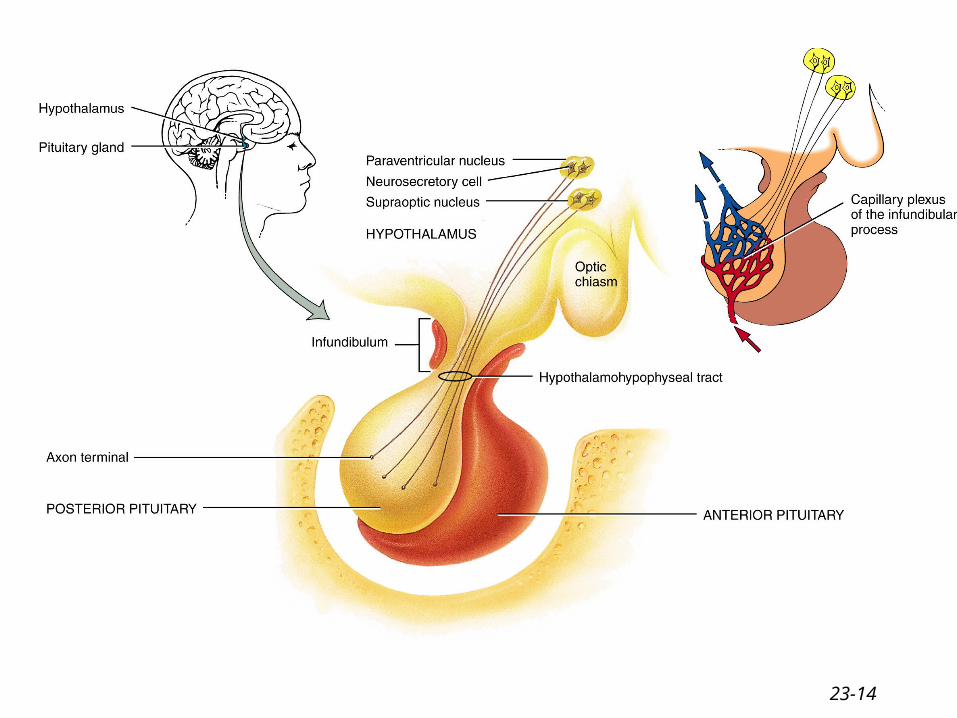
Hypothalamus and Pituitary Gland
• Both are master endocrine glands since their hormones control other endocrine glands
• Hypothalamus is a section of brain above where pituitary gland is suspended from stalk
• Hypothalamus receives input from cortex, thalamus, limbic system & internal organs
• Hypothalamus controls pituitary gland with 9 different releasing & inhibiting hormones

23-8
23-9
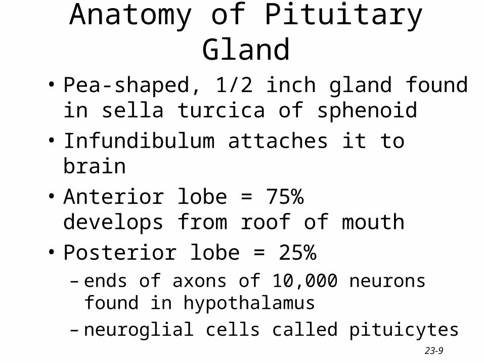
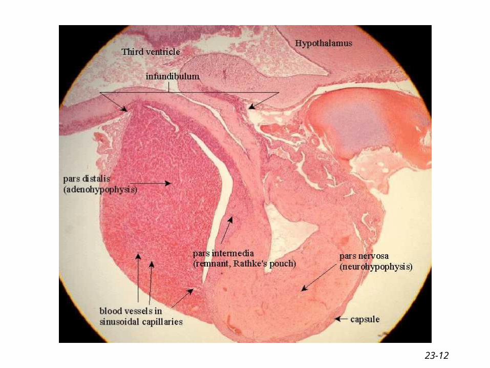
• Pea-shaped, 1/2 inch gland found in sella turcica of sphenoid
• Infundibulum attaches it to brain
• Anterior lobe = 75% develops from roof of mouth
• Posterior lobe = 25%– ends of axons of 10,000 neurons found in
hypothalamus– neuroglial cells called pituicytes
Anatomy of Pituitary Gland

23-10
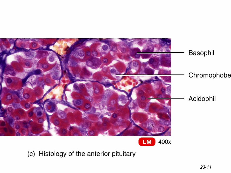
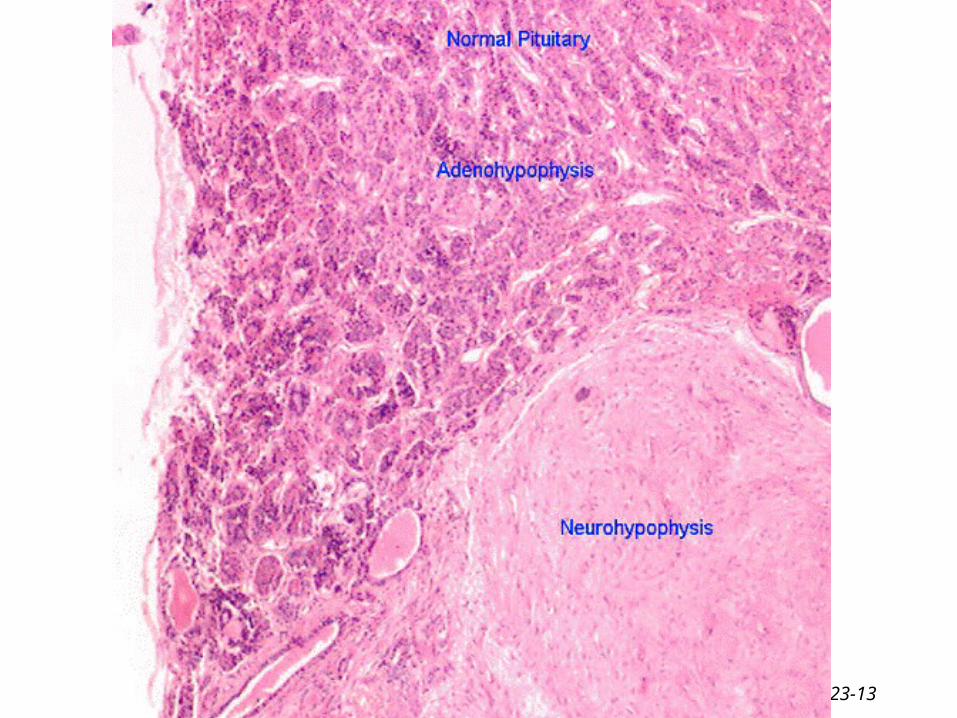
Cells of the anterior pituitary
• Five different types of cells classified according to their staining reaction
• Basophil- 10 % contain secretory granules (thyro, gonado, & corticotrophs)
• Acidophils- 40% contain secretory granules (somato, & lactotrophs)
• Chromophobes- 50% contain few or no secretory granules
23-11
23-12
23-13
23-14
23-15
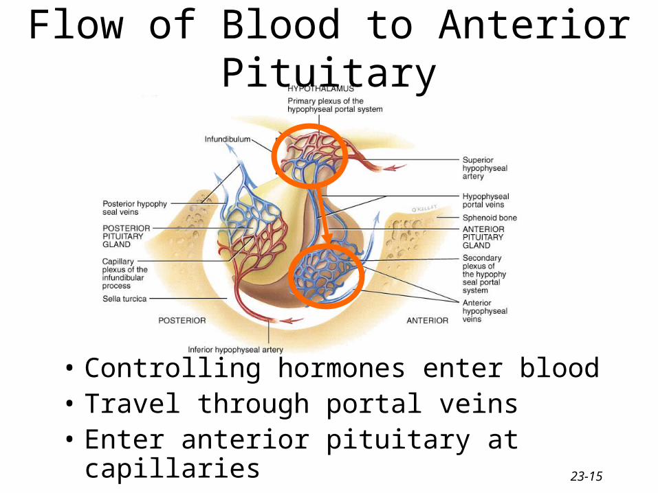
Flow of Blood to Anterior Pituitary
• Controlling hormones enter blood• Travel through portal veins• Enter anterior pituitary at capillaries
23-16
Human Growth Hormone• Produced by somatotrophs
• Within target cells increases synthesis of insulinlike growth factors that act locally or enter bloodstream– common target cells are liver, skeletal muscle,
cartilage and bone– increases cell growth & cell division by increasing
their uptake of amino acids & synthesis of proteins– stimulate lipolysis in adipose so fatty acids used for
ATP– retard use of glucose for ATP production so blood
glucose levels remain high enough to supply brain
23-17
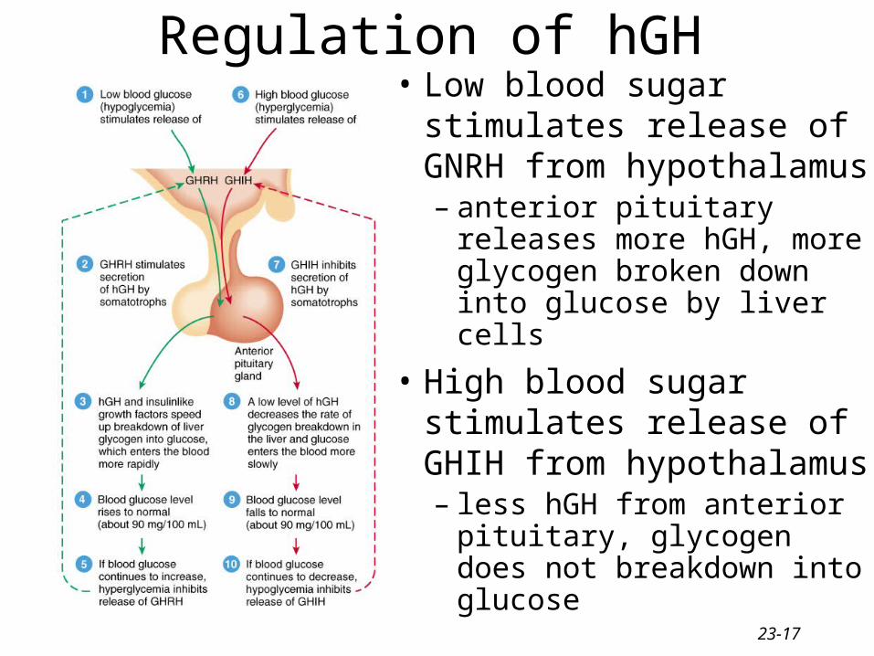
Regulation of hGH• Low blood sugar stimulates
release of GNRH from hypothalamus– anterior pituitary releases
more hGH, more glycogen broken down into glucose by liver cells
• High blood sugar stimulates release of GHIH from hypothalamus– less hGH from anterior
pituitary, glycogen does not breakdown into glucose

23-18
Diabetogenic Effect of Human Growth Hormone
• Excess of growth hormone– raises blood glucose concentration– pancreas releases insulin continually– beta-cell burnout
• Diabetogenic effect– causes diabetes mellitis if no insulin activity
can occur eventually
23-19
Thyroid Stimulating Hormone (TSH)
• Hypothalamus regulates thyrotroph cells
• Thyrotroph cells produce TSH
• TSH stimulates the synthesis & secretion of T3 and T4
• Metabolic rate stimulated
23-20
Follicle Stimulating Hormone (FSH)
• Releasing hormone from hypothalamus controls gonadotrophs
• Gonadotrophs release follicle stimulating hormone
• FSH functions – initiates the formation of follicles within the ovary– stimulates follicle cells to secrete estrogen– stimulates sperm production in testes
23-21
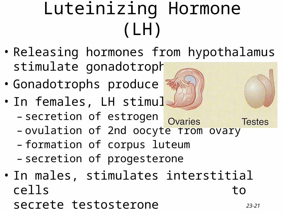
Luteinizing Hormone (LH)
• Releasing hormones from hypothalamus stimulate gonadotrophs
• Gonadotrophs produce LH
• In females, LH stimulates– secretion of estrogen– ovulation of 2nd oocyte from ovary– formation of corpus luteum– secretion of progesterone
• In males, stimulates interstitial cells to secrete testosterone
23-22
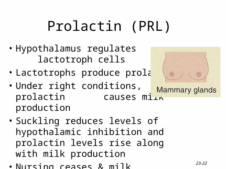
Prolactin (PRL)
• Hypothalamus regulates lactotroph cells
• Lactotrophs produce prolactin
• Under right conditions, prolactin causes milk production
• Suckling reduces levels of hypothalamic inhibition and prolactin levels rise along with milk production
• Nursing ceases & milk production slows
23-23
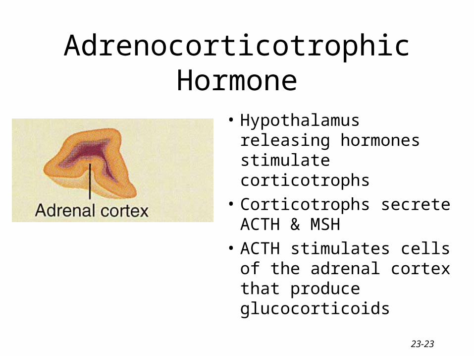
Adrenocorticotrophic Hormone
• Hypothalamus releasing hormones stimulate corticotrophs
• Corticotrophs secrete ACTH & MSH
• ACTH stimulates cells of the adrenal cortex that produce glucocorticoids
23-24
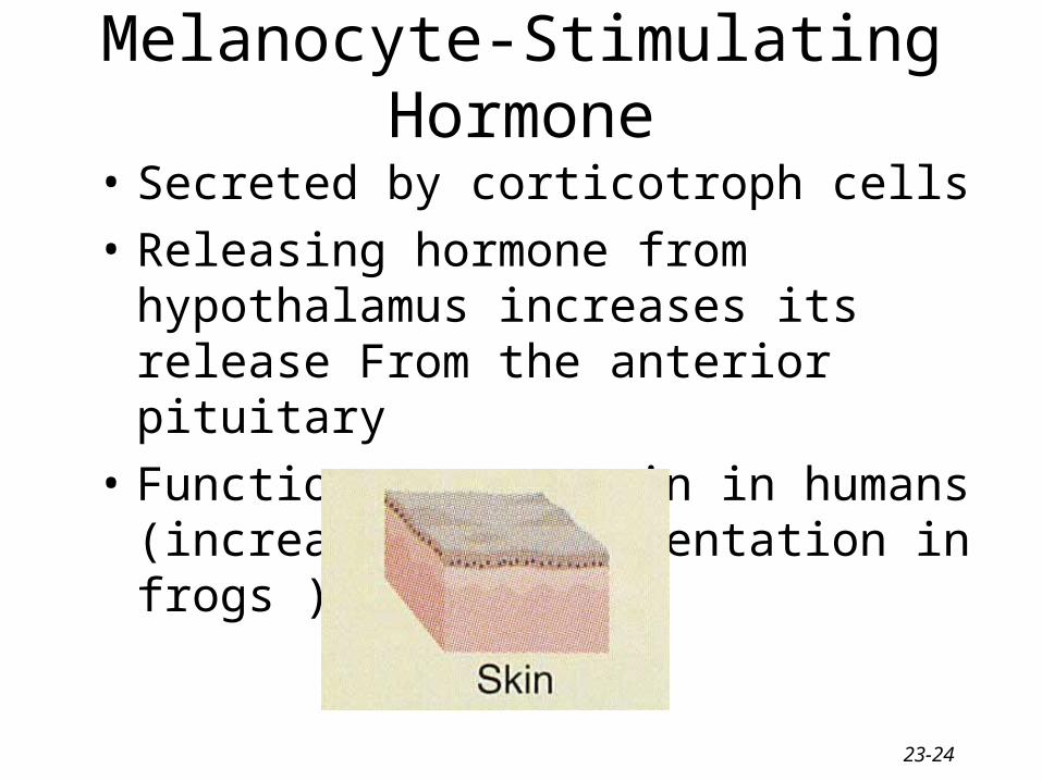
Melanocyte-Stimulating Hormone
• Secreted by corticotroph cells
• Releasing hormone from hypothalamus increases its release From the anterior pituitary
• Function not certain in humans (increase skin pigmentation in frogs )
23-25
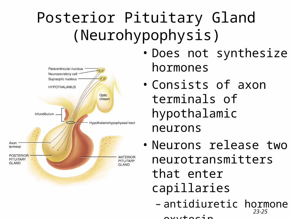
Posterior Pituitary Gland (Neurohypophysis)
• Does not synthesize hormones
• Consists of axon terminals of hypothalamic neurons
• Neurons release two neurotransmitters that enter capillaries– antidiuretic hormone– oxytocin
23-26
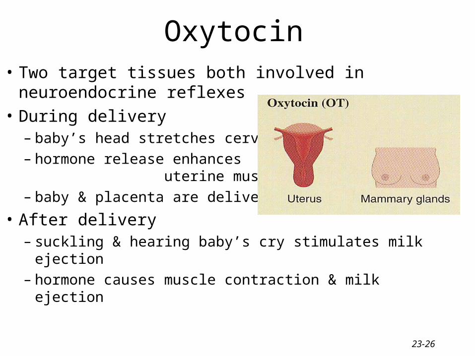
Oxytocin• Two target tissues both involved in neuroendocrine
reflexes
• During delivery– baby’s head stretches cervix– hormone release enhances uterine
muscle contraction– baby & placenta are delivered
• After delivery– suckling & hearing baby’s cry stimulates milk ejection– hormone causes muscle contraction & milk ejection
23-27
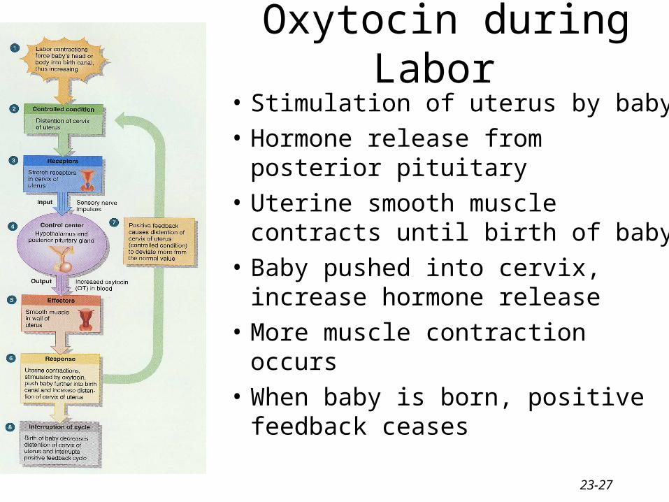
Oxytocin during Labor
• Stimulation of uterus by baby
• Hormone release from posterior pituitary
• Uterine smooth muscle contracts until birth of baby
• Baby pushed into cervix, increase hormone release
• More muscle contraction occurs
• When baby is born, positive feedback ceases
23-28
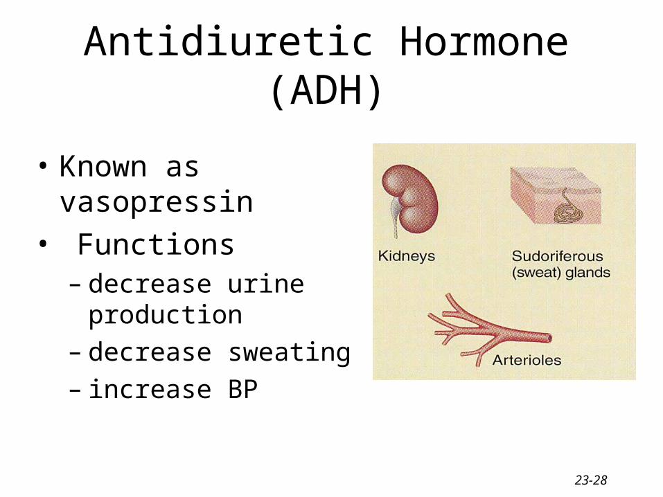
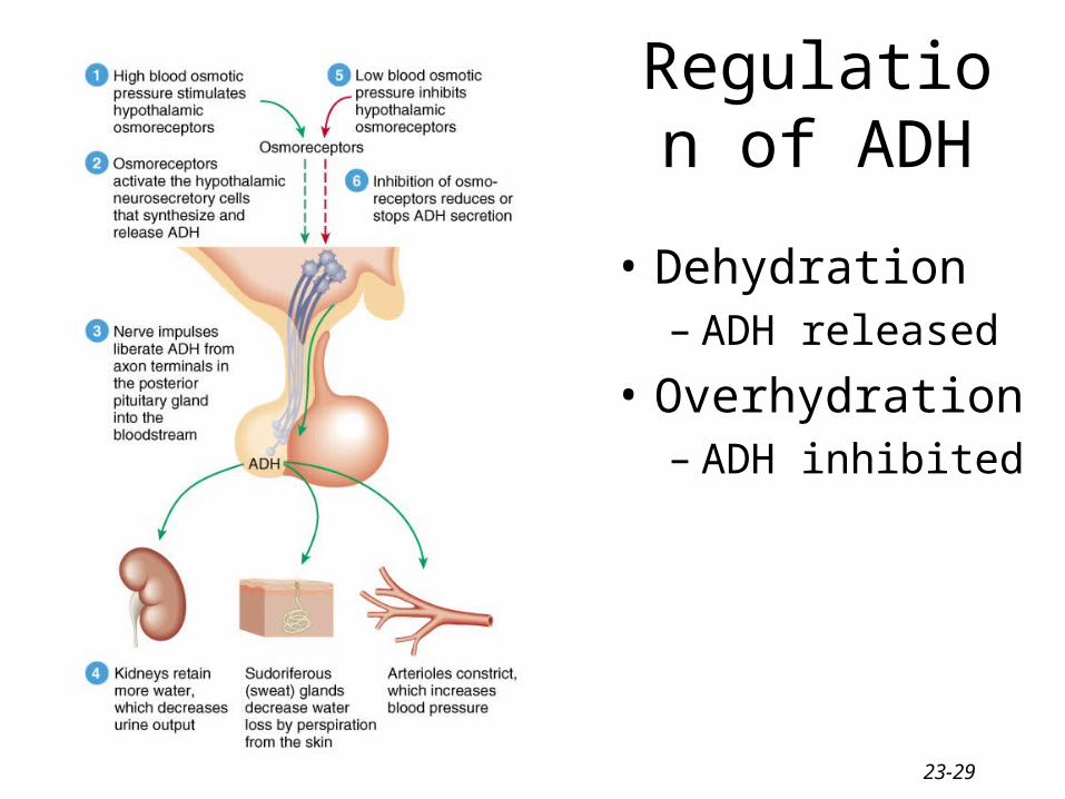
Antidiuretic Hormone (ADH)
• Known as vasopressin
• Functions– decrease urine production– decrease sweating– increase BP
23-29
Regulation of ADH
• Dehydration– ADH released
• Overhydration– ADH inhibited
23-30
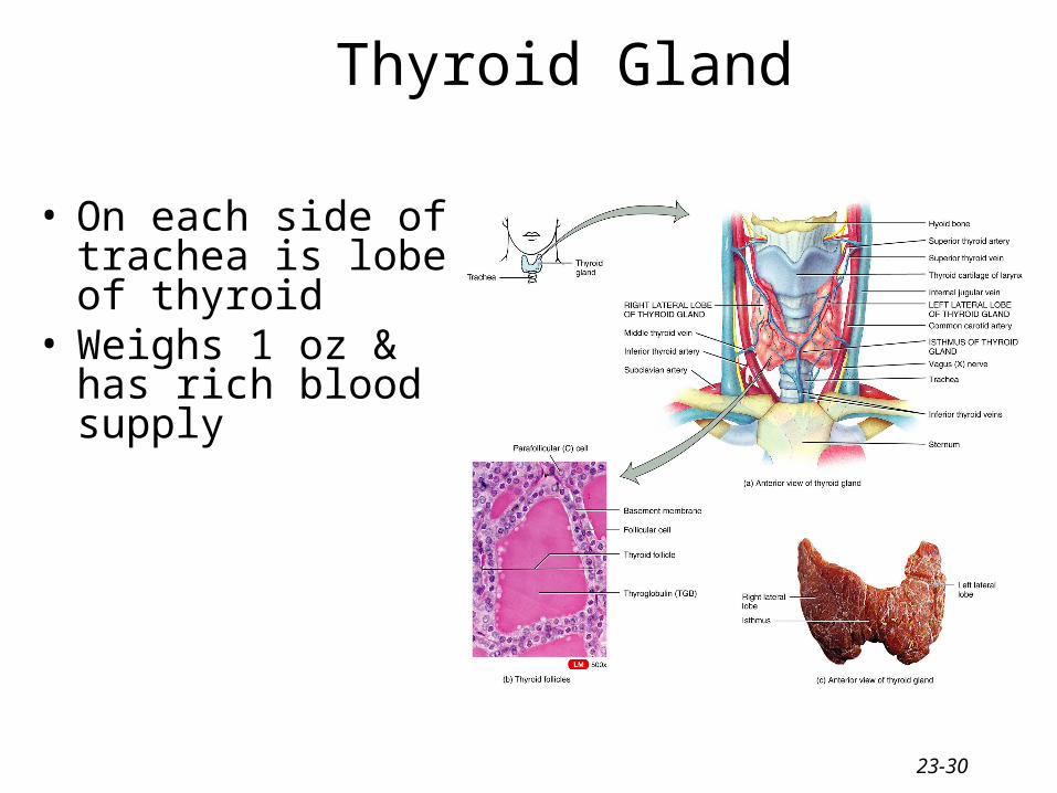
Thyroid Gland
• On each side of trachea is lobe of thyroid
• Weighs 1 oz & has rich blood supply
23-31
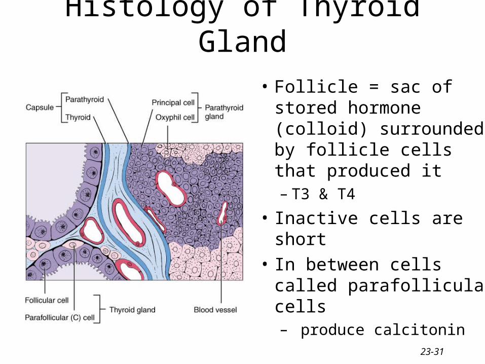
Histology of Thyroid Gland
• Follicle = sac of stored hormone (colloid) surrounded by follicle cells that produced it– T3 & T4
• Inactive cells are short
• In between cells called parafollicular cells– produce calcitonin
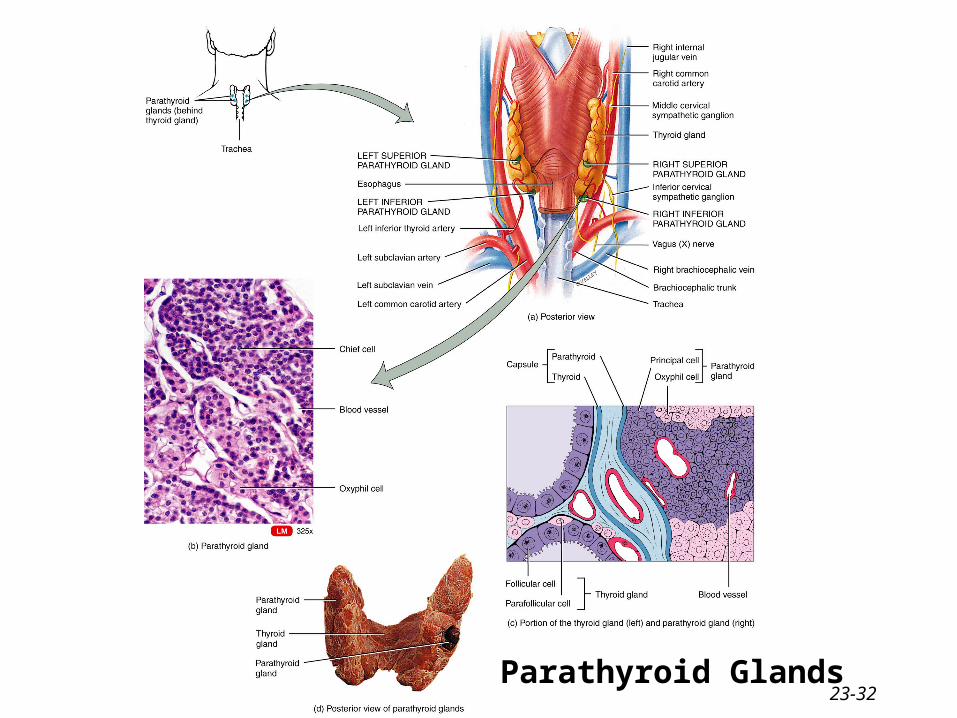
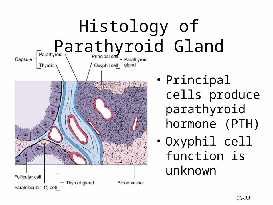
23-32Parathyroid Glands
23-33
Histology of Parathyroid Gland
• Principal cells produce parathyroid hormone (PTH)
• Oxyphil cell function is unknown
23-34
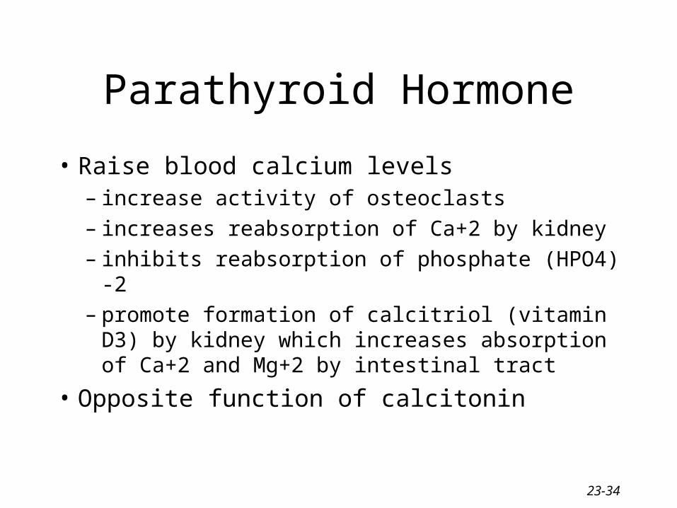
Parathyroid Hormone
• Raise blood calcium levels– increase activity of osteoclasts– increases reabsorption of Ca+2 by kidney– inhibits reabsorption of phosphate (HPO4) -2– promote formation of calcitriol (vitamin D3) by
kidney which increases absorption of Ca+2 and Mg+2 by intestinal tract
• Opposite function of calcitonin
23-35
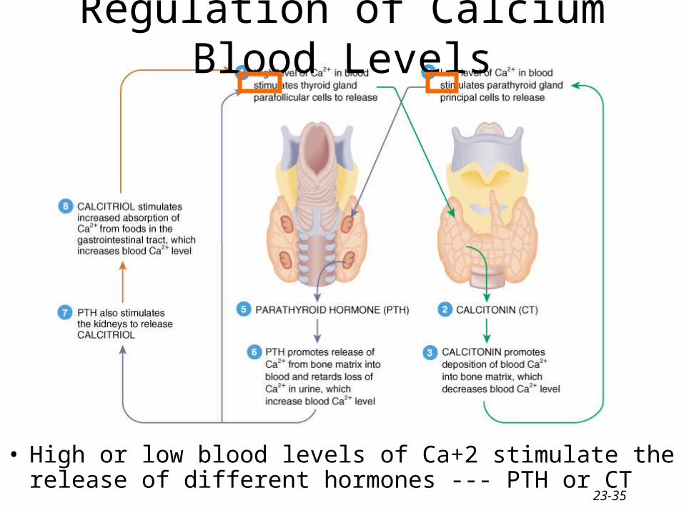
Regulation of Calcium Blood Levels
• High or low blood levels of Ca+2 stimulate the release of different hormones --- PTH or CT
23-36
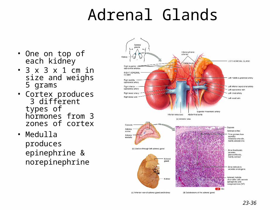
Adrenal Glands
• One on top of each kidney
• 3 x 3 x 1 cm in size and weighs 5 grams
• Cortex produces 3 different types of hormones from 3 zones of cortex
• Medulla produces epinephrine & norepinephrine
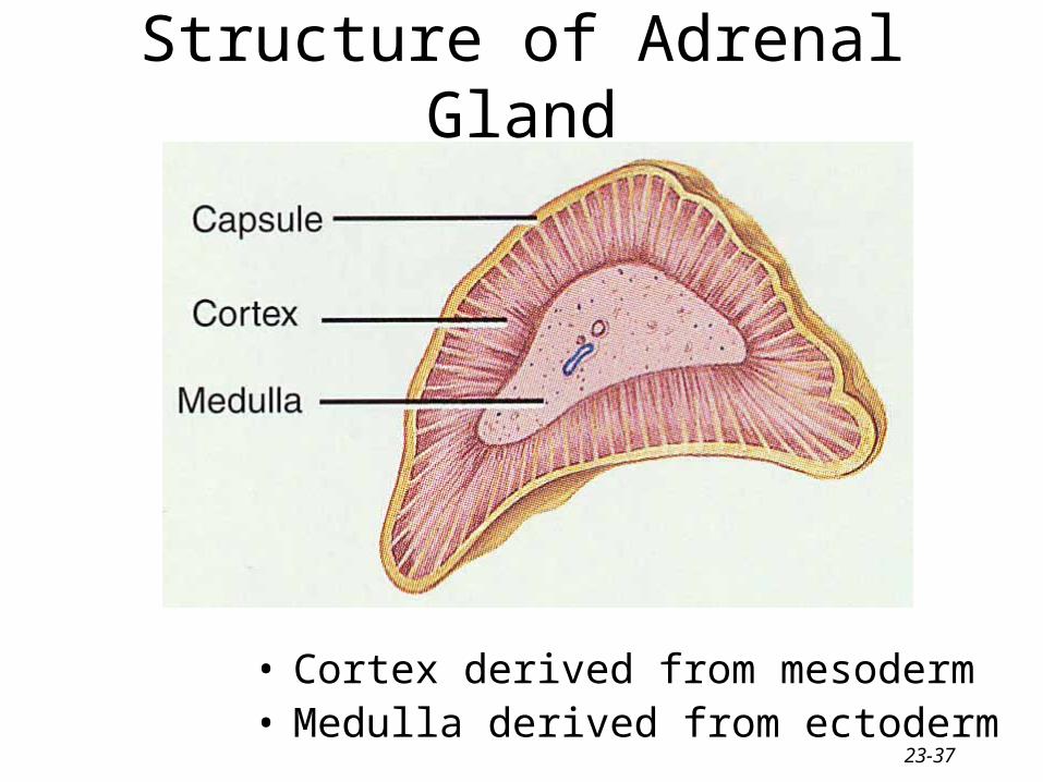
23-37
Structure of Adrenal Gland
• Cortex derived from mesoderm• Medulla derived from ectoderm
23-38
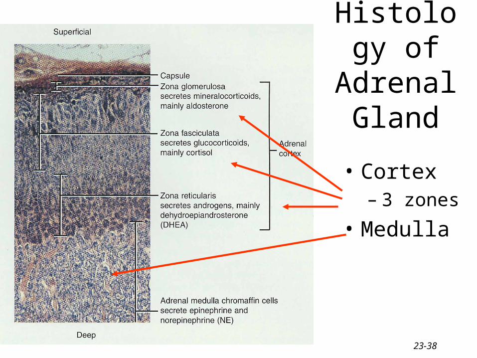
Histology of Adrenal
Gland
• Cortex– 3 zones
• Medulla
23-39
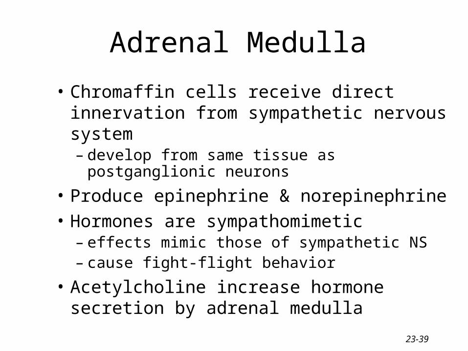
Adrenal Medulla
• Chromaffin cells receive direct innervation from sympathetic nervous system– develop from same tissue as postganglionic
neurons
• Produce epinephrine & norepinephrine• Hormones are sympathomimetic
– effects mimic those of sympathetic NS– cause fight-flight behavior
• Acetylcholine increase hormone secretion by adrenal medulla
23-40
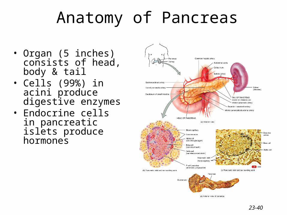
Anatomy of Pancreas
• Organ (5 inches) consists of head, body & tail
• Cells (99%) in acini produce digestive enzymes
• Endocrine cells in pancreatic islets produce hormones
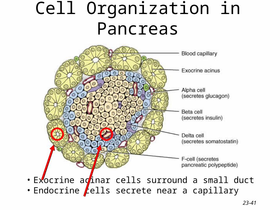
23-41
Cell Organization in Pancreas
• Exocrine acinar cells surround a small duct• Endocrine cells secrete near a capillary
23-42
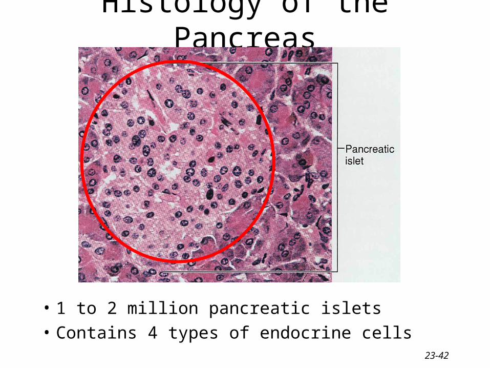
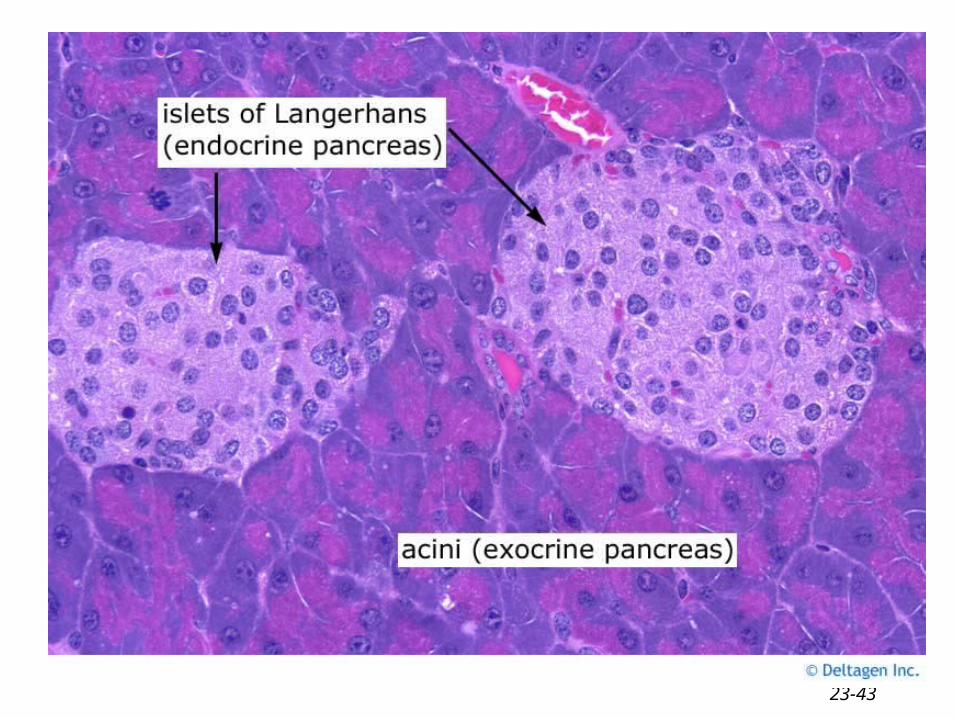
Histology of the Pancreas
• 1 to 2 million pancreatic islets
• Contains 4 types of endocrine cells
23-43
23-44
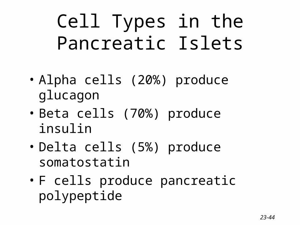
Cell Types in the Pancreatic Islets
• Alpha cells (20%) produce glucagon
• Beta cells (70%) produce insulin
• Delta cells (5%) produce somatostatin
• F cells produce pancreatic polypeptide
23-45
Ovaries and Testes
• Ovaries– estrogen, progesterone, relaxin & inhibin– regulate reproductive cycle, maintain pregnancy &
prepare mammary glands for lactation
• Testes– produce testosterone– regulate sperm production & 2nd sexual
characteristics
23-46
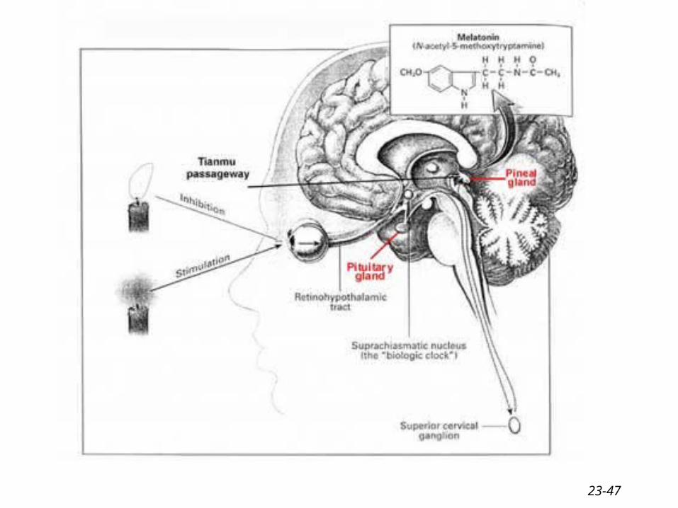
Pineal Gland
• Small gland attached to 3rd ventricle of brain
• Consists of pinealocytes & neuroglia
• Melatonin responsible for setting of biological clock
• Jet lag & SAD treatment is bright light
23-47
23-48
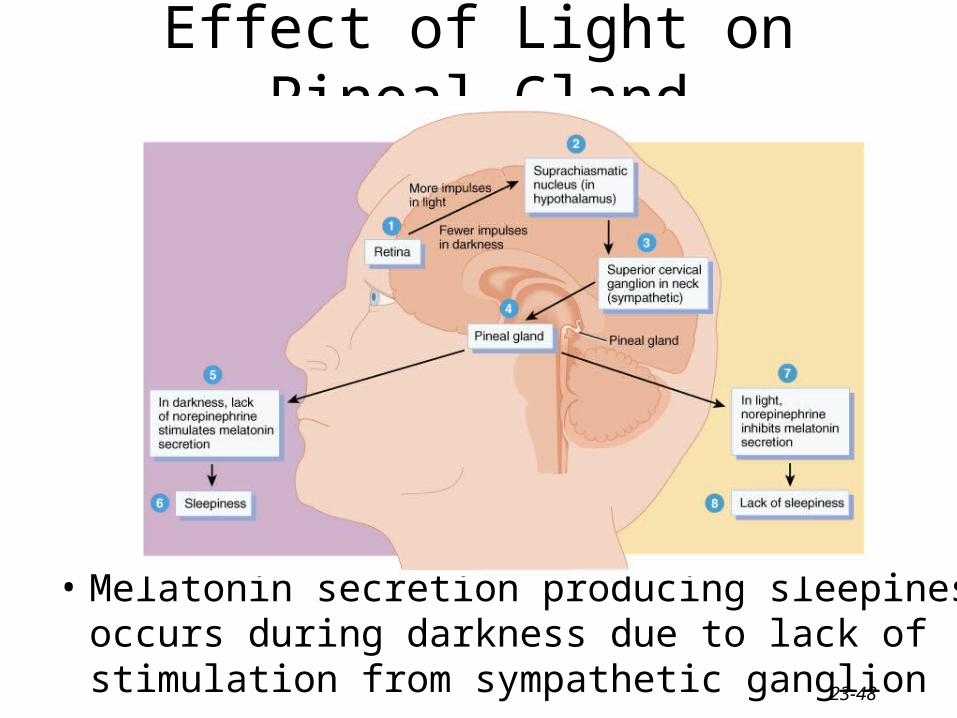
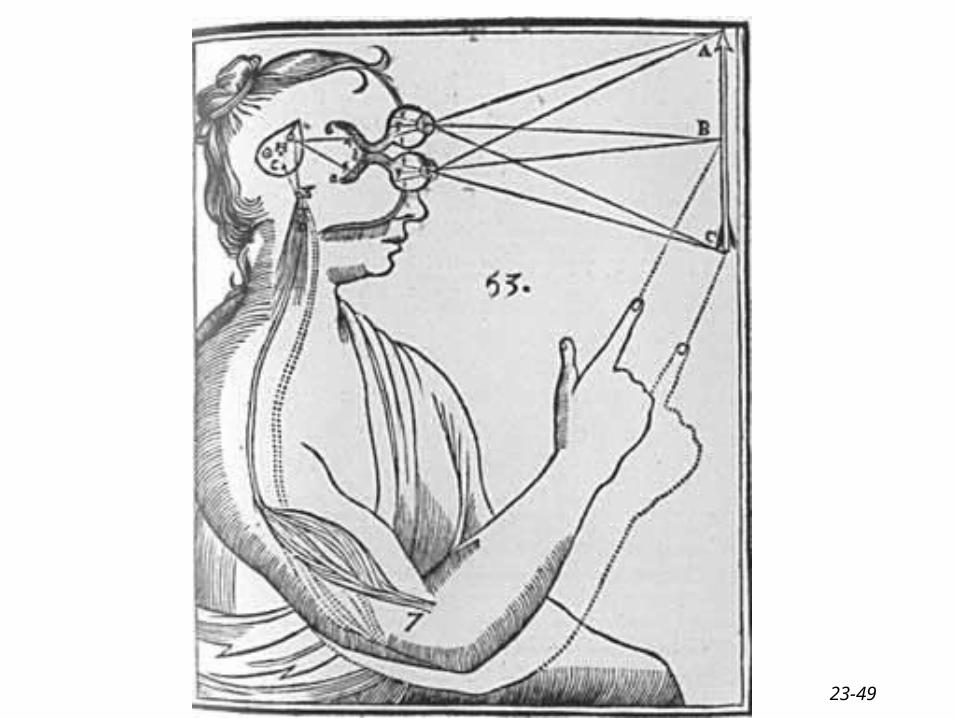
Effect of Light on Pineal Gland
• Melatonin secretion producing sleepiness occurs during darkness due to lack of stimulation from sympathetic ganglion
23-49
23-50
Seasonal Affective Disorder and Jet Lag
• Depression that occurs during winter months when day length is short
• Due to overproduction of melatonin
• Therapy– exposure to several hours per day of artificial light as
bright as sunlight– speeds recovery from jet lag
23-51
Thymus Gland
• Important role in maturation of T cells
• Hormones produced by gland promote the proliferation & maturation of T cells– thymosin– thymic humoral factor– thymic factor– thymopoietin
23-52
Miscellaneous Hormones Eicosanoids
• Local hormones released by all body cells• Leukotrienes influence WBCs & inflammation• Prostaglandins alter
– smooth muscle contraction, glandular secretion, blood flow, platelet function, nerve transmission, metabolism etc.
• Ibuprofen & other nonsteroidal anti-inflammatory drugs treat pain, fever & inflammation by inhibiting prostaglandin synthesis
23-53
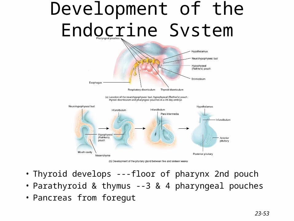
Development of the Endocrine System
• Thyroid develops ---floor of pharynx 2nd pouch• Parathyroid & thymus --3 & 4 pharyngeal pouches• Pancreas from foregut
23-54
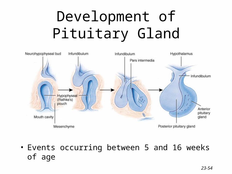
Development of Pituitary Gland
• Events occurring between 5 and 16 weeks of age
23-55
Aging and the Endocrine System• Production of human growth hormone decreases
– muscle atrophy
• Production of TSH increase with age to try and stimulate thyroid – decrease in metabolic rate, increase in body fat & hypothyroidism
• Thymus after puberty is replaced with adipose• Adrenal glands produce less cortisol & aldosterone• Receptor sensitivity to glucose declines• Ovaries no longer respond to gonadotropins
– decreased output of estrogen (osteoporosis & atherosclerosis)
23-56
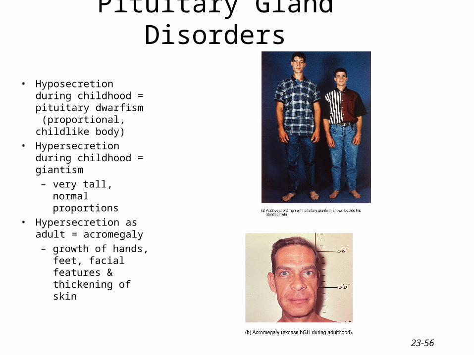
Pituitary Gland Disorders
• Hyposecretion during childhood = pituitary dwarfism (proportional, childlike body)
• Hypersecretion during childhood = giantism
– very tall, normal proportions
• Hypersecretion as adult = acromegaly
– growth of hands, feet, facial features & thickening of skin
23-57
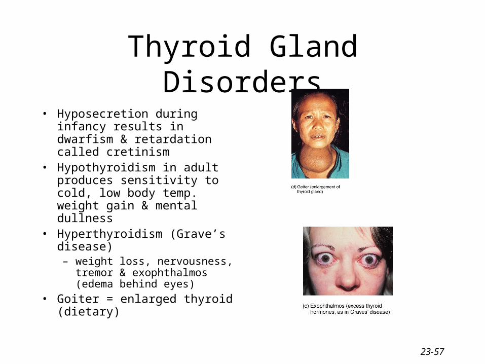
Thyroid Gland Disorders
• Hyposecretion during infancy results in dwarfism & retardation called cretinism
• Hypothyroidism in adult produces sensitivity to cold, low body temp. weight gain & mental dullness
• Hyperthyroidism (Grave’s disease)– weight loss, nervousness,
tremor & exophthalmos (edema behind eyes)
• Goiter = enlarged thyroid (dietary)
23-58
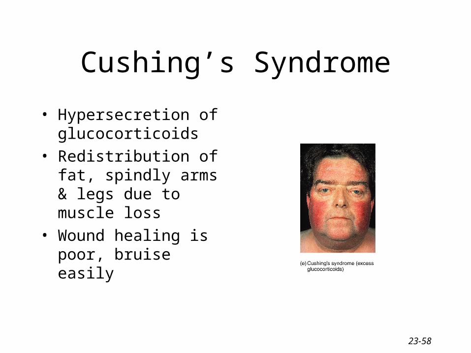
Cushing’s Syndrome
• Hypersecretion of glucocorticoids
• Redistribution of fat, spindly arms & legs due to muscle loss
• Wound healing is poor, bruise easily
23-59
Addison’s disease
• Hypersecretion of glucocorticoids– hypoglycemia, muscle weakness, low BP,
dehydration due to decreased Na+ in blood– mimics skin darkening effects of MSH– potential cardiac arrest
23-60
Diabetes Mellitus & Hyperinsulinism
• Diabetes mellitus marked by hyperglycemia– excessive urine production (polyuria)– excessive thirst (polydipsia)– excessive eating (polyphagia)
• Type I----deficiency of insulin (under 20)
• Type II---adult onset– drug stimulates secretion of insulin by beta cells– cells may be less sensitive to hormone