the endocrine system. electrochemical signals influences metabolic activities by means of hormones...
TRANSCRIPT
The Endocrine System
Electrochemical Signals• Influences metabolic activities by means of
hormones▫Chemical messengers released into the
bloodstream to be transported throughout the body.
•Lag time in response▫Nervous = (near) immediate▫Endocrine = seconds to days
•Once initiated, hormonal responses tend to be more prolonged than nervous responses
•Study of hormones & endocrine organs = endocrinology
Processes controlled by Hormones•Reproduction•Growth & development•Mobilizing body’s defense against stressors•Maintaining electrolyte balance•Maintaining nutrient balance•Regulating cellular metabolism•Maintaining an available energy source
Overview • Endocrine glands, compared to other systems:▫Called glands – main purpose: secrete▫Small glands, less than 2lbs worth▫Widely scattered throughout the body – usually near
their target organs• Endocrine vs. exocrine▫Exocrine – have ducts
Non hormonal products are routed to a membrane surface▫Endocrine – ductless
Release hormones into surrounding tissue of organs that are richly vascular Easy to be released into bloodstream of the appropriate organ.
Most are physically arranged into branching networks to maximize the spread of hormones
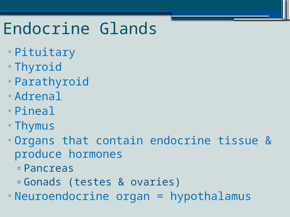
Endocrine Glands•Pituitary•Thyroid•Parathyroid•Adrenal•Pineal•Thymus•Organs that contain endocrine tissue & produce
hormones▫Pancreas▫Gonads (testes & ovaries)
•Neuroendocrine organ = hypothalamus
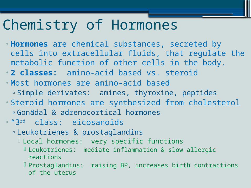
Chemistry of Hormones•Hormones are chemical substances, secreted by cells
into extracellular fluids, that regulate the metabolic function of other cells in the body.
•2 classes: amino-acid based vs. steroid•Most hormones are amino-acid based▫Simple derivates: amines, thyroxine, peptides
• Steroid hormones are synthesized from cholesterol▫Gonadal & adrenocortical hormones
• “3rd” class: eicosanoids▫Leukotrienes & prostaglandins
Local hormones: very specific functions Leukotrienes: mediate inflammation & slow allergic reactions Prostaglandins: raising BP, increases birth contractions of the
uterus
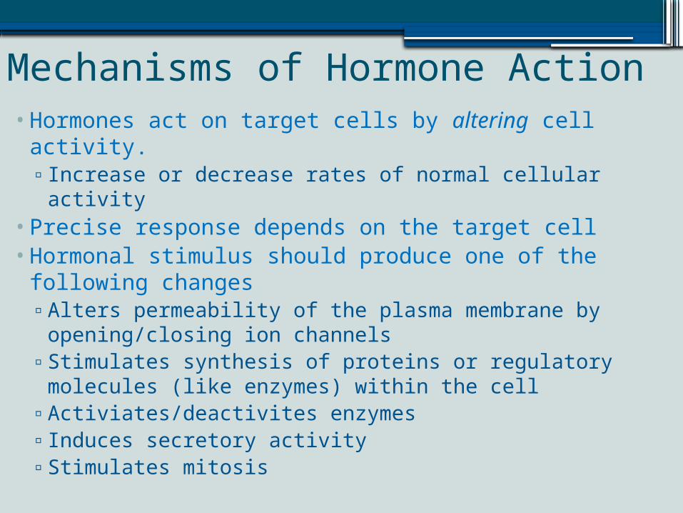
Mechanisms of Hormone Action•Hormones act on target cells by altering cell activity.▫Increase or decrease rates of normal cellular activity
•Precise response depends on the target cell•Hormonal stimulus should produce one of the
following changes▫Alters permeability of the plasma membrane by
opening/closing ion channels▫Stimulates synthesis of proteins or regulatory
molecules (like enzymes) within the cell▫Activiates/deactivites enzymes▫Induces secretory activity▫Stimulates mitosis
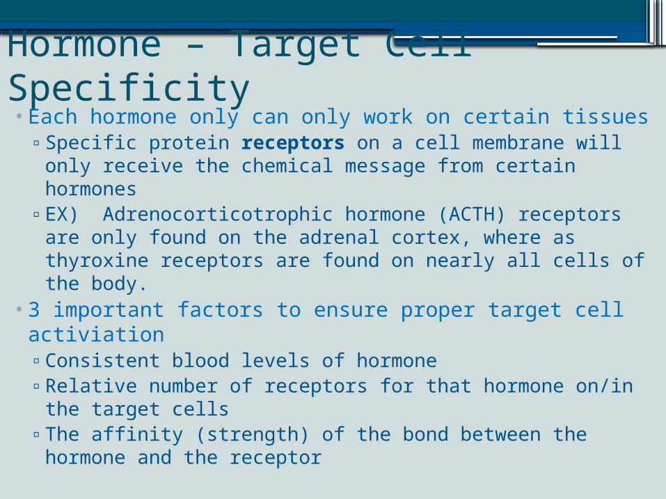
Hormone – Target Cell Specificity•Each hormone only can only work on certain tissues▫Specific protein receptors on a cell membrane will only
receive the chemical message from certain hormones▫EX) Adrenocorticotrophic hormone (ACTH) receptors are
only found on the adrenal cortex, where as thyroxine receptors are found on nearly all cells of the body.
•3 important factors to ensure proper target cell activiation▫Consistent blood levels of hormone▫Relative number of receptors for that hormone on/in the
target cells▫The affinity (strength) of the bond between the hormone
and the receptor
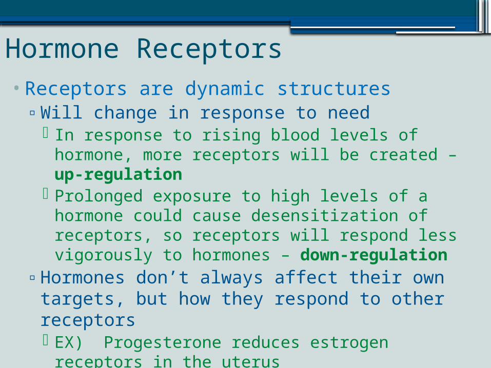
Hormone Receptors•Receptors are dynamic structures▫Will change in response to need
In response to rising blood levels of hormone, more receptors will be created – up-regulation
Prolonged exposure to high levels of a hormone could cause desensitization of receptors, so receptors will respond less vigorously to hormones – down-regulation
▫Hormones don’t always affect their own targets, but how they respond to other receptors EX) Progesterone reduces estrogen receptors in the
uterus Estrogen causes the same cells to produce more
progesterone receptors – enhancing the ability to respond to progesterone.
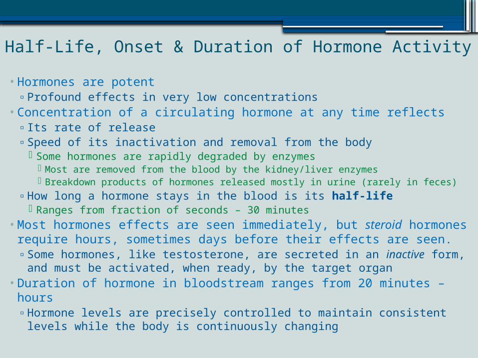
Half-Life, Onset & Duration of Hormone Activity
• Hormones are potent▫ Profound effects in very low concentrations
• Concentration of a circulating hormone at any time reflects▫ Its rate of release▫ Speed of its inactivation and removal from the body
Some hormones are rapidly degraded by enzymes Most are removed from the blood by the kidney/liver enzymes Breakdown products of hormones released mostly in urine (rarely in feces)
▫ How long a hormone stays in the blood is its half-life Ranges from fraction of seconds – 30 minutes
• Most hormones effects are seen immediately, but steroid hormones require hours, sometimes days before their effects are seen.▫ Some hormones, like testosterone, are secreted in an inactive form, and
must be activated, when ready, by the target organ• Duration of hormone in bloodstream ranges from 20 minutes – hours▫ Hormone levels are precisely controlled to maintain consistent levels
while the body is continuously changing
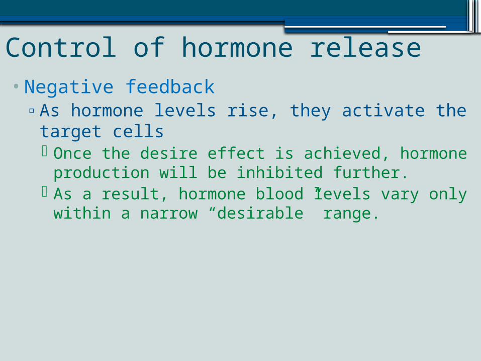
Control of hormone release•Negative feedback▫As hormone levels rise, they activate the target
cells Once the desire effect is achieved, hormone
production will be inhibited further. As a result, hormone blood levels vary only within a
narrow “desirable” range.
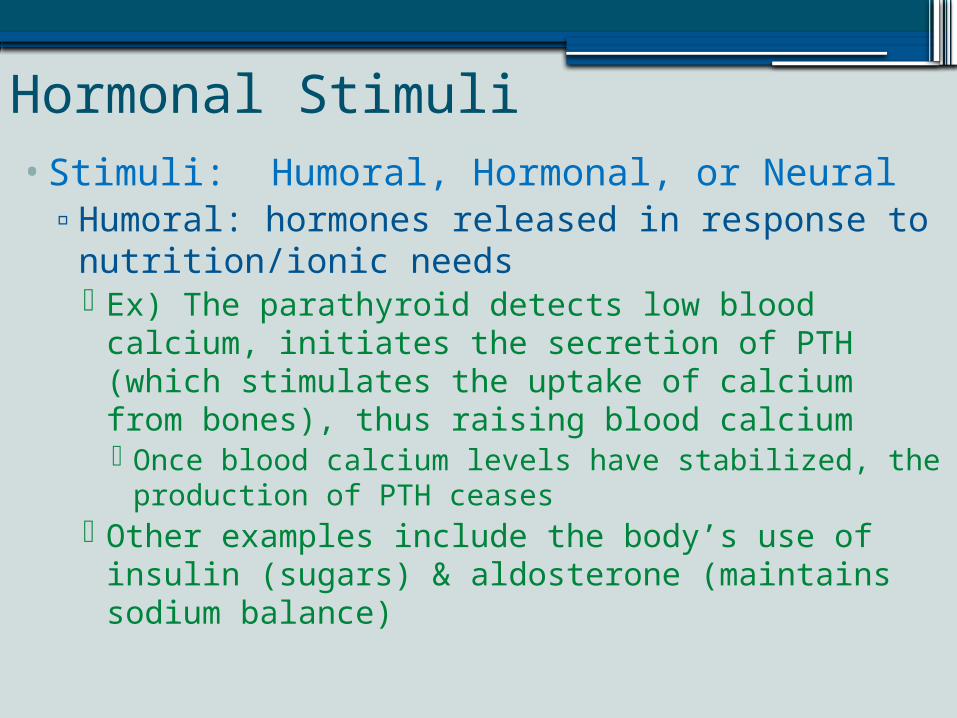
Hormonal Stimuli•Stimuli: Humoral, Hormonal, or Neural▫Humoral: hormones released in response to
nutrition/ionic needs Ex) The parathyroid detects low blood calcium,
initiates the secretion of PTH (which stimulates the uptake of calcium from bones), thus raising blood calcium Once blood calcium levels have stabilized, the
production of PTH ceases Other examples include the body’s use of insulin
(sugars) & aldosterone (maintains sodium balance)
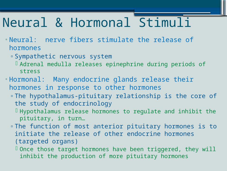
Neural & Hormonal Stimuli•Neural: nerve fibers stimulate the release of hormones▫Sympathetic nervous system
Adrenal medulla releases epinephrine during periods of stress•Hormonal: Many endocrine glands release their
hormones in response to other hormones▫The hypothalamus-pituitary relationship is the core of the
study of endocrinology Hypothalamus release hormones to regulate and inhibit the
pituitary, in turn…▫The function of most anterior pituitary hormones is to
initiate the release of other endocrine hormones (targeted organs) Once those target hormones have been triggered, they will
inhibit the production of more pituitary hormones
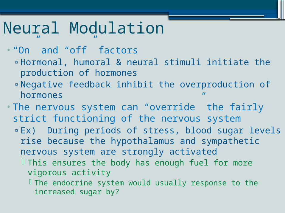
Neural Modulation•“On” and “off” factors▫Hormonal, humoral & neural stimuli initiate the
production of hormones▫Negative feedback inhibit the overproduction of
hormones•The nervous system can “override” the fairly strict
functioning of the nervous system▫Ex) During periods of stress, blood sugar levels rise
because the hypothalamus and sympathetic nervous system are strongly activated This ensures the body has enough fuel for more vigorous
activity The endocrine system would usually response to the
increased sugar by?
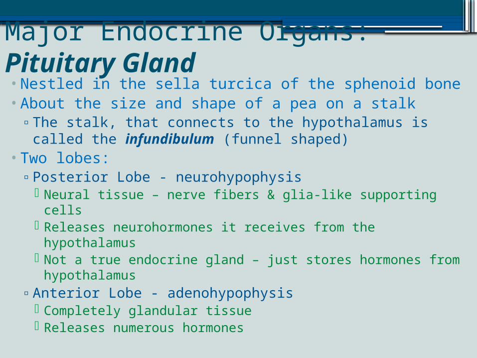
Major Endocrine Organs: Pituitary Gland•Nestled in the sella turcica of the sphenoid bone•About the size and shape of a pea on a stalk▫The stalk, that connects to the hypothalamus is called
the infundibulum (funnel shaped)•Two lobes:▫Posterior Lobe - neurohypophysis
Neural tissue – nerve fibers & glia-like supporting cells Releases neurohormones it receives from the
hypothalamus Not a true endocrine gland – just stores hormones from
hypothalamus▫Anterior Lobe - adenohypophysis
Completely glandular tissue Releases numerous hormones
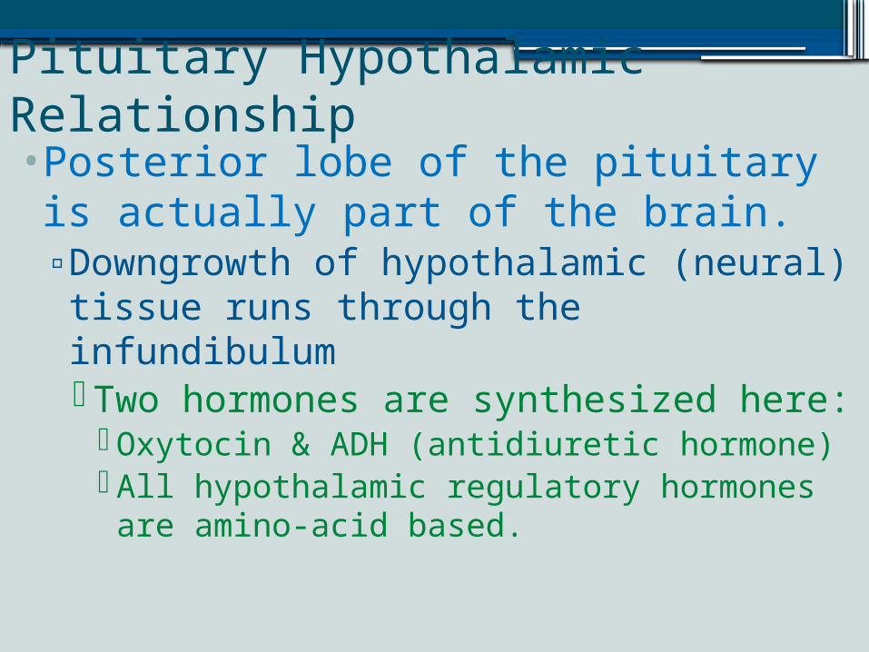
Pituitary Hypothalamic Relationship•Posterior lobe of the pituitary is actually part of the brain.▫Downgrowth of hypothalamic (neural) tissue runs through the infundibulum Two hormones are synthesized here:
Oxytocin & ADH (antidiuretic hormone) All hypothalamic regulatory hormones are amino-acid based.
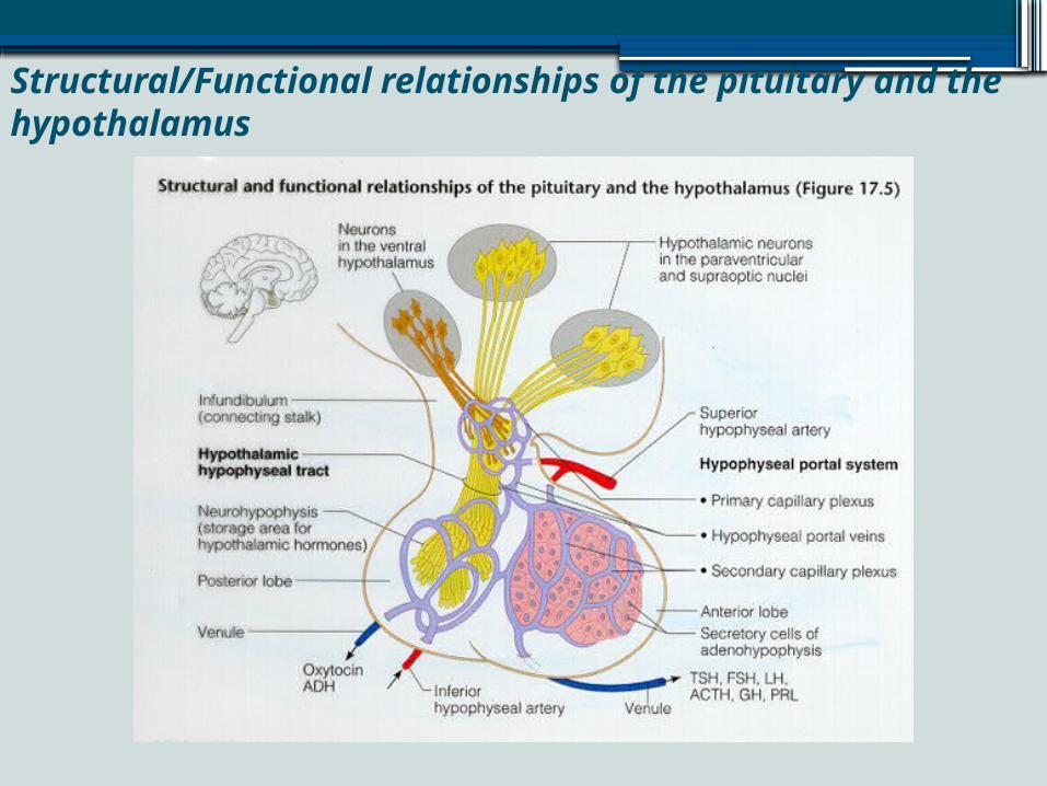
Structural/Functional relationships of the pituitary and the hypothalamus
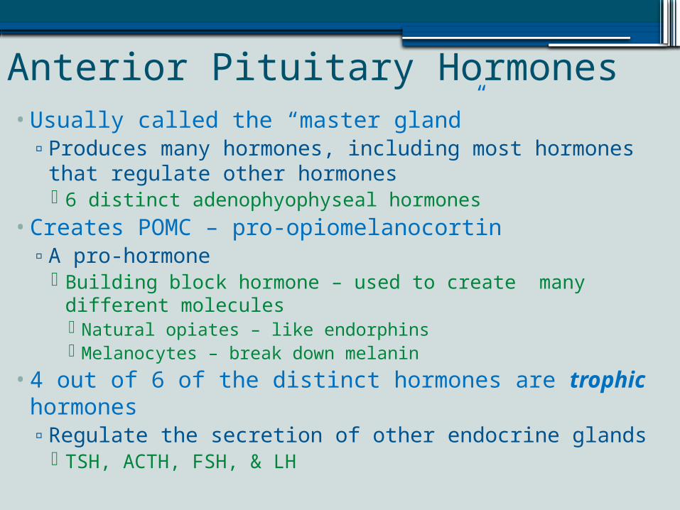
Anterior Pituitary Hormones•Usually called the “master gland”▫Produces many hormones, including most hormones
that regulate other hormones 6 distinct adenophyophyseal hormones
•Creates POMC – pro-opiomelanocortin▫A pro-hormone
Building block hormone – used to create many different molecules Natural opiates – like endorphins Melanocytes – break down melanin
•4 out of 6 of the distinct hormones are trophic hormones▫Regulate the secretion of other endocrine glands
TSH, ACTH, FSH, & LH
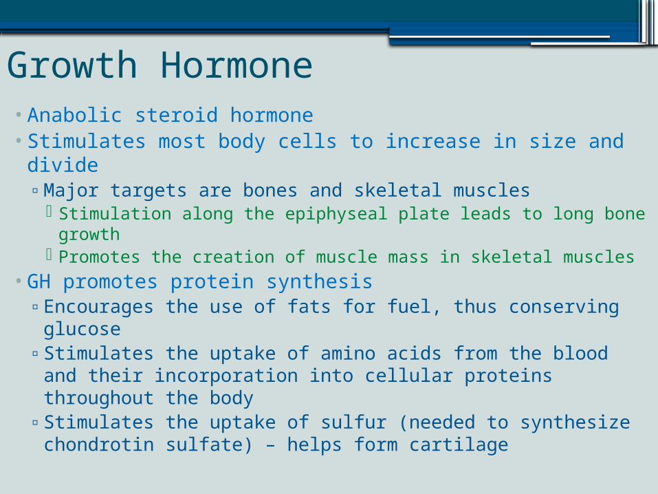
Growth Hormone•Anabolic steroid hormone•Stimulates most body cells to increase in size and divide▫Major targets are bones and skeletal muscles
Stimulation along the epiphyseal plate leads to long bone growth
Promotes the creation of muscle mass in skeletal muscles•GH promotes protein synthesis▫Encourages the use of fats for fuel, thus conserving glucose▫Stimulates the uptake of amino acids from the blood and
their incorporation into cellular proteins throughout the body
▫Stimulates the uptake of sulfur (needed to synthesize chondrotin sulfate) – helps form cartilage
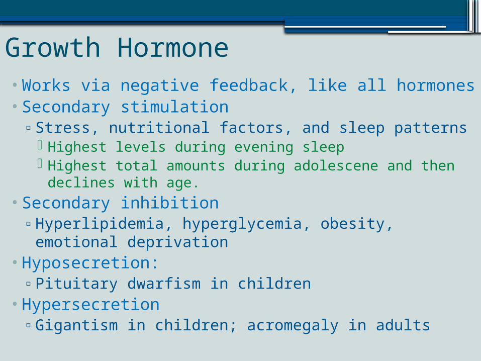
Growth Hormone•Works via negative feedback, like all hormones• Secondary stimulation▫Stress, nutritional factors, and sleep patterns
Highest levels during evening sleep Highest total amounts during adolescene and then
declines with age.• Secondary inhibition▫Hyperlipidemia, hyperglycemia, obesity, emotional
deprivation•Hyposecretion:▫Pituitary dwarfism in children
•Hypersecretion▫Gigantism in children; acromegaly in adults
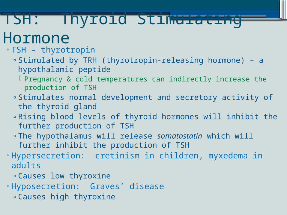
TSH: Thyroid Stimulating Hormone• TSH – thyrotropin▫Stimulated by TRH (thyrotropin-releasing hormone) – a
hypothalamic peptide Pregnancy & cold temperatures can indirectly increase the
production of TSH▫Stimulates normal development and secretory activity of the
thyroid gland▫Rising blood levels of thyroid hormones will inhibit the further
production of TSH▫The hypothalamus will release somatostatin which will further
inhibit the production of TSH•Hypersecretion: cretinism in children, myxedema in adults▫Causes low thyroxine
•Hyposecretion: Graves’ disease▫Causes high thyroxine
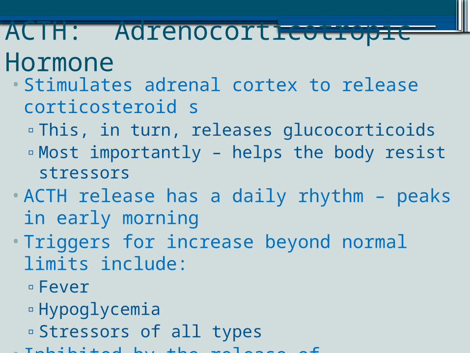
ACTH: Adrenocorticotropic Hormone•Stimulates adrenal cortex to release
corticosteroid s▫This, in turn, releases glucocorticoids▫Most importantly – helps the body resist stressors
•ACTH release has a daily rhythm – peaks in early morning
•Triggers for increase beyond normal limits include:▫Fever▫Hypoglycemia▫Stressors of all types
• Inhibited by the release of glucocorticoids•Hyposecretion: rare & idiopathic•Hypersecretion: Cushing’s disease
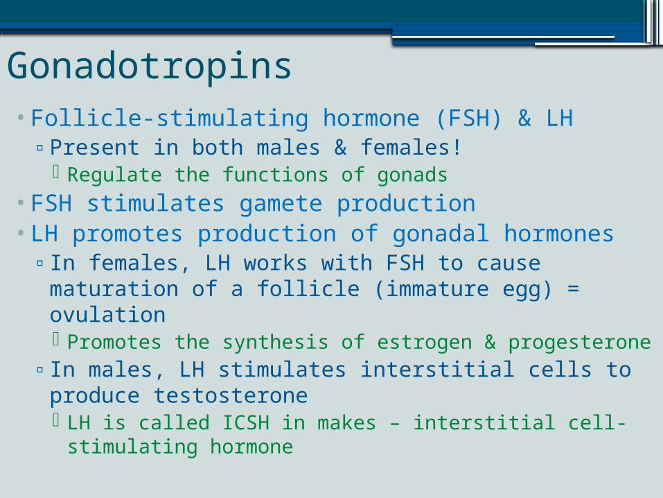
Gonadotropins•Follicle-stimulating hormone (FSH) & LH▫Present in both males & females!
Regulate the functions of gonads•FSH stimulates gamete production•LH promotes production of gonadal hormones▫In females, LH works with FSH to cause maturation
of a follicle (immature egg) = ovulation Promotes the synthesis of estrogen & progesterone
▫In males, LH stimulates interstitial cells to produce testosterone LH is called ICSH in makes – interstitial cell-
stimulating hormone
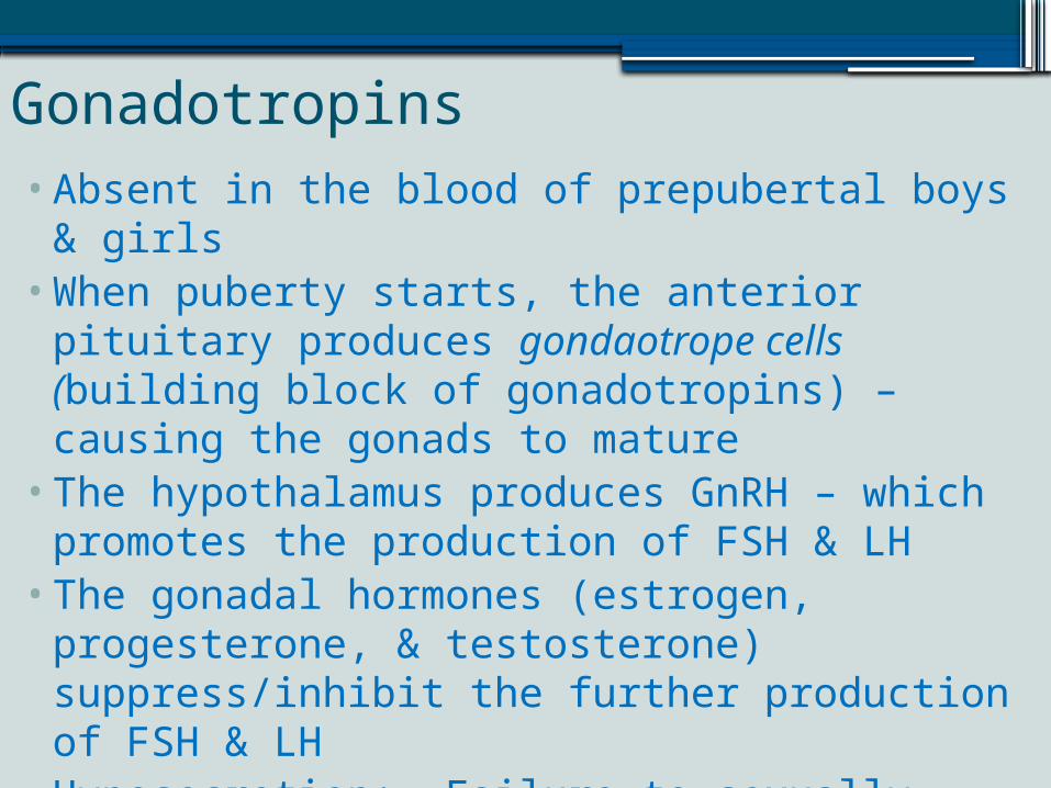
Gonadotropins•Absent in the blood of prepubertal boys & girls•When puberty starts, the anterior pituitary
produces gondaotrope cells (building block of gonadotropins) – causing the gonads to mature
•The hypothalamus produces GnRH – which promotes the production of FSH & LH
•The gonadal hormones (estrogen, progesterone, & testosterone) suppress/inhibit the further production of FSH & LH
•Hyposecretion: Failure to sexually mature•Hypersecretion: No important effects
Prolactin• PRL – protein hormone/similar to growth hormone• Produces by lactotropes – stimulates the gonads of
some mammals•Well-documented – production of breastmilk •Evidence that PRL enhances testosterone production• PRH released by hypothalamus to stimulate
prolactin production▫PIH (Prolactin-inhibiting hormone) IS dopamine –
prevents prolactin secretion In males, PIH predominates, but in women, prolactin
levels rise and fall with estrogen levels Low estrogen stimulates PIH release & high estrogen
promotes more prolactin production
Prolactin•Brief rise in prolactin levels accounts for breast
tenderness & swelling just before menstruation▫Since the PRL production is so brief, no milk is
produced• In pregnancy, prolactin rises dramatically in the
last trimester and milk production begins▫Fun fact: prolactin levels can remain high as much
as two years after breastfeeding ceases.•Hyposecretion: poor milk production in nursing
women•Hypersecretion: Galactorrhea, cessation of mense
in females, impotence and gynecomastia in males
Posterior Pituitary Hormones•Comprised largely of axons of hypothalamic
neurons•Stores oxytocin & antidiuretic hormone (ADH)•These hormones are left “on demand”, when
stimulated by nerve impulses from the hypothalamus
•ADH & oxytocin are protein based hormones▫Almost identical molecularly
VERY different functionally•ADH influences water balance•Oxytocin stimulates the contraction of smooth
muscle
Oxytocin•Released in significantly high amounts during childbirth
& nursing women•Oxytocin receptors peak near the end of pregnancy.•Stretching of the uterus and cervix as birth approaches
sends sensory impulses directly to the hypothalamus▫Hypothalamus makes more oxytocin and raises the blood
level of oxytocin Higher blood levels of oxytocin – expulsive contractions of
labor gain momentum & end with labor•Oxytocin triggers milk ejection (“let down”) in women
whose breasts actively produce milk in response to prolactin▫Positive feedback – as demand for milk increases, MORE
oxytocin is released, instead of being inhibited
Oxytocin•Synthetic oxytocin – Pitocin – can be used to
artificially progress labor▫Less frequently, oxytocics given to stop
uterine/vaginal bleeding post-delivery• In non-lactating females, the non-pregnant &
males:▫Potent peptide plays a role in sexual arousal,
when the body is primed for reproduction Responsible in satisfaction in the sexual interaction
▫Overall, it is now readily known as the “attachment” hormone.
Antidiuretic Hormone (Vasopressin)• Diuresis: urine production• ADH: Inhibits or prevents urine formation▫ Prevents wide swings in water balance
Helps to avoid water overload or water dehydration• Hypothalamic neurons called osmoreceptors continually
monitor solute & water concentration of the blood• When solutes make blood too concentrated▫ Ex) excessive perspiration, inadequate liquid intake, repeated
vomitting Osmoreceptors transmit excitatory impulses to the hypothalamic
neurons to release ADH This will tell the kidneys to reabsorb water into the bloodstream and
produce less urine
• When solute concentration declines, osmoreceptors are depolarized, stopping ADH production
• ADH can also be triggered by pain, low blood pressure, and certain drugs: nicotine, morphine and barbiturates (mild sedation to anesthesia)
ADH - Vasopression•Hyposecretion: Diabetes insipidus ▫ Characterized by excessive thirst and excretion of large
amounts of severely diluted urine, with reduction of fluid intake having no effect on the concentration of the urine.
•Drinking alcohol inhibits ADH = copious urine output• “hangover” – dehydrating effect of alcohol consumption
from suppression of ADH production•Diuretic drugs antagonize the effects of ADH and cause
water to be flushed from the body▫Used to manage hypertension, edema (retention of fluids in
tissues), typical in congestive heart failure• In high concentrations – ADH causes vasoconstriction –
raising BP▫Helpful in situations like severe blood loss
Thyroid Gland•Butterfly shape gland in the anterior neck, just
inferior to the larynx, on the trachea•Two lobes – connected by isthmus (piece of
tissue)• Internally:▫Composed of hollow, spherical follicles
Cuboidal & squamous cells – produce thyroglobulin▫Central cavity produces colloid, amber sticky
material that stores iodine▫Parafollicular cells: produce calcitonin
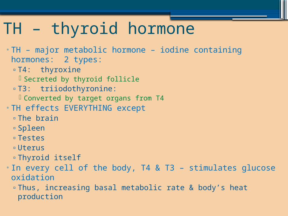
TH – thyroid hormone• TH – major metabolic hormone – iodine containing hormones:
2 types:▫T4: thyroxine
Secreted by thyroid follicle▫T3: triiodothyronine:
Converted by target organs from T4• TH effects EVERYTHING except▫The brain▫Spleen▫Testes▫Uterus▫Thyroid itself
• In every cell of the body, T4 & T3 – stimulates glucose oxidation▫Thus, increasing basal metabolic rate & body’s heat production
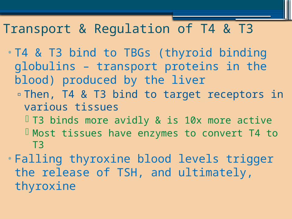
Transport & Regulation of T4 & T3•T4 & T3 bind to TBGs (thyroid binding globulins
– transport proteins in the blood) produced by the liver▫Then, T4 & T3 bind to target receptors in various
tissues T3 binds more avidly & is 10x more active Most tissues have enzymes to convert T4 to T3
•Falling thyroxine blood levels trigger the release of TSH, and ultimately, thyroxine
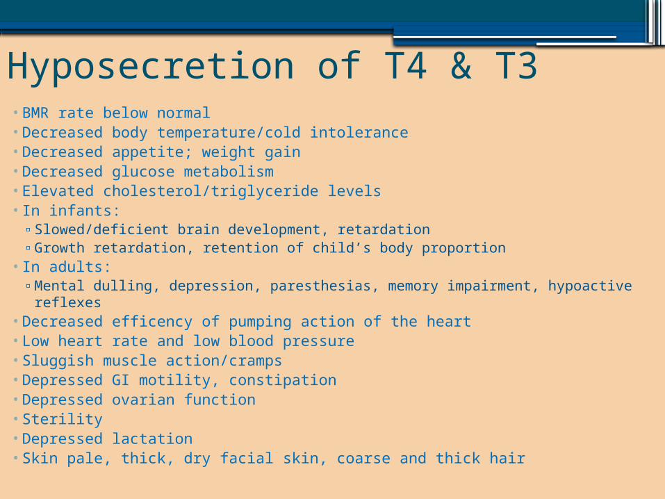
Hyposecretion of T4 & T3• BMR rate below normal• Decreased body temperature/cold intolerance• Decreased appetite; weight gain• Decreased glucose metabolism• Elevated cholesterol/triglyceride levels• In infants:
▫ Slowed/deficient brain development, retardation▫ Growth retardation, retention of child’s body proportion
• In adults:▫ Mental dulling, depression, paresthesias, memory impairment, hypoactive reflexes
• Decreased efficency of pumping action of the heart• Low heart rate and low blood pressure• Sluggish muscle action/cramps• Depressed GI motility, constipation• Depressed ovarian function• Sterility• Depressed lactation• Skin pale, thick, dry facial skin, coarse and thick hair
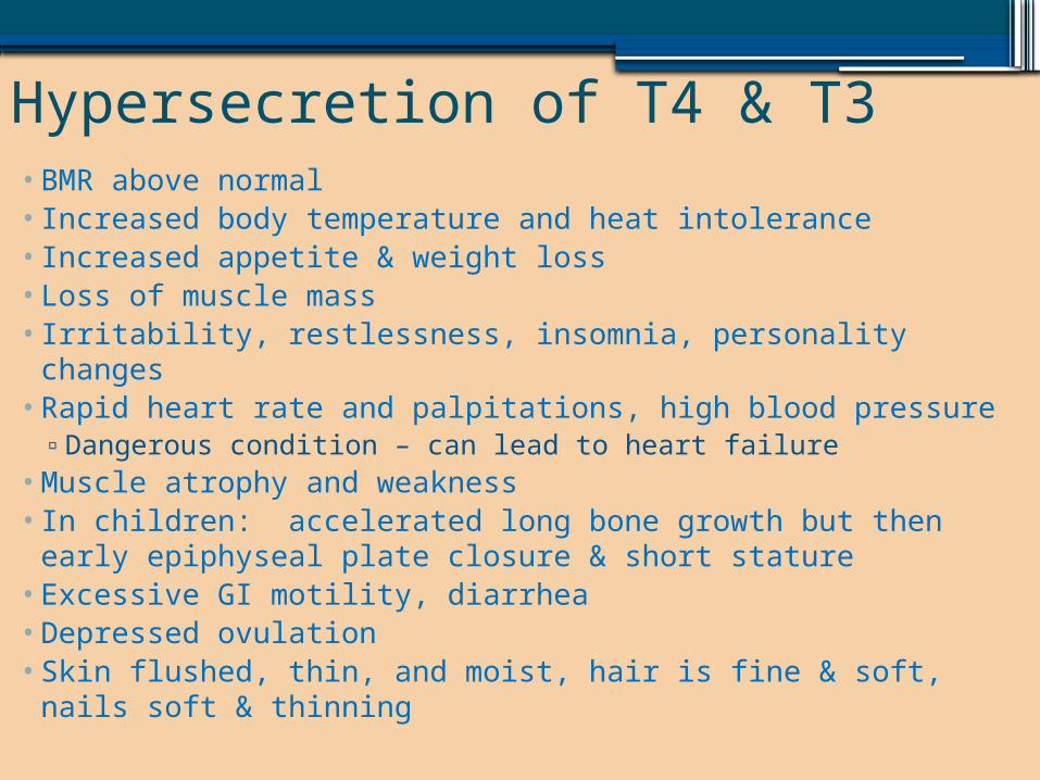
Hypersecretion of T4 & T3•BMR above normal• Increased body temperature and heat intolerance• Increased appetite & weight loss• Loss of muscle mass• Irritability, restlessness, insomnia, personality changes•Rapid heart rate and palpitations, high blood pressure▫Dangerous condition – can lead to heart failure
•Muscle atrophy and weakness• In children: accelerated long bone growth but then early
epiphyseal plate closure & short stature• Excessive GI motility, diarrhea•Depressed ovulation• Skin flushed, thin, and moist, hair is fine & soft, nails soft &
thinning
Calcitonin• Polypeptide hormone produced by parafollicular cells▫Lowers blood calcium
Direct antagonist to parathyroid hormone (PTH) which raises blood calcium
• Targets the skeleton▫ It inhibits osteoclast activity▫Stimulates calcium uptake and incorporation into the bone
matrix• Excessive blood calcium (over 20%) act as a humoral
stimulus for calcitonin release▫An extremely rapid process
• In children, calcitonin plays important role when skeleton is growing quickly
• In adults – weak hypocalcemic agent
Parathyroid Glands•Usually 4 glands on the posterior aspect of the
thyroid gland▫The parathyroid’s glandular cells are arranged in
thick branching cords containing oxyphil cells and large numbers of chief cells Chief cells – secrete PTH – parathyroid hormone
•PTH – protein hormone▫Triggered by falling blood calcium levels▫Inhibited by hypercalcemia
•3 target organs▫Skeleton, kidneys & intestines
PTH•Stimulates osteoclasts to digest some bony matrix to
increase blood calcium concentration•Enhances reabsorption of calcium by the kidneys• Increases absorption of calcium by intestinal mucosal
cells▫Enhanced by PTH’s vitamin D activation – better
calcium absorption For Vitamin D to work, the kidneys must turn it into
calcitriol – this is stimulated by the production of PTH•Stable calcium levels are important for:▫Nerve impulses, muscle contractions, blood clotting
•Hyposecretion: tetany, spasms of the larynx, respiratory paralysis, death
Adrenal (Suprarenal) Glands•Pyramid shaped organs perched atop the
kidneys – cushioned in fat▫Two glands in one
Adrenal medulla – more like nervous tissue than a gland A part of the sympathetic nervous system
Adrenal cortex – bulk of glandular tissue Encapsulates the medulla
•Medulla & cortex produce different hormones▫Both sets of hormones help cope with “extreme”
(stressful) situations•Adrenal glands
Adrenal Cortex•Synthesized from cholesterol – about 24 steroid
hormones are collectively called corticosteroids
•Mineralocorticoids ▫Regulation of electrolyte concentration (mineral
salts: sodium & potassium)▫Sodium is essential for homeostasis
Excessive sodium intake and retention cause high BP
▫Aldosterone – 95% mineralocorticoids produced Maintaining sodium balance is primary goal Reduces excretion of sodium from body Target: distal tubules of kidneys – stimulates
reabsorption of sodium ions from forming urine into the bloodstream
Aldosterone•Aldosterone also enhances sodium reabsorption
from perspiration, saliva & gastric juice▫Crucial for maintaining normal blood flow & BP
•Aldosterone’s effects are brief (about 20 mintues) ▫Therefore, electrolyte balance can be precisely
controlled and monitored continually•Secretion stimulated by:▫Rising blood levels of potassium▫Decreasing blood volume▫Decreasing BP
•Reverse conditions inhibit aldosterone secretion
Aldosterone•Hypersecretion: aldosteronism: results from
adrenal neoplasms▫Neoplasms = growths (both malignant & benign)▫Problems that arise: edema, accelerated
excretion of potassium ions Extreme potassium loss – neurons are
unresponsive, muscle weakness/paralysis may occur
•Hyposecretion: Addison’s disease▫Results from deficient mineralocorticoid &
glucocorticoid release
Regulation of Aldosterone•Renin-angiotensin system▫Major regulator of aldosterone
Specialized cells in the kidneys become “excited” when blood pressure/blood volume drops
Kidneys release renin into the bloodstream Renin reacts with angiotensinogen
Triggers an enzymatic cascade reaction to produce angiotensin II▫Angiotensin II stimulates aldosterone to be released by the adrenal
cortex▫Angiotensin II has widespread effects on BP
•Plasma concentrations of sodium & potassium▫Increased potassium & decreased sodium are
stimulatory▫Opposite conditions are inhibitory
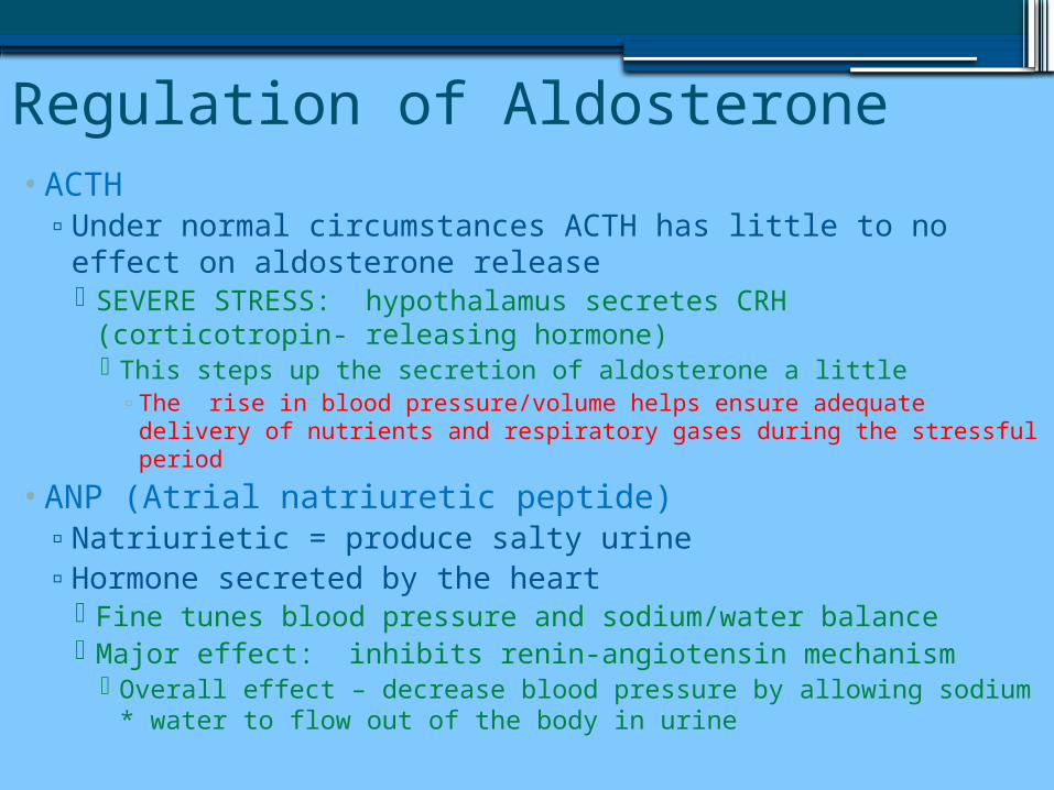
Regulation of Aldosterone•ACTH▫Under normal circumstances ACTH has little to no effect
on aldosterone release SEVERE STRESS: hypothalamus secretes CRH
(corticotropin- releasing hormone) This steps up the secretion of aldosterone a little▫The rise in blood pressure/volume helps ensure adequate delivery
of nutrients and respiratory gases during the stressful period
•ANP (Atrial natriuretic peptide)▫Natriurietic = produce salty urine▫Hormone secreted by the heart
Fine tunes blood pressure and sodium/water balance Major effect: inhibits renin-angiotensin mechanism
Overall effect – decrease blood pressure by allowing sodium * water to flow out of the body in urine
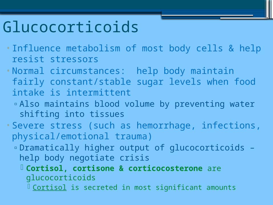
Glucocorticoids• Influence metabolism of most body cells & help resist
stressors•Normal circumstances: help body maintain fairly
constant/stable sugar levels when food intake is intermittent ▫Also maintains blood volume by preventing water
shifting into tissues•Severe stress (such as hemorrhage, infections,
physical/emotional trauma)▫Dramatically higher output of glucocorticoids – help
body negotiate crisis Cortisol, cortisone & corticocosterone are
glucocorticoids Cortisol is secreted in most significant amounts
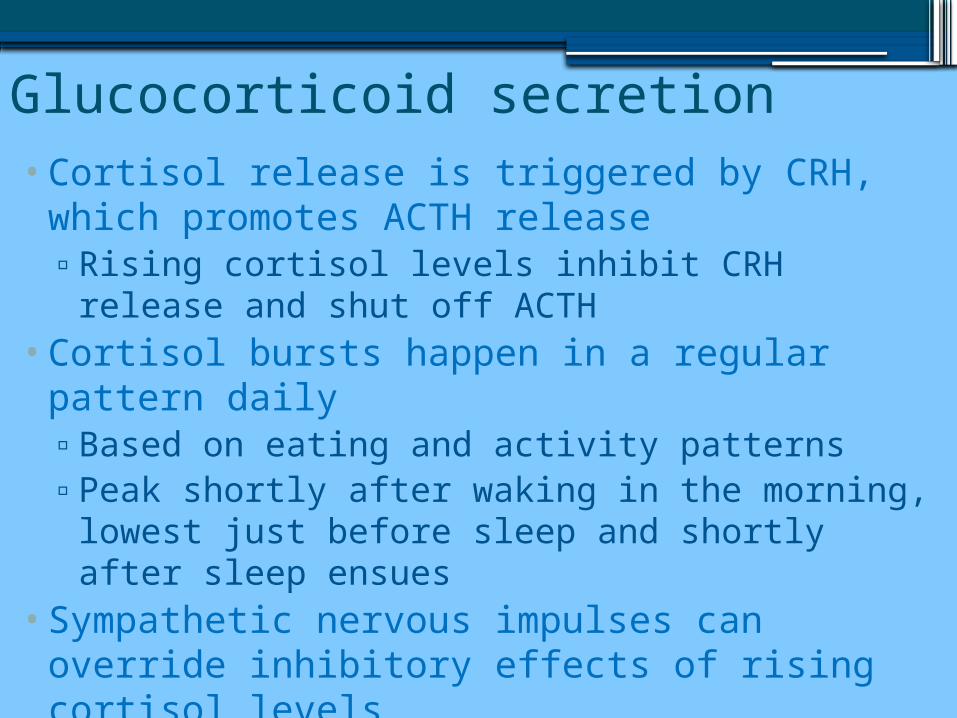
Glucocorticoid secretion•Cortisol release is triggered by CRH, which
promotes ACTH release▫Rising cortisol levels inhibit CRH release and shut
off ACTH•Cortisol bursts happen in a regular pattern daily▫Based on eating and activity patterns▫Peak shortly after waking in the morning, lowest
just before sleep and shortly after sleep ensues•Sympathetic nervous impulses can override
inhibitory effects of rising cortisol levels▫The resulting increase in ACTH causes an
outpouring of cortisol from the adrenal cortex.
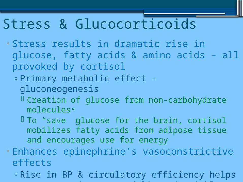
Stress & Glucocorticoids•Stress results in dramatic rise in glucose, fatty
acids & amino acids – all provoked by cortisol▫Primary metabolic effect – gluconeogenesis
Creation of glucose from non-carbohydrate molecules
To “save” glucose for the brain, cortisol mobilizes fatty acids from adipose tissue and encourages use for energy
•Enhances epinephrine’s vasoconstrictive effects▫Rise in BP & circulatory efficiency helps ensure
nutrients are delivered quickly to cells
Glucocorticoids•Ideal amounts of glucocorticoids promote normal function▫However:
Excessive glucocorticoids: Depress cartilage and bone formation Inhibit inflammation & prevent vasodilation Depress the immune system Promote changes in cardiovascular,
gastrointestinal and neural functioning
Hypersecretion & Hyposecretion•Hypersecretion helps treat chronic
inflammatory diseases like RA, or allergic responses▫May relieve some symptoms, also causes
undesirable effects Cushing’s disease:
Causes: ACTH tumor in pituitary, malignancy in lungs, pancreas, kidneys or tumor of the adrenal cortex▫Most often: pharmacological doses of glucocorticoids
(steroids) Characterized by persistent hyperglycemia, dramatic
loss in muscle mass, water/salt retention, leading to hypertension & edema
•Hyposecretion: Addison’s disease▫Weight loss, glucose & sodium levels drop &
potassium rises▫Severe dehydration & hypotension is common
Gondaocorticoids•Androgens – male hormones – secreted▫Small amounts of female hormones – estrogen –
secreted• Insignificant amount compared to amounts
made by gonads•Unknown significance▫Assumed contribution to onset of puberty▫Possible link of androgens to adult woman’s sex
drive After menopause, may be converted to estrogens
when ovaries no longer produce estrogen•Stimulation of secretion is unknown
Adrenal Medulla• Produce catecholamines – epinephrine & norepinephrine▫Brief responses
• Short term stress – sympathetic nervous system is activated:▫Blood glucose rises▫Blood vessels constrict▫Heart beats faster – raising BP▫Blood is temporarily diverted from nonessential organs to the
brain, heart, and skeletal muscles•Unequal amounts of hormones are released = 80%
epinephrine▫Epinephrine = more potent heart and metabolic activator
Clinical use: heart stimulant & bronchiodilator during asthma attack▫Norepinephrine = greater influence on peripheral
vasoconstriction & blood pressure
Pancreas• Located partially behind the stomach – triangular shaped
gland – both endocrine & exocrine▫Acinar cells – most of the gland
Produce enzyme rich juice that is ducted into small intenstines during food digestion This is the exocrine product
▫Pancreatic islets (islets of Langerhans) Tiny cell clusters that produce hormones
Two groups of hormone producing cells:▫Alpha cells: glucagon synthesizing▫Beta cells: insulin producing
Tiny fuel sensors – secreting glucagon & insulin appropriately during fasting and feeding states
Involved (but independently) in blood glucose regulation▫ Insulin is a hypoglycemic agent▫Glucagon is a hyperglycemic agent
Glucagon•Hyperglycemic hormone▫1 molecule of glucagon can release up to 1 million glucose
molecules into blood• Target: liver▫Breaks down glycogen into glucose (glycogenolysis)▫Synthesis of glucose from lactic acid & gluconeogenesis▫Release glucose into blood by liver cells, causes blood sugar to rise
• Secondary effect: fall in amino acids – liver cells sequester them to make new glucose molecules
• Secretion of glucagon – humoral stimulus▫Falling blood sugar levels stimulate glucagon production▫Eating a high protein meal is also stimulatory
•Glucagon suppression by rising sugar levels and somatostatin•Hypoglycemics: deficient glucagon, persistent low blood sugar
Insulin•Part of a larger molecule called proinsulin –
broken down into smaller useable molecules called insulin▫Particularly the middle of proinsulin
•Main effect: lower blood sugar▫Released just after eating▫Also influences protein & fat metabolism
•Circulating insulin lowers blood sugar by enhancing membrane transport of glucose in muscle & fat cells▫Does NOT accelerate glucose transport in brain,
liver & kidneys They have ready access to glucose
Insulin• Inhibits the breakdown of glycogen into glucose
& the conversion of amino acids & fats to glucose
•Counters any activity that would increase blood glucose
•Once energy needs are met by glucose, glycogen deposits begin to occur▫If excess glucose is still available, fat deposits
occur• Insulin also stimulates amino acid uptake and
protein synthesis in muscle tissue•Sweeps glucose out of the blood, causing it to
be used for energy or converted to other forms (glycogen or fats)
•Promotes protein synthesis or fat storage
Beta Cells•Beta cells are stimulated to secrete insulin by rising blood
glucose levels▫Rising amino acid & fatty acid levels in the blood can also
trigger insulin release• Lowered blood sugar suppresses insulin production• Indirect stimulation (when blood glucose drops)▫Glucagon▫Epinephrine▫GH▫Thyroxine
• Somatostatin depresses insulin release• Therefore – both humoral & hormonal stimulation▫Humoral stimulation: direct: blood glucose rising▫Hormonal stimulation: indirect: hormones that cause blood
sugar to increase
The Gonads• Produce steroidal sex hormones – identical to adrenal
cortex hormones• Paired ovaries produce estrogens & progesterone▫Located in abdominopelvic cavity
Estrogen: maturation of reproductive organs and appearance of secondary sex characteristics in females at puberty
Together with Progesterone: promote breast development & cyclic changes in uterine mucosa (menstrual cycle)
• Testes: extra-abdominal skin sac called scrotum ▫Produce sperm & male sex hormones, primarily
testosterone During puberty – testosterone: initiates maturation of male
sex organs and appearance of secondary sex characteristics Necessary for normal sperm production Maintains mature functioning of male reproductive organs
The Pineal Gland• Tiny, cone-shaped gland▫Located on the roof of third ventricle in diencephalon
•Secretory cells called pinealocytes▫Between clusters of cells: calcium salts
•Mysterious endocrine function▫Only secretory product is melatonin
Rise and fall in a daily cycle Peak at night, making us drowsy – lowest in daylight hours,
around noon
• Indirectly signaled from visual cues▫ Intensity and duration of sunlight inhibits melatonin
secretion• In children, antigonadotropic effect – inhibits precocious
puberty – delaying sexual maturation
Other hormone-producing structures•The placenta sustains a fetus during
pregnancy by secreting several hormones that influence the course of pregnancy▫Estrogens▫Progesterones▫hCG (Human chorionic gonadotropin)
•Kidneys secrete erythropoietin▫Signals bone marrow to increase production of
red blood cells