validation of zero te mra in the characterization of
TRANSCRIPT
ORIGINAL RESEARCHADULT BRAIN
Validation of Zero TE–MRA in the Characterization ofCerebrovascular Diseases: A Feasibility Study
X S. Shang, X J. Ye, X W. Dou, X X. Luo, X J. Qu, X Q. Zhu, X H. Zhang, and X J. Wu
ABSTRACT
BACKGROUND AND PURPOSE: Zero TE–MRA is less sensitive to field heterogeneity, complex flow, and acquisition noise. This studyaimed to prospectively validate the feasibility of zero TE–MRA for cerebrovascular diseases assessment, compared with TOF-MRA.
MATERIALS AND METHODS: Seventy patients suspected of having cerebrovascular disorders were recruited. Sound levels were esti-mated for each MRA subjectively and objectively in different modes. MRA image quality was estimated by 2 neuroradiologists. The degreeof stenosis (grades 0 – 4) and the z-diameter of aneurysms (tiny group �3 mm and large group �3 mm) were measured for furtherquantitative analysis. CTA was used as the criterion standard.
RESULTS: Zero TE–MRA achieved significantly lower subjective perception and objective noise reduction (37.53%). Zero TE–MRA imagesshowed higher signal homogeneity (3.29 � 0.59 versus 3.04 � 0.43) and quality of venous signal suppression (3.67 � 0.47 versus 2.75 � 0.46).The intermodality agreement was higher for zero TE–MRA than for TOF-MRA (zero TE, 0.90; TOF, 0.81) in the grading of stenosis. ZeroTE–MRA had a higher correlation than TOF-MRA (zero TE, 0.84; TOF, 0.74) in the tiny group and a higher consistency with CTA (intraclasscorrelation coefficient, 0.83; intercept, �0.5084 –1.1794; slope �0.4952 to �0.2093) than TOF-MRA (intraclass correlation coefficient, 0.64;intercept, 0.7000 –2.6133; slope �1.0344 to �0.1923). Zero TE–MRA and TOF-MRA were comparable in the large group. Zero TE–MRA hadmore accurate details than TOF-MRA of AVM and Moyamoya lesions.
CONCLUSIONS: Compared with TOF-MRA, zero TE–MRA achieved more robust performance in depicting cerebrovascular diseases.Therefore, zero TE–MRA was shown to be a promising MRA technique for further routine application in the clinic in patients withcerebrovascular diseases.
ABBREVIATIONS: ASL � arterial spin-labeling; AVM � arteriovenous malformation; MRA � magnetic resonance angiography; CTA � computed tomographyangiography; CE � contrast-enhanced; TOF � time-of-flight; zTE � zero echo time; MIP � maximum intensity projection; VR � volume rendering; MCA � middlecerebral artery; ICA � internal carotid artery
Cerebrovascular diseases are the main causes of ischemic or
hemorrhagic incidents within brain tissues and can lead to
neurologic deficits or even death. Imaging of the cerebral vascu-
lature plays a pivotal role in the initial diagnosis of cerebrovascu-
lar disorders, such as steno-occlusive arterial disease, cerebral an-
eurysm, AVM, and Moyamoya disease, in addition to treatment
decisions and follow-up evaluation.1,2 Although patients sus-
pected of having cerebrovascular disease are best diagnosed with
DSA or CTA due to their superior angiogram quality,3,4 these
protocols place patients at risk of ionizing radiation and contrast-
induced renal insufficiency.
MRA, a noninvasive and radiation-free imaging technique,
has been proposed as a promising alternative for vascular disease
assessment.5 In addition to conventional angiographic tech-
niques, such as TOF-MRA and contrast-enhanced MRA, MRA is
also able to use the principles of arterial spin-labeling (ASL) strat-
egies.6 ASL-MRA was introduced as a noninvasive approach that
uses inflowing blood as an endogenous contrast agent. Recent
technical advances in ASL-MRA have dramatically expanded its
clinical application. In particular, a newly developed zero TE ra-
dial ASL-MRA (zTE-MRA) method that integrates both a contin-
uous ASL strategy and a zTE radial acquisition readout has dem-
Received April 29, 2019; accepted after revision July 2.
From the Department of Radiology (S.S., J.Y., X.L., Q.Z., H.Z., J.W.), Clinical MedicalCollege, Yangzhou University, Yangzhou, Jiangsu, China; and MR Research China(W.D., J.Q.), GE Healthcare, Beijing China.
This work was supported by a grant from the National Natural Science Foundationof China (NSFC 81571652), “333 Project” of Jiangsu Province (BRA2017154), Scienceand technology project of Yangzhou (YZ2018059).
Please address correspondence to Jingtao Wu, MD, Department of Radiology, Clin-ical Medical College, Yangzhou University, No. 98, Nantong West Road, Guangling,Yangzhou 225009, Jiangsu, China; e-mail: [email protected]
Indicates open access to non-subscribers at www.ajnr.org
Indicates article with supplemental on-line photos.
http://dx.doi.org/10.3174/ajnr.A6173
1484 Shang Sep 2019 www.ajnr.org

onstrated potential for the follow-up assessment of coiled
aneurysms,7-10 highlighting the clinical value of this approach.
However, it remains unclear whether this technique is also
useful for the assessment of other cerebrovascular disorders.
Meanwhile, the acoustic noise reduction of zTE-MRA has still not
been assessed systematically, though this approach was described
as silent.7-9 To explore this issue, we applied this novel, nonen-
hanced MRA approach in a clinical population with suspected
cerebrovascular diseases. Sound-level assessment and imaging
properties were systematically assessed and compared with those
obtained using TOF-MRA. In addition, CTA measurements were
also performed and used as the criterion standard.
MATERIALS AND METHODSPatient CohortA single-center prospective study was performed with the ap-
proval from local institutional review board of Clinical Medical
College, Yangzhou University (2017-KY081). Patients suspected
of having cerebrovascular disorders were enrolled in this study
from May 2017 to February 2018. Cerebrovascular disorders were
suspected for the first time by 2 experienced neurologists (Yong
Zhen, with 24 years of experience; Long Yu, with 30 years of ex-
perience) on the basis of clinical symptoms (eg, dizziness, head-
ache, paralysis, paresthesia, conscious disturbance, dysmnesia,
dementia, and other psychiatric symptoms) and routine head CT
examinations (eg, infarction, hemorrhage, encephalatrophy, and
other lesions of abnormal density). Written informed consent was
obtained from each participant before MR imaging.
The following criteria for patient exclusion were applied in this
study: 1) contraindication for MR imaging, including severe
claustrophobia, ferromagnetic foreign bodies, or electronic im-
plants; 2) other intracranial lesions, such as tumor or hydroceph-
alus (traumatic or tumor-related); 3) an acute or subacute win-
dow for a vascular event (relatively large acute cerebral infarction
or acute cerebral infarction of the brain stem on DWI); 4) addi-
tional conditions that could potentially cause severe movement or
being uncooperative, such as psychiatric disorders or congenital/
acquired deafness; 5) estimated glomerular filtration rate �60
mL/min/1.73 m2; and 6) younger than 18 years of age. Finally, 69
patients (38 men and 31 women; range, 20 – 83 years of age; mean
age, 60.04 � 13.70 years) were enrolled in this study. One patient
had subarachnoid hemorrhage caused by aneurysm and under-
went only MRA because of an allergy to iodinated contrast media.
The MRA images (On-line Fig 1) were thus used only for image-
quality measurement. Finally, both MRA and CTA were success-
fully performed in 68 patients.
Image AcquisitionMR imaging measurements (DWI, MRA) were obtained 24 hours
before CTA acquisition on a 3T MR imaging scanner (Discovery
MR750w; GE Healthcare, Milwaukee, Wisconsin) using a 24-
channel head-neck united coil. The corresponding scan parame-
ters and source images are shown in Table 1 and On-line Fig 2.
CTA was acquired on a 64-slice spiral CT scanner (LightSpeed
VCT; GE Healthcare) using a contrast medium (iodixanol, Visi-
paque, 320 mg I/mL; GE Healthcare, Piscataway, New Jersey).
The scanning protocol was described in a previously reported
study.11 The corresponding scan parameters are shown in Table 2.
Sound Assessment of MRA MeasurementsMRAs were performed in random order and separated by a
5-minute interval during scanning.
After each measurement, the patient was asked to rate the
sound experience subjectively on a 5-point scale12: 0 � no noise,
1 � mild noise, 2 � moderate noise, 3 � loud noise, 4 � very loud
noise, and 5 � unacceptable noise/cancellation of MR imaging.
Objective measurements were obtained in an ambient mode
(without scanning) and scanning mode using a sound-level meter
(Type AS804; Smart Sensor, Guangdong, China) that was placed
1 m from the gantry. Each measurement period lasted for 30 sec-
onds, and measurements were repeated 5 times in different
directions.
Image AnalysisThe maximum intensity projection (MIP) and volume-rendering
(VR) methods were used to reconstruct both MRA and CTA data
by a neuroradiologist (Q.Z., with 9 years of experience) using a
commercially available dedicated workstation (Advantage Work-
station, software Version 4.6; GE Healthcare) We made the fol-
lowing standard projections of MIP and VR: 1) coronal view, 2)
lateral view, and 3) the optimal projection used at the lesions.
The corresponding CTA images (source image, MIP, and VR)
were independently and blindly evaluated by 2 experienced neu-
roradiologists (observer C, J.W., with 27 years of experience; ob-
server D, X.L., with 16 years of experience) at separate time points.
The corresponding MRA images (source image, MIP, and VR)
were independently and blindly evaluated by 2 experienced neu-
roradiologists (observer A, H.Z., with 25 years of experience; ob-
Table 1: Parameters of MRA sequences
TR/TE(ms)
FlipAngle
FOV(cm) Matrix
Thickness(mm) Slices NEX
Bandwidth(kHz) Slabs
LabelDuration
(sec) CoverageTime
(min:sec)zTE-MRA 862/0.016 3o 15 � 15 166 � 166 1.2 320 1 31.25 – 2 Calvarium-mandible 5:48TOF-MRA 25/3.4 15o 30 � 24 320 � 256 1.4 256 1 41.67 3 – Cingulate cortex–
mesencephalon5:08
Note:— –indicates no data available; TR, repetition time; TE, echo time; FOV, field of view; NEX, number of excitation.
Table 2: Parameters of CTA sequences
Collimation PitchFOV(cm)
GantryRotationTime (ms)
Thickness(mm) Slices
Tube Voltage(kV�peak)
TubeCurrent(mAs)
Dose-LengthProduct
(mGy × cm) CoverageCTA 128 � 0.625 0.758 17 � 17 400 0.6 715 100–120 100–450 220–608 Aortic arch–vertex
AJNR Am J Neuroradiol 40:1484 –90 Sep 2019 www.ajnr.org 1485

server B, J.Y., with 20 years of experience)) at separate time points.
All images were scored to determine signal homogeneity, lesion
conspicuity, quality of venous signal suppression, and diagnostic
confidence. A previously reported 4-point scale was applied13,14:
4 � excellent (excellent-quality diagnostic information with a
clearly detailed vascular architecture, no artifacts), 3 � good
(good-quality diagnostic information with adequate delineation
of the vascular architecture, minimal artifacts), 2 � poor (poor-
quality diagnostic information with ordinary delineation of the
vascular architecture, moderate artifacts), and 1 � not visible (al-
most no signal of the vascular architecture, severe artifacts). The
time interval between the qualitative analyses was 4 weeks. In
cases of disagreement, a consensus was established between
observers.
Observers were also asked to record additional vascular dis-
ease findings based on MRA and CTA images. The measurements
were performed on the VR images. For further quantitative anal-
ysis, the degree of stenosis was graded according to the standard
from NASCET: 0 for �9%, one for �30%, two for 30%– 69%,
three for 70%–99%, and 4 for 100%. In addition, aneurysms were
divided into tiny (�3 mm) and large (�3 mm) groups according
to the long-axis diameters of the lesions as determined on CTA
images.
Statistical AnalysisAll statistical analyses were performed using SPSS 19.0 software
(IBM, Armonk, New York).
The differences in subjective noise-level and image-quality
ratings between MRAs were analyzed using Wilcoxon signed rank
tests. One-way analysis of variance tests and least significant dif-
ference tests were used to estimate the difference in the objective
sound-level measurements between MRAs.
For the stenosis analysis, the Spearman correlation coefficient
(r) was used to evaluate the correlation of each MRA to CTA.
Weighted � statistics were used to assess interobserver agreement
and intermodality agreement between the results obtained with
each MRA and CTA.
The aneurysm measurements obtained on MRA were corre-
lated with those obtained on CTA using a Pearson analysis. Bland-
Altman analysis and intraclass correlation coefficients were used
to test the consistency of measurements obtained using the MRAs
and CTAs. A weighted � value or an intraclass correlation coeffi-
cient higher than 0.81 was interpreted as excellent agreement,
while 0.61– 0.81 indicated good, 0.41– 0.60 indicated moderate,
0.21– 0.40 indicated fair, and �0.21 indicated poor agreement.
The 95% confidence interval was calculated. A P value � .05 was
considered statistically significant.
RESULTSPatient CohortCTA revealed 32 steno-occlusive cases in 26 patients, 31 cerebral
aneurysm cases in 22 patients, 11 steno-occlusive cases and 8 ce-
rebral aneurysm cases in 8 patients, 2 patients with AVMS, 3 pa-
tients with Moyamoya disease, and 7 patients without evident
vascular diseases. The distribution of the steno-occlusive arteries
(cases) was as follows: middle cerebral artery (MCA), 18; internal
carotid artery (ICA), 10; posterior cerebral artery, 6; anterior ce-
rebral artery, 5; vertebral artery, 2; and basilar artery 2. The dis-
tribution of the aneurysms was as follows: posterior communicat-
ing artery, 14; anterior communicating artery, 10; ICA, 7; MCA, 4;
anterior cerebral artery, 2; posterior inferior cerebellar artery, 1;
and vertebral artery, 1. The 2 AVM lesions were located in the left
MCA and right ICA (1 each). The 3 Moyamoya lesions were lo-
cated at the origin of a unilateral MCA.
Sound-Level AssessmentMean sound-level perception based on subjective sound experi-
ence was significantly lower on zTE-MRA than on TOF-MRA
(1.36 � 0.48 versus 4.17 � 0.75, P �
.001). The intensities of sound measured
in different modes are shown in Table 3.
Noise levels recorded during zTE-MRA
scanning were 34.85 dB (37.53%) lower
in sound intensity. In addition, there
was a slight increase (3.12 dB, 5.68%,
P � .001) in sound levels over the ambi-
ent mode.
Image-Quality Evaluation ofzTE- and TOF-MRAThe mean scores for signal homogeneity
(3.29 � 0.59 versus 3.04 � 0.43, P � .03)
and the quality of venous signal suppres-
sion (3.67 � 0.47 versus 2.75 � 0.46,
P � .01) were significantly higher for
zTE-MRA than for TOF-MRA. zTE-
MRA was comparable with TOF-MRA
in lesion conspicuity (3.21 � 0.51 versus
Table 3: Intensity of sound levels in different modes for zTE-MRAand TOF-MRA
Mode Sound Level (dB) F PAmbient 54.89 � 0.41a 11,824.06 �.001zTE-MRA 58.01 � 0.32ab
TOF-MRA 92.86 � 0.64b
a A significant difference compared with the TOF-MRA mode.b A significant difference compared with the ambient mode.
Table 4: Cross-table of stenosis grade from observers for zTE-MRA (n � 44)a
zTE-MRAA Grade
zTE-MRA B Grade
0 1 2 3 4 Total0 1 0 0 0 0 1 (2.27%)1 0 7 3 0 0 10 (22.73%)2 0 0 14 0 0 14 (31.82%)3 0 0 4 8 0 12 (27.27%)4 0 0 0 1 6 7 (15.91%)Total 1 (2.27%) 7 (15.91%) 21 (47.73%) 9 (20.45%) 6 (13.64%) 44
a Grading criterion: NASCET. Data represent the number of cases. A and B are observers A and B.
Table 5: Cross-table of stenosis grade from observers for TOF-MRA (n � 44)a
TOF-MRAA Grade
TOF-MRA B Grade
0 1 2 3 4 Total0 0 0 0 0 0 0 (0.00%)1 1 9 0 0 0 10 (22.73%)2 0 1 8 4 0 13(29.55%)3 0 0 4 7 0 11 (25.00%)4 0 0 0 2 8 10 (22.73%)Total 1 (2.27%) 10 (22.73%) 12 (27.27%) 13 (29.55%) 8 (18.18%) 44
a Grading criterion: NASCET. Data represent the number of cases. A and B are observers A and B.
1486 Shang Sep 2019 www.ajnr.org
3.06 � 0.45, P � .08) and diagnostic confidence (3.23 � 0.46
versus 3.05 � 0.44, P � .06). Additionally, the peripheral vessels
presented on zTE-MRA were less well-defined than on
TOF-MRA.
Quantitative Analysis of zTE- and TOF-MRA ImagesThe interobserver agreement for CTA was excellent (weighted �,
0.94; 95% CI, 0.90 – 0.99). CTA revealed eleven (25%) cases with
grade 1, thirteen (29.55%) with grade 2, twelve (22.27%) with
grade 3, and seven (15.91%) with grade 4. One case (2.27%) was
wrongly categorized as grade 1 by TOF-MRA, while zTE-MRA,
consistent with CTA, showed that there was no steno-occlusive
lesion (On-line Fig 3).
The correlations of the results obtained using each MRA with
CTA results were both high (zTE, r � 0.94; TOF, r � 0.92). There
was excellent interobserver agreement for zTE-MRA (weighted �:
0.92; 95% CI, 0.87– 0.98) and TOF-MRA (weighted �: 0.89; 95%
CI, 0.84 – 0.96). The intermodality agreement between zTE-MRA
and CTA was excellent (weighted �: 0.90; 95% CI, 0.82– 0.99), and
it was good (weighted �: 0.81; 95% CI, 0.71– 0.92) between TOF-
MRA and CTA. These data are shown in Tables 4 – 6. Some le-
sions, especially at a critical point between 2 grades, may have
been overestimated on TOF-MRA, while on zTE-MRA, the ste-
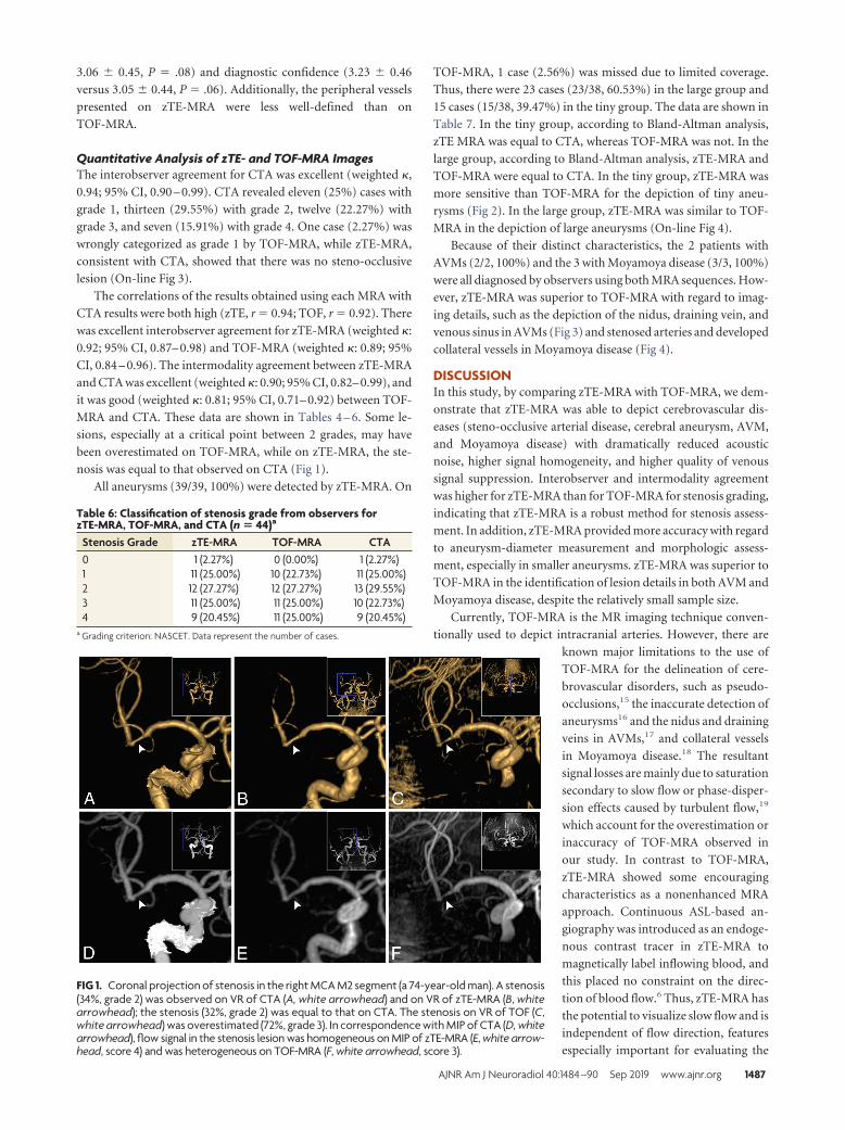
nosis was equal to that observed on CTA (Fig 1).
All aneurysms (39/39, 100%) were detected by zTE-MRA. On
TOF-MRA, 1 case (2.56%) was missed due to limited coverage.
Thus, there were 23 cases (23/38, 60.53%) in the large group and
15 cases (15/38, 39.47%) in the tiny group. The data are shown in
Table 7. In the tiny group, according to Bland-Altman analysis,
zTE MRA was equal to CTA, whereas TOF-MRA was not. In the
large group, according to Bland-Altman analysis, zTE-MRA and
TOF-MRA were equal to CTA. In the tiny group, zTE-MRA was
more sensitive than TOF-MRA for the depiction of tiny aneu-
rysms (Fig 2). In the large group, zTE-MRA was similar to TOF-
MRA in the depiction of large aneurysms (On-line Fig 4).
Because of their distinct characteristics, the 2 patients with
AVMs (2/2, 100%) and the 3 with Moyamoya disease (3/3, 100%)
were all diagnosed by observers using both MRA sequences. How-
ever, zTE-MRA was superior to TOF-MRA with regard to imag-
ing details, such as the depiction of the nidus, draining vein, and
venous sinus in AVMs (Fig 3) and stenosed arteries and developed
collateral vessels in Moyamoya disease (Fig 4).
DISCUSSIONIn this study, by comparing zTE-MRA with TOF-MRA, we dem-
onstrate that zTE-MRA was able to depict cerebrovascular dis-
eases (steno-occlusive arterial disease, cerebral aneurysm, AVM,
and Moyamoya disease) with dramatically reduced acoustic
noise, higher signal homogeneity, and higher quality of venous
signal suppression. Interobserver and intermodality agreement
was higher for zTE-MRA than for TOF-MRA for stenosis grading,
indicating that zTE-MRA is a robust method for stenosis assess-
ment. In addition, zTE-MRA provided more accuracy with regard
to aneurysm-diameter measurement and morphologic assess-
ment, especially in smaller aneurysms. zTE-MRA was superior to
TOF-MRA in the identification of lesion details in both AVM and
Moyamoya disease, despite the relatively small sample size.
Currently, TOF-MRA is the MR imaging technique conven-
tionally used to depict intracranial arteries. However, there are
known major limitations to the use of
TOF-MRA for the delineation of cere-
brovascular disorders, such as pseudo-
occlusions,15 the inaccurate detection of
aneurysms16 and the nidus and draining
veins in AVMs,17 and collateral vessels
in Moyamoya disease.18 The resultant
signal losses are mainly due to saturation
secondary to slow flow or phase-disper-
sion effects caused by turbulent flow,19
which account for the overestimation or
inaccuracy of TOF-MRA observed in
our study. In contrast to TOF-MRA,
zTE-MRA showed some encouraging
characteristics as a nonenhanced MRA
approach. Continuous ASL-based an-
giography was introduced as an endoge-
nous contrast tracer in zTE-MRA to
magnetically label inflowing blood, and
this placed no constraint on the direc-
tion of blood flow.6 Thus, zTE-MRA has
the potential to visualize slow flow and is
independent of flow direction, features
especially important for evaluating the
FIG 1. Coronal projection of stenosis in the right MCA M2 segment (a 74-year-old man). A stenosis(34%, grade 2) was observed on VR of CTA (A, white arrowhead) and on VR of zTE-MRA (B, whitearrowhead); the stenosis (32%, grade 2) was equal to that on CTA. The stenosis on VR of TOF (C,white arrowhead) was overestimated (72%, grade 3). In correspondence with MIP of CTA (D, whitearrowhead), flow signal in the stenosis lesion was homogeneous on MIP of zTE-MRA (E, white arrow-head, score 4) and was heterogeneous on TOF-MRA (F, white arrowhead, score 3).
Table 6: Classification of stenosis grade from observers forzTE-MRA, TOF-MRA, and CTA (n � 44)a
Stenosis Grade zTE-MRA TOF-MRA CTA0 1 (2.27%) 0 (0.00%) 1 (2.27%)1 11 (25.00%) 10 (22.73%) 11 (25.00%)2 12 (27.27%) 12 (27.27%) 13 (29.55%)3 11 (25.00%) 11 (25.00%) 10 (22.73%)4 9 (20.45%) 11 (25.00%) 9 (20.45%)
a Grading criterion: NASCET. Data represent the number of cases.
AJNR Am J Neuroradiol 40:1484 –90 Sep 2019 www.ajnr.org 1487

flow in severe stenosis (grade 3), microaneurysms, and collateral
vessels.
Acoustic noise induced by rapid switching of gradient coils
during long scan times is still the main drawback that causes pa-
tient discomfort and movement.20-22 The zTE technique, which
uses a sequence solution, attenuates acoustic noise by reducing
the slew rate and enabling minimal gradient variations.23,24 Al-
ibek et al12 assessed the noise level and image quality of silent
T1-weighted sequences on a clinical 3T MR imaging scanner.
However, whether a similar amount of acoustic noise reduction
could be achieved using zTE-MRA remains unclear because an
extra ASL-based angiography was integrated into this approach.
In this study, patients were much more comfortable during zTE-
MRA than TOF-MRA, and the reduction in sound intensity be-
tween the 2 was comparable with that of a silent T1-weighted
sequence. Thus, zTE-MRA would be an acceptable alternative an-
giography method for these patients.
In addition to acoustic noise attenuation, a zTE inside-out
radial acquisition could also minimize the phase dispersion of the
labeled blood flow signal in the voxel space, thereby making zTE-
MRA insensitive to turbulent flow.9,25 The flow signal inside the
large aneurysms was more homoge-
neous on zTE-MRA than on TOF-MRA
in our study, though the correlations
with CTA were both high and the con-
sistency with CTA was excellent for the
large group.
Susceptibility artifacts, which occur
mostly in regions adjacent to the skull
base or cavernous sinus, always induce
signal loss as a result of air–soft tissue or
bone–soft tissue boundary interfer-
ence.26 Because of its use of a constant
gradient and a long readout duration,zTE-MRA is less susceptible to field in-homogeneity and eddy current and canacquire more homogeneous flow signalsin vascular lesions, such as stenosis oraneurysms in the ICA siphon segment.Moreover, zTE-MRA is less sensitivethan TOF-MRA to motion effects,27 andit therefore minimizes the misregistra-tion and degradation of image qualityand achieves improved diagnosticperformance.
According to the principles of theASL strategy used by zTE-MRA, images
are acquired via subtraction to yield an angiographic image with
nearly zero background.7 The efficiency of postprocessing (ie, VR
or MIP reconstructions) can be improved with automatic sub-
traction, whereas the artifacts caused by skull and brain tissue on
TOF-MRA must be manually removed. Furthermore, zTE-MRA
demonstrated effective background suppression while a search
for aneurysms was performed in the setting of subarachnoid hem-
orrhage in the presence of residual T1-bright blood. An optimal
postlabel delay was set,6,28 ensuring that the labeled bolus flowed
into the target tissue in the imaging region and that the labeled
protons lost their signal before entering the veins. Thus, better
diagnostic image quality was achieved due to an acceptable signal-
to-noise ratio and the elimination of vein contamination. Addi-
tionally, zTE-MRA can detect arteriovenous shunting on the basis
of this technical feature. In this study, we identified a huge AVM
and found that zTE-MRA was capable of revealing its tubular
structures, nidus, and draining veins and even a venous sinus.
Contrast-enhanced MRA is another commonly used noninva-
sive MRA approach performed by administrating a T1-shorten-
ing paramagnetic contrast medium. Despite the controversy re-
lated to its diagnostic accuracy,15,16 contrast-enhanced MRA has
FIG 2. Oblique projection of aneurysms in the right ICA siphon segment (a 63-year-old woman).Two aneurysms were found on VR of CTA (A): The diameters were 3.0 � 3.2 mm (white arrow) and2.4 � 2.1 mm (white arrowhead), respectively. Equal findings were observed on VR of zTE (B, 3.3 �3.5 mm, white arrow; 2.� 2.4 mm, white arrowhead). On VR of TOF-MRA (C), the large one (3.1 �3.4 mm, white arrow) was equal to the one on CTA. However, the tiny one was not evident(1.5 � 1.4 mm, white arrowhead). Concerning MIP of CTA (D, white arrow and white arrowhead),the same results were observed on MIP of zTE-MRA (E, white arrow and white arrowhead) andTOF-MRA (F, white arrow and white arrowhead).
Table 7: Statistical analyses of aneurysms measured on MRA for groupsGroup MRA Sum ra Intercept (95% CI)b Slope (95% CI)b ICC
Group tinyc zTE 15 0.84 �0.5084–1.1794 �0.4952–0.2093 0.83; 95% CI, 0.57–0.94TOF 15 0.74 0.7000–2.6133 �1.0344 to �0.1923 0.64; 95% CI, 0.21–0.86
Group larged zTE 23 0.98 �0.6059–0.2130 �0.02625–0.1200 0.98, 95% CI, 0.97–0.99TOF 23 0.95 �1.1571–0.3062 �0.01380–0.2520 0.95, 95% CI, 0.89–0.98
Note:—ICC indicates intraclass correlation coefficient.a Pearson correlation coefficient.b Intercept and slope are by Bland-Altman analysis.c Group tiny �3 mm.d Group large �3 mm.
1488 Shang Sep 2019 www.ajnr.org

demonstrated some superiorities over TOF-MRA, such as it is lessvulnerable to blood flow disturbances, has a reduced risk of move-ment artifacts, and provides large coverage for additional find-ings, while TOF-MRA also has some advantages over contrast-enhanced MRA in that it requires no exogenous contrast agentand is repeatable and easy to manipulate. Although our resultsindicate that zTE-MRA integrates these potential aspects beyondthe reduction of venous artifacts, these characteristics need to beverified by further research.
In the present study, we demonstrated that zTE-MRA is a ro-bust sequence that maintains both zTE-like and ASL-like charac-
teristics. Several studies7-10 have focusedon zTE-MRA in the follow-up imagingof various aneurysm embolizations andhave revealed that this method achieveshigher image quality than is obtained byTOF-MRA. Our study enriches ourknowledge of zTE-MRA and highlightsthe clinical usefulness in evaluating cere-brovascular diseases.
Holdsworth et al24 noted that zTE-MRA is more prone to blurring of thevessel edge because of center-out radialsampling, as shown in Fig 1 and our pre-vious study.10 Qu et al29 implemented ahybrid ASL strategy in zTE-MRA thatled to a clearer depiction of vessels.Therefore, image quality could be fur-ther improved by the development ofnew techniques.
However, some limitations of thisstudy should be recognized. First, be-cause of the high accuracy of CTA andthe disadvantages of DSA, CTA was usedas the reference in this study. Further-more, DSA is not generally routinelyused for screening cerebrovascular dis-eases, and it would be very difficult toobtain approval of the ethics committeeto evaluate this issue in our patient pop-ulation. However, DSA would be moreaccurate if used as the reference. Second,contrast-enhanced MRA was not per-formed as a comparison method be-cause there is some concern regardingthe combined adverse effects of MR im-aging and CTA contrast media on renalfunction. In addition, we aimed to in-vestigate the validation of zTE-MRA innoncontrast MRA sequences. Third,when scan time was compromised, theresolution of peripheral vessels by zTE-MRA seemed to be lower than thatachieved by TOF-MRA, though largercoverage was obtained, and cerebrovas-cular lesions were visualized much bet-ter on zTE-MRA. The modification ofsome parameters would improve the res-
olution of zTE-MRA in future studies. Fourth, the data obtained forobjective sound-level measurements do not reflect the real situationinside the coil, though noise levels were statistically lower during thezTE-MRA scan. More advanced and accurate measurement tech-niques must be used in future studies.
CONCLUSIONSIn this study, we found that compared with TOF-MRA, zTE-
MRA achieved higher image quality and accuracy, provided a
more comfortable experience for the patient, and produced re-
sults more consistent with those obtained using CTA. Due to its
FIG 3. An AVM in the left MCA M1 segment (a 27-year-old man). For the AVM, the nidus (bluearrow), draining vein (white arrowhead), and venous sinus (transverse sinus and sigmoid sinus,white arrow) were clearly depicted on VR and MIP of CTA (A and B) and zTE-MRA (C and D),whereas they were not well-defined (nidus, blue arrow; draining vein, white arrowhead) and weremissed (venous sinus) on VR and MIP of TOF-MRA (E and F).
FIG 4. Oblique projection of Moyamoya disease in the left MCA M1 segment (a 43-year-old woman).The stenosed MCA (white arrowhead) and developed collateral vessels (white arrow) were scannedon VR and MIP of CTA (A and B) and zTE-MRA (C and D). A false occlusion in MCA (white arrowhead)and ill-defined collateral vessels (white arrow) were seen on VR and MIP of TOF-MRA (E and F).
AJNR Am J Neuroradiol 40:1484 –90 Sep 2019 www.ajnr.org 1489
robust performance, zTE-MRA is demonstrated to be a promis-
ing non-contrast-enhanced alternative MRA technique that
could be more routinely used in the clinic for patients with cere-
brovascular diseases. Future studies should also provide support-
ing evidence and verification in larger clinical populations for
zTE-MRA and should also include additional varieties of diseases.
REFERENCES1. Hollnagel DI, Summers PE, Poulikakos D, et al. Comparative veloc-
ity investigations in cerebral arteries and aneurysms: 3D phase-contrast MR angiography, laser Doppler velocimetry and compu-tational fluid dynamics. NMR Biomed 2009;22:795– 808 CrossRefMedline
2. Lavina B. Brain vascular imaging techniques. Int J Mol Sci 2016;18CrossRef Medline
3. Li Q, Lv F, Wei Y, et al. Automated subtraction CT angiography forvisualization of the whole brain vasculature: a feasibility study.Acad Radiol 2013;20:1009 –14 CrossRef Medline
4. Zhao DL, Wan Y, Wang GK, et al. Evaluation of image quality incarotid and cerebrovascular disease: a comparative study betweensubtraction and routine computed tomography angiography.Echocardiography 2016;33:1735– 40 CrossRef Medline
5. Roth C. Cerebrovascular diagnostics: imaging [in German]. Radio-loge 2012;52:1101– 06 CrossRef Medline
6. Wu H, Block WF, Turski PA, et al. Noncontrast-enhanced three-dimensional (3D) intracranial MR angiography using pseudocon-tinuous arterial spin labeling and accelerated 3D radial acquisition.Magn Reson Med 2013;69:708 –15 CrossRef Medline
7. Irie R, Suzuki M, Yamamoto M, et al. Assessing blood flow in anintracranial stent: a feasibility study of MR angiography using asilent scan after stent-assisted coil embolization for anterior circu-lation aneurysms. AJNR Am J Neuroradiol 2015;36:967–70 CrossRefMedline
8. Takano N, Suzuki M, Irie R, et al. Usefulness of non-contrast-en-hanced MR angiography using a silent scan for follow-up after Y-configuration stent-assisted coil embolization for basilar tip aneu-rysms. AJNR Am J Neuroradiol 2017;38:577– 81 CrossRef Medline
9. Takano N, Suzuki M, Irie R, et al. Non-contrast-enhanced silentscan MR angiography of intracranial anterior circulation aneu-rysms treated with a low-profile visualized intraluminal supportdevice. AJNR Am J Neuroradiol 2017;38:1610 –16 CrossRef Medline
10. Shang S, Ye J, Luo X, et al. Follow-up assessment of coiled intracra-nial aneurysms using zTE MRA as compared with TOF MRA: a pre-liminary image quality study. Eur Radiol 2017;27:4271– 80 CrossRefMedline
11. Yang L, Huang X, Duan S. Clinical application and technique of64-slice spiral CT subtraction angiography in head and neck. Vasa2012;41:27–33 CrossRef Medline
12. Alibek S, Vogel M, Sun W, et al. Acoustic noise reduction in MRIusing Silent Scan: an initial experience. Diagn Interv Radiol 2014;20:360 – 63 CrossRef Medline
13. Cong F, Zhuo Y, Yu S, et al. Noncontrast-enhanced time-resolved4D dynamic intracranial MR angiography at 7T: a feasibility study.J Magn Reson Imaging 2018;48:111–20 CrossRef Medline
14. Kim YK, Lin WC, Sung K, et al. Reducing artifacts during arterialphase of gadoxetate disodium-enhanced MR imaging: dilutionmethod versus reduced injection rate. Radiology 2017;283:429 –37CrossRef Medline
15. Boujan T, Neuberger U, Pfaff J, et al. Value of contrast-enhancedMRA versus time-of-flight MRA in acute ischemic stroke MRI.AJNR Am J Neuroradiol 2018;39:1710 –16 CrossRef Medline
16. HaiFeng L, YongSheng X, YangQin X, et al. Diagnostic value of 3Dtime-of-flight magnetic resonance angiography for detecting intra-cranial aneurysm: a meta-analysis. Neuroradiology 2017;59:1083–92CrossRef Medline
17. Fujima N, Osanai T, Shimizu Y, et al. Utility of noncontrast-en-hanced time-resolved four-dimensional MR angiography with avessel-selective technique for intracranial arteriovenous malfor-mations. J Magn Reson Imaging 2016;44:834 – 45 CrossRef Medline
18. Togao O, Hiwatashi A, Obara M, et al. Acceleration-selective arterialspin-labeling MR angiography used to visualize distal cerebral ar-teries and collateral vessels in Moyamoya disease. Radiology 2018;286:611–21 CrossRef Medline
19. Igase K, Igase M, Matsubara I, et al. Mismatch between TOF MRangiography and CT angiography of the middle cerebral artery maybe a critical sign in cerebrovascular dynamics. Yonsei Med J 2018;59:80 – 84 CrossRef Medline
20. Mcnulty JP, Mcnulty S. Acoustic noise in magnetic resonanceimaging: an ongoing issue. Radiography 2009;15:320 –26 CrossRef
21. Mollasadeghi A, Mehrparvar AH, Atighechi S, et al. Sensorineuralhearing loss after magnetic resonance imaging. Case Rep Radiol2013;2013:510258 CrossRef Medline
22. Munn Z, Jordan Z. Interventions to reduce anxiety, distress and theneed for sedation in adult patients undergoing magnetic resonanceimaging: a systematic review. Int J Evid Based Healthc 2013;11:265–74 CrossRef Medline
23. Weiger M, Brunner DO, Dietrich BE, et al. ZTE imaging in humans.Magn Reson Med 2013;70:328 –32 CrossRef Medline
24. Holdsworth SJ, Macpherson SJ, Yeom KW, et al. Clinical evaluationof silent T1-weighted MRI and silent MR angiography of the brain.AJR Am J Roentgenol 2018;210:404 –11 CrossRef Medline
25. Moon JI, Baek HJ, Ryu KH, et al. A novel non-contrast-enhancedMRA using silent scan for evaluation of brain arteriovenousmalformation: a case report and review of literature. Medicine (Bal-timore) 2017;96:e8616 CrossRef Medline
26. Choi CG, Lee DH, Lee JH, et al. Detection of intracranial atheroscle-rotic steno-occlusive disease with 3D time-of-flight magnetic reso-nance angiography with sensitivity encoding at 3T. AJNR Am J Neu-roradiol 2007;28:439 – 46 Medline
27. Weiger M, Wu M, Wurnig MC, et al. Rapid and robust pulmonaryproton ZTE imaging in the mouse. NMR Biomed 2014;27:1129 –34CrossRef Medline
28. Dai W, Garcia D, Bazelaire CD, et al. Continuous flow-driven inver-sion for arterial spin labeling using pulsed radio frequency and gra-dient fields. Magnet Reson Med 2008;60:1488 –97 CrossRef Medline
29. Qu J, Wu B, Zhou Z. Silent magnetic resonance angiography withhybrid arterial spin labeling techniques. In: Proceedings of the An-nual Meeting of the International Society for Magnetic Resonance inMedicine, Singapore. May 7–13, 2016
1490 Shang Sep 2019 www.ajnr.org