ruman rahman - introduction to brain tumours and neuroanatomy
TRANSCRIPT
Dr. Ruman RahmanAssistant Professor of Molecular Neuro-Oncology
University of Nottingham
BTC Nurse & AHP Study Day – Birmingham, 19.05.17
An Introduction to Brain Tumours and Neuroanatomy
Key Learning Objectives
• Be able to describe the common clinical presentations of intra-cranial tumours.
• Describe the most common primary brain tumours, their relevant neuroanatomy and their prognoses.
• List the common tumours (from outside the brain) which metastasise to the brain
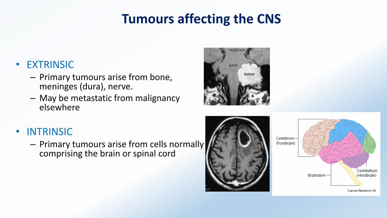
Tumours affecting the CNS
• EXTRINSIC– Primary tumours arise from bone,
meninges (dura), nerve.– May be metastatic from malignancy
elsewhere
• INTRINSIC– Primary tumours arise from cells normally
comprising the brain or spinal cord
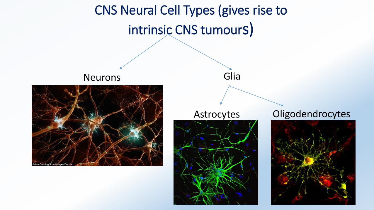
CNS Neural Cell Types (gives rise to
intrinsic CNS tumours)
Neurons Glia
Astrocytes Oligodendrocytes
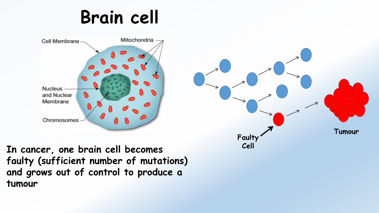
Brain cell
In cancer, one brain cell becomes faulty (sufficient number of mutations) and grows out of control to produce a tumour
Faulty Cell
Tumour
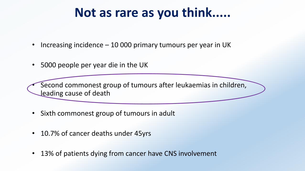
Not as rare as you think.....
• Increasing incidence – 10 000 primary tumours per year in UK
• 5000 people per year die in the UK
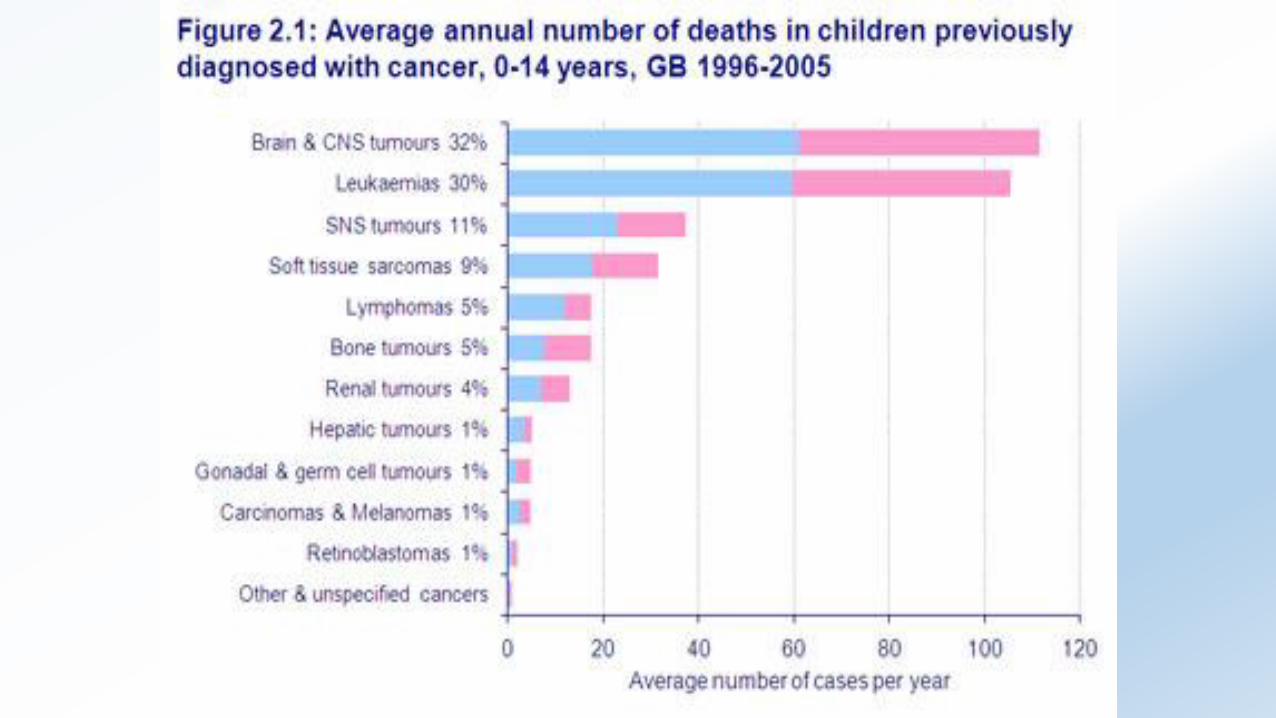
• Second commonest group of tumours after leukaemias in children, leading cause of death
• Sixth commonest group of tumours in adult
• 10.7% of cancer deaths under 45yrs
• 13% of patients dying from cancer have CNS involvement
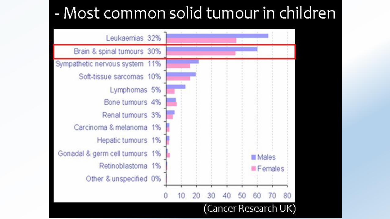

(Cancer Research UK)
Overall 5-year survival for adult brain cancer is also poor
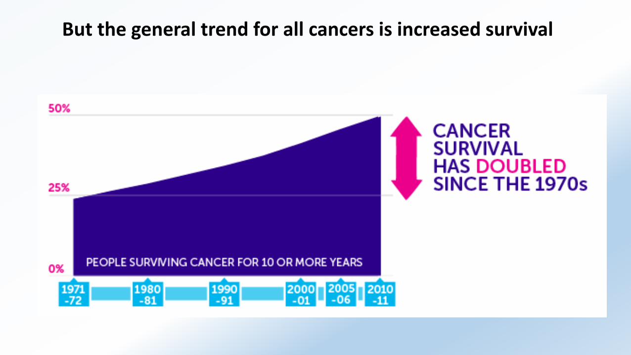
But the general trend for all cancers is increased survival
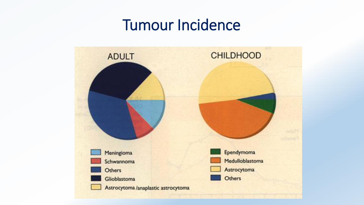
Tumour Incidence
Aetiology
• Unknown despite considerable research effort
• Links to mobile phone usage scientifically unproven as yet.
• Childhood Irradiation
• Genetic Factors
– NF1 and NF2 genes, Von Hippel Lindau syndrome
Childhood brain cancer – a developmental biology disorder
Adult brain cancer – multi-step progression, acquisition of mutations
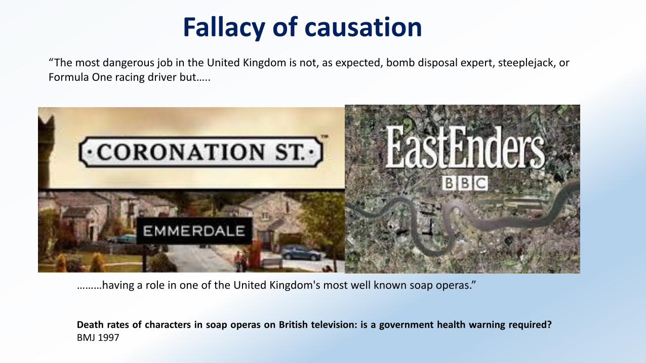
Fallacy of causation
………having a role in one of the United Kingdom's most well known soap operas.”
Death rates of characters in soap operas on British television: is a government health warning required?BMJ 1997
“The most dangerous job in the United Kingdom is not, as expected, bomb disposal expert, steeplejack, or Formula One racing driver but…..
Clinical Symptoms - extrinsic
• Extrinsic tumours compress underlying brain or spinal cord – focal neurological signs
• Extrinsic brain tumours cause symptoms of raised intracranial pressure
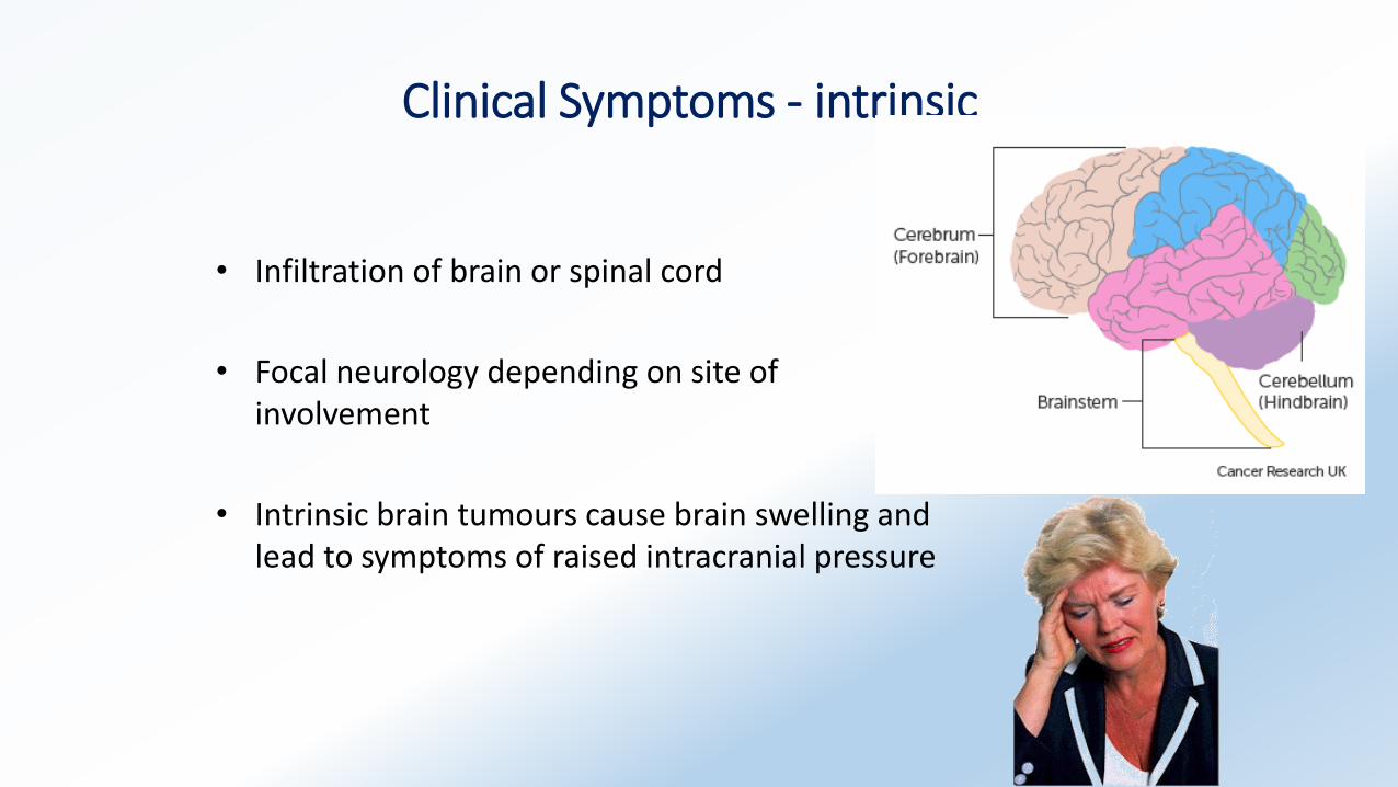
Clinical Symptoms - intrinsic
• Infiltration of brain or spinal cord
• Focal neurology depending on site of involvement
• Intrinsic brain tumours cause brain swelling and lead to symptoms of raised intracranial pressure
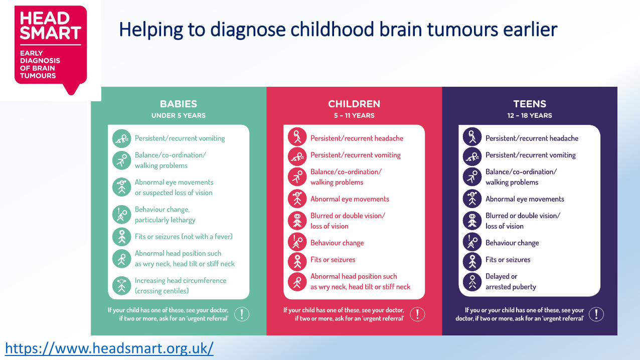
Helping to diagnose childhood brain tumours earlier
https://www.headsmart.org.uk/
Clinical Presentation
• Low grade tumours – brain can accommodate growth and slow pressure rise, so more likely to present with seizures or focal neurology
• High grade tumours – brain struggles with rapid pressure rise; more likely to present with pressure symptoms
Common Brain Tumours
• Secondary = metastases
• Primary –
• Astrocytoma / Glioma – High and low grade
• Meningioma
• Rarer – Ependymoma, Medulloblastoma/PNET, Choroid Plexus tumours Pineal
Metastases
• The most common type of brain tumour
• Most likely to come from breast, lung, bone, melanoma or renal. Others can spread to the brain but rarely
• Can be single or multiple tumours
• Treatment options are palliation, surgery, stereotactic radiosurgery or whole brain radiotherapy
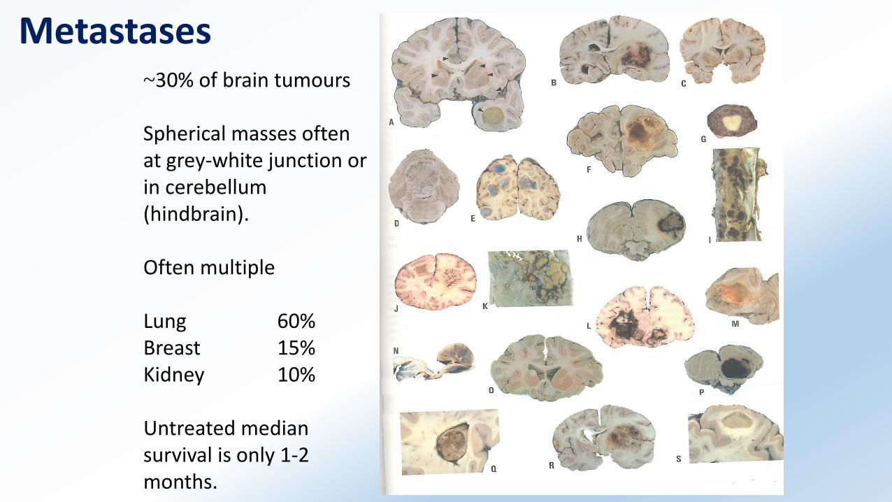
Metastases~30% of brain tumours
Spherical masses often at grey-white junction or in cerebellum (hindbrain).
Often multiple
Lung 60%Breast 15%Kidney 10%
Untreated median survival is only 1-2 months.
Primary Brain Tumours
High Grade AstrocytomaLow Grade AstrocytomaMeningioma
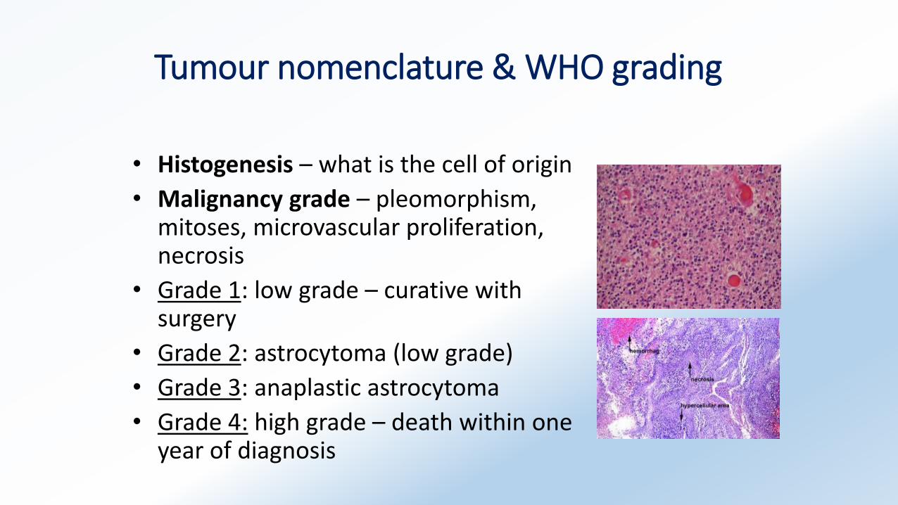
Tumour nomenclature & WHO grading
• Histogenesis – what is the cell of origin
• Malignancy grade – pleomorphism, mitoses, microvascular proliferation, necrosis
• Grade 1: low grade – curative with surgery
• Grade 2: astrocytoma (low grade)
• Grade 3: anaplastic astrocytoma
• Grade 4: high grade – death within one year of diagnosis
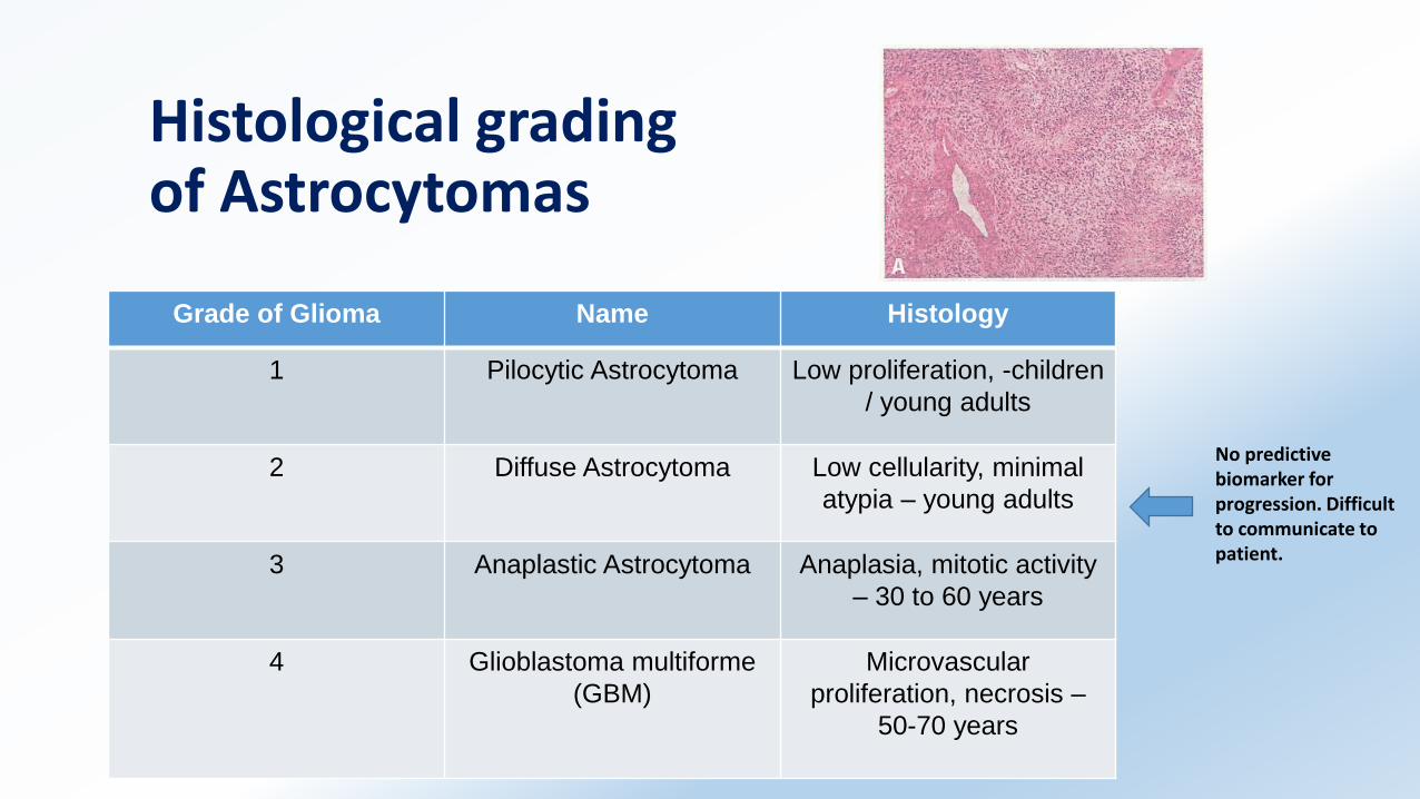
Histological gradingof Astrocytomas
Grade of Glioma Name Histology
1 Pilocytic Astrocytoma Low proliferation, -children
/ young adults
2 Diffuse Astrocytoma Low cellularity, minimal
atypia – young adults
3 Anaplastic Astrocytoma Anaplasia, mitotic activity
– 30 to 60 years
4 Glioblastoma multiforme
(GBM)
Microvascular
proliferation, necrosis –
50-70 years
No predictive biomarker for progression. Difficult to communicate to patient.
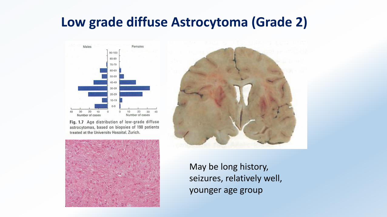
Low grade diffuse Astrocytoma (Grade 2)
May be long history, seizures, relatively well, younger age group
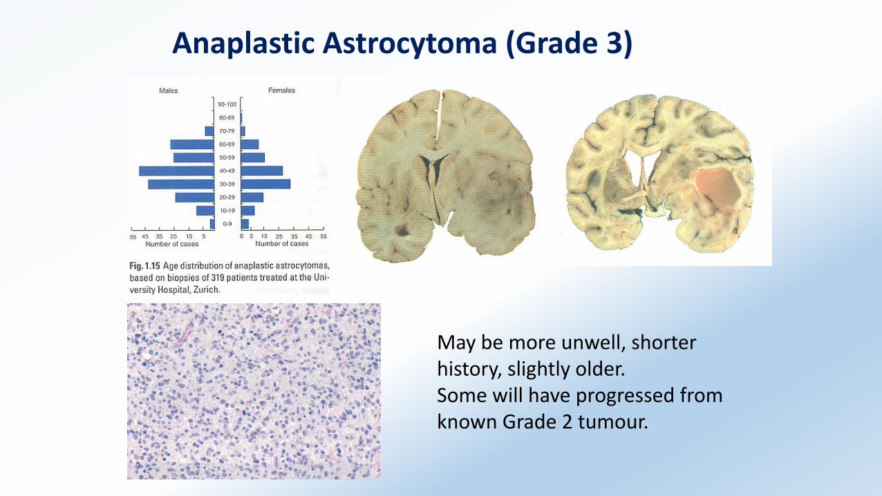
Anaplastic Astrocytoma (Grade 3)
May be more unwell, shorter history, slightly older.Some will have progressed from known Grade 2 tumour.
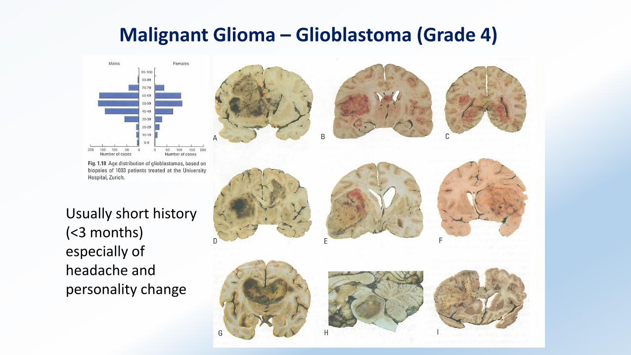
Malignant Glioma – Glioblastoma (Grade 4)
Usually short history (<3 months) especially of headache and personality change
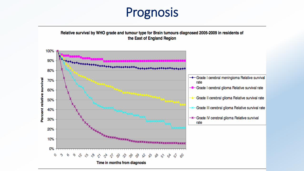
Prognosis
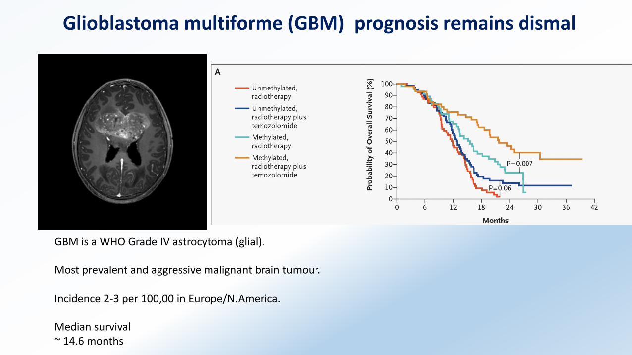
Glioblastoma multiforme (GBM) prognosis remains dismal
GBM is a WHO Grade IV astrocytoma (glial).
Most prevalent and aggressive malignant brain tumour.
Incidence 2-3 per 100,00 in Europe/N.America.
Median survival ~ 14.6 months
Treatment - Glioblastoma
• Radical surgery & steroids (dexamethasone)
• Radiotherapy – 60Gy in 30 fractions
• Chemotherapy – Temozolomide (some patients respond)
History
Bennett AH, Godlee RJ
“Excision of a tumour from the brain”
Lancet 1884
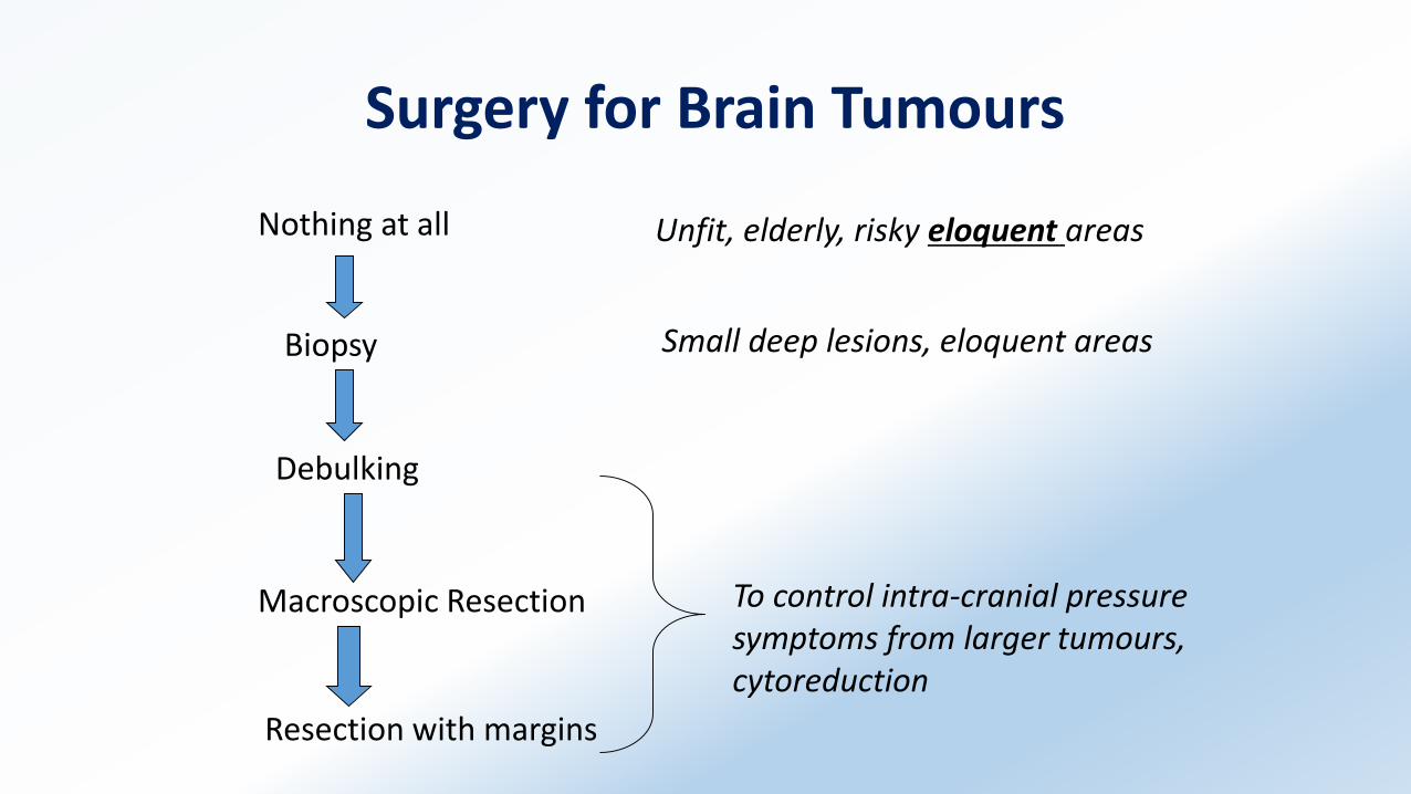
Surgery for Brain Tumours
Nothing at all
Biopsy
Debulking
Macroscopic Resection
Resection with margins
Unfit, elderly, risky eloquent areas
Small deep lesions, eloquent areas
To control intra-cranial pressure symptoms from larger tumours, cytoreduction

Image Guidance - StealthStation
CT / MRI scans of patients uploaded to StealthStation.
Fix patient’s head and register position of head (Pixar technology)
Neurosurgeon can decide where to remove fragment of skull and where to locate tumour using real-time feedback.
PROBLEMS – brain size and position shifts leading to inaccuracies

Fluorescence (5ALA)- guided surgery
Research
Improving diagnosis (MRI), treatment, prognostic markers,
palliative care / survivorship, basic and translational science e.g.
molecular biology
Genomic era (1972-2003)
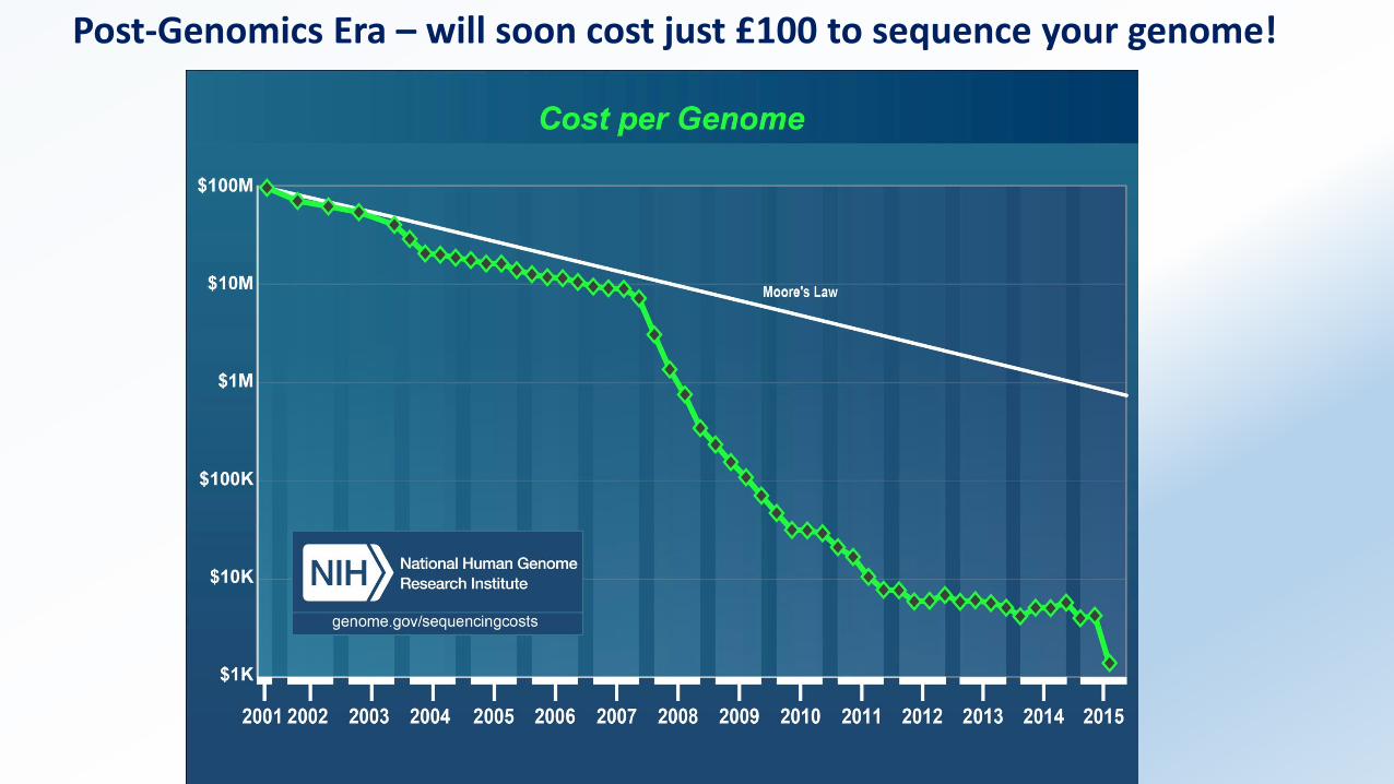
Post-Genomics Era – will soon cost just £100 to sequence your genome!
Pro
-ne
ura
l
Ne
ura
l
Cla
ssic
al
Me
sen
chym
al
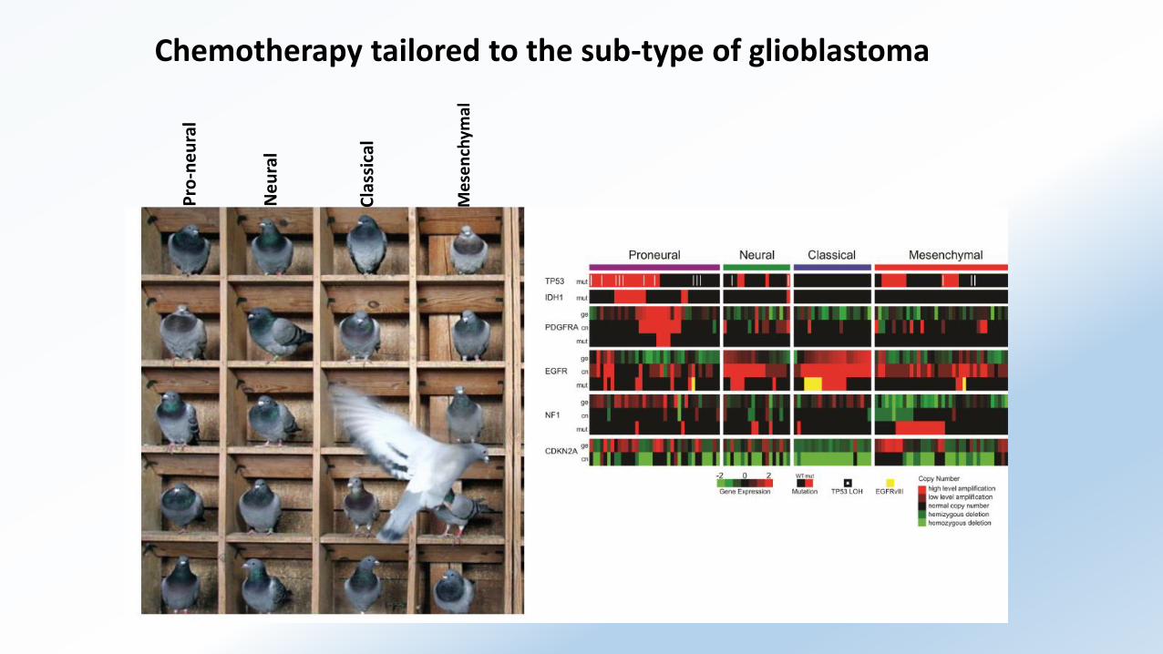
Chemotherapy tailored to the sub-type of glioblastoma

No objective response in any phase II clinical trial
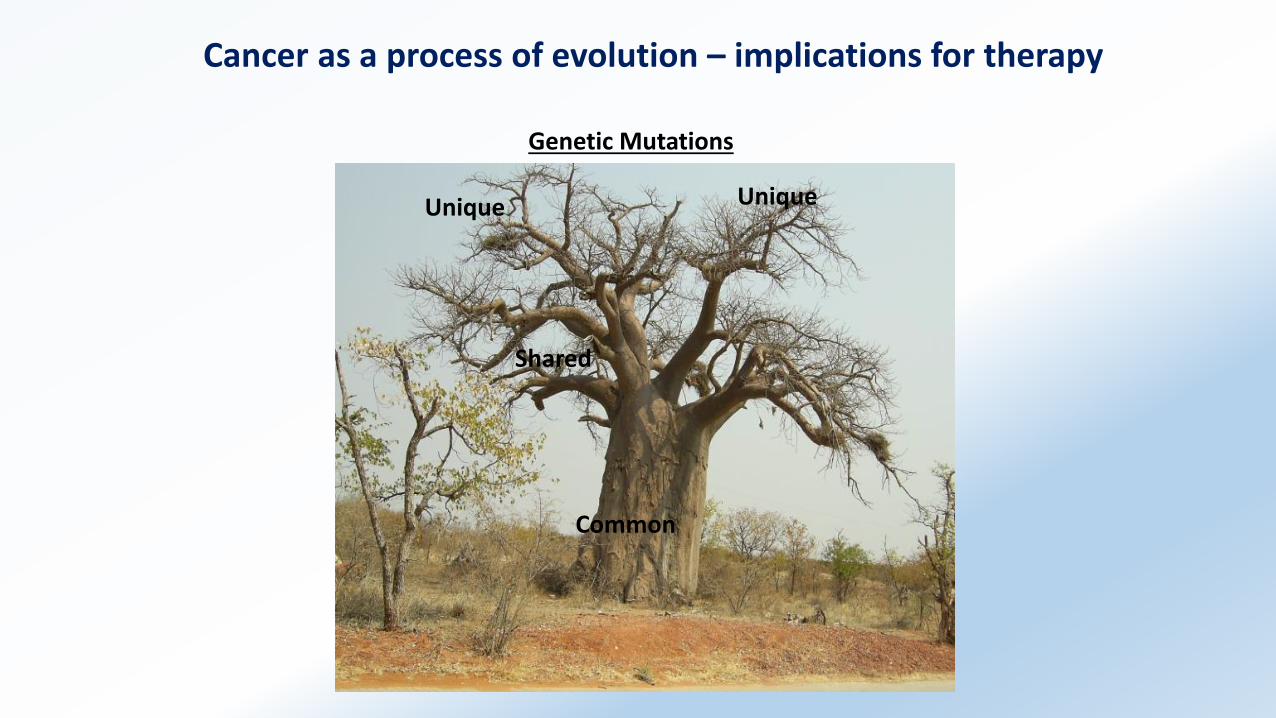
Cancer as a process of evolution – implications for therapy
Common
Shared
Unique Unique
Genetic Mutations