p21cipl/wafl fenl
TRANSCRIPT
Proc. Natl. Acad. Sci. USAVol. 93, pp. 11597-11602, October 1996Biochemistry
p21CiPl/Wafl disrupts the recruitment of human Fenl byproliferating-cell nuclear antigen into the DNAreplication complex
(cell cycle/maturation factor I/DNA repair)
JUNJIE CHEN, STEVEN CHEN, PARTHA SAHA, AND ANINDYA DUTTA*Department of Pathology, Division of Molecular Oncology, Brigham and Women's Hospital, Harvard Medical School, 75 Francis Street, Boston, MA 02115
Communicated by Peter M. Howley, Harvard Medical School, Boston, MA, August 1, 1996 (received for review April 17, 1996)
ABSTRACT Fenl or maturation factor 1 is a 5'-3' exo-nuclease essential for the degradation of the RNA primer-DNA junctions at the 5' ends of immature Okazaki fragmentsprior to their ligation into a continuous DNA strand. The geneis also necessary for repair of damaged DNA in yeast. Wereport that human proliferating-cell nuclear antigen (PCNA)associates with human Fenl with a Kd of 60 nM and anapparent stoichiometry of three Fenl molecules per PCNAtrimer. The Fenl-PCNA association is seen in cell extractswithout overexpression of either partner and is mediated by abasic region at the C terminus of Fenl. Therefore, the poly-merase 6-PCNA-Fenl complex has all the activities associ-ated with prokaryotic DNA polymerases involved in replica-tion: 5'-3' polymerase, 3'-5' exonuclease, and 5'-3' exonu-clease. Although p21, a regulatory protein induced by p53 inresponse to DNA damage, interacts with PCNA with a com-parable Kd (10 nM) and a stoichiometry of three molecules ofp21 per PCNA trimer, a p21-PCNA-Fenl complex is notformed. This mutually exclusive interaction suggests that theconformation of a PCNA trimer switches such that it caneither bind p21 or Fenl. Furthermore, overexpression of p21can disrupt Fenl-PCNA interaction in vivo. Therefore, besidesinterfering with the processivity of polymerase &-PCNA, p21also uncouples Fenl from the PCNA scaffold.
The eukaryotic DNA replication machinery has been exten-sively defined by biochemical fractionation of cell extracts andby genetic analysis in yeast. DNA polymerase a-primaseinitiates RNA primers on both strands ofDNA at the origin ofreplication, which are subsequently elongated as DNA strands(1-4). On both the leading and lagging strands, the initiatorDNA laid down by DNA polymerase a is subsequently elon-gated by DNA polymerase 8. The switch between the twopolymerases involves protein-protein interactions of the sin-gle-stranded DNA binding factor RPA, DNA polymerase a,RF-C, proliferating-cell nuclear antigen (PCNA), and DNApolymerase 8 itself (ref. 5 and references therein). PCNApromotes the processivity of DNA polymerase 8 allowing it tosynthesize long strands of DNA necessary for replicating theleading strand. The presence of the proofreading exonucleasein DNA polymerase 8 also suggests that this enzyme is bestsuited for replication with minimal errors. Due to the closesimilarity between the replication processes in prokaryotes andeukaryotes, it is somewhat surprising that the eukaryoticpolymerases are missing in one crucial activity, the 5'-3'exonuclease (6). This becomes particularly important on thelagging strand, where as each new Okazaki fragment reachesthe 5' end of the previous Okazaki fragment, a 5'-3' exonu-clease is required. While RNase H can remove most of theRNA primer, the ribonucleotide-deoxyribonucleotide bond
requires the activity of a special 5'-3' exonuclease that wascalled maturation factor 1 (MF1) or Fenl (5, 7-10).MF1/Fenl has been discovered in several different con-
texts. It was identified as a gene, rad2, that confers radiationsensitivity when mutated in Schizosaccharomyces pombe (11).Rad2 was found to be homologous to an anonymous openreading frame in Saccharomyces cerevisiae, YKL510 (renamedRAD27), that when mutated confers radiation sensitivity (12)and an instability in simple direct repeated sequences (13). Theinstability was primarily in the form of simple insertions,suggesting that an important repair mechanism in the repli-cation process was missing. In yeast missing two GI cyclins,CLN1 and CLN2, mutations in the RAD27 resulted in a celldivision cycle arrest consistent with a checkpoint being acti-vated by incomplete DNA replication during S phase (12, 14).Yeast with a deletion of the RAD27 gene demonstrate in-creased chromosome loss and a temperature-sensitive celldivision cycle block consistent with a requirement for the geneproduct in S phase. The Fenl exonuclease has also beenindependently purified by virtue of its ability to endonucleo-lytically cleave a flap DNA, a putative intermediate in theprocess of recombination (15). A similar cleavage may also beimportant for removing insertion mutations created by thereplication machinery.Human PCNA, the processivity factor for DNA polymerase
8 (16, 17), has a ring-shaped structure made up of threesubunits that assembles around DNA like a "ring around acurtain-rod." The structure suggests that its mechanism ofaction is to move along the DNA like a sliding clamp to whichthe polymerase 8 is tethered (18, 19). Besides interacting withpolymerase 8 and RF-C, PCNA associates with the cell-cycleregulatory factor p21. p21 associates with PCNA through its Cterminus, and with cyclin-cdks through its N terminus, therebyforming a bridge between the two protein complexes (20-24).By interacting with PCNA, p21 inhibits the activity of PCNAand disrupts the replication-machinery. Surprisingly, this in-teraction does not inhibit excision repair, which also utilizesPCNA (25, 26). Since p21 is transcriptionally induced by p53and since this pathway is activated after damage to DNA (27),the differential effect of p21 on the replication and repairfunctions of PCNA could be important for the pause in DNAreplication observed after DNA damage allowing the damageto be repaired before it is "fixed" by replication.
In view of the important role of PCNA in DNA replicationand repair, we searched for human gene products that interactwith PCNA in a two-hybrid interaction. The results reportedin this paper suggest that human MF1/Fenl 5'-3' exonucleaseassociates with human PCNA in vivo and in vitro. This asso-ciation completes the DNA polymerase holoenzyme, because
Abbreviations: PCNA, proliferating-cell nuclear antigen; GST, gluta-thione S-transferase.*To whom reprint requests should be addressed. e-mail:[email protected].
11597
The publication costs of this article were defrayed in part by page chargepayment. This article must therefore be hereby marked "advertisement" inaccordance with 18 U.S.C. §1734 solely to indicate this fact.
Dow
nloa
ded
by g
uest
on
Janu
ary
17, 2
022
Proc. Natl. Acad. Sci. USA 93 (1996)
the DNA polymerase 6-PCNA-Fenl complex has all threeactivities found in prokaryotic DNA polymerases. Throughthis association, PCNA recruits Fenl into replication complexand ensures the completion of DNA replication. These resultsmay explain why mutations in the Fenl subunit of the DNApolymerase holoenzyme result in incomplete DNA replicationand in the creation of insertion-deletion mutants in simpledirect repeats. p21 disrupts Fenl-PCNA interaction providingan additional mechanism by which p21 could inhibit DNAreplication.
MATERIALS AND METHODSYeast Two-Hybrid Screen. For the yeast two-hybrid screen,
the human PCNA open reading frame was cloned by PCR andinserted into the pAS2 vector (28) to create a fusion proteinwith the GAL4 DNA-binding domain. pAS2-hPCNA wastransformed into yeast strain Y190. For screening, a B-cellcDNA library in the pACT vector was transformed into Y190strain containing pAS2-hPCNA.Plasmid Construction and Protein Purification. pGST-
PCNA was constructed by ligating the full PCNA codingsequence into pGEX-5x-3 (Pharmacia) between BamHI andSall sites. pGST-p21C2 expresses the C-terminal 39 aminoacids of p21 fused to glutathione S-transferase (GST) (29).pGST-FenlC was constructed by ligating the sequence encod-ing the C-terminal amino acids of human Fenl (residues307-380, a BclI-SalI fragment) into the pGEX-5x-3 (BamHIand Sall). Plasmids were transformed into Escherichia coliBL21; cell lysis and purification of GST fusion proteins were
done as described (22).The cDNA encoding the full-length human Fenl was a gift
from J. Murray (University of Sussex, U.K.). pET-Fenl wasconstructed by ligating the Fenl full-length cDNA (as aNcoI-SalI fragment) into pET3a. pET-FenlB was constructedby ligating the NcoI-BamHI fragment of Fenl cDNA intopET3a, resulting in the deletion of C-terminal 17 amino acidsfrom full-length Fenl. All plasmids were transformed into E.coli BL21 for protein expression.
Bacterially expressed Fenl was purified by chromatographyon a DEAE-Sepharose column in buffer A (20 mM Hepes, pH8.0/1 mM EDTA/0.1% Nonidet P-40/1 mM DTT/1 mMphenylmethylsulfonyl fluoride/10% glycerol) containing 0.3M KCl (Flow-through) followed by binding to S-Sepharose at0.1 M KCl and elution with 0.3 M KCl. This crude preparationof Fenl protein was dialyzed against buffer A and loaded on5-ml Mono-Q FPLC column (Pharmacia). Proteins wereeluted with 50 ml of buffer A with a salt gradient of 0-1 M KCl.Fenl peak was followed by SDS/PAGE and Coomasie stain-ing.For FenlB purification, the flow through from the DEAE
column was loaded on Q-Sepharose column (Pharmacia)equilibrated with buffer A containing 0.3 M KCl. The flowthrough from the Q-Sepharose was loaded on a 5-ml hydrox-ylapatite column (0.1 M KCl) and eluted with 15 ml of bufferA/0.1M KCl/80 mM sodium phosphate, pH 8.0. ElutedFenlB was then dialyzed overnight against buffer A (pH 6.0)containing 25 mM KCl, loaded on a 5-ml S Sepharose column,and eluted with buffer A (pH 6.0) containing 100 mM KCl.Purified Fenl or FenlB were dialyzed against buffer A7.4 (20mM Tris-HCl, pH 7.4/1 mM EDTA/0.01% Nonidet P-40/10% glycerol/25 mM NaCl/1 mM DTT/0.1 mM phenylmeth-ylsulfonyl fluoride).pETp21His expresses human p21 with a six-histidine tag at
the N-terminal end. The purification of bacterially expressedHisp2l, Hisp27, and PCNA has been described (21, 30, 31).The fragments containing full-length Fenl and FenlB were
generated by PCR using Pfi polymerase (Stratagene) andcloned into pA3M vector (32) to generate pA3M-Fenl and
pA3M-FenlB for expression of myc epitope-tagged proteins inhuman cells.
Synthesis of Peptides. The synthetic peptide were purifiedusing C18 reverse-phase HPLC. p21C2 is the C-terminal 39amino acids of p21 that binds PCNA (29). CSH190 is negativecontrol peptide based on the sequence of Rpa2 (33).Pull-Down Assay. Affinity chromatography on glutathione
beads coated with various GST fusion proteins ("pull-down"assay) were done as described (34), except that washes weredone with buffer A7.4. Bound proteins were eluted by boilingin Laemmlis' buffer for 10 min, separated by SDS/polyacrylamide gel electrophoresis, and detected either byautoradiography (radiolabeled proteins) or by Western blotanalysis using appropriate antibodies. In each experiment, carewas taken to equalize the amount of input proteins, and GSTprotein was always included to serve as an negative control.
Gel Filtration and Glycerol Gradient. Proteins were incu-bated on ice for 15 min in A7.4 buffer before loading on a 25-mlSuperose 12 gel filtration column (Pharmacia). Proteins wereeluted from the column at flow rate 0.4 ml/min, and 0.5-mlfractions were collected.
Five-microliter 10-40% glycerol gradients were made withthe gradient maker; 50-100 ,ul of proteins was carefully addedon the top of the gradient. Gradients were centrifuged at55,000 rpm using SW55 rotor for 12-24 h in L8-M (Beckman).Fractions of 200 ,ul were collected starting from the top of thegradients.
Exonuclease and Endonuclease Assay. Exonuclease and flapendonuclease assays were performed as described (9, 15).Measurement ofBinding Constants by BlAcore Instrument
(Pharmacia). Detailed procedure of kinetic measurements bysurface plasmon resonance technique using BIAcore instru-ment is described in manufacturer's instruction manual. Therunning buffer used for all binding experiments is 20 mMHepes, pH 7.4/150 mM NaCl/3.4 mM EDTA/2 mM DTT/0.05% P-20 (Pharmacia). PCNA was immobilized on thesurface of sensor chip using carbodiimide method by followingmanufacturer's instruction. For measuring binding parame-ters, five concentrations of Fenl solutions were passed over thePCNA surface, and the affinity constant was estimated usingsoftware provided by the manufacturer.
Antibodies and Immunoprecipitations. Anti-Fenl poly-clonal rabbit antibody was raised against purified bacterialexpressed Fenl (Cocalico, Pennsylvania) and affinity-purifiedas described (35). PCNA was detected by Western blot analysisusing a monoclonal antibody (PC10; Santa Cruz Biotechnol-ogy). The Myc-tagged Fenl or FenlB were transfected into293T cells by the standard calcium phosphate coprecipitationmethod. Thirty-six hours after transfection, cells were lysed inNonidet P-40 lysis buffer (22) and Myc-tagged proteins wereimmunoprecipitated using antibody (9E10) against the Mycepitope.
RESULTSHuman Fenl Interacts with PCNA in a Two-Hybrid Assay.
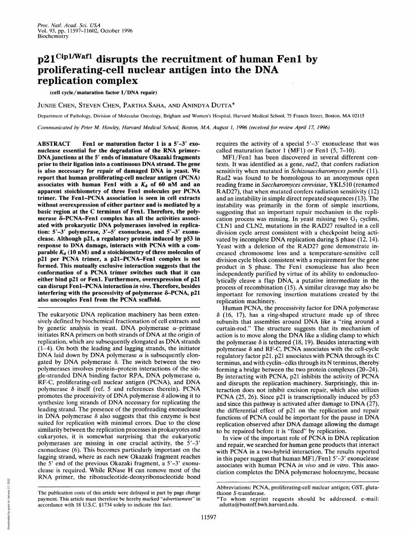
A two-hybrid screen (28, 36) was carried out to search forhuman gene products that interacted with human PCNA.Yeast Y190 expressing a Gal4 DNA binding domain fusedin-frame to human PCNA were transformed with a cDNAlibrary from human B cells in the pACT vector. From 2 x 10⁷transformants, 60 positive clones were analyzed. Ten encodedp21 (four clones), and eight encoded Fen 1 (two clones). Theamount of f3-galactosidase produced and the rate of growth onselective medium lacking histidine were equivalent for yeastexpressing Fenl or p21. The clones containing the humanFenl/Rad2 gene (11) expressed amino acids 249-380 (Cterminus) or amino acids 115-380 of Fenl (Fig. 1), suggestingthat the C-terminal portion of Fenl could be involved in the
11598 Biochemistry: Chen et al.
Dow
nloa
ded
by g
uest
on
Janu
ary
17, 2
022
Proc. Natl. Acad. Sci. USA 93 (1996) 11599
Interactionwith PCNA
Fenl (1-380)
A Cat ADH BSA
20 21 22 23 24 25 26 27 28
-----j
L-I im +
+
s7 +
PCNA-___
Feni-
Feni1..........
PCNA-
FIG. 1. Summary of two-hybrid screen and of Fenl deletions usedin this paper. (Upper) Two cDNAs (eight clones) encoded amino acids115-380 and amino acids 249-380 of human Fenl were isolated bytwo-hybrid screen using human PCNA as the bait. None of the FenlcDNA isolated interacts with any other baits tested in the two-hybridassay. (Lower) Deletion derivatives used in this paper. The domainsconserved between Fenl and other exonucleases including XP-G areindicated by shaded and solid boxes in the Fenl schematic.
interaction. None of the clones interacted with a negativecontrol bait comprising the Gal4 DNA binding domain alone.Fenl Binds PCNA Through Its C-Terminal Tail. To verify
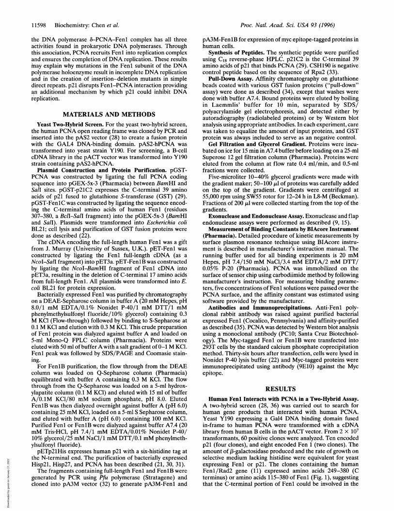
the direct interaction between PCNA and Fenl, both proteinswere expressed in E. coli. When PCNA was expressed as a GSTfusion protein, Fenl formed a stable complex with PCNA inthe pull-down assay (Fig. 2A). But FeniB, which lacks the basicC-terminal 17 amino acids of Fenl, fails to associate withPCNA (Fig. 2A). GST-FenlC, which contains the C-terminal77 amino acids, binds to PCNA (Fig. 2B, also see Fig. SA).These results suggest that Fenl associates with PCNA throughits C-terminal tail.Three Molecules of Fenl Form a Complex with the PCNA
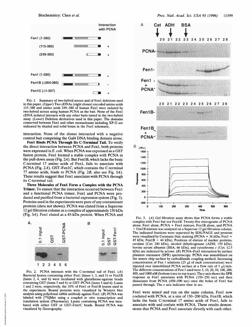
Trimer. To ensure that the interaction occurred between Feniand a functional PCNA trimer, Fenl and PCNA were pro-duced and purified from a bacterial expression system (Fig. 3).Proteins used in the experiments were pure of any contaminantproteins (data not shown). PCNA was eluted from a Superose12 gel filtration column as a complex of approximately 150 kDa(Ficr 'IA Ft-.nl ethiti-l qz n A1i_ Tns nrnti-in Whpn PCNA nnil
A.40.
CL
Bz
cL
cn (1nO ca
Bacterial lysates containing either Fenl (lanes 1, 3,(lanes 2, 4, and 6) were incubated with glutathiorcontaining GST (lanes 3 and 4) or GST-PCNA (lane1 and 2 were, respectively, the 10% of Fenl or Fenlthe experiment. Bound proteins were visualized Ianalysis using polyclonal rabbit antibody against Fenlabeled with [35S]Met using a coupled in vitro ttranslation system (Pharmacia). Lysate containingbated with either GST or GST-FenlC beads. Bcvisualized by fluorography.
20 21 22 23 24 25 26 27 28
Fen1B-
FeniB+
PCNA-
X (RU)looo -
140 -
1200 -
1000-~
200-
o-
fr0
11911 sr%11 A dilukFIG. 3. (A) Gel filtration assay shows that PCNA forms a stable
complex with Fenl but not FenlB. Twenty-five micrograms of PCNAalone, Fenl alone, PCNA + Fenl mixture, FenlB alone, and PCNA+ FenlB mixture was analyzed on a Superose 12 gel filtration column.
{, a) The indicated fractions were separated by SDS/PAGE and proteinsC LF Fwere visualized by Coomasie blue staining (PCNA = 36 kDa; Fenl =
45 kDa; FenlB = 44 kDa). Positions of elution of marker proteins,o catalase (Cat; 240 kDa), alcohol dehydrogenase (ADH; 150 kDa),
bovine serum albumin (BSA; 66 kDa), and cytochrome c (Cyt; 12.5kDa) are indicated by arrows. (B) PCNA-Fenl interaction by surfaceplasmon resonance (SPR) spectroscopy. PCNA was immobilized onthe sensor chip surface by carbodiimide coupling method. Increasingconcentration of Fen 1 solutions (25 ,ul of each concentration) were
injected over immobilized PCNA surface at a flow rate of 5 p,l/min.ail of Fenl. (LA ) 5, 10, 20, 50, 100, 200,
and5o Fn(B 400, and 1000 nM (bottom trace to top trace). They axis shows the SPRand 5) or Fenib Fenl associates with PCNA (150-250 sec) and then
ne-agarose beads dissociates from PCNA (450-500 sec) the bolus of Fenl has
B lysates used inpassed through. The x axis indicates time in sec.
LBy Wesatern bloti1. (B) PCNA Fenl were mixed and run on the same column, Fenl now
ranscription and coeluted with PCNA, at a size of 150-200 kDa. FenlB, whichPCNA was incu- lacks the basic C-terminal 17 amino acids of Fenl, fails to)und PCNA was associate with and coelute with PCNA. These results demon-
strate that PCNA and Fenl associate directly with each other.
(115-380)
(249-380)
FenI (1-380)
Fenl B (A364-380) 0
Fenl C (Al -307)
Biochemistry: Chen et al.
kl-lg. J-11). 1-Ulll CUUMU db d PIULUIII- VVJ
P,L
Dow
nloa
ded
by g
uest
on
Janu
ary
17, 2
022
Proc. Natl. Acad. Sci. USA 93 (1996)
The interaction occurs through the basic tail of Fenl and doesnot disrupt the trimeric structure of PCNA that is essential forits function as a processivity factor for DNA polymerase 8.The stoichiometry of the Fenl-PCNA interaction was de-
termined by mixing Fenl with PCNA at various molar ratiosand determining by sedimentation on a glycerol gradientwhether free Fenl (sedimenting at 45 kDa) was present in themixture. Free Fenl was detected only when the ratio of Fenlto PCNA exceeded three molecules of Fenl per PCNA trimer(data not shown). Therefore, three molecules of Fenl bind toeach PCNA trimer.
Native molecular mass of PCNA trimer and Fenl-PCNAcomplex were calculated based on the results of the gelfiltration experiments and glycerol gradient sedimentationexperiments. The calculated molecular mass of Fenl-PCNAcomplex is 209 kDa, while that of PCNA trimer is 123 kDa(although the theoretical molecular weight of PCNA trimer is88 kDa). This would suggest two Fenl molecules associate withone PCNA trimer. Because of the abnormal behavior ofPCNAin gel filtration experiments and the results presented aboveand following, we favor that three molecules of Fenl associatewith one PCNA trimer.The affinity of the Fenl-PCNA interaction was determined
by surface plasmon resonance technique using BlAcore in-strument. The 2627 resonance units (RU) of PCNA trimerwere immobilized on the sensor chip and solutions containingFenl at various concentrations was passed over PCNA-coatedchip in the BiaCore machine. The sensorgrams (Fig. 3B)indicated that the Kd of the interaction was about 60 nM at25°C. The stoichiometry of the interaction (based on themaximum number of resonance units of Fenl that bind to afixed number of resonance units of PCNA) was about 2.47molecules of Fenl per PCNA trimer. Since all the PCNAimmobilized on the chip may not remain functionally active,this ratio is consistent with the stoichiometry determined byglycerol-gradient sedimentation (three molecules of Fenl perPCNA trimer).PCNA Interacts with Fenl in Vivo and the Interaction Is
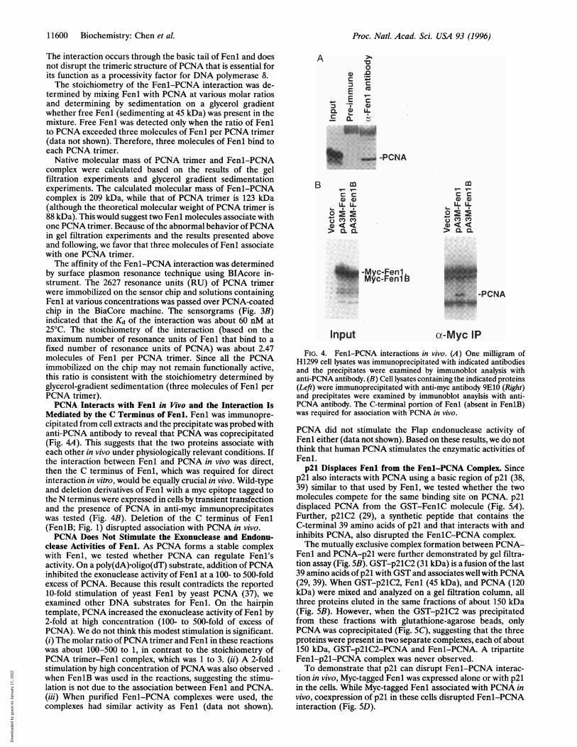
Mediated by the C Terminus of Fenl. Fenl was immunopre-cipitated from cell extracts and the precipitate was probed withanti-PCNA antibody to reveal that PCNA was coprecipitated(Fig. 4A). This suggests that the two proteins associate witheach other in vivo under physiologically relevant conditions. Ifthe interaction between Fenl and PCNA in vivo was direct,then the C terminus of Fenl, which was required for directinteraction in vitro, would be equally crucial in vivo. Wild-typeand deletion derivatives of Fenl with a myc epitope tagged tothe N terminus were expressed in cells by transient transfectionand the presence of PCNA in anti-myc immunoprecipitateswas tested (Fig. 4B). Deletion of the C terminus of Fenl(FenIB; Fig. 1) disrupted association with PCNA in vivo.PCNA Does Not Stimulate the Exonuclease and Endonu-
clease Activities of Fenl. As PCNA forms a stable complexwith Fenl, we tested whether PCNA can regulate Fenl'sactivity. On a poly(dA).oligo(dT) substrate, addition of PCNAinhibited the exonuclease activity of Fenl at a 100- to 500-foldexcess of PCNA. Because this result contradicts the reported10-fold stimulation of yeast Fenl by yeast PCNA (37), weexamined other DNA substrates for Fenl. On the hairpintemplate, PCNA increased the exonuclease activity of Fenl by2-fold at high concentration (100- to 500-fold of excess ofPCNA). We do not think this modest stimulation is significant.(i) The molar ratio of PCNA trimer and Fenl in these reactionswas about 100-500 to 1, in contrast to the stoichiometry ofPCNA trimer-Fenl complex, which was 1 to 3. (ii) A 2-foldstimulation by high concentration of PCNA was also observedwhen FenlB was used in the reactions, suggesting the stimu-lation is not due to the association between Fenl and PCNA.(iii) When purified Fenl-PCNA complexes were used, thecomplexes had similar activity as Fenl (data not shown).
A0
.0
r- Pc
'
- _U.....
B _ _
U. U.
>0.0Q
-Myc-FenlMyc-Fenlb
Input
t- ,-lc c0)G)LL LL
L- I IEo .0.
-PCNA
aX-Myc IPFIG. 4. Fenl-PCNA interactions in vivo. (A) One milligram of
H1299 cell lysates was immunoprecipitated with indicated antibodiesand the precipitates were examined by immunoblot analysis withanti-PCNA antibody. (B) Cell lysates containing the indicated proteins(Left) were immunoprecipitated with anti-myc antibody 9E10 (Right)and precipitates were examined by immunoblot anaylsis with anti-PCNA antibody. The C-terminal portion of Fenl (absent in Fen1B)was required for association with PCNA in vivo.
PCNA did not stimulate the Flap endonuclease activity ofFenl either (data not shown). Based on these results, we do notthink that human PCNA stimulates the enzymatic activities ofFenl.p21 Displaces Fenl from the Fenl-PCNA Complex. Since
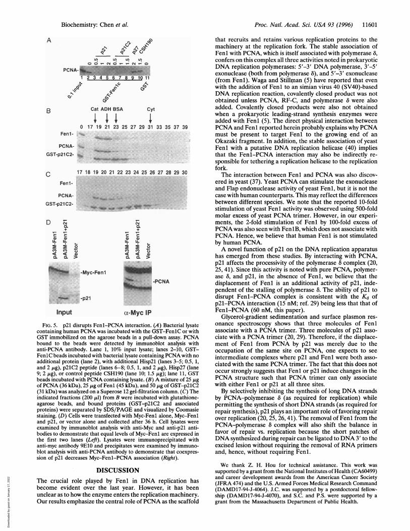
p21 also interacts with PCNA using a basic region of p21 (38,39) similar to that used by Fenl, we tested whether the twomolecules compete for the same binding site on PCNA. p21displaced PCNA from the GST-FenlC molecule (Fig. SA).Further, p21C2 (29), a synthetic peptide that contains theC-terminal 39 amino acids of p21 and that interacts with andinhibits PCNA, also disrupted the FenlC-PCNA complex.The mutually exclusive complex formation between PCNA-
Fenl and PCNA-p21 were further demonstrated by gel filtra-tion assay (Fig. SB). GST-p21C2 (31 kDa) is a fusion of the last39 amino acids of p21 with GST and associates well with PCNA(29, 39). When GST-p21C2, Fenl (45 kDa), and PCNA (120kDa) were mixed and analyzed on a gel filtration column, allthree proteins eluted in the same fractions of about 150 kDa(Fig. SB). However, when the GST-p21C2 was precipitatedfrom these fractions with glutathione-agarose beads, onlyPCNA was coprecipitated (Fig. SC), suggesting that the threeproteins were present in two separate complexes, each of about150 kDa, GST-p21C2-PCNA and Fenl-PCNA. A tripartiteFenl-p21-PCNA complex was never observed.To demonstrate that p21 can disrupt Fenl-PCNA interac-
tion in vivo, Myc-tagged Fenl was expressed alone or with p21in the cells. While Myc-tagged Fenl associated with PCNA invivo, coexpression of p21 in these cells disrupted Fenl-PCNAinteraction (Fig. SD).
11600 Biochemistry: Chen et al.
ALI-.....
.1l,iillw.11 -PCNA11 ".
Dow
nloa
ded
by g
uest
on
Janu
ary
17, 2
022
Proc. Natl. Acad. Sci. USA 93 (1996) 11601
A &S, 90
LO Ili LO t
PCNA- 45 8410111 2 34 5 6 7 " 01
40:
Cat ADH BSAB cyt
0 17 19 21 23 25 27 29 31 33 35 37 39Fen1 -
PCNA-
GST-p21C2- s
17 18 19 20 21 22 23 24 25 26 27 28 29 30cFenl-
PCNA-
GST-p21C2-
+L_C
C C
_L IL U.
< 0 4
r5 -Myc-Fenl-p1-PCNA
-p2l
Input ct-Myc IP
FIG. 5. p21 disrupts Fenl-PCNA interaction. (A) Bacterial lysatecontaining human PCNA was incubated with the GST-FenlC or withGST immobilized on the agarose beads in a pull-down assay. PCNAbound to the beads were detected by immunoblot analysis withanti-PCNA antibody. Lane 1, 10% input lysate; lanes 2-10, GST-FenlC beads incubated with bacterial lysate containing PCNA with noadditional protein (lane 2), with additional Hisp2l (lanes 3-5; 0.5, 1,and 2 ,ug), p21C2 peptide (lanes 6-8; 0.5, 1, and 2 ,ug), Hisp27 (lane9; 2 ,ug), or control peptide CSH190 (lane 10; 1.5 ,g); lane 11, GSTbeads incubated with PCNA containing lysate. (B) A mixture of 25 ,ugof PCNA (36 kDa), 25 Aig of Fenl (45 kDa), and 50 ,ig of GST-p21C2(31 kDa) was analyzed on a Superose 12 gel-filtration column. (C) Theindicated fractions (200 p.l) from B were incubated with glutathione-agarose beads, and bound proteins (GST-p21C2 and associatedproteins) were separated by SDS/PAGE and visualized by Coomasiestaining. (D) Cells were transfected with Myc-Fenl alone, Myc-Fenland p21, or vector alone and collected after 36 h. Cell lysates were
examined by immunoblot analysis with anti-Myc and anti-p21 anti-bodies to demonstrate that equal levels of Myc-Fenl are expressed inthe first two lanes (Left). Lysates were immunoprecipitated withanti-myc antibody 9E10 and precipitates were examined by immuno-blot analysis with anti-PCNA antibody to demonstrate that coexpres-sion of p21 decreases Myc-Fenl-PCNA association (Right).
DISCUSSIONThe crucial role played by Fenl in DNA replication hasbecome evident over the last year. However, it has beenunclear as to how the enzyme enters the replication machinery.Our results emphasize the central role of PCNA as the scaffold
that recruits and retains various replication proteins to themachinery at the replication fork. The stable association ofFenl with PCNA, which is itself associated with polymerase 8,confers on this complex all three activities noted in prokaryoticDNA replication polymerases: 5'-3' DNA polymerase, 3'-5'exonuclease (both from polymerase 8), and 5'-3' exonuclease(from Fenl). Waga and Stillman (5) have reported that evenwith the addition of Fenl to an simian virus 40 (SV40)-basedDNA replication reaction, covalently closed product was notobtained unless PCNA, RF-C, and polymerase 8 were alsoadded. Covalently closed products were also not obtainedwhen a prokaryotic leading-strand synthesis enzymes wereadded with Fenl (5). The direct physical interaction betweenPCNA and Fenl reported herein probably explains why PCNAmust be present to target Fenl to the growing end of anOkazaki fragment. In addition, the stable association of yeastFenl with a putative DNA replication helicase (40) impliesthat the Fenl-PCNA interaction may also be indirectly re-sponsible for tethering a replication helicase to the replicationfork.The interaction between Fenl and PCNA was also discov-
ered in yeast (37). Yeast PCNA can stimulate the exonucleaseand Flap endonuclease activity of yeast Fenl, but it is not thecase with human counterparts. This may reflect the differencesbetween different species. We note that the reported 10-foldstimulation of yeast Fenl activity was observed using 500-foldmolar excess of yeast PCNA trimer. However, in our experi-ments, the 2-fold stimulation of Fenl by 100-fold excess ofPCNA was also seen with FenlB, which does not associate withPCNA. Hence, we believe that human Fenl is not stimulatedby human PCNA.A novel function of p21 on the DNA replication apparatus
has emerged from these studies. By interacting with PCNA,p21 affects the processivity of the polymerase 8 complex (20,25, 41). Since this activity is noted with pure PCNA, polymer-ase 8, and p21, in the absence of Fenl, we believe that thedisplacement of Fenl is an additional activity of p21, inde-pendent of the stalling of polymerase 8. The ability of p21 todisrupt Fenl-PCNA complex is consistent with the Kd ofp21-PCNA interaction (15 nM; ref. 29) being less that that ofFenl-PCNA (60 nM, this paper).
Glycerol-gradient sedimentation and surface plasmon res-onance spectroscopy shows that three molecules of Fenlassociate with a PCNA trimer. Three molecules of p21 asso-ciate with a PCNA trimer (20, 29). Therefore, if the displace-ment of Fenl from PCNA by p21 was merely due to theoccupation of the same site on PCNA, one expects to seeintermediate complexes where p21 and Fenl were both asso-ciated with the same PCNA trimer. The fact that this does notoccur strongly suggests that Fenl or p21 induce changes in thePCNA structure such that PCNA trimer can only associatewith either Fenl or p21 at all three sites.By selectively inhibiting the synthesis of long DNA strands
by PCNA-polymerase 8 (as required for replication) whilepermitting the synthesis of short DNA strands (as required forrepair synthesis), p21 plays an important role of favoring repairover replication (20, 25, 26, 41). The removal of Feni from thePCNA-polymerase 8 complex will also shift the balance infavor of repair vs. replication because the short patches ofDNA synthesized during repair can be ligated to DNA 3' to theexcised lesion without requiring the removal of RNA primersand, hence, without requiring Fenl.
We thank Z. H. Hou for technical assistance. This work wassupported by a grant from the National Institutes of Health (CA60499)and career development awards from the American Cancer Society(JFRA 474) and the U.S. Armed Forces Medical Research Command(DAMD17-94-J-4064). J.C. was supported by a postdoctoral fellow-ship (DAMD17-94-J-4070), and S.C. and P.S. were supported by agrant from the Massachusetts Department of Public Health.
Biochemistry: Chen et al.
Dow
nloa
ded
by g
uest
on
Janu
ary
17, 2
022
Proc. Natl. Acad. Sci. USA 93 (1996)
1. Kelly, T. J. (1988) J. Biol. Chem. 263, 17889-17892.2. Stillman, B. (1989) Annu. Rev. Cell. Bio. 5, 197-245.3. Hurwitz, J., Dean, F. B., Kwong, A. D. & Lee, S.-H. (1990)
J. Biol. Chem. 265, 18043-18046.4. Campbell, J. L. (1993) J. Biol. Chem. 268, 25261-25264.5. Waga, S. & Stillman, B. (1994) Nature (London) 369, 207-212.6. Weiser, T., Gassmann, M., Thommes, P., Ferrari, E., Hafke-
meyer, P. & Hubscher, U. (1991) J. Biol. Chem. 266, 10420-10428.
7. Ishimi, Y., Claude, A., Bullock, P. & Hurwitz, J. (1988) J. Biol.Chem. 263, 19723-19733.
8. Goulian, M., Richards, S. H., Heard, C. J. & Bigsby, B. M. (1990)J. Biol. Chem. 265, 18461-18471.
9. Turchi, J. J. & Bambara, R. A. (1993) J. Biol. Chem. 268, 15136-15141.
10. Murante, R. S., Huang, L., Turchi, J. J. & Bambara, R. A. (1994)J. Biol. Chem. 269, 1191-1196.
11. Murray, J. M., Tavassoli, M., Al-Harithy, R., Sheldrick, K. S.,Lehmann, A. R., Carr, A. M. & Watts, F. Z. (1994) Mol. Cell.Biol. 14, 4878-4888.
12. Reagan, M. S., Pittenger, C., Siede, W. & Friedberg, E. C. (1995)J. Bacteriol. 177, 364-371.
13. Johnson, R. E., Kowali, G. K., Prakash, L. & Prakash, S. (1995)Science 269, 238-240.
14. Vallen, E. A. & Cross, F. R. (1995) Mol. Cell. Biol. 15,4291-4302.15. Harrington, J. J. & Lieber, M. R. (1994) Genes Dev. 8, 1344-
1355.16. Tan, C. K., Castillo, C., So, A. G. & Downey, K. M. (1986)J. Biol.
Chem. 261, 12310-12316.17. Prelich, G., Kostura, M., Marshak, D. R., Mathews, M. B. &
Stillman, B. (1987) Nature (London) 326, 471-475.18. Kuriyan, J. & O'Donnell, M. (1993) J. Mol. Bio. 234, 915-925.19. Krishna, T. S., Kong, X. P., Gary, S., Burgers, P. M. & Kuriyan,
J. (1994) Cell 79, 1233-1243.20. Flores-Rozas, H., Kelman, Z., Dean, F. B., Pan. Z.-Q., Harper,
J. W., Elledge, S. J., O'Donnell, M. & Hurwitz, J. (1994) Proc.Natl. Acad. Sci. USA 91, 8655-8659.
21. Waga, S., Hannon, G. J., Beach, D. & Stillman. B. (1994) Nature(London) 369, 574-578.
22. Chen, J., Jackson, P. K., Kirschner, M. W. & Dutta, A. (1995)Nature (London) 374, 386-388.
23. Luo, Y., Hurwitz, J. & Massague, J. (1995) Nature (London) 375,159-161.
24. Nakanishi, M., Robetorye, R. S., Adami, G. R., Pereirasmith,0. M. & Smith, J. R. (1995) EMBO J. 14, 555-563.
25. Li, R., Waga, S., Hannon, G. J., Beach, D. & Stillman, B. (1994)Nature (London) 371, 534-537.
26. Shivji, M., Grey, S. J., Strausfeld, U. P., Wood, R. D. & Blow, J. J.(1994) Curr. Biol. 4, 1062-1068.
27. el Deiry, W. S., Harper, J. W., Oconnor, P. M., Velculescu, V. E.,Canman, C. E., Jackman, J., Pietenpol, J. A., Burrell, M., Hill,D. E., Wang, Y. S., Wiman, K. G., Mercer, W. E., Kastan, M. B.,Kohn, K. W., Elledge, S. J., Kinzler, K. W. & Vogelstein, B.(1994) Cancer Res. 54, 1169-1174.
28. Harper, J. W., Adami, G. R., Wei, N., Keyomarsi, K. & Elledge,S. J. (1993) Cell 75, 805-816.
29. Chen, J., Peters, R., Saha, P., Lee, P., Theodoras, A., Pagano, M.,Wagner, G. & Dutta, A. (1996) NucleicAcids Res. 24, 1727-1733.
30. Polyak, K., Lee, M. H., Erdjumentbromage. H., Koff, A., Rob-erts, J. M., Tempst, P. & Massague, J. (1994) Cell 78, 59-66.
31. Fien, K. & Stillman, B. (1992) Mol. Cell. Biol. 12, 155-163.32. Makela, T. P., Parvin, J. D., Kim, J., Huber, L. J., Sharp, P. A. &
Weinberg, R. A. (1995) Proc. Natl. Acad. Sci. USA 92,5174-5178.33. Dutta, A. & Stillman, B. (1992) EMBO J. 11, 2189-2199.34. Dutta, A., Ruppert, J. M., Aster, J. C. & Winchester, W. (1993)
Nature (London) 365, 79-82.35. Nishitani, H. & Nurse, P. (1995) Cell 83, 397-405.36. Fields, S. & Song, 0. (1989) Nature (London) 340, 245-246.37. Li, X., Li, J., Harrington, J., Lieber, M. R. & Burgers, P. M.
(1995) J. Biol. Chem. 270, 22109-22112.38. Goubin, F. & Ducommun, B. (1995) Oncogene 10, 2281-2287.39. Warbrick, E., Lane, D. P., Glover, D. M. & Cox, L. S. (1995)
Curr. Bio. 5, 275-282.40. Budd, M. E. & Campbell, J. L. (1995) Proc. Natl. Acad. Sci. USA
92, 7642-7646.41. Podust, V. N., Podust, L. M., Goubin, F., Ducommun, B. &
Hubscher, U. (1995) Biochemistry 34, 8869-8875.
11602 Biochemistry: Chen et al.
Dow
nloa
ded
by g
uest
on
Janu
ary
17, 2
022