ocular traumaocular trauma - mahidol university ocular...acute eye conditions traumatic hyphema...
TRANSCRIPT
traumaACUTE EYE CONDITIONS
Ocular traumaOcular trauma
Taweekit Nimvorapunanatomy 1anatomy

traumaACUTE EYE CONDITIONS
PRESENTING SYMPTOMPRESENTING SYMPTOM
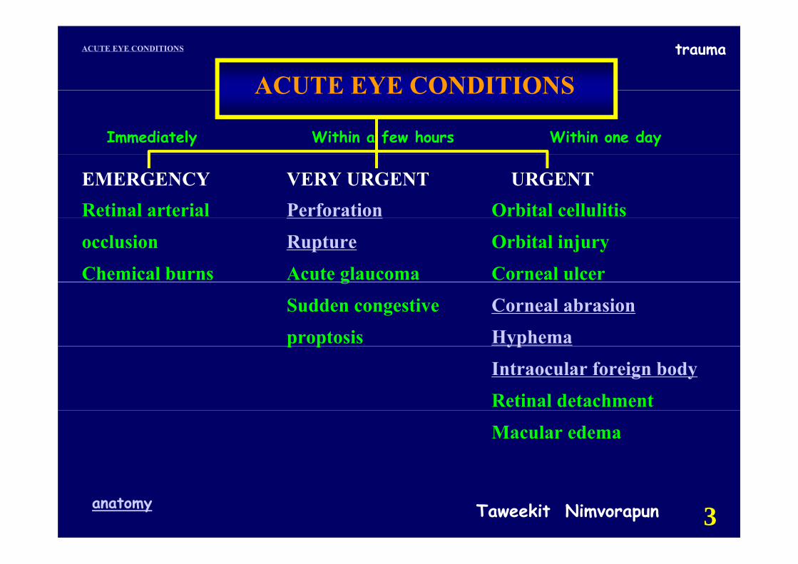
EMERGENCY VERY URGENT URGENT
Sudden Trauma Acute redness of the eye- loss of vision Suddenly unequal pupilloss of vision Suddenly unequal pupil- onset of pain - blurring of vision
“Something in the eye”Something in the eye
Evaluation needed:Evaluation needed:Immediately Within a few hours Within one day
Taweekit Nimvorapunanatomy 2
traumaACUTE EYE CONDITIONS
ACUTE EYE CONDITIONSACUTE EYE CONDITIONS
Immediately Within a few hours Within one day
EMERGENCY VERY URGENT URGENTRetinal arterial Perforation Orbital cellulitisocclusion Rupture Orbital injuryChemical burns Acute glaucoma Corneal ulcerg
Sudden congestive Corneal abrasionproptosis Hyphemap p yp
Intraocular foreign bodyRetinal detachmentMacular edema
Taweekit Nimvorapunanatomy 3
traumaACUTE EYE CONDITIONS
anatomy
Taweekit Nimvorapunanatomy 4
traumaACUTE EYE CONDITIONS
Taweekit Nimvorapunanatomy 5
traumaACUTE EYE CONDITIONS
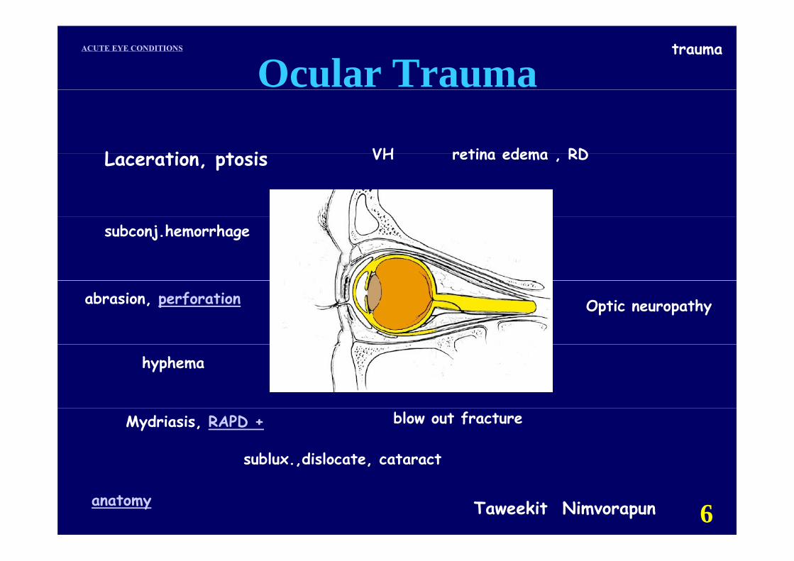
Ocular Trauma
L ti t i VH retina edema RDLaceration, ptosis VH retina edema , RD
subconj.hemorrhage
abrasion, perforation Optic neuropathy
hyphema
Mydriasis, RAPD +
sublux.,dislocate, cataract
blow out fracture
Taweekit Nimvorapunanatomy 6
traumaACUTE EYE CONDITIONS
VISION HISTORY
☺ I ff t d b th?☺ Is one eye affected, or both?
☺ What is your current level of vision?☺ What is your current level of vision?
☺ Was vision normal prior to trauma?☺ Was vision normal prior to trauma?
Taweekit Nimvorapunanatomy 7
traumaACUTE EYE CONDITIONS
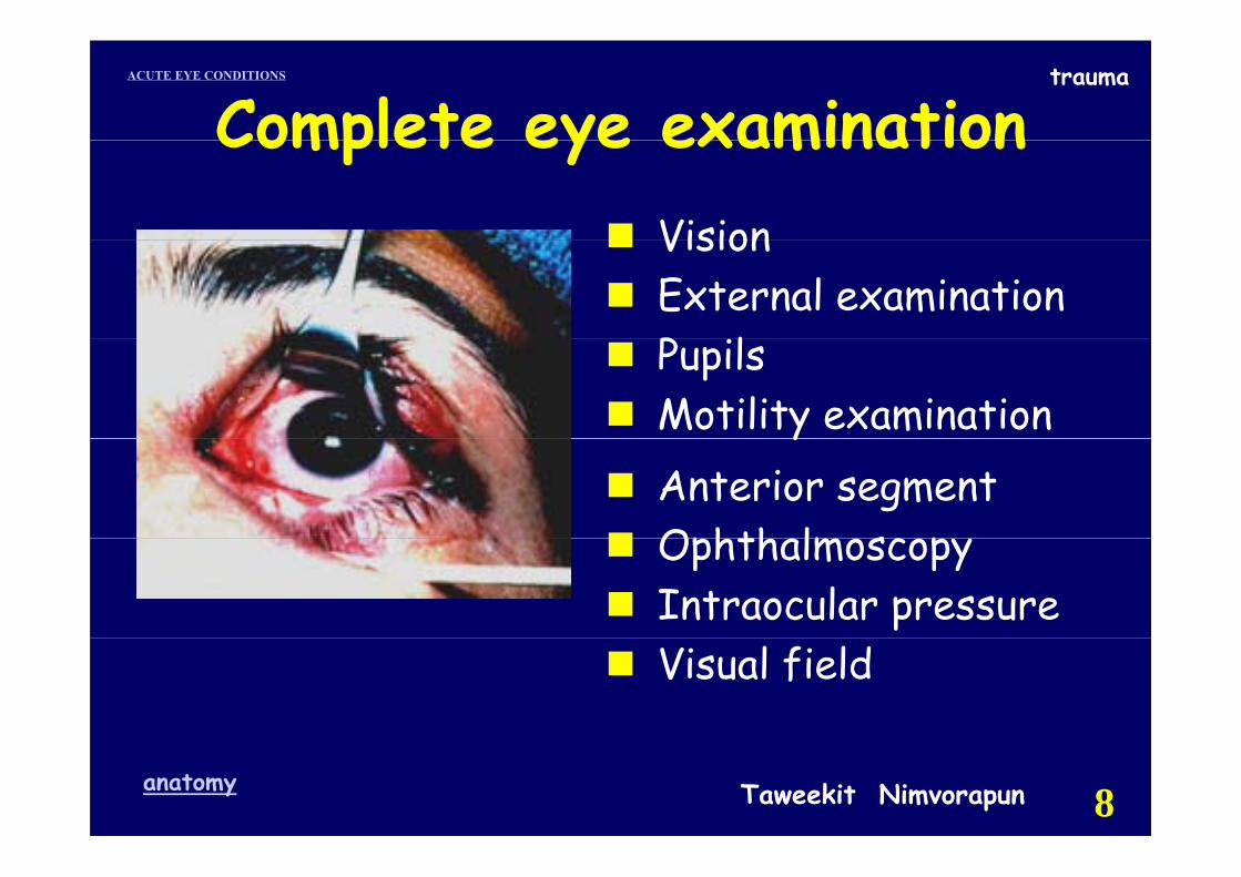
Complete eye examinationVision
Complete eye examinationVisionExternal examinationP ilPupilsMotility examinationyAnterior segmentOphth lm sc pOphthalmoscopyIntraocular pressureVisual field
Taweekit Nimvorapunanatomy 8
traumaACUTE EYE CONDITIONS
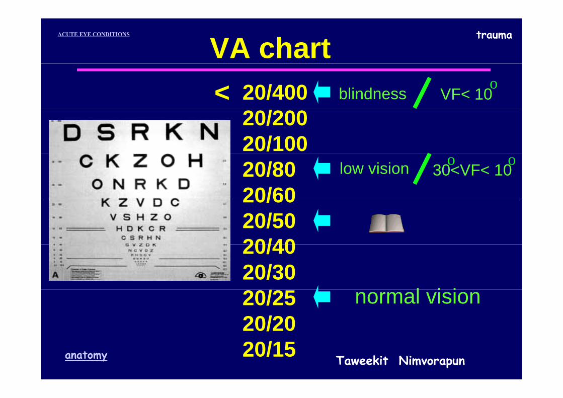
VA chart blindness20/400 VF< 10< o
20/20020/100
low vision20/8020/60
30<VF< 10o o
20/6020/5020/40
l i i
20/4020/30
normal vision20/2520/20
Taweekit Nimvorapunanatomy 20/15
traumaACUTE EYE CONDITIONS
Final visual outcome depends on p
prompt appropriate prompt, appropriate
☺ diagnosis
☺ treatment.
Taweekit Nimvorapunanatomy 10
traumaACUTE EYE CONDITIONS
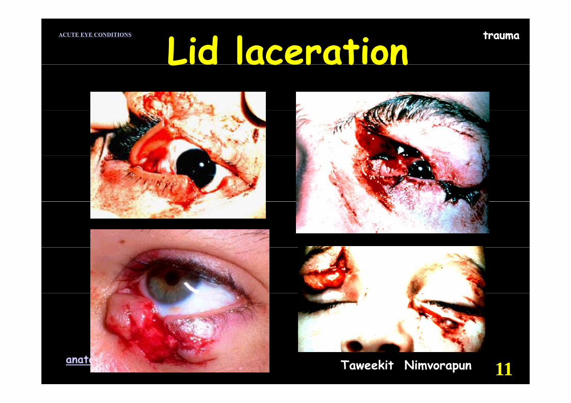
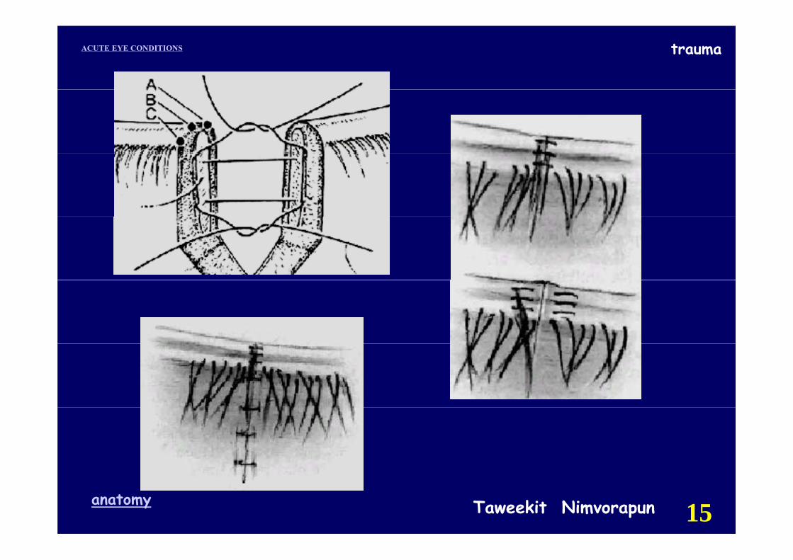
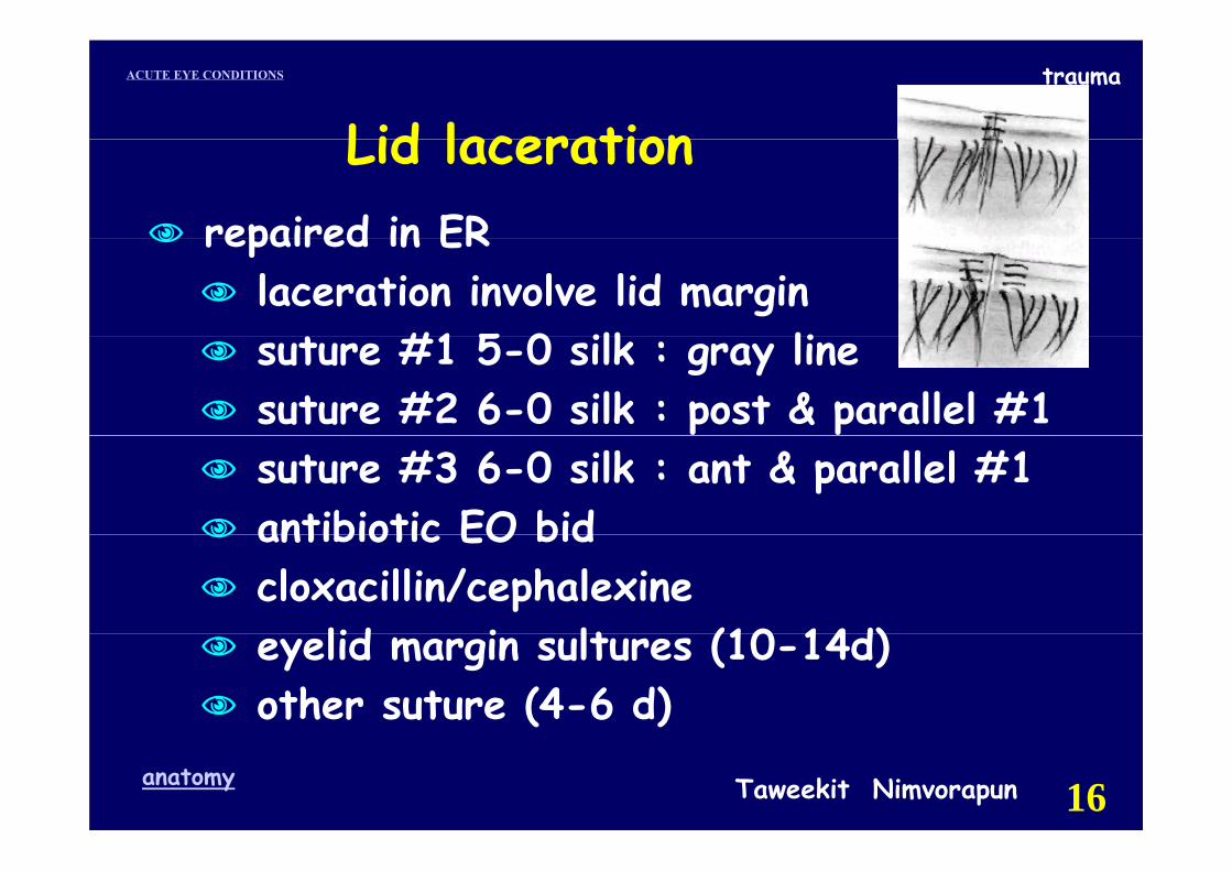
Lid laceration traumaLid laceration
Taweekit Nimvorapunanatomy 11
traumaACUTE EYE CONDITIONS
Lid lacerationLid laceration
C l f h bl Can result from sharp or blunt traumaRule out associated ocular injuryRule out associated ocular injuryAvoid lid margin retractiongRemove superficial foreign bodiesRule out deeper foreign bodiesGi h l iGive tetanus prophylaxis
Taweekit Nimvorapunanatomy 12

traumaACUTE EYE CONDITIONS
Lid lacerationLid lacerationf t hth lm l i t if i t d refer to ophthalmologist if associated
ocular injuryruptured globelacrimal drainage systemlacrimal drainage systemlevator aponeurosis,SRmedial canthal tend n medial canthal tendon extensive tissue loss (>1/3)FB
Taweekit Nimvorapunanatomy 13
traumaACUTE EYE CONDITIONS
Lid lacerationLid lacerationdelayed repaired in
significant risk for contaminationhuman biteshuman bites
clean with betadinei i t ith li irrigate with saline search FBdebridge infected or necrotic tissueleave the wound open & topical antibioticleave the wound open & topical antibiotic3-4 d later : repair
t i tibi tiTaweekit Nimvorapunanatomy 14
systemic antibiotic
traumaACUTE EYE CONDITIONS
Taweekit Nimvorapunanatomy 15
traumaACUTE EYE CONDITIONS
Lid lacerationLid lacerationrepaired in ERrepaired in ER
laceration involve lid margin t #1 5 0 ilk li suture #1 5-0 silk : gray line
suture #2 6-0 silk : post & parallel #1suture #3 6-0 silk : ant & parallel #1antibiotic EO bidantibiotic EO bidcloxacillin/cephalexine
lid i lt (10 14d)eyelid margin sultures (10-14d)other suture (4-6 d)
Taweekit Nimvorapunanatomy 16
traumaACUTE EYE CONDITIONS
Subconjunctival hemorrhage
Taweekit Nimvorapunanatomy 17
traumaACUTE EYE CONDITIONS trauma
Taweekit Nimvorapunanatomy 18

traumaACUTE EYE CONDITIONS
Taweekit Nimvorapunanatomy 19
traumaACUTE EYE CONDITIONS
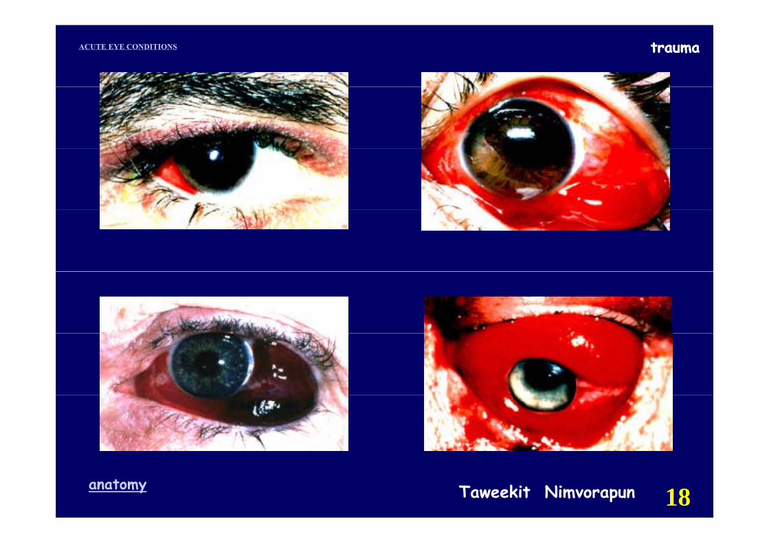
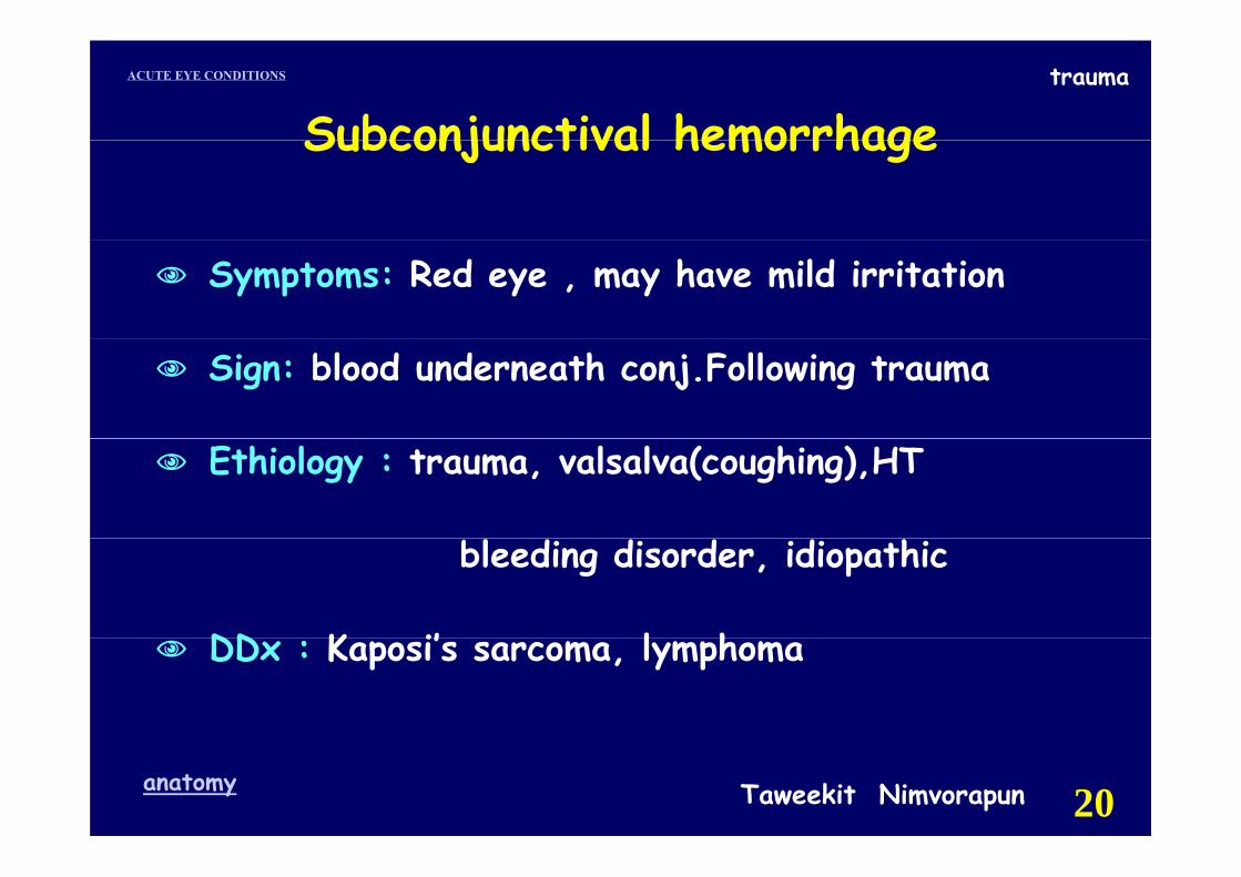
Subconjunctival hemorrhageSubconjunctival hemorrhage
Symptoms: Red eye , may have mild irritation
Sign: blood underneath conj.Following trauma
Ethiology : trauma, valsalva(coughing),HT
bleeding disorder, idiopathic
DD K i’ l hDDx : Kaposi’s sarcoma, lymphoma
Taweekit Nimvorapunanatomy 20
traumaACUTE EYE CONDITIONS
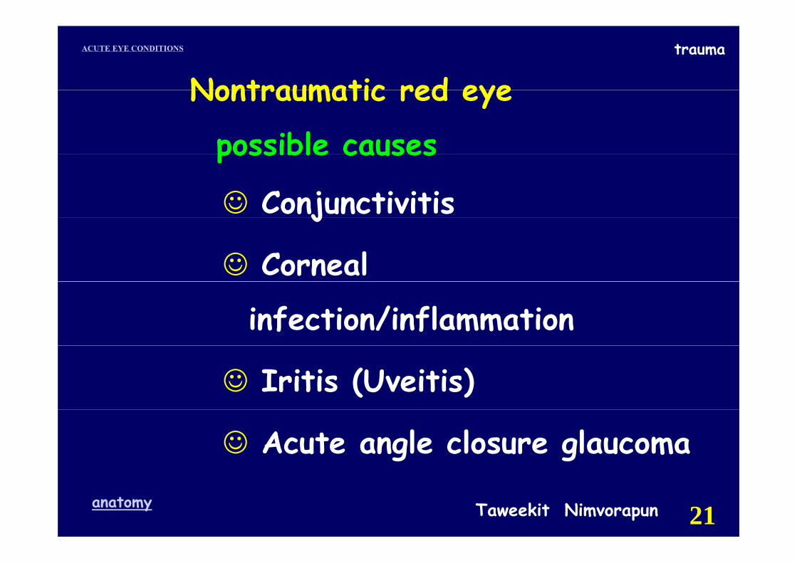
Nontraumatic red eye Nontraumatic red eye
possible causespossible causes
☺ Conjunctivitisj
☺ Corneal
infection/inflammation
☺ Iritis (Uveitis)
☺ Acute angle closure glaucoma
Taweekit Nimvorapunanatomy 21
traumaACUTE EYE CONDITIONS
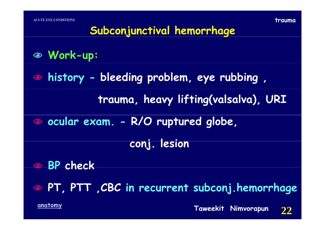
Subconjunctival hemorrhage
Work-up:
j g
history - bleeding problem, eye rubbing ,
trauma, heavy lifting(valsalva), URI
ocular exam. - R/O ruptured globe,
j l iconj. lesion
BP checkBP check
PT, PTT ,CBC in recurrent subconj.hemorrhage
Taweekit Nimvorapunanatomy 22
j g
traumaACUTE EYE CONDITIONS
Subconjunctival hemorrhageSubconjunctival hemorrhage
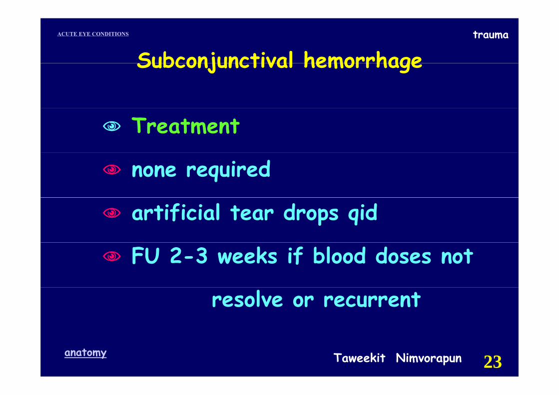
Treatment
none required
artificial tear drops qid
FU 2-3 weeks if blood doses not
resolve or recurrent
Taweekit Nimvorapunanatomy 23

traumaACUTE EYE CONDITIONS
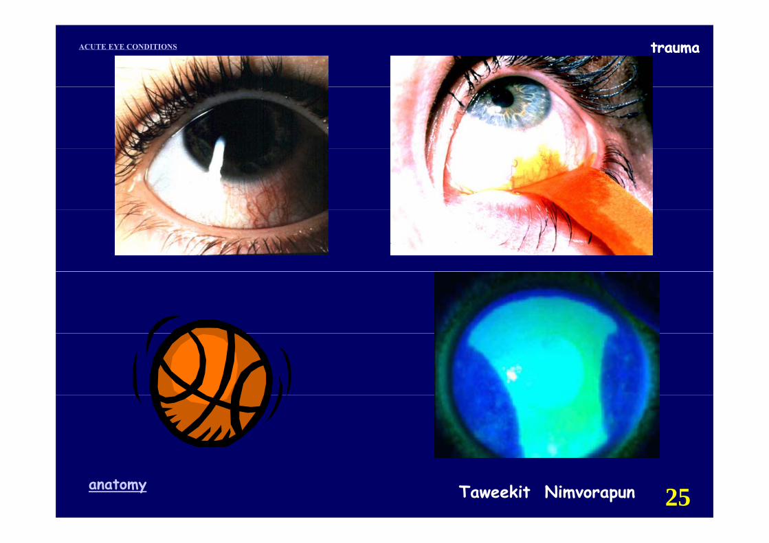
Corneal abrasionCorneal abrasion
Taweekit Nimvorapunanatomy 24
traumaACUTE EYE CONDITIONS trauma
Taweekit Nimvorapunanatomy 25
traumaACUTE EYE CONDITIONS
Corneal abrasion
Abrasion : absence of epithelium pthat is caused by trauma.
H f t hi th Hx : of scratching the eye Symptoms: Pain , photophobia , Symptoms Pain , photophobia ,
FB sensation, tearing h l l d f h Sign : epithelial staining defect with
fluorescein conjunctival injection fluorescein, conjunctival injection, swollen eyelid
Taweekit Nimvorapunanatomy 26
traumaACUTE EYE CONDITIONS
C l b iCorneal abrasion
Work-up fl iuse fluorescein
measure the size of the abrasionmeasure the size of the abrasiondiagram its locationevert eyelid : search foreign body
Taweekit Nimvorapunanatomy 27
traumaACUTE EYE CONDITIONS
Corneal abrasion
Treatment
Corneal abrasion
Treatmentcycloplegic EDy p gantibiotic EOpressure patch for 24 hr
not applied PP at significantnot applied PP at significantrisk for infection
Taweekit Nimvorapunanatomy 28
traumaACUTE EYE CONDITIONS
Corneal abrasion
FU daily
Corneal abrasion
FU daily abrasion เล็กลง antibiotic ED q 1-2 hr q
ไม path ให antibiotic EO hsabrasion ใหญ:antibiotic EO+ PP 24 hr.observe infection ทกวันจนกวาจะหายobserve infection ทุกวนจนกวาจะหาย
return if symptoms persist or worsey p pwhite corneal infiltration refer
d lTaweekit Nimvorapunanatomy 29
immediately
traumaACUTE EYE CONDITIONS
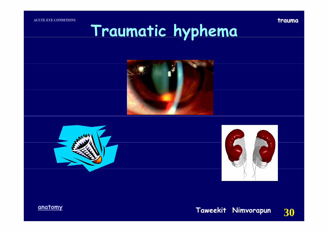
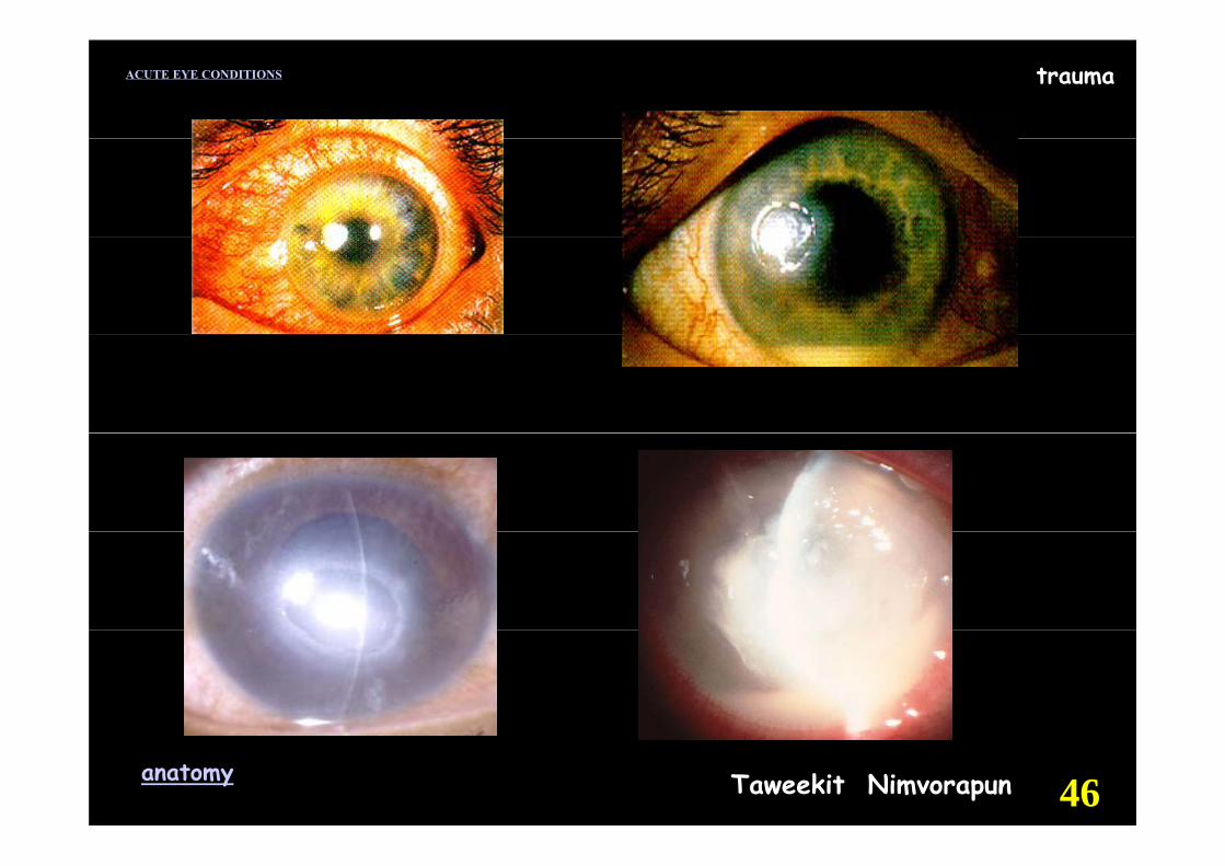
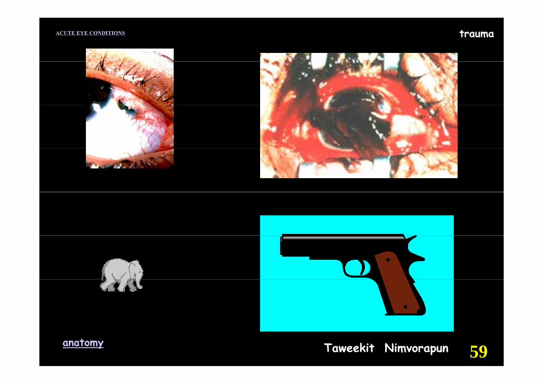
Traumatic hyphematrauma
yp
Taweekit Nimvorapunanatomy 30
traumaACUTE EYE CONDITIONS
Taweekit Nimvorapunanatomy
traumaACUTE EYE CONDITIONS
Taweekit Nimvorapunanatomy 32
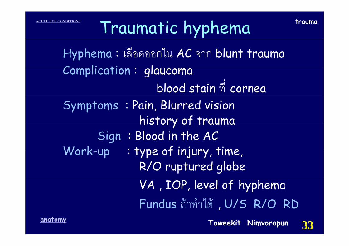
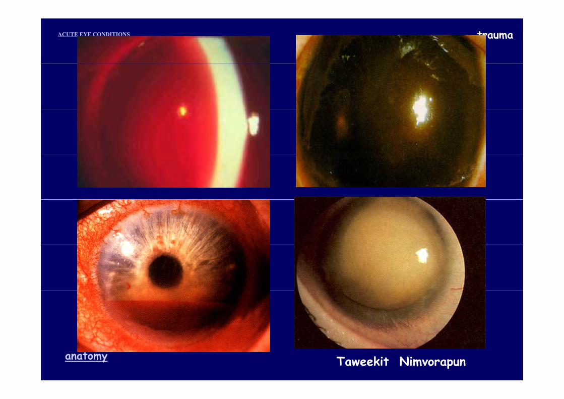
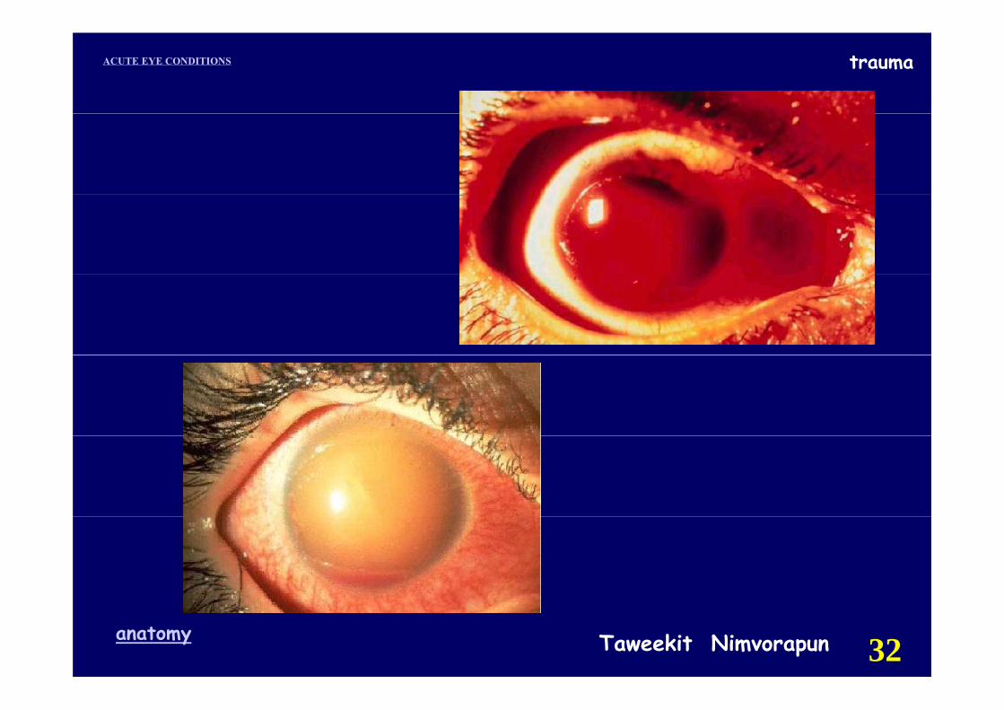
traumaACUTE EYE CONDITIONS Traumatic hyphemaHyphema : เลือดออกใน AC จาก blunt trauma C li ti lComplication : glaucoma
blood stain ที่ corneaSymptoms : Pain, Blurred vision
history of traumahistory of traumaSign : Blood in the AC
Work up : type of injury time Work-up : type of injury, time, R/O ruptured globe VA , IOP, level of hyphema Fundus ถาทําได U/S R/O RD
Taweekit Nimvorapunanatomy 33
Fundus ถาทาได , U/S R/O RD
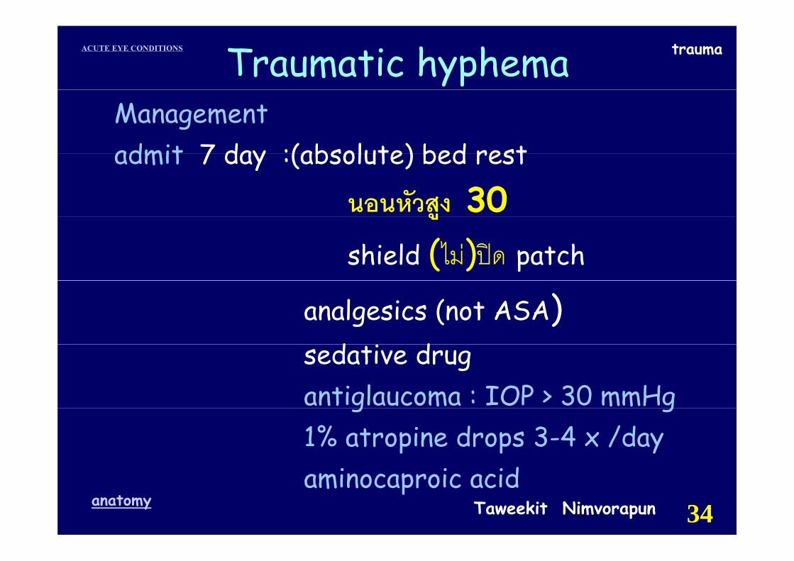
traumaACUTE EYE CONDITIONS Traumatic hyphemaManagementadmit 7 day :(absolute) bed restadmit 7 day :(absolute) bed rest
นอนหวัสูง 30ูshield (ไม)ปด patch
analgesics (not ASA) d dsedative drug
antiglaucoma : IOP > 30 mmHgg g1% atropine drops 3-4 x /day
i i idTaweekit Nimvorapunanatomy 34
aminocaproic acid
traumaACUTE EYE CONDITIONS Traumatic hyphema
Management
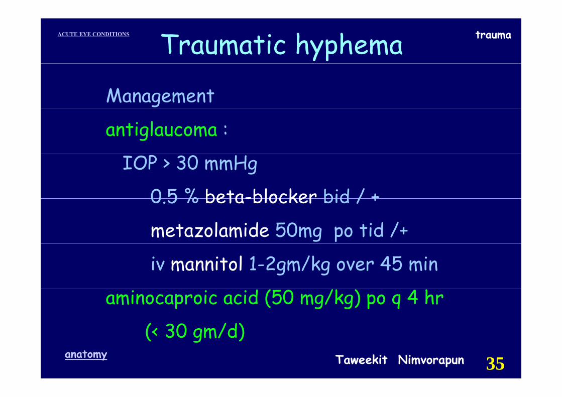
antiglaucoma :
IOP > 30 mmHg
0 5 % beta-blocker bid / +0.5 % beta-blocker bid / +
metazolamide 50mg po tid /+
iv mannitol 1-2gm/kg over 45 min
aminocaproic acid (50 mg/kg) po q 4 hr
(< 30 gm/d)Taweekit Nimvorapunanatomy 35
(< 30 gm/d)
traumaACUTE EYE CONDITIONS Traumatic hyphema
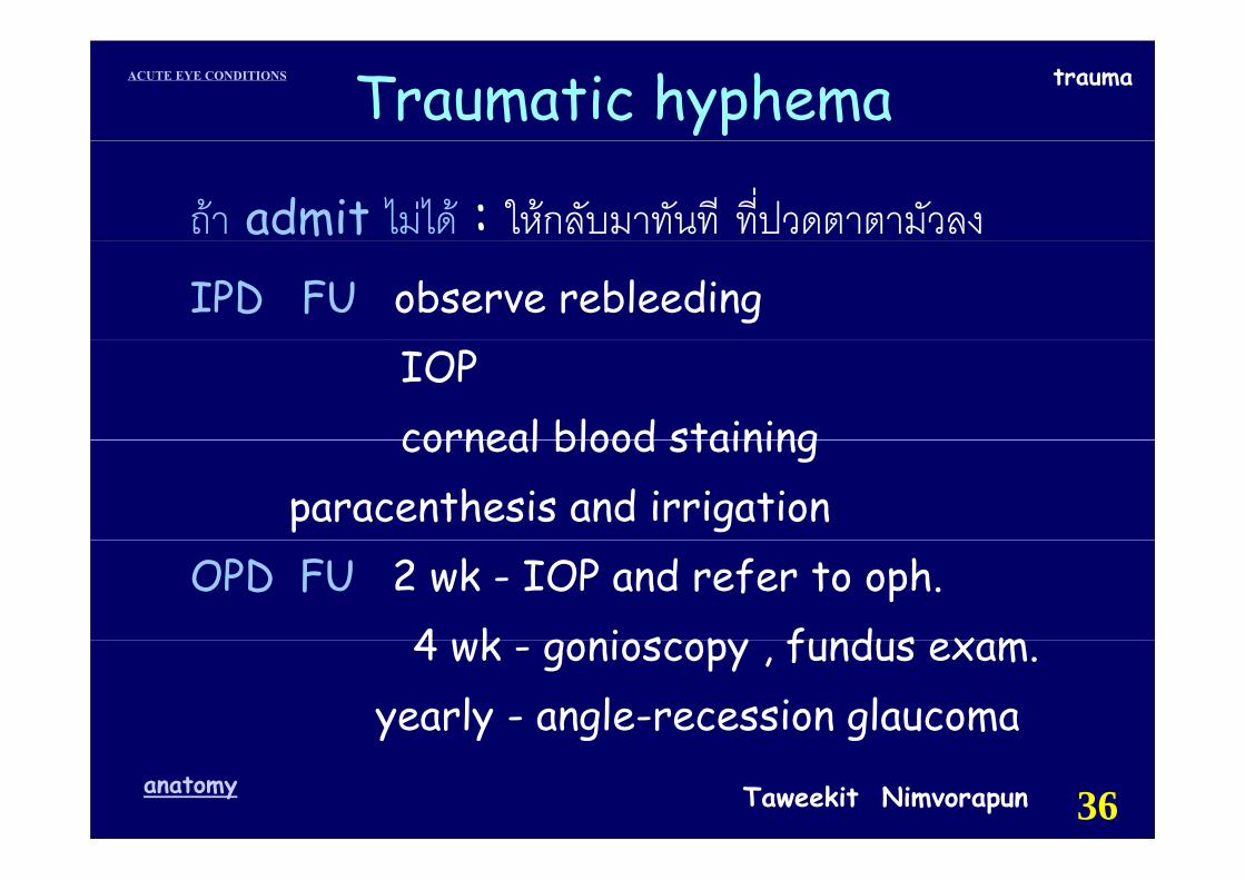
ถา admit ไมได : ใหกลับมาทันที ที่ปวดตาตามัวลงIPD FU observe rebleeding
IOP corneal blood stainingcorneal blood staining
paracenthesis and irrigation OPD FU 2 wk - IOP and refer to oph.
4 wk gonioscopy fundus exam4 wk - gonioscopy , fundus exam.yearly - angle-recession glaucoma
Taweekit Nimvorapunanatomy 36
y y g g

traumaACUTE EYE CONDITIONS
Hyphema managementyp g
☺ R/O globe is ruptured☺ R/O globe is ruptured☺ Shield eye y☺ Restricted activity☺ symptomatic Rx☺ T i l l l i & ti t id☺ Topical cycloplegic & corticosteroids☺ Possibly aminocaproic acid ☺ Possibly aminocaproic acid ☺ refer to ophthalmologist
Taweekit Nimvorapunanatomy 37
p g
traumaACUTE EYE CONDITIONS
Ocular foreign body
Taweekit Nimvorapunanatomy 38
traumaACUTE EYE CONDITIONS
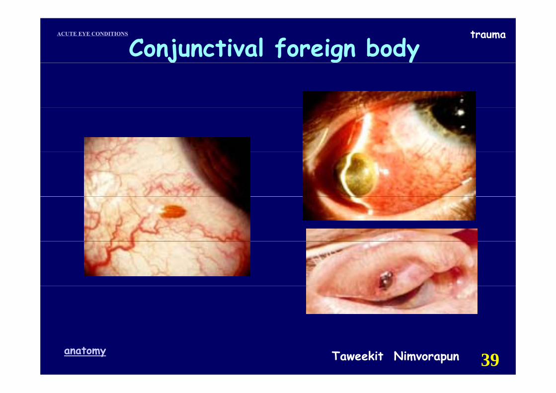
Conjunctival foreign body
Taweekit Nimvorapunanatomy 39
traumaACUTE EYE CONDITIONSConjunctival foreign bodyj g ySymptoms : Ocular irritation or pain
FB sensation tearing red eyetearing , red eye
Hx: trauma or FB to the eye.m ySigns : Linear,vertical scratches
(FB- upper eyelid )subconj hemorrhagesubconj.hemorrhage
Dx tests: fluorescein - scratchTaweekit Nimvorapunanatomy 40
traumaACUTE EYE CONDITIONSConjunctival foreign body
Managementgtopical anesthesia
irrigationirrigationcotton-tipped applicatorfi ffine forceps
artificial tear
antibiotic EO : PEE , abrasion
FU วันรุงขึน้ในรายทีม่ี residual FB
Taweekit Nimvorapunanatomy 41
traumaACUTE EYE CONDITIONS
Taweekit Nimvorapunanatomy 42
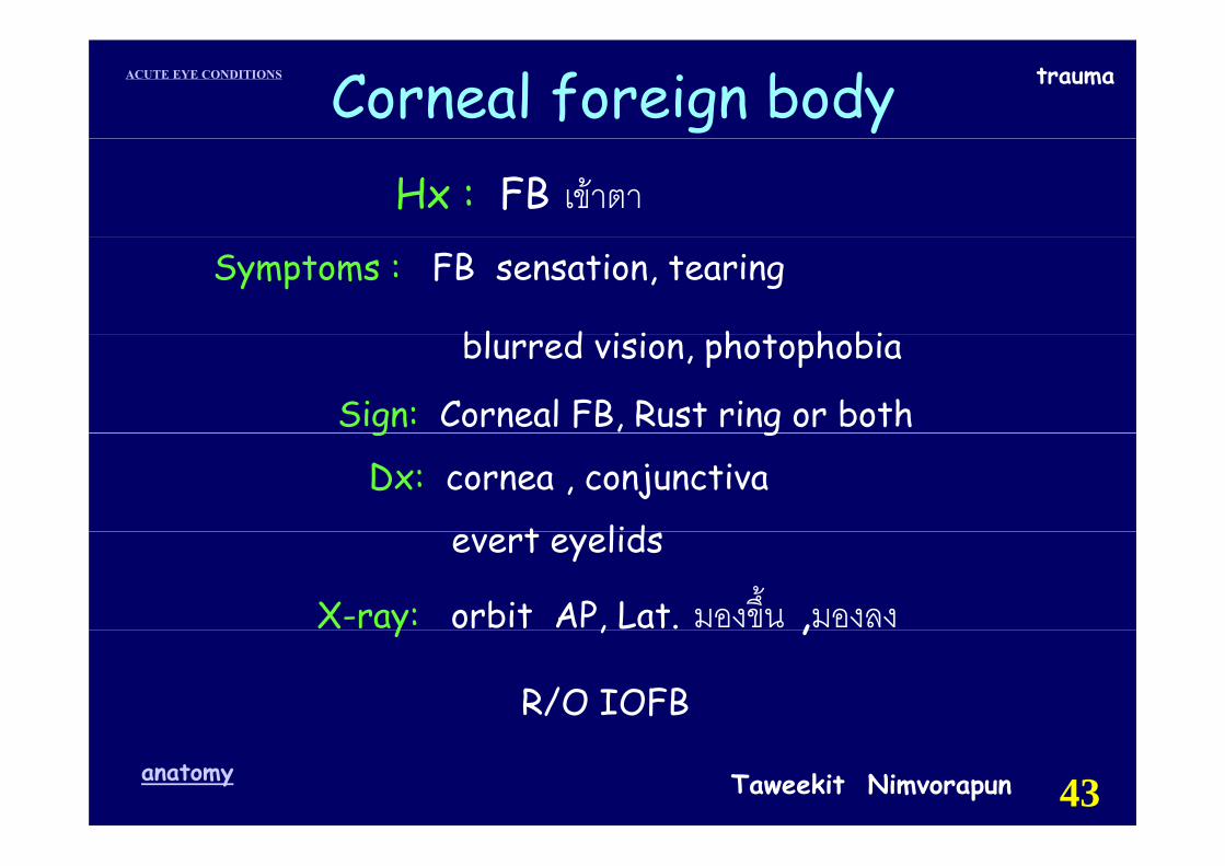
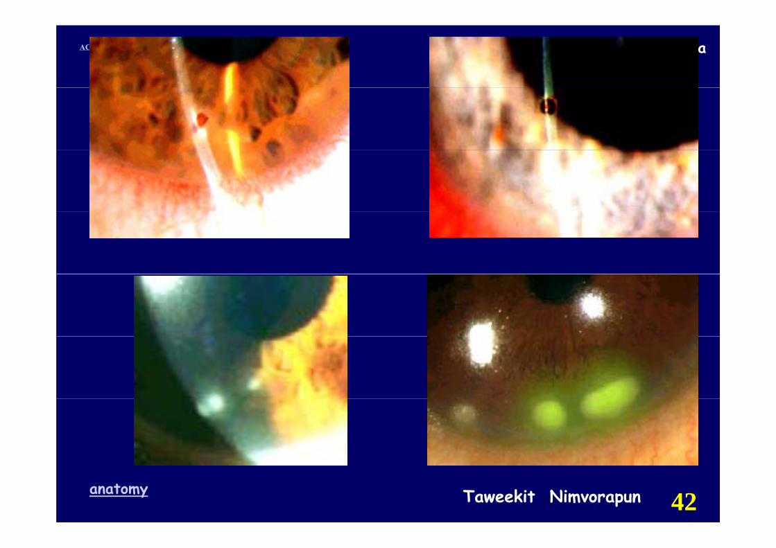
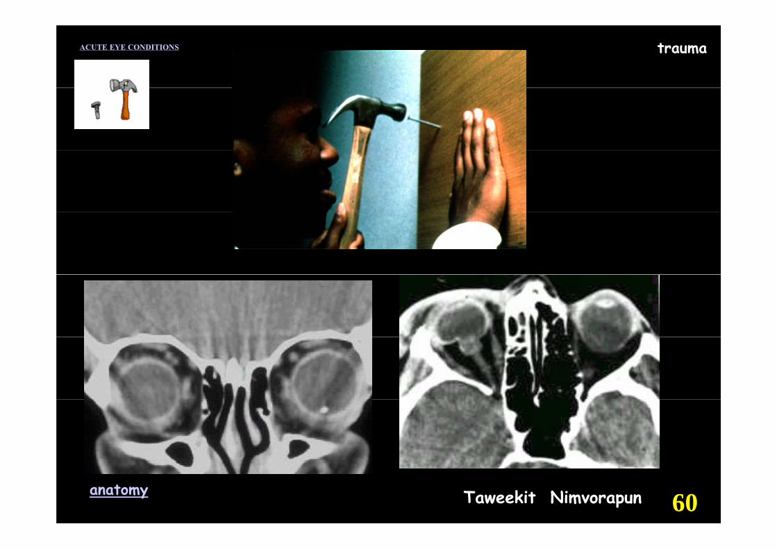
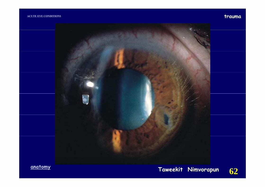
traumaACUTE EYE CONDITIONS Corneal foreign bodyHx : FB เขาตา
Symptoms : FB sensation, tearing
bl d i i h t h biblurred vision, photophobia
Sign: Corneal FB, Rust ring or bothg g
Dx: cornea , conjunctiva
t lidevert eyelids
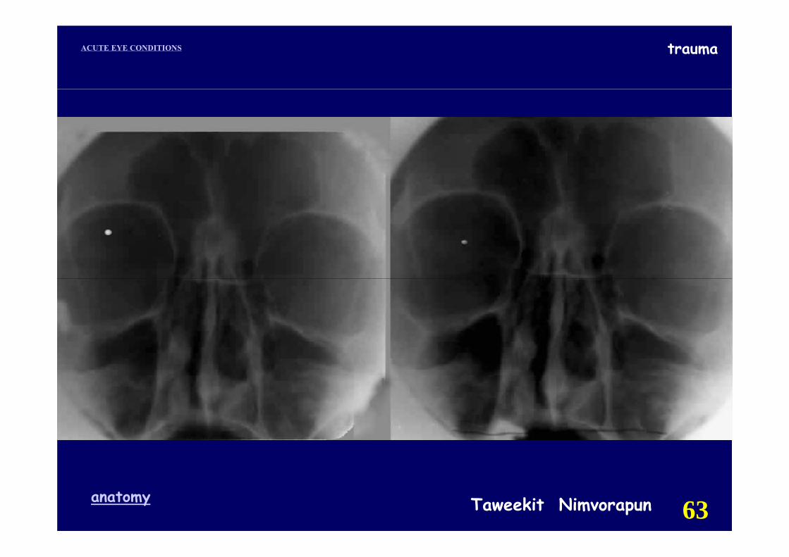
X-ray: orbit AP, Lat. มองขึ้น ,มองลงy , ,
R/O IOFB
Taweekit Nimvorapunanatomy 43
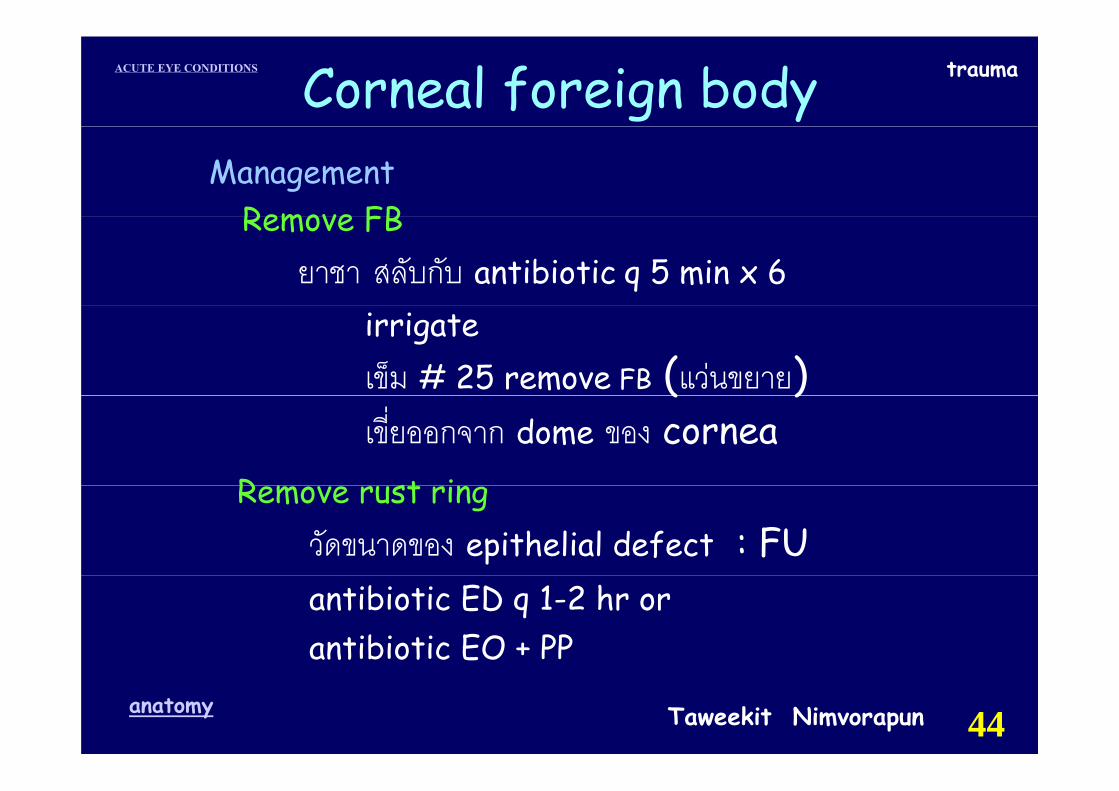
traumaACUTE EYE CONDITIONS Corneal foreign bodyManagement
Remove FBRemove FBยาชา สลับกับ antibiotic q 5 min x 6
irrigate เข็ม # 25 remove FB (แวนขยาย)( )เขี่ยออกจาก dome ของ cornea
R m st iRemove rust ringวัดขนาดของ epithelial defect : FUantibiotic ED q 1-2 hr orantibiotic EO + PP
Taweekit Nimvorapunanatomy 44
traumaACUTE EYE CONDITIONS
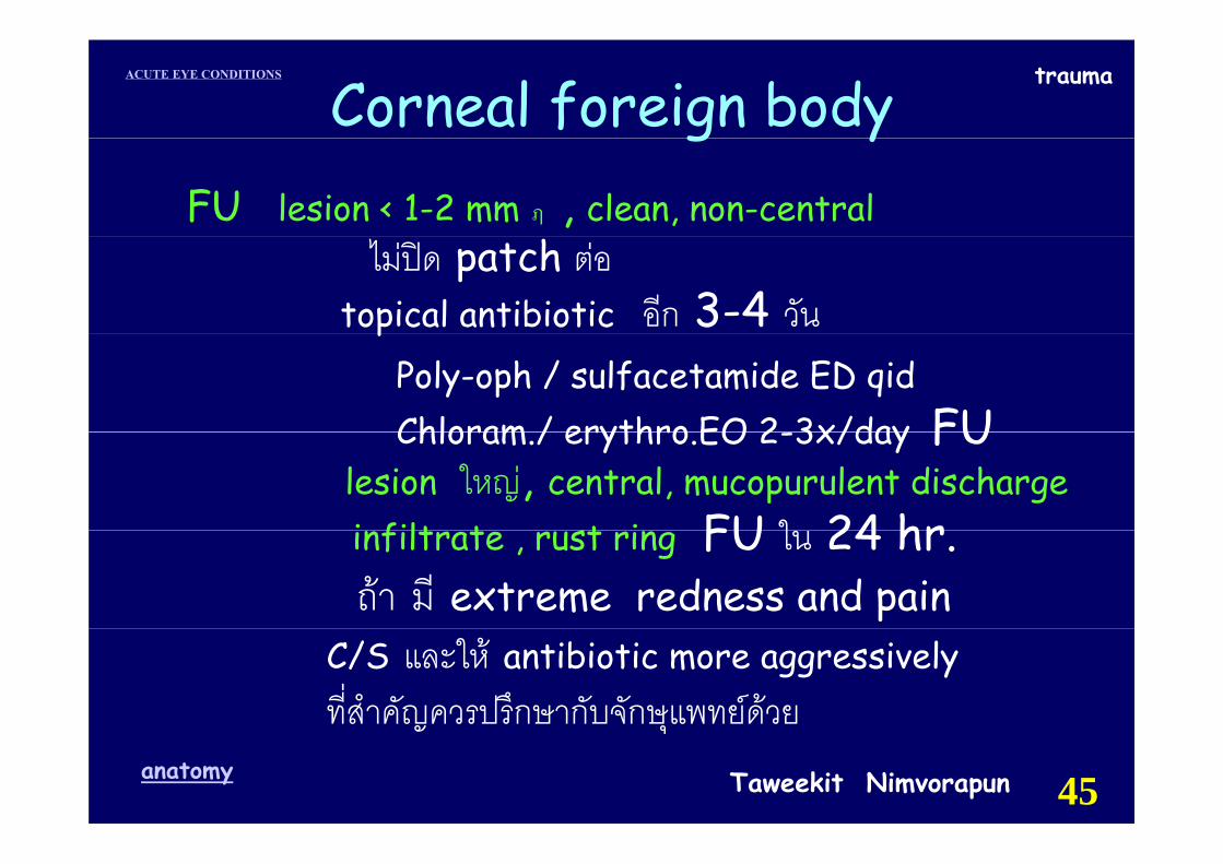
Corneal foreign bodyFU lesion < 1-2 mm ฦ , clean, non-central
g y
ไมปด patch ตอtopical antibiotic อีก 3-4 วันp
Poly-oph / sulfacetamide ED qidChloram / erythro EO 2 3x/day FUChloram./ erythro.EO 2-3x/day FU
lesion ใหญ, central, mucopurulent discharge i filt t st i FU ใ 24 hrinfiltrate , rust ring FU ใน 24 hr.ถา มี extreme redness and pain
C/S และให antibiotic more aggressivelyที่สําคัญควรปรึกษากับจักษุแพทยดวย
Taweekit Nimvorapunanatomy 45
ญ ุ
traumaACUTE EYE CONDITIONS
Taweekit Nimvorapunanatomy 46
traumaACUTE EYE CONDITIONS
Taweekit Nimvorapunanatomy 47
traumaACUTE EYE CONDITIONS
Intraorbital foreign bodyIntraorbital foreign body
Taweekit Nimvorapunanatomy 48
traumaACUTE EYE CONDITIONS
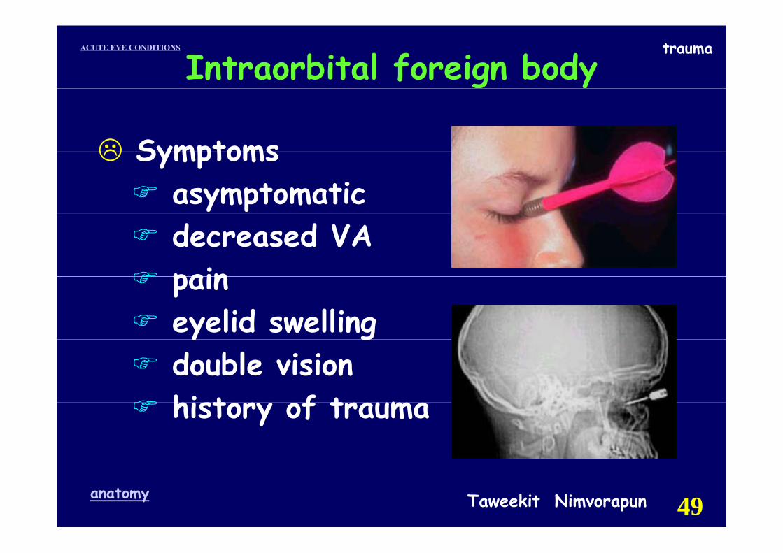
Intraorbital foreign body
SymptomsSymptomsasymptomatic decreased VA painpaineyelid swellingy gdouble vision hi t f t mhistory of trauma
Taweekit Nimvorapunanatomy 49
traumaACUTE EYE CONDITIONS
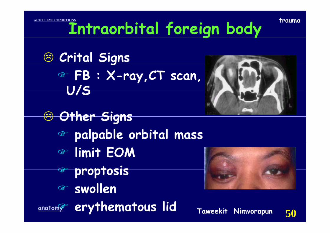
Intraorbital foreign bodyg y
Crital SignsgFB : X-ray,CT scan,
U/S U/S
Other SignsOther Signspalpable orbital masslimit EOMproptosisproptosisswollen
Taweekit Nimvorapunanatomy 50erythematous lid
traumaACUTE EYE CONDITIONS
Intraorbital foreign bodyg y
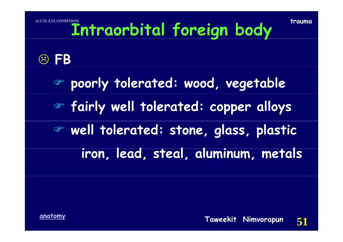
FB
poorly tolerated: wood, vegetable
fairly well tolerated: copper alloys
well tolerated: stone, glass, plastic
i l d l l i l iron, lead, steal, aluminum, metals
Taweekit Nimvorapunanatomy 51
traumaACUTE EYE CONDITIONS
Intraorbital foreign bodyg y
Work upWork-up
history : FB ? timehistory : FB ?, time
complete exam : RAPD IOP Funduscomplete exam.: RAPD , IOP , Fundus
CT scan: R/O ruptured globe CT scan: R/O ruptured globe
location of FB location of FB
Taweekit Nimvorapunanatomy 52
traumaACUTE EYE CONDITIONS
Intraorbital foreign bodyg y
Indication : exploration & extractionIndication : exploration & extractionsigns of infection signs of infection fistula formationsigns of optic nerve compressionsevere inflammationlarge/sharp FB & easily extractedlarge/sharp FB & easily extracted
Taweekit Nimvorapunanatomy 53
traumaACUTE EYE CONDITIONS
Intraorbital foreign bodyTreatment
h i li i
g y
hospitalizationsystemic antibioticssystemic antibioticstetanus toxoidsurgery when indicated
F llFollow-upVA , RAPD , IOP, EOM , proptosis, , , , p poral antibiotic 10-14 d
Taweekit Nimvorapunanatomy 54
traumaACUTE EYE CONDITIONSACUTE EYE CONDITIONS
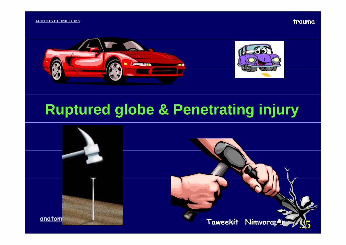
Ruptured globe & Penetrating injury
Taweekit Nimvorapunanatomy 55
traumaACUTE EYE CONDITIONS
Taweekit Nimvorapunanatomy 56
traumaACUTE EYE CONDITIONS trauma
Taweekit Nimvorapunanatomy 57

traumaACUTE EYE CONDITIONS
Taweekit Nimvorapunanatomy 58
traumaACUTE EYE CONDITIONS
Taweekit Nimvorapunanatomy 59
traumaACUTE EYE CONDITIONS
Taweekit Nimvorapunanatomy 60

traumaACUTE EYE CONDITIONS
Taweekit Nimvorapunanatomy 61
traumaACUTE EYE CONDITIONS
Taweekit Nimvorapunanatomy 62
traumaACUTE EYE CONDITIONS
Taweekit Nimvorapunanatomy 63
traumaACUTE EYE CONDITIONS
Taweekit Nimvorapunanatomy 64
traumaACUTE EYE CONDITIONS
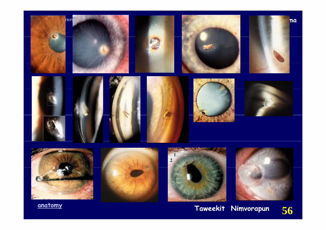
Ruptured globe & Penetrating injuryp g g j y☺ Symptoms : pain , decreased vision,
history of trauma
☺ Signs: Ruptured globe
hypotony (not always present)
se ere s bconj edema & hemorrhagesevere subconj. edema & hemorrhage
intraocular contents may be outside the globey g
limitation of extraocular motility
Taweekit Nimvorapunanatomy 65
traumaACUTE EYE CONDITIONS
Ruptured globe & Penetrating injuryp g g j y☺ Penetrating injury
full thickness scleral and corneal laceration
sign of ruptured globe
history of sharp object entering the globehistory of sharp object entering the globe
☺ Other Signs☺ Other Signs
irregular pupil , iridodialysisg p p , y
periorbital echymosis, subluxed lens
Taweekit Nimvorapunanatomy 66
traumaACUTE EYE CONDITIONS
Ruptured globe & Penetrating injury
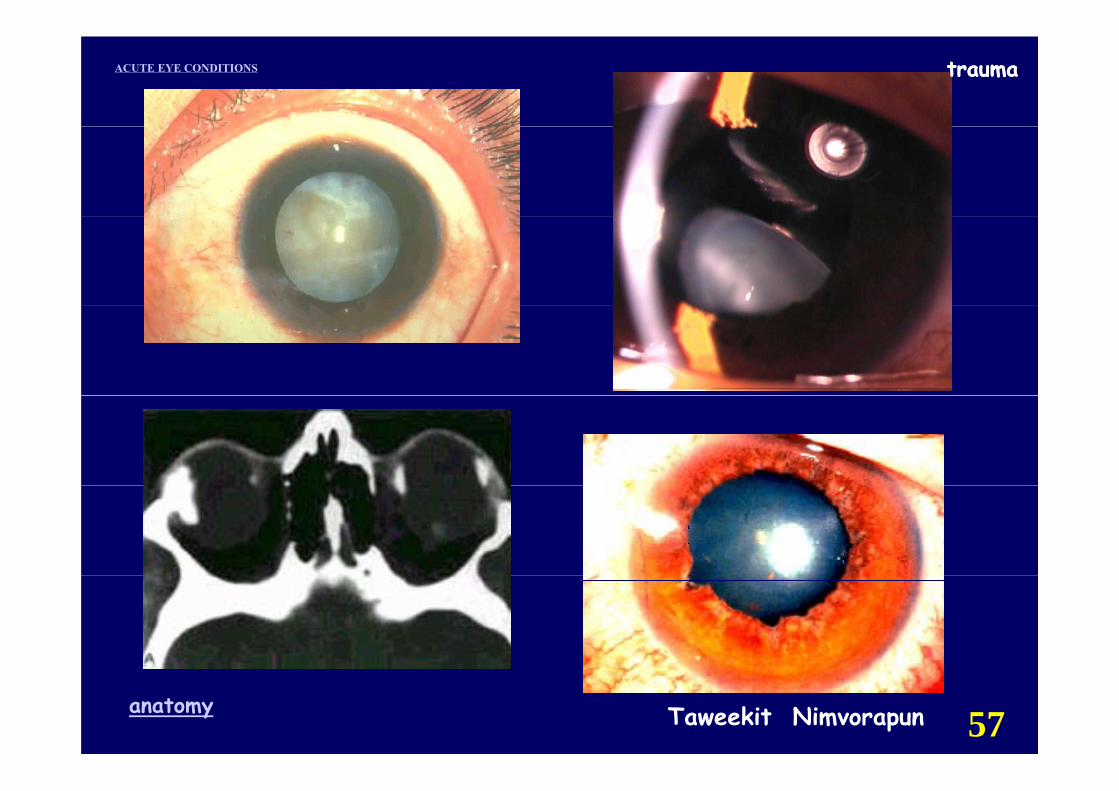
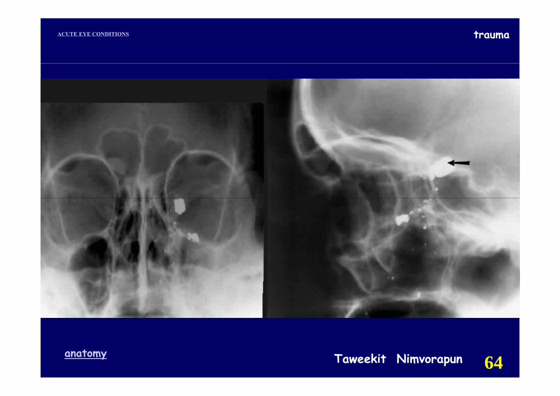
☺ Diagnostic test☺ Diagnostic test
film orbit AP & Lateral : R/O IOFBfilm orbit AP & Lateral : R/O IOFB
CT scanning may be helpfulCT scanning may be helpful.
show a shrunken globe. g
shows subconjunctival edema.
R/O IOFB
Taweekit Nimvorapunanatomy 67
traumaACUTE EYE CONDITIONS
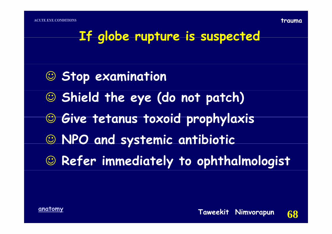
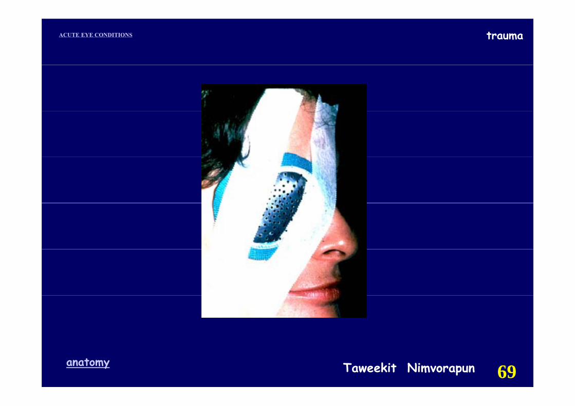
If globe rupture is suspectedIf globe rupture is suspected
☺ Stop examination☺ Shield the eye (do not patch)☺ Give tetanus toxoid prophylaxis☺ Give tetanus toxoid prophylaxis☺ NPO and systemic antibiotic☺ NPO and systemic antibiotic☺ Refer immediately to ophthalmologist
Taweekit Nimvorapunanatomy 68
traumaACUTE EYE CONDITIONS
Taweekit Nimvorapunanatomy 69
traumaACUTE EYE CONDITIONS trauma
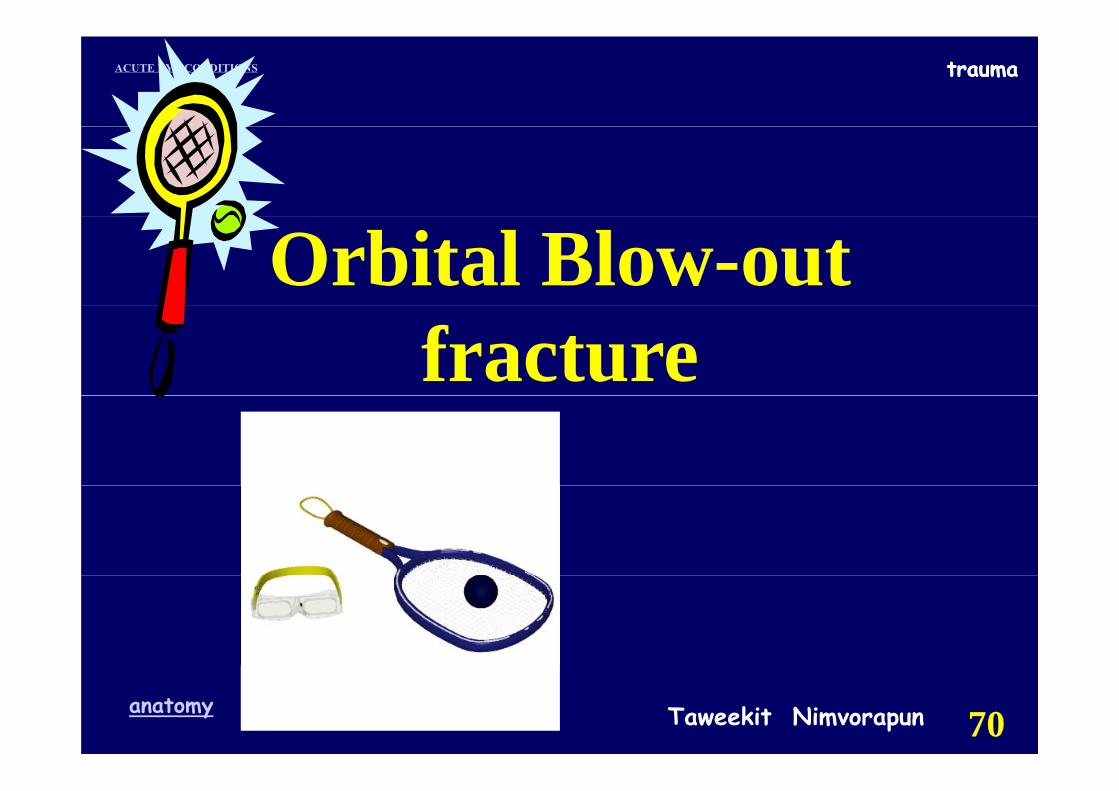
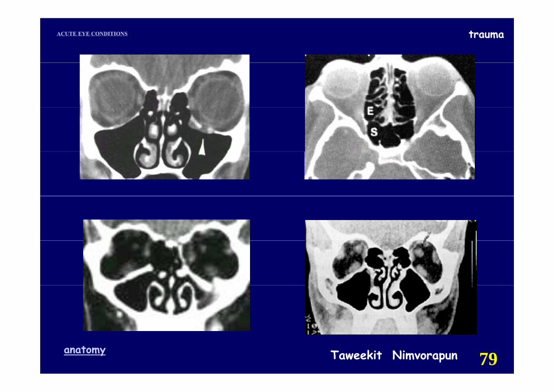
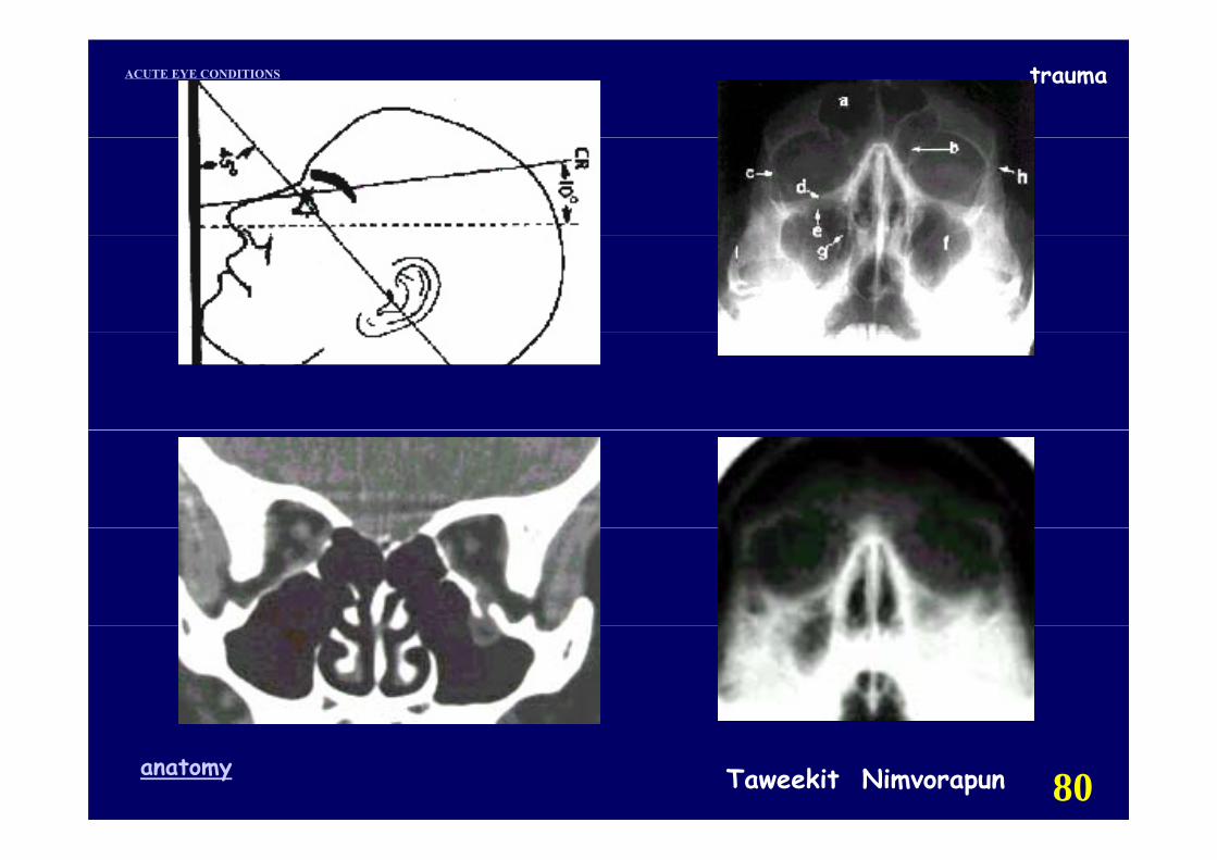
Orbital Blow-out fracture
Taweekit Nimvorapunanatomy 70

traumaACUTE EYE CONDITIONS
Taweekit Nimvorapunanatomy 71
traumaACUTE EYE CONDITIONS
Taweekit Nimvorapunanatomy 72
traumaACUTE EYE CONDITIONS
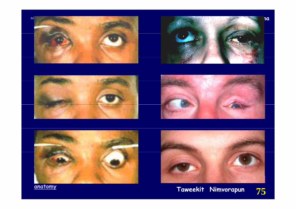
Orbital Blow-out fractureOrbital Blow-out fracture
☺ S t☺ SymptomPain (vertical eye movement)( y )local tendernessbi l d bl i ibinocular double visioneyelid swelling after nose blowingeyelid swelling after nose blowingrecent history of trauma.
Taweekit Nimvorapunanatomy 73
traumaACUTE EYE CONDITIONS
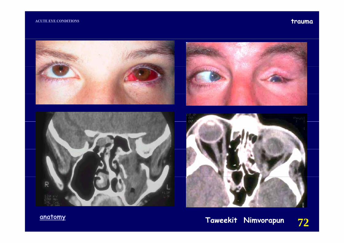
Orbital Blow-out fracture
☺ Signs
restricted eye movement (upward/lateral gaze)
orbital subcutaneous emphysema
hypesthesia (intraorbital nerve)hypesthesia (intraorbital nerve)
enophthalmos (masked by orbital edema).
☺ Other Signs
nosebleed, lid edema, ecchymosis, ptosis
Taweekit Nimvorapunanatomy 74
traumaACUTE EYE CONDITIONS
Taweekit Nimvorapunanatomy 75
traumaACUTE EYE CONDITIONS
Orbital Blow-out fracture
☺ Diff ti l Di i☺ Differential DiagnosisOrbital edema and hemorrhage(limitation of EOM, but resolve > 7-10 d.)Cranial nerve palsyCranial nerve palsy(limitation of EOM, no restriction on forced d ti t ti )duction testing.)
Taweekit Nimvorapunanatomy 76
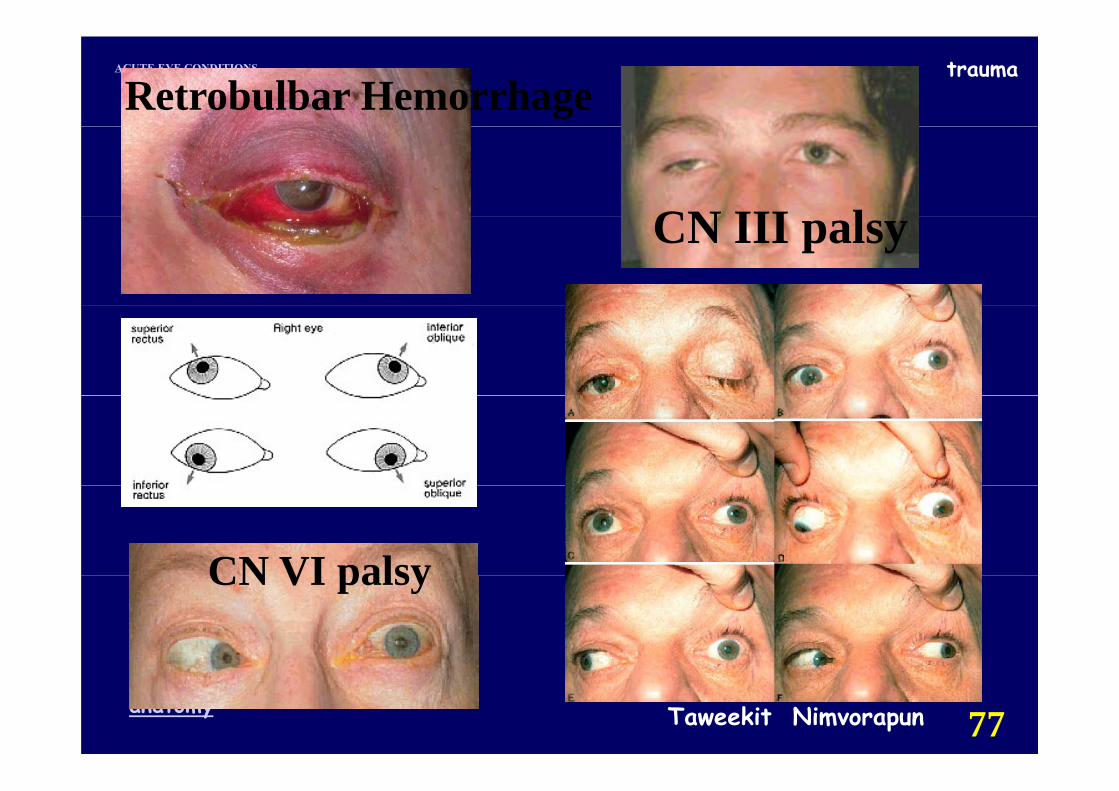
traumaACUTE EYE CONDITIONS
Retrobulbar Hemorrhage
CN III lCN III palsy
CN VI palsyCN VI palsy
Taweekit Nimvorapunanatomy 77
traumaACUTE EYE CONDITIONS
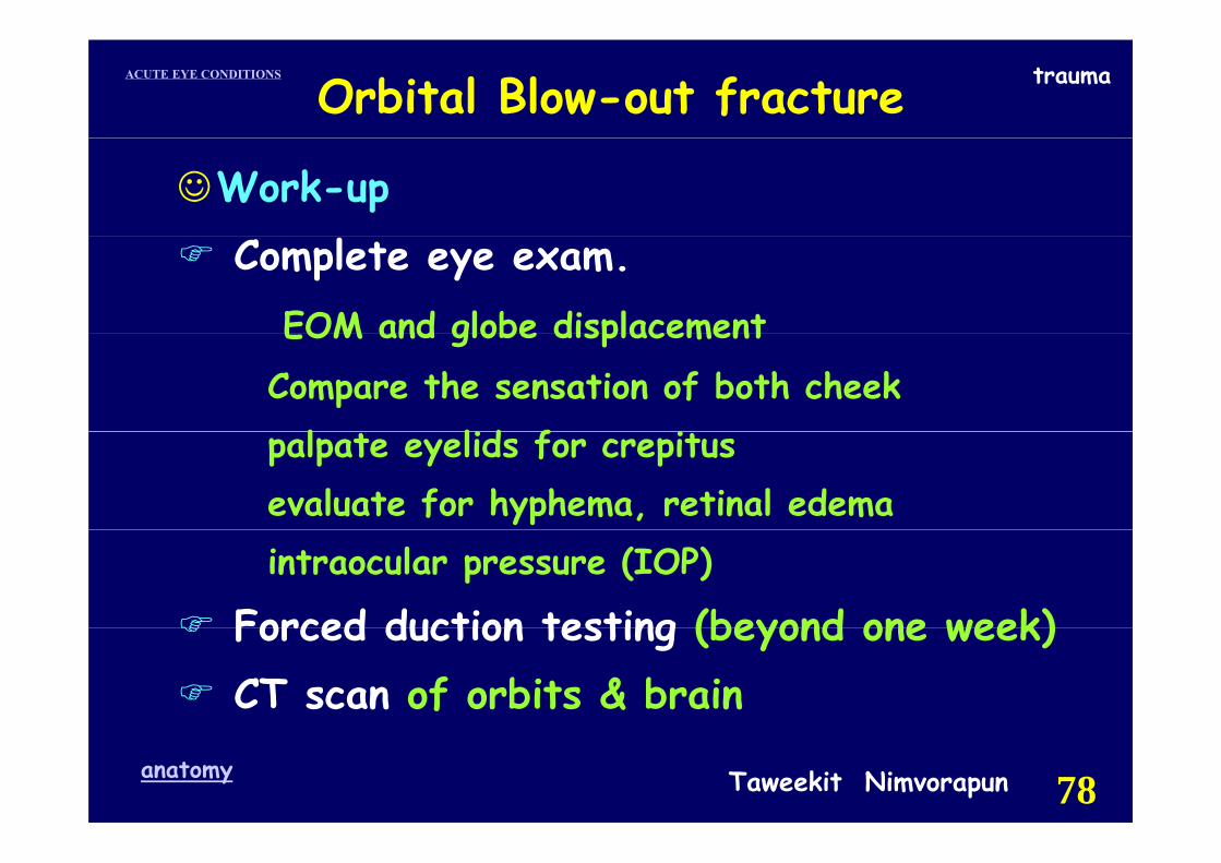
Orbital Blow-out fracture
☺Work-upComplete eye exam.
EOM and globe displacementEOM and globe displacementCompare the sensation of both cheek
l lid f ipalpate eyelids for crepitusevaluate for hyphema, retinal edemaintraocular pressure (IOP)
Forced duction testing (beyond one week)Forced duction testing (beyond one week)CT scan of orbits & brain
Taweekit Nimvorapunanatomy 78
traumaACUTE EYE CONDITIONS
Taweekit Nimvorapunanatomy 79
traumaACUTE EYE CONDITIONS
Taweekit Nimvorapunanatomy 80
traumaACUTE EYE CONDITIONS
Orbital Blow-out fracture
☺Treatment 10-14 d☺Treatment 10 14 dNasal decongestants(Afrin nasal spray bid)Broad-spectrum oral antibiotics:
cephalexin250-500 mg po qid orcephalexin250-500 mg po qid orerythromycin 250-500 mg po qid
Instruct the patient not to blow his nose.Ice packs to the orbit for the first 24 48hr Ice packs to the orbit for the first 24-48hr. Re-examination at 10-14 d after trauma
Taweekit Nimvorapunanatomy 81
traumaACUTE EYE CONDITIONS
Orbital Blow out fractureOrbital Blow-out fracture
☺ Surgical indications (controversy)persist entrapment of orbital contents persist entrapment of orbital contents diplopia within 30 degrees of primary positionpositive forced duction test & X-ray :entrapmentcosmetically unacceptable enophthalmosfractures (1/2 of orbital floor ,large medial wall
fibrosis & contracture of prolapsed tissuefibrosis & contracture of prolapsed tissue
Taweekit Nimvorapunanatomy 82
traumaACUTE EYE CONDITIONS
T ti O ti N thTraumatic Optic Neuropathy
Taweekit Nimvorapunanatomy 83
traumaACUTE EYE CONDITIONS trauma
Taweekit Nimvorapunanatomy 84
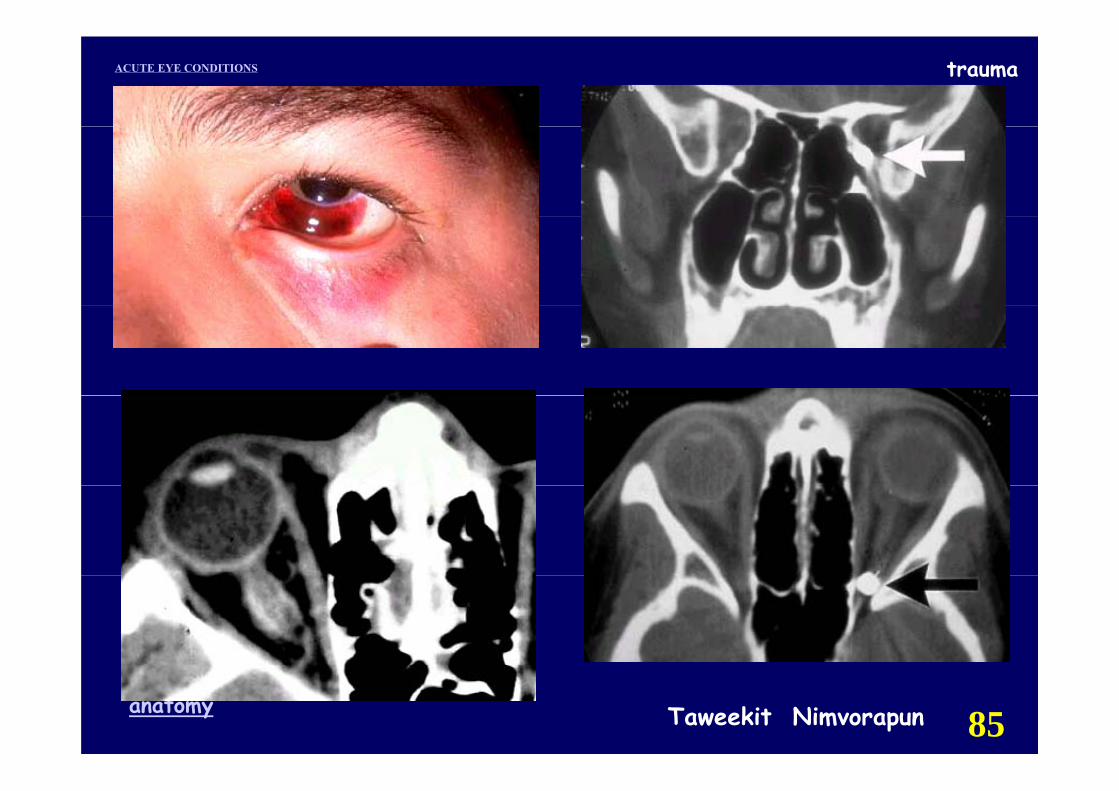
traumaACUTE EYE CONDITIONS
Taweekit Nimvorapunanatomy 85
traumaACUTE EYE CONDITIONS
Taweekit Nimvorapunanatomy 86
traumaACUTE EYE CONDITIONS
Taweekit Nimvorapunanatomy 87
traumaACUTE EYE CONDITIONS
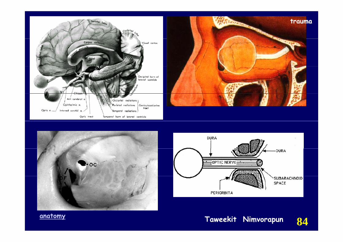
Traumatic Optic NeuropathyTraumatic Optic Neuropathy
☺ Symptoms : VA after trauma
☺ Critical Signs: new RAPD +
☺ Others Signs: relatively poor color
vision VF defect
Taweekit Nimvorapunanatomy 88
traumaACUTE EYE CONDITIONS
Traumatic Optic Neuropathy
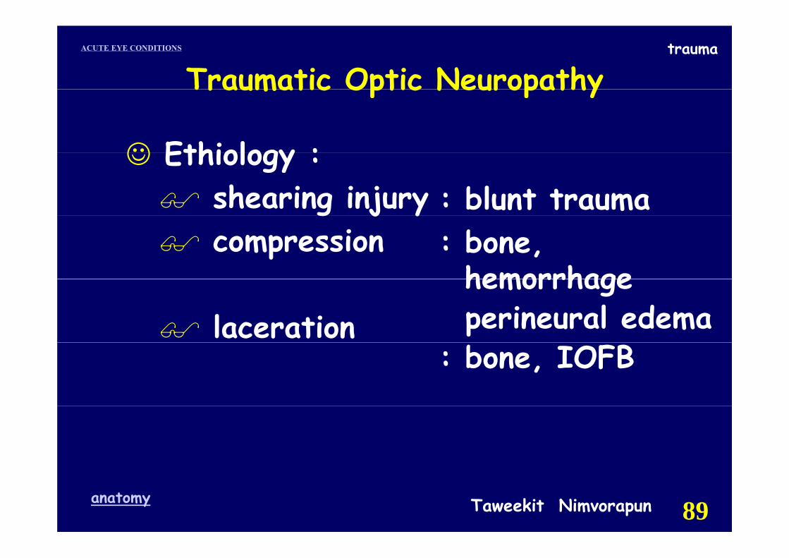
☺ Ethiology :
Traumatic Optic Neuropathy
☺ Ethiology :shearing injury : blunt traumag j ycompression : bone,
hemorrhagelaceration
hemorrhageperineural edema
: bone, IOFB
Taweekit Nimvorapunanatomy 89
traumaACUTE EYE CONDITIONS
Traumatic Optic NeuropathyTraumatic Optic Neuropathy
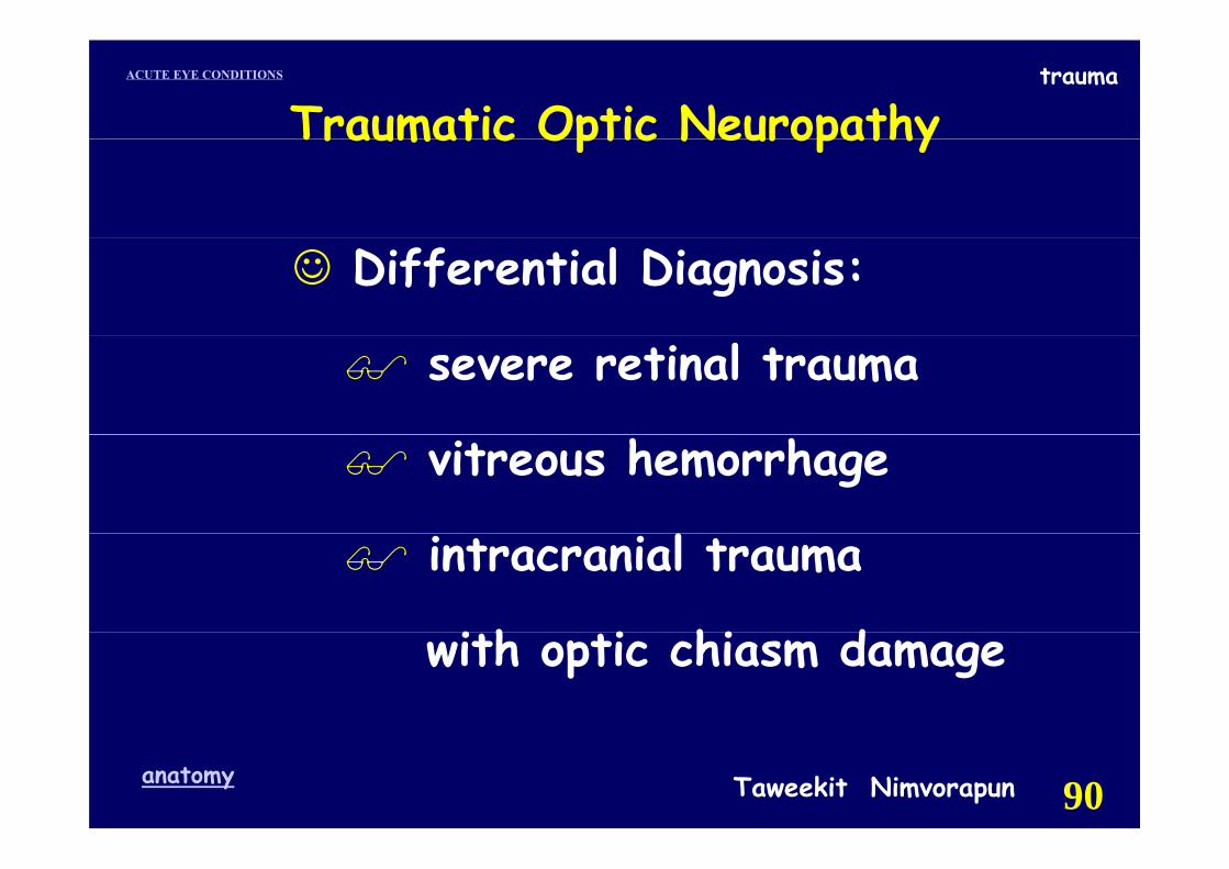
☺ Differential Diagnosis:
severe retinal trauma
vitreous hemorrhage
intracranial trauma
i h i hi d with optic chiasm damage
Taweekit Nimvorapunanatomy 90
traumaACUTE EYE CONDITIONS
Traumatic Optic Neuropathy
☺ Work up :
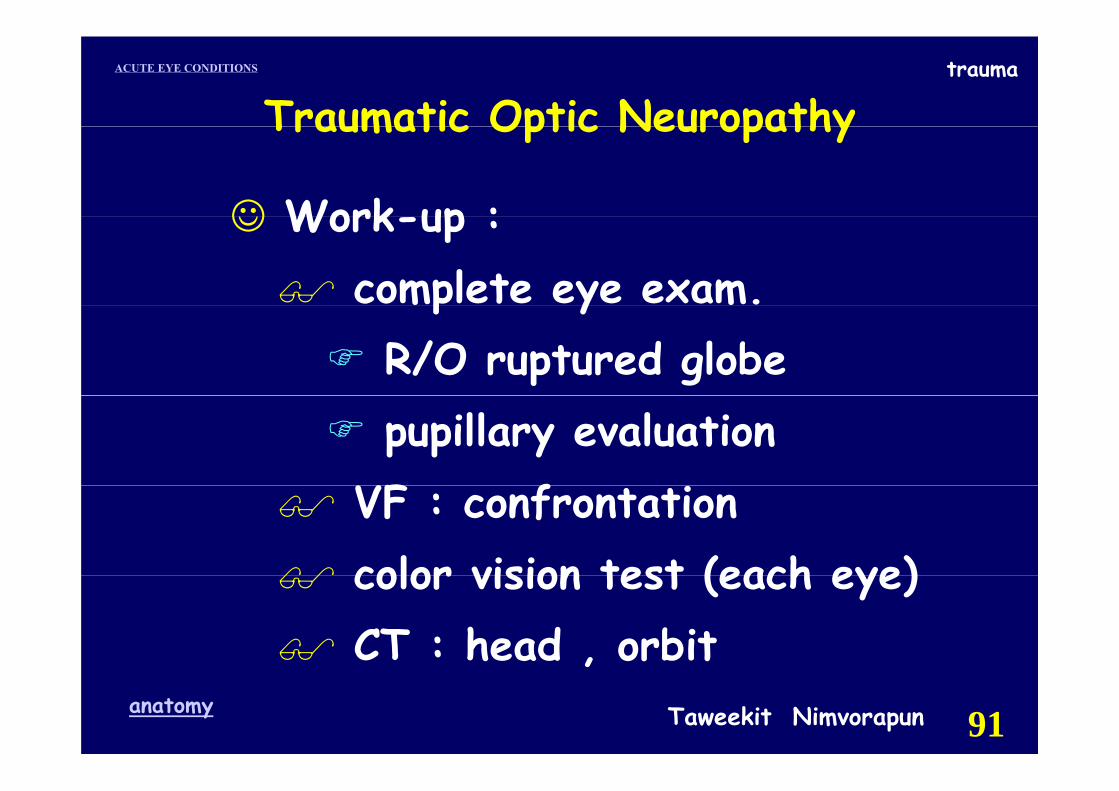
Traumatic Optic Neuropathy
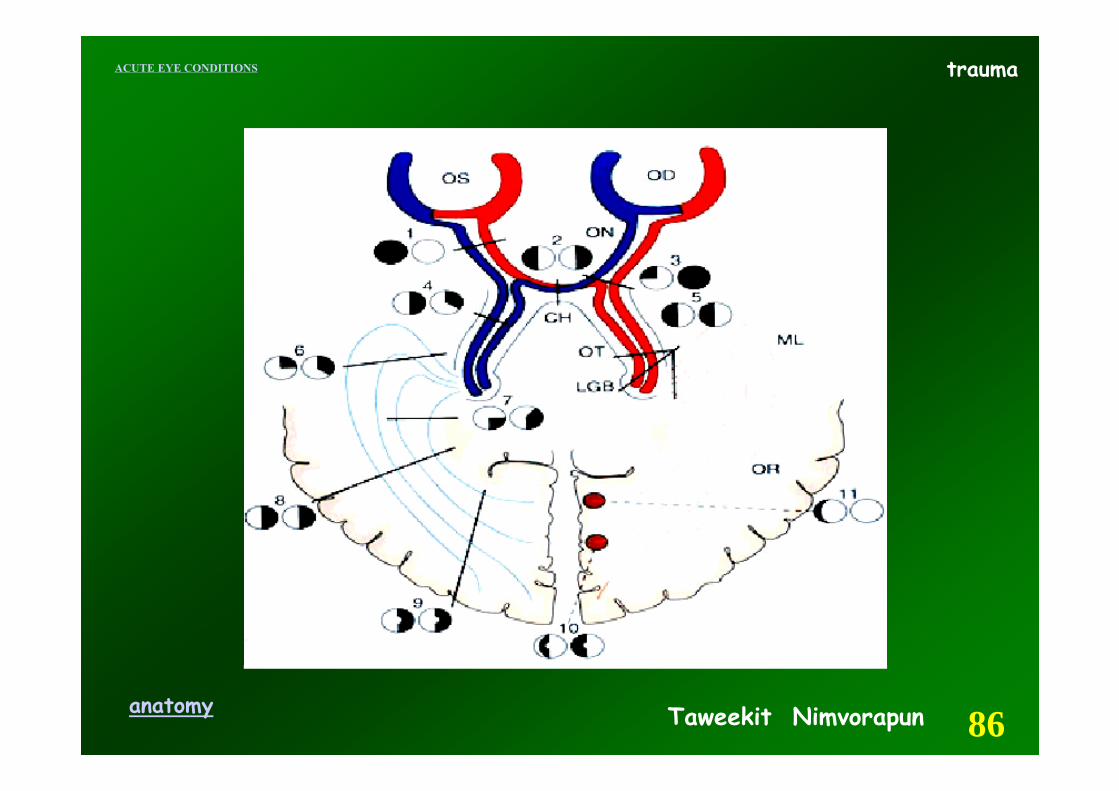
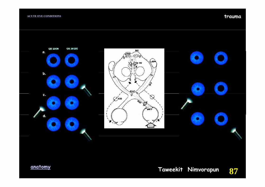
☺ Work-up :complete eye exam.p yR/O ruptured globepupillary evaluation
VF fVF : confrontationcolor vision test (each eye)color vision test (each eye)CT : head , orbit
Taweekit Nimvorapunanatomy 91
,
traumaACUTE EYE CONDITIONS
Traumatic Optic Neuropathy
☺ Treatment :
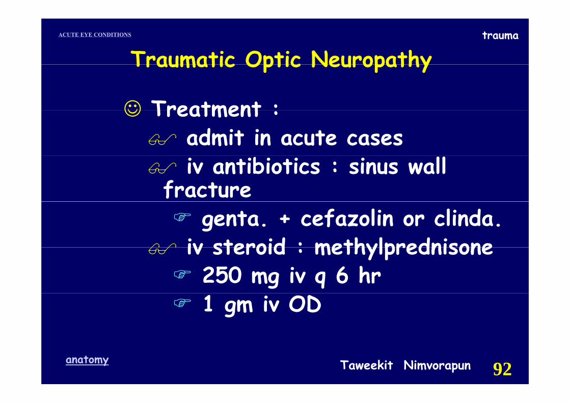
Traumatic Optic Neuropathy
☺ Treatment :admit in acute cases iv antibiotics : sinus wall
fracture genta. + cefazolin or clinda.
iv steroid : methylprednisone iv steroid : methylprednisone 250 mg iv q 6 hr1 gm iv OD
Taweekit Nimvorapunanatomy 92
traumaACUTE EYE CONDITIONS
Traumatic Optic NeuropathyTraumatic Optic Neuropathy
☺ FU :daily daily evaluate VA if refer evaluate VA if refer pupillary reactionscolor vision
Taweekit Nimvorapunanatomy 93