low back pain in adolescent athletes -...
TRANSCRIPT

Low back pain in Adolescent
Athletes
Claes Göran Sundell
Department of Community Medicine and Rehabilitation Umeå 2019
1
Responsible publischer under Swedish law: The Dean of the Medical Faculty This work is protected by the Swedish Copyright Legislation (Act 1960:729) Dissertation for PhD ISBN: 978-91-7855-024-1 ISSN-0346-6612 New series no: 2014 Copyright © Claes Göran Sundell, 2019 Cover illustration: Stina Norgren, Illustre.se Illustrations: Stina Norgren, Illustre.se, page 1,3,4,12,13,41 Illustration page 2: Permission Encyplopedica Brittanica, page 2 Pictures: 3D4 Medical; www.3D4Medical.com, page 1,3,5,6,7
Article nr .3 "© Georg Thieme Verlag KG."
All previously published papers were reproduced with the kind permission of the publishers
Electronic version available at: http://umu.diva-portal.org/ Printed by: UmU Print Service, Umeå University Umeå, Sweden, 2019

“Listen to the Sound of Silence”
Unknown
To my family:
Doris, Cecilia, David, Johan

i
Contents Abstract .......................................................................................... iv Sammanfattning på svenska ........................................................... vi Preface .......................................................................................... viii Abbreviations ................................................................................. ix Introduction .................................................................................... 1
1.Anatomy ......................................................................................................................... 1 1.1 The skeleton .......................................................................................................... 2 1.2 The Spine ...............................................................................................................3 1.3 The lower back ..................................................................................................... 4 1.4 The Functional Unit of the Spine ........................................................................ 4 1.5 The vertebrae ........................................................................................................ 5 1.6 The Pars interarticularis of the Vertebrae ......................................................... 6 1.7 The Zygapophysial facets of the Vertebrae ......................................................... 7 1.8 Apophysis .............................................................................................................. 7
2. Functional anatomy ...................................................................................................... 7 2.1 Spinal Load .......................................................................................................... 8
3. The historical perspective of Low back Pain .............................................................. 8 3.1 The natural history of LBP .................................................................................. 9 3.2 The history of Spondylolysis .............................................................................. 9
4. Low Back Pain.............................................................................................................. 9 4.1 Non-Specific Low Back Pain (NLBP) ................................................................ 10 4.2 Specific Low back Pain ...................................................................................... 10 4.3 Adolescent Low Back Pain (ALBP).................................................................... 10 4.4 Low Back Pain and Disability ........................................................................... 10
5. Low Back Pain in Adolescent athletes ....................................................................... 11 5.1 Spondylolysis ...................................................................................................... 11 5.2 Spondylolysis-Aetiology .................................................................................... 11 5.3 Spondylolysis-Pathogenesis .............................................................................. 11 5.4 Spondylolysis- Stress reaction .......................................................................... 12 5.5 Spondylolysis- Stressfracture (Incomplete/Complete) .................................... 13 5.6 Spondylolysis/Pseudoarthrosis ......................................................................... 13 5.7 Spondylolisthesis ................................................................................................ 13 5.8 A differential diagnosis ..................................................................................... 14
6. Aims ............................................................................................................................ 14 7. Summary of Papers ..................................................................................................... 15
Paper 1 ...................................................................................................................... 15 Paper 2 ...................................................................................................................... 15 Paper 3 ...................................................................................................................... 16 Paper 4 ...................................................................................................................... 16 Paper 5 ...................................................................................................................... 17

ii
Methods ......................................................................................... 18 Questionnaries – Study I and II ..................................................................................... 18
Epidemiology/Prevalence study - Study I .............................................................. 18 Retrospective questionnaire study – Study II ........................................................ 19
Physical examination – Study III, IV, V ....................................................................... 20 Physical examination - Study III............................................................................ 20 Functional movement test ....................................................................................... 21 Specific provocation tests ........................................................................................ 21 Physical examination – Study IV ............................................................................ 21 Physical examination – Study V ............................................................................ 22
MRI and CT – study II, III, IV, V .................................................................................. 22 Study II .................................................................................................................... 23 Study III ................................................................................................................... 23 Study IV ................................................................................................................... 24 Study V ..................................................................................................................... 24
Differential diagnosis .................................................................................................... 24 Statistical Methods ........................................................................................................ 24
Statistical analysis .................................................................................................. 24 Etichs ...............................................................................................................................25
Results ........................................................................................... 26 Results – Study I-V ........................................................................................................ 26
Study I ...................................................................................................................... 26 Prevalence ..................................................................................... 26 Duration ........................................................................................ 26 Consequences ................................................................................ 27
Study II Retrospective ................................................................................................... 30 Study III. Clinical study .................................................................................................. 31
Clinical investigation ............................................................................................... 31 Study IV. Subgroup Study III – Classification ...................................................... 33
Study V. Differential diagnostic ..................................................................................... 37 Discussion ..................................................................................... 39
Low back pain ................................................................................................................ 39 Prevalence. ............................................................................................................... 39 Low Back Pain in adolescence ................................................................................ 39 The Maturity of the skeleton (i.e lower back) ........................................................ 40 Spinal Load .............................................................................................................. 40
Low Back Pain in Adolescent athletes ............................................ 41 Injuries to Pars Interarticularis of the low back ........................................................... 42
Etiology .................................................................................................................... 42 Pathogenesis ............................................................................................................ 42 Spondylolisthesis ..................................................................................................... 43 Physical examination .............................................................................................. 43
Treatment ...................................................................................................................... 43 Differential diagnosis .................................................................................................... 44

iii
Summary ........................................................................................................................ 44 Perspective ..................................................................................................................... 45
Acknowledgments .......................................................................... 46 References ..................................................................................... 47
iv
Abstract Abstract
Background:
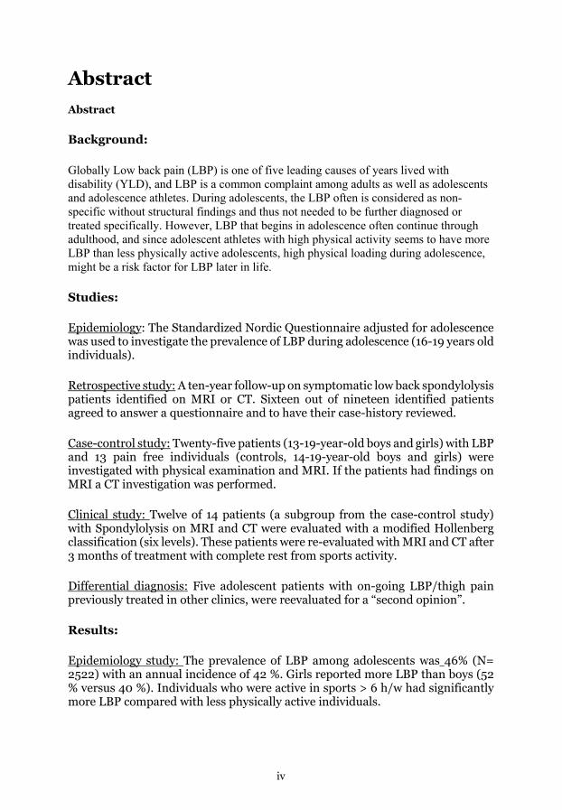
Globally Low back pain (LBP) is one of five leading causes of years lived with disability (YLD), and LBP is a common complaint among adults as well as adolescents and adolescence athletes. During adolescents, the LBP often is considered as non-specific without structural findings and thus not needed to be further diagnosed or treated specifically. However, LBP that begins in adolescence often continue through adulthood, and since adolescent athletes with high physical activity seems to have more LBP than less physically active adolescents, high physical loading during adolescence, might be a risk factor for LBP later in life.
Studies:
Epidemiology: The Standardized Nordic Questionnaire adjusted for adolescence was used to investigate the prevalence of LBP during adolescence (16-19 years old individuals).
Retrospective study: A ten-year follow-up on symptomatic low back spondylolysis patients identified on MRI or CT. Sixteen out of nineteen identified patients agreed to answer a questionnaire and to have their case-history reviewed.
Case-control study: Twenty-five patients (13-19-year-old boys and girls) with LBP and 13 pain free individuals (controls, 14-19-year-old boys and girls) were investigated with physical examination and MRI. If the patients had findings on MRI a CT investigation was performed.
Clinical study: Twelve of 14 patients (a subgroup from the case-control study) with Spondylolysis on MRI and CT were evaluated with a modified Hollenberg classification (six levels). These patients were re-evaluated with MRI and CT after 3 months of treatment with complete rest from sports activity.
Differential diagnosis: Five adolescent patients with on-going LBP/thigh pain previously treated in other clinics, were reevaluated for a “second opinion”.
Results:
Epidemiology study: The prevalence of LBP among adolescents was 46% (N= 2522) with an annual incidence of 42 %. Girls reported more LBP than boys (52 % versus 40 %). Individuals who were active in sports > 6 h/w had significantly more LBP compared with less physically active individuals.

v
Retrospective study: Thirteen of the 16 individuals had a second MRI/CT investigation and thus 52 out of the initial 64 pars interarticularis where investigated a second time (mean 3 months after the first investigation). These patients had had different treatments, most commonly rest for 3 months. In total, 7 out of 16 individuals healed (44%).
Case control study: In 22/25 patients (88%) there were clinical findings and MRI findings such as spondylolysis, disc herniated discs, disc degeneration and injuries to the vertebral body. In 13/25 patients (52%) spondylolysis of different stages were found. No clinical test alone, or in combination, could reliably diagnose spondylolysis, and could thus not be used as a selection test for patients who need further diagnostics with MRI and CT.
Clinical study: Using a combination of MRI and CT examination the early signs of skeletal injury (oedema, incomplete fracture) to the pars interarticularis were detected. Patients diagnosed with these early stages healed significantly better than if diagnosed in later stages (complete fracture and pseudoarthrosis).
Differential diagnosis: The second opinion with careful physical examination and MRI examination diagnosed stress reactions in the pelvic ischial tuberosity in the patient seeking help for pain in the lower back/thigh. After guided rehabilitation, all these patients returned to previous sport activities.
Conclusions:
LBP is common during adolescence, more common in girls and in highly active individuals. LBP must be taken seriously, especially in adolescent athletes who not seldom have fractures in pars interarticularis (Spondylolysis). If diagnosed early, there seems to be good potentials to heal the fracture with 3 months´ of rest from loading (sport activity). Differential diagnoses such as stress reactions in the pelvic ischial tuberosity should be taken into consideration.
vi
Sammanfattning på svenska
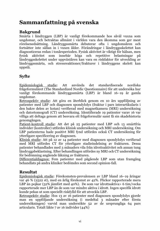
Bakgrund Smärta i ländryggen (LBP) är vanligt förekommande hos såväl vuxna som ungdomar, och betraktas allmänt i världen vara den åkomma som ger mest rörelsenedsättning. Ländryggssmärta debuterar ofta i ungdomsåren och fortsätter inte sällan in i vuxen ålder. Förändringar i ländryggsskelettet kan diagnosticeras redan i tonårsperioden. Fysisk aktivitet är viktigt för hälsan, men fysisk aktivitet som innebär höga och repetitive belastningar på ländryggsskelettet under uppväxtåren kan vara en riskfaktor för utveckling av ländryggssmärta, och stressreaktioner/frakturer i ländryggens skelett kan uppstå. Syfte Epidemiologisk studie: Att använda det standardiserade nordiska frågeformuläret (The Standardized Nordic Questionnaire) för att undersöka hur vanligt förekommande ländryggssmärta (LBP) är bland 16–19 år gamla ungdomar. Retrospektiv studie: Att göra en återblick genom en 10 års uppföljning av patienter med LBP och diagnosen spondylolys (fraktur i pars interartikularis i den bakre delen av kotan) verifierad med magnetkamera (MRI) undersökning och datortomografi (CT) undersökning. Identifierade 19 patienter varav 16 var villiga att deltaga genom att besvara ett frågeformulär samt få sin skadehistoria genomgången. Patient-kontroll studie: Att det på 25 patienter med LBP och 13 smärtfria individer (kontroller) utfördes klinisk undersökning och MRI undersökning. Om LBP patienterna hade positivt MRI fynd utfördes också CT undersökning för ytterligare specificering av diagnosen. Klinisk studie: Att på 12 av 14 patienter med diagnosen spondylolys verifierad med MRI utfördes CT för ytterligare stadieindelning av frakturen. Dessa patienter behandlades med 3 månaders vila från idrottsaktivitet och annan tung ländryggsbeklastning. Efter behandlingen utfördes ny MRI och CT undersökning för bedömning angående läkning av frakturen. Differentialdiagnos: Fem patienter med pågående LBP som utan framgång behandlats på andra kliniker bedömdes som second opinion-fall. Resultat Epidemiologisk studie: Förekomsten-prevalensen av LBP bland 16–19 åringar var 46 % (2522 st), med en årlig förekomst av 42%. Flickor rapporterade mera LBP än pojkar (52% jämfört med 40%). De som var idrottsaktiva> 6 tim/vecka rapporterade mer LBP än de som var mindre aktiva i idrott. Ingen specifik idrott kunde pekas ut som speciellt riskfylld för att utveckla LBP. Retrospektiv studie: Hos 13 av 16 patienter med diagnosen spondylolys gjorde man en uppföljande undersökning (i medeltal 3 månader efter första undersökningen) varvid man undersökte 52 av de ursprungliga 64 pars articularis. Totalt läkte 7 av 16 individer (44%)

vii
Patient-Kontroll studie: Hos 22/25 (88%) av LBP patienterna förelåg positiva fynd vid klinisk undersökning och MRI undersökning. Hos 13 patienter visade CT undersökningen spondylolys i olika stadier. Ingen enskild klinisk test, eller i combination med andra tester, kunde med säkerhet diagnosticera spondylolys, och kliniska tester kan därför ej användas för att välja ut patienter där det är indicerat att gå vidare med MRI och CT undersökning. Klinisk studie: Genom att använda MRI i combination med CT undersökning kunde man upptäcka tidiga tecken på skelettskada som ödem och inkomplett fraktur. Hos patienter där skelettskadan diagnosticerades i tidigt stadium såg man bättre läkning på behandling än om skelettskadan diagnosticerades i ett senare stadium (komplett fraktur och pseudartros). Differentialdiagnos: LBP patienter och patienter med LBP och smärta ner över höftens baksida bedömdes för “second opinion”. Noggrann klinisk undersökning kombinerad med MRI undersökning visade diagnosen stressreaktion/frakturtecken i Tuber Ischiadicum. Efter specifik rehabilitering kunde dessa patienter återvända till full idrottsaktivitet.
Sammanfattning
LBP är vanligt förekommande hos 16–19 åringar, och mer förekommande hos flickor och högaktiva ungdomar. LBP hos ungdomar måste tas på allvar då framförallt högaktiva idrottare kan lida av frakturer i pars interarticularis i den bakre delen av kotan. Det finns ingen speciell klinisk test att använda för att diagnosticera spondylolys. Om positiva fynd vid MRI undersökning kan komplettering med CT undersökning användas för stadieindelning av frakturen. Vid tidig diagnos, i tidigt frakturstadium, verkar det finnas goda förutsättningar för läkning på behandling med 3 månaders vila från idrott och annan tung ländryggsbelastning. En differentialdiagnos vid LBP kan vara stressreaktion/fraktur i bäckenets skelett.

viii
Preface
More than twenty years ago I had a 15-year-old sports active patient suffering from LBP especially after sport activity. Clinical examination indicated facet joint pain, and with my education in manual therapy I was thinking about treatment with manipulation. By a co-incidence, this patient instead first had an MRI examination, followed by a CT examination due to positive findings on the MRI. These examinations showed that the patient suffered from spondylolysis (incomplete fracture) in the L5 vertebrae. At that time, I had sparse knowledge about spondylolysis, and did not at all know how to treat the condition. Discussions with my future tutor and most scientific papers suggested treatment in a brace for 6 months. However, in one of the papers (1) the treatment was rest from physical activity for three months, and to me that seemed better to suggest for this patient. Two months later another CT examination showed that the fracture had healed, and after some additional rehabilitation exercises this patient was pain-free and returned to previous physical activity level. This started my interest in doing research in this field. Suddenly I recognized other young and mostly sport actives suffering from LBP, were further examinations showed the diagnosis spondylolysis. By just starting to think about this diagnosis I have now seen around 100 patients with spondylolysis.
ix
Abbreviations ADL – Activities of Daily Living ALBP – Adolescent Low Back Pain BMD- Body Mass Density CT – Computer Tomography FSU - Functional Spinal Unit Inferior Articular Process (IAP) IMD - inter malleolar distance. LBP- Low Back Pain NSLBP – Non-Specific Low Back Pain NSALBP – Non-Specific Adolescent Low Back Pain ICD - Intercondylar distance YLD - years lived with disability OR – Odds Ratio PE- Physical Examination PBM - Peak Bone Mass PHV - Peak Hight Velocity MRI – Magnetic Resonance Imaging MVPA – Moderate to Vigorous Physical Activity IT– Ischial Tuberosity SRBI – Stress Related Bone Injuries PGR – Personally Graded Rehabilitation PI – Pars Interarticularis VAS – Visual Analogue Scale STIR - sequence and a coronal T2-weighted inversion recovery SAP -Superior Articular Process PED - Posterior Element Disturbanc

1
Introduction
1.Anatomy
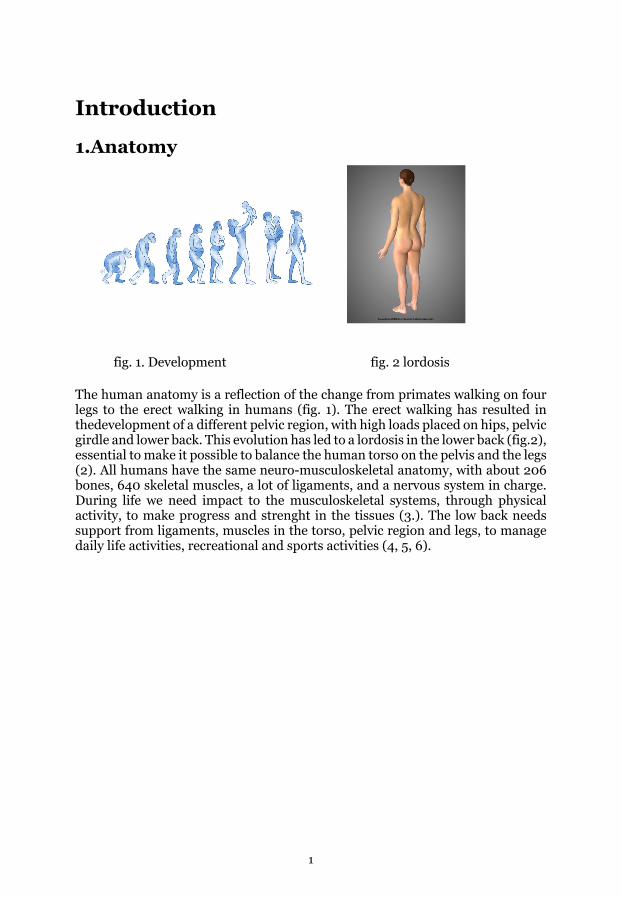
fig. 1. Development fig. 2 lordosis
The human anatomy is a reflection of the change from primates walking on four legs to the erect walking in humans (fig. 1). The erect walking has resulted in thedevelopment of a different pelvic region, with high loads placed on hips, pelvic girdle and lower back. This evolution has led to a lordosis in the lower back (fig.2), essential to make it possible to balance the human torso on the pelvis and the legs (2). All humans have the same neuro-musculoskeletal anatomy, with about 206 bones, 640 skeletal muscles, a lot of ligaments, and a nervous system in charge. During life we need impact to the musculoskeletal systems, through physical activity, to make progress and strenght in the tissues (3.). The low back needs support from ligaments, muscles in the torso, pelvic region and legs, to manage daily life activities, recreational and sports activities (4, 5, 6).
2
1.1 The skeleton
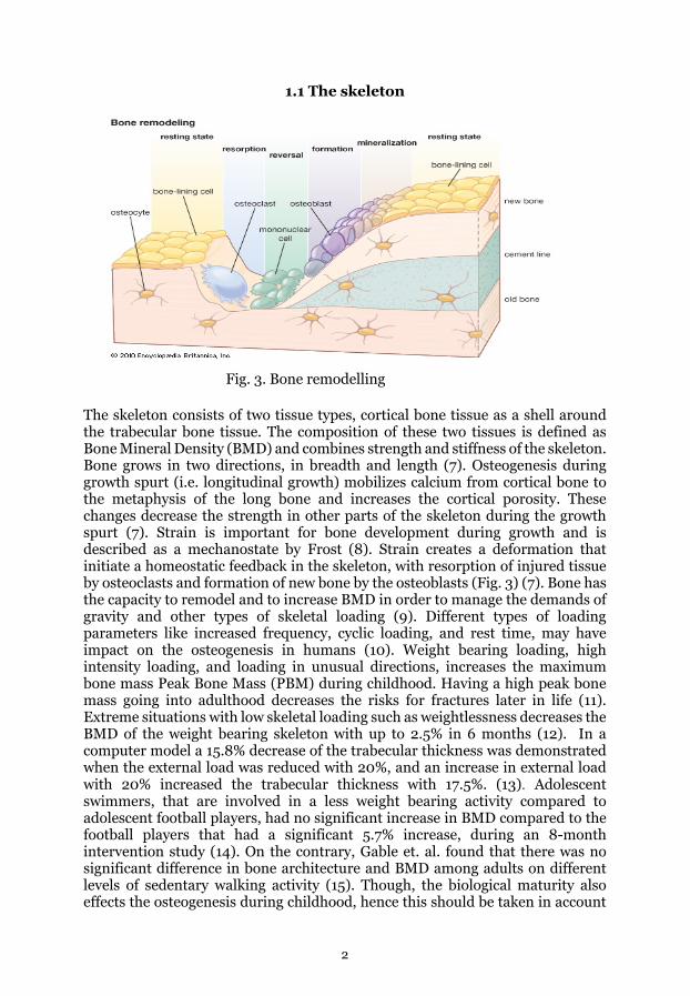
Fig. 3. Bone remodelling
The skeleton consists of two tissue types, cortical bone tissue as a shell around the trabecular bone tissue. The composition of these two tissues is defined as Bone Mineral Density (BMD) and combines strength and stiffness of the skeleton. Bone grows in two directions, in breadth and length (7). Osteogenesis during growth spurt (i.e. longitudinal growth) mobilizes calcium from cortical bone to the metaphysis of the long bone and increases the cortical porosity. These changes decrease the strength in other parts of the skeleton during the growth spurt (7). Strain is important for bone development during growth and is described as a mechanostate by Frost (8). Strain creates a deformation that initiate a homeostatic feedback in the skeleton, with resorption of injured tissue by osteoclasts and formation of new bone by the osteoblasts (Fig. 3) (7). Bone has the capacity to remodel and to increase BMD in order to manage the demands of gravity and other types of skeletal loading (9). Different types of loading parameters like increased frequency, cyclic loading, and rest time, may have impact on the osteogenesis in humans (10). Weight bearing loading, high intensity loading, and loading in unusual directions, increases the maximum bone mass Peak Bone Mass (PBM) during childhood. Having a high peak bone mass going into adulthood decreases the risks for fractures later in life (11). Extreme situations with low skeletal loading such as weightlessness decreases the BMD of the weight bearing skeleton with up to 2.5% in 6 months (12). In a computer model a 15.8% decrease of the trabecular thickness was demonstrated when the external load was reduced with 20%, and an increase in external load with 20% increased the trabecular thickness with 17.5%. (13). Adolescent swimmers, that are involved in a less weight bearing activity compared to adolescent football players, had no significant increase in BMD compared to the football players that had a significant 5.7% increase, during an 8-month intervention study (14). On the contrary, Gable et. al. found that there was no significant difference in bone architecture and BMD among adults on different levels of sedentary walking activity (15). Though, the biological maturity also effects the osteogenesis during childhood, hence this should be taken in account

3
when comparing BMD during adolescents. Instead BMD should be related to Peak Hight Velocity (PHV) biological level (+- 1year in 95% of cases) and be compared to chronological ages (16).
1.2 The Spine
Fig.4 The Spine
The spine consists of 24 vertebrae (7 neck, 12 thoracic and 5 lumbar vertebrae). Sacrum is also a part of the spine with 5 vertebrae melted together and it finishes with the coccyx. The sacrum is connected to the pelvic girdle through the joints between the right and the left os. ileum in the pelvic girdle – the SI joints, and rests on the hip joints. The spine is designed with cervical lordosis, thoracic kyphosis and Lumbar lordosis which gives the spine less compression forces to the structures. The spine is central in the movement of the muscles in the torso which have their origin and insertions in the spine, the pelvic region and the hips. The muscles in the torso area act together to balance the trunk and pelvic girdle on the legs. The stability of the spine is established by the intervertebral discs, the zygapophysial joint, ligaments and muscles, intrinsic stability by the intervertebral discs, zygapophysial joint and ligaments and extrinsic stability by muscles. The vertebrae in the low back functions together with the vertebrae in the rest of the back as they are connected to each other in the Functional Unit of the Spine (FSU) described below (17,18).

4
1.3 The lower back
The Low Back is the region bordered abowe by the 12th rib and below the gluteal folds (19, 20, 21), and is represented by the five lowest vertebrae in the back. It is closely connected to the pelvic girdle. The single vertebrae form an anterior part with the vertebral bodies and intervertebral discs, and a posterior part with its neural arch, zygapophysial joints and the spinous process. The low back consists of 5 functional spinal units (fig.3) joining L1/L2, L2/L3, L3/L4, L4/L5 and L5/S1, respectively. There is a lordosis between Th12 and sacrum that is an important part that lower the compressive loads acting on the low back during during daily life activities, as well as more strenuous load during work, physical activity and sports (17,18). The low back develops during the first 2 decades of life and is estimated to have matured when the adolescent individual becomes adult around 20 years of age (22).
1.4 The Functional Unit of the Spine
Fig.5. Functional Unit of the Spinal (FSU)
The FSU (fig.5) can be described in a ventral and dorsal portion where the ventral portion consists of the vertebral body, intervertebral discs, ligaments, intervertebral foramina the spinal cord and the nerve root. The dorsal portion consists of lig. flavum, the superior and inferior facet joints, joint capsules, transvers processes, spinosus processes and the ligaments and small muscles in-between (23). The posterior part of the functional spinal unit forms the spinal canal where the spinal nerves passes. It is a complex part with several processes acting as insertion points for muscles and ligaments. Pars interarticularis (PI) is a part of the dorsal vertebrae previously called “The neck of the scotty dog”. It is the bone area between the superior and inferior zygapophysial facets on the same vertebrae, on the same side. and can be seen on the figure that shows the dorsal part of the vertebrae (Fig 7). The height of the intervertebral disc is important as
5
it makes a free space for the nerve root to pass on its way to become a peripheral nerve. Stability is formed by the facet joints. Thru the whole spine, the small muscles between two vertebrae, as well as the bigger muscles in the different muscle layers, initiate and control movements both locally as well as with another FSU (17, 18).
1.5 The vertebrae
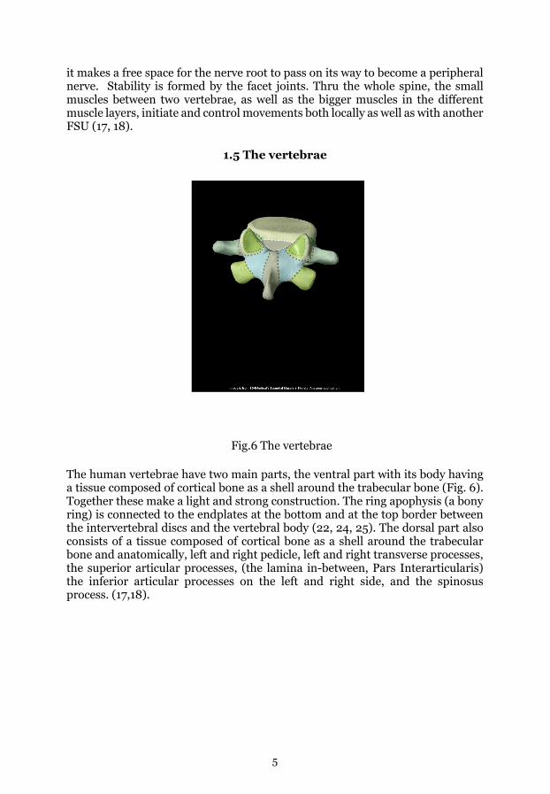
Fig.6 The vertebrae
The human vertebrae have two main parts, the ventral part with its body having a tissue composed of cortical bone as a shell around the trabecular bone (Fig. 6). Together these make a light and strong construction. The ring apophysis (a bony ring) is connected to the endplates at the bottom and at the top border between the intervertebral discs and the vertebral body (22, 24, 25). The dorsal part also consists of a tissue composed of cortical bone as a shell around the trabecular bone and anatomically, left and right pedicle, left and right transverse processes, the superior articular processes, (the lamina in-between, Pars Interarticularis) the inferior articular processes on the left and right side, and the spinosus process. (17,18).
6
1.6 The Pars interarticularis of the Vertebrae
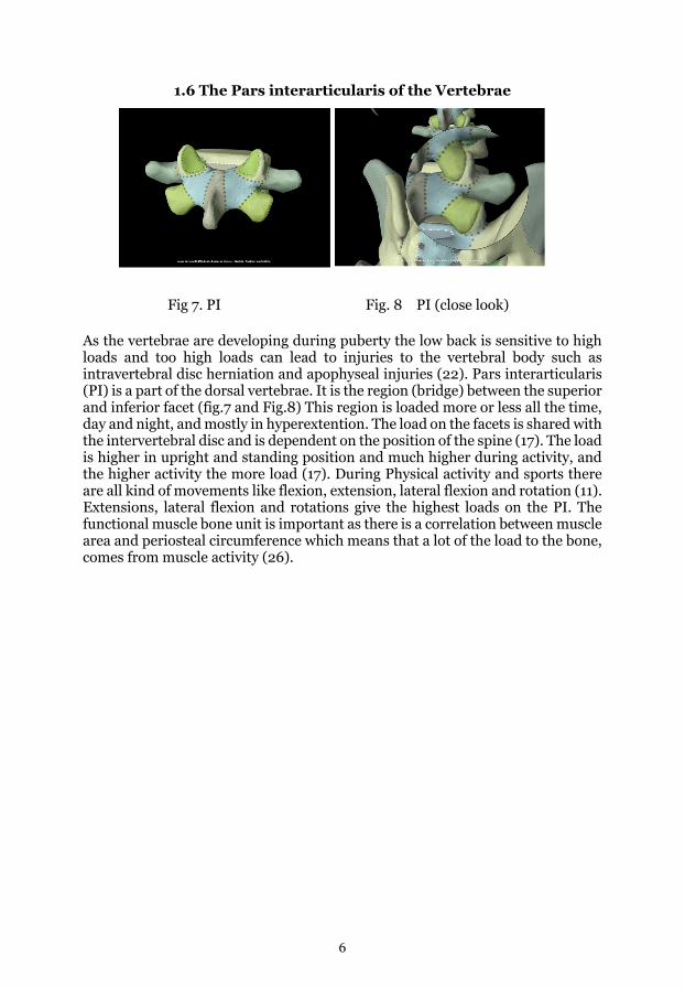
Fig 7. PI Fig. 8 PI (close look)
As the vertebrae are developing during puberty the low back is sensitive to high loads and too high loads can lead to injuries to the vertebral body such as intravertebral disc herniation and apophyseal injuries (22). Pars interarticularis (PI) is a part of the dorsal vertebrae. It is the region (bridge) between the superior and inferior facet (fig.7 and Fig.8) This region is loaded more or less all the time, day and night, and mostly in hyperextention. The load on the facets is shared with the intervertebral disc and is dependent on the position of the spine (17). The load is higher in upright and standing position and much higher during activity, and the higher activity the more load (17). During Physical activity and sports there are all kind of movements like flexion, extension, lateral flexion and rotation (11). Extensions, lateral flexion and rotations give the highest loads on the PI. The functional muscle bone unit is important as there is a correlation between muscle area and periosteal circumference which means that a lot of the load to the bone, comes from muscle activity (26).
7
1.7 The Zygapophysial facets of the Vertebrae
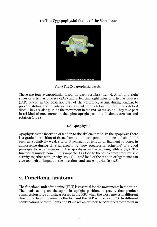
Fig. 9 The Zygapophysial facets
There are four zygapophysial facets on each vertebra (fig. 9). A left and right superior articular process (SAP) and a left and right inferior articular process (IAP) placed in the posterior part of the vertebrae, acting during loading to prevent sliding and in rotation too prevent to much load on the intervertebral discs. They are also guiding the movement in the FSU of the spine. They take part in all kind of movements in the spine upright position, flexion, extension and rotation (17, 18).
1.8 Apophysis
Apophysis is the insertion of tendon to the skeletal tissue. In the apophysis there is a gradual transition of tissue from tendon or ligament to bone and should be seen as a relatively weak site of attachment of tendon or ligament to bone, in adolescence during physical growth. A “slow progression principle” is a good principle to avoid injuries in the apophysis in the growing athlete (27). The functional muscle bone unit is important as load to thebone comes from muscle activity together with gravity (26,27). Rapid load of the tendon or ligaments can give too high an impact to the insertions and cause injuries (27, 28)
2. Functional anatomy
The functional unit of the spine (FSU) is essential for the movements in the spine. The loads acting on the spine in upright position, is gravity that produce compression force and shear forces in the FSU when the torso moves in different directions. In all movements the IAP and the SAP is in action (22). In different combinations of movements, the PI makes an obstacle to continued movement in
8
the FSU. The intervertebral disc is also an obstacle to these movements (18). The erector spinae muscles seem to be inactive in full flexion while the counteraction is made up by the posterior ligaments of the spine (17). Rotation in the low back are small movements in each FSU with the total of about 10 degrees. There is a couple of degrees in each segment and direction, with most of the rotation taking part at L5 level with about 5 degrees to each side (18). In lateral flexion the cranial vertebrae are bending to the same side and SAP glides down on IAP to the side the bending is (convergence) and on the other side there is a divergence as the SAP glides up on the IAP, on the same side. This action stretches ligaments and capsule on the side you are bending from and relaxes structures on the side you are bending towards.
2.1 Spinal Load
The load subjected to the spine differs depending on the type of activity, from activities of daily life to strenuous work and sport activity. . Loading produces strain in the tissues and activates osteogenesis in the skeleton (11 ), lumbrication in the joints (29), the development of the muscle-bone-unit (27), the tendon strength (30), the muscle tendon unit and the growing of the muscle fiber thickness (31). Studies have reported loads on the lower back up to 800 N in upright standing (17, 32) and about 1000 N in upright sitting and if you make a lift of an object of 800N (80kg) there will be a load of about 10000N (1000kg) on the FSU in lumbar level. This load is produced by the external objects, the weight of the upper body as well as load produced by concentric and eccentric muscle contractions (17 In vivo and in vitro studies has shown that the load on the spine differs depending on spinal posture and movements, and mainly load the intervertebral disc and the facet joints (25). Combination of movements like extension, lateral flexion and rotation increase the load of the posterior part of the vertebra (17, 25, 33).
3. The historical perspective of Low back Pain
The historical perspective of Low Back Pain
Allan, 1989, stated that “there has always been back pain” in his “Understanding and management of low back pain”. The history of Low Back Pain (LBP) starts many thousand years ago and was recognized as a disease in papyrus about 1500 BC. Degenerative changes in the spine were seen in the earliest remains of humans, and during decades with studies on the spine there has been similar findings all over the world. Hippocrates (400 BC) also wrote about LBP, but at that time LBP was a symptom of many illnesses. In the 19th century, parallel to the industrial revolution when building of the railways started, serious back injuries were seen, and injury was established as a cause to back pain. During decades the understanding and management of LBP has changed, and Wadell (1989) concluded that there is an “iatrogenic disability” explained as health care being a part of the development of LBP conditions (34). This opinion has been discussed and criticized thanks to the fact that specific LBP

9
now has been able to be diagnosed with the use of new examination technologies. (35,36, 37)
3.1 The natural history of LBP
The natural history of LBP is still under discussion. The picture is very heterogenous and there is no consensus about where the pain comes from, what it is, how serious it is to humans, social interactions and what the best practice is (38, 39, 40). Different opinions present different solutions to this condition (36, 41, 42, 43, 44, 45). Some authors describe a natural history of recovery within 3 months for 90% of the individuals (46), while others claim that LBP is not a self-limiting condition (47, 48, 49, 50, 51, 52). Recurrence of LBP seems to be very common since as many as 73% of those with an earlier episode of LBP will have another one. Thus, it seems to be difficult to state that the LBP conditions have an excellent prognosis (36). Nowadays the discussion is about Non-Specific Low Back Pain (NSLBP) and Specific Low Back Pain (SLBP) (53). Luschka (1858) proposed that neuro-, and musculoskeletal changes are supposed to be the reason for LBP, related to prolapses of the intervertebral disc. Schmorl (1929) and Andrea (1929) had a similar proposal related to posterior disc protrusion and protrusions of the disc into the vertebral body. Sciatica was actualized by Domenico Cotugno (1764). The prolapse of the intervertebral disc was later established in the beginning of the 20th century by Goldthwaite (1911), Middeton & Teacher (1911), Dandy (1929) and Mixter & Barr (1934) (34).
3.2 The history of Spondylolysis
Ever since Herbiniaux (1782) introduced spondylolisthesis, Robert zu Coblenz (1855) recognized the pars defect, and Neugebauer (1884) established the term Spondylolysis, there have been a lot of discussions about different reasons for the development of spondylolysis. Anomalies and mechanical failure have been proposed. Archaeology findings in North Alabama, USA showed as much as 17-20 % spondylolysis changes (54). In population studies just about 6% develop some kind of spondylolysis in bone tissue in the low back and many of these lesions seems to be non-symptomatic (55). Vigorous athletic activity seems to be involved in symptomatic spondylolysis (stressreaction/stressfracture) (56) Spondylolysis can be seen in different parts of the spine
4. Low Back Pain
The definition of LBP is heterogeneous; it can be based on extent or impact of LBP and would benefit from measurements of severity and activity limitation (57). LBP is a common problem in all ages (35,58,). It is often divided in acute (<3 months duration) and chronic (>3 months duration) which is an over-simplification since 30% of acute LBP persist into disability (21,59).
10
4.1 Non-Specific Low Back Pain (NLBP)
Non-specific low back is defined as low back pain with no known specific pathology (53). There are attempts to sub-classify this condition to find the right intervention in different levels of NLBP (60).
4.2 Specific Low back Pain
Many authors have described specific Low Back Pain as many diagnoses (35, 53 61). In this thesis I have focused on Spondylolysis.
4.3 Adolescent Low Back Pain (ALBP)
ALBP is difficult to define because it´s heterogeneous. In an attempt to define different stages of ALBP Milanese suggests three stages General LBP, chronic/recurrent LBP and severe disabling LBP. (61) There is a need for more uniform and standardized definitions of ALBP (35, 41). The first episode occure in as early age as 12 years of age and escalates very quickly during puberty. The different stages are quite the same for girls and boys with the exception that the girls start one or two years earlier than boys (12-13 for girls, 13-14 for boys) (62, 63)
4.4 Low Back Pain and Disability
Today, it is a common problem in the world population to have LBP. Up to 84 % of the population has that experience, during life (53,64) It starts early and increases through life, mostly during puberty (62). Odds ratio for persistent LBP in to adulthood, if you experience LBP during adolescence, is high, up to 4.7 (65). There is a difference in the reason for LBP in adulthood and in adolescence. While adults mostly have pain from lumbosacral strain and impact on discs (75 out of 100), adolescent athletes were found to have mostly spondylolysis or mechanical back pain (73 out of 100) Spondylolysis was found in 48 of the adolescents and in 5 adults (39). The same amount of spondylolysis is seen in other studies (66). Low back pain in occupational medicine was recognized as Low Back Disability (LBD) in relation to work, by Ramazzini (1705) (34). LBD is when LBP does impact on work, ADL, recreational and sport activities. About 12% of those that have a lifetime prevalence of LBP (84%) experienced work, ADL and physical activity related LBD (53,64)

11
5. Low Back Pain in Adolescent athletes
5.1 Spondylolysis
(Spondylo = vertebrae, lysis=disintegration)
An early start in athletic activities seems to be a risk factor for both LBP and spinal abnormalities (22,23,66,67). Repetitive stress both in flexion and extension, and in combination with rotation of the low back, can result in impacts to the pars interarticularis resulting in skeletal stress reactions. Rapid and oscillating movements in extension, as in certain sports, can result in a mechanical stop between the neural arches. These movements can develop into Spondylolysis (18). Spondylolysis is defined as a bony defect fatigue failure in the lumbar pars interarticularis (68). Today it is a description of the end of a process visible thanks to modern techniques in radiology (SPECT, MRI and CT). Early stage findings include edema visible on MRI, and incomplete fracture visible on CT. Later stage findings include complete fracture visible on CT, and pseudo arthrosis visible on CT. Spondylolysis is defined as a bony defect (stress reaction in the skeleton) in the lumbar pars interarticularis (68,69,70). Early stage findings include edema visible on MRI, and incomplete fracture visible on CT. Later stage findings include complete fracture visible on CT, and pseudo arthrosis visible on CT (71).
5.2 Spondylolysis-Aetiology
There is good evidence that spondylolysis is a fatigue fracture in Pars interarticularis and is more common in adolescent athletes compared with non-athletes (68). The incidence of Spondylolysis has been reported to differ between sports, but many sports are associated with spondylolysis. Certain sports like cricket, weight lifting, American football, wrestling, gymnastics and tennis are particularly associated with pars interarticularis injuries (72,73,74, 75).
5.3 Spondylolysis-Pathogenesis
Spondylolysis during childhood and adolescence is defined as a defect in the pars interarticularis of the human vertebrae. An important pathogenetic criterion is athletic activities with extension/flexion movements. A combination of movements can produce stress reactions in the lumbar segments (L4, L5) during complex activities and ADL where there are recognized microdamage in the PI of vertebrae on these levels. This is also the most common site for spondylolysis (25,33,76)
12
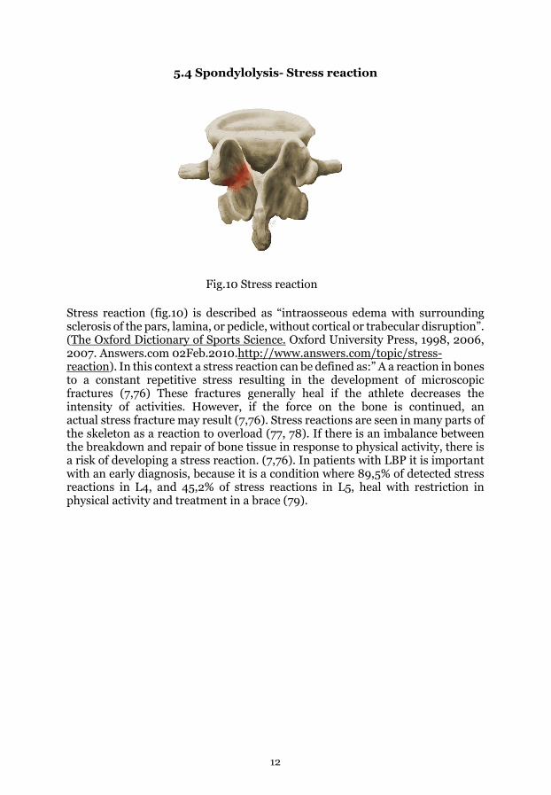
5.4 Spondylolysis- Stress reaction
Fig.10 Stress reaction
Stress reaction (fig.10) is described as “intraosseous edema with surrounding sclerosis of the pars, lamina, or pedicle, without cortical or trabecular disruption”. (The Oxford Dictionary of Sports Science. Oxford University Press, 1998, 2006, 2007. Answers.com 02Feb.2010.http://www.answers.com/topic/stress-reaction). In this context a stress reaction can be defined as:” A a reaction in bones to a constant repetitive stress resulting in the development of microscopic fractures (7,76) These fractures generally heal if the athlete decreases the intensity of activities. However, if the force on the bone is continued, an actual stress fracture may result (7,76). Stress reactions are seen in many parts of the skeleton as a reaction to overload (77, 78). If there is an imbalance between the breakdown and repair of bone tissue in response to physical activity, there is a risk of developing a stress reaction. (7,76). In patients with LBP it is important with an early diagnosis, because it is a condition where 89,5% of detected stress reactions in L4, and 45,2% of stress reactions in L5, heal with restriction in physical activity and treatment in a brace (79).
13
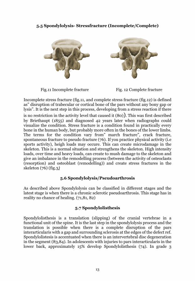
5.5 Spondylolysis- Stressfracture (Incomplete/Complete)
Fig.11 Incomplete fracture Fig. 12 Complete fracture
Incomplete stress fracture (fig.11, and complete stress fracture (fig.12) is defined as” disruption of trabecular or cortical bone of the pars without any bony gap or lysis”. It is the next step in this process, developing from a stress reaction if there is no restriction in the activity level that caused it (80)). This was first described by Briethaupt (1855) and diagnosed 42 years later when radiographs could visualize the condition. Stress fracture is a condition found in practically every bone in the human body, but probably more often in the bones of the lower limbs. The terms for the condition vary from” march fracture”, crack fracture, spontaneous fracture to pseudo fracture (76). If you practice physical activity (i.e sports activity), heigh loads may occure. This can create microdamage in the skeleton. This is a normal situation and strengthens the skeleton. High intensity loads, over time and heavy loads, can create to mush damage to the skeleton and give an imbalance in the remodelling process (between the activity of osteoclasts (resorption) and osteoblast (remodelling)) and create stress fractures in the skeleton (76) (fig.3)
5.6 Spondylolysis/Pseudoarthrosis
As described above Spondylolysis can be classified in different stages and the latest stage is when there is a chronic sclerotic pseudoarthrosis. This stage has in reality no chance of healing. (71,81, 82)
5.7 Spondylolisthesis
Spondylolisthesis is a translation (slipping) of the cranial vertebrae in a functional unit of the spine. It is the last step in the spondylolysis process and the translation is possible when there is a complete disruption of the pars interarticularis with a gap and surrounding sclerosis at the edges of the defect ref. Spondylolistesis is accentuated when there is an intervertebral disc degeneration in the segment (83,84). In adolescents with injuries to pars interarticularis in the lower back, approximately 15% develop Spondylolisthesis (74). In grade 3

14
(according to Pfirrman scale 1-5) disc degeneration, there is a potential risk for progression of Spondylolisthesis, and also a progression of symptoms (83, 85, 86)
5.8 A differential diagnosis
A differential diagnosis in injuries to Pars Interarticularis of the lower back is stress reactions and fractures in the Ischial Tuberosity (IT) of the pelvic girdle (87,88). The condition is seen in adolescent athletes, similar to injuries to the back of the tight, not seldom missed in clinic (87,88,89,90)
6. Aims
The general aim of the present thesis was to investigate the prevalence of low back pain (LBP) among adolescents, with particular focus on adolescent athletes.
Specific aims were:
• To focus on the diagnosis pars interarticularis injury-Spondylolysis in adolescents with LBP.
• To evaluate clinical tests to diagnose pars interarticularis injuries in adolescents with LBP.
• To describe MRI and CT findings to diagnose pars interarticularis injuries in adolescents with LBP.
• To describe the different fracture stages in pars interarticularis injuries found on MRI and CT in adolescents with LBP.
• To try to evaluate whether the fracture stage in pars interarticularis affected the potential for healing of the injury, using treatment with 3 months of rest.
• To describe a differential diagnosis to pars interarticularis injury-Spondylolysis.

15
7. Summary of Papers
Paper 1
Aim: The aim with this epidemiology study was to evaluate the prevalence of low back pain (LBP) and associated physical disability in a male and female adolescent population on different activity levels.
Material and Methods: The Standardized Nordic Questionnaire for musculoskeletal symptoms adjusted for adolescence was used to investigate the prevalence of LBP during adolescence (16-19 years old individuals). From 3076 questionnaires there were 2550 answers (83%) from adolescents (1304 boys and 1234 girls) with a mean age of 18.2 years.
Main results: LBP was reported by 46% out of 2522 subjects, 42 % had had LBP during the last year, and among those 45% had experienced LBP the last 7 days. More girls reported LBP than boys (52 % and 40 %, respectively, p< 0.001). Significantly more adolescents active in sports > 6 hours/week reported LBP, compared to less physically active adolescents.
Conclusions: LBP is common among adolescents, more common in girls and in highly active individuals.
Paper 2
Aim: The aim with this retrospective study was to study the history, clinical findings, MRI and CT findings in patients with the diagnosis Spondylolysis.
Material and Methods: In retrospective study based on patients treated at the local hospital between 1995-2006, for 19 patients (17 men and 2 women, mean age 16 years, range 12-21 years at the time for diagnosis) there was information about clinical findings, MRI or CT findings. Sixteen patients agreed to have their history reviewed and answered a questionnaire-the Standardized Nordic Questionnaire.
Main results: In 13/16 sports active patients with diagnosed spondylolysis, MRI and CT showed fractures in althogether 30 pars interarticularis. The patients had had different treatments, most commonly rest for 3 months. In total 22/30 (73%) diagnosed pars interarticularis fractures healed.
Conclusions: LBP needs to be taken seriously, especially in adolescent athletes that not seldom have fractures in pars interarticularis (Spondylolysis).
16
Paper 3
Aim: To in a case control study evaluate clinical tests to diagnose pars interarticularis injuries-Spondylolysis, and to evaluate MRI and CT findings in patients with pars interarticularis injuries-Spondylolysis.
Material and Methods: Physical examination (specific clinical tests) and MRI examination was performed in 25 sports active (>6h/w) patients (14 boys and 11 girls, mean age 15 years, range 13-20) with > 3 weeks of LBP prevent them from sports, and 13 sports active pain-free controls (5 girls and 8 boys, mean age 17 years, 14–19). If the patients had findings on MRI a CT investigation was performed for further diagnostics.
Main results: No clinical test alone, or in combination, could reliably diagnose pars interarticularis injury-spondylolysis, and could thus not be used as a selection test for further diagnostics with MRI and CT. In 22/25 patients (88%) there were clinical and MRI (pars interarticularis injury) findings. In 13/25 patients (52%) pars interarticularis fracture in different stages was demonstrated on CT examination.
Conclusions: There is no single clinical test, or combination of clinical tests, that reliably can diagnose pars interarticularis injury-Spondylolysis. CT examination in addition to MRI provides a staging of the pars interarticularis fracture.
Paper 4
Aim: In a clinical study, we tried to evaluate whether the fracture stage in the pars interarticularis affected the potential for healing of the injury, using treatment with 3 months of rest.
Material and Methods: In 12/14 patients (10 boys, mean age 14.7 years range 14-17 and 4 girls mean age 15, range13-17) with the diagnosis pars interarticularis injury-Spondylolysis verified on MRI, a CT examination was performed with the purpose to stage the fracture. Two patients decided to not participate in the CT examination. The 12 remaining patients were re-evaluated with MRI and CT after 3 months of treatment with complete rest from sports activity.
Main results: Using a combination of MRI and CT examination the early signs of skeletal injury (oedema, incomplete fracture) were detected. After treatment with 3 months of rest, patients diagnosed in the early stages (oedema, incomplete fracture) healed significantly better than if diagnosed in later stages (complete fracture and pseudoarthrosis).
17
Conclusions: CT examination in addition to MRI provides a staging of the pars interarticularis fracture. If diagnosed early, in the early fracture stages, there seems to be good potentials to heal the fracture with 3 months of rest from loading (sport activity).
Paper 5
Aim: To describe a differential diagnosis to pars interarticularis injury-Spondylolysis.
Material and Methods: Five sports active patients (5 boys, mean age 15 years, range 13-17) suffering from LBP that had been treated at other clinics without success, were evaluated for a “second opinion”.
Main results: Physical Examination and MRI examination diagnosed stress reactions in the pelvic ischial tuberosity. After guided rehabilitation, all patients returned to previous sport activities.
Conclusions: Differential diagnoses such as stress reactions in the pelvic ischial tuberosity should be taken in consideration.

18
Methods Table. 1
Questionnaries – Study I and II
Epidemiology/Prevalence study - Study I
This study comprises 23 items from a total of 73 items from the Standardized Nordic Questionnaire for the analysis of musculoskeletal symptoms (19) modified for students. The first page contains individual characteristics and questions about physical activity level in hours/ week and kind of sport performed. The two following pages are questions about symptoms from the musculoskeletal system in general, related to a drawing that explains the areas referred to.
The following questions were used;
1. “Have you ever had pain, ache or felt unpleasantness in your low back?”
2. “Have you had pain or ache, or felt unpleasantness in your low back anytime the last 12 months?”
If the answers to these questions were YES, the following questions were asked:
3. “Have you had pain or ache, or felt unpleasantness in your low back some time the last 7 days?”
Questionnary Physical exmination
MRI CT
Epidemiology (Study 1)
accepted x
Retro study (Study 2)
manuscript x X (retro)
X (retro)
Clinic 1 (Study 3)
published x x x x
Clinic 2 (Study 4)
published x x x x
Different diag. (Study 5)
published x x
19
The answer options to these questions were no or yes.
4. “How many days in total have you experienced pain in the low back the last 12 months?”
The answer alternatives to this question were 0 days, 1-7 days, 8-30 days, > then 30 days but not every day and day in day out.
5. “Have you, due to LBP, reduced your spare time physical activity the last 12 months?”
6. “How many days in all have you experienced that you were unable to perform your daily activities due to pain in the low back the last 12 months?”
The alternatives were 0 days, 1-7 days, 8-30 days and >30 days.
7. “Have you, due to low back pain, been examined or treated by a physician, physiotherapist or chiropractor the last 12 months?”
The answer options to this question were no or yes.
Before the investigation, information was delivered to all registered students in high school about the purpose of the investigation. Students younger than 18 years of age were informed that they should take the information home to their parents.
The questionnaires were delivered to each class separately and distributed by the homeroom teacher or by the physical educational teacher. The questionnaires were returned within two to four weeks. The school nurse delivered questionnaires to those who had not answered the questionnaire during the first round. A total of twenty-eight questionnaires were collected in this manner.
Retrospective questionnaire study – Study II
The subjects were asked to answer a questionnaire (the Standardized Nordic Questionnaire for the analysis of musculoskeletal symptoms, modified for students) about their low back pain problems today, the last week with registration on Visual Analog Scale (VAS) (91) and the last year and about their present physical activity level. Seventeen individuals returned the questionnaire and agree to examinations of MRI investigations. We also asked permission to take part of their medical records and 16 individuals agreed to this in order for us to establish date of symptom debut, time of diagnosis and follow-up time.
20
Physical examination – Study III, IV, V
Physical examination - Study III
Clinical tests for low back pain and lower limb examination often used in sports medicine and orthopedic manual therapy were performed before the MRI and CT investigation to blind the examiner to the structural anatomical findings. The tests were standardized and performed by the same physiotherapist according to (92) unless otherwise stated. The performance tests were performed in the following order:
Gait pattern: limping, toe-off, heel-strike, step length.
Inspection: Scoliosis, lordosis, atrophy of muscles (legs, but- tocks and lower back), foot position (e. g. pronation, supination, outflare and inflare) and leg length difference (93).
Neurological tests: Straight leg raise, sensibility, reflexes and muscle strength (heel walking, walking on toes). Slump tests were performed in the standing and seated position.
21
Functional movement test
Standing: Squat, hyperextension of the knees (standing), floor- finger distance in flexion, ROM of feet, knees and hips, Trende- lenburg sign, side bending left and right, one leg lumbar hyperextension test, thump test in neutral, flexion and exten- sion, MCI pattern (standing up from sitting position and sitting down from standing position) (94)
Sitting: Back movement tests for coupled movement in thoracic spine and lower back. Supine: Leg muscle strain tests, trunk muscle control test and standardized sit-ups (hands across chest, in the neck and over the head).
Specific provocation tests
Supine: SI joint separation and sacrum ventrally.
On the side: Segmental movement in flexion, extension and rotation, and nerve tension test for n.femoralis and n.obtorato-rius. Prone: Palpation of muscle in the gluteus region, in the lower back, provocation of sacrum in nutation, hyperextension test with fixation of the pelvis, coin test, percussion test on the spinous process, hook test and rocking test, joint play L5-Th12 with springing test technique, and joint play in the SI joint with slight flexion of hips. Standing: Intercondylar distance (ICD) and intermalleolar dis- tance (IMD). After the clinical investigation, information was sent to an ortho- pedic surgeon who wrote the referral for an MRI/CT investigation.
Physical examination – Study IV
The subjects included in this study is a subgroup from Study III. Fourteen adolescent athletes (6 football players, 3 icehockey players, 1 floorball player, 1 basketball player, 1 volleyball player, 1 swimmer and 1 track and field athlete) were diagnosed with stress reactions (using CT and MRI) in the PI ranging from just oedema without fractures to pseudoarthrosis (Spondylolysis) Paper IV. The twelve patients that gave their informed consent to participate in the study were recommended refraining from physical activity and sports but not from Activities of Daily Living (ADL) for 3 months, without a brace, and met a physiotherapist once a month to ensure adherence/compliance. At each of these meetings the perception of pain was registered with Visual Analog Scale (VAS) [Sugiura 2014]. After three months, the same physical examination as described above in study III, was performed.
22
Physical examination – Study V
After anamnesis was taken, a physical examination was performed with an eccentric test of the insertion of the hamstrings muscles with hip flexion > 100 degrees, slightly flexed knee, and pain provocation when palpating the tuber ischiadicum (TI) insertion of the hamstring muscles. The eccentric test was scored as positive if pain in TI was recognized as a symptom by the patient. The palpation test was also considered positive if the patient recognized the pain in TI as a symptom.
MRI and CT – study II, III, IV, V
All images were examined/read by a doctor with a long experience of MRI and CT diagnostics. MRI in study III and IV were performed on a standard 1.5T Philips Achieva system and CT on a General Electric Light Speed VCT 64 row detector system. The MRI protocol was different from the standard adult protocol and focused on sequences sensitive to bone marrow and soft tissue edema for detection of the secondary bone marrow changes that accompany a fatigue fracture (a sagittal T1-weighted FSE sequence, sagittal T2-weighted fat suppressed FSE and coronal T2-weighted inversion recovery (STIR) sequences). In cases with disc disease, the axial T2-weighted FSE sequence was added. In search of changes compatible with a fatigue fracture, the presence of a high signal in the bone marrow in the pars interarticularis of the vertebrae on the T2-weighted sequences and a low signal on the T1-weighted sequence was assessed. Other pathological changes were recorded such as disc degeneration, disc herniation, olisthesis, congenital disorders, etc.
The initial CT was performed in all cases on the L4 and L5 vertebrae level regardless of MRI findings. If there were any findings suspicious to fatigue fracture in levels superior to L4 on the MRI, the intention was to include this level on the CT. The CT control after three months of rest was only performed on the level of pathology. Consecutive 0.6 mm thick slices with a bone algorithm were used and evaluated on a workstation. A radiologist with long experience in MRI and CT diagnostic imaging read the MRI and CT results. MRI was performed on a standard 1.5 T Philips Achieve systems and CT was performed on a General Electric Light Speed VCT 64 row detector system. The MRI examination was evaluated in search of changes compatible with a fatigue fracture, which was the presence of high signal in the bone marrow in the PI of the vertebrae on T2 weighted fat suppressed sequences and low signal on T1 weighted sequences. The MRI and CT investigations were performed after 11 – 20 weeks following the start of the intervention (median 14 weeks).
The Hollenberg classification on MRI was as follows:
→0 = no signal abnormalities,
→1= T2 signal abnormalities in PI – marrow oedema,

23
→2 = T2 signal abnormalities – thinning, fragmentation or irregularity of PI,
→3 = T2 signal abnormalities – visible complete unilateral or bilateral Spondylolysis,
→4 = No abnormal T2 signal - complete Spondylolysis not united fracture of PI was our basic classification, but we added CT in our investigation to get a more specified diagnosis.
Study II
Reinvestigation of MRI or CT identified symptomatic spondylolysis in physically active subjects found in the archive of the local hospital. The MRI/CT diagnosis was classified on the L4/L5 level with a classification by Hollenberg and Dunn and modified with a sixth class.
1. Normal: Normal marrow signal and intact cortical margins (stage 0)
2. Stress reaction: Marrow edema and intact cortical margins (stage 1)
3. Incomplete fracture: Marrow edema and cortical fracture incompletely extending
through pars (stage 2)
4. Complete active fracture: Marrow edema and fracture completely extends through pars (stage 3)
5. Fracture non-union: No marrow edema. and fracture completely extends through pars
(stage 4)
(Hollenberg 2002, Dunn AJ et al., 2008).
A sixth level was added in this article:
6. Healed stress fracture: Signs of healed stress fracture or stress fracture with small rests of fracture (stage 5)
Study III
Three – six weeks after the physical examination, MRI investigations were performed in both case and control groups (see above). CT was also performed in all cases at the level of the L4 and L5 vertebrae, regardless of the MRI findings. If any findings suggesting fatigue fracture were seen on MRI at any other levels or

24
in controls, CT was performed at these levels or on these subjects. The case groups CT exams were performed (0.6mm thick slices with a bone algorithm) over the L4 and L5 vertebrae, to assess the bone changes compatible with fatigue fracture. The findings in the vertebral arc were categorized using a five-grade system reflecting the different stages in the development of a fatigue fracture [8], but with a 6th grade added to reflect secondary signs of a healed fracture; that is, sclerosis with small remnants of fracture and absence of bone marrow oedema.
Study IV
The cases in study III diagnosed with spondylolys/stress fractures of the pars interarticularis where invited to take part in a follow up of treatment after three months. After these three months of intervention, a new MRI and CT according to the above were performed.
Study V
Differential diagnosis
An MRI was performed on 1.5 Tesla Philips Achiva system using sequences for both good anatomical resolution and sequences with high sensitivity for water in order to depict oedema and bleeding in bone marrow and surrounding soft tissue. A radiologist with extensive experience of MRI reads the examinations.
Statistical Methods
Statistical analysis
No.1 Analyses were made using SPSS 11.0 for PC. To compare frequency difference between groups, chi‐square likelihood ratio was calculated. Differences were considered significant if P < 0.05. We also used logistic regression analysis. Percentage of answers to questions 3 and 4 are calculated in relation to the number of “Yes” to question two
No.2 Due to the small number of subjects the Fischer exact was used. The significance level was 0,05 and the program we used was SPSS 15,0 for Windows and SPSS 17.0 for Mac.
No.3 The SPSS Statistics software, v.19 for Mac, and Fischer’s exact test were used to calculate differences between groups. Partial least squares (PLS) multivariate regression analy- sis was also performed.
No.4 Non-parametric related Samples Wilcox on Sign Rank Test was used in our estimates of p value for changes in the perception of pain. To analyse differences in healing among the different stages X2 test with Yates correction method was used due to the small number of cases.
25
No.5 No statistical analysis was performed
Etichs
No.1 The study was approved by the Regional Ethical Board of Umeå University, Sweden (No. 97-248 FEK). Questionnaries were anonymous and if the participants were under 18 years, they had to inform their parents about the questionarie.
No.2 The ethics committee of Umeå University approved this investigation No.
(No. 97-248 FEK).
No.3 The investigation was approved by the regional ethics committee at the Umeå University (06–014).
No.4 See above.
No.5 The investigation was approved by the regional ethics committee at the Umea University (06–014) as an addendum (2011/36-32M added to 06– 014M). All procedures performed in studies involving human participants were in accordance with the ethical standards of the institutional and/or national research committee and with the 1964 Helsinki declaration and its later amendments or comparable ethical standards.” For the retrospective study we had formal consent from the participants, eventhough this is not required. Informed consent: Informed consent was obtained from all individual participants, and their parents if under the age of 18 year
26
Results
Results – Study I-V
Study I
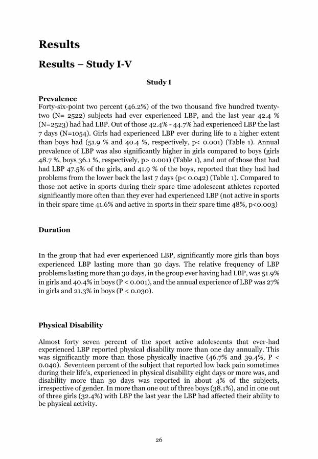
Prevalence Forty-six-point two percent (46.2%) of the two thousand five hundred twenty-two (N= 2522) subjects had ever experienced LBP, and the last year 42.4 % (N=2523) had had LBP. Out of those 42.4% - 44.7% had experienced LBP the last 7 days (N=1054). Girls had experienced LBP ever during life to a higher extent than boys had (51.9 % and 40.4 %, respectively, p< 0.001) (Table 1). Annual prevalence of LBP was also significantly higher in girls compared to boys (girls 48.7 %, boys 36.1 %, respectively, p> 0.001) (Table 1), and out of those that had had LBP 47.5% of the girls, and 41.9 % of the boys, reported that they had had problems from the lower back the last 7 days (p< 0.042) (Table 1). Compared to those not active in sports during their spare time adolescent athletes reported significantly more often than they ever had experienced LBP (not active in sports in their spare time 41.6% and active in sports in their spare time 48%, p<0.003)
Duration
In the group that had ever experienced LBP, significantly more girls than boys experienced LBP lasting more than 30 days. The relative frequency of LBP problems lasting more than 30 days, in the group ever having had LBP, was 51.9% in girls and 40.4% in boys (P < 0.001), and the annual experience of LBP was 27% in girls and 21.3% in boys (P < 0.030).
Physical Disability
Almost forty seven percent of the sport active adolescents that ever-had experienced LBP reported physical disability more than one day annually. This was significantly more than those physically inactive (46.7% and 39.4%, P < 0.040). Seventeen percent of the subject that reported low back pain sometimes during their life’s, experienced in physical disability eight days or more was, and disability more than 30 days was reported in about 4% of the subjects, irrespective of gender. In more than one out of three boys (38.1%), and in one out of three girls (32.4%) with LBP the last year the LBP had affected their ability to be physical activity.
27
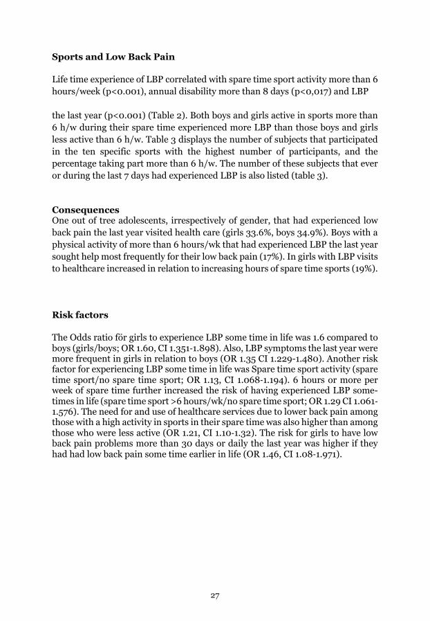
Sports and Low Back Pain
Life time experience of LBP correlated with spare time sport activity more than 6 hours/week (p<0.001), annual disability more than 8 days (p<0,017) and LBP
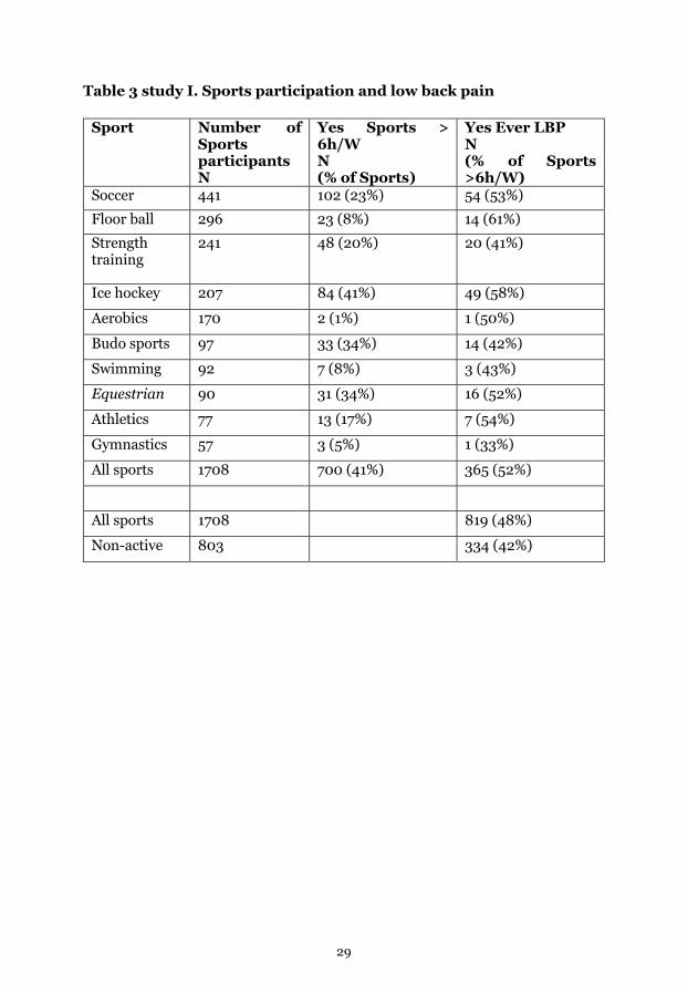
the last year (p<0.001) (Table 2). Both boys and girls active in sports more than 6 h/w during their spare time experienced more LBP than those boys and girls less active than 6 h/w. Table 3 displays the number of subjects that participated in the ten specific sports with the highest number of participants, and the percentage taking part more than 6 h/w. The number of these subjects that ever or during the last 7 days had experienced LBP is also listed (table 3).
Consequences One out of tree adolescents, irrespectively of gender, that had experienced low back pain the last year visited health care (girls 33.6%, boys 34.9%). Boys with a physical activity of more than 6 hours/wk that had experienced LBP the last year sought help most frequently for their low back pain (17%). In girls with LBP visits to healthcare increased in relation to increasing hours of spare time sports (19%).
Risk factors
The Odds ratio för girls to experience LBP some time in life was 1.6 compared to boys (girls/boys; OR 1.60, CI 1.351‐1.898). Also, LBP symptoms the last year were more frequent in girls in relation to boys (OR 1.35 CI 1.229-1.480). Another risk factor for experiencing LBP some time in life was Spare time sport activity (spare time sport/no spare time sport; OR 1.13, CI 1.068‐1.194). 6 hours or more per week of spare time further increased the risk of having experienced LBP some- times in life (spare time sport >6 hours/wk/no spare time sport; OR 1.29 CI 1.061‐1.576). The need for and use of healthcare services due to lower back pain among those with a high activity in sports in their spare time was also higher than among those who were less active (OR 1.21, CI 1.10‐1.32). The risk for girls to have low back pain problems more than 30 days or daily the last year was higher if they had had low back pain some time earlier in life (OR 1.46, CI 1.08‐1.971).
28
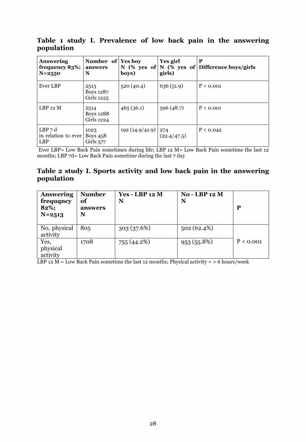
Table 1 study I. Prevalence of low back pain in the answering population
Ever LBP= Low Back Pain sometimes during life; LBP 12 M= Low Back Pain sometime the last 12 months; LBP 7d= Low Back Pain sometime during the last 7 day
Table 2 study I. Sports activity and low back pain in the answering population
Answering frequqncy 82%; N=2513
Number of answers N
Yes - LBP 12 M N
No - LBP 12 M N
P
No, physical activity
805 303 (37.6%) 502 (62.4%) P < 0.001 Yes,
physical activity
1708 755 (44.2%) 953 (55.8%)
LBP 12 M = Low Back Pain sometime the last 12 months; Physical activity = > 6 hours/week
Answering frequency 83%; N=2550
Number of answers N
Yes boy N (% yes of boys)
Yes girl N (% yes of girls)
P Difference boys/girls
Ever LBP
2513 Boys 1287 Girls 1225
520 (40.4) 636 (51.9) P < 0.001
LBP 12 M
2514 Boys 1288 Girls 1224
465 (36.1) 596 (48.7) P < 0.001
LBP 7 d in relation to ever LBP
1023 Boys 458 Girls 577
192 (14.9/41.9) 274 (22.4/47.5)
P < 0.042
29
Table 3 study I. Sports participation and low back pain
Sport Number of Sports participants N
Yes Sports > 6h/W N (% of Sports)
Yes Ever LBP N (% of Sports >6h/W)
Soccer 441 102 (23%) 54 (53%)
Floor ball 296 23 (8%) 14 (61%)
Strength training
241 48 (20%) 20 (41%)
Ice hockey 207 84 (41%) 49 (58%)
Aerobics 170 2 (1%) 1 (50%)
Budo sports 97 33 (34%) 14 (42%)
Swimming 92 7 (8%) 3 (43%)
Equestrian 90 31 (34%) 16 (52%)
Athletics 77 13 (17%) 7 (54%)
Gymnastics 57 3 (5%) 1 (33%)
All sports 1708 700 (41%) 365 (52%)
All sports 1708 819 (48%)
Non-active 803 334 (42%)
30
Study II Retrospective
During the period between 1995 And 2006, Nineteen cases of spondylolysis were identified in the archives of the department of radiology of the local hospital and were invited to participate in the investigation. Sixteen of the cases accepted to take part in the whole investigation (84,2%), 1 women/15 men with the mean age of 20.8 years at the time of the investigation and mean age of 15,5 years (12-21 years) at the time for their MR diagnosis. Follow-up took part 16-27 years after diagnosis.
Seven fractures at L4 level was found at diagnosis (6 incomplete and 1 pseudoarthrosis) and 19 fractures at the L5 level (1 stress reaction, 10 incomplete fractures, 4 complete fractures, 4 pseudoarthrosis). Four heeled fractures in PA were also found. Thirteen out of the 16 cases that took part in the study had had a second MRI/CT investigation and thus 52 out of the initial 64 pars interarticularis where investigated a second time (mean 3 months after the first investigation). In these MRI/CT investigations there was found two fractures at the L4 level, two pseudoarthrosis, and five healed pars interarticularis. At the L5 level we found nine fractures (1 stress reaction, 1 incomplete fracture, 1 complete and 6 pseudoarthrosis) and eight healed stress fractures was identified. In total we found 30 stress reaction/fractures on MRI/CT and out of these 22 heeled during the MRI/CT follow up time (mean 3 months after first investigation). This results in a heeling frequency of 73% of the 64 separate pars interarticularis. Since there are subject with more than one stress fracture a complete heeling of all found fractures in all investigated pars interarticularis was found in only 7 out 16 subjects (43.8%).
This study was made 1-12 years after diagnosis and mean time between the first symptoms of back pain and diagnosis was according to the medical journals, 28 weeks (0-127 weeks). Three months of rest from physical activity except ADL was the most common treatment found in the medical records, but two individuals had been given the advice to be physical active and out of these two one healed and one developed pseudoarthrosis.
The questionnaire was answered by 17 subjects (16 boys and one girl) with a mean age of 20,8 years at the time of answering the questionnaire. At the time of answering the questionnaire nine had had LBP more than 8 days the last 12 month, which had led to a change in work or school activities for six individuals. Three had to change their sport activity due to LBP while 14 had kept their normal activity the last year. The fourteen subjects that had answered the question about number of hours of physical activity each week had a mean activity level of 8.9 hours a week. Ten were physically active more than 6 hours per week and four were active 5 hours or less while three did not answered the question. Seven had LBP at the day of questionnaire with a mean VAS estimation value of 11.4 (range 0-48). Four of those individuals scored more than 30 mm on VAS, which corresponds to moderate pain. Three of the individuals with physical activity 5 hours or less per week had minor LBP problems while 6 with 6 hours or more in physical activity had minor problems and 3 had frequent problems. We did not
31
find any significance correlation between time span between first symptom and initial X ray and LBP today (r=0,608), time span between first symptom and x ray an LBP the last year (r=0,282) or in the rest 3 month as treatment and LBP the last year (r=0,476).
Study III. Clinical study
Clinical investigation
Case/Control – clinical tests – LBP
Even though 8 clinical tests differed significantly in regard to the number of positive tests between the case and the control group w no single test could fully discriminated between LBP and no LBP (Table 1). However, if all these test eight tests were analyzed in combination they could discriminate between LBP and no LBP when the case and control groups were tested together.
Case/Control and diagnostic imaging
Significantly more subjects in the case group (22 out of 25) had findings on their MRI than did the control group (3 out of 13) (p < 0.001). In the control group one boy had a healed stress fracture, one boy with a healed stress fracture and arthrosis of the intervertebral joints L4-L5, and one girl with disc degeneration and L5-S1 level hernia without effect on the nerve roots. (Table 2).
Clinical tests – stress fracture/no stress fracture – all subjects The number of positive tests in the groups with and without stress reaction/stress fracture, irrespective of LBP differed significantly in some of the tests: the coin test (p=0.015), the hook test (p=0.040), and the MCI (p = 0.042) (Table 3). Though, if further analyzed none of the tests could discriminate between stress fracture and other conditions, found in these groups.
Clinical tests – stress fracture/no stress fracture – case group The number of subjects with a positive one-legged lumbar hyper- extension test was significantly higher in subjects with than without stress fracture in the case group. However, 5 cases with stress fracture did not have a positive one-legged lumbar hyperextension test. The test can thus not be used on its own for the clinical diagnosis of stress fractures in the lumbar spine. The multivariate regression analysis (PLS) showed that none of the tests alone or in combination could discriminate all cases of stress fractures from other conditions in the case group.
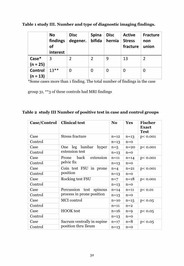
32
Table 1 study III. Number and type of diagnostic imaging findings.
No findings of interest
Disc degener.
Spina bifida
Disc hernia
Active Stress fracture
Fracture non union
Case* (n = 25)
3 2 2 9 13 2
Control (n = 13)
13** 0 0 0 0 0
*Some cases more than 1 finding. The total number of findings in the case
group 31, **3 of these controls had MRI findings
Table 2 study III Number of positive test in case and control groups
Case/Control Clinical test No Yes Fischer Exact Test
Case Stress fracture n=12 n=13 p< 0.001 Control n=13 n=0 Case One leg lumbar hyper
extension test n=5 n=20 p< 0.001
Control n=13 n=0 Case Prone back extension
pelvic fix n=11 n=14 p< 0.001
Control n=13 n=0 Case Coin test FSU in prone
position n=4 n=21 p< 0.001
Control n=13 n=0 Case Rocking test FSU n=7 n=18 p< 0.001 Control n=13 n=0 Case Percussion test spinous
process in prone position n=14 n=11 p< 0.01
Control n=13 n=0 Case MCI control n=10 n=15 p< 0.05 Control n=11 n=2 Case HOOK test n=16 n=9 p< 0.05 Control n=13 n=0 Case Sacrum ventrally in supine
position thru Ileum n=17 n=8 p< 0.05
Control n=13 n=0
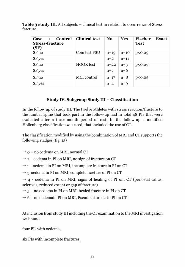
33
Table 3 study III. All subjects – clinical test in relation to occurrence of Stress fracture.
Case + Control Stress-fracture (SF)
Clinical test No Yes Fischer Exact Test
SF no Coin test FSU n=15 n=10 p<0.05 SF yes n=2 n=11 SF no HOOK test n=22 n=3 p<0.05 SF yes n=7 n=6
SF no MCI control n=17 n=8 p<0.05 SF yes n=4 n=9
Study IV. Subgroup Study III – Classification
In the follow up of study III. The twelve athletes with stress reaction/fracture to the lumbar spine that took part in the follow-up had in total 48 PIs that were evaluated after a three-month period of rest. In the follow-up a modified Hollenberg classification was used, that included the use of CT.
The classification modified by using the combination of MRI and CT supports the following stadges (fig. 13)
→ 0 – no oedema on MRI, normal CT
→ 1 – oedema in PI on MRI, no sign of fracture on CT
→ 2 - oedema in PI on MRI, incomplete fracture in PI on CT
→ 3-oedema in PI on MRI, complete fracture of PI on CT
→ 4 - oedema in PI on MRI, signs of healing of PI om CT (periostal callus, sclerosis, reduced extent or gap of fracture) → 5 – no oedema in PI on MRI, healed fracture in PI on CT
→ 6 – no oedemain PI on MRI, Pseudoarthrosis in PI on CT
At inclusion from study III including the CT examination to the MRI investigation we found:
four PIs with oedema,
six PIs with incomplete fractures,
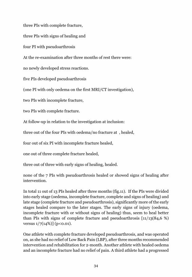
34
three PIs with complete fracture,
three PIs with signs of healing and
four PI with pseudoarthrosis
At the re-examination after three months of rest there were:
no newly developed stress reactions.
five PIs developed pseudoarthrosis
(one PI with only oedema on the first MRI/CT investigation),
two PIs with incomplete fracture,
two PIs with complete fracture.
At follow-up in relation to the investigation at inclusion:
three out of the four PIs with oedema/no fracture at , healed,
four out of six PI with incomplete fracture healed,
one out of three complete fracture healed,
three out of three with early signs of healing, healed.
none of the 7 PIs with pseudoarthrosis healed or showed signs of healing after intervention.
In total 11 out of 13 PIs healed after three months (fig.11). If the PIs were divided into early stage (oedema, incomplete fracture, complete and signs of healing) and late stage (complete fracture and pseudoarthrosis), significantly more of the early stages healed compare to the later stages. The early signs of injury (oedema, incomplete fracture with or without signs of healing) thus, seem to heal better than PIs with signs of complete fracture and pseudoarthrosis [11/13(84,6 %) versus 1/7(14%)] (p<0.01).
One athlete with complete fracture developed pseudoarthrosis, and was operated on, as she had no relief of Low Back Pain (LBP), after three months recommended intervention and rehabilitation for 2-month. Another athlete with healed oedema and an incomplete fracture had no relief of pain. A third athlete had a progressed
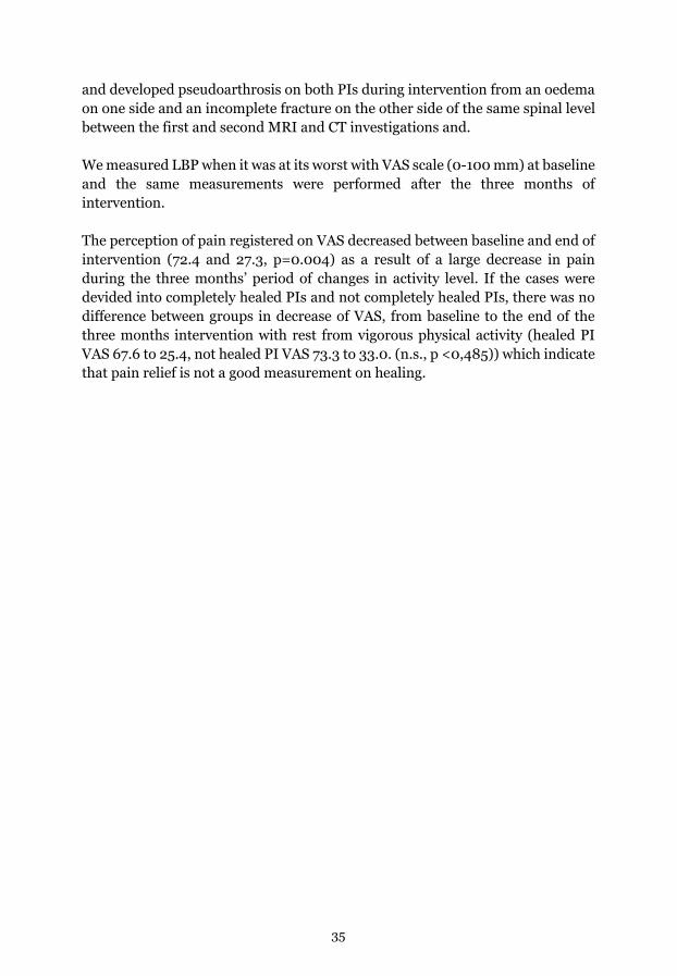
35
and developed pseudoarthrosis on both PIs during intervention from an oedema on one side and an incomplete fracture on the other side of the same spinal level between the first and second MRI and CT investigations and.
We measured LBP when it was at its worst with VAS scale (0-100 mm) at baseline and the same measurements were performed after the three months of intervention.
The perception of pain registered on VAS decreased between baseline and end of intervention (72.4 and 27.3, p=0.004) as a result of a large decrease in pain during the three months’ period of changes in activity level. If the cases were devided into completely healed PIs and not completely healed PIs, there was no difference between groups in decrease of VAS, from baseline to the end of the three months intervention with rest from vigorous physical activity (healed PI VAS 67.6 to 25.4, not healed PI VAS 73.3 to 33.0. (n.s., p <0,485)) which indicate that pain relief is not a good measurement on healing.
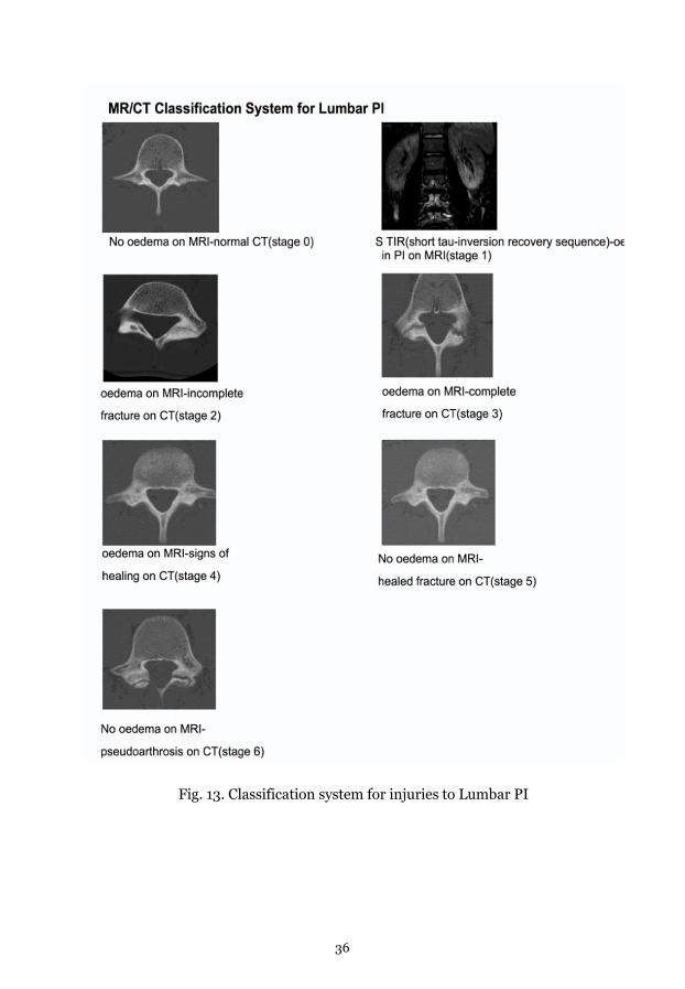
36
Fig. 13. Classification system for injuries to Lumbar PI
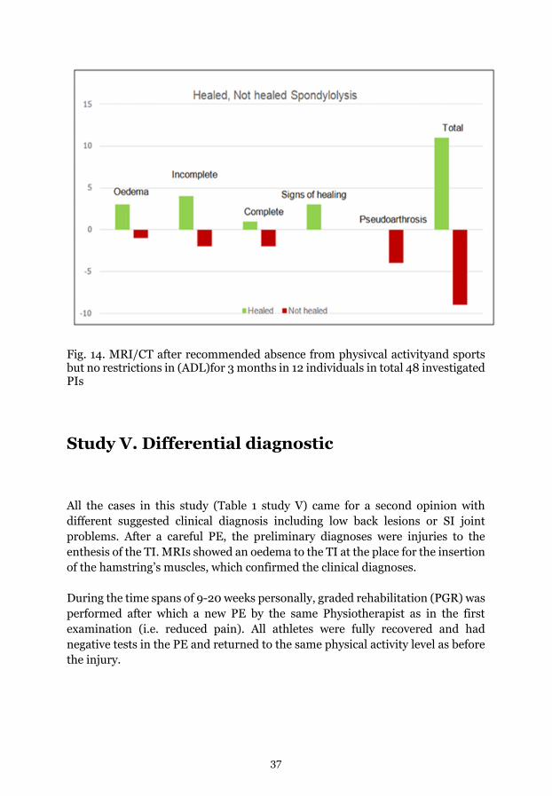
37
Fig. 14. MRI/CT after recommended absence from physivcal activityand sports but no restrictions in (ADL)for 3 months in 12 individuals in total 48 investigated PIs
Study V. Differential diagnostic
All the cases in this study (Table 1 study V) came for a second opinion with different suggested clinical diagnosis including low back lesions or SI joint problems. After a careful PE, the preliminary diagnoses were injuries to the enthesis of the TI. MRIs showed an oedema to the TI at the place for the insertion of the hamstring’s muscles, which confirmed the clinical diagnoses.
During the time spans of 9-20 weeks personally, graded rehabilitation (PGR) was performed after which a new PE by the same Physiotherapist as in the first examination (i.e. reduced pain). All athletes were fully recovered and had negative tests in the PE and returned to the same physical activity level as before the injury.

38
Table 1 Study V. Case history, adolescent male athletes, stress reaction/fracture in tuber ischiadicum
PE=physical examination, a. Eccentric test hamstrings with hip flexion > 100 and slightly flexed knee – eccentric test with positive pain provocation recognized as a symptom by patient, b. Pain provocation when palpating tuber ischiadicum insertion. Hamstring muscles recognized as a symptom by patient, c. In the second MRI of the TI we could see that the first injury was completely healed
Case Case I Case II Case III Case IVa Case IVb Gender/Age
Male/17 Male/14 Male/13 Male/15 Male/15
Main Sport
Football Ice hockey Track and field Ice hockey Ice hockey
Injury activity
Playing football
Explosive running
Explosive running
Ice hockey Ice hockey
Symptom Pain back of thigh
Pain back of thigh
Pain back of thigh
Pelvic pain, Pain back of thigh right side
Groin pain, Pain back of thigh left side
Previous PE
Back pain, sprain tight
Groin injury
Initial PE a, b
a+b a+b a+b a+b a+b
MRI affected side
Stress reaction (oedema) in the skeleton in tuber ischiadicum and in surrounding soft tissues
Avulsion fracture on tuber ischiadicum 15 mm diastasis
Stress reaction oedema in the skeleton on tuber ischiadicum, and discrete reaction in the surrounding soft tissues
Stress reaction (great oedema)in the skeleton in tuber ischiadicum and discrete reaction in the surrounding soft tissues
Stress reaction (great oedema) in the skeleton in tuber ischiadicum.
MRI contralateral side
No reaction No reaction No reaction No reaction No reaction. c Left TI healed
Rest time 6 days 3 days 5 weeks 3 weeks 2 weeks Rehabilitation
Controlled rehabilitation 20 weeks. Stepwise return to normal physical activity
Controlled rehabilitation 11 weeks Stepwise return to normal physical activity
Controlled rehabilitation 4 weeks. Stepwise return to normal physical activity
Controlled rehabilitation 9 weeks. Stepwise return to normal physical activity
Controlled rehabilitation 7 weeks. Stepwise return to normal physical activity
Final PE a, b
a+b negative a+b negative a+b negative a+b negative a+b negative
Outcome Back to normal sport activity
Back to normal sport activity
Back to normal sport activity
Back to normal sport activity
Back to normal sport activity
39
Discussion
Low back pain
Prevalence.
In a wide perspective LBP is common all over the world. The lifetime prevalence of LBP is reported to be as high as 84% (64, 53) and based on 7.6 billion people in the world, about 6.4 billion people have experienced LBP sometimes in their life. Individuals suffering from disability because of LBP have been estimated to be 8-12 % (about 768 million individuals) (53,64,95). Globally LBP seems to be the leading cause of years lived with disability (YLD) in both men and women (96)Different forms of physical workload and awkward positions are risk factors for low back pain in adults (97) together with many other factors like absence from physical activity (98). LBP is commonly divided into acute <3 months´ pain duration and chronic>3 months´ pain duration but is better seen as a condition that fluctuate over time, including low or medium intensity periods and periods with more severe pain (36,59). If LBP is divided into specific and non-specific LBP significantly higher pain scores and longer pain duration have been demonstrated in the specific LBP groups (99). LBP early in life tends to remain as chronic LBP or recurrent LBP over years (36 57).
Low Back Pain in adolescence
LBP during adolescence has been demonstrated already around 10 years of age, and then escalate to reach almost the same prevalence as in adults by the age of 1 (62, 100, 101, 102). LBP in adolescence has been shown to increase, and LBP with disability seems to continue into adulthood (103) presenting a high risk to suffer from LBP later in life (65 104). In 25-years follow-up study of adolescents (14 years of age at inclusion) there was a positive correlation between adolescent LBP and LBP in adulthood (105) In our prevalence study on 16-19 years old individuals (paper I) we found high numbers (46%) suffering from LBP, more girls than boys reported LBP, and highly sports active had significantly higher prevalence of LBP compared with less sports active. High level of sport activity (>6 h/w) was associated with higher odds ratio of having LBP, a longer duration suffering from LBP, and more often disability due to LBP (Paper I). In a study on Australian football players compared with non-football players it was shown that one out of 4 football players reported that LBP had a negative impact on their sport performance (106). In a study in Japan, on first year male and female university students on different activity levels (non- actives, medium activity, and high levels of sports activity) significantly more students that were on a high activity level reported LBP (21). In a large cross-sectional study among 9-15-year-old boys and girls (divided into non-sport group and sport group) the odds ratio for having LBP was 1.57 in the sport group (100). Althogether, it seems that highly sport actives sustain a higher risk to suffer from LBP.

40
The Maturity of the skeleton (i.e lower back)
The whole skeleton needs to be loaded during youth and adolescence to become strong (7,107). However, to much load can harm the growing skeleton. In adolescent athletes it is important to find an individual loading approach (long term project) considering the maturity of the skeleton (108). Especially the spine needs to mature before tolerating high loads (22,23, 32,67)Paper IV.
Spinal Load
The spine is always under load, (109). It manages high loads during daily life as well as during physical activity. For the growing skeleton activitiescontaining weight bearing, high intensity activity, and movements in unusual directions is important to build optimal peak bone mass (PBM) (11). Both gravity and muscle force produce mechanical loading of the skeleton (110). Studying the intra-discal pressure in different positions and during different exercises showed that during a forward bend, during jogging and when holding 20 kg close to body, these activities produce an intra-discal pressure that is twice as high as seen during unloading positions (109). In a torsion like a golf swing the load on the lumbar spine can be as high as 7500N and during a half squat lift of a weight that is 1.6 times body weight a compressive lumbar disc load of 10 times body weight has been demonstrated (111). In an in vitro model estimating loads on the lumbar spine facet joints, it was shown that a combination of lateral bending and rotation resulted in loads up to 154.2 N on the facet joints at L4 level (25). High levels of sport activities produce high levels of spinal load and for the adolescent athlete this can overload the lower back and cause stress reactions like microfractures, incomplete or complete pars interarticularis fractures most often seen at L4 or L5 levels (112). This is in line with our results in Paper III, were we found spondylolysis-fractures in pars interarticularis in highly active patients with LBP.

41
Low Back Pain in Adolescent athletes
Fig. 15 U shape
Low Back Pain in Adolescent athletes has its own identity, not to compare with adult low back pain. The reason for this is puberty, as adolescents is in a developing stage were the living body and its tissues develops in an individual manner. The skeletal development is not always correlating to the actual chronological age, but more in its stage of maturity (10,39,113, 114, 115) LBP seems to have a U shape relation to physical activity, as no physical activity or low activity is related to LBP, but also a high activity levels can be related to LBP (fig. 15) (97).High level of competitive sport activities has to be adjusted depending on the adolescent maturity stage, this is not always the chronologic age, because the impacts on the growing low back skeleton from high loads and complex movements (22,23,25,32,111,). The low back has development stages where some parts of the FSU is weaker compared to other tissues. The endplate in the ventral part of the FSU is a weaker part compared to the disc until around 20 years of age (22,23,32). Pars interarticularis in the dorsal part of the L4 and L5 is due to cortical porosity during puberty (7), also more vulnerable to repeated load in extension and rotation (25). Unilateral defects in pars interarticularis has been shown to produce greater stress on the contralateral pars, and bilateral defects change the load in the whole FSU (disc, ligaments and endplates) (116). In our case/control study 88% of the adolescent athletes with LBP had posterior element

42
pathology and 50% had injuries in pars interarticularis in the L4 or L5 vertebrae (Paper III).
Injuries to Pars Interarticularis of the low back
Etiology
The functional unit of the spine in the lower back, and its assignment to control the complex loads and movements of the torso, place high demands on the structures involved. The ventral and dorsal parts of the vertebrae, the disc, ligaments and muscles, are extensively involved (80). Daily activity as well as physical activity and sports produce different types of load on the low back skeleton and require muscular and ligament control. The total daily load from axial load, shear forces, and cyclic load with different intensity, is depending on the type of activity. Low physical activity is a risk factor for LBP as well as to much physical activity (97,117) (fig.15). Also, biomechanical variations and the vertebral anatomy can be involved in the etiology. The sacro-horizontal angle has been shown to be larger in adolescents with spondylolysis and spondylolisthesis (116, 118). The vertebral bodies are more “sensitive” too compressive high loads (47Swärd 1990, 23Lundin 2001, 22 Baranto 2006, 24 Thorenson 2016), while the dorsal part and especially the pars interarticularis is more sensitive to intensive cyclic movements causing stress reactions and fatigue fractures (97 117). A genetic predisposition might play a role in the etiology. The sulfate anion transporter possibly plays a role in the development of cartilage in the lumbosacral spine (119,120) has found a possible genetic predisposition of spondylolysis in certain families.
Pathogenesis
Cyclic repetitive loading in high frequencies has been found to be vulnerable to pars interarticularis and can lead to fatigue fractures (56,74,84,121,123) There is a “pincer theory” about the pathogenesis where the inter facet distance between facets in the low back segments normally can increase, but in patients with pars interarticularis fractures there is no capacity to increase. This decreased interface distances were found in patients with pars interarticularis fractures and created a pinch on the pars interarticularis in the caudal vertebra in the FSU when performing flexion, extension, side bending and rotation movements during daily living activities, physical activity and sport activities (84,122,124,). In a study of the fibrocartilage in defected pars interarticularis the authors found both Ruffino receptors (movement transmitters) and free nerve endings (nociceptors) (125). This can possibly explain the background to pain caused by pars interarticularis fractures. However, in another similar study a more ligamentous structure and no neural structures were found (126).
43
Spondylolisthesis
Pars interarticularis fractures that don´t heal can develop in to a pseudarthrosis and spondylolisthesis (74) (PaperIII). A relationship between pars interarticularis fractures and spondylolisthesis has been shown, and about 15% of the pars interarticularis fractures develop into spondylolisthesis, often together with disc degeneration (74,83,86) Spondylolisthesis is considered to be seen as a specific cause of LBP (127).
Physical examination
Medical history together with a physical examination is a traditional tool used in the investigation of LBP (35, 39,128,129It often contains many questions and many functional tests in an attempt to gain a careful clinical diagnosis and agrees with reference standard diagnosis more often than by chance (130). Although more specific diagnosis in the lower back is hard to find with a Physical Examination (128). The causes of LBP in Adolescent Athletes compared with an adult population is significantly different (35,39). Injuries to the lower back such as conditions in Pars Interarticularis in Adolescent Injuries to the lower back such as pars interarticularis fractures in adolescent athletes is no exception. The one leg lumbar hyper extension test is said to be the physical examination test that discriminate injuries in pars interarticularis in the low back from other conditions in the low back in adolescent athletes (1). When looking at the specific diagnosis spondylolysis, we could not find a significant difference between patients and controls using physical tests including the one leg lumbar hyper extension test (Paper III). To establish an early diagnosis, we found that the best way to find pars interarticularis fractures was to focus on the medical history (symptoms), and to use MRI and CT examination (Papers III and IV). This has later also been confirmed by others (68)
Treatment
In Paper IV we describe the different fracture stages that was found using MRI and CT examination. The injury can progress from a stress reaction in the skeleton, to an incomplete fracture, a complete fracture, and to a pseudoarthrosis. Spondylolysis should be seen as the end of this process and the stages before the manifest fracture-spondylolysis, are the stages were there seeming to be a good potential to heal the injury (Paper IV). Therefore, early diagnosis is important. This has also been reported by others (56,79,80,82, 131, 132). The best options for healing are an early detection in the stress reaction or incomplete fracture stage (56,79, 82,132). We used conservative treatment with rest from aggravating activities for 3 months (Paper IV) and this has also been successfully used by others (56,132,133). Another treatment used by others is immobilization in a brace (56,81,131,).
44
Differential diagnosis
An important differential diagnosis to injuries to Pars Interarticularis of the lower back is stress reactions and avulsion fractures in Tuber Ischiadicum (TI) of the pelvis. In our study of LBP among adolescent athletes some cases of stress reactions/ avulsion fractures in TI of the pelvis was found. As described in paper V they had earlier been give the diagnose LBP (paper V). The diagnosis of avulsion fractures to TI was detected by a Physical Examination and confirmed with an MRI. The subjects were recommended different times of rest. After that, they all got a Physically Graded Rehabilitation during different length and back to the same sport activity. Paper V is a case report with few participants but the same is found in another report (90). The condition is seen in adolescent athletes but often missed in clinic. It has its similarities with injuries to the proximal hamstrings muscle and tendon (87, 88, 89,90)
Summary
Adolescence ages have in general a high prevalence of Low back Pain and it increases with age and Adolescents with a high level of competitive Sports have significantly more Low Back Pain during longer time, more disabling and more among girls than boys. The growing spine mature individual and does not follows chronologic age. High loads and many hours of Sports/week can be vulnerable to the growing spine It is important to adjust the level of hours spent in competitive sports and the content in practice together with time for rest. It is important to take LBP in adolescent athletes seriously as we found specific conditions that explained the LBP symptoms. This makes it possible to secure early diagnosis and early intervention to heal injuries and make these young people come back to their normal physical activity and sports and in the long run prevent LBP in adult ages. It is also important to see that it is not always LBP and that differential diagnosis is the answer.
-LBP is common among adolescents
-LBP is more common in girls than in boys
-LBP is more common among highly sports active adolescents
-LBP in adolescents should be taken seriously, especially in adolescent athletes that not seldom have fractures in pars interarticularis (Spondylolysis).
-There is no single clinical test, or combination of clinical tests, that reliably can diagnose a pars interarticularis fracture-Spondylolysis.
-CT examination in addition to MRI provides a staging of the pars interarticularis fracture.
45
-Pars interarticularis fractures diagnosed early, in the early fracture stages, seems to have a good potential to heal with 3 months of rest from loading (sport activity).
-Differential diagnoses such as stress reactions in the pelvic ischial tuberosity should be taken in consideration.
Perspective
To be Physically active is good for your health and your body both the nervous system, the muscle system and the skeletal system. On the contrary, physical inactivity is bad for the nervous system, the muscle system and the skeletal system. Adolescents with an ambition to be high level athletes with many hours in practice, high intensity and high load exercises are close to a danger-zon to injury because of too much total load to the body. There should be an awareness that the maturity stage is more important than the chronologic age to avoid injuries of high load and high intensity and there should be a slow progression arrangement in practice, over time. This should be a guiding principle in the design of exercises and long-term planning and make the training more individualized the more hours there are, on the sport schedule.
46
Acknowledgments Karin Larsén, my head tutor, thank you for your never-ending help and support. During the years I have learned a lot about research from you. Thank you!
Håkan Jonsson, assistant tutor, very supportive and you gave me many interesting discussions in specific issues
Lars Ådin, radiologist, good freind and expert in his field
Erik Bergström, helpful in the preparation and management of the first paper
Håkan Alfredsson, the one and only, always there with help and discussions
Linda Nilsson, a really nice person always with a smile, helpful. Assisted me in the clinical studies
Doris Sundell, my wife, and help in research as she assisted me to bring in all the facts from the questionnaries in to the computer 2550 individuals, what can I say!
Margareta Rullander, neighbor and a good help with the English language
Colleagues in Umeå University and all the people in the Sport Medicine Unite, Lisbeth Fritzen, Lars Stigell, Kajsa Gilenstam,Kerstin Ekman, Gunilla Solander, Pär Liljeholm, Magnus Högström, Lars Göran Fjällborg, Jonas Lindberg, Jonas Lorentzon, Per Jonsson
Thank you for all the help during the production of the thesis, Mikael Kohkoinen.
47
References
1. Jackson DW, Wiltse LL, Dingeman RD, Hayes M. Stress reactions involving the pars interarticularis in young athletes. Am J Sports Med 1981: Vol. 9. Doi: 10.1177/036354658100900504
2. Kurtén B, Lindhart og Ringhof (1981, 2018). Våra älsta Förfäder. Copyright©1981, 2018 Förlag A/S, ISBN: 9788711879771
3. Fausto‐Sterling A. "The Bare Bones of Sex: Part 1—Sex and Gender," Signs: Journal of Women in Culture and Society 30;2:1491-1527. https://doi.org/10.1086/424932
4. Marjolein C.H. van der Meulena,b, Rik Huiskesc, Why mechanobiology? A survey article. Journal of Biomechanics 2002;35:401–414
5. Heiniö L, Nikander R, Sievänen H. Association between long-term exercise loading and lumbar spine trabecular bone score (TBS) in different exercise loading groups. J Musculoskelet Neuronal Interact 2015;15(3):279-285
6. Damm P, Kutzner I, Bergmann G, Rohlmann A, Schmidt H. Comparison of in vivo measured loads in knee, hip and spinal implants during level walking. Journal of Biomechanics 51. 2017;128–132. http://dx.doi.org/10.1016/j.jbiomech.2016.11.060
7. Parfitt AM. The Two Faces of Growth: Benefits and Risks to Bone Integrity. Osteoporosis Int. 1994;4:382-398
8. Frost HM. Bone’s Mechanostat: A 2003 Update. The anatomical record part a 275A. 2003:1081–1101
9. Wolff, J., 1892. The Law of Bone Remodeling. (Das Gesetz der Transformation der Knochen, Kirschwald, 1892). Translated by Maquet, P., Furlong, R. Springer, Berlin, 1986.
10. Tiwaria A K, Kumara R, Tripathia D, Badhyalb S. In silico modeling of bone adaptation to rest-inserted loading: Strain energy density versus fluid flow as stimulus. Journal of Theoretical Biology. 2018;446: 110–127
48
11. Nordström A, Tervo T , Högström M. The Effect of Physical Activity on Bone Accrual, Osteoporosis and Fracture Prevention. The Open Bone Journal. 2011;3:11-21
12. Whedon, 1984 G. Donald Whedon. Disuse Osteoporosis: Physiological Aspects. Calcif Tissue Int. 1984;36:146-S150
13. Huiskes R, Ruimerman R G, van Lenthe H, Janssen J D. Effects of mechanical forces on maintenance and adaptation of form in trabecular bone. Nature vol. 405, 2000
14. Ferry B, Lespessaillesc E, Rochcongar P, Duclose M, Courteix D. Bone health during late adolescence: Effects of an 8-month training program on bone geometry in female athletes. Joint Bone Spine. 2013;80:57–63.
15. Gabel L A, McKay H, Nettelefold L, Race D , MacDonald HM. Bone Architecture and Strength in the Growing Skeleton: The Role of Sedentary Time. Medicine & Science in Sports & Exercise. 2014; 363-372 DOI:10.1249/MSS.0000000000000418
16. Mirwald RL, Baxter-Jones ADG, Baily DA, Beunen GP. An assessment of maturity from anthropometric measurements. Medicine & Science in Sports & Exercise. 2002;34(4): 689-694
17. Frankel VH, Nordin M Basic Biomechanics of the Musculoskeletal System/edited Margareta Nordin, - 3rd ed. 2001. ISBN-683-30247-7, Lippincott Williams & Wilkins.
18. Bogduk A, Burton D. Biomechanics of Back Pain, 2002/2004. Churchill Livingstone. ISBN 0 433 06207 2
19. Kuorinka I, Jonsson B, Kilbom A, Vinterberg H, Biering-Sørensen F, Andersson G, Jørgensen K. Standardised Nordic questionnaires for the analysis of musculoskeletal symptoms. Appl Ergon. 1987;18: 233–237
20. Thomas E, Silman AJ, Croft PR, Papageorgiou A C, I V Jayson M, Macfarlane GJ. Predicting who develops chronic low back pain in primary care: a prospective study BMJ. 1999;318:1662–7
21. Hangai M, Kaneoka K, Okubo Y, Miyakawa S, Hinotsu S, Mukai N, Sakane M, Ochiai N. Relationship Between Low Back Pain and Competitive Sports Activities During Yout. The American Journal of Sports Medicine. 2010;38(4):791-796. DOI: 10.1177/0363546509350297
49
22. Baranto O, Traumatic high-load injuries in the adolescent spine: clinical, radiological and experimental studies. University of Gothenburg, 2005. ISBN 91-628-6604-4
23. Lundin O. Effects of high load on the growing spine. Experimental studies and long-term follow-up of top athletes. University of Gothenburg, 2000. ISBN 91-628-4086-X
24. Thoreson O, Baranto A, Ekström L, Holm S, Hellström M, Swärd L. The immediate effect of repeated loading on the compressive strength of young porcine lumbar spine. Knee Surg Sports Traumatol Arthrosc. 2010;18:694–701. DOI 10.1007/s00167-009-z
25. Popovich J M Jr, Welcher JB, Hedman T P, Tawackoli W, Anand N, Chen TC, Kulig K. Lumbar facet joint and intervertebral disc loading during simulated pelvic obliquity. The Spine Journal 2013;13:1581–1589
26. Schoenau E. From mechanostat theory to development of the "Functional Muscle-Bone-Unit". J Musculoskelet Neuronal Interact. 2005; 5(3):232-238
27. Joza LG, Kannus P. Human Tendons. Anatomy, Physiology and Pathology 1997. Human Kinetics. ISBN: 0-87322-484-1
28. Naughton G, Farpour-Lambert NJ, Carlson J, Bradney M, Van Praagh. Physiological Issues Surrounding the Performance of Adolescent Athletes. Sports Med. 2000;30(5): 309-325
29. Singh N. Synovial Joints and Lubrication mechanisms International Journal of Computational and Applied Mathematics. ISSN 1819-4966. 2017;12(1):29-33
30. Benjamin M, E. Kaiser, S. Milz. Structure-function relationships in tendons: a review. J. Anat. 2008;212:211–228. doi: 10.1111/j.1469-7580.2008.00864.
31. Lloyd,RS, Faigenbaum A D, Stone M H, Oliver J L, Jeffreys I, Moody J A, Brewer C, Pierce K C, McCambridge TM, Howard R, Herrington L, Hainline B, Micheli L J, Jaques R, Kraemer W J, McBride MG, Best TM, Chu D A, Alvar BA, Gregory D Myer GD. Position statement on youth resistance training: the 2014 International Consensus. Br J Sports Med 2014;48:498–505.
32. Thoreson O. On the effect of repetitive loading on the spine of young elite athletes – Clinical and experimental studies. University of
50
Gothenburg. ISBN-978-91-628-9820-5(PRINT), ISBN-978-91-628-9821-2(PDF)
33. Actis JA, Honegger JD , Gates DH , Petrella AJ, Nolasco LA , Silverman AK. Validation of lumbar spine loading from a musculoskeletal model including the lower limbs and lumbar spine. Journal of Biomechanics. 2018:68:107–114.
34. Allan DB, Wadell G. An historical perspective on low back pain and disability. Acta Ortopedica. 1989;60(3):1-23
35. Hartvigsen J, Hancock MJ, Kongsted A, Louw Q, Ferreira ML, Genevay S, Hoy D, Karppinen J, Pransky G, Sieper J, Smeets RJ, Underwood M, on behalf of the Lancet Low Back Pain Series Working Group. What low back pain is and why we need to pay attention. Lancet 2018;391:2356–67
36. Donelson R, McIntosh G, Hall H. Is It Time to Rethink the Typical Course of Low Back Pain? PM R 2012;4:394-401. DOI: 10.1016/j.pmrj.2011.10.015
37. Rampersaud YR, Bidos A, Fanti C, Perruccio AV, The Need for Multidimensional Stratification of Chronic Low Back Pain (LBP). SPINE 2017;42(22):E1318–E1325. DOI: 10.1097/BRS.0000000000002237
38. Apeldoorn AT, Raymond W, Ostelo RW, van Helvoirt H, Fritz JM, de Vet H CW, van Tulder MW The cost-effectiveness of a treatment-based classification system for low back pain: design of a randomised controlled trial and economic evaluation. BMC Musculoskeletal Disorders 2010;11:58
39. Micheli LJ, Wood R. Back pain in young athletes. Significant differences from adults in causes and patterns. Arch Pediatr Adolesc Med. 1995;149(1):15-18
40. Masci L, Pike J, Malara F, Phillips B, Bennell K, Brukner P. Use of the one-legged hyperextension test and magnetic resonance imaging in the diagnosis of active spondylolysis. Br J Sports Med. 2006;40(11):940–6.
41. Von Korff, Saunders K. The Course of Back Pain in Primary Care. SPINE 1996;21(24):2833-2839
42. Hestbaek L, Leboeuf Yde, Engberg M, Lauritzen T, Bruun NH, Manniche C. The course of low back pain in a general population.

51
Results from a 5-year prospective study. Journal of Manipulative and Physiological Therapeutics 2003;26(4):213–9.
43. Hegmann K, Talmage J. Practice guidelines low back disorders. In: Occupational Medicine Practice Guidelines. Revised ed. Elk Grove Village, IL: American College of Occupational and Environmental Medicine; 2007, 368
44. Van Tulder M, Becker A, Bekkering T, Breen A, Gil del Real M, Hutchinson A, Koes B, Laerum, E, Malmivaara A. European guidelines for the managament of acute nonspecific low back pain in primary care European spine journal : official publication of the European Spine Society, the European Spinal Deformity Society, and the European Section of the Cervical Spine Research Society. 2006;15(2):169-91
45. Koes BW, van Tulder M W, Thomas S. Diagnosis and treatment of low back pain. BMJ 2006;332:1430-4
46. Dillane JB, Kalton G. Acute back syndrome-a study from general practice Brit. Med. J. 1966;2:82-84
47. Henschke N, Maher C, Refshauge K, et al. Prognosis in patients with recent onset low back pain in Australian primary care: Inception cohort study. BMJ. 2008;337:a171. doi:10.1136/bmj.a171
48. van den Hoogen HJM, Koes BW, Devillé W, van Eijk JTM, Bouter L M. The Prognosis of Low Back Pain in General Practice. SPINE 1997;22(13):1515-1521
49. Phillips H, Grant L. The evolution of chronic back pain problems: A longitudinal study. Behav Res Ther 1991;29:435-441
50. Cherkin D, Deyo R, Street J, Barlow W. Predicting poor outcomes for back pain seen in primary care using patients’ own criteria. Spine (Phila Pa 1976) 1996;21:2900-2907.
51. Costa L, Maher C, McAuley J, et al. Prognosis for patients with chronic low back pain: Inception cohort study. BMJ 2009;339\b3829
52. Young AE, Wasia R, Phillips L, Douglas P. Gross DP. Workers’ perspectives on low back pain recurrence: ‘‘It comes and goes and comes and goes, but it’s always there’’. PAIN 2011;152:204-211
53. Balagué F, Manninon AF, Pellisé F, Cederaschi C. Non-specific low back pain. Lancet 2012;379:482-91. DOI:10.1016/S0140- 6736(11)60610-7
52
54. Newell RM, Historical Perspective Spondylolysis, An Historical Review. SPINE 1995;20(17):1950-1956
55. Belfi LM, Orlando Ortiz A, Katz DS. Computed Tomography Evaluation of Spondylolysis and Spondylolisthesis in Asymptomatic Patients. SPINE 2006;33(24):907–910
56. Standaert CJ, Herring SA. Spondylolysis: a critical review. Br J Sports Med. 2000;34:415–422
57. Kjaer P, Korsholm L, Leboeuf-Yde C, Hestbaek L, Bendix T. Individual courses of low back pain in adult Danes: a cohort study with 4-year and 8-year follow-up. BMC Musculoskeletal Disorders 2017;18:28. DOI 10.1186/s12891-016-1377-0
58. Buchbinder R, van Tulder M, Öberg B, Costa LM, Woolf A, Schoene M, Croft P, on behalf of the Lancet Low Back Pain Series Working Group. Low back pain: a call for action. Lancet 2018;391:2384–88.
59. Kongsted A, Kent P, Axen I, Downie AS, Dunn KM Open Access. What have we learned from ten years of trajectory research in low back pain? BMC Musculoskeletal Disorders 2016;17:220. DOI 10.1186/s12891-016-1071-2
60. Murphy DR, Hurwitz EL. A theoretical model for the development of a diagnosis-based clinical decision rule for the management of patients with spinal pain. BMC Musculoskeletal Disorders 2007;8:75. doi:10.1186/1471-2474-8-75
61. Milanese S, Grimmer-Somers K. What is adolescent low back pain? Current definitions used to define the adolescent with low back pain. Journal of Pain Research 2010;3: 57-66
62. Leboeuf-Yde C, Ohm Kyvik K. At what age does Low Back Pain become a common problem? A study of 29,424 Individuals aged 12-41 years. 1998 SPINE. 23;2:228-234
63. Grimmer K, Nyland L, Milanese S. Longitudinal investigation of low back pain in Australian adolescents: a five-year study. Physiother. Res. Int. 2006;11: 161–172 DOI: 10.1002/pri
64. Airaksinen O, Brox J.I., Cedraschi C, Hildebrandt J, Klaber-Moffett J, Kovacs F, Mannion A.F, Reis S, Staal J.B, Ursin H, Zanoli G. Chapter 4.European guidelines for the management of chronic nonspecific low back pain. Eur Spine J. 2006;15 (2): 192–300. DOI 10.1007/s00586-006-1072-1

53
65. Hestbaek L, Leboeuf‐Yde C. The course of low back pain from adolescence to adulthood eight‐year follow-up of 9600 twins. Spine. 2006;4:468‐472.
66. Kujala UM, Salminen JJ, Taimela S, Oksanen A, Jaakola Laura. Subject characteristics and low back pain in young athletes and nonathletes. Medicine and Science in Sports 1992;24(6):627-632
67. Swärd L, Hellström M, Jacobsson B, Peterson L. Back Pain and Radiologic Changes in the thoraco-Lumbar Spin of Athletes. SPINE 1990;15(2):124-129
68. Crawford CH, Ledonio CGT, Shay Bess R, Buchowski JM, Douglas C. Burton DC, Hu SS, Lonner BS, Polly DW, Smith JS, MD, Sanders JO. Current Evidence Regarding the Etiology, Prevalence, Natural History and Prognosis of Pediatric Lumbar Spondylolysis: A Report from the Scoliosis Research Society Evidence-Based Medicine Committee. Spine Deformity 2015:3:12-29. http://dx.doi.org/10.1016/j.jspd.2014.06.005
69. Herman MJ, Pizzutillo PD. Spondylolysis and Spondylolisthesis in the Child and Adolescent. A New Classification. CLINICAL ORTHOPAEDICS AND RELATED RESEARCH. 2005: 434:46–54
70. Tofte JF, CarlLee TL, Holte AJ, Sitton SE, Weinstein SL. Imaging Pediatric Spondylolysis. A Systematic Review SPINE 2017;42(10); 777–782
71. Kountouris A, Saw R, Saw A. Management of Lumbar Spondylolysis in Athletes: Role of Imaging. Curr Radiol Rep. 2018;6:39. https://doi.org/10.1007/s40134-018-0299-z
72. Rossi F, Dragoni S. Acute avulsion fractures of the pelvis in adolescent competitive athletes: prevalence, location and sports distribution of 203 cases collected Skeletal Radiol. 2001;30:127–131
73. Ranawat VS J.K. Dowell, M.B. Heywood-Stress fractures of the lumbar pars interarticularis in athletes: a review based on long-term results of 18 professional cricketers Injury, Int. J. Care Injured 34 (2003) 915–919
74. Hu SS, Tribus CB, Diab M, Ghanayem AJ Spondylolisthesis and Spondylolysis J Bone Joint Surg Am. 2008;90:656-671
75. Ladenhauf HN, Fabricant PD, Grossman E, Widmann RF, Green DW. Athletic Participation in Childrenwith Symptomatic Spondylolysis in

54
the New York Area. Medicine & Science inSports & Exercise 2013;45(10):1971-1974.DOI: 10.1249/MSS.0b013e318294b4ed
76. Benell KL, Malcolm SA, Brukner PD. Models for pathogenesis of stress fractures in athletes. Br J SportsMed 1996;30:200-204
77. Warden SJ, Creaby MW, Bryant AL, Crossley KM. Stress fracture risk factors in female football players and their clinical implications. Br J SportsMed. 007;41(Suppl I):i38–i43. doi: 10.1136/bjsm.2007.037804
78. Lee JC, Malara FA, Wood T, Hoy G, Saifuddin A, Connell DA. MRI of Stress Reaction of the Distal Humerus in Elite Tennis Players. AJR. 2006;187:901–904. DOI:10.2214/AJR.05.1058
79. Fujii K, Katoh S, Sairyo K, Ikata T. Union of defects in the pars interar- ticularis of the lumbar spine in children and adolescents. The radio-logical outcome after conservative treatment. J Bone Joint Surg Br. 2004; 86: 225–231
80. Motley M, Nyland MJ, Jacobs J, Caborn DMN. The Pars Interarticularis Stress Reaction, Spondylolysis, and Spondylolisthesis Progression. Journal of Athletic Training 1998;33(4):351-358
81. Morita T, Ikata T, Katoh S, Miyake R. Lumbar spondylolysis in children and adolescents. J Bone Joint Surg Br. 1995;77: 620–62
82. Sairyo K, Sakai T, Yasui N. Conservative treatment of lumbar spondylolysis in childhood and adolescence. The Radiological Signs Which Predict Healing. Conservative treatment of lumbar spondylolysis in childhood and adolescence The Radiological Signs Which Predict Healing. J Bone Joint Surg. 2009;91-B:206-9.
83. Floman Y. Progression of lumbosacral isthmic spondylolisthesis in adults. Spine. 2000;25:342–347.
84. Foreman P, Griessenauer CJ, Watanabe K, Conklin M, Shoja MM, Rozzelle CJ, Loukas M, Tubbs RS. L5 spondylolysis/spondylolisthesis: a comprehensive review with an anatomic focus. Childs Nerv Syst. 2013;29:209–216 DOI 10.1007/s00381-012-1942-2
85. Aono K, MD, Kobayashi T, Jimbo S, AtsutaY, Matsuno T. Radiographic Analysis of Newly Developed Degenerative Spondylolisthesis in a Mean Twelve-Year Prospective Study. SPINE 2010 35;(8):887–891
86. Iguchi T, Nishida K, Ozaki T, Kitagawa A, Tsumura N, Kakutani K, Takashi, Yurube T, Kuroda R. Grade three disc degeneration is a

55
critical stage for anterior spondylolisthesis in lumbar spine. Eur Spine J. 2012:21;2134–2139
87. Lazovi´c D, Wegner G, Gossé PF: Ultrasound for diagnosis of apophyseal injuries. Knee Surg, Sports Traumatol, Arthroscopy. 1996;3:234-237
88. Brittenden J, Robinson JP. Imaging of pelvic injuries in athletes. The British Journal of Radiology, 2005;78:457–468 DOI: 10.1259/bjr/73355272
89. Askling CM, Tengvar M, Saartok. Proximal Hamstring Strains of Stretching Type in Different Sports: Injury Situations, Clinical and Magnetic Resonance Imaging Characteristics, and Return to Sport. 2008;36 :1799-1804
90. Ferlic PW, Sadoghi P, Singer G, Kraus T Eberl R. Treatment for ischial tuberosity avulsion fractures in adolescent athletes. Knee Surg Sports Traumatol Arthrosc. 2014;22:893–897. DOI 10.1007/s00167-013-2570-4
91. Collins SL, Moore RA, McQuayHJ. The visual analogue pain intensity scale: what is moderate pain in millimetres? Pain 72 (1997) 95–97
92. Kaltenborn F. Manual mobilization of the joints. Volume II. The Spine 5th edition 2009 , Norli, Oslo, Norway
93. Paatelma M, Karvonen E, Heinonen A. Inter- and intra-tester reliability of selected clinical tests in examining patients with early phase lum- bar spine and sacroiliac joint pain and dysfunction. Adv Physiother. 2010;12: 74–80
94. Dankaerts W, O’Sullivan P. The validity of O’Sullivan’s classification system (CS) for a sub-group of NS-CLBP with motor control impairment (MCI). Overview of a series of studies and review of the literature. Man Ther. 2011; 16: 9–14
95. Goubert L, Crombez G, De Bourdeaudhuij I. Low back pain, disability and back pain myths in a community sample: prevalence and interrelationships. European Journal of Pain. 2004;8:385–394
96. GBD 2016 Disease and Injury Incidence and Prevalence Collaborators. Global, regional, and national incidence, prevalence, and years lived with disability for 328 diseases and injuries for 195 countries, 1990–2016: a systematic analysis for the Global Burden of Disease Study 2016. Lancet 2017; 390: 1211–59

56
97. Heneweer H, Vanhees L, Picavet HSJ. Physical activity and low back pain: A U-shaped relation? PAIN 2009;143:21–25
98. Hills AP, Street SJ, Byrne NM. Physical Activity and Health: “What is Old is New Again”. Advances in Food and Nutrition Research. Volume 75, 2015. ISSN 1043-4526. http://dx.doi.org/10.1016/bs.afnr.2015.06.001
99. Kim G, Yi C, Cynn H. Factors Influencing Disability due to Low Back Pain Using the Oswestry Disability Questionnaire and the Quebec Back Pain Disability Scale. Physiother. Res. Int. 2015;20:16–21
100. Sato T, Ito T, Hirano T, Morita O, Kikuchi R, Endo N, Tanabe N. Low back pain in childhood and adolescence: assessment of sports activities Eur Spine J. 2008;17:1441–1447. DOI 10.1007/s00586-008-0788-5
101. Calvo-Muñoz I, Gómez-Conesa A, Sánchez-Meca J. Prevalence of low back pain in children and adolescents: a meta-analysis. BMC Pediatrics. 2013 13:14. doi:10.1186/1471-2431-13-14
102. Calvo-Munoz I, Kovacs FM, Roquè M, Fernández IG, Calvo JS. Risk Factors for Low Back Pain in Childhood and Adolescence: A Systematic Review. Clin J Pain. 2018;34(5):468-484. doi:10.1097/AJP.0000000000000558.
103. Coenen P, Smith A, Paananen M, O`Sullivan P, Beales D, Straker L. Trajectories of Low Back Pain from Adolescenceto Young Adulthood. Arthritis Care & Research. 2017;69 (3):403–412. DOI 10.1002/acr.22949
104. Sjolie AN. Associations between activities and low back pain in adolescents. Scand J Med Sci Sports. 2004;14: 352–359
105. Harreby M, Neergaard K, Hasselsøe G, Kjer J. Are Radiologic Changes in the Thoracic and Lumbar Spine of Adolescents Risk Factors for Low Back Pain in Adults. A 25-Year Prospective Cohort Study of 640 School Children. SPINE 1995;20 (21):2298-2302
106. Hoskins WPollard H, Daff C, Odell A Garbutt P McHardy AHardy KDragasevic G. Low back pain in junior Australian Rules football: a cross-sectional survey of elite juniors, non-elite juniors and non-football playing controls. BMC Musculoskeletal Disorders. 2010;11:241
107. Karlsson MK, Rosengren BE. Training and bone – from health to injury. Scand J Med Sci Sports. 2012;22:15–23 doi: 10.1111/j.1600-0838.2012.01461.x

57
108. Murray A. Managing the Training Load in Adolescent Athletes. IJSPP. 2017;12: Suppl. 2,
109. Wilke HJ, Neef P, Caimi M, Hoogland T, Claes LE. New In Vivo Measurements of Pressures in the Intervertebral Disc in Daily Life. SPINE 1999;24(8):755–762
110. Kohrt WM, Barry DW, Schwartz RS. Muscle Forces or Gravity: What Predominates Mechanical Loading on Bone? Med. Sci. Sports Exerc. 2009:41(11):2050–2055
111. Mortazavi J, Zebardast J, Mirzashahi B. Low Back Pain in Athletes Asian J Sports Med. 2015;6(2): e24718. DOI: 10.5812/asjsm.6(2)2015.24718
112. Patel DR, Kinsella E. Evaluation and management of lower back pain in young athletes. Transl Pediatr 2017;6(3):225-235. doi:10.21037/tp.2017.06.01
113. Huiskes R, Ruimerman RG, van Lenthe H, Janssen JD. Effects of mechanical forces on maintenance and adaptation of form in trabecular bone. NATURE 2000;405;704-706
114. Faigenbaum AD, Myer GD. Pediatric Resistance Training: Benefits, Concerns, and Program Design Considerations. Current Sports Medicine Reports. Current Sports Medicine Reports. 2010;9(3):161-168
115. Mills K, Baker D, Pacey V, Wollin M, Michael K. Drew MK. What is the most accurate and reliable methodological approach for predicting peak height velocity in adolescents? A systematic review. Journal of Science and Medicine in Sport. 2017;20:572–577
116. Wang JP, Zhong ZC, Cheng CK, Chen-Sheng Chen CS, Chung-hung Yu C, Chang TK, Wei SH. Finite element analysis of the spondylolysis in lumbar spine. Bio-Medical Materials and Engineering. 2006;16:301–308
117. Shurley J, Newman JK. Spondylolysis in American Football Players: Etiology, Symptoms, and Implications for Strength and Conditioning Specialists. Strength and Conditioning Journal. 2016;38;5:40-51
118. Swärd L, Hellström M, Jacobsson, Peterson L. Spondylolysis and the Sacro-Horizontal Angle in Athletes. Acta Radiologica 1989;30:359-364,Fasc. 4

58
119. Cai T, Yang L, Cai W, Guo S, Yu P, Li J, Hu X, Yan M, Shao Q, Jin Y, Zhong Sheng Sun ZS, Luo ZJ. Dysplastic spondylolysis is caused by mutations in the diastrophic dysplasia sulfate transporter gene. PNAS. 2015;112(26):8064–8069. www.pnas.org/cgi/doi/10.1073/pnas.1502454112
120. Yurube T, Kakutani K, Okamoto K, Manabe M, Maeno K, Yoshikawa M, Sha Kuroda NR, Nishida K. Lumbar spondylolysis: A report of four cases from two generations of a family. Journal of Orthopaedic Surgery. 2017; 25(2):1–5
121. Wiltze LL, Widell H, Jackson DW. The Basic Leasion in Isthmic Spondylolisthesis. Journal of Bone and Joint Surgery 1976; VOL. 57-A, NO.1
122. Haun DW, Kettner NW. Spondylolysis and spondylolisthesis: a narrative review of etiology, diagnosis, and conservative management Journal of Chiropractic Medicine. 2005;4 (4): 206-217
123. Cyron B, Hutton W. The fatigue strength of the lumbar neural arch in spondylolysis. J Bone Joint Surg Br. 1978;60(2):234–8.
124. Ward CV, Latimer B, Alander DH, Parker J, Ronan JA, Holden AD, Sanders C. Radiographic Assessment of Lumbar Facet Distance Spacing and Spondylolysis. SPINE 2007;32 (2): 85–88
125. Hasegawa S, Hiroshi Yamamoto H, Morisawa Y, Michinaka Y. A study of mechanoreceptors in fibrocartilage masses in the defect of pars interarticularis J Orthop Sci. 1999;4:413–420
126. Miyauchi A, Baba I, Sumida T, Manabe H, Hayash Yi, Ochi M. Relationship Between the Histological Findings of Spondylolytic Tissue, Instability of the Loose Lamina, and Low Back Pain. SPINE 2008;33(6):687– 693
127. Raastad J, Reiman M, Coeytaux R, Ledbetter L, Goode AP. The association between lumbar spine radiographic features and low back pain: A systematic review and meta-analysis Seminars in Arthritis and Rheumatism. 2015;44:571–585
128. Billis E, McCarthy CJ, Roberts C, Gliatis J, Papandreou M, Gioftsos G, Oldham JA. Sub-Grouping Patients with Non-Specific Low Back Pain Based on Cluster Analysis of Discriminatory Clinical Items J Rehabil Med. 2013; 45: 177–185
59
129. Alqarni AM, Schneiders AG, Cook CE, Hendrick PA. Clinical tests to diagnose lumbar spondylolysis and spondylolisthesis: A systematic review Physical Therapy in Sport 2015;16:268-275
130. Laslett M, McDonald B, Tropp H, Aprill CN, Öberg B. Agreement between diagnoses reached by clinical examination and available reference standards: a prospective study of 216 patients with lumbopelvic pain. BMC Musculoskeletal Disorders. 2005;6:28
131. Leone A, Cianfoni A, CeraseA, Magarelli N, Bonomo L. Lumbar spondylolysis: a review. Skeletal Radiol 2011;40:683–700. DOI 10.1007/s00256-010-0942-0
132. Ruiz-Cotorro A, Balius-Matas R, Estruch-Massana A, Vilaro ́Angulo JV. Spondylolysis in Tennis Players. Br J Sports Med. 2006;40:441–446. doi:10.1136/bjsm.2005.023960
133. El Rassi G, Takemitsu M, Glutting J, Shah SA: Effect of sports modification on clinical outcome in children and adolescent athletes with symptomatic lumbar spondylolysis. Am J Phys Med Rehabil. 2013;92:1070-1074.