total shoulder arthroplasty -...
TRANSCRIPT
Total Shoulder Arthroplasty Clinical and radiological studies on the implant positioning and fixation
Ph.D. Thesis in Orthopaedics by
Bakir Kadum M.D.
Department of Surgical and Perioperative sciences Umeå 2015

Prinicipal tutor Associate professor Göran Sjödén Co-tutor Associate professor Arkan S. Sayed-Noor Faculty opponent Professor Lars Adolfsson Published articles have been reprinted with permission from the publisher Responsible publisher under Swedish law: the Dean of the Medical Faculty This work is protected by the Swedish Copyright Legislation (Act 1960:729) ISBN: 978-91-7601-327-4 ISSN: 0346-6612 New series nr: 1753 Elektronisk version tillgänglig på http://umu.diva-portal.org/ Tryck/Printed by: Print & Media, Umeå universitet, SE-901 87 Umeå Umeå, Sverige 2015


To my parents, Whose immeasurable sacrifices I have never forgotten- I thank you.
"I don't have any regrets. I consider myself really privileged to belong to medicine and do what I do. I would do it all again." ~ Magdi Yacoub


Table of Contents
Abstract 1
Abstrakt på svenska (Abstract in Swedish) 3
List of papers 5
List of abbreviations 6
1. Introduction 7
1.1 Historical background 7
1.2 Anatomical and biomechanical rationale 10
1.3 RSA 12
1.4 Implant fixation 15
1.5 Stemless shoulder arthroplasty 16
1.6 Surgical approaches 18 2. Aim of the studies 20
3. Hypotheses of the studies 21
4. Material and methods 22
5. Results 40
6. Discussion 52
7. Conclusion 60
8. Implications for future research 61
9. Acknowledgements 62
10. References 64
11. Appendix 73
Reprinted studies I-V 76

Abstract Shoulder arthroplasty surgery has shown remarkable progress during the last few decades. A
number of factors affect postoperative range of motion, pain and prosthetic durability. Among
these factors, the length of the lever arm and joint stability is the ones that can be altered by the
selected prosthetic component. It is uncertain how much of the normal anatomy needs to be re-
established. Stemless prostheses with total reliance on metaphyseal fixation were introduced in
France in 2004 (TESS, Zimmer Biomet). The goals were to avoid stem-related complications.
Stemless implants have other potential benefits, including the ability to restore shoulder
anatomy.
The aims of this thesis were to:
(1) Investigate the functional outcome and radiological stability of stemless shoulder prostheses.
(2) Study the effect of prosthesis positioning in reverse shoulder arthroplasty on radiological
and clinical outcome.
(3) Study the reliability of measurement of the lateral humeral offset (LHO) i.e. the distance
between the medial edge of the base of the coracoid process to the most lateral point of the
greater tubercle, using CT or X-ray.
(4) Study the ability of stemless shoulder prosthesis to restore shoulder anatomy.
(5) Clinical importance of LHO restoration in optimizing the functional outcome after shoulder
arthroplasty.
Study I: This is a prospective cohort study of 49 patients with one of two versions of the TESS
prosthesis (anatomic or reverse) with clinical and radiological follow-up ranging from 9–24
months. The TESS prosthesis showed short-term results that were comparable with other
shoulder prosthetic systems.
Study II: This is a prospective comparative non-randomised study of 37 patients (40 shoulders)
who underwent reverse shoulder arthroplasty (RSA) with a follow-up ranging from 15–66
months. We found a significant improvement in functional outcome and reduction of pain in
both stemmed and stemless groups. Glenoid overhang influenced the occurrence of scapular
notching (SN).
Study III: This is a radiological study showing that CT had a good reliability and
reproducibility in estimating LHO.
Study IV: This is a prospective radiological study of 69 patients (70 shoulders) with primary
osteoarthritis (OA) who had undergone stemless anatomical total shoulder arthroplasty
(TSA). This study showed that stemless anatomical TSA could be useful in restoring shoulder
anatomy.
Study V: This is a prospective study of 44 patients with OA who had undergone stemless
anatomical TSA with a clinical and radiological follow up ranging from 12 – 50 months. Our
study showed that LHO reconstruction close to the anatomy of a healthy contralateral shoulder
improved shoulder function. Stemless anatomical TSA help to restore LHO. Increasing LHO
may have a negative effect on shoulder function at three months but had no effect at 12 months.
The main conclusions of this thesis are:
1. TSA (anatomic and reverse) using stemless humeral components is reliable if bone quality is
adequate. The complication rate is comparable with other shoulder prosthetic systems.
2. Glenoid overhang decreased complications in RSA.
3. LHO measurement on AP radiographs is less reliable and underestimates the distance when
compared with CT.
4. Stemless TSA could be of help in reconstructing shoulder anatomy.
5. Shoulder reconstruction close to the anatomy of a healthy contralateral shoulder improves
shoulder function.
Keywords
Total shoulder arthroplasty, reverse shoulder arthroplasty, stemless shoulder arthroplasty, TESS
shoulder prosthesis, comprehensive shoulder prosthesis, scapular notching, arm lengthening,
Quick DASH, lateral humeral offset, glenohumeral offset, shoulder anatomy, CT shoulder,
shoulder anatomy reconstruction.
Abstrakt på svenska (Abstract in Swedish) Axelprotes kirurgi har visat avsevärd utveckling under de senaste decennierna. Ett antal faktorer
påverkar postoperativt rörelseomfång, smärta och proteshållbarhet. Bland dessa faktorer utgör
längden av hävarmen och ledstabilitet de faktorer som kan ändras genom val av
proteskomponent. Det är osäkert om den normala anatomin måste återupprättas. Oskaftad
protes med eliminering av humerusstamm och tillit till metafysär fixering introducerades i
Frankrike år 2004 (TESS, Zimmer Biomet). Målen var att undvika stam relaterade
komplikationer. Oskaftat implantat har andra potentiella fördelar, inklusive möjligheten att
återställa axelnsanatomi.
Syftet med denna avhandling var:
(1) Att undersök radiologisk stabilitet av oskaftade axelproteser.
(2) Att studera effekten av protes placering vid omvänd axelartroplastik både radiologiska och
kliniskt utfall.
(3) Att studera tillförlitlighet av mätningen av den laterala humeral offset (LHO), avståndet
mellan processus coracoideus till laterala kanten av tuberkulum majus, med användning av CT
eller röntgen.
(4) Att studera oskaftad axelprotes förmåga att återställa axelnsanatomi.
(5) Att studera den kliniska betydelsen av LHO återställning i för det funktionella resultatet
efter axelartroplastik.
Studie I: Detta är en prospektiv kohortstudie av 49 patienter med en av de två versionerna av
TESS (anatomisk eller omvänd) med klinisk och radiologisk uppföljning från 9-24 månader.
TESS protes visade lovande resultat på kort sikt med komplikationer som var jämförbar med
andra axelprotessystem.
Studie II: Detta är en prospektiv jämförande icke-randomiserad studie av 37 patienter (40
skuldror) som opererades med TESS omvänd axelartroplastik med en uppföljning från 15-66
månader. Vi fann en signifikant förbättring av funktion och minskning av smärta i både skaftad
och oskaftad grupper. Glenoid overhang bedöms påverka risken för scapular notching (SN).
Studie III: Detta är en radiologisk studie som visade att CT hade god tillförlitlighet och
reproducerbarhet att mäta LHO.
Studie IV: Detta är en prospektiv radiologisk studie av 69 patienter (70 skuldror) med primär
artros som hade genomgått oskaftad total anatomisk axelprotes. Denna studie visade att
oskaftad axelprotes kan vara till hjälp att återställa axelnsanatomi.
Studie V: Detta är en prospektiv studie av 44 patienter med unilateral primär artros som hade
genomgått oskaftad total axelprotes med en klinisk och radiologisk uppföljning från 12 - 50
månader. Vår studie visade att LHO rekonstruktion till den friska axeln förbättrar axelfunktion.
Oskaftat implantat kan vara av hjälp till att återställa LHO. Ökad LHO kan ha en negativ effekt
på axelnsfunktion vid tre månader, men denna effekt påvisade ej vid 12 månader.
De viktigaste slutsatserna i denna avhandling är:
1. Oskaftad total axel artroplastik (anatomisk och omvänd) är tillförlitlig om benkvalitén är god
med komplikationer som var jämförbar med andra axelprotessystem.
2. Glenoid overhang minskar komplikationer vid omvänd axelartroplastik.
3. LHO mätningen på röntgen är mindre tillförlitlig och underskattar avståndet jämfört med CT.
4. Oskaftad axelprotes skulle kunna vara till hjälp för att rekonstruera axelnsanatomi.
5. Axel rekonstruktion inom anatomi till att efterlikna anatomi på den friska kontralaterala axeln
förbättrar axelfunktion.
LIST OF PAPERS
This thesis is based on the following papers, which are indicated in the text by their Roman
numerals (Studies I-V).
I. Results of the Total Evolutive Shoulder System (TESS): a single-centre study of 56
consecutive patients.
Kadum B, Mafi N, Norberg S, Sayed-Noor AS. Arch Orthop Trauma Surg. 2011 Dec; 131(12): 1623-9. doi: 10.1007/s00402-011-1368-4.
II. Clinical and radiological outcome of the Total Evolutive Shoulder System (TESS®)
reverse shoulder arthroplasty: a prospective comparative non-randomised study.
Kadum B, Mukka S, Englund E, Sayed-Noor A, Sjödén G. Int Orthop. 2014 May; 38(5): 1001-6. doi: 10.1007/s00264-013-2277-7.
III. Radiologic assessment of glenohumeral relationship: reliability and reproducibility of
lateral humeral offset.
Kadum B, Sayed-Noor AS, Perisynakis N, Baea S, Sjödén G. Surg Radiol Anat. 2015 May; 37(4): 363-8. doi: 10.1007/s00276-015-1424-9.
IV. Geometrical analysis of stemless shoulder arthroplasty: a radiological study of seventy
TESS total shoulder prostheses.
Kadum B, Hassany H, Wadsten M, Sayed-Noor A, Sjödén G. Int Orthop. 2015 Aug 11.
V. Association of lateral humeral offset with functional outcome in total shoulder
arthroplasty: a study on 44 stemless implants
Kadum B, Khoschnau S, Wahlström P, Sjödén G, Sayed-Noor A In manuscript
LIST OF ABBREVIATIONS
AS Anterosuperior
COR Center of rotation
CT Computerized tomography
CTA Cuff tear arthropathy
DASH Disabilities of the arm, shoulder and hand score
DP Deltopectoral
GH Gleno-humeral
EQ-5D EuroQol index
HRQoL Health-related quality of life
ICC Intra-class correlation coefficient
NSA Neck shaft angle
OA Osteoarthritis
RA Rheumatoid arthritis
RCo Reversed corolla
ROM Range of motion
RSA Reverse shoulder arthroplasty
SBI Scapular bone impression
SD Standard deviation
SN Scapular notching
TESS Total Evolutive Shoulder System
TSA Total shoulder arthroplasty
VAS Visual analogue scale
1. INTRODUCTION
1. 1 Historical background Pioneers of shoulder arthroplasty Jules Emil Péan performed the first shoulder replacement in March 11, 1893, at the Hospital
International in Paris, preceding the first prosthetic hip joint replacement by 26 years. The
patient was a 37 year old baker who had tuberculosis of the shoulder joint. The patient had
refused amputation, so Péan was left only with the possibility of excising the infected tissue and
inserting a prosthesis. The prosthesis was designed and constructed by Dr. J. Porter Michaels, a
Parisian dentist. The prosthesis was made of platinum for the humeral shaft and rubber for the
head of the humerus. The intervention was not a complete success since Péan had to remove the
artificial joint after two years due to a persistent tubercular infection (Lugli 1978). However, in
his original report Péan refers to the work of Themistocles Gluck as being the inspiration for his
shoulder prosthesis (Bankes et al 1995). Gluck designed a number of shoulder replacements,
including a simple prosthesis consisting of an ivory humeral component, which articulated by
hooking on to an ivory eye screwed into the glenoid. He also developed more complex hinge
and ball and socket joints using ivory and cadaveric bone (Gluck 1891). However, he did not
describe the results of these operations or state definitively that they were performed in living
human beings (Eynon-Lewis et al 1995).
In the 1930s and 1940s Baron and Judet used an acrylic prosthesis to replace the proximal
humerus. These prostheses failed due to material indurability. Frederick Krueger in 1950
performed the first modern shoulder arthroplasty using a Vitallium prosthesis. This was used to
treat a patient with avascular necrosis (Fealy et al 2008)
Charles S. Neer introduced the concept of using prosthesis for treatment of proximal humerus
fractures in the early 1950s. His clinical series was first published in the Journal of Bone and
Joint Surgery in 1953 (Neer et al 1953). The success that he made with Neer I marked the
beginning of shoulder arthroplasty, becoming part of mainstream treatment in orthopedic
surgery.
Evolution of the design of shoulder prostheses Neer designed his humeral prosthesis in 1951 for treatment of four-part fracture dislocation of
the proximal humerus. The prosthesis was monoblock with one press fit stem and head size.
The stem had additional holes in the upper lateral flange in order to stabilize the tuberosities.
Further development resulted in 5 different stem sizes.
Neer and Averil, in the early 1970s designed fully constrained or fixed fulcrum prosthesis. The
prosthesis consisted of a ball and socket. The indication for using this prosthesis was treating
severe shoulder OA and CTA and was basically a salvage procedure because of a high failure
rate.
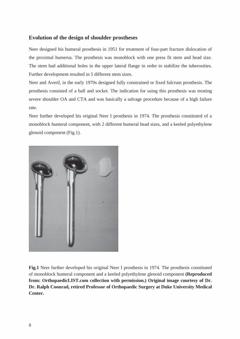
Neer further developed his original Neer I prosthesis in 1974. The prosthesis constituted of a
monoblock humeral component, with 2 different humeral head sizes, and a keeled polyethylene
glenoid component (Fig.1).
Fig.1 Neer further developed his original Neer I prosthesis in 1974. The prosthesis constituted of monoblock humeral component and a keeled polyethylene glenoid component (Reproduced from: OrthopaedicLIST.com collection with permission.) Original image courtesy of Dr. Dr. Ralph Coonrad, retired Professor of Orthopaedic Surgery at Duke University Medical Center.
These designs showed good results in the treatment of shoulder OA in terms of improving pain
and function (Neer 1974). The Neer II prosthesis was the first complete unconstrained shoulder
arthroplasty and marked the birth of modern day TSA. Neer continued to use the Neer II
prosthesis until his retirement in 1990.
A flurry of activity began in the 1970s with the creation of new shoulder prosthesis designs. The
designs were basically divided into three types: a fully-constrained prosthesis, a semi-
constrained design and a non-constrained prosthesis. Various constrained prostheses were
developed but most of the systems were abandoned because of high complication rates.
The second generation of unconstrained shoulder arthroplasty adopted the concept of
modularity. The humeral component was split into two parts, a stem and a head, connected by a
morse taper. This new generation of prosthesis tried to better address the challenges in shoulder
anatomy and to make it more feasible to reconstruct the anatomy (Moeckel et al 1992).
Anatomic studies in the late 1980s lead to further development in implant design and
appearance of a third generation. The newer designs attempted to give the surgeon more
feasibility to adapt the prosthesis to the individual anatomy. This new generation of humeral
components is commonly referred to as adaptable and modular.
The Aequalis prosthesis which was developed by Walch and Boileau was one of the earlier
prosthesis of this kind. The humeral component of the prosthesis was made of three parts (stem,
neck and head). The stem was of 3 diameters and 2 different lengths. The head was of 7
different sizes with additionally 4 neck angles. The glenoid polyethylene component was
available in three different sizes in order to fit different humeral sizes. This allowed greater
flexibility, creating a more anatomical implant for each individual patient (Walch et al 1999).
1. 2 Anatomical and biomechanical rationale
Shoulder anatomy consideration Normal shoulder anatomy varies considerably among different individuals as well as in the left
and right shoulders of the same individual (Pearl et al 2005, Moeckel et al 1992).
The neck is inclined relative to the humeral shaft (NSA) by 130-150o. The head is retroverted
with a highly variable angle which ranges from 0° to 55°. COR of the humeral head is displaced
by 6 mm medially (medial offset) and 3-5 mm posteriorly (posterior offset), relative to the axis
of the humeral shaft (Severt et al 1993, Pearl et al 1995). In addition, the offset of the humerus
in relation to the glenoid may vary in three dimensions, and the radius of curvature ranges from
20 to 30 mm (Pearl et al 1995, Pearl et al 2005). All of these variations should be considered
when an arthroplasty is performed because changes in any of these parameters adversely affect
the biomechanics of the shoulder.
TSA biomechanics consideration TSA can basically be divided into total arthroplasty or hemi-arthroplasty depending on whether
a glenoid component is involved. TSA is subdivided into three groups depending on the degree
of stability: unconstrained, semiconstrained or fully constrained. The classification can be
further subdivided according to degree of constraint and conformity of the articular surface into:
a) low constraint and low conformity b) low constraint and high conformity c) high constraint
and low conformity and d) high constraint and high conformity (Severt et al 1993, Zadeh et al
1998).
Success in shoulder arthroplasty is dependent on a number of factors. Reproducing normal GH
anatomy and biomechanical environment is an important factor (Severt et al 1993). Several
factors should be considered by the surgeon in order to restore the anatomy. The surgeon must
ensure accurate soft tissue tensioning by deciding on accurate alignments and choosing the right
size of the components. Humeral head version is critical and most shoulder arthroplasty systems
recommend 20 to 30 degree retroversion. The top of the humeral component should protrude
above the tuberosity to avoid impingement. Larger humeral head size improves stability but
increases soft tissue tensioning which reduces the range of motion. One of the tests that can be
done by the surgeon to determine the humeral head height is testing the displacement of the
humeral head relative to the glenoid. It should not be displaced downward more than 50%.

Displacement more than this means the prosthesis is too low, while the opposite means the
prosthesis is too high with increased risk of subacromial impingement (Pearl et al 1995).
1.3 RSA
Historical backgrounds Although successful results had been achieved using the Neer II TSA in treatment of shoulder
OA, these unconstrained prostheses had higher failure rates in treatment of cuff deficient
arthritis. CTA was first termed by Neer as a massive rotator cuff tear associated with proximal
humeral head migration and secondary arthritic changes in the glenoid (Jazayeri et al 2011).
The first RSA was designed in the early 1970s by Neer (Mark I). The prosthesis is composed of
a large spherical glenoid implant with a neck to stabilize the humerus. The prosthesis had a high
failure rate and poor functional outcome due to the absence of cuff function. The prosthesis was
further modified (Mark II) with a smaller glenosphere in order to permit rotator cuff
reconstruction and decrease superior impingement. The new prosthesis was abandoned because
of a high glenoid loosening rate. Further development led to Mark III, which allowed axial
rotation between the humeral stem and the diaphysis in order to limit constraint and improve
range of motion. Unfortunately this new design was also unsuccessful and in 1974 Neer
abandoned it. After that, many designs were developed but all resulted in catastrophic failure of
the glenoid implants (Grammont et al 1987, Jazayeri et al 2011).
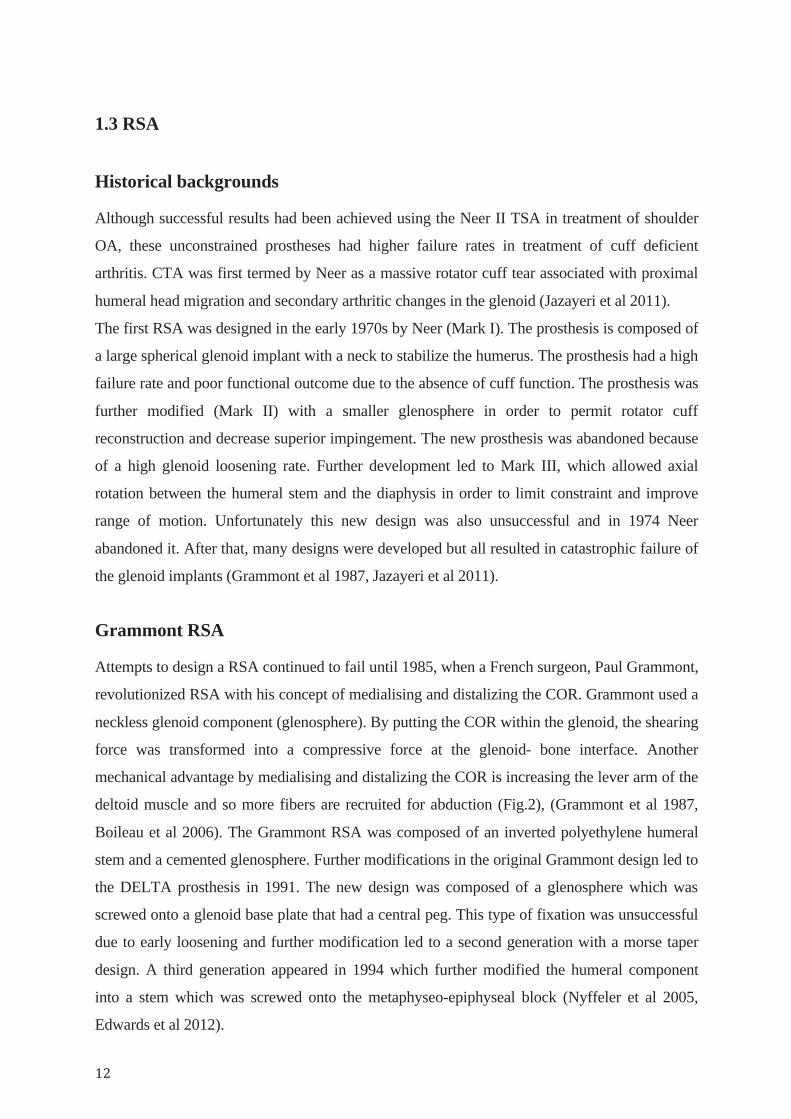
Grammont RSA Attempts to design a RSA continued to fail until 1985, when a French surgeon, Paul Grammont,
revolutionized RSA with his concept of medialising and distalizing the COR. Grammont used a
neckless glenoid component (glenosphere). By putting the COR within the glenoid, the shearing
force was transformed into a compressive force at the glenoid- bone interface. Another
mechanical advantage by medialising and distalizing the COR is increasing the lever arm of the
deltoid muscle and so more fibers are recruited for abduction (Fig.2), (Grammont et al 1987,
Boileau et al 2006). The Grammont RSA was composed of an inverted polyethylene humeral
stem and a cemented glenosphere. Further modifications in the original Grammont design led to
the DELTA prosthesis in 1991. The new design was composed of a glenosphere which was
screwed onto a glenoid base plate that had a central peg. This type of fixation was unsuccessful
due to early loosening and further modification led to a second generation with a morse taper
design. A third generation appeared in 1994 which further modified the humeral component
into a stem which was screwed onto the metaphyseo-epiphyseal block (Nyffeler et al 2005,
Edwards et al 2012).
Fig. 2 COR and position of the humerus and the deltoid muscle with the arm at the side (A) and in abduction (B) in normal shoulder anatomy. C and D, RSA medializes the center of rotation, distalizes the humerus, and elongates the deltoid. The lever arm of the deltoid muscle (dotted line) is lengthened so that for any given angular displacement of the humerus, shortening of the deltoid is greater than in total shoulder arthroplasty. (Reproduced from: Gerber C, Pennington SD, Nyffeler RW. Reverse total shoulder arthroplasty. J Am Acad Orthop Surg. 2009 May; 17(5): 284-95. © 2009 by the American Academy of Orthopaedic Surgeons. With permission.)
Complications of RSA The main complications of RSA are: SN, periprosthetic infection, instability, acromion stress
fracture, mechanical failure and brachial plexus neuropathy.
Medialising the COR is not without biomechanical disadvantages. One possible mechanical
disadvantage may be insufficient tensioning of external rotators which may lead to weakness in
external rotation. Another potential problem is that the concave humeral component can glide
medially over the edge of the convex glenoid component in adduction position. This repetitive
contact can lead to bone loss under the inferior aspect of the glenoid and SN development. The
incidence of this phenomenon is variable in the literature and up to 96 % has been reported
(Levigne et al 2008). The clinical significance of SN is still unclear although glenoid
components failure and polyethylene wear have been reported as possible consequences. To
avoid or decrease the incidence of SN, a number of technical modifications in component
positioning have been suggested. These recommendations have included: inferior glenoid
component position, a larger glenosphere, a lateralized COR, tilting the glenoid component 10
degrees inferiorly and a varus humeral cut. However, the best way to avoid scapular notching is
still debatable (Simovitch et al 2007, Levigne et al 2008, Edwards 2012).
Putting the humerus more distally with subsequent arm lengthening can be another source of
complication. Excessive lengthening can lead to high deltoid muscle tension with increasing
risk of acromion stress fracture and neuropathy. On the other hand, inadequate deltoid
tensioning can lead to instability (Lädermann et al 2014).
The large subacromial dead space with formation of a hematoma can be a possible source of
infection. Periprosthetic infection has been reported in up to 5.1% of primary RSA (Jazayeri R
et al 2011).
1. 4 Implant fixation
Humeral component fixation Humeral stems can be used both in uncemented press-fit or cemented fashion. A cemented
prosthesis is recommended where there is risk of implant subsidence like fracture, RA and
osteoporosis. To enhance biological fixation, hydroxyapatite coatings have been added by some
manufacturers. Regardless of the mode of fixation, aseptic loosening remains remarkably
uncommon (Cofield 1994).
Glenoid component fixation Glenoid fixation remains the weakest link in TSA. The revision rate for the glenoid component
is 3.2% compared with 1.8% for the humeral component (Torchia et al 1997). A cemented
glenoid component has been shown to be an effective treatment for glenohumeral arthritis.
Radiolucent lines and potential for glenoid loosening remain a major concern. Most cemented
glenoid components with lucent lines are present from the immediate postoperative period and
do not progress (Blevins et al 1997). These concerns resulted in development of metal-backed,
bone-ingrowth prostheses which potentially could offer a more stable fixation. Another
potential benefit is the ability to convert an anatomical TSA to a RSA, in cases of revision due
to rotator cuff failure, without compromising the fixation of the glenoid baseplate component
(Rodosky et al 1996). Unfortunately, the results of metal-backed components in anatomical
TSA have been associated with a higher revision rate (Boileau et al 2002, Clitherow et al 2014).
1.5 Stemless shoulder arthroplasty
Background Complications related to the humeral stem account for 10-20% of postoperative complications
(Zumstein 2011). The reported complications in the literature that are related to insertion of a
stemmed prosthesis can be divided into intraoperative complications (malpositioning, false
route, periprosthetic fracture) or postoperative complications (loosening, migration,
disassembly, peri-prosthetic fracture, stem fracture). In the presence of a long stem elbow
prosthesis it is difficult or sometimes even impossible to implant a humeral stem. Increasing
stress increases the risk of fracture when there is an ipsilateral elbow prosthesis. When revision
of a stemmed implant is necessary, the removal of well fixed implant can be a challenge and
can lead to bone damage (Sahota et al 2014). With the aim of reducing stem related
complications, many designs of prosthesis have progressively shortened the humeral stem.
Stemless arthroplasty, with complete humeral stem elimination and reliance on metaphyseal
fixation, was first introduced in 2004 (Churchill RS 2014).
Types of stemless prosthesis There are two different types of stemless prosthesis; one obtained by impaction of a fin system
and in the other implants system, cage screw fixation is used. In sclerotic bone the cage screw
system seems to be advantageous whereas impaction using the fin system seems to be
preferable in osteoporotic bone and in conditions with defects of the bone. Because of the small
number of cases and short period of observation at the present time, none of the implant systems
show a decisive superiority. A total number of almost 10,000 stemless implants have been used
in shoulder arthroplasty since 2004 (Petriccioli et al 2015).
Today, at least five stemless shoulder implants are available on the worldwide market: 1. TESS (Zimmer Biomet), the world’s first stemless shoulder arthroplasty system, first
implanted in Europe in 2004 that evolved into the Comprehensive shoulder system (Zimmer
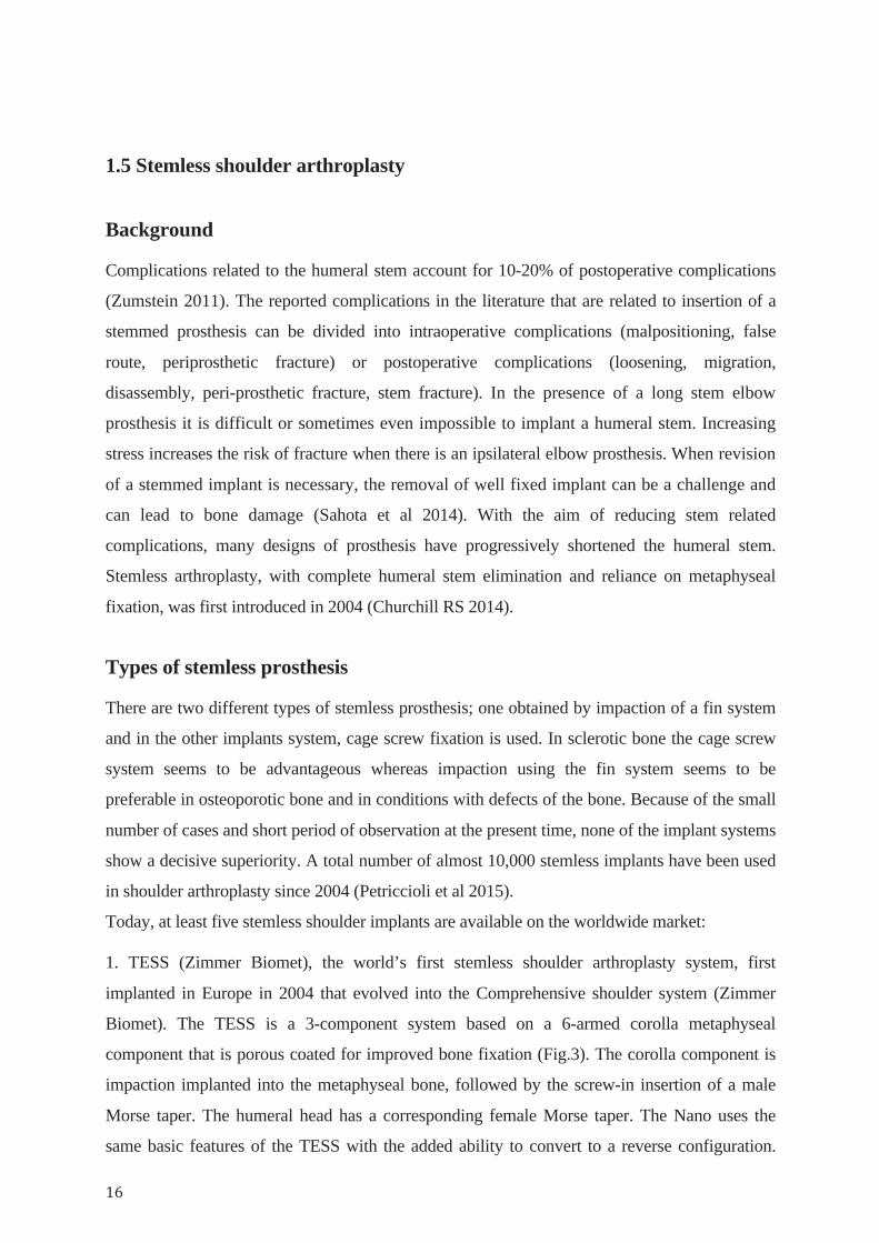
Biomet). The TESS is a 3-component system based on a 6-armed corolla metaphyseal
component that is porous coated for improved bone fixation (Fig.3). The corolla component is
impaction implanted into the metaphyseal bone, followed by the screw-in insertion of a male
Morse taper. The humeral head has a corresponding female Morse taper. The Nano uses the
same basic features of the TESS with the added ability to convert to a reverse configuration.
The Nano system is similar in that the 6-armed corolla is impaction implanted. One major
difference between the TESS and the Nano, however, is that the Nano has a female Morse taper
within the corolla and a corresponding male Morse taper on the humeral head (Churchill 2014,
Petriccioli et al 2015).
Fig.3 The TESS is a 3-component system based on a 6-armed corolla metaphyseal component that is porous coated for improved bone fixation (This figure is the property of Zimmer Biomet Inc or its affiliates, which have granted their permission for usage only on this publication and solely for educational and scientific purposes).
2. Eclipse stemless shoulder arthroplasty (Arthrex, Naples, FL, USA), which was first
introduced in Europe in 2005.
3. Affinis stemless arthroplasty (Mathys, Bettlach, Switzerland), introduced in 2009.
4. Simpliciti by Tournier available only for the European market, since 2010.
5. Sidus stemless shoulder system (Zimmer, Warsaw, IN, USA) which was recently introduced.
Indications and contraindications
The indications for an anatomical stemless TSA are the same as conventional anatomical TSA
i.e. OA, RA, post-traumatic OA or osteonecrosis that is not amenable to non-surgical treatment.
The contraindications for stemless shoulder prosthesis include proximal humeral fractures and
inadequate bone stock (Huguet et al 2010, Berth 2013).
1. 6 Surgical approaches The questions concerning any surgical approach are its utility, advantages, and disadvantages.
The two most common approaches in implanting shoulder prostheses are the deltopectoral (DP)
and antero-superior (AS) approaches. The choice between the DP and AS approach depends
primarily on the experience of the surgeon. Secondarily, it is guided by the indication and
analysis of the advantages and disadvantages of both techniques.
AS approach The main advantages of the AS approach are simplicity, ease of axial preparation of the
humerus, quality of the frontal exposure of the glenoid, and preservation of the subscapularis
tendon. The main drawback of the AS approach is the potential risk of weakening of the
anterior deltoid due to either mechanical stretching by retractors or direct damage to the axillary
nerve which lies in the lower part of the incision (Mole ́ et al 2007). Many authors consider the
AS approach for treatment of complex fractures of the proximal humerus because, in addition to
the quality of exposure from the proximal humerus to the glenoid, it offers the advantages of
better conditions of release and fixation of the greater tuberosity when compared to the DP
approach (Bufquin et al 2007). AS is the preferable approach when implanting a reverse
prosthesis by many surgeons because of better visualization of the glenoid surface which
enhances the precision and accuracy of component positioning (Valenti et al 2008).
Nonetheless, some surgeons continue to use the deltopectoral approach when doing RSA.
DP approach The main advantages of the deltopectoral approach are the preservation of the deltoid muscle,
exposure of the lower pole of the glenoid to facilitate glenoid implant positioning, possibility of
inferior extension to control the proximal humerus, and ability to perform a latissimus dorsi
transfer (Boileau et al 2008). The main drawbacks of the DP approach are the interruption of the
continuity of the subscapularis tendon (the main anterior stabilizer), need for an extended
capsular release (which is a factor for instability), lack of control of posterior cuff and in glenoid
screwing.
The two approaches were compared in a multicenter study of the French Society of Trauma and
Orthopaedic Surgery, which reported on 527 primary RSA with a follow-up of more than 2
years in 11 European specialist centers. The authors found that the AS was associated with less
instability and comparable risk for scapular notching and periprosthetic fractures. Outcome was

similar between the two approaches. However, loosening tended to occur more often with the
anterosuperior approach (Mole ́ et al 2007).
2. Aims of the studies The general aim of this thesis was to report the functional and radiological outcome of TSA in
relation to anatomical placement and implant stability.
The specific aims of these studies were: Study I The primary aim was to assess the functional outcome after TESS shoulder arthroplasty. The
secondary aim was to investigate the radiological stability of stemless shoulder prostheses.
Study II The primary aim was to report the functional outcome after RSA. The secondary aim was to
evaluate the effect of glenoid component positioning on occurrence of SN, to address the effect
of arm lengthening on shoulder function and to assess the radiological stability of the humeral
component.
Study III The aim of the study was to evaluate the reliability and reproducibility of LHO measurements
on plain radiographs in comparison to CT.
Study IV The aim of this study was to investigate the ability of stemless shoulder prosthesis to restore
proximal humerus anatomy in relation to premorbid anatomy.
Study V The primary aim was to assess the importance of LHO restoration in optimising the functional
outcome after anatomical TSA. The secondary aim was to investigate the ability of stemless
implants in restoration of LHO.

3. Hypotheses of the studies
Study I The functional and radiological outcome after TESS shoulder arthroplasty is comparable with
other shoulder prosthetic systems. Stemless shoulder prostheses have a good radiological
stability.
Study II RSA provides a reliable treatment option and prosthesis component position affects the rate of
occurrence of SN.
Study III CT is more reliable than X-ray in estimating LHO.
Study IV The use of stemless shoulder prostheses results in acceptable restoration of shoulder anatomy.
Study V LHO restoration within accepted anatomical limits improves postoperative function.

4. Material and methods
Ethics All studies were conducted according to the Helsinki declaration and were approved by the
Ethics Committee of Umeå University. All patients gave their informed consent to participate.
Study I During the study period 56 consecutive patients were included who underwent shoulder
arthroplasty with one version of TESS (Zimmer Biomet) at the Orthopaedic department at
Sundsvall Teaching Hospital, Sweden between October 2007 and December 2009. There was
no age limits for inclusion.
Before the operation, the patient’s functional impairment was evaluated by Quick DASH
index, while the affection of health status was evaluated by EQ-5D self report questionnaire,
including a 10 cm VAS for the level of life quality. Preoperative radiographic evaluation was
obtained by anteroposterior, axillary and lateral views. When indicated, MRI was performed
to evaluate the rotator cuff. Regardless of the version of shoulder prosthesis implanted, the AS
approach according to Mackenzie was used in all patients. Postoperative radiographic and
clinical assessment was evaluated in the same way as preoperatively. For the postoperative
rehabilitation, patients are allowed to start exercises directly after the operation under the
supervision of a physiotherapist. Patients operated with anatomical TSA were instructed to
avoid external rotation more than 20 degrees for 4 weeks while patients with RSA were
instructed to avoid extension against resistance for 4 weeks. Six weeks postoperatively, the
patients were allowed to use their arms as tolerated. The postoperative follow-up plan
included: a 2 weeks visit to check the operative wound, a 3 and 12 months visit to check the
clinical and radiographic outcome and an additional visit in September 2010 to check the
clinical and radiographic outcome as a final control for this study.
We compared the results in fracture patients (3 and 4 fragments proximal humeral fractures)
with sex- and age-matched OA patients from the same cohort. Reports of the ROM before and
after the operation were not documented in a standardized way and therefore were not
included in this study.
Quick DASH score The DASH score is a region-specific outcome instrument developed as a measure of upper
extremity disability and symptoms (Hudak et al 1996). We used the 11-item Quick DASH,
which has been developed from the original 30-item DASH. The Quick DASH has been shown
to have similar precision as the original DASH and can replace it with similar precision in upper
extremity disorders (Gummesson et al 2006). The items are used to calculate a score ranging
from 0 (no disability) to 100 (most severe disability). The Swedish version of DASH has been
shown to have good reliability, validity and responsiveness (Atroshi et al 2000).
EQ-5D score The EQ-5D, the health status component of the Euro-Qol assessment (EuroQol Group,
Rotterdam, The Netherlands), is a non-disease-specific instrument for describing and evaluating
HRQOL. The EuroQol instrument has been designed for self-completion by the respondent.
There are four components of the instrument: description of the respondent’s own health (EQ-
5D), rating of own health by means of the EuroQol thermometer, evaluation of a standard set of
health states, and background information about the respondent. The respondents describe their
own health state on five dimensions -mobility, self-care, usual activities, pain/discomfort, and
anxiety/depression. One of three levels is chosen for each dimension and thus the resulting
health state can be defined by a five-digit number (EuroQol Group 1990). The reliability and
validity of the EQ-5D has been evaluated in different patient populations including the Swedish
population and it was concluded that there was good evidence for the validity, reliability, and
responsiveness of the EQ-5D (Brooks 1991). It was demonstrated that the EQ-5D had good
internal and external responsiveness in patients with shoulder injuries and can therefore be
recommended for use as an outcome measure for evaluating the HRQOL in both clinical studies
and health-care assessments (Olerud et al 2011).
Radiographic views For accuracy and reproducibility, a true AP X-ray view of the glenohumeral joint was
obtained under the following conditions:
The AP view was taken with the patient erect with the arm by the trunk in neutral position and
with the palm facing forward. All examinations were made under fluoroscopy control in order
to obtain views without overlap of the glenoid and humeral head (Takase et al 2004). All
views included the whole scapula and the space between scapula and spine in order to give
optimum exposure. A 30 mm spherical marker placed lateral to the greater tubercle was used
to control magnification. All radiographs were obtained on a computerised radiography
system (Siemens, Erlangen, Germany). The images were digitally acquired using the Picture
Archiving and Communication System (PACS) (Impax: Agfa, Antwerp, Belgium).
Surgical approach: We used the AS approach, regardless of the version of TESS implanted, according to
Mackenzie (Burgess et al 2009). The anatomical details are described below.
1. Anatomical landmarks The acromion is rectangular. Its bony dorsum and lateral border are easy to palpate on the outer
aspect of the shoulder.
2. Incision The longitudinal skin incision runs from just posterior to the acromioclavicular joint down the
lateral aspect of the arm for about 7- 9 cm.
3. Superficial surgical dissection There is no true internervous plane. The fibers of the anterior deltoid muscle are split
longitudinally from the acromion about 5 cm downward. To decrease the accidental extension
and axillary nerve injury, a suture at the inferior border was occasionally used. The deltoid
muscle was the detached at its origin on the anterior part of the acromion. The axillary nerve
leaves the posterior wall of the axilla by penetrating the quadrangular space. Then it winds
around the humerus with the posterior circumflex artery. The nerve enters the deltoid muscle
posteriorly from its deep surface, about 7 cm below the tip of the acromion (Stanley Hoppenfeld
2009).
4. Deep surgical dissection The lateral aspect of the upper humerus and its attached rotator cuff lie directly under the
deltoid muscle and subacromial bursa. An acromioplasty was regularly performed. After further
excision of the bursa, the cuff was evaluated. The rotator interval was divided longitudinally
and the subscapularis muscle detached at its insertion. If the tendon of the long portion of the
biceps was still present, its intra-articular portion was resected and a tenodesis was sometimes
performed. The inferior aspect of the capsule was released along the humeral neck while the
shoulder was progressively externally rotated to approximately 80° to 90°. Proximally directed
force on the elbow while the arm is in extension allowed the humeral head to luxate superiorly
and the humerus was prepared for prosthesis. Any osteophyte on the glenoid rim was removed
to improve exposure of the glenoid.
Three general orthopaedic surgeons with a special interest in shoulder surgery operated on the
patients. All patients were given 2g cloxacillin preoperatively, followed by 2 additional doses
during the first 24 hours. General anaesthesia with interscalene blockade was used. The
patient lies in the beach chair position with a 40-60° tilt of the chest position in a theatre with
laminar airflow. A sandbag or other form of support was placed under the medial edge of the
scapula to facilitate exposure. The arm was placed on an armrest and was draped free.
1. Anatomical TSA
After dislocation of the humeral head, osteophytes were removed with a ronguer for
visualisation of rotator cuff insertion and anatomical neck. The cutting guide was held parallel
to the anatomical neck. This adjusted the inclination and head version automatically. The final
decision as to whether a stemmed or stemless humeral implant would be used was made
intraoperatively depending on bone quality and stability of the humeral component. We chose
the stemmed version if primary stability of the humeral implants could not be achieved. If it
was planned implant the stemmed version then the medullary canal was prepared. If a
stemless version was considered appropriate then corolla preparation proceeded. The size of
the corolla broach was chosen using a humeral sizing template. Preparation of the glenoid
begins with an assessment of wear and loss of bone stock. The surgeon then completes
glenoid exposure, labrum resection, and peripheral capsular release. The inferior labrum is
carefully released with a knife while maintaining contact with the bony rim and avoiding
electric cautery, considering the proximity of the axillary nerve, which is not visualized. This
allows the positioning of a hooked retractor that presses the humeral epiphysis, which is
protected by its trial prosthesis. Once the glenoid implant was in place, the humerus again was
subluxated superiorly and anteriorly. The metaphyseal fixation, i.e. the corolla implant, was

introduced after broach removal, and a trial head was tested. There were six symmetrical and
six asymmetrical head sizes available (41, 43, 45, 48, 50, 52 mm). Several factors determine
the appropriate choice of head size: size of the head cut, range of motion (ROM) and
translation of the humeral head on the glenoid, which should be <50 %. In all anatomical
TESS implants, a symmetrical head was chosen. After reducing the joint, the subscapularis
was reinserted back into the lesser tuberosity, and the deltoid was reattached to the acromion
using osteosutures. The skin was closed using intracutaneus sutures.
2. RSA
The design of the TESS RSA prosthesis is based on Grammont’s concept. For the glenoid, an
asymmetric reamer increased the height of the central portion to improve osteointegration and
preserved the peripheral rim to optimize the stability of the baseplate. The glenosphere (sized
36 or 41 mm in diameter) is made of cobalt-chrome and is bolted to a glenoid baseplate fixed
by 4 screws. The humeral stemless cup has been developed from the anatomic corolla. The
RCO is made of cobalt-chrome with a titanium plasma spray and hydroxyapatite coating. It
has six anti-rotational wings to optimize the rotational stability and is available in 4 sizes. The
bone cut is done with the help of a cutting guide with an angulation of 150 degrees. A pin was
centred on the bone resection using a template and a humeral reamer was centered on the
resected humeral cut. The RCO was retroverted by 20 degrees and was press fitted without
cement. If bone quality was deemed to be inadequate, a stem of adequate diameter was
inserted after prereaming of the medullary canal. The polyethylene component is prevented
from dislocating by a ring-lock system. Trial humeral inserts were tested and the final
polyethylene insert was impacted. After testing the amplitude and stability, the subscapularis
muscle was reattached to the lesser tuberosity.
Statistical analysis SPSS version 16 (SPSS, Chicago, IL, USA) was used for the statistical analysis. The
Student’s paired t test was used to compare the Quick DASH, EQ-5D and VAS for life
quality before and after the operation. A p value\ 0.05 was considered statistically significant.
Study II The study period was between October 2007–January 2012 at Sundsvall teaching hospital. The
inclusion criteria were: 1. All patients with CTA, primary OA with rotator cuff dysfunction,
RA and proximal humeral fracture sequelae who had undergone TESS RSA (Zimmer
Biomet), both stemmed and stemless at Sundsvall Hospital, Sweden during the study period 2.
Intact cognitive function (no diagnosis of dementia, with the patient being lucid and fully
oriented). 3. No previous neurological disorder that affects the operated side. There was no
age limits for inclusion.
Functional impairment was evaluated by the Quick DASH index, the EQ-5D score was used for
the estimation of quality of life and global VAS for evaluation of overall health status. Pre- and
post-operative active ROM was measured by visual estimation in degrees of abduction and
flexion, while internal rotation was measured as the ability to reach behind the back. VAS was
used for assessing pain.
The VAS pain is a continuous scale comprised of a 100 mm line, anchored by 2 verbal
descriptors, one for each symptom extreme. For pain intensity, the scale is most commonly
anchored by “no pain” (score of 0) and “pain as bad as it could be” or “worst imaginable pain”
(score of 100), (Huskisson et al 1974, Jensen et al 1986).
Radiographic assessment with anteroposterior (AP), axillary and lateral scapular radiographs
was performed pre- and post-operatively by standardised views at three months and then
annually
SN was assessed on the latest radiograph using the AP view according to Sirveaux (Sirveaux
1997). Grade 1 indicated a notch limited to the scapular pillar, grade 2 reached the inferior
screw of the baseplate, grade 3 extended beyond the inferior screw and grade 4 reached the
central peg of the baseplate.
Glenoid loosening was defined as radiolucencies under the baseplate or around the peg or
screws, screw breakage or glenoid migration.
The peg-glenoid rim distance was measured as the distance from the uppermost border of the
central peg to the inferior glenoid margin on post-operative AP radiographs (Fig.3 a)
(Simovitch et al 2007).
Glenoid component craniocaudal positioning Two horizontal lines were drawn, one from the
inferior margin of the glenoid sphere parallel to another line which was drawn from the inferior

margin of the glenoid. The distance between the two lines was calculated and values more than
0 mm were regarded as overhang (Fig.3 b) (Nyffeler et al 2005).
Glenoid component inclination was described as the angle between the baseplate and a
horizontal line drawn from the upper margin of the glenoid An inferior tilt was defined as an
angle measuring more than 90°; a superior tilt was under 90°, while 90° was considered neutral
(Fig.3 c) (Lévigne et al 2011).
Fig. 3 a The peg-glenoid rim distance (PGRD) was measured as the distance from the uppermost border of the central peg to the inferior glenoid margin on the post-operative AP radiograph. b The craniocaudal glenoid component position was measured by drawing two horizontal lines, one from the inferior margin of the glenoid sphere parallel to another line which was drawn from the inferior margin of the glenoid. The distance between the two lines was calculated and values more than 0 mm were regarded as overhang. c Glenoid baseplate inclination was described as the angle between the baseplate and a horizontal line drawn from the upper margin of the glenoid.
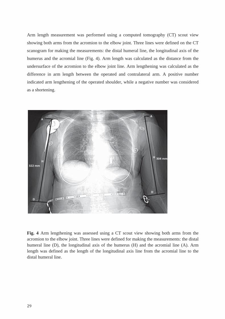
Arm length measurement was performed using a computed tomography (CT) scout view
showing both arms from the acromion to the elbow joint. Three lines were defined on the CT
scanogram for making the measurements: the distal humeral line, the longitudinal axis of the
humerus and the acromial line (Fig. 4). Arm length was calculated as the distance from the
undersurface of the acromion to the elbow joint line. Arm lengthening was calculated as the
difference in arm length between the operated and contralateral arm. A positive number
indicated arm lengthening of the operated shoulder, while a negative number was considered
as a shortening.
Fig. 4 Arm lengthening was assessed using a CT scout view showing both arms from the acromion to the elbow joint. Three lines were defined for making the measurements: the distal humeral line (D), the longitudinal axis of the humerus (H) and the acromial line (A). Arm length was defined as the length of the longitudinal axis line from the acromial line to the distal humeral line.
Statistical analysis Statistics were obtained using SPSS for Windows statistical program release 21 (SPSS Inc.,
Chicago, IL, USA). The Wilcoxon signed rank test was used to compare pre- and post-
operative values of Quick DASH, EQ-5D, VAS pain and ROM. The Mann-Whitney test was
used to compare inclination and overhang between categories of glenoid notching. The
Spearman correlation was used to evaluate relations between arm lengthening and outcome.
Kruskal-Wallis analysis of variance (ANOVA) was used to evaluate relations between
notching and glenoid loosening. Values for continuous data are presented as median (min.,
max.). The statistical significance level was designated at P<0.05.
Study III This prospective study was performed at Sundsvall Teaching Hospital, Sweden between May
2011 and January 2013. A power analysis for 4 observers and a minimum value of 0.7 for the
interclass correlation coefficient (ICC) indicated that 22 shoulders were required. Thus, 26
consecutive patients that underwent anatomical TESS TSA (Zimmer Biomet) were included
in order to provide a safe margin of error.
Inclusion criteria were primary OA of the shoulder and scheduled to have anatomical TSA.
Patients with previous shoulder surgery were excluded as well as those with secondary OA
such as posttraumatic OA or CTA. The 4 observers were two shoulder surgeons, one
orthopaedic resident and one radiologist. The four observers had access to written instructions
and illustrations of LHO measurements and they independently examined all radiographs and
CT scans. After 6 weeks and blinded to their results, each observer repeated the same
measurements.
Plain radiograph measurements LHO was evaluated by measuring the distance between the medial edge of the base coracoid
process and the most lateral point of the greater tubercle in AP X-ray view (Fig.5).
CT measurements CT imaging was done with the patient in supine position and the arms by the side with palms
facing upward (anatomical position). Both shoulders were included in the axial CT sections.
LHO was measured in the axial section by measuring the distance between the medial edge of
the base of the coracoid process to the most lateral point of the greater tubercle, (LHO, CT)
(Fig. 6).
Statistical analysis The ICC (with 95 % CI) was used to evaluate the interobserver reliability and intraobserver
reproducibility of the obtained measures while paired t-tests were used to compare their
means. For ICC the value of 0.00–0.20 was considered slight, 0.21–0.40 was considered fair,
0.41–0.60 was considered moderate, 0.61–0.80 was considered substantial and 0.81–1.00 was
considered excellent (Landis 1977). The distributions of variables were tested for normality
using the Shapiro–Wilk test (Shapiro et al 1965). Correlation between LHO (X-ray) and LHO

(CT) measurements were analysed using Bland–Altman plots (Bland et al 1986). A scatter
plot with linear regression was performed to relate corresponding measurements. Statistical
analysis was carried out using SPSS for Windows version 20.0 (SPSS Inc., Chicago, Illinois)
and statistical significance was set at p\ 0.05.
Fig. 5 LHO measured in the A-P view as the distance (c) between the medial edge of the base of coracoid process (a) to the most lateral point of the greater tubercle (b).
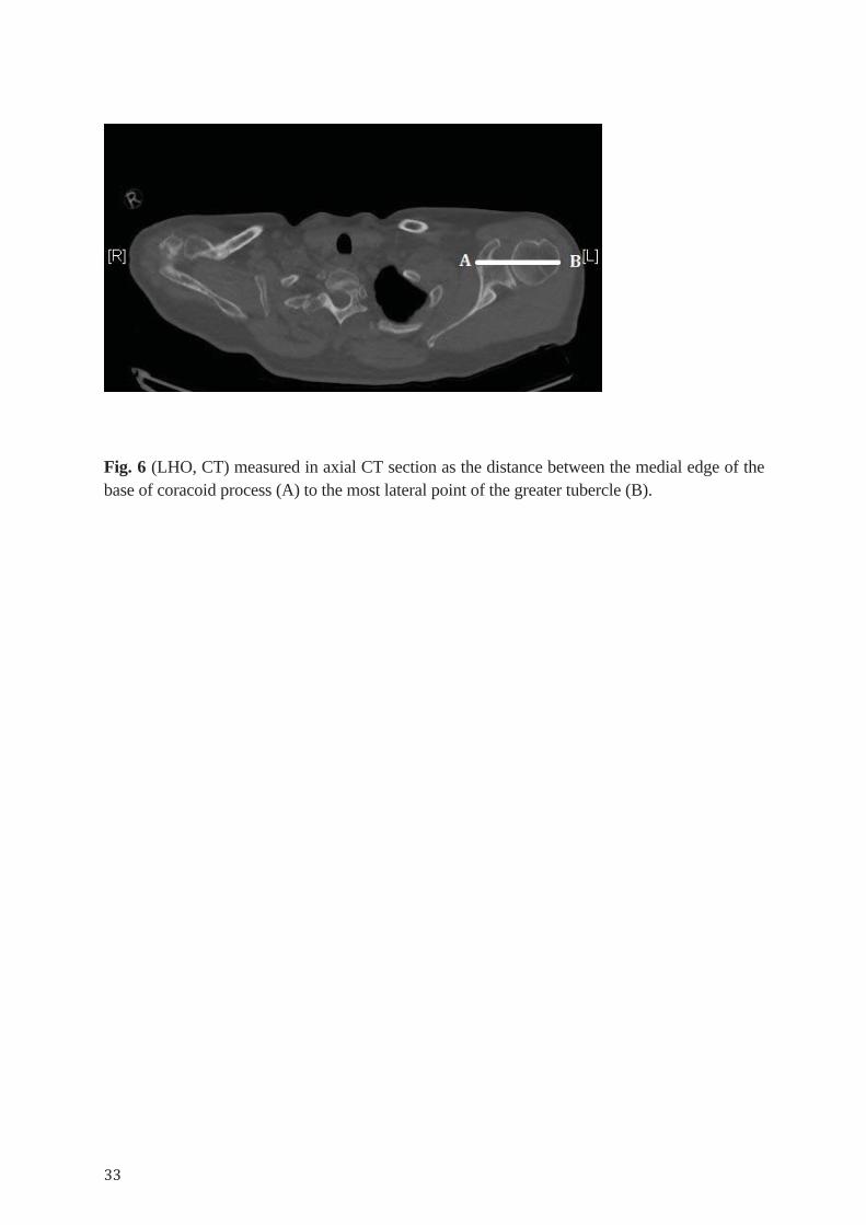
Fig. 6 (LHO, CT) measured in axial CT section as the distance between the medial edge of the base of coracoid process (A) to the most lateral point of the greater tubercle (B).

Study IV This prospective study was performed between May 2007 and December 2013 at Sundsvall
teaching hospital. During that period, 216 shoulders were given an anatomic TSA or RSA
TESS (Zimmer Biomet) implant for pain after OA, CTA, fractures, revision (due to failure of
other implants) or RA. Inclusion criteria were patients with primary OA who had undergone
stemless total anatomic shoulder arthroplasty. Patients with post-traumatic OA, inflammatory
arthropathies, bone-stock insufficiency, revision, previous surgeries or stemmed humeral
implants were excluded.
Radiographic parameters Pre-operative measurements: Premorbid humeral head anatomy was estimated by a best-fit circle method according to
previous studies (Alolabi et al 2014,Youderian et al 2014). A circle was mapped in the AP view
and matched to three preserved nonarticular bone landmarks: the lateral cortex below the flare
of the greater tuberosity, the medial footprint of the rotator cuff on the greater tuberosity and the
medial calcar at the inflection point where the calcar meets the articular surface (Fig. 7a).
COR was then identified from the circle and the distance to the anatomical neck was calculated
in millimetres (Fig. 7a).
Humeral head height (HH) is defined as the perpendicular linear distance from the anatomic
neck to the apex of the circle (Fig. 7b).
NSA was measured as the angle between a line perpendicular to the anatomical neck and the
long axis of the humeral diaphysis, which was defined by a proximal and distal point in the
centre of the intramedullary canal (Fig. 7c).
Post-operative measurements: COR was identified by placing a circle fitted to the curvature of the HH prosthesis. The
anatomical neck was supposed to be the same as that identified during surgery (lower margin of
the prosthetic head). Deviation of post-operative COR >3 mm from the normal anatomy was
considered as being clinically
significant (Alolabi et al 2014). HH was measured from the anatomical neck to the top of the
prosthetic HH. Deviation of postoperative HH >5 mm from the normal anatomy was considered
clinically significant (Harryman et al 1995). NSA was measured in the same manner as pre-

operatively. Shoulders with post-operative NSA <130° were considered as varus (Takase et al
2002). We excluded shoulders that already had pre-operative NSA <130° from the varus group.
Statistical analysis Data was tested for normality with the Shapiro–Wilk test. Descriptive statistics are reported as
means, standard deviations (SD) and ranges for continuous data. We used the Pearson
correlation coefficient (r) to calculate the correlation between premorbid and postoperative
parameters (COR, HH and NSA).
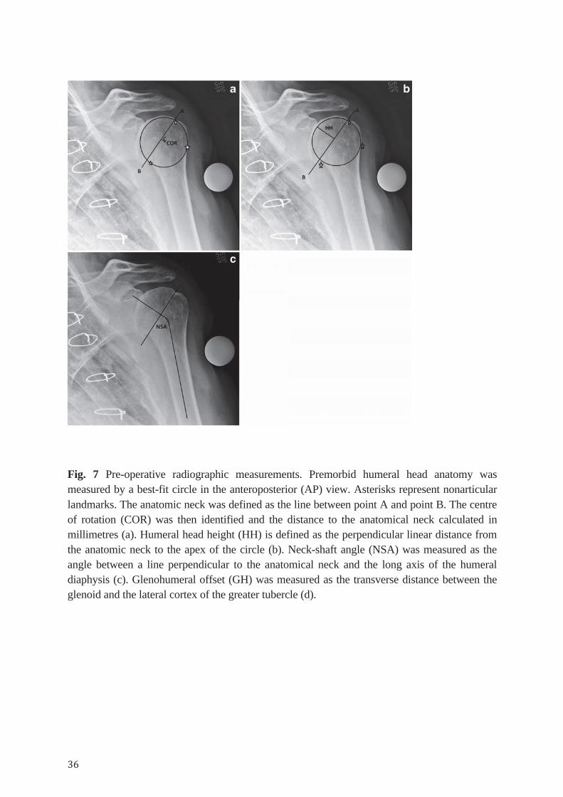
Fig. 7 Pre-operative radiographic measurements. Premorbid humeral head anatomy was measured by a best-fit circle in the anteroposterior (AP) view. Asterisks represent nonarticular landmarks. The anatomic neck was defined as the line between point A and point B. The centre of rotation (COR) was then identified and the distance to the anatomical neck calculated in millimetres (a). Humeral head height (HH) is defined as the perpendicular linear distance from the anatomic neck to the apex of the circle (b). Neck-shaft angle (NSA) was measured as the angle between a line perpendicular to the anatomical neck and the long axis of the humeral diaphysis (c). Glenohumeral offset (GH) was measured as the transverse distance between the glenoid and the lateral cortex of the greater tubercle (d).
Study V This prospective study was performed between May 2011 and August 2014 at Sundsvall
teaching hospital, Sundsvall, Sweden. All patients with symptomatic unilateral primary OA
scheduled for stemless anatomical TSA were considered for inclusion. Patients with previous
shoulder surgery, cognitive impairment or neurological disorder were excluded.
Functional measurements Within 6 weeks preoperatively, functional impairment was measured by the Quick DASH
score, quality of life by EQ-5D, a measure of health status using the VAS scale component of
EQ-5D, pain both at rest and exertion using a VAS scale and active ROM.
Postoperatively, the patients were assessed with the same functional parameters at 3 months, 12
months and then once annually. One independent observer performed all functional
measurement to ensure objectivity.
CT measurements
CT imaging was done in the same way as in study III. Preoperative CT was done within 6
weeks before surgery and used to rule out the presence of any osteoarthritic changes in the
contralateral shoulder and therefore to confirm the diagnosis of unilateral OA. The
postoperative CT was done at three months follow-up to measure the LHO bilaterally.
LHO was measured in the same way as in study III. LHO was measured for the contralateral
healthy shoulder, (LHO contra), while LHO for the operated shoulder, (LHO op) (Fig.7). The
difference between LHO op and LHO contra, (LHO post), was calculated and a positive value
was obtained when the LHO op was longer than the contralateral side, whereas a negative value
indicated the opposite.
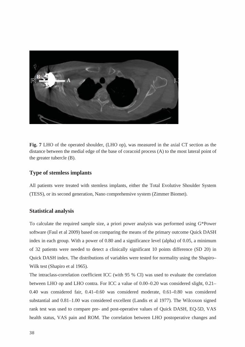
Fig. 7 LHO of the operated shoulder, (LHO op), was measured in the axial CT section as the distance between the medial edge of the base of coracoid process (A) to the most lateral point of the greater tubercle (B).
Type of stemless implants
All patients were treated with stemless implants, either the Total Evolutive Shoulder System
(TESS), or its second generation, Nano comprehensive system (Zimmer Biomet).
Statistical analysis To calculate the required sample size, a priori power analysis was performed using G*Power
software (Faul et al 2009) based on comparing the means of the primary outcome Quick DASH
index in each group. With a power of 0.80 and a significance level (alpha) of 0.05, a minimum
of 32 patients were needed to detect a clinically significant 10 points difference (SD 20) in
Quick DASH index. The distributions of variables were tested for normality using the Shapiro–
Wilk test (Shapiro et al 1965).
The intraclass-correlation coefficient ICC (with 95 % CI) was used to evaluate the correlation
between LHO op and LHO contra. For ICC a value of 0.00–0.20 was considered slight, 0.21–
0.40 was considered fair, 0.41–0.60 was considered moderate, 0.61–0.80 was considered
substantial and 0.81–1.00 was considered excellent (Landis et al 1977). The Wilcoxon signed
rank test was used to compare pre- and post-operative values of Quick DASH, EQ-5D, VAS
health status, VAS pain and ROM. The correlation between LHO postoperative changes and

functional parameters was calculated by linear regression analysis. Statistical analysis was
carried out using SPSS for Windows version 20.0 (SPSS Inc., Chicago, Illinois) and statistical
significance was set at p = 0.05.
5. Results
Study I Patient flow and baseline data Forty-nine patients were available with a mean follow-up 14 months (range 9–24 months).
There were 36 females and 13 males with ages at operation ranging between 50 and 83 years
(mean 71). The right side was operated in 27 patients and the left side in 22 patients.
There were 30 patients with OA (19 with good cuff function and 11 with cuff dysfunction), 9
with RA (6 with cuff dysfunction and 3 with good cuff function) and 10 with proximal humerus
fracture sequelae (6 with good cuff and 4 with cuff dysfunction).
Type of implants and operation indications There were 28 anatomical TSA and 21 RSA. There were 39 stemless shoulder prostheses (22
anatomical TSA and 17 RSA) and 10 stemmed (6 anatomical TSA and 4 RSA).
The shoulder disorders that were operated on using stemless TSA (n= 22) were as follows:
1. OA (n=19).
2. RA (n=3).
The shoulder disorders that were operated on using stemless RSA were as follows:
1.OA with cuff dysfunction (n=11).
2. RA with cuff dysfunction (n=6)..
Cemented stemmed prosthesis, both anatomical and RSA, were used only in fracture patients.
Patients with malunion or non-union proximal humerus fracture with cuff dysfunction received
cemented stemmed RSA while those with fracture sequelae with intact cuff received anatomical
TSA.
Patient outcome measurements All functional parameters (Quick DASH, EQ-5D and VAS for life quality) were significantly
improved (Table. 1). None of the patients operated on using reverse TESS (n = 21) showed
radiolucencies or scapular notching during the study period. There were no radiolucencies
around any stemless prosthesis (n=39). The outcome in fracture patients was compared with
age- and sex-matched patients operated for primary/ posttraumatic osteoarthritis with the same
TESS version from the same cohort. We found that the Quick DASH index was worse in
fracture patients (43, SD 18) compared to osteoarthritis patients (28, SD 26). However, this
difference did not reach a statistical significance (p = 0.08).
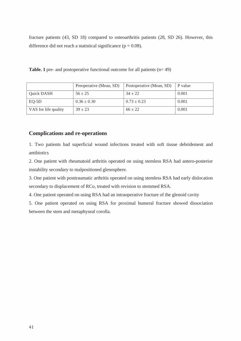
Table. 1 pre- and postoperative functional outcome for all patients (n= 49)
Preoperative (Mean, SD) Postoperative (Mean, SD) P value
Quick DASH 56 ± 25 34 ± 22 0.001
EQ-5D 0.36 ± 0.30 0.73 ± 0.23 0.001
VAS for life quality 39 ± 23 66 ± 22 0.001
Complications and re-operations 1. Two patients had superficial wound infections treated with soft tissue debridement and
antibiotics
2. One patient with rheumatoid arthritis operated on using stemless RSA had antero-posterior
instability secondary to malpositioned glenosphere.
3. One patient with posttraumatic arthritis operated on using stemless RSA had early dislocation
secondary to displacement of RCo, treated with revision to stemmed RSA.
4. One patient operated on using RSA had an intraoperative fracture of the glenoid cavity
5. One patient operated on using RSA for proximal humeral fracture showed dissociation
between the stem and metaphyseal corolla.
Study II Patient flow and baseline data The study group comprised 37 patients (23 women and 14 men; mean age at surgery 72.0 years;
age range 60–88 years). In total 40 shoulders were operated on. The mean duration of follow-up
was 39 months (range 15–66 months). Indications were CTA (n=14), primary OA with rotator
cuff dysfunction (n= 10), RA (n=7) and proximal humeral fracture sequelae (n=9).
Three patients died during the study from causes unrelated to the surgery at 20, 35 and 40
months post-operatively. For these patients, results were obtained from their last follow-up.
Type of implants and operation indications There were 37 patients (40 shoulders) who underwent TESS RSA. There were 16 stemless and
24 stemmed.
The shoulder disorders that were operated on using stemmed RSA (n=24) were as follows:
1. CTA (n= 7).
2. OA with cuff dysfunction (n= 5).
3. RA (n= 3).
4. Proximal humeral fracture sequelae (n= 9).
Cemented stemmed RSA prostheses were used only in fracture patients.
The shoulder disorders that were operated on using stemless RSA (n= 16) were as follows:
1. CTA (n= 7).
2. OA (n=5).
3. RA (n=5).
Patient outcome measurements There was a marked improvement in shoulder function, quality of life and reduction of pain for
both stemmed and stemless versions and for all included diagnoses (Table. 2)
When we looked at the stemmed and stemless RSA in arthritis patients (i.e. no fracture patients
included), we found the two groups to be comparable except that more women received
stemmed implants (< 0.05). At radiological follow-up we found no signs of humeral implant
loosening except for one stemmed shoulder where thin zones of resorption of the proximal
humerus were detected.
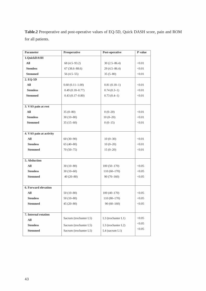
Table.2 Preoperative and post-operative values of EQ-5D, Quick DASH score, pain and ROM
for all patients.
Parameter Preoperative Post-operative P value
1.QuickDASH
All
Stemless
Stemmed
68 (4.5–93.2)
67 (38.6–88.6)
56 (4.5–55)
30 (2.5–86.4)
29 (4.5–86.4)
35 (5–80)
<0.01
<0.01
<0.01
2. EQ-5D
All
Stemless
Stemmed
0.60 (0.11–1.00)
0.49 (0.18–0.77)
0.43 (0.17–0.80)
0.81 (0.18–1)
0.74 (0.3–1)
0.73 (0.4–1)
<0.01
<0.01
<0.01
3. VAS pain at rest
All
Stemless
Stemmed
35 (0–80)
30 (10–80)
35 (15–60)
0 (0–20)
10 (0–20)
0 (0–15)
<0.01
<0.01
<0.01
4. VAS pain at activity
All
Stemless
Stemmed
60 (30–90)
65 (40–80)
70 (50–75)
10 (0–30)
10 (0–20)
15 (0–20)
<0.01
<0.01
<0.01
5. Abduction
All
Stemless
Stemmed
30 (10–80)
30 (10–60)
40 (20–80)
100 (50–170)
110 (60–170)
90 (70–160)
<0.05
<0.05
<0.05
6. Forward elevation
All
Stemless
Stemmed
50 (10–80)
50 (10–80)
45 (20–80)
100 (40–170)
110 (80–170)
90 (60–160)
<0.05
<0.05
<0.05
7. Internal rotation
All
Stemless
Stemmed
Sacrum (trochanter L5) Sacrum (trochanter L5)
Sacrum (trochanter L5)
L3 (trochanter L1)
L3 (trochanter L2)
L4 (sacrum L1)
<0.05
<0.05
<0.05
Radiological parameters and clinical correlation SN SN was seen in 12 shoulders. Seven shoulders had SBI already on the first post-operative X ray
(day one to four). In the remaining five shoulders, SN appeared at a mean of seven months
(range 3 to 12 months). Patients with CTA had more SBI than other diagnoses (five of seven)
P<0.01. Three of the four baseplate loosenings occurred concurrent with grade 1 SN. However,
SN was not correlated with glenoid loosening and did not affect final ROM or reported quality
of life.
Glenoid component positioning The inclination of the glenoid baseplate was 93° (range 80– 105°). No correlation was observed
between inclination and SN. The mean glenoid overhang was 1.3 mm (range 5–6 mm). With no
overhang there was a higher incidence of SN (10 of 12 shoulders; P<0.001). The peg-glenoid
rim distance was 20 mm (range 15–28 mm). The peg-glenoid distance correlated with SN.
When the distance was more than 20 mm, SN was evident in 9/12 shoulders, while 3/12
occurred when the distance was less than 20 mm (P<0.01).
Arm lengthening The lengthening of the upper extremity was 16 mm (range 0–32 mm). We compared those with
arm lengthening 15 mm or less (15 shoulders) to those with lengthening over 15 mm (12
shoulders). Those with arm lengthening more than 15 mm showed greater improvement in EQ-
5D (preoperative mean = 0.41 vs 0.80 post-operatively) as compared with the others
(preoperative mean = 0.51 vs 0.66 postoperatively; P<0.05). However, lengthening did not
correlate with degree of post-operative pain, ROM, Quick DASH or SN.
Complications and re-operations Of the 16 stemless implants, two prostheses were revised at three and four days post-operatively
due to corolla displacement. There were two RSA dislocations in two patients with fracture
sequelae. These were treated with exchange of the polyethylene insert. Also, four glenoid
loosenings were revised with exchange of the baseplate.

Study III
Patient flow and baseline data Fifteen females and 11 males were recruited into the study. The mean age was 70 years (range
54–83). Ten patients had OA on the right side, 8 on the left side and 8 had bilateral OA.
Preoperative CT of 26 patients (52 shoulders) and X-ray of 34 shoulders were available for
examination.
Radiological parameters The interobserver reliability of LHO (CT) measurements among the four observers was
excellent (0.93) while LHO (X-ray) was moderate (0.48). The intraobserver reproducibility of
LHO (X-ray) was variable among the observers. It was excellent for observers 1 and 4 while
it was moderate and fair for observers 2 and 3, respectively. The intraobserver reliability was
excellent for LHO (CT) for all four observers. A Bland–Altman plot was used for the analysis
of correlation between LHO (CT) and LHO (X-ray). Disagreement in assessment of LHO
between CT and X-ray was within 5 mm. The mean LHO (X-ray) was significantly lower
than the mean LHO (CT) (p = 0.001, paired sample t-test) with a mean difference of 5.0 mm
(95 % CI 2.3–7.1).

Study IV
Patient flow and baseline data 69 patients (70 shoulders) were available for the study. The patients (33 men and 36 women)
had a mean age of 69 years (range 52–88 years) at time of the surgery
Type of implants and operation indications Patients with primary OA who had undergone stemless anatomical TESS TSA (Zimmer
Biomet) were included in the study.
Radiological parameters COR The mean difference between premorbid and post-operative COR was 1±2 mm (range −3 to
5.8 mm). There were 13/70 (19 %) shoulders with increased post-operative COR >3 mm
(3.9±0.5 mm).
HH The mean difference between premorbid and post-operative HH was −1±3 mm (range −9.7 to
8.5 mm). There were 8/70 (11 %) shoulders with a post-operative HH difference >5 mm.
Of the eight, six had decreased post-operative HH >5 mm (range −9.7 to −5.7 mm) and two
had increased postoperative HH >5 mm.
NSA The mean difference between premorbid and post-operative NSA was −3±12° (range −26 to
20°). There were 25/70 (36 %) shoulders with post-operative NSA <130° (Fig. 8) (Table. 3).
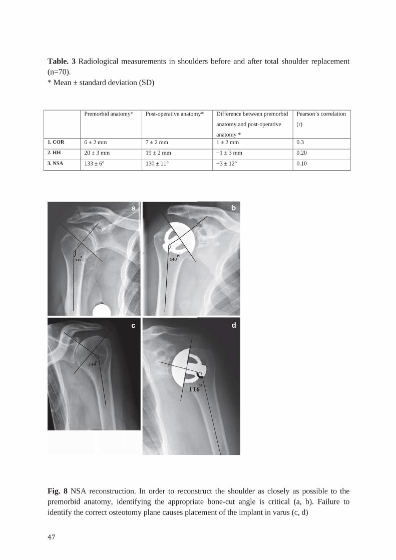
Table. 3 Radiological measurements in shoulders before and after total shoulder replacement (n=70). * Mean ± standard deviation (SD)
Premorbid anatomy* Post-operative anatomy* Difference between premorbid
anatomy and post-operative
anatomy *
Pearson’s correlation
(r)
1. COR 6 ± 2 mm 7 ± 2 mm 1 ± 2 mm 0.3
2. HH 20 ± 3 mm 19 ± 2 mm −1 ± 3 mm 0.20
3. NSA 133 ± 6° 130 ± 11° −3 ± 12° 0.10
Fig. 8 NSA reconstruction. In order to reconstruct the shoulder as closely as possible to the premorbid anatomy, identifying the appropriate bone-cut angle is critical (a, b). Failure to identify the correct osteotomy plane causes placement of the implant in varus (c, d)

Study V Patient flow and baseline data Forty-four patients with a minimum follow up of 12 months (range 12 – 50 months) were
available for the study. The patients (22 men and 22 women) had a mean age of 69 years (range
52 to 82) at time of the surgery.
Type of implants and operation indications Forty-four patients with primary OA who had undergone stemless total anatomic shoulder
arthroplasty were included in the study. There were 20 TESS and 24 Comprehensive stemless
implants (Zimmer Biomet).
Radiological parameters
LHO
The mean difference between LHO contra and LHO op was 1.3 mm ± 4.6. Deviation of LHO
op of more than 5 mm from LHO contra was considered as being clinically significant
(Mechlenburg 2013). There were 36/44 (82%) patients whose LHO ops were restored within 5
mm from LHO contra (mean 0 mm ± SD 3.8) and 8/44 (18%) patients who had more than 5
mm (mean 7.5 mm ± SD 1.85). There were 26/44 (59%) patients with LHO op > 0 mm (mean
4.4 mm ± SD 2.5), and 18/44 (41%) patients with LHO ≤ 0 (mean − 3 mm ± SD 2.8). The
correlation between (LHO contra) and (LHO op) was substantial (ICC 0.8, CI 95% 0.62 - 0.87,
P = 0.001).
Functional outcome
All functional measures showed significant improvement (Table.3). We compared values of
functional parameters at the three follow-up occasions: 3 months, 12 months and at last follow-
up visit in order to investigate whether the shoulder function would be different. All parameters
showed significant improvement at 3 and 12 months post-surgery but not after 12 months
follow-up (Table. 4).
Correlation between LHO post and Quick DASH
LHO post was correlated with Quick DASH at 3 months (Pearson correlation = 0.36, P = 0.01)
in which lengthening of LHO showed a tendency for increasing Quick DASH i.e. worsening
shoulder function (coefficient =1.09). LHO post showed no correlation with Quick DASH at 12
months (Pearson correlation = − 0.01, P = 0.49).
Correlation between LHO post and VAS pain
LHO post was correlated with VAS pain (at rest) at 3 months (Pearson correlation= 0.30, P
=0.03) in which lengthening of LHO showed tendency for increasing pain at rest
(coefficient=0.65) but not at 12 months (Pearson correlation = − 0.18, P = 0.12).
LHO post was correlated with VAS pain (at exertion) at 3 months (Pearson correlation= 0.34,
95%, P = 0.01) in which lengthening of LHO showed tendency for increasing pain at exertion
(coefficient=1.68) but not at 12 months (Pearson correlation = − 0.01, p = 0.47).
Correlation between LHO post and other functional outcome measures
There was no correlation between LHO post and other outcome measures (EQ-5D, VAS health,
ROM) at either 3 or 12 months follow-up. Age and gender did not correlate with any of the
functional outcome measures.
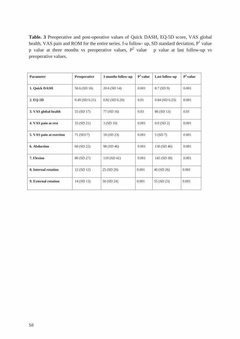
Table. 3 Preoperative and post-operative values of Quick DASH, EQ-5D score, VAS global health, VAS pain and ROM for the entire series. f-u follow- up, SD standard deviation, P1 value
p value at three months vs preoperative values, P2 value p value at last follow-up vs preoperative values.
Parameter Preoperative 3 months follow-up P1 value Last follow-up P2 value
1. Quick DASH 56.6 (SD 16) 20.6 (SD 14) 0.001 8.7 (SD 9) 0.001
2. EQ-5D 0.49 (SD 0.21) 0.82 (SD 0.20) 0.01 0.84 (SD 0.23) 0.001
3. VAS global health 55 (SD 17) 77 (SD 16) 0.03 86 (SD 13) 0.01
4. VAS pain at rest 33 (SD 21) 3 (SD 10) 0.001 0.0 (SD 2) 0.001
5. VAS pain at exertion 71 (SD17) 18 (SD 23) 0.001 5 (SD 7) 0.001
6. Abduction 60 (SD 22) 98 (SD 46) 0.001 130 (SD 46) 0.001
7. Flexion 80 (SD 27) 110 (SD 41) 0.001 145 (SD 38) 0.001
8. Internal rotation 12 (SD 12) 25 (SD 20) 0.001 40 (SD 26) 0.001
9. External rotation 14 (SD 13) 50 (SD 24) 0.001
55 (SD 23) 0.001
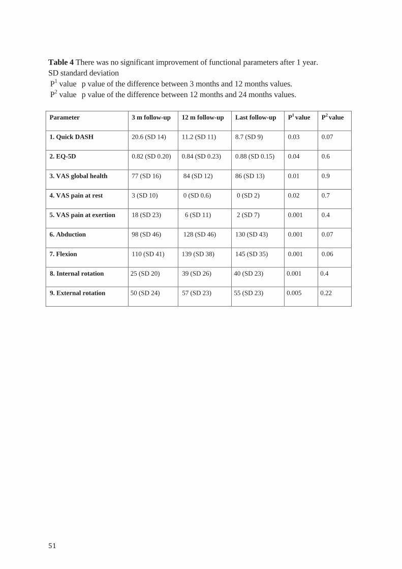
Table 4 There was no significant improvement of functional parameters after 1 year. SD standard deviation P1 value p value of the difference between 3 months and 12 months values. P2 value p value of the difference between 12 months and 24 months values. Parameter 3 m follow-up 12 m follow-up Last follow-up P1 value P2 value
1. Quick DASH 20.6 (SD 14) 11.2 (SD 11) 8.7 (SD 9) 0.03 0.07
2. EQ-5D 0.82 (SD 0.20) 0.84 (SD 0.23) 0.88 (SD 0.15) 0.04 0.6
3. VAS global health 77 (SD 16) 84 (SD 12) 86 (SD 13) 0.01 0.9
4. VAS pain at rest 3 (SD 10) 0 (SD 0.6) 0 (SD 2) 0.02 0.7
5. VAS pain at exertion 18 (SD 23) 6 (SD 11) 2 (SD 7) 0.001 0.4
6. Abduction 98 (SD 46) 128 (SD 46) 130 (SD 43) 0.001 0.07
7. Flexion 110 (SD 41) 139 (SD 38) 145 (SD 35) 0.001 0.06
8. Internal rotation 25 (SD 20) 39 (SD 26) 40 (SD 23) 0.001 0.4
9. External rotation 50 (SD 24) 57 (SD 23) 55 (SD 23) 0.005 0.22
6. Discussion
Stemless prostheses with total elimination of the humeral stem and total reliance on
metaphyseal fixation were developed to decrease shaft-related complications, e.g. periprosthetic
fracture and bone loss (Huguet et al 2010). The great individual variation in shoulder anatomy
makes it a challenge to design implants that fit most anatomical variations (Pearl et al 2002,
Iannotti et al 2005). The factors that affect pain range of motion, stability and wear rate after
shoulder reconstruction are multifactorial. For instance, the length of the lever arm of the
deltoid and rotator cuff muscles and tension of the soft tissue are both important and can be
adjusted by proper component position and size selection (Iannotti et al 1998, Takase et al
2004). Stemless implants should provide other potential benefits, including the ability to restore
shoulder anatomy regardless of the posterior offset of the proximal humerus (Sassoon et al
2013, Churchill et al 2014). This thesis evaluated the implication of the type of implant fixation
and positioning of prosthesis on both clinical and radiological outcome.
Evaluation of the type of implant fixation In studies I and II, we reported clinical and radiological results using the TSA and RSA (TESS,
Zimmer Biomet) shoulder system, both stemless and stemmed, for various shoulder disorders.
Both stemmed and nonstemmed implants showed improvement in clinical outcome with no
sign of radiological loosening after stemless implantation. In contrast, there were zones around
one stemmed implant. We did not find a specific diagnosis where the use of stemless implants
was mandatory. Instead the decision to use stemmed or stemless humeral implants depends on
bone quality and judgment of the stability achieved during the initial preparation of the
proximal humerus. In fracture surgery we recommend that only stemmed implants should be
used since implant-bone stability is not expected in osteoporotic bone. We chose to cement the
stems in the fracture group to secure the fixation of the prosthesis and to maintain the proximal
humeral length. We used the anterosuperior surgical approach in all cases. We experienced no
technical difficulty with this approach and had no axillary nerve injury.
We had two patients in the stemless group with early instability secondary to a malpositioned or
displaced corolla component. This can be the effect of limited surgical technical experience,
limited operative exposure or poor bone quality. We reported also in the first study, a
complication in the fracture group with dissociation between the metaphyseal corolla and the
stem. The presence of an unhealed humeral neck fracture might have applied excessive
rotational forces on the humeral component leading to mechanical failure in the coupling
between the corolla and the stem.
The first study on TESS implants was in 2010 from the TESS group. The authors reported the
results of 63 TESS implants with a minimum follow-up of three years. A good clinical and
radiological outcome was achieved with no evidence of subsidence or loosening (Huguet et al
2010). In the first report regarding the Eclipse stemless implant, the authors concluded that it is
possible to implant the prosthesis without a stem (Schoch et al 2011). Another study compared
clinical and radiological outcomes of three different prosthetic designs: the Neer II system, the
Bigliani-Flatow, and TESS stemless prosthesis. The authors concluded that TESS implants
resulted in significant functional improvement and the results were comparable to other
stemmed prosthesis (Razmjou et al. 2013). In a prospective randomized trial that compared the
stemless TESS and stemmed implants, the authors found that the functional improvement and
radiological stability of the stemless shoulder prosthesis were comparable with those provided
by a standard anatomical shoulder prosthesis (Berth 2013). Brunner et al. in 2012 presented the
results obtained in 233 patients treated with the Arthrex Eclipse stemless prosthesis for various
indications. In this study the Eclipse prosthesis showed a secure bony fixation and no evidence
of loosening.
We had two dislocations after fracture sequelae who had undergone RSA; both of them had
proximal humeral bone loss. Boshali et al. found that glenohumeral instability was the second
leading cause of complications associated with total shoulder arthroplasty, with a reported
prevalence of 4% and accounting for 30% of all complications. Malpositioning of the prosthetic
components, inadequate tensioning of the periprosthetic soft tissues, bone defects and rupture of
the subscapularis tendon are some of the underlying factors (Boshali et al 2006). The above
mentioned complications took place early in the study period and the authors think that this is
most probably due to the learning curve of the procedure. Kempton et al. reported that 30–40
cases are needed as a learning curve to improve the rate of early complications associated with
reverse shoulder replacement (Kempton et al 2011). The complications encountered in our
studies are comparable to that reported by other authors using other shoulder prosthesis systems
(Boshali et al 2006, Chin et al 2006, Farng et al 2011).
Despite the early good results obtained with the use of stemless anatomical implants, the use of
stemless reverse prostheses is still limited. This could be due to greater forces on the implant
because the joint constraint leads to increased forces at the bone interfaces and hence possible
failure of bone fixation.
Ballas et al in 2013 studied 56 stemless TESS RSA with a mean follow-up period of 58 months.
There was a significant functional improvement in all parameters and the results were
comparable to conventional prostheses with a stem. They had only one revision surgery due to
early instability, no humeral loosening and 5 cases (9 %) of scapular notching. This study
showed the possibility of safely implanting a stemless humeral cup for reverse shoulder
arthroplasty.
Teissier et al in 2015 reported the results of 105 stemless TESS RSA with a minimum follow-
up period of 24 months. There was neither radiographic evidence of nor glenoid nor humeral
component. SN occurred in 17 cases (19 %) which was higher than their first published series
which had only 9 %. The authors explained this as being due to the larger sample size.
Von Engelhardt et al recently published a report of 67 patients (56 stemless, 11 stemmed) with a
mean follow-up of 17.5 months. The study showed again a significant functional improvement
with no loosening of the non-stemmed humeral component in the CTA group. On the other
hand one loosening was observed in the revision group. The SN rate was 13.4 % in this series.
Three patients were revised immediately after surgery because of an intraoperative
malpositioning of one humeral component and 2 glenoid components.
A number of developments in prosthetic design occurred after the early reports, to prevent the
reported complications, like:
A. Downsizing the size of corolla, which decreased intraoperative periprosthetic fracture.
B. Development of the fracture system (stem) into a monoblock to overcome the weak link
between the corolla and the shaft
C. Improvement of the fixation of the glenosphere into the base plate to avoid early dissociation
and an increase in the number of fixation screws from 2 to 4.
D. Obliteration of the metal backed glenoid component from the anatomical system because of
increased incidence of polyethylene wear.
Study I and II might have a number of limitations. The sample size was probably relatively
small. However, all consecutive patients were operated upon using the same implant and
approach. This is the usual situation in middle sized county hospitals with general orthopaedic
surgeons interested in shoulder surgery, i.e. a situation resembling most of the hospitals around
the world.
In spite of the relatively short follow-up period, these studies reported the early results after
surgery which have special importance when evaluating a new prosthesis. This will provide
surgeons with valuable information on how to improve their early results of the operation.
The absence of control of the range of shoulder movements in study I is another limitation. At
the time of writing the first report there was discussion within the authors group about using
goniometry as a standard method to increase the precision. The included patients were
measured by visual estimation, therefore, we chose not to include this part in our report despite
its importance. After a thorough discussion within our research group, consulting other
researchers within the same field and doing a literature review, we concluded that visual
estimation is an acceptable method. It has good inter- and intrarater reliability as shown by
many studies (Riddle et al 1987, Williams et al 1990, Hayes et al 2001). Therefore, we decided
to continue measuring ROM by visual estimation and included this important measurement of
shoulder function evaluation in the other reports.
Although a standardised positioning protocol was used in obtaining AP radiographs, the
method for obtaining the radiographs remains a source of bias as both arm rotation and
positioning of the scapula depend on the judgment of the technician.
In spite of mentioned limitations, there were a number of strengths. These studies, reports
results of a stemless version of both anatomical TSA and RSA, where the available data in the
literature is still sparse. The studies showed the importance of the learning curve as a possible
way to avoid early complications especially in the accurate assessment of suitable bone quality
that permits implantation of a stemless implant. This will hopefully encourage further research
on the development of an objective tool that assesses bone quality. Rehabilitation was
standardised and all patients were followed up prospectively.
Evaluation of the implication of the positioning of the prosthesis in RSA In study II, we studied the positioning of the glenoid components in RSA and its radiological
and clinical correlation. We found SN in 30 % of our patients. However, SN did not influence
the clinical or the radiological outcome. The incidence of SN is variable in the literature,
ranging from 0 to 96 %, and increases with longer follow-up (Nyffeler et al 2005, Simovitch et
al 2007, Lévigne et al 2011). In the first study, we did not observe SN. This could be explained
by the smaller sample size (21 RSA) and shorter follow-up (mean 14 months), hence we
recommended in the first study further investigations with repeated clinical and radiological
controls. Another possible explanation is that SN was not well identifiable to the authors at the
time of writing the first study.
The underlying cause, clinical significance and ways to avoid notching are still under debate
(Nyffeler et al 2005, Simovitch et al 2007). We found that glenoid overhang decreases SN. This
finding is consistent with several studies that found that it is a critical factor in order to avoid
SN (Sirveaux et al 1997, deWilde et al 2010). We found also a peg-glenoid distance less than 20

mm significantly decreased SN which is consistent with Simovitch et al. (Simovitch et al 2007,
De Biase et al 2013).
We did not find any correlation between glenoid inclination and SN. However, this could be
due to the small number of patients with inclination over 10° from neutral. There are divergent
findings about the effect of inferior inclination on the incidence of SN. Some deny its effect
(Kempton et al 2011, Edwards et al 2012), while others report decreased (Gutiérrez et al 2007,
Gutiérrez et al 2011) or increased incidence of SN (Simovitch et al 2007).
We also noticed that some patients already had SBI on the post-operative X-ray that later
developed into SN. A possible explanation could be the use of the anterosuperior approach.
This approach necessitates retractors to be placed under the glenoid rim to dislocate the
humerus downwards creating deformative forces on the scapular edge. Other possible
explanations cannot be excluded, e.g. the forward scapular rotation in the early post-operative
period or a prominent anatomical notch of the circumflex scapular vessels.
In the same study, we reported the effect of arm lengthening on radiological and clinical
outcome. We found that arm lengthening improved the quality of life, but we were unable to
note effects on function, ROM or pain. This is in accordance with previous reports that found
that arm lengthening could influence the outcome (Lädermann et al 2012, Lädermann et al
2014). Evaluation of the implication of the prosthesis positioning on LHO in stemless anatomical TSA One important radiological parameter is LHO as shown by many studies that its
reconstruction is an important element in achieving efficient shoulder function (Takase et al
2004). Iannotti et al. measured LHO offset as the distance between the coracoid base to the
most lateral part of the greater tubercle and showed that it correlated both with the size of the
humeral head and the moment arm of the deltoid and rotator muscles (Iannotti et al 1992,
Iannotti et al 1998). It is still unclear how much restoration of the LHO is needed, and whether
this restoration would affect the prosthetic long-term survival (Pearl et al 2002, Pearl et al
2009). Takase et al studied LHO offset in AP radiography and showed also that it is related to
other parameters like humeral head diameter, radius of curvature of the humeral head and the
distance between the acromion and the greater tubercle (Takase et al 2004).
Plain radiographs are routinely used for preoperative templating and postoperative evaluation.
However, despite their availability, easy interpretation, low cost and low radiation exposure,
plain radiographs have limited accuracy compared to CT scans (Ho et al 2013).
In study III we evaluated reliability of LHO performed on both AP radiographs and axial CT
of the shoulder in patients with primary OA. The interobserver reliability using AP
radiography was moderate while the intraobserver reproducibility was variable among the
observers. A possible explanation is the variable experience among the observers. Another
possible explanation is the difficulty in finding the same point on the medial border of the
coracoid process among the observers.
Thomas et al. studied reliability of LHO in plain radiography using the same standardised
protocol that we used in our study. They found it to be unreliable with a tendency of observers
to disagree on the medial/ lateral location of the base of the coracoid (Thomas et al 2005).
We found excellent inter- and intraobserver agreement of the CT measurements among all 4
observers. The correlation between CT and X-ray in measuring LHO was moderate with a
significant underestimation of LHO on AP radiographs with a mean difference of 5 mm. This
indicates that CT is more precise in identifying the base of the coracoid process than plain
radiographs and thus is the recommended method for measuring LHO. Differences in the
estimation between CT and X-ray could be also explained by the measurements having been
done in different planes, i.e. coronal and sagittal. Possibly also the patient positioning could
have a role, being prone in CT and erect in X-ray examinations. Deladerrie`re et al analysed
the geometry of 42 resurfacing shoulder implants using CT. They used a modified method to
measure LHO on axial CT scans. The study showed that CT was a reliable way to identify
these two points and of measuring LHO (Deladerrie`re et al 2012).
In Study V we evaluated the ability of stemless implants to restore LHO and its correlation
with functional results. In order to assess the ability of an implant to restore shoulder anatomy,
we chose to use the contralateral healthy shoulder in the axial CT, as a reference in our
measurements.
The correlation between LHO contra and LHO op was substantial (ICC 0.8, CI 95% 0.62 - 0.87,
P = 0.001). This supports the hypothesis that stemless implants could restore the linear
glenohumeral relationship, LHO, close to the normal anatomy.
Variation of the LHO op from the LHO contra > 5 mm was considered as being clinically
significant (Mechlenburg 2013). There were 8/44 (18%) of patients with > 5 mm (mean 6.8 mm
± SD 2).
Regarding the postoperative outcome, all included measures showed significant improvement
compared with the preoperative state, regardless of LHO. This improvement was not detectable
after 12 months postoperatively. Therefore, the authors think that 12 months follow-up is
adequate to make a reasonable evaluation of the postoperative outcome. The LHO was
correlated at three months with Quick DASH (Pearson correlation = 0.36, P = 0.12), VAS pain
(at rest) (Pearson correlation = 0.33, P = 0.05) and VAS pain (at exertion) (Pearson correlation
= 0.33, P = 0.05), where increased LHO op negatively affected these measures. No correlation
at 12 months was found. The LHO showed no correlation with any of the other outcome
measures (EQ-5D, VAS health status, ROM) during the follow-up period.
One possible explanation for this negative effect of increased LHO is soft-tissue over-tensioning
of the newly operated shoulder joint, especially if the joint had excessive long lasting narrowing
of the joint space and capsule tightening secondary to OA. Differences in the laxity of the joint
capsule among patients might also affect the degree of association between the LHO increment
and the outcome measures. Gradual adaptation of the soft-tissue to the new postoperative LHO
after the first 3 months can explain the lack of association between the LHO increment and the
outcome measures at 12 months follow-up. The LHO seemed not to affect quality of life, health
status or ROM at any time during the first year postoperatively.
Evaluation of the implication of the prosthesis positioning on proximal humeral anatomy in stemless anatomical TSA In study IV we investigated the ability of a stemless prosthesis to restore the premorbid
proximal humerus anatomy using the best-fit-circle method (Youderian et al. 2014)
COR is considered by many studies to be an important parameter to reconstruct (Pearl et al
2009). We considered a 3-mm deviation from the premorbid COR to be clinically significant, as
proposed by (Alolabi et al 2014). In our study, the difference was within 3 mm in 82 % of
cases.
HH is similarly of paramount importance. In our study, post-operative HH was 19±2 mm,
which was less than pre-operative values but close to results of other studies (Iannotti JP et al
1992, Nyffeler RW et al 2004, Nyffeler RW et al 2005). Deviation of ≥5mm of the HH from the
pre-operative measurement was considered of clinical importance as shown by other studies
(Harryman DT et al 1995). We had 8/70(11 %) shoulders with postoperative HH difference >5
mm.
NSA was 133±6° pre-operatively compared with 130±11° post-operatively. There were 25/70
(36 %) shoulders with postoperative NSA <130°, which shows a tendency to put the prosthesis
in varus. NSA has a wide range of variation, as shown by other studies (Iannotti JP et al 1992,
Boileau P et al 1997). Takase et al. studied NSA in 519 shoulders, and it ranged from 130° to
152°, with a mean of 140.5°± 4.0° (Takase et al 2002).
We used the Pearson correlation coefficient (r) to calculate the correlation between premorbid
and post-operative parameters (COR, HH and NSA). According to Hornij et al , r values >0.75
represent excellent agreement, 0.4–0.75 fair to good agreement and <0.4 poor agreement.
Rheault et al. used criteria recommended by Landis and Koch (0.00–0.20 slight agreement,
0.21–0.40 fair agreement, 0.41–0.60 moderate agreement, 0.61–0.80 substantial agreement,
0.81–1.00 excellent agreement. Lindell et al considered the value of 0.60 as a limit of
acceptability for application in clinical practice. In this paper, mean r values were used, leaving
evaluation of the degree of their reliability and reproducibility to the judgment of the readers.
These studies (III, IV and V) have some limitations. The use of CT can be associated with high
cost and radiation dose. We included only patients with primary OA. In secondary OA, many
cases have flattening of the humeral head causing a medial shift of the rotational centre in
relation to the glenoid, and hence decreasing LHO with soft-tissue contracture. The technical
demands for shoulder anatomy restoration also differ considerably in both groups. For these
reasons, we chose to exclude patients with secondary OA in order to have a homogenous group.
This can be considered as a limitation since the results of these studies cannot be applied to
patients with secondary OA.
In spite of these limitations there were also strengths like:
The CT measurements have a proven reliability and reproducibility and it was used in
measuring LHO instead of plain radiographs. To our knowledge, these are the first studies
designed to investigate the ability of stemless implants to restore LHO and proximal humerus
anatomy using the same prosthesis and the same surgical approach.
7. Conclusion
1. The results of stemless shoulder prostheses are promising with a complication rate that is
comparable with other shoulder prosthetic systems with the advantage of bone stock
preservation and avoidance of stem-related complications. Long-term follow-up is required to
confirm the results of this innovative system in the long run.
2. The anterosuperior approach can adequately be used for the implantation of different versions
of shoulder prostheses.
3. RSA can successfully treat different shoulder problems. Glenoid overhang can reduce SN
and arm lengthening has positive effects on outcome. Our method of arm lengthening
measurement needs further studies to ensure validity.
4. LHO measurement on AP radiographs is less reliable and underestimates the distance when
compared with CT. Also, CT is a reliable tool to measure LHO supporting its use in
preoperative planning. Outcome measures were improved regardless of the LHO. At 3 months
follow-up, increased LHO had negative effect on shoulder function and gave more shoulder
pain at rest and exertion but did not affect quality of life, health status or ROM. At 12 months
follow-up, LHO had no relation with the outcome measures. Further studies are warranted to
investigate the influence of LHO on long-term prosthetic survival.
5. Stemless implants could restore the shoulder anatomy in an acceptable manner. This thesis
shows that there are some challenges to be addressed when attempting to ensure optimal
implant positioning. The critical step is to determine the correct level of bone cut to avoid
varus or valgus HH inclination and version. Surgical instruments and technique may require
modifications to optimise the surgeon’s ability to replicate normal anatomic parameters,
allowing easier identification of the proper head-cut level.
8. Implications for future research
Future research on stemless implants To investigate remodelling characteristics and bone quality at the prosthetic-bone interface in
stemless implants and correlate it to functional outcome and implant survival.
Future research on shoulder anatomy restoration To investigate the other factors that could play a role in shoulder anatomy restoration such as
the type of approach, BMI, type of diagnosis e.g. secondary OA.
Future research on RSA To study the reliability and reproducibility of our method of measuring arm lengthening in CT
in comparison to methods in the literature.
9. Acknowledgements
I wish to express my sincere gratitude to: Göran Sjödén My principal tutor. For guidance, encouragement, patience and leading me through the path of this thesis. Arkan Sayed-Noor My co-tutor and my best friend, who has always made himself available to share his extensive knowledge of biomechanics and orthopaedics with me and for his continued support leading to the completion of this thesis. Sebastian Mukka My colleague and co-author. For his enthusiasm, never ending assistance and readiness and friendship. Sarwar Mahmood My colleague, for support and encouragement
Mats Wadsten My colleague and co-author, who has been a wonderful co-worker through the years. Nader Mafi and Sigge Norberg My colleagues and co-authors. For your contribution in study I and for your support and teaching me shoulder surgery. Hamid Hassany My colleague and co-author. For your contribution in study IV, good talks, your support, encouragement and good friendship.
Nikolaos Perisynakis and Saida Baea My colleagues and co-authors. For your contribution in study III. Per Wahlström and Shwan Khoschnau My colleagues and co-authors. For your contribution in study V and your valuable input. All my other colleagues at the department of orthopaedics at Sundsvall hospital, for their support, encouragement and understanding during these years.
Raymond Pollock, for language correction.
Erling Englund, for invaluable statistical advice and analysis as well as patient explanation at
any time of the day.
Helene Sörell, Anne-Charlotte Söderlind and Elieann Broman
The research nurses at the Orthopedic Department of Sundsvall Hospital, who were always there when needed and helped to examine the patients.
All the patients included in the studies.
The Swedish Orthopedic Association, Emil Anderssons fond, The Swedish shoulder and elbow surgery association and Research and development center (FoU) Landstinget Västernorrland, for financial support Last but not least, To my beloved wife, Hala Aljuboori, all who know you understand why I cherish you, thank you for your assistance, encouragement, and patience during this onerous project. To my children Mohammed, Ali, Ammar and Yosef, Thanks for always reminding me that I need balance in life, and thanks for being that balance.
10. References
A Alolabi B, Youderian AR, Napolitano L, Szerlip BW, Evans PJ, Nowinski RJ et al. Radiographic assessment of prosthetic humeral head size after anatomic shoulder arthroplasty. J Shoulder Elbow Surg. 2014; 23(11): 1740–6. doi: 10.1016/j.jse.2014.02.013. Atroshi I, Gummesson C, Andersson B, Dahlgren E, Johansson A. The disabilities of the arm, shoulder and hand (DASH) outcome questionnaire: reliability and validity of the Swedish version evaluated in 176 patients. Acta Orthop Scand. 2000 Dec; 71(6): 613-8. B Ballas R, Béguin L. Results of a stemless reverse shoulder prosthesis at more than 58 months mean without loosening. J Shoulder Elbow Surg. 2013 Sep; 22(9): e1-6. doi: 10.1016/j.jse.2012.12.005 Bankes MJ, Emery RJ. Pioneers of shoulder replacement: Themistocles Gluck and Jules Emile Péan. J Shoulder Elbow Surg. 1995 Jul-Aug; 4(4): 259-62. Baulot E, Valenti P, Garaud P, Boileau P, Neyton L, Sirveaux F, et al. R_esultats des proth_eses invers_ees. Revue de Chirurgie Orthop_edique et R_eparatrice de l’Appareil Moteur 2007; 93(Suppl 1): 63-92. http:// dx.doi.org/10.1016/S0035-1040(07) 92712-9. Berth A, Pap G. Stemless shoulder prosthesis versus conventional anatomic shoulder prosthesis in patients with osteoarthritis: a comparison of the functional outcome after a minimum of two years follow-up. J Orthop Traumatol. 2013; 14:31-37. Bland JM, Altman DG. Statistical methods for assessing agreement between two methods of clinical measurement. Lancet. 1986; 1:307–310. Blevins FT, Deng X, Torzilli PA, Dines D, Warren RF. Dissociation of modular humeral head components: a biomechanical and implant retrieval study. J Shoulder Elbow Surg 1997; 6: 113-124. Boileau P, Walch G .The three-dimensional geometry of the proximal humerus. Implications for surgical technique and prosthetic design. J Bone Joint Surg (Br) 1997; 79(5):857–65 Boileau P, Avidor C, Krishnan SG, Walch G, Kempf JF, Molé D. Cemented polyethylene versus uncemented metal-backed glenoid components in total shoulder arthroplasty: a prospective, double-blind, randomized study. J Shoulder Elbow Surg 2002; 11:351-9. http://dx. doi.org/10.1067/mse.2002.125807. Boileau P, Watkinson D, Hatzidakis AM, Hovorka I. Neer Award 2005: The Grammont reverse shoulder prosthesis: results in cuff tear arthritis, fracture sequelae, and revision arthroplasty. J Shoulder Elbow Surg. 2006 Sep- Oct; 15(5): 527-40.
Bohsali KI, Wirth MA, Rockwood CA Jr. Complications of total shoulder arthroplasty. J Bone Joint Surg Am. 2006; 88: 2279-2292. Brooks RG, Jendteg S, Lindgren B, Björk S. EuroQol: health-related quality of life measurement. Results of the Swedish questionnaire exercise. Health Policy. 1991 Jun; 18(1): 37-48. Brunner UH, Fruth M, Rückl K, Magosch P, Tauber M, Resch H, et al. Die schaftfreie Eclipse-Prothese-Indikation und mittelfristige Ergebnisse. Obere Extremität. 2012; 7:22-28. Bufquin T, Hersan A, Hubert L, Massin P. Reverse shoulder arthroplasty for the treatment of three- and four-part fractures of the proximal humerus in the elderly: a prospective review of 43 cases with a short-term follow-up. J Bone Joint Surg Br. 2007; 89:516–520. Burgess DL, McGarth MS, Bonutti PM, Marker DR, Delanois RE, Mont MA. Current concepts review: shoulder resurfacing. J Bone J Surg Am. 2009; 91:1228–1238. C Chin PYK, Sperling JW, Cofield RH, Schleck C. Complications of total shoulder arthroplasty: are they fewer or different? J Should Elbow Surg. 2006; 15(1):19–22 Churchill RS. Stemless shoulder arthroplasty: current status. J Shoulder Elbow Surg. 2014; 23:1409-1414. DOI: 10.1016/ j.jse.2014.05.005. Clitherow HD, Frampton CM, Astley TM. Effect of glenoid cementation on total shoulder arthroplasty for degenerative arthritis of the shoulder: a review of the New Zealand National Joint Registry. J Shoulder Elbow Surg. 2014 Jun; 23(6): 775-81. doi: 10.1016/j.jse.2013.08.022. Cofield RH. Uncemented total shoulder arthroplasty: a review. Clin Orthop 1994; 307:86 93. D De Biase CF, Ziveri G, Delcogliano M, de Caro F, Gumina S, Borroni M, Castagna A, Postacchini R . The use of an eccentric glenosphere compared with a concentric glenosphere in reverse total shoulder arthroplasty: two-year minimum follow-up results. Int Orthop.2013; 37(10):1949–1955 Dieckmann R, Liem D, Gosheger G, HenrichsMP, Höll S, Hardes J, Streitbürger A. Evaluation of a reconstruction reverse shoulder for tumour surgery and tribological comparison with an anatomical shoulder arthroplasty. Int Orthop. 2013: 37(3):451–456 Deladerrie`re JY, Szymanski C, Vervoort T, Budzik JF, Maynou C. Geometrical analysis results of 42 resurfacing shoulder prostheses: a CT scan study. Orthop Traumatol Surg Res. 2012; 98(5): 520–527. doi:10.1016/j.otsr.2012.03.010
Dines DM, Warren RE Modular shoulder hemiarthroplasty for acute fractures: surgical considerations. Clin Orthop 1994; 307: 18-26. de Wilde LF, Poncet D,Middernacht B, Ekelund A.Prosthetic overhang is the most effective way to prevent scapular conflict in a reverse total shoulder prosthesis. Acta Orthop. 2010; 81(6):719–726 E Edwards TB, Trappey GJ, Riley C, O'Connor DP, Elkousy HA, Gartsman GM. Inferior tilt of the glenoid component does not decrease scapular notching in reverse shoulder arthroplasty: results of a prospective randomized study. J Shoulder Elbow Surg. 2012 May; 21(5): 641-6. doi: 10.1016/j.jse.2011.08.057. EuroQol Group. EuroQol--a new facility for the measurement of health-related quality of life. Health Policy. 1990 Dec; 16(3): 199-208. Eynon-Lewis NJ, FerryD, PearseMF. ThemistoclesGluck: an unrecognised genius. BMJ 1992; 305:1534-6. F Farng E, Zingmond D, Krenek L, Soohoo NF. Factors predicting complication rates after primary shoulder arthro- plasty. J Shoulder Elbow Surg. 2011; 20:557-563. Faul F, Erdfelder E, Buchner A. Statistical power analyses using G*Power 3.1: tests for correlation and regression analyses. Behav Res Methods 2009; 41:1149. G Gerber C, Pennington SD, Nyffeler RW.Reverse total shoulder arthroplasty. J Am Acad Orthop Surg. 2009; 17:284–295 Gluck T. Referat iJber die durch das moderne chirurgische Experfment gewonnenen positiven Resultate, betreffend die Naht und den Ersatzvan Defecten hoherer Gewebe, sowie Jberdie Verwerthungresorbirbarerund lebendigerTamponsin der Chirurgie. Archivfor KlinischeChirurgie 1891; 41:187- 239. Grammont P, Trouilloud P, Laffay JP, Deries X. Concept study and realization of a new total shoulder prosthesis. Rhumatologie. 1987; 39:407-18. Gummesson C, Ward MM, Atroshi I. The shortened disabilities of the arm, shoulder and hand questionnaire (QuickDASH): validity and reliability based on responses within the full-length DASH. BMC Musculoskelet Disord. 2006 May 18; 7:44. Gutiérrez S, Greiwe RM, Frankle MA, Siegal S, Lee WE III. Biomechanical comparison of component position and hardware failure in the reverse shoulder prosthesis. J Shoulder Elbow Surg. 2007; 16(3 Suppl):S9–S12.
Gutiérrez S, Levy JC, Frankle MA, Cuff D, Keller TS, Pupello DR, Lee WE 3rd Evaluation of abduction range of motion and avoidance of inferior scapular impingement in a reverse shoulder model. J Should Elbow Surg. 2008; 17:608–615. H Harryman DT, Sidles JA, Harris SL, Lippitt SB, Matsen FA. The effect of articular conformity and the size of the humeral head component on laxity and motion after glenohumeral arthroplasty. A study in cadavera. J Bone Joint Surg Am. 1995; 77(4): 555–63. Hayes K, Walton JR, Szomor ZR, Murrell GA. Reliability of five methods for assessing shoulder range of motion. Randomized controlled trial. Aust J Physiother. 2001; 47(4): 289-49. H. G. Zadeh, R T. Calvert. Recent advances in shoulder arthroplasty. Current Orthopaedics (1998) 12, 122-134 Ho JC, Youderian A, Davidson IU, Bryan J, Iannotti JP. Accuracy and reliability of postoperative radiographic measurements of glenoid anatomy and relationships in patients with total shoulder arthroplasty. J Shoulder Elbow Surg. 2013; 8:1068–1077. doi:10.1016/j.jse.2012.11.015 Horneij E, Holmström E, Hemborg B, Isberg PE, Ekdahl C. Interrater reliability and between-days repeatability of eight physical performance tests. Adv Physiother. 2002; 4:146–60. doi:10.1080/ 14038190260501596 Hudak PL, Amadio PC, Bombardier C. Development of an upper extremity outcome measure: the DASH (disabilities of the arm shoulder and hand). The Upper Extremity Collaborative Group (UECG). Am J Ind Med. 1996 Jun; 29(6):602-8. Huguet D, DeClercq G, Rio B, Teissier J, Zipoli B, TESS Group. Results of a new stemless shoulder prosthesis: radiologic proof of maintained fixation and stability after a minimum of three years' follow-up. J Shoulder Elbow Surg. 2010; 19:847-852. Huskisson EC. Measurement of pain. Lancet 1974; 2: 1127–31. I Iannotti JP, Gabriel JP, Schneck SL, Evans BG, Misra S. The normal glenohumeral relationships. An anatomical study of one hundred and forty shoulders. J Bone Joint Surg Am. 1992; 74(4):491–500 Iannotti JP, Williams GR, Total shoulder arthroplasty. Factors influencing prosthetic design. Orthop Clin North Am. 1998; 29(3): 377–391 Iannotti JP, Spencer EE, Winter U, Deffenbaugh D, Williams G Prosthetic positioning in total shoulder arthroplasty. J Shoulder Elbow Surg. 2005; 14(1 Suppl S): 111S–121S. doi:10.1016/j. jse.2004.09.026
J Jazayeri R, Kwon YW. Evolution of the reverse total shoulder prosthesis. Bull NYU Hosp Jt Dis. 2011;69(1):50-5. Jensen MP, Karoly P, Braver S. The measurement of clinical pain intensity: a comparison of six methods. Pain 1986; 27: 117–26. K Kempton LB, Balasubramaniam M, Ankerson E, Wiater JM. A radiographic analysis of the effects of glenosphere position on scapular notching following reverse total shoulder arthroplasty. J Shoulder Elbow Surg. 2011; 20(6):968–974 Kempton LB, Ankerson E, Wiater JM.A complication-based learning curve from 200 reverse shoulder arthroplasties. Clin Orthop Relat Res. 2011 Sep;469(9):2496-504. doi: 10.1007/s11999-011-1811-4. Kim MS, Kovacevic D, Milks RA, Jun BJ, Rodriguez E, Delozier KR, Derwin KA, Iannotti JP. Bone Graft Substitute Provides Metaphyseal Fixation for a Stemless Humeral Implant. Orthopedics. 2015 Jul 1;38(7):e597-603. doi: 10.3928/01477447-20150701-58. L Landis JR, Koch GG. The measurement of observer agreement for categorical data. Biometrics. 1977; 33:159–174 Levigne C, Boileau P, Favard L, et al. Scapular notching in reverse shoulder arthroplasty. J Shoulder Elbow Surg. 2008; 17: 925-35. Lévigne C, Garret J, Boileau P, Alami G, Favard L, Walch G. Scapular notching in reverse shoulder arthroplasty: is it important to avoid it and how? Clin Orthop Relat Res.2011; 469(9):2512–2520 Lindell O, Eriksson L, Strender L-E. The reliability of a 10- test package for patients with prolonged back and neck pain: could an examiner without formal medical education be used without loss of quality? A methodological study. BMC Musculoskelet Disord. 2007; 8: 31. doi:10.1186/1471-2474-8-31 Lugli T. Artificial shoulder joint by Péan (1893): the facts of an exceptional intervention and the prosthetic method. Clin Orthop Relat Res. 1978 Jun;(133): 215-8. Lädermann A, Walch G, Lubbeke A, Drake GN, Melis B, Bacle G, Collin P, Edwards TB, Sirveaux F .Influence of arm lengthening in reverse shoulder arthroplasty. J Shoulder Elbow Surg. 2012; 21(3): 336–341 Lädermann A, Edwards TB, Walch G. Arm lengthening after reverse shoulder arthroplasty: a review. Int Orthop. 2014 May; 38(5):991-1000. doi: 10.1007/s00264-013-2175-z.
M Mackenzie DB. The antero-superior exposure for total shoulder replacement. Orthop Traumatol. 1993; 2:690–700 Moeckel BH , Dines DM, Warren RF, Altchek DW. Modular hemiarthroplasty for fractures of the proximal part of the humerus. J Bone Joint Surg Am. 1992 Jul;74(6):884-9. Mole ́ D, Favard L. Excentered scapulohumeral osteoarthritis,[in French. Rev Chir Orthop Reparatrice Appar Mot. 2007;93(6 Suppl):37–94. Molé D, Wein F, Dézaly C, Valenti P, Sirveaux F.Surgical technique: the anterosuperior approach for reverse shoulder arthroplasty. Clin Orthop Relat Res. 2011 Sep; 469(9):2461-8. doi: 10.1007/s11999-011-1861-7. Mechlenburg I, Amstrup A, Klebe T, Jacobsen SS, Teichert G, Stilling M. The Copeland resurfacing humeral head implant does not restore humeral head anatomy. A retrospective study. Arch Orthop Trauma Surg. 2013;133(5):615-9. doi: 10.1007/s00402-013-1715-8. N Neer CS, Brown Th, Mclaughlin Hl (1953) Fracture of the neck of the humerus with dislocation of the head fragment. Am J Surg 85(3): 252–258. Neer CS 2nd. Replacement arthroplasty for glenohumeral osteoarthritis. J Bone Joint Surg Am. 1974 Jan; 56(1):1-13. 10. Nicholson GP, Strauss EJ, Sherman SL.Scapular notching: Recognition and strategies to minimize clinical impact. Clin Orthop Relat Res. 2011 Sep; 469(9):2521-30. doi: 10.1007/s11999-010-1720-y. Nyffeler RW, Sheikh R, Jacob HA, Gerber C (2004) Influence of humeral prosthesis height on biomechanics of glenohumeral abduction, An in vitro study. J Bone Joint Surg Am 86-A(3):575–80 Nyffeler RW, Werner CM, Gerber C. Biomechanical relevance of glenoid component positioning in the reverse Delta III total shoulder prosthesis. J Shoulder Elbow Surg. 2005; 14:524-8. O Ortmaier R, Resch H, Matis N, Blocher M, Auffarth A, Mayer M, Hitzl W, Tauber M.Reverse shoulder arthroplasty in revision of failed shoulder arthroplasty-outcome and follow-up. Int Orthop.2013: 37(1):67–75 P Pearl ML, Volk AG.Retroversion of the proximal humerus in relationship to prosthetic replacement arthroplasty. J Shoulder Elbow Surg 1995; 4: 286-289.
Pearl ML, Kurutz S, Robertson DD, Yamaguchi K .Geometric analysis of selected press fit prosthetic systems for proximal humeral replacement. J Orthop Res. 2002; 20(2):192–7. doi:10.1016/ s0736-0266 (01) 00111-5 Pearl ML. Proximal humeral anatomy in shoulder arthroplasty: implications for prosthetic design and surgical technique. J Shoulder Elbow Surg. 2005;14(1 Suppl S):99S-104S. Pearl ML, Kurutz S, Postacchini R. Geometric variables in anatomic replacement of the proximal humerus: how much prosthetic geometry is necessary? J Shoulder Elbow Surg. 2009; 18(3):366–70. doi:10.1016/j.jse.2009.01.011 Petriccioli D, Bertone C1, Marchi G .Stemless shoulder arthroplasty: a literature review. Joints. 2015 Jun 8;3(1):38-41 R Razmjou H, Holtby R, Christakis M, Axelrod T, Richards R. Impact of prosthetic design on clinical and radiologic outcomes of total shoulder arthroplasty: a prospective study. J Shoulder Elbow Surg. 2013; 22:206-214. Rheault W, Albright B, Beyers C, Franta M, Johnson A, Skowronek M, Dougherty J.Intertester reliability of the cervical range of motion device. J Orthop Sports Phys Ther. 1992; 15(3):147–50 Riddle DL, Rothstein JM and Lamb RL. Goniometric reliability in a clinical setting. Physical Therapy .1987;67: 668-673. Roberts SNJ, Foley APJ, Swallow HM, Wallace WA, Coughlan DR.The geometry of the humeral head and the design of prostheses. J Bone Joint Surg 1991; 73-B: 647-650. Roche C, Flurin PH, Wright T, Crosby LA, Mauldin M, Zuckerman JD An evaluation of the relationships between reverse shoulder design parameters and range of motion, impingement, and stability. J Should Elbow Surg. 2009;18:734–741 Rodosky MW, Bigliani LU. Indications for glenoid resurfacing in shoulder arthroplasty. J Shoulder Elbow Surg 1996; 5: 231-248 S Sassoon A, Schoch B, Rhee P, Schleck CD, HarmsenWS, Sperling JW, Cofield RH. The role of eccentric and offset humeral head variations in total shoulder arthroplasty. J Shoulder Elbow Surg. 2013; 22(7):886–93. doi:10.1016/j.jse.2012.09.008 Sahota S, Sperling JW, Cofield RH. Humeral windows and longitudinal splits for component removal in revision shoulder arthroplasty. J Shoulder Elbow Surg. 2014; 23:1485-91. pii: S1058-2746(14)00113-X. DOI: 10.1016/j.jse.2014.02.004.
Severt R, Thomas B J, Tsenter M J, Amstutz HC, Kabo JM.The influence of conformity and constraint on translational forces and frictional torque in total shoulder arthroplasty. Clin Orthop 1993; 292:151 158. Shapiro SS, Wilk MB (1965) An analysis of variance test for normality (complete samples). Biometrika 52(3/4):591–611 Simovitch RW, Zumstein MA, Lohri E, et al. Predictors of scapular notching in patients managed with the Delta III reverse total shoulder replacement. J Bone Joint Surg Am. 2007;89:588-600. Sirveaux F. Grammont prosthesis in the treatment of shoulder arthropathies with massive cuff tear. Multicenter series of 42 cases (in French). Medical doctor thesis, Nancy I University.1997; 245 pp. Stanley Hoppenfeld MD , Piet deBoer MA FRCS , Richard Buckley MD FRCSC. Surgical Exposures in Orthopaedics: The Anatomic Approach (Hoppenfeld, Surgical Exposures in Orthopaedics), chapter one pp. 37- 47. Fourth Edition. 2009 by Lippincot Williams and Wilkins. Stephen Fealy (Author, Editor), John Sperling (Author), John W. Sperling (Editor), Russell F. Warren (Editor), Edward V. Craig (Editor) Shoulder Arthroplasty: Complex Issues in the Primary and Revision Setting . New York: Thieme Medical Publishers, Inc.; 2008. p. 1-3. (ISBN: 978-158890-505-5) T Takase K, Imakiire A, Burkhead WZ Jr. Radiographic study of the anatomic relationships of the greater tuberosity. J Shoulder Elbow Surg. 2002; 11(6):557–61 Takase K, Yamamoto K, Imakiire A, Burkhead WZ Jr. The radiographic study in the relationship of the glenohumeral joint. J Orthop Res. 2004; 22(2):298–305 Thomas SR, Sforza G, Levy O, Copeland SA. Geometrical analysis of Copeland surface replacement shoulder arthroplasty in relation to normal anatomy. J Shoulder Elbow Surg. 2005; 14(2):186–192 Torchia ME, Cofield RH, Settergren CR. Total shoulder arthroplasty with the Neer prosthesis: long term results. J Shoulder Elbow Surg 1997; 6: 495-505. Teissier P, Teissier J, Kouyoumdjian P, Asencio G.The TESS reverse shoulder arthroplasty without a stem in the treatment of cuff-deficient shoulder conditions: clinical and radiographic results. J Shoulder Elbow Surg. 2015 Jan;24(1):45-51. doi: 10.1016/j.jse.2014.04.005. V Valenti P, Sauzieres P, Cogswell L, O’Toole G, Katz D. The reverse shoulder prosthesis—surgical technique. Tech Hand Up Extrem Surg. 2008; 12:46–55. Von Engelhardt LV , Manzke M, Filler TJ, Jerosch J. Short-term results of the reverse Total Evolutive Shoulder System (TESS) in cuff tear arthropathy and revision arthroplasty cases. Arch Orthop Trauma Surg. 2015 Jul;135(7):897-904. doi: 10.1007/s00402-015-2218-6
W Walch G, Boileau P. Prosthetic adaptability: a new concept for shoulder arthroplasty. J Shoulder Elbow Surg. 1999 Sep-Oct;8(5): 443-51. Williams JG and Callaghan M. Comparison of visual estimation and goniometry in determination of a shoulder joint angle. Physiotherapy.1990;76: 655-657. Y Youderian AR, Ricchetti ET, Drews M, Iannotti JP. Determination of humeral head size in anatomic shoulder replacement for glenohumeral osteoarthritis. J Shoulder Elbow Surg. 2014: 23(7): 955–63. doi:10.1016/j.jse.2013.09.005 Z Zippel J, Meyer-Ralfs M. Themistocles Gluck (1853-1942) Wegbereiter der Endoprothetik. Z Orthop Ihre Grenzgeb 1975; 113:134-9. Zumstein MA, Pinedo M, Old J, Boileau P. Problems, complications, reoperations, and revisions in reverse total shoulder arthroplasty: a systematic review. J Shoulder Elbow Surg 2011; 20:146-57. http://dx.doi.org/10.1016/j.jse.2010.08.001
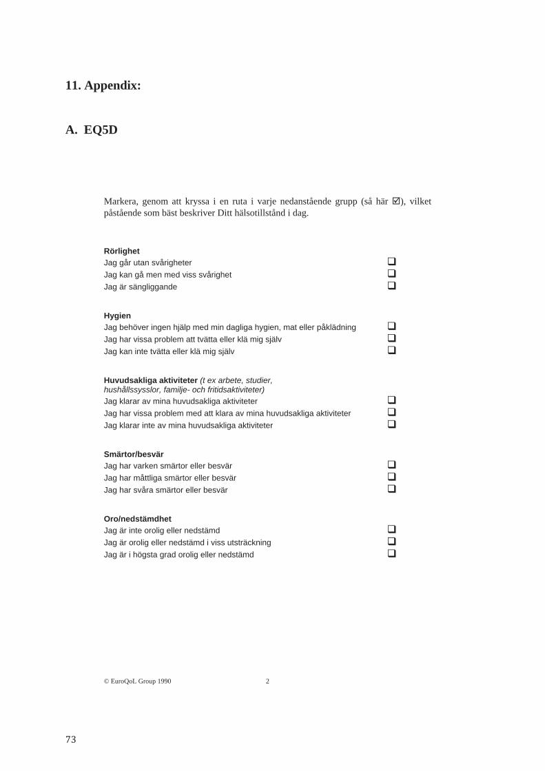
11. Appendix:
A. EQ5D
© EuroQoL Group 1990
2
Markera, genom att kryssa i en ruta i varje nedanstående grupp (så här ), vilket påstående som bäst beskriver Ditt hälsotillstånd i dag. Rörlighet Jag går utan svårigheter Jag kan gå men med viss svårighet Jag är sängliggande Hygien Jag behöver ingen hjälp med min dagliga hygien, mat eller påklädning Jag har vissa problem att tvätta eller klä mig själv Jag kan inte tvätta eller klä mig själv Huvudsakliga aktiviteter (t ex arbete, studier, hushållssysslor, familje- och fritidsaktiviteter) Jag klarar av mina huvudsakliga aktiviteter Jag har vissa problem med att klara av mina huvudsakliga aktiviteter Jag klarar inte av mina huvudsakliga aktiviteter Smärtor/besvär Jag har varken smärtor eller besvär Jag har måttliga smärtor eller besvär Jag har svåra smärtor eller besvär Oro/nedstämdhet Jag är inte orolig eller nedstämd Jag är orolig eller nedstämd i viss utsträckning Jag är i högsta grad orolig eller nedstämd
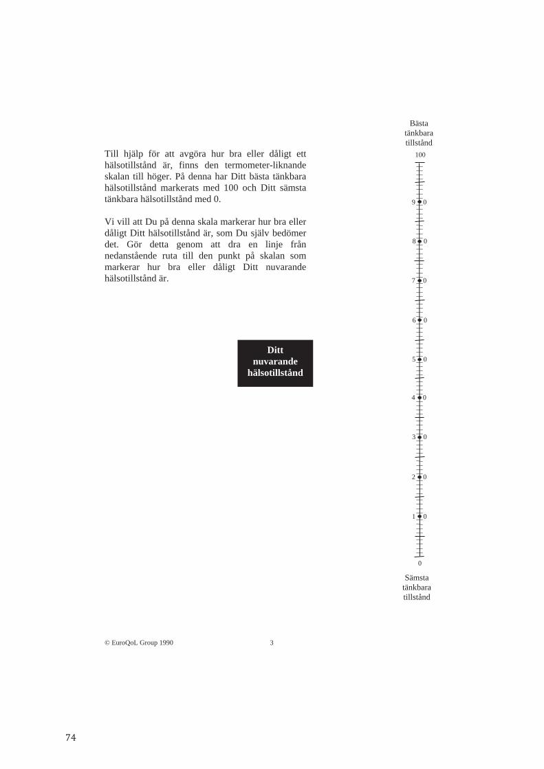
Bästa
9 0
8 0
7 0
6 0
5 0
4 0
3 0
2 0
1 0
100
Sämsta tänkbara tillstånd
0
tänkbara tillstånd
Till hjälp för att avgöra hur bra eller dåligt ett hälsotillstånd är, finns den termometer-liknande skalan till höger. På denna har Ditt bästa tänkbara hälsotillstånd markerats med 100 och Ditt sämsta tänkbara hälsotillstånd med 0. Vi vill att Du på denna skala markerar hur bra eller dåligt Ditt hälsotillstånd är, som Du själv bedömer det. Gör detta genom att dra en linje från nedanstående ruta till den punkt på skalan som markerar hur bra eller dåligt Ditt nuvarande hälsotillstånd är.
Ditt nuvarande
hälsotillstånd
© EuroQoL Group 1990
3
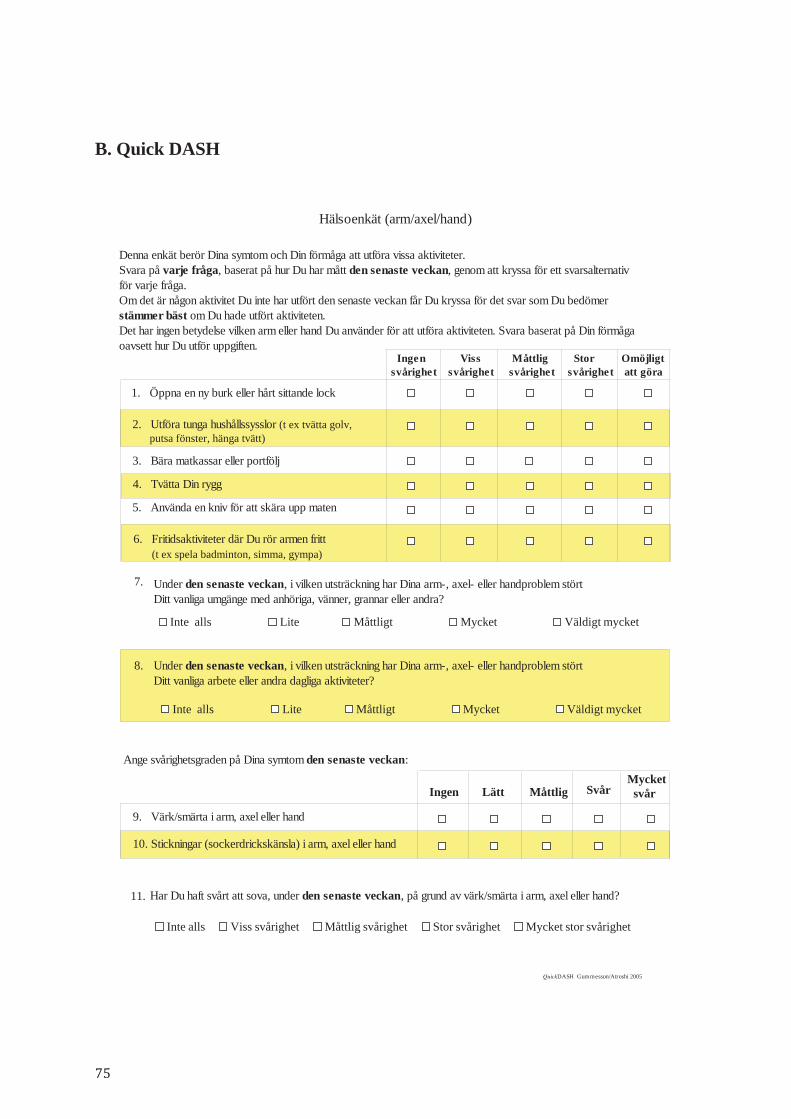
B. Quick DASH
Denna enkät berör Dina symtom och Din förmåga att utföra vissa aktiviteter.Svara på varje fråga, baserat på hur Du har mått den senaste veckan, genom att kryssa för ett svarsalternativför varje fråga.Om det är någon aktivitet Du inte har utfört den senaste veckan får Du kryssa för det svar som Du bedömerstämmer bäst om Du hade utfört aktiviteten.Det har ingen betydelse vilken arm eller hand Du använder för att utföra aktiviteten. Svara baserat på Din förmågaoavsett hur Du utför uppgiften.
Storsvårighet
Visssvårighet
Måttligsvårighet
Omöjligt att göra
1. Öppna en ny burk eller hårt sittande lock
2. Utföra tunga hushållssysslor (t ex tvätta golv, putsa fönster, hänga tvätt)
3. Bära matkassar eller portfölj
4. Tvätta Din rygg
5. Använda en kniv för att skära upp maten
6. Fritidsaktiviteter där Du rör armen fritt (t ex spela badminton, simma, gympa)
Ingensvårighet
QuickDASH Gummesson/Atroshi 2005
Hälsoenkät (arm/axel/hand)
7. Under den senaste veckan, i vilken utsträckning har Dina arm-, axel- eller handproblem störtDitt vanliga umgänge med anhöriga, vänner, grannar eller andra? Inte alls Lite Måttligt Mycket Väldigt mycket
Under den senaste veckan, i vilken utsträckning har Dina arm-, axel- eller handproblem störtDitt vanliga arbete eller andra dagliga aktiviteter?
8.
Inte alls Lite Måttligt Mycket Väldigt mycket
Ange svårighetsgraden på Dina symtom den senaste veckan:
LättIngen Måttlig SvårMycket svår
9. Värk/smärta i arm, axel eller hand
10. Stickningar (sockerdrickskänsla) i arm, axel eller hand
11. Har Du haft svårt att sova, under den senaste veckan, på grund av värk/smärta i arm, axel eller hand?
Inte alls Viss svårighet Måttlig svårighet Stor svårighet Mycket stor svårighet