intramuscular hemangiomas on the masseter muscle and ... · intramuscular hemangioma (imh) is a...
TRANSCRIPT

125
metry occurs in adulthood. Several non-surgical treatments
including cryotherapy, sclerosant injection, and arterial liga-
ture have been described, but complete surgical resection is
the curative intervention7,8. In the case of a large feeding ves-
sel, embolization of the feeding vessels is recommended to
reduce the risk of perioperative and postoperative bleeding9.
We present two rare cases of IMH—one IMH case in the
masseter muscle feeding from the transverse facial artery
(TFA) and a second IMH case in the orbicularis oris muscle
feeding from the superior labial artery.
II. Cases Report
Written informed consent was obtained from the patients
for publication of this case report and any accompanying im-
ages.
1. Case 1
In August 2014, a 48-year-old male visited our clinic with
the chief complaint of swelling of the left buccal area.(Fig.
1) On palpation, a painless and compressible mass was found
I. Introduction
Intramuscular hemangioma (IMH) is a rare vascular dis-
ease involving skeletal muscle, comprising only 0.8% of
all hemangiomas1. About 10% to 15% of IMHs occur in
the head and neck region, mostly involving the masseter
muscle2,3. IMH of the head and neck may be localized mainly
in the masseter muscle (36%), trapezius (24%), periorbital
muscles (12%), sternocleidomastoid muscle (10%), tempora-
lis muscle, or orbicularis oris muscle4. IMH in the orbicularis
oris muscle is very rare5,6.
IMH occurs mostly in childhood. However, it often is not
found until unexpected enlargement, pain, or cosmetic asym-
CASE REPORT
Il-Kyu KimDepartment of Oral and Maxillofacial Surgery, Inha University Hospital, Inha University College of Medicine, 27 Inhang-ro, Jung-gu, Incheon 22332, KoreaTEL: +82-32-890-2470 FAX: +82-32-890-2475E-mail: [email protected]: http://orcid.org/0000-0003-3930-766X
This is an open-access article distributed under the terms of the Creative Commons Attribution Non-Commercial License (http://creativecommons.org/licenses/by-nc/4.0/), which permits unrestricted non-commercial use, distribution, and reproduction in any medium, provided the original work is properly cited.
CC
Intramuscular hemangiomas on the masseter muscle and orbicularis oris muscle: a report of two cases
Il-Kyu Kim1, Ji-Hoon Seo1, Hyun-Young Cho1, Dong-Hwan Lee1, Jun-Min Jang1,
Joon Mee Kim2, In Suh Park2
1Department of Oral and Maxillofacial Surgery, Inha University College of Medicine, 2Department of Pathology, Inha University School of Medicine, Incheon, Korea
Abstract (J Korean Assoc Oral Maxillofac Surg 2017;43:125-133)
Intramuscular hemangioma (IMH) is a rare vascular disease involving skeletal muscle, comprising only 0.8% of hemangiomas. About 10% to 15% of IMHs occur in the head and neck region, mostly involving the masseter muscle. IMH occurs mostly in childhood, but is often not found until unex-pected enlargement, pain, or cosmetic asymmetry occurs in adulthood. Several non-surgical treatments including cryotherapy, sclerosant injection, and arterial ligature have been described, but complete surgical resection is the curative intervention. In this report, we present two rare cases of IMH. One IMH case in a 48-year-old male occurred in the masseter muscle feeding from the transverse facial artery. Embolization of the distal branch of the fa-cial artery was first conducted, and then the buccal mass was removed surgically via the intraoral approach. A second IMH case in a 58-year-old female occurred in the orbicularis oris muscle feeding from the superior labial artery, and the mass was excised surgically without embolization.
Key words: Intramuscular hemangioma, Hemangioma, Vascular tumor, Vascular malformations[paper submitted 2016. 6. 30 / revised 2016. 9. 3 / accepted 2016. 9. 21]
Copyright Ⓒ 2017 The Korean Association of Oral and Maxillofacial Surgeons. All rights reserved.
https://doi.org/10.5125/jkaoms.2017.43.2.125pISSN 2234-7550·eISSN 2234-5930

J Korean Assoc Oral Maxillofac Surg 2017;43:125-133
126
lateral view of angiography, the prosthesis of a dental implant
on the upper left first molar overlapped the mass. The mass
became blushed and then disappeared over time as contrast
agent diffused through the blood vessels.
Based on clinical and radiographic examinations, the pa-
tient was provisionally diagnosed with IMH of the left mas-
seter muscle feeding from the left TFA (main) and maxillary
artery. To reduce the risk of perioperative and postoperative
bleeding, embolization of the distal TFA branch was per-
formed.(Fig. 5) A guiding catheter (diameter of 5 Fr, Enboy;
DePuy Synthes, West Chester, PA, USA) was inserted in the
left ECA, then selection of the distal branch of the TFA was
performed by Prowler select plus (Cordis, Miami, FL, USA).
(Fig. 5. A) An embolizing agent, polyvinyl alcohol (PVA)
particle (contour of 150-200 nm), was injected at slow speed
near the left masseter muscle. There was no wattle sign or
beating, and only bloody discharge was observed in fine
needle aspiration. Immediately following fine needle aspira-
tion, the patient reported increased swelling of the left cheek.
Initial diagnosis was a vascular lesion, such as hemangioma
or vascular malformation.
On magnetic resonance imaging (MRI), a mass about 2.2×
3.3×3.4 cm at the left masseter muscle was strongly enhanced
with heterogeneous T2 hyperintensity.(Fig. 2) There were
multiple intratumoral and peritumoral vascularities visualized
as signal voids. This mass was suggestive of IMH of the left
masseter muscle.
External carotid angiography (ECA) was performed for
further evaluation. A vascular mass feeding from the left
TFA and the maxillary artery was identified.(Fig. 3, 4) In the
A B
Fig. 1. Case 1. Preoperative clini-cal view. Swelling of the patient’s left cheek is shown (arrow). A. Facial view. B. Lateral view.Il-Kyu Kim et al: Intramuscular hemangiomas on the masseter muscle and orbicularis oris muscle: a report of two cases. J Korean Assoc Oral Maxillofac Surg 2017
A B
Fig. 2. Case 1. Preoperative magnetic resonance imaging. A well-marginated mass (arrows) was found in the left masseter muscle. A. Axial view. B. Coronal view.Il-Kyu Kim et al: Intramuscular hemangiomas on the masseter muscle and orbicularis oris muscle: a report of two cases. J Korean Assoc Oral Maxillofac Surg 2017

IMHs on the masseter muscle and orbicularis oris muscle
127
into the selected site. PVA particles are invisible in angiogra-
phy, so the contrast agent was injected locally to the selected
site to verify embolization. Because the pathway was blocked
at the selected site, back pressure was observed intensively
at the backside of the selected site instead of the mass.(Fig.
5. B) To confirm effective embolization, contrast agent was
injected into the ECA again. The mass was faintly visualized
although sufficient time had passed.(Fig. 5. C) That repre-
sents successful blocking of blood supply to the mass. Blood
supply from the maxillary artery was not significant, so em-
bolization of the vessel was not performed.
The following day, there was no change in the strongly
enhancing mass at the left masseter muscle on post-emboli-
zation MRI.(Fig. 6) The swelling of the patient’s left cheek
also remained unchanged. Two days after embolization, the
buccal mass was excised via intraoral approach under general
anesthesia. After insertion of a fine tube to Stensen’s duct for
position marking, mucosal incision anterior to Stensen’s duct
was performed. Local control of bleeding and direct resection
of the mass were achieved.(Fig. 7) Based on biopsy result,
IMH was confirmed.(Fig. 8) After the operation, swelling
A
B
CD
E
F
G
Fig. 3. Case 1. Preoperative external carotid angiography, lateral view. The main mass (‘A’) is shown near the overlapped image of a dental prosthesis (‘B’). It is feeding from the transverse facial ar-tery (‘G’) and maxillary artery (‘E’). (‘A’: main mass, ‘B’: overlapped image of dental prosthesis [maxillary left implant and maxillary right 3-unit bridge], ‘C’: external carotid artery, ‘D’: facial artery, ‘E’: maxillary artery, ‘F’: superficial temporal artery, ‘G’: transverse facial artery [main])Il-Kyu Kim et al: Intramuscular hemangiomas on the masseter muscle and orbicularis oris muscle: a report of two cases. J Korean Assoc Oral Maxillofac Surg 2017
A
B
A B
C D
Fig. 4. Case 1. Preoperative external carotid angiography, lateral view. The mass (‘A’) became blushed by contrast agent injection (A-C), then gradually disappeared with time (D). (‘A’: main mass, ‘B’: overlapped image of dental prosthesis [maxillary left implant and maxillary right 3-unit bridge])Il-Kyu Kim et al: Intramuscular hemangiomas on the masseter muscle and orbicularis oris muscle: a report of two cases. J Korean Assoc Oral Maxillofac Surg 2017
J Korean Assoc Oral Maxillofac Surg 2017;43:125-133
128
and patient discomfort resolved without any significant com-
plications. At the eight month check up, the patient reported
no symptoms and was thereafter lost to follow-up.
2. Case 2
In April 2015, a 58-year-old female visited our clinic. The
patient’s upper lip was swollen in the right angular region.
(Fig. 9) In another dental clinic, she had undergone laser
therapy at that area more than 10 years prior for the same
symptom, but the mass had progressively enlarged. There
was neither wattle sign nor beating.
A 2.7 cm ovoid submucosal mass was well defined on MRI
STIR (short-T1 inversion recovery) and T2 images (Fig. 10),
and the mass was feeding from small branches of the superior
labial artery on angiography.(Fig. 11) The mass was excised
A
B
A B C
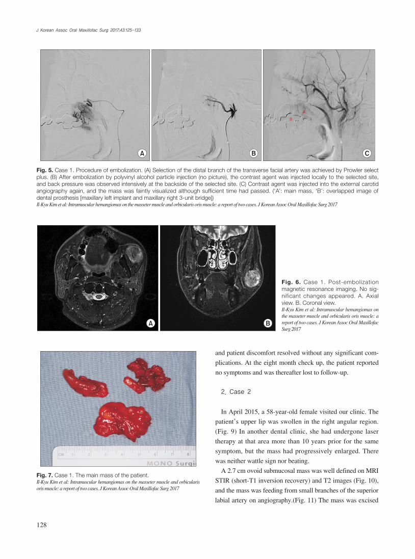
Fig. 5. Case 1. Procedure of embolization. (A) Selection of the distal branch of the transverse facial artery was achieved by Prowler select plus. (B) After embolization by polyvinyl alcohol particle injection (no picture), the contrast agent was injected locally to the selected site, and back pressure was observed intensively at the backside of the selected site. (C) Contrast agent was injected into the external carotid angiography again, and the mass was faintly visualized although sufficient time had passed. (‘A’: main mass, ‘B’: overlapped image of dental prosthesis [maxillary left implant and maxillary right 3-unit bridge])Il-Kyu Kim et al: Intramuscular hemangiomas on the masseter muscle and orbicularis oris muscle: a report of two cases. J Korean Assoc Oral Maxillofac Surg 2017
BA
Fig. 6. Case 1. Post-embolization magnetic resonance imaging. No sig-nificant changes appeared. A. Axial view. B. Coronal view.Il-Kyu Kim et al: Intramuscular hemangiomas on the masseter muscle and orbicularis oris muscle: a report of two cases. J Korean Assoc Oral Maxillofac Surg 2017
Fig. 7. Case 1. The main mass of the patient.Il-Kyu Kim et al: Intramuscular hemangiomas on the masseter muscle and orbicularis oris muscle: a report of two cases. J Korean Assoc Oral Maxillofac Surg 2017

IMHs on the masseter muscle and orbicularis oris muscle
129
A B
C D
Fig. 8. Case 1. Microscopic view of the mass. A. Developed vessels with proliferation of endothelial cells between skeletal muscle tissue can be identified (H&E staining, ×100). B. Capillaries are predominant, and large vessels are seen occasionally (H&E staining, ×200). Many adipose tissues can be observed (C: H&E staining, ×40), and large vessels (possibly from feeding vessels) are also observed within the mass (D: H&E staining, ×40).Il-Kyu Kim et al: Intramuscular hemangiomas on the masseter muscle and orbicularis oris muscle: a report of two cases. J Korean Assoc Oral Maxillofac Surg 2017
Fig. 9. Case 2. Preoperative clinical view of the patient. The patient’s upper lip is shown with swelling in the right angular region.Il-Kyu Kim et al: Intramuscular hemangiomas on the masseter muscle and orbicularis oris muscle: a report of two cases. J Korean Assoc Oral Maxillofac Surg 2017

J Korean Assoc Oral Maxillofac Surg 2017;43:125-133
130
hemangiomas versus vascular malformations. In 1992, the
International Society for the Study of Vascular Anomalies
adopted Mulliken and Glowacki’s classification system and
modified the classification of vascular malformations versus
vascular tumors10.
Vascular malformations are present at birth and enlarge in
proportion to the growth of the child11. They are subcatego-
rized as slow-flow vascular malformations (capillary malfor-
mation, venous malformation, and lymphatic malformation),
fast-flow vascular malformations (including arteriovenous
malformation), and complex-combined vascular malforma-
tions. They radiographically show cystic or dysplastic ves-
sels, usually involving multiple tissue planes12. Phlebolith
(especially in venous malformation on computed tomography
[CT]) and signal void (especially in arteriovenous malforma-
tion on MRI) are common features. Histologically, vascular
malformations have quiescent endothelium. They do not in-
volute and persist throughout life10,14. There is often a wattle
sign, beating, and thrilling in vascular malformations.
On the other hand, vascular tumors are true neoplasms
with cellular hyperplasia and include infantile hemangioma,
congenital hemangioma, and hemangioendothelioma. On
radiographic images, they show a well-defined mass in the
majority of patients13. Arteriovenous shunting and phlebolith
are unusual in vascular tumors. Histologically, they grow by
endothelial proliferation, and tumors can grow, regress, or
persist. Adipose tissue can be observed, because regressed tu-
mors are replaced by adipose tissue10,14. Wattle sign, beating,
and thrilling are uncommon in vascular tumor.
IMH is a special form of hemangioma involving skeletal
muscle and comprises only 0.8% of all hemangiomas1. About
10% to 15% of IMHs occur in the head and neck region,
mostly involving the masseter muscle2,3. IMH has developed
under general anesthesia without embolization. Via the in-
traoral approach, mucosal incision on the buccal mucosa and
undermining of the mass were conducted under local bleed-
ing control. IMH was confirmed on biopsy.(Fig. 12, 13)
III. Discussion
The term “hemangioma” has been applied to a wide vari-
ety of vascular lesions and continues to be used to describe
vascular malformations. Several authors have recently sug-
gested that vascular malformations should be distinguished
from hemangiomas10-13. In 1982, Mulliken and Glowacki pro-
posed a simple classification system of vascular anomalies—
Fig. 10. Case 2. Preoperative mag-netic resonance imaging (MRI). A well-marginated mass was found in the right upper lip on MRI STIR (short-T1 inversion recovery) axial images (A) and T2 coronal images (B).Il-Kyu Kim et al: Intramuscular hemangiomas on the masseter muscle and orbicularis oris muscle: a report of two cases. J Korean Assoc Oral Maxillofac Surg 2017
A B
A
B
C
D
Fig. 11. Case 2. Preoperative angiography of the patient. The mass was feeding from small branches of the superior labial artery. (‘A’: external carotid artery, ‘B’: facial artery, ‘C’: superior labial artery, ‘D’: main mass)Il-Kyu Kim et al: Intramuscular hemangiomas on the masseter muscle and orbicularis oris muscle: a report of two cases. J Korean Assoc Oral Maxillofac Surg 2017

IMHs on the masseter muscle and orbicularis oris muscle
131
vessels embedded within muscle tissue usually in the deep
layer. They therefore have somewhat different characters
from other types of hemangiomas.
Due to the rarity of these tumors and their deep location
and unfamiliar presentation, inaccurate diagnosis, inap-
propriate excision, and unnecessary risk to the facial nerve
occur when IMH is present in the face4. Over 90% of all
IMHs are misdiagnosed15. Differential diagnosis of IMH in-
cludes lymphangioma, lymphomas, rhabdomyosarcoma, and
schwannomas9. In particular, IMH arising from the masseter
muscle is frequently confused with parotid tumor7,15.
IMH occurs mostly in childhood. However, it is often not
found until unexpected enlargement, pain, or cosmetic asym-
metry occurs in adulthood. Trigger factors include hormonal
change, infection, or trauma.
In 1972, Allen and Enzinger16 retrospectively reviewed
the histopathological findings of a large group of patients
described as having IMH. They classified these patients into
three categories based on vessel size and histological fea-
tures: small-vessel type (capillary type, less than 140 μm),
large-vessel type (cavernous type, more than 140 μm), and
mixed-vessel type. However, Yilmaz et al.13 argued that the
description of the small-vessel type was identical to that of
intramuscular capillary-type hemangioma, while the descrip-
tion of large-vessel type was more consistent with what is
now designated as venous malformation. Capillary-type IMH
accounts for 50% of all IMH, and 68% of these tumors are in
the head and neck4.
To diagnosis IMH, ultrasound and CT are used along
with clinical examination. MRI is considered the gold stan-
dard10-12,15 and typically shows a well-marginated soft tissue
mass. On T2 and STIR images, lesions appear homogeneous
and moderately hyperintense during the proliferative phase
and increased heterogeneity during the involution phase ow-
ing to areas of fat replacement. Flow voids may be seen at
the periphery of the lesion on spin-echo sequences, reflecting
high-flow arterial feeders10.
Angiography is useful for diagnosis of arteriovenous mal-
formation and is not routinely performed for diagnosis of
hemangioma. However, angiography may be helpful when
the feeding artery is large. Fine needle aspiration is not rec-
A B
Fig. 12. Case 2. Microscopic view of the mass of the patient. Large vessels (may be from feeding vessels) are observed in the lateral side of the mass (A: H&E staining, ×40), and developed vessels with proliferation of endothelial cells between skeletal muscle tissues can be identified (B: H&E staining, ×100).Il-Kyu Kim et al: Intramuscular hemangiomas on the masseter muscle and orbicularis oris muscle: a report of two cases. J Korean Assoc Oral Maxillofac Surg 2017
Fig. 13. Case 2. Immunohistochemical view of the mass. The en-dothelial cells are positive for CD34 (×100).Il-Kyu Kim et al: Intramuscular hemangiomas on the masseter muscle and orbicularis oris muscle: a report of two cases. J Korean Assoc Oral Maxillofac Surg 2017

J Korean Assoc Oral Maxillofac Surg 2017;43:125-133
132
ommended due to the risk of hemorrhage9.
Non-surgical treatments such as cryotherapy, sclerosant
injection, embolization, and arterial ligature have been de-
scribed, but there is dispute about the results of these treat-
ments. Therefore, non-surgical treatments are currently rec-
ommended only when surgery is contraindicated or refused7.
Complete surgical resection is the curative intervention. For
intramasseteric cases, the intraoral approach for surgical re-
section may be useful as it avoids any visible scar or facial
nerve dissection7,8.
Localized recurrence rate ranges from 9% to 28%, despite
wide resection of the tumor due to the absence of capsule
surrounding the tumor2. Minor feeding vessels and residual
tumor may be responsible for the recurrence rate16.
In case 1, there was no wattle sign or beating palpation.
Phlebolith was not found. A mass with well-defined border,
strongly enhanced with heterogeneous T2 hyperintensity
was found on MRI.(Fig. 2) On angiography, a vascular mass
feeding from the left TFA (main) and the maxillary artery
was found.(Fig. 3) TFA is a branch arising from the superfi-
cial temporal artery, and it anastomoses with the facial artery,
mesenteric artery, infraorbital artery, and others17. No re-
markable changes were found by preoperative embolization
of TFA (Fig. 5), and the mass was surgical excised by intra-
oral approach two days later.(Fig. 7) In case 2, there was a
similar radiographic exam (Fig. 10, 11), and there was wattle
sign or beating.
On microscopic exam of case 1, well developed vessels
with proliferation of endothelial cells between skeletal muscle
tissue were found.(Fig. 8. A) Capillaries were predominant,
and large vessels were seen occasionally.(Fig. 8. B) A large
amount of adipose tissue was observed because regressed tu-
mors are replaced by adipose tissue.(Fig. 8. C) Larger vessels
(possibly from feeding vessels) were also observed within the
mass, but there was no obvious arteriovenous shunting.(Fig. 8.
D) This tumor was therefore considered mixed-type IMH.
In case 2, large vessels (possibly from feeding vessels)
were observed in the lateral side of the mass (Fig. 12. A),
and developed vessels between skeletal muscle tissues were
found.(Fig. 12. B) However, there was only a small amount
of adipose tissue in this case. Immunohistochemically, endo-
thelial cells were positive for CD34 (Fig. 13), an antigen pro-
tein that shows expression on immature hematopoietic cells
and endothelial cells. This case was therefore considered
IMH.
In conclusion, IMHs can be diagnosed using MRI, angiog-
raphy, and excisional biopsy. Preoperative embolization and
surgical excision are recommended. In these two rare cases
of IMH in masseter and orbicularis oris muscle, the intraoral
approach provided a direct approach and allowed removal of
the tumor without any visible scar or facial nerve dissection.
Conflict of Interest
No potential conflict of interest relevant to this article was
reported.
Acknowledgements
Authors’ contributions: IKK carried out the operation and
related treatment, contribution of conception of the report,
HYC and JHS carried out the operation and critical revising.
JHS, DHL, and JMJ participated in the treatment, collection
of data and drafting the manuscript. JMK, ISP carried out
histological examination and contribution of the histological
opinion. All authors read and approved the final manuscript.
ORCID
Il-Kyu Kim, http://orcid.org/0000-0003-3930-766XJi-Hoon Seo, http://orcid.org/0000-0003-3618-6380Hyun-Young Cho, http://orcid.org/0000-0003-3055-0591Dong-Hwan Lee, http://orcid.org/0000-0002-1091-2543Jun-Min Jang, http://orcid.org/0000-0002-2484-2718Joon Mee Kim, http://orcid.org/0000-0003-1355-4187In Suh Park, http://orcid.org/0000-0002-9415-2923
References
1. Watson WL, McCarthy WD. Blood and lymph vessel tumors. Srug Gynecol Obstet 1940;71:569-88.
2. Wolf GT, Daniel F, Krause CJ, Kaufman RS. Intramuscular hem-angioma of the head and neck. Laryngoscope 1985;95:210-3.
3. Gordon JS, Mandel L. Masseteric intramuscular hemangioma: case report. J Oral Maxillofac Surg 2014;72:2192-6.
4. Odabasi AO, Metin KK, Mutlu C, Başak S, Erpek G. Intramuscular hemangioma of the masseter muscle. Eur Arch Otorhinolaryngol 1999;256:366-9.
5. Chan MJ, McLean NR, Soames JV. Intramuscular haemangioma of the orbicularis oris muscle. Br J Oral Maxillofac Surg 1992;30:192-4.
6. Kinni ME, Webb RI, Christensen RE. Intramuscular heman-gioma of the orbicularis oris muscle: report of case. J Oral Surg 1981;39:780-2.
7. Righini CA, Berta E, Atallah I. Intramuscular cavernous heman-gioma arising from the masseter muscle. Eur Ann Otorhinolaryngol Head Neck Dis 2014;131:57-9.
8. Ichimura K, Nibu K, Tanaka T. Essentials of surgical treatment for intramasseteric hemangioma. Eur Arch Otorhinolaryngol 1995;252:125-9.

IMHs on the masseter muscle and orbicularis oris muscle
133
9. Righi S, Boffano P, Malvè L, Rossi P, Zanardi F, Pateras D. Intra-mural perimasseteric hemangiomas of the inner cheek. J Craniofac Surg 2015;26:959-60.
10. Chaudry MI, Manzoor MU, Turner RD, Turk AS. Diagnostic imag-ing of vascular anomalies. Facial Plast Surg 2012;28:563-74.
11. Donnelly LF, Adams DM, Bisset GS 3rd. Vascular malformations and hemangiomas: a practical approach in a multidisciplinary clinic. AJR Am J Roentgenol 2000;174:597-608.
12. Flors L, Leiva-Salinas C, Maged IM, Norton PT, Matsumoto AH, Angle JF, et al. MR imaging of soft-tissue vascular malformations: diagnosis, classification, and therapy follow-up. Radiographics 2011;31:1321-40.
13. Yilmaz S, Kozakewich HP, Alomari AI, Fishman SJ, Mulliken JB,
Chaudry G. Intramuscular capillary-type hemangioma: radiologic-pathologic correlation. Pediatr Radiol 2014;44:558-65.
14. Rosbe KW, Hess CP, Dowd CF, Frieden IJ. Masseteric venous mal-formations: diagnosis, treatment, and outcomes. Otolaryngol Head Neck Surg 2010;143:779-83.
15. Lee SK, Kwon SY. Intramuscular cavernous hemangioma arising from masseter muscle: a diagnostic dilemma (2006: 12b). Eur Ra-diol 2007;17:854-7.
16. Allen PW, Enzinger FM. Hemangioma of skeletal muscle. An analysis of 89 cases. Cancer 1972;29:8-22.
17. Yang HJ, Gil YC, Lee HY. Topographical anatomy of the trans-verse facial artery. Clin Anat 2010;23:168-78.