imaging findings of central nervous system …...with cns vasculitis. in our case, the diagnosis of...
TRANSCRIPT
Korean J Radiol 8(6), December 2007 545
Imaging Findings of Central NervousSystem Vasculitis Associated withGoodpasture’s Syndrome: a Case Report
Glomerulonephritis and pulmonary hemorrhage are features of Goodpasture’ssyndrome. Goodpasture’s syndrome accompanied with central nervous system(CNS) vasculitis is extremely rare. Herein, we report a rare case of CNS vasculitisassociated with Goodpasture’s syndrome in a 34-year-old man, who presentedwith a seizure and sudden onset of right sided weakness. He also had recurrenthemoptysis of one month’s duration. Goodpasture’s syndrome is histologicallydiagnosed by intense linear deposits of IgG along the glomerular basement mem-brane in both renal and lung tissues.
oodpasture’s syndrome is a rare disease, characterized by rapidlyprogressive glomerulonephritis, diffuse pulmonary hemorrhage andcirculating antiglomerular basement membrane antibody (anti-GBM
antibody). Central nervous system (CNS) manifestations in Goodpasture’s syndromeare extremely rare, with only a few cases having been reported in the literature (810). Therefore, we present our imaging findings of CNS vasculitis associated withGoodpasture’s syndrome, together with a review of the relevant literature.
CASE REPORT
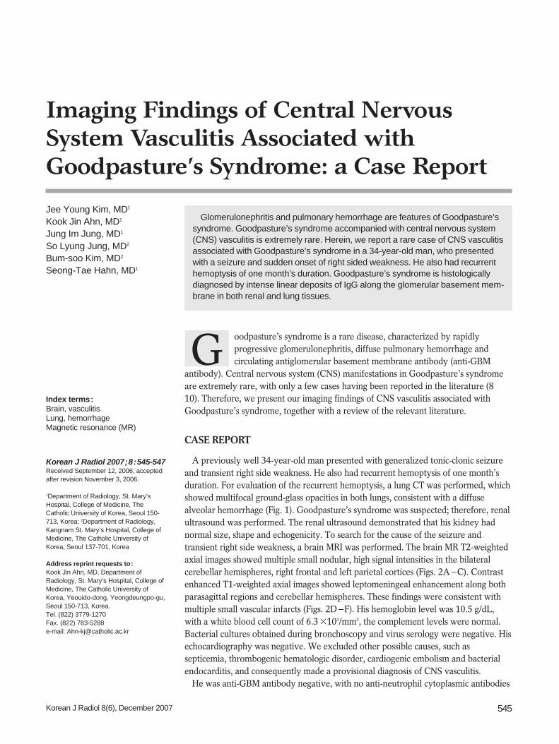
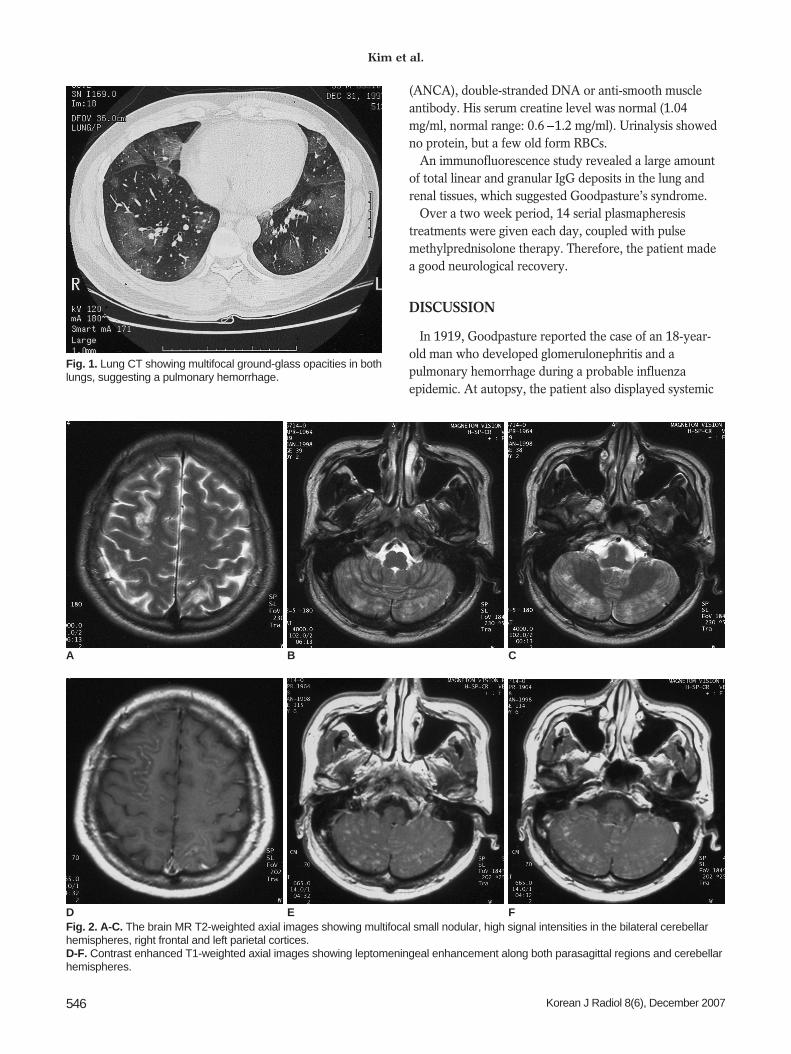
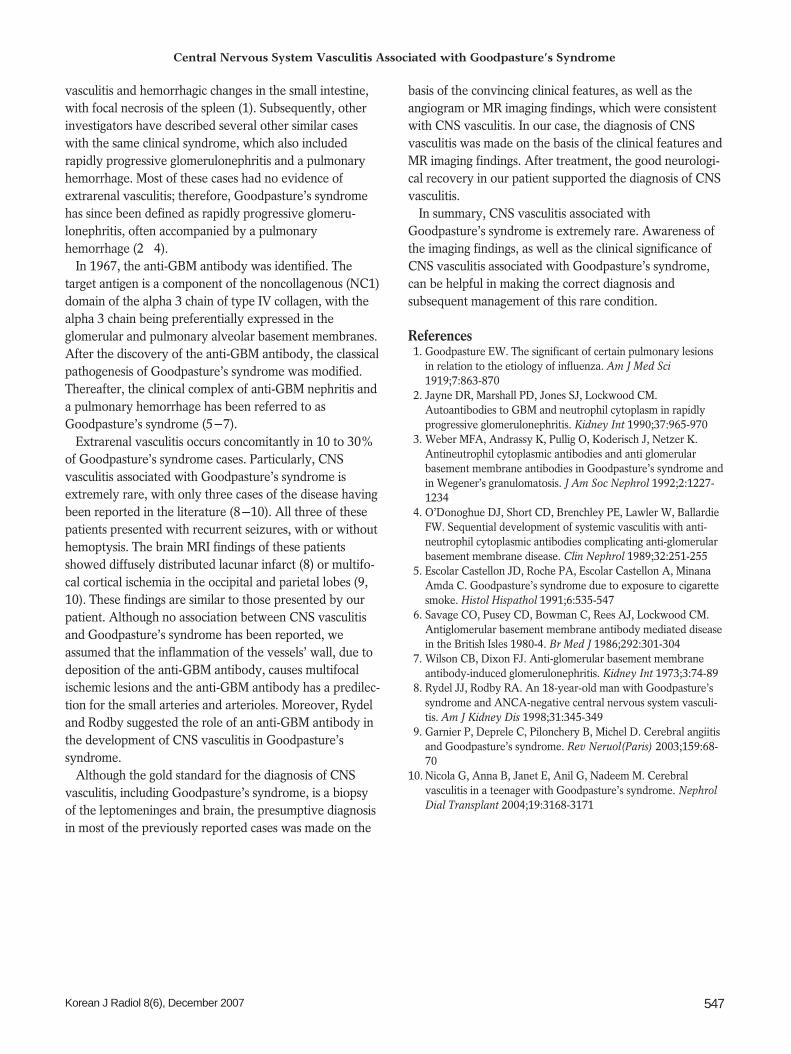
A previously well 34-year-old man presented with generalized tonic-clonic seizureand transient right side weakness. He also had recurrent hemoptysis of one month’sduration. For evaluation of the recurrent hemoptysis, a lung CT was performed, whichshowed multifocal ground-glass opacities in both lungs, consistent with a diffusealveolar hemorrhage (Fig. 1). Goodpasture’s syndrome was suspected; therefore, renalultrasound was performed. The renal ultrasound demonstrated that his kidney hadnormal size, shape and echogenicity. To search for the cause of the seizure andtransient right side weakness, a brain MRI was performed. The brain MR T2-weightedaxial images showed multiple small nodular, high signal intensities in the bilateralcerebellar hemispheres, right frontal and left parietal cortices (Figs. 2A C). Contrastenhanced T1-weighted axial images showed leptomeningeal enhancement along bothparasagittal regions and cerebellar hemispheres. These findings were consistent withmultiple small vascular infarcts (Figs. 2D F). His hemoglobin level was 10.5 g/dL,with a white blood cell count of 6.3 103/mm3, the complement levels were normal.Bacterial cultures obtained during bronchoscopy and virus serology were negative. Hisechocardiography was negative. We excluded other possible causes, such assepticemia, thrombogenic hematologic disorder, cardiogenic embolism and bacterialendocarditis, and consequently made a provisional diagnosis of CNS vasculitis.
He was anti-GBM antibody negative, with no anti-neutrophil cytoplasmic antibodies
Jee Young Kim, MD1
Kook Jin Ahn, MD1
Jung Im Jung, MD1
So Lyung Jung, MD2
Bum-soo Kim, MD2
Seong-Tae Hahn, MD1
Index terms:Brain, vasculitisLung, hemorrhageMagnetic resonance (MR)
Korean J Radiol 2007;8:545-547Received September 12, 2006; accepted after revision November 3, 2006.
1Department of Radiology, St. Mary’sHospital, College of Medicine, TheCatholic University of Korea, Seoul 150-713, Korea; 2Department of Radiology,Kangnam St. Mary’s Hospital, College ofMedicine, The Catholic University ofKorea, Seoul 137-701, Korea
Address reprint requests to:Kook Jin Ahn, MD, Department ofRadiology, St. Mary’s Hospital, College ofMedicine, The Catholic University ofKorea, Yeouido-dong, Yeongdeungpo-gu,Seoul 150-713, Korea.Tel. (822) 3779-1270 Fax. (822) 783-5288e-mail: [email protected]
G
(ANCA), double-stranded DNA or anti-smooth muscleantibody. His serum creatine level was normal (1.04mg/ml, normal range: 0.6 1.2 mg/ml). Urinalysis showedno protein, but a few old form RBCs.
An immunofluorescence study revealed a large amountof total linear and granular IgG deposits in the lung andrenal tissues, which suggested Goodpasture’s syndrome.
Over a two week period, 14 serial plasmapheresistreatments were given each day, coupled with pulsemethylprednisolone therapy. Therefore, the patient madea good neurological recovery.
DISCUSSION
In 1919, Goodpasture reported the case of an 18-year-old man who developed glomerulonephritis and apulmonary hemorrhage during a probable influenzaepidemic. At autopsy, the patient also displayed systemic
Kim et al.
546 Korean J Radiol 8(6), December 2007
Fig. 1. Lung CT showing multifocal ground-glass opacities in bothlungs, suggesting a pulmonary hemorrhage.
A B C
Fig. 2. A-C. The brain MR T2-weighted axial images showing multifocal small nodular, high signal intensities in the bilateral cerebellarhemispheres, right frontal and left parietal cortices.D-F. Contrast enhanced T1-weighted axial images showing leptomeningeal enhancement along both parasagittal regions and cerebellarhemispheres.
D E F
vasculitis and hemorrhagic changes in the small intestine,with focal necrosis of the spleen (1). Subsequently, otherinvestigators have described several other similar caseswith the same clinical syndrome, which also includedrapidly progressive glomerulonephritis and a pulmonaryhemorrhage. Most of these cases had no evidence ofextrarenal vasculitis; therefore, Goodpasture’s syndromehas since been defined as rapidly progressive glomeru-lonephritis, often accompanied by a pulmonaryhemorrhage (2 4).
In 1967, the anti-GBM antibody was identified. Thetarget antigen is a component of the noncollagenous (NC1)domain of the alpha 3 chain of type IV collagen, with thealpha 3 chain being preferentially expressed in theglomerular and pulmonary alveolar basement membranes.After the discovery of the anti-GBM antibody, the classicalpathogenesis of Goodpasture’s syndrome was modified.Thereafter, the clinical complex of anti-GBM nephritis anda pulmonary hemorrhage has been referred to asGoodpasture’s syndrome (5 7).
Extrarenal vasculitis occurs concomitantly in 10 to 30%of Goodpasture’s syndrome cases. Particularly, CNSvasculitis associated with Goodpasture’s syndrome isextremely rare, with only three cases of the disease havingbeen reported in the literature (8 10). All three of thesepatients presented with recurrent seizures, with or withouthemoptysis. The brain MRI findings of these patientsshowed diffusely distributed lacunar infarct (8) or multifo-cal cortical ischemia in the occipital and parietal lobes (9,10). These findings are similar to those presented by ourpatient. Although no association between CNS vasculitisand Goodpasture’s syndrome has been reported, weassumed that the inflammation of the vessels’ wall, due todeposition of the anti-GBM antibody, causes multifocalischemic lesions and the anti-GBM antibody has a predilec-tion for the small arteries and arterioles. Moreover, Rydeland Rodby suggested the role of an anti-GBM antibody inthe development of CNS vasculitis in Goodpasture’ssyndrome.
Although the gold standard for the diagnosis of CNSvasculitis, including Goodpasture’s syndrome, is a biopsyof the leptomeninges and brain, the presumptive diagnosisin most of the previously reported cases was made on the
basis of the convincing clinical features, as well as theangiogram or MR imaging findings, which were consistentwith CNS vasculitis. In our case, the diagnosis of CNSvasculitis was made on the basis of the clinical features andMR imaging findings. After treatment, the good neurologi-cal recovery in our patient supported the diagnosis of CNSvasculitis.
In summary, CNS vasculitis associated withGoodpasture’s syndrome is extremely rare. Awareness ofthe imaging findings, as well as the clinical significance ofCNS vasculitis associated with Goodpasture’s syndrome,can be helpful in making the correct diagnosis andsubsequent management of this rare condition.
References1. Goodpasture EW. The significant of certain pulmonary lesions
in relation to the etiology of influenza. Am J Med Sci1919;7:863-870
2. Jayne DR, Marshall PD, Jones SJ, Lockwood CM.Autoantibodies to GBM and neutrophil cytoplasm in rapidlyprogressive glomerulonephritis. Kidney Int 1990;37:965-970
3. Weber MFA, Andrassy K, Pullig O, Koderisch J, Netzer K.Antineutrophil cytoplasmic antibodies and anti glomerularbasement membrane antibodies in Goodpasture’s syndrome andin Wegener’s granulomatosis. J Am Soc Nephrol 1992;2:1227-1234
4. O’Donoghue DJ, Short CD, Brenchley PE, Lawler W, BallardieFW. Sequential development of systemic vasculitis with anti-neutrophil cytoplasmic antibodies complicating anti-glomerularbasement membrane disease. Clin Nephrol 1989;32:251-255
5. Escolar Castellon JD, Roche PA, Escolar Castellon A, MinanaAmda C. Goodpasture’s syndrome due to exposure to cigarettesmoke. Histol Hispathol 1991;6:535-547
6. Savage CO, Pusey CD, Bowman C, Rees AJ, Lockwood CM.Antiglomerular basement membrane antibody mediated diseasein the British Isles 1980-4. Br Med J 1986;292:301-304
7. Wilson CB, Dixon FJ. Anti-glomerular basement membraneantibody-induced glomerulonephritis. Kidney Int 1973;3:74-89
8. Rydel JJ, Rodby RA. An 18-year-old man with Goodpasture’ssyndrome and ANCA-negative central nervous system vasculi-tis. Am J Kidney Dis 1998;31:345-349
9. Garnier P, Deprele C, Pilonchery B, Michel D. Cerebral angiitisand Goodpasture’s syndrome. Rev Neruol(Paris) 2003;159:68-70
10. Nicola G, Anna B, Janet E, Anil G, Nadeem M. Cerebralvasculitis in a teenager with Goodpasture’s syndrome. NephrolDial Transplant 2004;19:3168-3171
Central Nervous System Vasculitis Associated with Goodpasture’s Syndrome
Korean J Radiol 8(6), December 2007 547