effect of bottle height on the corneal endothelium during phacoemulsification
TRANSCRIPT
Effect of bottle height on the corneal endotheliumduring phacoemulsification
Hisaharu Suzuki, MD, PhD, Kotaro Oki, MD, PhD, Toshihiko Shiwa, MD, PhD,Hideaki Oharazawa, MD, PhD, Hiroshi Takahashi, MD, PhD
PURPOSE: To directly measure intraocular pressure (IOP) in simulated phacoemulsification and toassess the usefulness of lowering the bottle height in protecting the corneal endothelium in clinicalphacoemulsification.
SETTING: Nippon Medical School Hospital, Tokyo, Japan.
METHODS: Simulated phacoemulsification was performed in porcine eyes with 2 bottle heights,65.0 cm (BH 65 group) and 19.0 cm (BH 19 group). The IOP was continuously measured witha microprobe. In a clinical study, phacoemulsification was performed with a bottle height of60.0 cm (BH 60 group) and of 30.0 cm (BH 30 group). One day, 1 week, and 1 and 3 months aftersurgery, cell density and corneal volumes were measured using specular microscopy and rotatingScheimpflug photography, respectively.
RESULTS: In the simulation study, IOP fluctuated between 50 mm Hg and 60 mm Hg in the BH 65group and between 20 mm Hg and 30 mm Hg in the BH 19 group. In the clinical study of 31 eyes, therate of cell density decrease was significantly lower in the BH 30 group than in the BH 60 group at alltime points. The rate of increase in corneal volume was significantly lower in the BH 30 group than inthe BH 60 group at 1 month.
CONCLUSIONS: Intraoperative IOP in phacoemulsification with a usual bottle height appeared toexceed the normal range. Phacoemulsification with a low bottle height was less harmful to thecorneal endothelium.
J Cataract Refract Surg 2009; 35:2014–2017 Q 2009 ASCRS and ESCRS
LABORATORY SCIENCE
The corneal endothelium can be damaged during pha-coemulsification by factors such as excessive durationof phacoemulsification1–3; localized temperature in-creases4; or damage from lens nucleus fragmentscaused by the turbulent flow of the irrigating solu-tion,5,6 air bubbles,7,8 or free radicals associated withultrasound oscillation.9–13 However, little attention
Submitted: March 23, 2009.Final revision submitted: May 15, 2009.Accepted: May 17, 2009.
From the Department of Ophthalmology, Nippon Medical School,Tokyo, Japan.
No author has a financial or proprietary interest in any material ormethod mentioned.
Corresponding author: Hiroshi Takahashi, MD, PhD, Department ofOphthalmology, Nippon Medical School, 1-1-5 Sendagi, Bunkyo-ku, Tokyo 113-8602, Japan. E-mail: [email protected].
Q 2009 ASCRS and ESCRS
Published by Elsevier Inc.
2014
has been paid to intraocular pressure (IOP) during sur-gery as a cause of corneal endothelial damage. Intrao-perative IOP is determined by a balance between theirrigating (influx) pressure, which depends on theheight of the irrigating bottle, and the outflow volume,which depends on the vacuum pressure, aspirationrate, and fluid leakage from the incision cite. Thus,the intraoperative IOP at one moment can be consid-ered an instantaneous measure of the fluid flow intothe anterior chamber.
Although turbulent flow of the irrigating solution isa known cause of corneal endothelial damage,5,6 it hasbeen discussed mainly in terms of the lens nucleuscolliding with the corneal endothelium. The effect ofelevated IOP or alteration of intraoperative IOP onthe corneal endothelium has not been fully elucidated.In this study, we directly measured intraoperativeIOP in simulated phacoemulsification in animaleyes and evaluated the effect of lowering the bottleheight on corneal endothelial damage in clinicalphacoemulsification.
0886-3350/09/$dsee front matter
doi:10.1016/j.jcrs.2009.05.057
2015LABORATORY SCIENCE: EFFECT OF BOTTLE HEIGHT ON THE CORNEAL ENDOTHELIUM
MATERIALS AND METHODS
Simulation Study
Phacoemulsification was performed in porcine eyes ob-tained from a local abattoir using a Sovereign WhiteStar sys-tem (Abbott Medical Optics). The surgery was performedvia a superior 2.4mmsclerocorneal incisionwith an aspirationrate of 22mL/minandavacuumpressure of 150mmHg. Twobottle height settings, 65.0 cm (BH 65 group) and 19.0 cm (BH19group),wereused. Because thebottle height value that is setanddisplayedby thephacodevice isnot the truedistance fromthe eye but is the distance from a certain point of the device tothebottle, actualdistances fromtheeye to thebottle (truebottleheight) were measured and found to be 88.0 cm in the BH 65group and 40.5 cm in the BH 19 group. Intraoperative IOPwas measured using a universal pressure meter (DPM-2Plus, Fluke Corp.). The microprobe of the device was insertedfrom the 6 o’clock limbus into the anterior chamber, and theIOP was recorded continuously during the procedure.
Clinical Study
The clinical study included patients who had phacoemul-sification and intraocular lens implantation over a 3-monthperiod starting in November 2005 at NipponMedical SchoolHospital. In all cases, lens nucleus hardness was approxi-mately 2 according to the Emery-Little classification.
The same surgeon (H.S.) operated on all patients using thesame type of phaco systemused in the simulation study. Pha-coemulsification was performed through a superior sclero-corneal incision using sodium hyaluronate 2.3% (Healon5),an ultrasound power output of 20%, and vacuum pressureof 150 mm Hg. Two bottle height settings, 60.0 cm (BH 60group) and 30.0 cm (BH 30 group), were used; both bottleheightsweredisplayedon thedevice. These settingsweredif-ferent from those in the simulation study. The reason is thata bottle height of 19.0 cm was considered too low and therewas concern that it may cause anterior chamber collapse inclinical phacoemulsification; in addition, the 60.0 cm heightis the standard setting used at the hospital where the surgerywas performed. The aspiration rate was set at 20 mL/min inthe BH 60 group and 18 mL/min in the BH 30 group to pre-vent anterior chamber collapse.
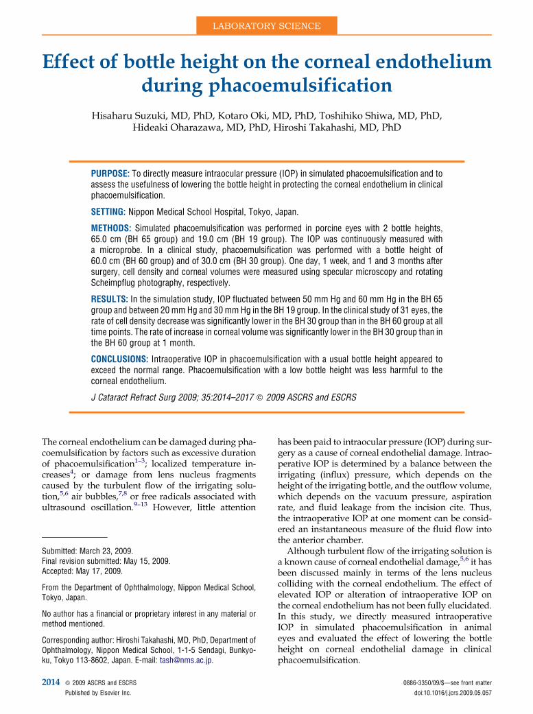
Figure 1. Intraoperative IOP in the BH 65 and BH 19 groups. In theBH 65 group, intraoperative IOP oscillated between 50 mm Hgand 60 mm Hg and in the BH 19 group, between 20 mm Hgand 30 mm Hg (BH19 Z bottle height at 19.0 cm; BH65 Z bottleheight at 65.0 cm; IOP Z intraocular pressure).
J CATARACT REFRACT SURG
A specular microscope (Noncon Robo, Konan Medical)and a rotating Scheimpflug camera (Pentacam, Oculus)were used to analyze the cornea before surgery and 1 day,1 week, and 1 and 3 months after surgery. Specular micros-copy was performed in the central cornea to measure celldensity and to analyze polymegathism (coefficient of varia-tion) and pleomorphism (hexagonal cell ratio 6A) using thecenter method. The Scheimpflug camera was used to calcu-late the corneal volume within 3.0 and 10.0 mm circles inthe central cornea.14,15
Statistical analysis was performed using the unpaired Stu-dent t test, F test, 1-way analysis of variance (ANOVA), orDunnett test, when appropriate.
RESULTS
Simulation Study
The intraoperative IOP fluctuated in an oscillatingfashion (Figure 1). The IOP in the BH 65 group(mean 52.72 G 7.94 mm Hg) was statistically signifi-cantly higher than in the BH 19 group (mean 21.96G 4.98 mm Hg) (P!.05, Student t test). Moreover,the range between the maximum IOP and minimumIOP was significantly greater in the BH 65 groupthan in the BH 19 group (P!.05, F test).
Clinical Study
The clinical study comprised 63 patients (63 eyes);there were 31 patients in the BH 60 group and 32 pa-tients in the BH 30 group. In all cases, the surgerylasted fewer than 20 minutes and the effective phacotime was generally fewer than 10 seconds.
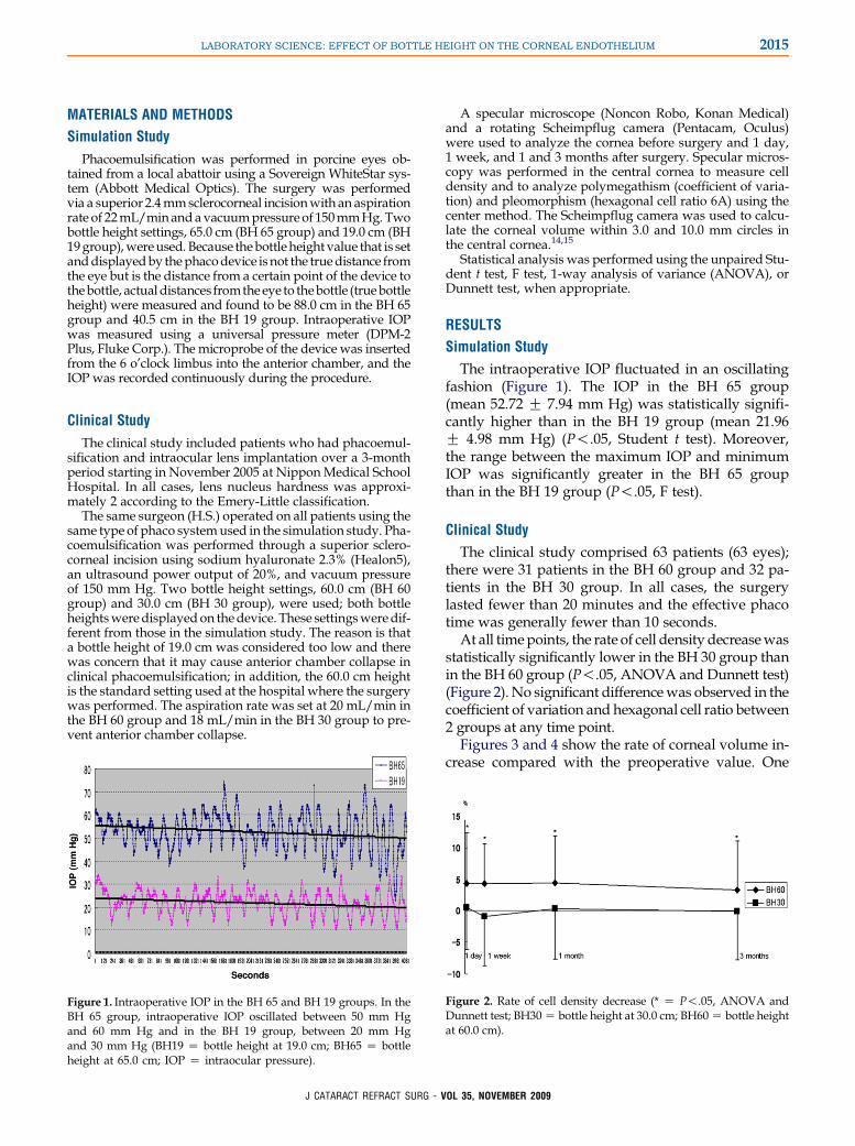
At all timepoints, the rate of cell density decreasewasstatistically significantly lower in the BH 30 group thanin the BH 60 group (P!.05, ANOVA and Dunnett test)(Figure 2). No significant differencewas observed in thecoefficient of variation and hexagonal cell ratio between2 groups at any time point.
Figures 3 and 4 show the rate of corneal volume in-crease compared with the preoperative value. One
Figure 2. Rate of cell density decrease (* Z P!.05, ANOVA andDunnett test; BH30 Z bottle height at 30.0 cm; BH60 Z bottle heightat 60.0 cm).
- VOL 35, NOVEMBER 2009

2016 LABORATORY SCIENCE: EFFECT OF BOTTLE HEIGHT ON THE CORNEAL ENDOTHELIUM
month after surgery, the corneal volumes within the3.0 mm and 10.0 mm circles were significantly smallerin the BH 30 group than in the BH 60 group (P!.05,ANOVA and Dunnett test).
DISCUSSION
When the caliber or the resistance of the irrigating tubeis not taken into consideration, the irrigating pressurefor the eye can be roughly estimated by the followingformula:
Irrigating pressureZTBHðcmÞ�10=13:5 ðz0:74Þ mmHg
where TBH is the true bottle height and 13.5 is thespecific gravity of mercury. Using animal eyes, we di-rectly measured IOP during simulated phacoemulsifi-cation under 2 bottle heights, 65.0 cm (BH 65 group)and 19.0 cm (BH 19 group). The true bottle heightwas 88.0 cm in the BH 65 group and 40.5 cm in theBH 19 group. Thus, the rough estimate of irrigatingpressure is 65 (88 � 0.74) mm Hg in the BH 65 groupand 30 (40.5 � 0.74) mm Hg in the BH 19 group. Re-sults of measurements using a microprobe for themost part agreed with these estimates and clearlyshowed that intraoperative IOP with the usual bottleheight setting (as in the BH 65 group) was higherthan the normal range. We also found that the intra-operative IOP fluctuated in a sawtooth wave patternand that the range between the peak and trough ofthe wave was significantly greater in the BH 65 groupthan in the BH 19 group, suggesting that the higher theirrigating pressure, the more significant the alterationin the fluid flow in the anterior chamber.
In our clinical study, the postoperative corneal en-dothelial damage was significantly less with a lowbottle height than with the usual height setting, asshown by parameters such as the rate of cell densityincrease and the increase in corneal volume, whichwe have proposed as a new parameter of corneal
Figure 3. Rate of 3.0 mm corneal volume increase (* Z P!.05,ANOVA and Dunnett test; BH30 Z bottle height at 30.0 cm; BH60Z bottle height at 60.0 cm).
J CATARACT REFRACT SURG
endothelial damage after phacoemulsification.14,15
Unlike the cell density value, the change in cornealvolume, with the 10.0 mm volume in particular,could reflect the total histologic alteration in thecornea, induced not only by endothelial damagebut also by mechanical trauma at an incision site.We found little alteration in corneal volume witha lower bottle height, suggesting minimum damageto the entire cornea.
It is well known that in acute glaucoma, extremelyhigh IOP can be hazardous to the corneal endothe-lium and that the duration of acute glaucoma epi-sodes may be a risk factor for corneal endothelialdamage.16 In our study, however, all surgery wascompleted within 20 minutes, suggesting that the ef-fect of elevated IOP on the corneal endothelium dur-ing the surgery may not be significant. Results in thesimulation study suggest that the beneficial effectsof a lower bottle height is the result of a smaller fluc-tuation in intraoperative IOP, which implies a moresubtle alteration in irrigating fluid flow than thatcaused by the usual bottle height. In addition, im-proved retention of the ophthalmic viscosurgical de-vice (OVD) in the anterior chamber during surgerywith a lower bottle height likely plays a role in pro-tecting the corneal endothelium. In addition, withslow fluid flow, movements of the phacoemulsifica-tion probe are restrained. Probe movements can alsocontribute to damage at the incision site, as suggestedby the corneal volume analysis. Furthermore, we low-ered the aspiration rate in the BH 30 group to preventanterior chamber collapse, which contributed to theslow fluid flow associated with lower irrigating pres-sure. In any case, our results clearly show that lower-ing the bottle height helps to protect the cornealendothelium. However, whether lowering intraopera-tive IOP contributes to the effect remains to be eluci-dated. To evaluate this, we are planning a histologicanalysis to determine how intraoperative IOP affectsthe corneal endothelium.
Figure 4. Rate of 10.0 mm corneal volume increase (* Z P!.05,ANOVA and Dunnett test; BH30 Z bottle height at 30.0 cm; BH60Z bottle height at 60.0 cm).
- VOL 35, NOVEMBER 2009
2017LABORATORY SCIENCE: EFFECT OF BOTTLE HEIGHT ON THE CORNEAL ENDOTHELIUM
In conclusion, phacoemulsification with a low bottleheight was less harmful to the corneal endotheliumthan phacoemulsification with a normal bottle height.Thebeneficial effectsof loweringbottleheightmayresultfrom minimizing the impact of lens nucleus fragmentswith the corneal endothelium in the settingofmilder tur-bulence in the anterior chamber, restrainedmovementofthe phacoemulsification probe, better retention of theOVD in the anterior chamber, or decreased IOP.
REFERENCES1. Olson LE, Marshall J, Rice NSC, Andrews R. Effects of ultrasound
on the corneal endothelium: I. The acute lesion. Br J Ophthalmol
1978; 62:134–144. Available at: http://www.pubmedcentral.nih.
gov/picrender.fcgi?artidZ1043167&blobtypeZpdf. Accessed July
14, 2009
2. Beesley RD, Olson RJ, Brady SE. The effects of prolonged pha-
coemulsification time on the corneal endothelium. Ann Ophthal-
mol 1986; 18:216–219; 222
3. Pirazzoli G, D’Eliseo D, Ziosi M, Acciarri R. Effects of phaco-
emulsification time on the corneal endothelium using phacofrac-
ture and phaco chop techniques. J Cataract Refract Surg 1996;
22:967–969
4. Sippel KC, Pineda R II. Phacoemulsification and thermal wound
injury. Semin Ophthalmol 2002; 17:102–109
5. Hayashi K, Hayashi H, Nakao F, Hayashi F. Risk factors for cor-
neal endothelial injury during phacoemulsification. J Cataract
Refract Surg 1996; 22:1079–1084
6. MiyataK,NagamotoT,MaruokaS,TanabeT,NakaharaM,AmanoS.
Efficacy and safety of the soft-shell technique in cases with a hard
lens nucleus. J Cataract Refract Surg 2002; 28:1546–1550
7. Kim EK, Cristol SM, Geroski DH, McCarey BE, Edelhauser HF.
Corneal endothelial damage by air bubbles during phacoemulsi-
fication. Arch Ophthalmol 1997; 115:81–88; correction, 630
8. Kim EK, Cristol SM, Kang SJ, Edelhauser HF, Kim H-L, Lee JB.
Viscoelastic protection from endothelial damage by air bubbles.
J Cataract Refract Surg 2002; 28:1047–1053
9. Shimmura S, Tsubota K, Oguchi Y, Fukumura D, Suematsu M,
Tsuchiya M. Oxiradical-dependent photoemission induced by
J CATARACT REFRACT SURG
a phacoemulsification probe. Invest Ophthalmol Vis Sci 1992;
33:2904–2907. Available at: http://www.iovs.org/cgi/reprint/33/
10/2904. Accessed July 14, 2009
10. Holst A, Rolfsen W, Svensson B, Ollinger K, Lundgren B. Forma-
tion of free radicals during phacoemulsification. Curr Eye Res
1993; 12:359–365
11. Cameron MD, Poyer JF, Aust SD. Identification of free radicals
produced during phacoemulsification. J Cataract Refract Surg
2001; 27:463–470
12. Takahashi H, Sakamoto A, Takahashi R, Ohmura T,
Shimmura S, Ohara K. Free radicals in phacoemulsification
and aspiration procedures. Arch Ophthalmol 2002; 120:1348–
1352. Available at: http://archopht.ama-assn.org/cgi/reprint/
120/10/1348.pdf. Accessed July 14, 2009
13. Murano N, Ishizaki M, Sato S, Fukuda Y, Takahashi H. Corneal
endothelial cell damage by free radicals associated with ultra-
sound oscillation. Arch Ophthalmol 2008; 126:816–821. Available
at: http://archopht.ama-assn.org/cgi/reprint/126/6/816?ckZnck.
Accessed July 14, 2009
14. Suzuki H, Takahashi H, Hori J, Hiraoka M, Igarashi T, Shiwa T.
Phacoemulsification associated corneal damage evaluated by
corneal volume. Am J Ophthalmol 2006; 142:525–528
15. Suzuki H, Oki K, Takahashi K, Shiwa T, Takahashi H. Func-
tional evaluation of corneal endothelium by combined
measurement of corneal volume alteration and cell density
after phacoemulsification. J Cataract Refract Surg 2007; 33:
2077–2082
16. Tham CCY, Kwong YYY, Lai JSM, Lam DSC. Effect of a previous
acute angle closure attack on the corneal endothelial cell density
in chronic angle closure glaucoma patients. J Glaucoma 2006;
15:482–485
First author:Hisaharu Suzuki, MD, PhD
Department of Ophthalmology, NipponMedical School, Tokyo, Japan
- VOL 35, NOVEMBER 2009