specular microscopy ofthe corneal endotheliumbjo.bmj.com/content/bjophthalmol/62/12/809.full.pdf ·...
TRANSCRIPT
British Journal of Ophthalmology, 1978, 62, 809-814
Specular microscopy of the corneal endotheliumG. D. STURROCK, E. S. SHERRARD, AND N. S. C. RICEFrom the Pocklington Eye Transplantation Research Unit, Institute of Ophthalmology, London, andMoorfields Eye Hospital, City Road, London
SUMMARY The endothelium of the normal corneas of 67 human subjects was studied in vivo withthe specular microscope in order to quantify the method as a means of sampling the cell densityof the tissue. It was found that (1) axial cell counts of the endothelium are reproducible in thesame cornea after an interval of time; (2) the cell counts of the centre and periphery of the same
cornea are similar; (3) the axial cell counts of pairs of eyes are similar; and (4) there is a gradualreduction of cell number with increasing age. The significance of these data is discussed.
Although it is possible to observe the individualcells of the corneal endothelium with the use of thebiomicroscope and specular illumination (Vogt,1919), eye movements and the limited magnificationavailable virtually preclude the use of this techniquefor systematic study of the endothelium. Theseproblems have been largely overcome by the intro-duction of the specular microscope (Maurice, 1968),which was subsequently adapted for clinical use byreplacing the original water immersion lens with adipping cone objective which applanates the cornea(Laing et al., 1975). With this instrument it is possibleto examine and photograph the corneal endotheliumat high magnification in vivo. Several authors havepublished their findings with the clinical specularmicroscope (Laing et al., 1976a, b; Bourne andKaufman, 1976a, b, c; Bourne et al., 1976), and theoptical principles have been discussed (Bourne andEnoch, 1976). Recently the use of an auxiliary lensto obtain a wider field of view has been described(Sherrard, 1976).The purpose of the present study was to quantify
the method of sampling corneal endothelial celldensity with the specular microscope. Endothelialcell counts were performed on 67 subjects of variousages and the following 4 parameters were investi-gated-the reproducibility of axial cell counts, therelationship between axial and peripheral cell countsin the same eye, the relationship between axial cellcounts from pairs of eyes, and the variation in axialcell density with age.
Materials and methods
APPARATUS AND PHOTOGRAPHYThe construction of the prototype specular micro-
Address for reprints: Mr G. D. Sturrock, FRCS, UniversitatsAugenklinik, Mittlerestrasse 91, 4056 Basle, Switzerland
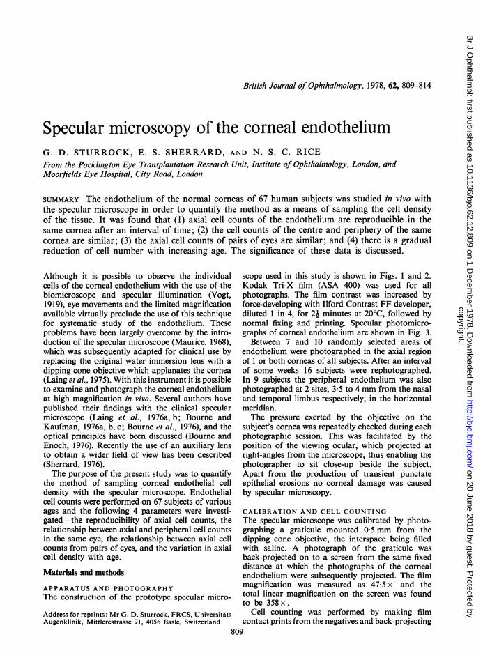
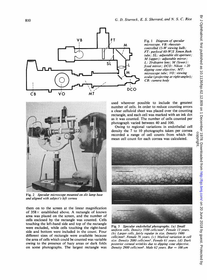
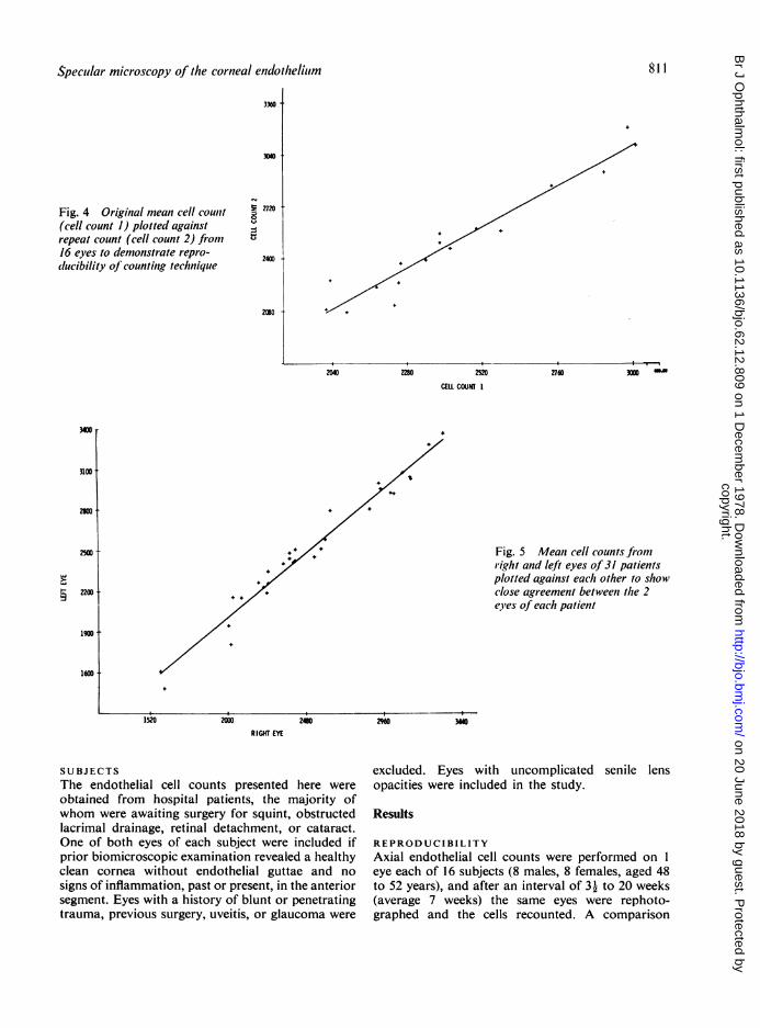
scope used in this study is shown in Figs. 1 and 2.Kodak Tri-X film (ASA 400) was used for allphotographs. The film contrast was increased byforce-developing with Ilford Contrast FF developer,diluted 1 in 4, for 21 minutes at 20°C, followed bynormal fixing and printing. Specular photomicro-graphs of corneal endothelium are shown in Fig. 3.Between 7 and 10 randomly selected areas of
endothelium were photographed in the axial regionof 1 or both corneas of all subjects. After an intervalof some weeks 16 subjects were rephotographed.In 9 subjects the peripheral endothelium was alsophotographed at 2 sites, 3-5 to 4 mm from the nasaland temporal limbus respectively, in the horizontalmeridian.The pressure exerted by the objective on the
subject's cornea was repeatedly checked during eachphotographic session. This was facilitated by theposition of the viewing ocular, which projected atright-angles from the microscope, thus enabling thephotographer to sit close-up beside the subject.Apart from the production of transient punctateepithelial erosions no corneal damage was causedby specular microscopy.
CALIBRATION AND CELL COUNTINGThe specular microscope was calibrated by photo-graphing a graticule mounted 0 5 mm from thedipping cone objective, the interspace being filledwith saline. A photograph of the graticule wasback-projected on to a screen from the same fixeddistance at which the photographs of the cornealendothelium were subsequently projected. The filmmagnification was measured as 47-5 x and thetotal linear magnification on the screen was foundto be 358x.
Cell counting was performed by making filmcontact prints from the negatives and back-projecting
809
copyright. on 20 June 2018 by guest. P
rotected byhttp://bjo.bm
j.com/
Br J O
phthalmol: first published as 10.1136/bjo.62.12.809 on 1 D
ecember 1978. D
ownloaded from
G. D. Sturrock, E. S. Sherrard, and N. S. C. Rice
Fig. 1 Diagram of specularmicroscope. VB: rheostat-controlled 15-W viewing bulb;FT: parfocal 60- W/S Xenon flashtube; SL: adjustable slit aperture;M (upper): adjustable mirror;L: 20-dioptre lens; M (lower):fixed mirror; DCO: Nikon x 20dipping cone objective; MT:microscope tube; VO: viewingocular (projecting at right-angles);CB: camera body
used wherever possible to include the greatestnumber of cells. In order to reduce counting errorsa clear celluloid sheet was placed over the countingrectangle, and each cell was marked with an ink dotas it was counted. The number of cells counted perphotograph varied between 40 and 100.Owing to regional variations in endothelial cell
density the 7 to 10 photographs taken per cornearecorded a range of cell counts from which themean cell count for each cornea was calculated.
Fig. 2 Specular microscope mounted on slit lamp baseand aligned with subject's left cornea
them on to the screen at the linear magnificationof 358x established above. A rectangle of knownarea was placed on the screen, and the number ofcells enclosed by the rectangle was counted. Cellstouching the left-hand side and top of the rectanglewere excluded, while cells touching the right-handside and bottom were included in the count. Fourdifferent sizes of rectangle were available becausethe area of cells which could be counted was variableowing to the presence of hazy areas or dark foldson some photographs. The largest rectangle was
tig. 3 Specular endothelial photographs. (a) Smalluniform cells. Density 3100 cells/mm2. Female 13 years.(b) Larger cells, fairly regular in size. Density 1980cells/mm2. Female 76 years. (c) Marked variation in cellsize. Density 2080 cells/mM2. Female 61 years. (d) Darkposterior corneal wrinkles due to dipping cone objective.Density 2900 cells/mm2. Male 62 years. Bar = 100 [im
810
copyright. on 20 June 2018 by guest. P
rotected byhttp://bjo.bm
j.com/
Br J O
phthalmol: first published as 10.1136/bjo.62.12.809 on 1 D
ecember 1978. D
ownloaded from
Specular microscopy of the cornealendotheliim8n
3360 t
J 272Fig. 4 Original mean cell count(cell count 1) plotted againstrepeat count (cell count 2) from16 eyes to demonstrate repro-dlucibility of counting technique
2400 -
230 t
2040 20 2520
CELL COUN I
2760
Fig. 5 Mean cell counts fromlright and left eyes of 31 patientsplotted against each other to showclose agreement betweeni the 2eyes of each patient
RIGHT EYE
SUBJECTS
The endothelial cell counts presented here wereobtained from hospital patients, the majority ofwhom were awaiting surgery for squint, obstructedlacrimal drainage, retinal detachment, or cataract.One of both eyes of each subject were included ifprior biomicroscopic examination revealed a healthyclean cornea without endothelial guttae and nosigns of inflammation, past or present, in the anteriorsegment. Eyes with a history of blunt or penetratingtrauma, previous surgery, uveitis, or glaucoma were
excluded. Eyes with uncomplicated senile lensopacities were included in the study.
Results
REPRODUCIBILITYAxial endothelial cell counts were performed on 1eye each of 16 subjects (8 males, 8 females, aged 48to 52 years), and after an interval of 3-1 to 20 weeks(average 7 weeks) the same eyes were rephoto-graphed and the cells recounted. A comparison
I
811
300 NS
copyright. on 20 June 2018 by guest. P
rotected byhttp://bjo.bm
j.com/
Br J O
phthalmol: first published as 10.1136/bjo.62.12.809 on 1 D
ecember 1978. D
ownloaded from
C. D. Stiurraock, E. S. Sherrard, anid N. S. C. Rice
4 4.
4. 4.
4. 4. 4. +
+ 4.4. 4. 4.
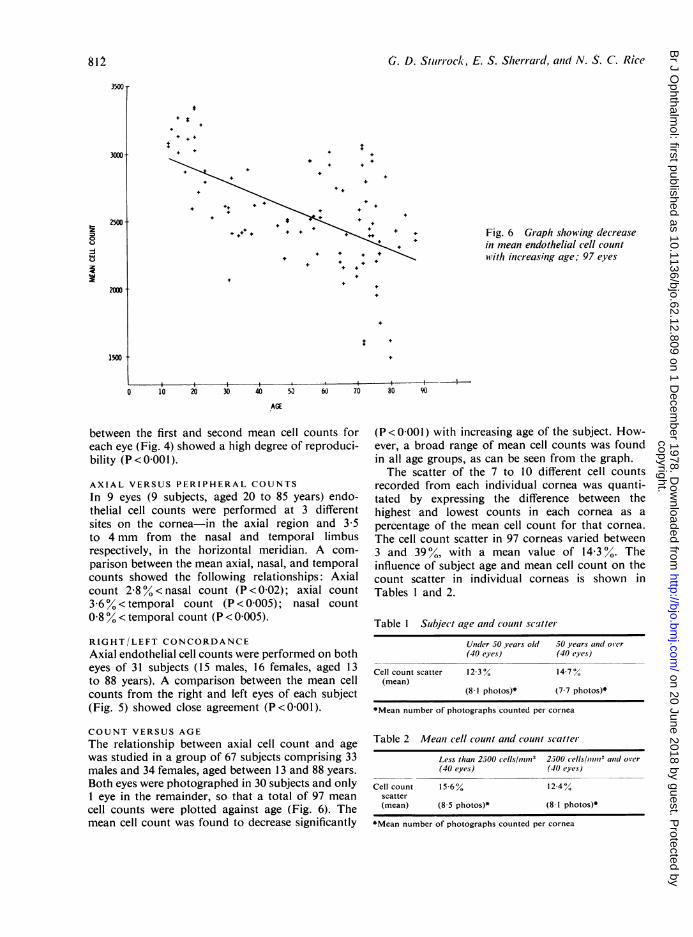
Fig. 6 Graph showing decreasein mean endothelial cell countwith inicreasing age; 97 eyes
0 10 20 30 40 50 60 70 80 90
AGE
between the first and second mean cell counts foreach eye (Fig. 4) showed a high degree of reproduci-bility (P<000l).
AXIAL VERSUS PERIPHERAL COUNTS
In 9 eyes (9 subjects, aged 20 to 85 years) endo-thelial cell counts were performed at 3 differentsites on the cornea-in the axial region and 3 5to 4 mm from the nasal and temporal limbusrespectively, in the horizontal meridian. A com-
parison between the mean axial, nasal, and temporalcounts showed the following relationships: Axialcount 2-8% < nasal count (P < 0 02); axial count36 %0< temporal count (P <0005); nasal count0 8% < temporal count (P < 0 005).
RIGHT/LEFT CONCORDANCEAxial endothelial cell counts were performed on botheyes of 31 subjects (15 males, 16 females, aged 13to 88 years). A comparison between the mean cellcounts from the right and left eyes of each subject(Fig. 5) showed close agreement (P<0001).
COUNT VERSUS AGE
The relationship between axial cell count and age
was studied in a group of 67 subjects comprising 33males and 34 females, aged between 13 and 88 years.
Both eyes were photographed in 30 subjects and only1 eye in the remainder, so that a total of 97 mean
cell counts were plotted against age (Fig. 6). Themean cell count was found to decrease significantly
(P <0OOl) with increasing age of the subject. How-ever, a broad range of mean cell counts was foundin all age groups, as can be seen from the graph.The scatter of the 7 to 10 different cell counts
recorded from each individual cornea was quanti-tated by expressing the difference between thehighest and lowest counts in each cornea as apercentage of the mean cell count for that cornea.The cell count scatter in 97 corneas varied between3 and 39%, with a mean value of 143o%. Theinfluence of subject age and mean cell count on thecount scatter in individual corneas is shown inTables I and 2.
Table 1 Subject age and outit scaqtterUndler 50 years old 50 years anid over(40 eyes) (40 eyes)
Cell count scatter 12-3% 14 7%(mean)
(8 1 photos)* (7 7 photos)*
*Mean number of photographs counted per cornea
Table 2 Meant cell count and count scatter
Less than 2500 cells/nun112 2500 cells/lmm712 anid over(40 eyes) (40 eyes)
Cell count 15-6% 1244%scatter(mean) (8 5 photos)* (8 1 photos)*
*Mean number of photographs counted per cornea
81.
3500 T
+ +.4
4. +t4.+
4. 4.4. 4.
4. *
4.4.4. + 4.
2500t!E0Q
-1L.jQ
ig
2000f
1500
copyright. on 20 June 2018 by guest. P
rotected byhttp://bjo.bm
j.com/
Br J O
phthalmol: first published as 10.1136/bjo.62.12.809 on 1 D
ecember 1978. D
ownloaded from
Specul/ar microscopy of the corneal e,idotheliuim
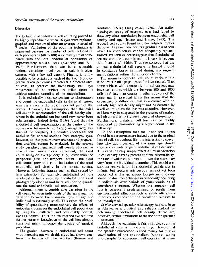
Discussion
The technique of endothelial cell counting proved tobe highly reproducible when 16 eyes were rephoto-graphed and recounted after an average interval of7 weeks. Validation of the counting technique isimportant because the number of cells included ineach photograph (40 to 100) is extremely small com-pared with the total endothelial population ofapproximately 400 000 cells (Svedberg and Bill,1972). Furthermore, there can be considerableregional variations in cell size, particularly in oldercorneas with a low cell density. Finally, it is im-possible to be certain that each of the 7 to 10 photo-graphs taken per cornea represents a different area
of cells. In practice the involuntary small eyemovements of the subject are relied upon toachieve random sampling of the endothelium.
It is technically most convenient to photographand count the endothelial cells in the axial region,which is clinically the most important part of thecornea. However, the assumption that the axialcell count is representative of the cell density else-where in the endothelium has until now never beensubstantiated. Indeed Irvine (1956) found that theendothelial cell concentration in the centre of thecornea was between 6 and 480% (average 160%) lessthan at the periphery. He counted endothelial cellnuclei in flat corneal sections from necropsy eyes,
however, so that post mortem changes and prepara-
tion artefacts cannot be excluded. In the presentstudy peripheral and axial cell counts obtained invivo showed much closer agreement, the axialcount being on average only 32 0% lower than theperipheral (nasal and temporal) count. Thus axialcell counts provide a good indication of the totalendothelial cell density in the normal cornea.
However, following trauma such as that caused bylens extraction, for example, endothelial cell lossis almost certainly unevenly distributed, and axialphotography alone cannot be relied upon to quanti-tate the total endothelial cell population.Although there is considerable variation in the
cell count between individuals of the same age, thevariation between the 2 normal eyes of any one
individual is extremely small. This raises the possi-bility of quantitating retrospectively the effects ofuniocular trauma on the endothelial cell populationby using the unaffected, and presumably normal,eye as a control. Thus, if a traumatised eye requiredfurther surgery, knowledge of the cell loss alreadysustained might influence the choice of surgicalprocedure.The gradual decrease in endothelial cell count
with increasing age which this study has shown con-
firms the findings of other workers (Bourne and
Kaufman, 1976a; Laing et al., 1976a). An earlierhistological study of necropsy eyes had failed toshow any clear correlation between endothelial celldensity and age (Irvine and Irvine, 1953). Thereduced cell counts found in older subjects impliesthat over the years there occurs a gradual loss of cellswhich the endothelium cannot adequately replace.Indeed, available evidence suggests that if endothelialcell division does occur in man it is very infrequent(Kaufman et al., 1966). Thus the concept that thecorneal endothelial cell reserve is limited shouldbe constantly borne in mind during any surgicalmanipulations within the anterior chamber.The normal endothelial cell count varies within
wide limits in all age groups so far investigated. Thussome subjects with apparently normal corneas mayhave cell counts which are between 800 and 1000cells/mm2 less than counts in other subjects of thesame age. In practical terms this means that theoccurrence of diffuse cell loss in a cornea with aninitially high cell density might not be detected bya cell count unless the loss was excessive. However,cell loss may be suspected in the presence of markedcell pleomorphism (Sturrock, personal observation).Furthermore, unilateral cell loss can be readilydiagnosed by demonstrating a higher count in thefellow eye.On the assumption that the lower cell counts
found in older corneas are indeed due to the gradualloss of cells throughout life it is interesting to specu-late why adult corneas of the same age shouldshow such a wide range of endothelial cell densities.This variation may simply reflect a similar variationin cell density already present at birth. Alternatively,the rate at which cells 'drop out' over the years mayvary from one individual to another. This would pre-suppose less variation in endothelial cell density ininfants, but specular microscopy has not yet beenperformed in this age group. Long-term follow-upstudies to document changes in cell density occurringin individuals over periods of years would be ofconsiderable interest. Whether the apparent cellloss is genetically predetermined or results fromenvironmental influences such as subtle alterationsin aqueous composition and circulation remains tobe investigated.
In vivo corneal specular microscopy has now beenestablished as a practical and reliable method fordetermining endothelial cell density. There are,however, certain limitations to the use of the specularmicroscope.Although the technique is fairly simple, counting
endothelial cells is time-consuming. However, ifthe specular microscope is used merely for in vivoexamination of the endothelium (without takingphotographs for subsequent cell counting) it is not
813
copyright. on 20 June 2018 by guest. P
rotected byhttp://bjo.bm
j.com/
Br J O
phthalmol: first published as 10.1136/bjo.62.12.809 on 1 D
ecember 1978. D
ownloaded from
G. D. Sturrock, E. S. Sherrard, and N. S. C. Rice
always possible to visualise the cells adequatelyowing to constant small movements of the eye insome subjects. Then it is necessary either to takephotographs which can be studied later or use asuction contact lens (Laing et al., 1975), but thelatter restricts the examination to the centre of thecornea.The information obtained from specular micro-
scopy is basically morphological and gives littleindication of the functional capacity of the endo-thelium to maintain corneal deturgescence. Further-more, absolute cell counts are of limited value, sincethe minimum cell density compatible with cornealclarity is unknown, and indeed this hypotheticalfigure probably depends upo: .Lhe health or 'pumpingcapacity' of the cells. It has been found that thecornea can remain thir and clear when the axial celldensity has fallen to le.s thF n 500/mm2 (Bourne andKaufman, 1976b; Binkhorst- et al., 1977).
It would clearly b2 of gneat interest to be able tostudy selected areas of endothelial cells over pro-longed periods of time (days or weeks). Although thechanging appearance of individual cells in the rabbitendothelium have been followed in vivo for periodsof up to 9 hours (Sherrard, 1976), in vivo studies inman are impracticable owing to the combinationof high magnification, eye movement, and therelative uniformity of the endothelium, which makeorientation impossible.
Lastly it is possible to perform specular micro-scopy only when the cornea is optically clear. Thusthe presence of oedema, scarring, or infiltration ofthe cornea precludes examination of the underlyingendothelium. The precise role of corneal endothelialspecular microscopy in clinical ophthalmology hasyet to be defined, but it clearly represents a significantadvance in our ability to study this vital layer ofcells.Statistical analysis of the cell counts was performed by Mr H.Donovan, Institute of Ophthalmology. Thanks are due toProfessor Barrie Jones for permission to examine andphotograph patients under his care.
References
Binkhorst, C. D., Loones, L. H., and Nygaard, P. (1977).The clinical specular microscope. Documenta Ophthal-mologica, 44, 57-75.
Bourne, W. M., and Enoch, J. M. (1976). Some opticalprinciples of the clinical specular microscope. InvestigativeOphthalmology, 15, 29-32.
Bourne, W. M., and Kaufman, H. E. (1976a). Specularmicroscopy of human corneal endothelium in vivo.American Journal of Ophthalmology, 81, 319-323.
Bourne, W. M., and Kaufman, H. E. (1976b). Endothelialdamage associated with intraocular lenses. AmericanJournal of Ophthalmology, 81, 482-485.
Bourne, W. M., and Kaufman, H. E. (1976c). Cataractextraction and the corneal endothelium. Americani Journalof Ophthalmology, 82, 44-47.
Bourne, W. M., McCarey, B. E., and Kaufman, H. E. (1976).Clinical specular microscopy. Transactions of the AmericanAcademy of Ophthalmology and Otolaryngology, 81, 743-753.
Irvine, A. R. (1956). The role of the endothelium in bullouskeratopathy. Archives of Ophthalmology, 56, 338-351.
Irvine, A. R., and Irvine, A. R. (1953). Variations in normalhuman corneal endothelium. American Journal of Ophthal-mology, 36, 1279-1285.
Kaufman, H. E., Capella, J. A., and Robbins, J. E. (1966).The human corneal endothelium. American Journal ofOphthalmology, 61, 835-84'.
Laing, R. A., Sandstrom, M. M., and Leibowitz, H. M.(1975). In vivo photomicrography of the corneal endo-thelium. Archives of Ophthalmology, 93, 143-145.
Laing, R. A., Sandstrom, M. M., Benospi, A. R., andLeibowitz, H. M. (1976a). Changes in the corneal endo-thelium as a function of age. Experimental Eye Research,22, 587-594.
Laing, R. A., Sandstrom, M. M., Benospi, A. R., andLeibowitz, H. M. (1976b). Morphological changes incorneal endothelial cells after penetrating keratoplasty.American Journal of Ophthalmology, 82, 459-464.
Maurice, D. M. (1968). Cellular membrane activity in thecorneal endothelium of the intact eye. Experientia, 24,1094-1095.
Sherrard, E. S. (1976). The corneal endothelium in vivo itsresponse to mild trauma. Experimental Eye Research, 22347-357.
Svedberg, B., and Bill, A. (1972). Scanning electron micro-scopic studies of the corneal endothelium in man andmonkeys. Acta Ophthalmologica, 50, 321-336.
Vogt, A. (1919). Die Sichtbarkeit des lebenden Hornhauten-dothels im Lichtbuschel der gullstrandschen Spaltlampe.Klinische Monatsblattenfiir Augenheilkunde, 63, 233-234.
814
copyright. on 20 June 2018 by guest. P
rotected byhttp://bjo.bm
j.com/
Br J O
phthalmol: first published as 10.1136/bjo.62.12.809 on 1 D
ecember 1978. D
ownloaded from