prof. dr. huda al khateeb - comed.uobaghdad.edu.iq a very thik basement membrane. it is produced by...
TRANSCRIPT


Prof. Dr. Huda Al Khateeb
EYE:THE PHOTORECEPTOR SYSTEM
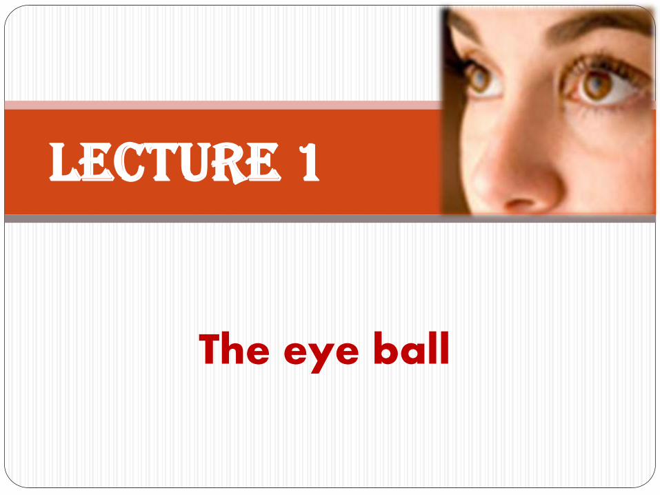
The eye ball
Lecture 1

By the end of this lecture the student should:
1. List the layers and chambers of the eye ball
2. Describe the sclera
3. Name and describe the layers of the cornea
4. Define parts of uvea
5. Discuss the choroid, ciliary body and iris
6. Outline the lens and its parts
Objectives

The eye is a complex and highly
developed photosensitive organ that analyses light reflected from objects, providing the sense of sight..
EYE

The eye is composed of
1. eye ball-(about 22 mm in adults)
2. its protective structures…
What are they?
EYE

The protective structures of the eye ball are:
1. The bony orbit
2. The adipose tissue between eye ball and orbit
3. the eyelids
4. the conjunctiva
5. the lacrimal apparatus
HOW THEY PROTECT THE EYE BALL???????
EYE

Eye ball consist of three layers
1. Outer layer (Sclera and cornea)
2. Middle layer (Uvea)
3. Inner layer (Retina)
EYE
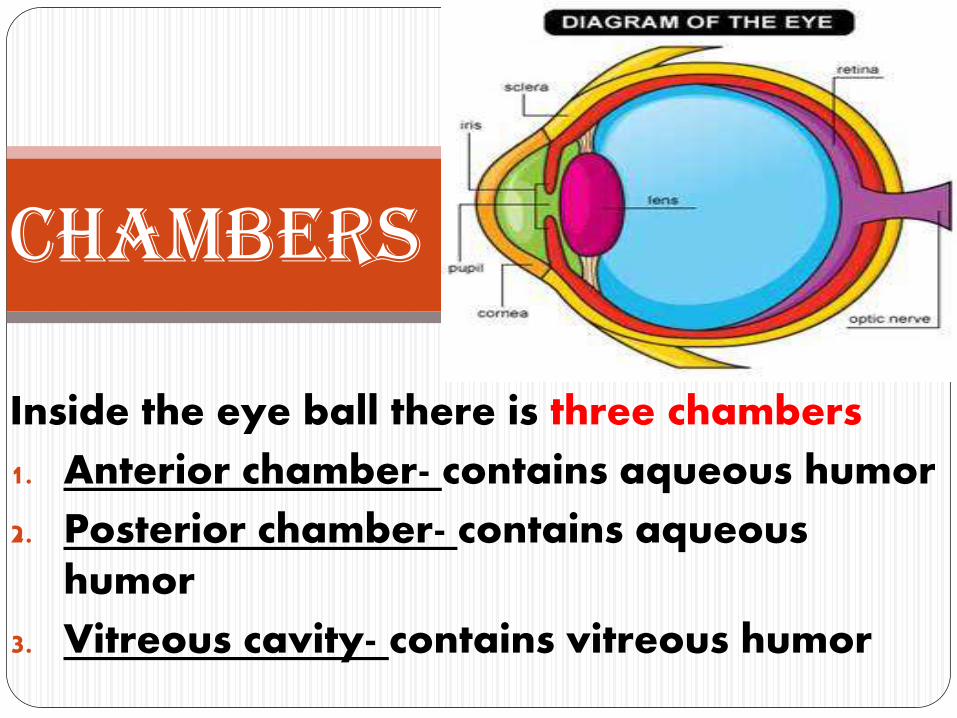
Inside the eye ball there is three chambers
1. Anterior chamber- contains aqueous humor
2. Posterior chamber- contains aqueous humor
3. Vitreous cavity- contains vitreous humor
Chambers

The lens is placed in front of vitreous cavity and posterior to posterior chamber
Lens

• It is a white, fibrous, external layer of the eyeball that protects the more delicate internal structures and provides sites for muscle insertion
• It makes the white posterior five-sixths of the external layer of the outer layer of the eye ball
Sclera

• The sclera is relatively avascular, and consists of dense connective tissue containing flat type I collagen bundles
• Tendons of the extra-ocular muscles that move the eyes insert into anterior areas of the sclera
• Posteriorly the sclera joins with the epineurium covering the optic nerve
Sclera
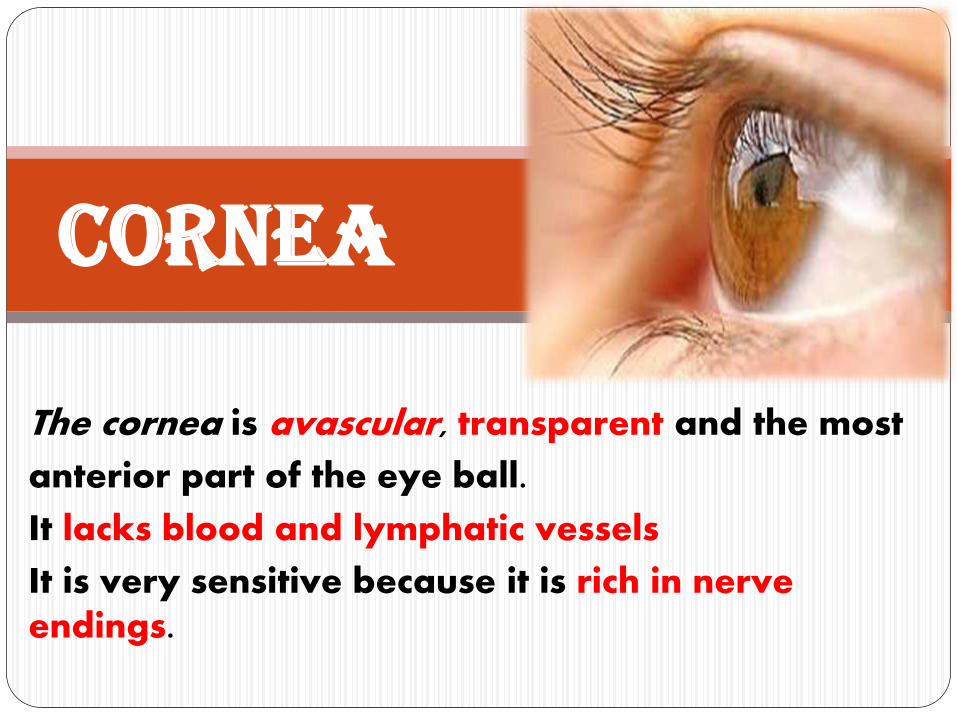
The cornea is avascular, transparent and the most
anterior part of the eye ball.
It lacks blood and lymphatic vessels
It is very sensitive because it is rich in nerve endings.
Cornea

The cornea consists of five layers
• Corneal epithelium –
• Bowman’s membrane
• Corneal stroma (90 %)- (substantia propria)
• Descemet’s membrane
• Corneal endothelium – single layer
Cornea
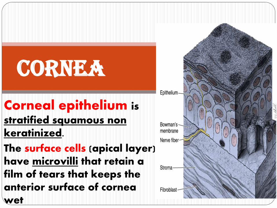
Corneal epithelium is
stratified squamous non keratinized.
The surface cells (apical layer) have microvilli that retain a film of tears that keeps the anterior surface of cornea wet
Cornea

The basement membrane of this
epithelium is very thick (8–12 µm) and contributes to the stability and strength of the cornea, helping to protect against infection of the underlying stroma
Cornea

The corneal stroma is transparent
It represents 90% of the corneal thickness
Composed of type I and V collagen fibers that are regularly arranged. Fibers and layers are separated by extracellular matrix rich in prteoglycans containing chondrotin and keratin sulfate
Cornea
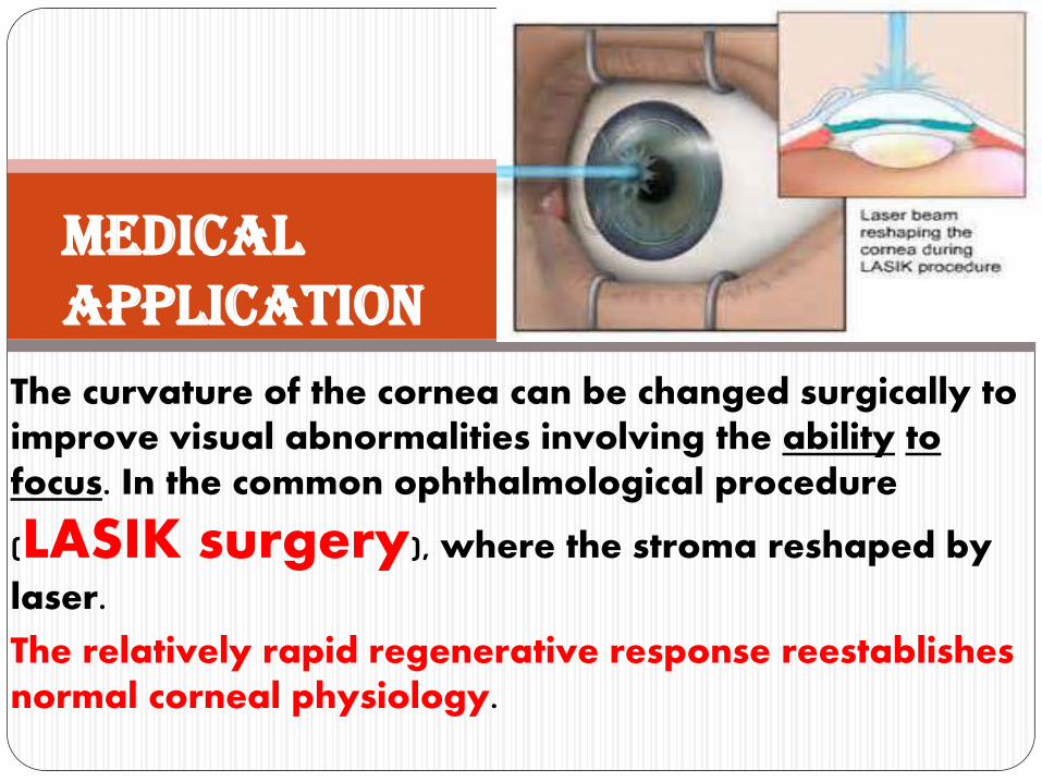
The curvature of the cornea can be changed surgically to improve visual abnormalities involving the ability to focus. In the common ophthalmological procedure
(LASIK surgery), where the stroma reshaped by
laser.
The relatively rapid regenerative response reestablishes normal corneal physiology.
MEDICAL APPLICATION

Descement membrane is a very thik
basement membrane. It is produced by the corneal endothelium and contains type VII collagen
Cornea

The corneal endothelium lines the
posterior surface of the cornea and faces the anterior chamber of the eye. It consists of singlelayer of squamous epithelial cells.
Cornea
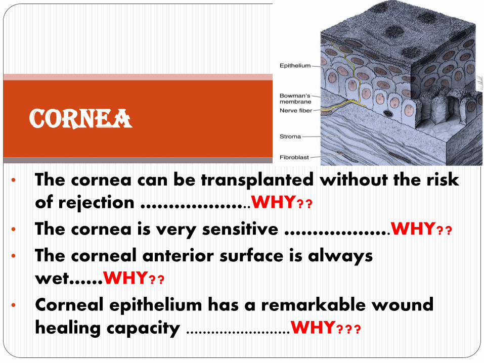
• The cornea can be transplanted without the risk of rejection ………………..WHY??
• The cornea is very sensitive ……………….WHY??
• The corneal anterior surface is always wet……WHY??
• Corneal epithelium has a remarkable wound healing capacity .........................WHY???
Cornea

Encircling the cornea is the Corneo-scleral junction, or Limbus, a transitional area where the transparent stroma
merges with the opaque sclera.
This region has micro-vasculature which, along with aqueous humor in the anterior chamber, provides metabolites for the corneal cells by diffusion
Discuss the modes of nutrition of cornea?????
Limbus

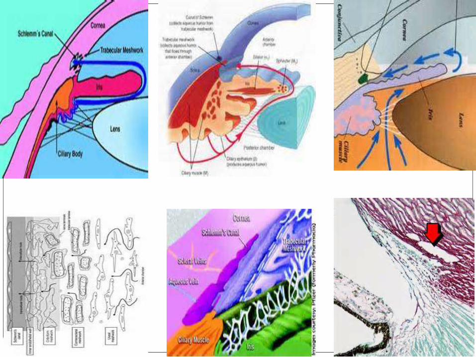
• It is a circular canal that lie in the corneo-scleral junction (limbus).
• It drains the aquoes humor from the anterior chamber through the meshwork trabeculae into the veins that drain the eye ball
Canal of Schlemm

It consists of three parts, from posterior to anterior:
1. the choroid
2. the ciliary body
3. the iris
Middle layer: UVEAVascular Layer
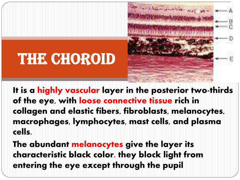
It is a highly vascular layer in the posterior two-thirds of the eye, with loose connective tissue rich in collagen and elastic fibers, fibroblasts, melanocytes, macrophages, lymphocytes, mast cells, and plasma cells.
The abundant melanocytes give the layer its characteristic black color. they block light from entering the eye except through the pupil
The choroid

It is an anterior expansion of the choroid.
It is a thickened ring of tissue lying just inside the anterior portion of the sclera.
In transverse section the ciliary body is roughly a triangle, with its long base contacting the sclera, another side in contact with the vitreous body, and the third facing the posterior chamber.
The ciliary body

It is composed of Ciliary muscle(smooth muscle) that is surrounded by a
stroma of loose connective tissue, rich in
microvasculature, elastic fibers, and
melanocytes,
The ciliarybody
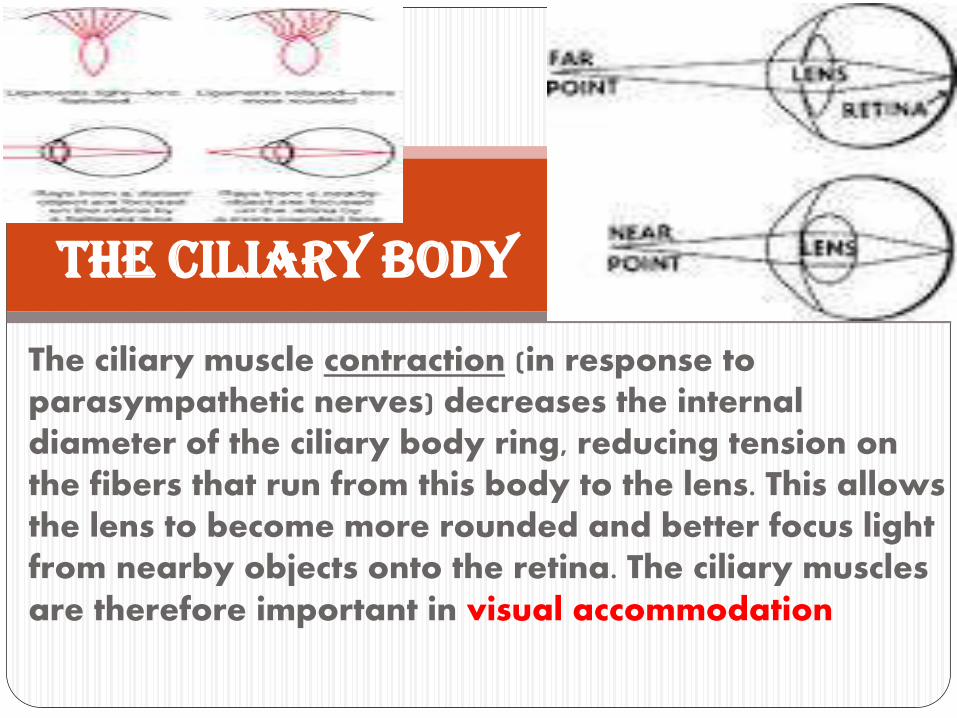
The ciliary muscle contraction (in response to parasympathetic nerves) decreases the internal diameter of the ciliary body ring, reducing tension on the fibers that run from this body to the lens. This allows the lens to become more rounded and better focus light from nearby objects onto the retina. The ciliary muscles are therefore important in visual accommodation
The ciliary body

The surfaces of the ciliary body that face the vitreous body, posterior chamber, and lens has the ciliary processes (a series of about 75 radial ridges)
The ciliarybody

The surface epithelial cells is connected to the lens capsule by zonular fibers (suspensory ligament of the lens.)
The ciliary body

Both ciliary body and processes are covered by a double layer of low columnar epithelium, the Ciliaryepithelium.
The epithelial cells directly covering the ciliary stroma are rich in melanin (Pigmented Layer).
The surface layer of cells lacks melanin (Secretory Layer) (Non-pigmented layer)- secrets the aquoushumor to the posterior chamber .
The ciliary body

Secreted into the posterior chamber, aqueous humor flows between the lens and the iris to reach the anterior chamber through the pupil.
The aqueous then flows into the channels of the trabecular meshwork at the corneo-scleral junction then to the Canal of Schlemm then drained to scleral venous sinus.
The ciliary body

Aqueous humor is produced continuously. If its drainage is impeded, typically by obstruction of the trabecular meshwork or scleral venous sinus, intraocular pressure
can increase, causing the condition called glaucoma.
Untreated glaucoma can cause pressing of the vitreous body against the retina, affecting visual function.
MEDICALAPPLICATION

It is the most anterior extension of the uvea (middle layer) that partially covers the lens, leaving a round
opening in the center called the pupil.
Iris

Pupil Pupil

Iris: Flowers

Iris: Goddess of Rainbow

The anterior surface of the iris is covered
by irregular, discontinuous layer of fibroblastsand melanocytes, densely packed and with inter-digitating processes.
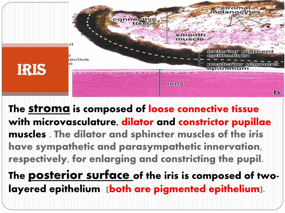
The stroma is composed of loose connective tissue
with microvasculature, dilator and constrictor pupillae muscles . The dilator and sphincter muscles of the iris have sympathetic and parasympathetic innervation, respectively, for enlarging and constricting the pupil.
The posterior surface of the iris is composed of two-
layered epithelium (both are pigmented epithelium).
Iris

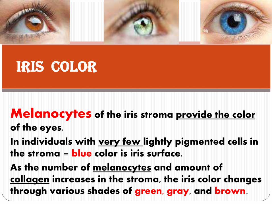
Melanocytes of the iris stroma provide the color
of the eyes.
In individuals with very few lightly pigmented cells in the stroma = blue color is iris surface.
As the number of melanocytes and amount of collagen increases in the stroma, the iris color changes through various shades of green, gray, and brown.
Iris color

Individuals with albinismhave almost no pigment and the pink color of their irises is due to the reflection of incident light from the blood vessels of the stroma.
Iris

The lens is a transparent biconvex structure immediately behind the iris, used to focus light on the retina.
The lens is a unique avascular tissue. It is highly elastic, a feature that is lost with age as lens tissue hardens.
The lens has three principal components (capsule, anterior epithelium and lens fibers).
Lens

Lens Capsule
The lens is covered by a thick, homogeneous capsule rich in proteoglycans and type IV collagen.
The lens capsule protects the underlying cells and provides the place of attachment for zonular fibers
Lens

Lens Epithelium
Subcapsular lens epithelium consists of a single layer of cuboidal epithelial cells and is present only on the anterior surface of the lens. The basal ends of the epithelial cells attach to the lens capsule and their apical surfaces have interdigitations that bind the epithelium to the internal lens fibers..
Lens

Lens fibers
At the posterior edge of this epithelium, near the equator of the lens, the cells divide to provide new cells that differentiate as lens fibers. This process allows for growth of the lens and continues at a slow, decreasing rate near the equator of the lens throughout adult life
Lens

Lens Fibers
Lens fibers are highly elongated and appear as thin, flattened structures.
Differentiating lens fibers eventually lose their nuclei and other organelles, fill the cytoplasm with a group of proteins called crystallins, and become very long.
Lens
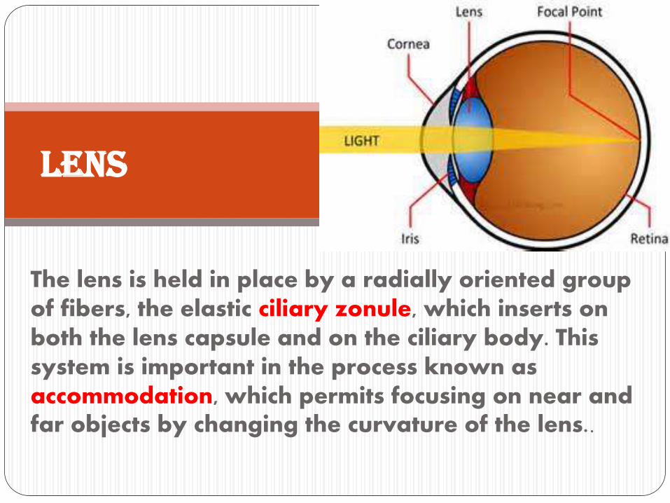
The lens is held in place by a radially oriented group of fibers, the elastic ciliary zonule, which inserts on both the lens capsule and on the ciliary body. This system is important in the process known as accommodation, which permits focusing on near and far objects by changing the curvature of the lens..
Lens

When the eye is at rest or gazing at distant objects, the lens is kept stretched by the zonule in a plane perpendicular to the optical axis.
To focus on a near object, the ciliary muscles contract. This relieves some of the tension exerted by the zonule on the lens, allowing the latter to round up and become thicker, keeping the object in focus
Lens

Advancing age reduces the elasticity of the lens, making accommodation for near objects difficult. This
is a normal process (presbyopia, Gr. eyes of
elders), which can be corrected by wearing glasses with convex lenses (reading glasses).
MEDICALAPPLICATION

In old individuals, denaturation of crystallins commonly begins to occur in lens fibers, making them less transparent. When areas of the lens become opaque or cloudy and vision is impaired, the condition is termed
cataract.
Other causes of cataract include exposure to ultraviolet light or other radiation, trauma, and as secondary effects in diseases such as diabetes mellitus and hypertension.
MEDICAL APPLICATIOn
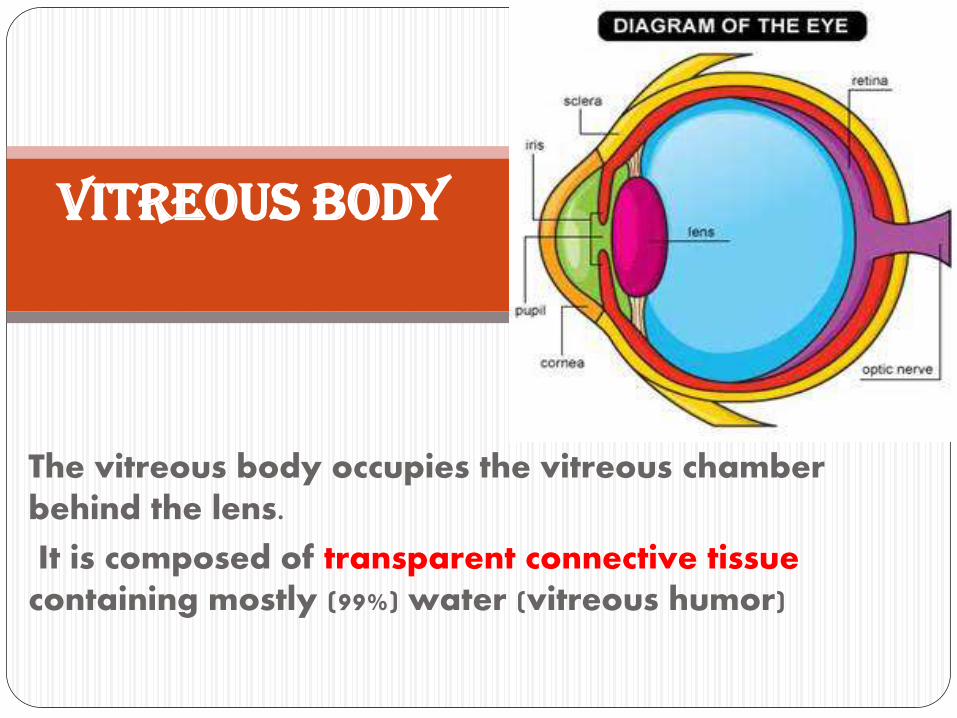
The vitreous body occupies the vitreous chamber behind the lens.
It is composed of transparent connective tissue containing mostly (99%) water (vitreous humor)
Vitreous Body

• Eye ball consist of three layers and three chambers
• Cornea has five layers
• Uvea composed of choroid, ciliary body and iris
• Lens
summary

A 56-year-old woman complains of decreased vision in her left eye of 4 years duration. White discoloration of her left pupil is noted on physical examination (shown in the image). The white appearance of the pupil in this patient represents a pathological change affecting:
1. Conjunctiva
2. Cornea
3. Iris
4. Lens
5. Vitreous body
Quiz
