eau guidelines on renal cell carcinoma - uroweb.org · 4 renal cell carcinoma - limited update...
TRANSCRIPT

B. Ljungberg (Chair), L. Albiges, K. Bensalah, A. Bex (Vice-chair), R.H. Giles (Patient Advocate), M. Hora,
M.A. Kuczyk, T. Lam, L. Marconi, A.S. Merseburger, T. Powles, M. Staehler, A. Volpe
Guidelines Associates: S. Dabestani, S. Fernández-Pello Montes, F. Hofmann, R. Tahbaz
Renal CellCarcinoma
EAU Guidelines on
© European Association of Urology 2017

RENAL CELL CARCINOMA - LIMITED UPDATE MARCH 20172
TABLE OF CONTENTS PAGE1. INTRODUCTION 5 1.1 Aims and scope 5 1.2 Panel composition 5 1.3 Available publications 5 1.4 Publication history and summary of changes 5 1.4.1 Publication history 5 1.4.2 Summary of changes 5
2. METHODS 7 2.1 Data identification 7 2.2 Review 8 2.3 Future goals 8
3. EPIDEMIOLOGY, AETIOLOGY AND PATHOLOGY 9 3.1 Epidemiology 9 3.1.1 Summary of evidence and recommendation 9 3.2 Histological diagnosis 9 3.2.1 Clear cell renal cell cancer 9 3.2.2 Papillary renal cell cancer 10 3.2.3 Chromophobe (chRCC) 10 3.3 Other renal tumours 10 3.3.1 Carcinoma associated with end-stage renal disease; acquired cystic disease-associated RCC 10 3.3.2 Papillary adenoma 10 3.3.3 Hereditary kidney tumours 10 3.3.4 Angiomyolipoma 11 3.3.4.1 Treatment 11 3.3.4.2 Summary 13 3.4 Summary of evidence and recommendations for the management of other renal tumours 13
4. STAGING AND CLASSIFICATION SYSTEMS 13 4.1 Staging 13 4.2 Anatomic classification systems 14
5. DIAGNOSTIC EVALUATION 15 5.1 Symptoms 15 5.1.1 Physical examination 15 5.1.2 Laboratory findings 15 5.2 Imaging investigations 15 5.2.1 Presence of enhancement 15 5.2.2 Computed tomography or magnetic resonance imaging 15 5.2.3 Other investigations 16 5.2.4 Radiographic investigations to evaluate RCC metastases 16 5.2.5 Bosniak classification of renal cystic masses 16 5.3 Renal tumour biopsy 16 5.4 Summary of evidence and recommendations for the diagnostic assessment of renal cell cancer 17
6. PROGNOSTIC FACTORS 18 6.1 Classification 18 6.2 Anatomical factors 18 6.3 Histological factors 18 6.4 Clinical factors 19 6.5 Molecular factors 19 6.6 Prognostic systems and nomograms 20 6.7 Summary of evidence and recommendations for prognostic factors 20

3RENAL CELL CARCINOMA - LIMITED UPDATE MARCH 2017
7. DISEASE MANAGEMENT 22 7.1 Treatment of localised RCC 22 7.1.1 Introduction 22 7.1.2 Surgical treatment 22 7.1.2.1 Nephron-sparing surgery vs. radical nephrectomy 22 7.1.2.2 Associated procedures 23 7.1.2.2.1 Adrenalectomy 23 7.1.2.2.2 Lymph node dissection for clinically negative lymph nodes (cN0) 23 7.1.2.2.3 Embolisation 23 7.1.2.2.4 Summary of evidence and recommendations 24 7.1.3 Radical and partial nephrectomy techniques 24 7.1.3.1 Radical nephrectomy techniques 24 7.1.3.2 Partial nephrectomy techniques 24 7.1.3.3 Positive margins on histopathological specimens of resected tumours 25 7.1.3.4 Summary of evidence and recommendations 25 7.1.4 Therapeutic approaches as alternatives to surgery 26 7.1.4.1 Surgical versus non-surgical treatment 26 7.1.4.2 Surveillance 26 7.1.4.3 Ablative therapies 26 7.1.4.3.1 Cryoablation 26 7.1.4.3.2 Cryoablation versus partial nephrectomy 26 7.1.4.3.3 Radiofrequency ablation 27 7.1.4.3.4 Radiofrequency ablation versus partial nephrectomy 27 7.1.4.3.5 Cryoablation versus radiofrequency ablation 27 7.1.4.3.6 Other ablative techniques 27 7.1.4.3.7 Summary of evidence and recommendation for therapeutic approaches as alternative to surgery 27 7.2 Treatment of locally advanced RCC 28 7.2.1 Introduction 28 7.2.2 Management of clinically positive lymph nodes (cN+) 28 7.2.3 Management of locally advanced unresectable RCC 28 7.2.4 Management of RCC with venous tumour thrombus 28 7.2.4.1 The evidence base for surgery in patients with venous tumour thrombus 28 7.2.4.2 The evidence base for different surgical strategies 28 7.2.4.3 Summary of evidence and recommendations for the management of RCC with venous tumour thrombus 28 7.2.5 Adjuvant therapy 29 7.2.5.1 Summary of evidence and recommendations for adjuvant therapy 29 7.3 Advanced/metastatic RCC 29 7.3.1 Local therapy of advanced/metastatic RCC 29 7.3.1.1 Cytoreductive nephrectomy 29 7.3.1.1.1 Embolisation of the primary tumour 29 7.3.1.1.2 Summary of evidence and recommendation for local therapy of advanced/metastatic RCC 30 7.3.2 Local therapy of metastases in mRCC 30 7.3.2.1 Complete versus no/incomplete metastasectomy 30 7.3.2.2 Local therapies for RCC bone metastases 30 7.3.2.3 Local therapies for RCC brain metastases 30 7.3.2.4 Embolisation of metastases 31 7.3.2.5 Summary of evidence and recommendations for local therapy of metastases in metastatic RCC 31 7.4 Systemic therapy for advanced/metastatic RCC 31 7.4.1 Chemotherapy 31 7.4.1.1 Summary of evidence and recommendation for systemic therapy for advanced/metastatic renal cell cancer 31 7.4.2 Immunotherapy 32 7.4.2.1 IFN-α monotherapy and combined with bevacizumab 32

RENAL CELL CARCINOMA - LIMITED UPDATE MARCH 20174
7.4.2.2 Interleukin-2 32 7.4.2.3 Vaccines and targeted immunotherapy 32 7.4.2.4 Immune checkpoint blockade 32 7.4.2.5 Summary of evidence and recommendations for immunotherapy in mRCC 33 7.4.3 Targeted therapies 33 7.4.3.1 Tyrosine kinase inhibitors 34 7.4.3.1.1 Sorafenib 34 7.4.3.1.2 Sunitinib 34 7.4.3.1.3 Pazopanib 34 7.4.3.1.4 Axitinib 34 7.4.3.1.5 Cabozantinib 35 7.4.3.1.6 Lenvatinib 35 7.4.4 Monoclonal antibody against circulating VEGF 35 7.4.4.1 Bevacizumab monotherapy and bevacizumab + IFN-α 35 7.4.5 mTOR inhibitors 35 7.4.5.1 Temsirolimus 35 7.4.5.2 Everolimus 35 7.4.6 Therapeutic strategies 36 7.4.6.1 Therapy for treatment-naïve patients with clear-cell mRCC 36 7.4.6.1.1 Sequencing targeted therapy 36 7.4.6.1.1.1 Following progression of disease with one or more lines of VEGF-targeted therapy 36 7.4.6.1.1.2 Treatment after progression of disease with mTOR inhibition 36 7.4.6.1.1.3 Treatment after progression of disease with cytokines 36 7.4.6.1.1.4 Treatment after second-line targeted therapy 36 7.4.6.1.1.4.1 Treatment after two VEGF-targeted therapies 36 7.4.6.1.1.4.2 Treatment after VEGFR- and mTOR inhibition 37 7.4.6.1.1.4.3 Combination of targeted agents 37 7.4.6.2 Non-clear-cell renal cancer 37 7.4.6.3 Summary of evidence and recommendations for systemic therapy in metastatic renal cell cancer 39 7.5 Recurrent RCC 40 7.5.1 Introduction 40 7.5.2 Summary of evidence and recommendation for advanced/metastatic RCC 40
8. FOLLOW-UP IN RCC 41 8.1 Introduction 41 8.2 Which investigations for which patients, and when? 41 8.3 Summary of evidence and recommendations for surveillance following RN or PN or ablative therapies in RCC 42 8.4 Research priorities 42
9. REFERENCES 43
10. CONFLICT OF INTEREST 66

5RENAL CELL CARCINOMA - LIMITED UPDATE MARCH 2017
1. INTRODUCTION1.1 Aims and scopeThe European Association of Urology (EAU) Renal Cell Cancer (RCC) Guidelines Panel has compiled these clinical guidelines to provide urologists with evidence-based information and recommendations for the management of RCC.
It must be emphasised that clinical guidelines present the best evidence available to the experts but following guideline recommendations will not necessarily result in the best outcome. Guidelines can never replace clinical expertise and judgement when making treatment decisions for individual patients, but rather help to focus decisions whilst also taking personal values and preferences/individual circumstances of patients into account. Guidelines are not mandates and do not purport to be a legal standard of care.
1.2 Panel compositionThe RCC Guidelines Panel is an international group of clinicians consisting of urological surgeons, oncologists, methodologists, a pathologist and a radiologist, with particular expertise in the field of renal cancer care. Since 2015, the panel has incorporated a patient advocate to provide a consumer perspective for its guidelines.
All experts involved in the production of this document have submitted potential conflict of interest statements, which can be viewed on the EAU website Uroweb: http://uroweb.org/guideline/renal-cell-carcinoma/.
AcknowledgementThe RCC Guidelines Panel is most grateful for the methodological and scientific support provided by Prof.Dr. O. Hes (pathologist, Pilzen, Czech Republic) for two sections of this document: Histological diagnosis and Other renal tumours.
1.3 Available publicationsA quick reference document (Pocket Guidelines) is available, both in print and in a number of versions for mobile devices, presenting the main findings of the RCC Guidelines. These are abridged versions which may require consultation together with the full text version. Several scientific publications are available, as are a number of translations of all versions of the EAU RCC Guidelines [1, 2]. All documents can be accessed on the EAU website: http://uroweb.org/guideline/renal-cell-carcinoma/.
1.4 Publication history and summary of changes1.4.1 Publication historyThe EAU RCC Guidelines were first published in 2000. This 2017 RCC Guidelines document presents a limited update of the 2016 publication.
1.4.2 Summary of changesAll chapters of the 2017 RCC Guidelines have been updated, based on the 2016 version of the guideline. References have been added throughout the document.
Key changes in this 2017 print: • Section 3.3.3 - Hereditary kidney tumours: This section has been expanded• Section 5.2 - Imaging evaluations: The findings of a systematic review have been incorporated.
New data and recommendations have been included in the following sections:
5.4 Summary of evidence and recommendations for the diagnostic assessment of renal cell cancer
Summary of evidence LE
Contrast enhanced multi-phasic CT has a high sensitivity and specificity for characterisation
and detection of RCC, invasion, tumour thrombus and metastatic RCC.
2
MRI has a slightly higher sensitivity and specificity for small renal masses and tumour
thrombus as compared to CT.
2
CEUS has a high sensitivity and specificity for characterisation of renal masses. 2
US, Power-Doppler US and PET-CT have a low sensitivity and specificity for detection and
characterisation of RCC.
2

RENAL CELL CARCINOMA - LIMITED UPDATE MARCH 20176
Recommendations grade
Use multi-phasic contrast-enhanced computed tomography (CT) for general staging
and detection of renal cell cancer (RCC).
strong ↑↑
Use axial abdominal imaging and CT of the chest for staging of RCC. strong ↑↑
Use non-ionising modalities, mainly contrast enhanced ultrasound (CEUS), for
further characterisation of small renal masses, tumour thrombus and differentiation
of unclear renal masses.
weak ↑
Do not use bone scan and/or positron-emission tomography (PET)-CT for staging of
RCC.
weak ↓
Perform a renal tumour biopsy before ablative therapy and systemic therapy without
previous pathology.
strong ↑↑
Perform a percutaneous biopsy in select patients who are considered for active
surveillance.
weak ↑
When performing a renal tumour biopsy technique, use a coaxial technique. strong ↑↑
Do not perform a renal tumour biopsy of cystic renal masses. weak ↓
7.2.5.1 Summary of evidence and recommendations for adjuvant therapy
Summary of evidence LE
Adjuvant cytokines do not improve survival after nephrectomy. 1b
Adjuvant sunitinib improved disease-free survival in one of the two available studies, but not
overall survival, after nephrectomy in selected high-risk patients.
1b
Recommendations grade
Do not offer adjuvant therapy with sorafenib. strong ↓↓
Do not offer adjuvant sunitinib following surgically resected high-risk clear-cell renal
cell cancer.
weak ↓
7.3.2.5 Recommendations for local therapy of metastases in metastatic RCC
Recommendation grade
Consider local therapy for metastatic disease (including metastasectomy) in patients
with a favourable risk profile in whom complete resection is achievable or when
local symptoms need to be controlled.
weak ↑
7.4.1.1 Summary of evidence and recommendation for systemic therapy for advanced/metastatic renal cell cancer
Summary of evidence LE
In metastatic RCC, 5-FU combined with immunotherapy has equivalent efficacy to INF-α. 1b
In metastatic RCC, chemotherapy is otherwise not effective with the exception of gemcitabine
and doxorubicine in sarcomatoid and rapidly progressive disease.
3
Recommendations grade
Do not offer chemotherapy as first-line therapy in patients with metastatic clear-cell
renal cell cancer (RCC).
strong ↓↓
Consider offering a combination of gemcitabine and doxorubicin to patients with
sarcomatoid or rapidly progressive RCC.
weak ↑
7.4.6.3 Summary of evidence and recommendations for systemic therapy in metastatic renal cell cancer
Summary of evidence LE
First line pazopanib is not inferior to sunitinib in clear-cell mRCC patients. 1b
Cabozantinib is superior to everolimus in terms of PFS and OS in patients failing one or more
lines of VEGF-targeted therapy.
1b
Everolimus prolongs PFS in patients who have previously failed or are intolerant of VEGF-
targeted therapy when compared to placebo.
1b
No combination has proven to be better than single-agent therapy, with the exception of the
combination of lenvatinib plus everolimus.
1a
7RENAL CELL CARCINOMA - LIMITED UPDATE MARCH 2017
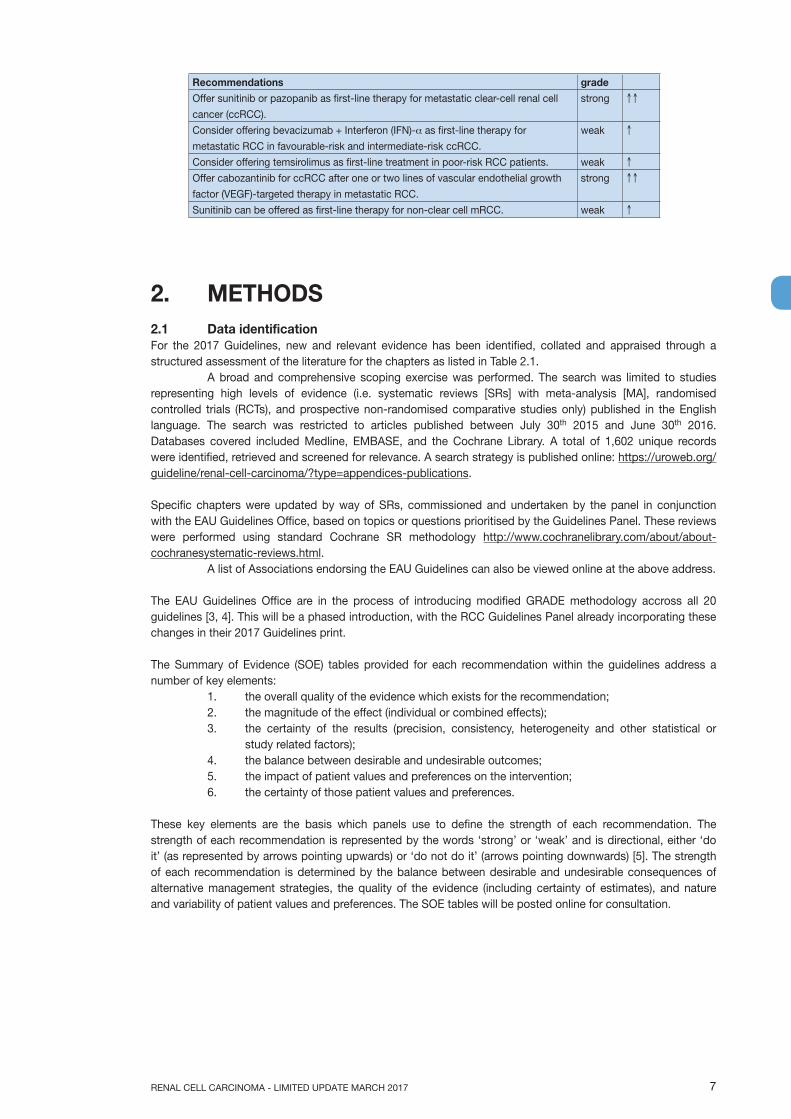
Recommendations grade
Offer sunitinib or pazopanib as first-line therapy for metastatic clear-cell renal cell
cancer (ccRCC).
strong ↑↑
Consider offering bevacizumab + Interferon (IFN)-α as first-line therapy for
metastatic RCC in favourable-risk and intermediate-risk ccRCC.
weak ↑
Consider offering temsirolimus as first-line treatment in poor-risk RCC patients. weak ↑
Offer cabozantinib for ccRCC after one or two lines of vascular endothelial growth
factor (VEGF)-targeted therapy in metastatic RCC.
strong ↑↑
Sunitinib can be offered as first-line therapy for non-clear cell mRCC. weak ↑
2. METHODS2.1 Data identificationFor the 2017 Guidelines, new and relevant evidence has been identified, collated and appraised through a structured assessment of the literature for the chapters as listed in Table 2.1. A broad and comprehensive scoping exercise was performed. The search was limited to studies representing high levels of evidence (i.e. systematic reviews [SRs] with meta-analysis [MA], randomised controlled trials (RCTs), and prospective non-randomised comparative studies only) published in the English language. The search was restricted to articles published between July 30th 2015 and June 30th 2016. Databases covered included Medline, EMBASE, and the Cochrane Library. A total of 1,602 unique records were identified, retrieved and screened for relevance. A search strategy is published online: https://uroweb.org/guideline/renal-cell-carcinoma/?type=appendices-publications.
Specific chapters were updated by way of SRs, commissioned and undertaken by the panel in conjunction with the EAU Guidelines Office, based on topics or questions prioritised by the Guidelines Panel. These reviews were performed using standard Cochrane SR methodology http://www.cochranelibrary.com/about/about-cochranesystematic-reviews.html. A list of Associations endorsing the EAU Guidelines can also be viewed online at the above address.
The EAU Guidelines Office are in the process of introducing modified GRADE methodology accross all 20 guidelines [3, 4]. This will be a phased introduction, with the RCC Guidelines Panel already incorporating these changes in their 2017 Guidelines print.
The Summary of Evidence (SOE) tables provided for each recommendation within the guidelines address a number of key elements:
1. the overall quality of the evidence which exists for the recommendation;2. the magnitude of the effect (individual or combined effects);3. the certainty of the results (precision, consistency, heterogeneity and other statistical or
study related factors);4. the balance between desirable and undesirable outcomes;5. the impact of patient values and preferences on the intervention;6. the certainty of those patient values and preferences.
These key elements are the basis which panels use to define the strength of each recommendation. The strength of each recommendation is represented by the words ‘strong’ or ‘weak’ and is directional, either ‘do it’ (as represented by arrows pointing upwards) or ‘do not do it’ (arrows pointing downwards) [5]. The strength of each recommendation is determined by the balance between desirable and undesirable consequences of alternative management strategies, the quality of the evidence (including certainty of estimates), and nature and variability of patient values and preferences. The SOE tables will be posted online for consultation.

RENAL CELL CARCINOMA - LIMITED UPDATE MARCH 20178
Table 2.1: Description of update and summary of review methodology
Chapter Brief description of review methodology1. Introduction Not applicable2. Methods Not applicable3. Epidemiology, aetiology and pathology This chapter was updated by a traditional narrative
review, based on a structured literature assessment.4. Staging and grading classification systems This chapter was updated by a traditional narrative
review, based on a structured literature assessment.5. Diagnostic evaluation Section 5.2 (Diagnostic imaging) was revised based
on a SR [6]. The remainder of the chapter was updated by a structured literature assessment.
6. Prognosis This chapter was updated by a traditional narrative review, based on a structured literature assessment.
7. Treatment (Disease management) Chapters 7.1.2 and 7.2.4 (Treatment of localised and locally advanced disease) were revised based on an updated SR. The remainder of the chapter was updated using a structured literature assessment.Systemic therapy for metastatic disease: this section was updated by a SR.
8. Surveillance following radical or partial nephrectomy or ablative therapies
This chapter was updated by a traditional narrative review, based on a structured literature assessment.
The findings of a number of SR topics have been incorporated in this 2017 update:• Imaging in Suspected Renal Cell Carcinoma: A Systematic Review [6]• What is the best surgical treatment option for clinical > T2, N0M0 tumours? What is the best way of
performing this procedure? [7];• A Systematic Review and Meta-analysis Comparing the Effectiveness and Adverse Effects of Different
Systemic Treatments for Non-clear Cell Renal Cell Carcinoma [8].
2.2 ReviewChapter 7 ‘Disease management’ was peer reviewed prior to publication. Publications ensuing from SRs have all been peer reviewed. The other sections of the RCC Guidelines were peer reviewed prior to publication in 2015.
2.3 Future goalsFor their future updates, the RCC Guideline Panel aims to focus on patient-reported outcomes.
The use of clinical quality indicators is an area of interest for the RCC Panel. A number of key quality indicators for this patient group have been selected:• thorax computed tomography (CT) for staging of pulmonary metastasis;• proportion of patients with T1aN0M0 tumours undergoing nephron-sparing surgery as first treatment;• the proportion of patients treated within six weeks after diagnosis;• the proportion of patients with metastatic RCC offered treatment with targeting agents;• proportion of patients who undergo minimally invasive or operative treatment as first treatment who die
within 30 days;Panel members have set up a database to capture current practice of follow-up of RCC patients in a number of European Centres. Assessing patterns of recurrence and use of imaging techniques are primary outcomes for this project.
The results of ongoing and new SRs will be included in the 2018 update of the RCC Guidelines.
Topics of ongoing SRs:• What is the best treatment option for T1-T2 tumours? (updated review);• What is the best treatment option for T1a tumours?;• What is the best treatment option for T1b-T2a tumours? (updated review);• What is the best treatment option for T2b tumours;• Systematic review and meta-analysis of systemic therapy of renal tumours (Cochrane Review).

9RENAL CELL CARCINOMA - LIMITED UPDATE MARCH 2017
3. EPIDEMIOLOGY, AETIOLOGY AND PATHOLOGY
3.1 EpidemiologyRenal cell cancer represents 2-3% of all cancers [9], with the highest incidence in Western countries. Over the last two decades the incidence of RCC increased by about 2%, both worldwide and in Europe. In Western European countries this incidence stabilised over the past decade [10]. In 2012, there were approximately 84,400 new cases of RCC and 34,700 kidney-cancer-related deaths in the European Union [11]. In Europe, overall mortality rates for RCC increased up to the early 1990s, and stabilised or declined thereafter [12]. Mortality has decreased since the 1980s in Scandinavian countries and since the early 1990s in France, Germany, Austria, the Netherlands, and Italy. However, in some European countries (Croatia, Estonia, Greece, Ireland, Slovakia), mortality rates still show an upward trend [12]. Data from the United States also show increased incidence [13].
There is a 1.5:1 male predominance, with a peak incidence between 60 and 70 years. Aetiological factors include smoking, obesity [14] and hypertension. Having a first-degree relative with RCC also increases the risk of RCC [15]. A number of other factors associated with higher or lower RCC-risk include specific dietary habits, occupational exposure to specific carcinogens, acetaminophen and non-aspirin non-steroidal anti-inflammatory drugs [16], cruciferous vegetables [17], nephrolithiasis [18], and viral hepatitis [19-23]. However, data from the literature are still inconclusive [24, 25]. Moderate alcohol consumption appears to have a protective effect for unknown reasons [26-28]. Effective prophylaxis includes avoidance of cigarette smoking and obesity.
Due to increased detection of tumours by ultrasound (US) and computed tomography (CT), the number of incidentally diagnosed RCCs has increased. These tumours are usually smaller and of lower stage [29-31].
3.1.1 Summary of evidence and recommendation
Summary of evidence LESeveral verified risk factors have been identified including smoking, obesity and hypertension. These are considered definite risk factors for RCC.
2a
Recommendation gradeFor the most important primary prevention of RCC, eliminate cigarette smoking and reduce weight.
strong ↑↑
3.2 Histological diagnosisRenal cell carcinomas comprise a broad spectrum of histopathological entities described in the 2016 World Health Organization (WHO) classification [32, 33]. There are three main RCC types: clear cell (ccRCC), papillary (pRCC - type I and II) and chromophobe (chRCC). Renal cell cancer type classification has been confirmed by cytogenetic and genetic analyses [32, 33] (LE: 2b). Collecting duct carcinoma and other infrequent renal tumours are discussed in Section 3.3.
Histological diagnosis includes, besides RCC type; evaluation of nuclear grade, sarcomatoid features, vascular invasion, tumour necrosis, and invasion of the collecting system and peri-renal fat, pT or even pN categories. The four-tiered WHO/ISUP (International Society of Urological Pathology) grading system has replaced the Fuhrman grading system [32, 33].
3.2.1 Clear cell renal cell cancer Overall, clear-cell RCC (ccRCC) is well circumscribed and a capsule is usually absent. The cut surface is golden-yellow, often with haemorrhage and necrosis. Loss of chromosome 3p and mutation of the von Hippel-Lindau (VHL) gene at chromosome 3p25 are frequently found, including additional tumour suppressor genes including SETD2, BAP1, and PBRM1; all genes are identified near the VHL gene within a region that is frequently deleted in ccRCC [34]. In general, ccRCC has a worse prognosis compared to pRCC and chRCC [35, 36] even after stratification for stage and grade [37]. The five-year cancer-specific-survival (CSS) rate was 91%, 74%, 67% and 32% for TNM stages I, II, III and IV (patients treated between 1987-1998) [38]. For more details, see Section 6.3 - Histological factors.

RENAL CELL CARCINOMA - LIMITED UPDATE MARCH 201710
3.2.2 Papillary renal cell cancerPapillary RCC (pRCC) is the second most commonly encountered morphotype of RCC. Papillary RCC has traditionally been subdivided into two types [33]. Type 1 and 2 pRCC, which were shown to be clinically and biologically distinct; pRCC type 1 is associated with activating germline mutations of MET and pRCC type 2 is associated with activation of the NRF2-ARE pathway with at least three subtypes [39]. Macroscopically, pRCC is well circumscribed with pseudocapsule, yellow or brown in colour, and a soft structure. Compared to ccRCC, pRCC has a significantly higher rate of organ-confined tumour (pT1-2N0M0) and a higher five-year CSS rate [40]. Papillary RCC type 1 is more common and generally considered to have a better prognosis than pRCC type 2 [33, 41]. Exophytic spherical growth, pseudo-necrotic changes and pseudo-capsule are typical signs of pRCC type 1. Tumours are fragile. On post-contrast CT, a hypodense central area of tumour surrounded by vital tumour tissue is seen, presented as a serpiginous contrast-enhancing margin on CT [42].
3.2.3 Chromophobe (chRCC)Overall, chRCC is a pale tan, relatively homogenous and tough, well-demarcated mass without a capsule. Chromophobe RCC cannot be graded (by the Fuhrman grading system), because of its innate nuclear atypia. An alternative grading system has been proposed, but has yet to be validated [32, 33]. Loss of chromosomes Y, 1, 2, 6, 10, 13, 17 and 21 are typical genetic changes [32, 33]. The prognosis is relatively good, with high five-year recurrence-free survival (RFS), and ten-year CSS [43]. The new WHO/ISUP Grading system merges former entity hybrid oncocytic chromophobe tumour with chRCC.
3.3 Other renal tumoursOther renal tumours constitute the remaining 10-15% of renal cortical tumours. These include a variety of uncommon, sporadic, and familial carcinomas, some only recently described, as well as a group of unclassified carcinomas. A summary of these tumours is provided in Table 3.1, but some clinically relevant tumours and extremely rare entities are mentioned below.
3.3.1 Carcinoma associated with end-stage renal disease; acquired cystic disease-associated RCCCystic degenerative changes (acquired cystic kidney disease [ACKD]) and a higher incidence of RCC are typical features of ESKD (end-stage kidney disease). Renal cell cancers of native end-stage kidneys are found in about 4% of patients. Their lifetime risk of developing RCCs is at least ten times higher than in the general population. Compared with sporadic RCCs, RCCs associated with ESKD are generally multicentric and bilateral, found in younger patients (mostly male), and are less aggressive [44, 45]. The relatively indolent outcome of tumours in ESKD is due to the mode of diagnosis and a specific ACKD-related molecular pathway which has still to be determined [45]. Although the histological spectrum of ESKD tumours is similar to that of sporadic RCC, the predominant form is pRCC. The remaining tumours are mostly ccRCC [44-46]. A specific subtype of RCC occurring only in end-stage kidneys has been described as Acquired Cystic Disease-associated RCC (ACD-RCC) [47] with indolent clinical behaviour, likely due to early detection in patients with ESKD on periodic follow-up [33].
3.3.2 Papillary adenomaThese tumours have a papillary or tubular architecture of low nuclear grade and may be up to 15 mm in diameter, or smaller [48], according to the WHO 2016 classification [33].
3.3.3 Hereditary kidney tumoursFive to eight percent of RCC is hereditary; to date there are ten hereditary RCC syndromes known, associated with specific germline mutations, RCC histology, and comorbidities. Hereditary RCC syndromes are often suggested by family history, age of onset and presence of other lesions typical for the respective syndromes. Median age for hereditary RCC is 37 years; 70% of hereditary RCC tumours are found in the lowest decile (< 46 years old) of all RCC tumours [49]. Hereditary kidney tumours are found in the following entities: VHL syndrome, hereditary pRCC, Birt-Hogg-Dubé syndrome (see Hybrid oncocytoma-chromophobe carcinoma), hereditary leiomyomatosis and RCC (HLRCC), tuberous sclerosis (TS), germline succinate dehydrogenase (SDH) mutation, non-polyposis colorectal cancer syndrome, hyperparathyroidism-jaw tumour syndrome, phosphatase and tensin homolog (PTEN) harartoma syndrome (PHTS), constitutional chromosome 3 translocation, and familial nonsyndromic ccRCC. Renal medullary carcinoma can be included because of its association with hereditary haemoglobinopathies [47, 48, 50, 51].
Patients with hereditary kidney cancer syndromes may require repeated surgical interventions [52, 53]. Appropriately timed nephron-sparing approaches are recommended with the exception of Hereditary Leiomyomatosis and RCC (HLRCC) and succinate dehydrogenase (SDH) syndromes, for which surveillance is recommended until the largest solid tumour reaches 3 cm in diameter, to reduce interventions [54]. Active
11RENAL CELL CARCINOMA - LIMITED UPDATE MARCH 2017
surveillance for VHL, BDH and HPRCC should, in individual patients, follow the growth kinetics, size and location of the tumours rather than apply a standardised fixed follow-up interval. Regular screening for both renal and extra-renal lesions should follow international guidelines for these syndromes. Multi-disciplinary and co-ordinated care should be offered, where appropriate [55].
Although not hereditary, somatic fusion translocations of TFE3 and TFEB may affect 15% of patients with RCC younger than 45 years and 20-45% of children and young adults with RCC [56].
3.3.4 AngiomyolipomaAngiomyolipoma (AML) is a benign mesenchymal tumour, which can occur sporadically, and is four times more common in females [57]. Angiomyolipoma also occurs in tuberous sclerosis and accounts for approximately 1% of surgically removed tumours. Ultrasound, CT, and magnetic resonance imaging (MRI) often lead to diagnosis due to the presence of adipose tissue. Biopsy is rarely useful. Pre-operatively, it may be difficult to differentiate between smooth muscle cell tumours and epithelial tumours. Angiomyolipoma can be found in tuberous sclerosis in lymph nodes (LNs), but it is not metastasis, and has a multicentric genesis. Angiomyolipoma can be due to angiotrophic-type growth extending into the renal vein or the inferior vena cava. Angiomyolipoma with LN involvement and tumorous thrombus is benign. Only epithelioid AML is potentially malignant [48, 58]. Angiomyolipoma has a slow and consistent growth rate, and minimal morbidity [59]. The main complications of renal AML are retroperitoneal bleeding or bleeding into the urinary collection system, which can be life-threatening [60]. The bleeding tendency is related to the angiogenic component of the tumour that includes irregular and aneurysmatic blood vessels [60]. The major risk factors for bleeding are tumour size, grade of the angiogenic component, and the presence of tuberous sclerosis [60, 61]. Indications for intervention are pain, bleeding, or suspected malignancy.
3.3.4.1 TreatmentActive surveillance (AS) is the most appropriate option for most AMLs [57, 59, 62] (LE: 3). Risk factors for delayed intervention include tumour size > 4 cm and symptoms at diagnosis [62]. Selective arterial embolisation (SAE) seems to be the first-line option used for active treatment after AS is discontinued [62] (LE: 3). Selective arterial embolisation is an efficient treatment for AML devascularisation, but only for volume reduction [63].
Although SAE controls haemorrhage in the acute setting, it has limited value long-term [64, 65]. If surgery is selected, most cases of AML can be managed by conservative nephron-sparing surgery (NSS), although some patients may require complete nephrectomy [61] (LE: 3). Radiofrequency ablation (RFA) can be an option as well [59, 60, 66]. The volume of AML can be reduced by the mammalian target of rapamycin (mTOR) inhibitor everolimus [67]. A clinical phase II trial and its open-label extension of medical management with everolimus in AMLs not requiring surgical intervention, showed a response rate of 81.6 (64.5%) (> 50% or 30% tumour volume reduction) by week 96, confirming the long-term safety profile of everolimus [67]. Sirolimus can be combined with deferred surgery [68].

RENAL CELL CARCINOMA - LIMITED UPDATE MARCH 201712
Table 3.1: Other renal cortical tumours, and recommendations for treatment (grade: weak) [32, 33]
Entity Clinical relevant notes Malignant potential Treatment of localised tumour/metastatic tumour
Sarcomatoid variants of RCC
Sign of high-grade transformation without being a distinct histological entity.
High Surgery.Sunitinib, gemcitabine plus doxorubicin is also an option [69].
Multilocular cystic renal neoplasm of low malignant potential
Formerly multilocular cystic RCC
Benign Surgery, nephron-sparing surgery (NSS).
Carcinoma of the collecting ducts of Bellini
Rare, often presenting at an advanced stage (N+ 44% and M1 33% at diagnosis). The hazard ratio (HR) CSS in comparison with ccRCC is 4.49 [36].
High, very aggressive. Median survival 30 months [70].
Surgery. Response to targeted therapies is poor [71].
Renal medullary carcinoma
Very rare. Mainly young black men with sickle cell trait.
High, very aggressive, median survival is five months [70].
Surgery.Different chemotherapy regimes, radiosensitive.
Translocation RCC (TRCC) Xp11.2
Rare, mainly younger patients < 40, more common in females. It constitutes with TRCC 6p21 MiT translocation RCCs [72].
High Surgery.Vascular endothelial growth factor (VEGF)-targeted therapy.
Translocation RCC t(6;11)
Low/intermediate Surgery, NSS.VEGF-targeted therapy.
Mucinous tubular and spindle cell carcinoma
Tumour is associated with the loop of Henle.
Intermediate Surgery, NSS.
Acquired cystic disease-associated RCC
Low Surgery.
Clear cell papillary RCC Also reported as renal angiomyomatous tumour (RAT).
Low Surgery, NSS.
Hereditary leiomyomatosis and RCC-associated RCC
Rare, new entity in the 2016 WHO classification, caused by a germline mutation of the fumarate hydratase gene [33].
Aggressive Surgery.No data about treatment of metastatic disease.
Tubulocystic RCC Mainly men, imaging can be Bosniak III or IV.
Low (90% indolent) Surgery, NSS.
Succinate dehydrogenase-deficient RCC
Rare. Low Surgery.
Metanephric tumours Divided into metanephric adenoma, adenofibroma, and metanephric stromal tumours.
Benign Surgery, NSS.
Cystic nephroma/Mixed Epithelial and Stromal Tumour
Term renal epithelial and stromal tumours (REST) is used as well. Imaging – Bosniak type III or II/IV.
Low/benign Surgery, NSS.
Oncocytoma 3-7% of all renal tumours. Imaging characteristics alone are unreliable when differentiating between oncocytoma and RCC. Histopathological diagnosis remains the reference standard [73, 74].
Benign Observation (when histologically confirmed) [75-77].NSS.

13RENAL CELL CARCINOMA - LIMITED UPDATE MARCH 2017
Hereditary kidney tumours
Details see above. High Surgery, NSS.
Angiomyolipoma Details see above. Benign Consider treatment only in very well selected patients.
Unclassified RCC RCC that cannot be assigned to any other category of RCC-type carcinoma [48].
Variable Surgery, NSS.
3.3.4.2 SummaryA variety of renal tumours exist, and about 15% are benign. All kidney lesions require examination for malignant behaviour.
3.4 Summary of evidence and recommendations for the management of other renal tumours
Recommendations gradeTreat Bosniak type III or IV cysts the same as RCC. strong ↑↑
Treat angiomyolipoma (AML) with selective arterial embolisation or nephron-sparing surgery, in:• large tumours (a recommended threshold of intervention does not exist, the formerly
recommended size of > 4 cm wide is disputed);• females of childbearing age;• patients in whom follow-up or access to emergency care may be inadequate.
weak ↑
Treat AMLs that are not candidates for active treatment with active surveillance. weak ↑
In AML > 3 cm not requiring surgical intervention, medical treatment with everolimus can be considered.
weak ↑
Offer active surveillance to patients with biopsy-proven oncocytomas. weak ↑
For advanced uncommon renal tumours, develop individualised oncological treatment plans for each patient.
strong ↑↑
4. STAGING AND CLASSIFICATION SYSTEMS4.1 StagingThe Tumour Node Metastasis (TNM) classification system is recommended for clinical and scientific use [78], but requires continuous re-assessment [79] with the latest version published in 2017. A supplement was published in 2012 (Table 4.1), and the latter’s prognostic value was confirmed in single and multi-institution studies [80, 81]. Tumour size, venous invasion, renal capsular invasion, adrenal involvement, and LN and distant metastasis are included in the TNM classification system (Table 4.1). However, some uncertainties remain:• The sub-classification of T1 tumours using a cut-off of 4 cm might not be optimal in NSS for localised
cancer.• The value of size stratification of T2 tumours has been questioned [82].• Since the 2002 version, tumours with renal sinus fat invasion have been classified as pT3a.• Renal sinus fat invasion might carry a worse prognosis than perinephric fat invasion but is nevertheless
included in the same pT3a stage group [83-85] (LE: 3).• Sub T-stages (pT2b, pT3a, pT3c and pT4) may overlap [81].• For adequate M staging, accurate pre-operative imaging (chest and abdominal CT) should be performed
[86, 87] (LE: 4).

RENAL CELL CARCINOMA - LIMITED UPDATE MARCH 201714
Table 4.1: 2017 TNM classification system [78] and TNM supplement 2012 [88]
T - Primary TumourTX Primary tumour cannot be assessedT0 No evidence of primary tumourT1 Tumour < 7 cm or less in greatest dimension, limited to the kidney
T1a Tumour < 4 cm or lessT1b Tumour > 4 cm but < 7 cm
T2 Tumour > 7 cm in greatest dimension, limited to the kidneyT2a Tumour > 7 cm but < 10 cm T2b Tumours > 10 cm, limited to the kidney
T3 Tumour extends into major veins or perinephric tissues but not into the ipsilateral adrenal gland and not beyond Gerota fasciaT3a Tumour grossly extends into the renal vein or its segmental (muscle-containing) branches,
or tumour invades perirenal and/or renal sinus fat (peripelvic fat), but not beyond Gerota fascia
T3b Tumour grossly extends into the vena cava below diaphragmT3c Tumour grossly extends into vena cava above the diaphragm or invades the wall of the vena
cavaT4 Tumour invades beyond Gerota fascia (including contiguous extension into the ipsilateral adrenal
gland)N - Regional Lymph NodesNX Regional lymph nodes cannot be assessedN0 No regional lymph node metastasisN1 Metastasis in regional lymph node(s) M - Distant MetastasisM0 No distant metastasisM1 Distant metastasisTNM stage groupingStage I T1 N0 M0Stage II T2 N0 M0Stage III T3 N0 M0
T1, T2, T3 N1 M0Stage IV T4 Any N M0
Any T Any N M1
A help desk for specific questions about TNM classification is available at http://www.uicc.org/tnm.
4.2 Anatomic classification systemsObjective anatomical classification systems, such as the Preoperative Aspects and Dimensions Used for an Anatomical (PADUA) classification system, the R.E.N.A.L. nephrometry score, the C-index, an Arterial Based Complexity (ABC) Scoring System and Zonal NePhRO scoring system, have been proposed to standardise the description of renal tumours [89-91]. These systems include assessment of tumour size, exophytic/endophytic properties, proximity to the collecting system and renal sinus, and anterior/posterior or lower/upper pole location.
The use of such a system is helpful as it allows objective prediction of potential morbidity of NSS and tumour ablation techniques. These tools provide information for treatment planning, patient counselling, and comparison of partial nephrectomy (PN) and tumour ablation series. However, when selecting the most optimal treatment option, anatomic scores must always be considered together with patient features and surgeon experience.
15RENAL CELL CARCINOMA - LIMITED UPDATE MARCH 2017
5. DIAGNOSTIC EVALUATION5.1 SymptomsMany renal masses remain asymptomatic until the late disease stages. More than 50% of RCCs are detected incidentally by non-invasive imaging investigating various non-specific symptoms and other abdominal diseases [81, 92] (LE: 3). The classic triad of flank pain, visible haematuria, and palpable abdominal mass is rare (6-10%) and correlates with aggressive histology and advanced disease [93, 94] (LE: 3).
Paraneoplastic syndromes are found in approximately 30% of patients with symptomatic RCCs (LE: 4). Some symptomatic patients present with symptoms caused by metastatic disease, such as bone pain or persistent cough [95] (LE: 3).
5.1.1 Physical examinationPhysical examination has a limited role in RCC diagnosis. However, the following findings should prompt radiological examinations:• palpable abdominal mass;• palpable cervical lymphadenopathy;• non-reducing varicocele and bilateral lower extremity oedema, which suggests venous involvement.
5.1.2 Laboratory findingsCommonly assessed laboratory parameters are serum creatinine, glomerular filtration rate (GFR), complete cell blood count, erythrocyte sedimentation rate, liver function study, alkaline phosphatase, lactate dehydrogenase (LDH), serum corrected calcium [96], coagulation study, and urinalysis (LE: 4). For central renal masses abutting or invading the collecting system, urinary cytology and possibly endoscopic assessment should be considered in order to exclude urothelial cancer (LE: 4).
Split renal function should be estimated using renal scintigraphy in the following situations [97, 98] (LE: 2b):• when renal function is compromised, as indicated by increased serum creatinine or significantly
decreased GFR;• when renal function is clinically important - e.g., in patients with a solitary kidney or multiple or bilateral
tumours.
Renal scintigraphy is an additional diagnostic option in patients at risk of future renal impairment due to comorbid disorders.
5.2 Imaging investigationsMost renal tumours are diagnosed by abdominal US or CT performed for other medical reasons [92] (LE: 3). Renal masses are classified as solid or cystic based on imaging findings.
5.2.1 Presence of enhancementWith solid renal masses, the most important criterion for differentiating malignant lesions is the presence of enhancement [99] (LE: 3). Traditionally, US, CT and MRI are used for detecting and characterising renal masses. Most renal masses are diagnosed accurately by imaging alone. Contrast-enhanced US can be helpful in specific cases [100-102] (LE: 3).
5.2.2 Computed tomography or magnetic resonance imagingComputed tomography or MRI are used to characterise renal masses. Imaging must be performed before and after administration of intravenous contrast material to demonstrate enhancement. In CT imaging, enhancement in renal masses is determined by comparing Hounsfield units (HUs) before and after contrast administration. A change of fifteen, or more, HUs demonstrates enhancement [103] (LE: 3). Computed tomography or MRI allow accurate diagnosis of RCC, but cannot reliably distinguish oncocytoma and fat-free AML from malignant renal neoplasms [73, 104-106] (LE: 3). Abdominal CT provides information on [107]:• function and morphology of the contralateral kidney [108] (LE: 3);• primary tumour extension;• venous involvement;• enlargement of locoregional LNs;• condition of the adrenal glands and other solid organs (LE: 3).
Abdominal contrast-enhanced CT angiography is useful in selected cases for detailed information on renal vascular supply [109, 110].
If the results of CT are indeterminate, contrast enhanced ultrasound (CEUS) is a valuable alternative to further characterise renal lesions [6] (LE: 1b).
RENAL CELL CARCINOMA - LIMITED UPDATE MARCH 201716
Magnetic resonance imaging may provide additional information on venous involvement if the extent of an inferior vena cava (IVC) tumour thrombus is poorly defined on CT [111-114] (LE: 3).
Magnetic resonance imaging is indicated in patients who are allergic to intravenous CT contrast medium and in pregnancy without renal failure [112, 115] (LE: 3). Advanced MRI techniques such as diffusion-weighted and perfusion-weighted imaging are being explored for renal mass assessment [116].
In patients with hereditary RCC who are worried about the radiation exposure of frequent CT scans, MRI may be offered as alternative.
5.2.3 Other investigationsRenal arteriography and inferior venacavography have a limited role in the work-up of selected RCC patients (LE: 3). In patients with any sign of impaired renal function, an isotope renogram and total renal function evaluation should be considered to optimise treatment decision making [97, 98] (LE: 2a).
Positron-emission tomography (PET) is not recommended [6, 117] (LE: 1b).
5.2.4 Radiographic investigations to evaluate RCC metastasesChest CT is accurate for chest staging [86, 87, 118-120] (LE: 3). There is a consensus that most bone metastases are symptomatic at diagnosis; thus, routine bone imaging is not generally indicated [118, 121, 122] (LE: 3). However, bone scan, brain CT, or MRI may be used in the presence of specific clinical or laboratory signs and symptoms [121, 123, 124] (LE: 3).
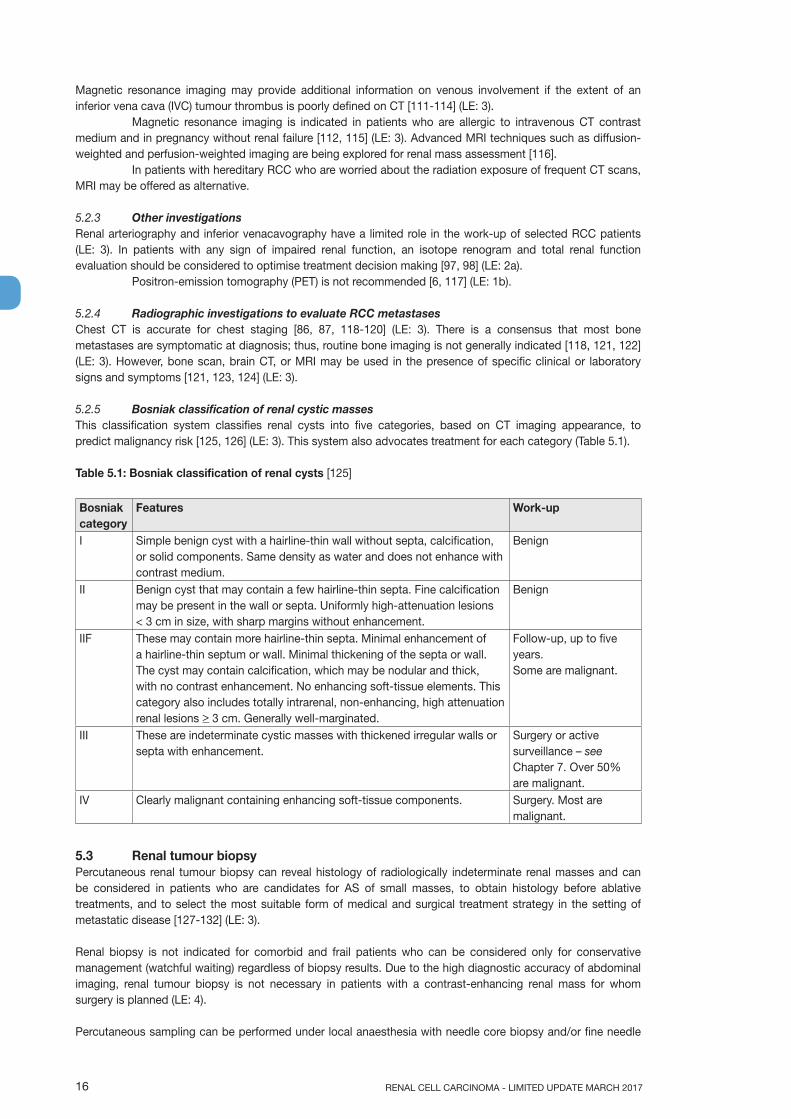
5.2.5 Bosniak classification of renal cystic massesThis classification system classifies renal cysts into five categories, based on CT imaging appearance, to predict malignancy risk [125, 126] (LE: 3). This system also advocates treatment for each category (Table 5.1).
Table 5.1: Bosniak classification of renal cysts [125]
Bosniak category
Features Work-up
I Simple benign cyst with a hairline-thin wall without septa, calcification, or solid components. Same density as water and does not enhance with contrast medium.
Benign
II Benign cyst that may contain a few hairline-thin septa. Fine calcification may be present in the wall or septa. Uniformly high-attenuation lesions < 3 cm in size, with sharp margins without enhancement.
Benign
IIF These may contain more hairline-thin septa. Minimal enhancement of a hairline-thin septum or wall. Minimal thickening of the septa or wall. The cyst may contain calcification, which may be nodular and thick, with no contrast enhancement. No enhancing soft-tissue elements. This category also includes totally intrarenal, non-enhancing, high attenuation renal lesions > 3 cm. Generally well-marginated.
Follow-up, up to five years. Some are malignant.
III These are indeterminate cystic masses with thickened irregular walls or septa with enhancement.
Surgery or active surveillance – see Chapter 7. Over 50% are malignant.
IV Clearly malignant containing enhancing soft-tissue components. Surgery. Most are malignant.
5.3 Renal tumour biopsyPercutaneous renal tumour biopsy can reveal histology of radiologically indeterminate renal masses and can be considered in patients who are candidates for AS of small masses, to obtain histology before ablative treatments, and to select the most suitable form of medical and surgical treatment strategy in the setting of metastatic disease [127-132] (LE: 3).
Renal biopsy is not indicated for comorbid and frail patients who can be considered only for conservative management (watchful waiting) regardless of biopsy results. Due to the high diagnostic accuracy of abdominal imaging, renal tumour biopsy is not necessary in patients with a contrast-enhancing renal mass for whom surgery is planned (LE: 4).
Percutaneous sampling can be performed under local anaesthesia with needle core biopsy and/or fine needle
17RENAL CELL CARCINOMA - LIMITED UPDATE MARCH 2017
aspiration (FNA). Biopsies can be performed with US or CT guidance, with a similar diagnostic yield [130, 133] (LE: 2b). Eighteen-gauge needles are ideal for core biopsies, as they result in low morbidity and provide sufficient tissue for diagnosis [127-131, 134] (LE: 2b). A coaxial technique allowing multiple biopsies through a coaxial cannula should always be used to avoid potential tumour seeding [127-131] (LE: 3).
Core biopsies should be preferred for the characterisation of solid renal masses (LE: 2a). A SR and meta-analysis of the diagnostic performance and complications of renal tumour biopsy (RTB) was recently performed by this Panel. Fifty-seven articles including a total of 5,228 patients were included in the analysis. Needle core biopsies were found to have better accuracy for the diagnosis of malignancy compared with FNA [135]. Other studies showed that solid pattern, larger tumour size and exophytic location are predictors of a diagnostic core biopsy [127, 130, 133] (LE: 2b).
In experienced centres, core biopsies have a high diagnostic yield, specificity, and sensitivity for the diagnosis of malignancy. The above-mentioned meta-analysis showed that sensitivity and specificity of diagnostic core biopsies for the diagnosis of malignancy are 99.1% and 99.7%, respectively [135] (LE: 2b). However, 0-22.6% of core biopsies are non-diagnostic (8% in the meta-analysis) [128-134, 136] (LE: 2a). If a biopsy is non-diagnostic, and radiologic findings are suspicious for malignancy, a further biopsy or surgical exploration should be considered (LE: 4). Repeat biopsies have been reported to be diagnostic in a high proportion of cases (83-100%) [127, 137-139].
Accuracy of RTBs for the diagnosis of tumour histotype is good. The median concordance rate between tumour histotype on RTBs and on the surgical specimen of the following PN or radical nephrectomy (RN) was 90.3% in the pooled analysis [135].
Assessment of tumour grade on core biopsies is challenging. In the pooled analysis the overall accuracy for nuclear grading was poor (62.5%), but significantly improved (87%) using a simplified two-tier system (high grade vs. low grade) [135] (LE: 2a).
The ideal number and location of core biopsies are not defined. However, at least two good quality cores should be obtained, and necrotic areas should be avoided to maximise diagnostic yield [127, 130, 140, 141] (LE: 4). Peripheral biopsies are preferable for larger tumours, to avoid areas of central necrosis [142] (LE: 2b). In cT2 or greater renal masses multiple core biopsies taken from at least four separate solid enhancing areas in the tumour were shown to achieve a higher diagnostic yield and a higher accuracy to identify sarcomatoid features without increasing the complication rate [143].
Core biopsies of cystic renal masses have a lower diagnostic yield and accuracy and are not recommended alone, unless areas with a solid pattern are present (Bosniak IV cysts) [127, 130, 135] (LE: 2b). Combined FNA and core biopsies can provide complementary results, especially for complex cystic lesions [131, 136, 137, 144, 145] (LE: 3).
Overall, percutaneous biopsies have a low morbidity [135]. Tumour seeding along the needle tract is anecdotal. Spontaneously resolving subcapsular/perinephric haematomas are reported in 4.3% of cases in a pooled analysis, but clinically significant bleeding is unusual (0-1.4%; 0.7% in the pooled analysis) and generally self-limiting [135].
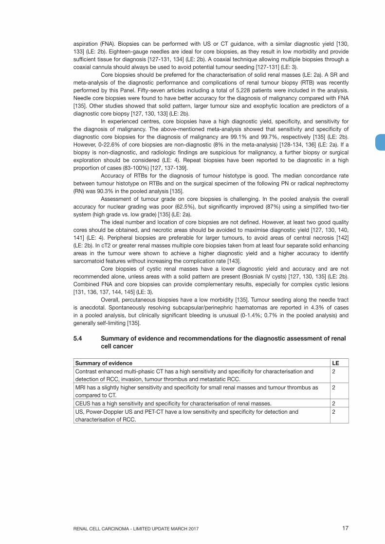
5.4 Summary of evidence and recommendations for the diagnostic assessment of renal cell cancer
Summary of evidence LEContrast enhanced multi-phasic CT has a high sensitivity and specificity for characterisation and detection of RCC, invasion, tumour thrombus and metastatic RCC.
2
MRI has a slightly higher sensitivity and specificity for small renal masses and tumour thrombus as compared to CT.
2
CEUS has a high sensitivity and specificity for characterisation of renal masses. 2US, Power-Doppler US and PET-CT have a low sensitivity and specificity for detection and characterisation of RCC.
2
RENAL CELL CARCINOMA - LIMITED UPDATE MARCH 201718
Recommendations gradeUse multi-phasic contrast-enhanced computed tomography (CT) for general staging and detection of RCC.
strong ↑↑
Use axial abdominal imaging and CT of the chest for staging of RCC. strong ↑↑
Use non-ionising modalities, mainly contrast enhanced ultrasound (CEUS), for further characterisation of small renal masses, tumour thrombus and differentiation of unclear renal masses.
weak ↑
Do not use bone scan and/or positron-emission tomography (PET)-CT for staging of RCC. weak ↓
Perform a renal tumour biopsy before ablative therapy and systemic therapy without previous pathology.
strong ↑↑
Perform a percutaneous biopsy in select patients who are considered for active surveillance. weak ↑
Use a coaxial technique when performing a renal tumour biopsy. strong ↑↑
Do not perform a renal tumour biopsy of cystic renal masses. weak ↓
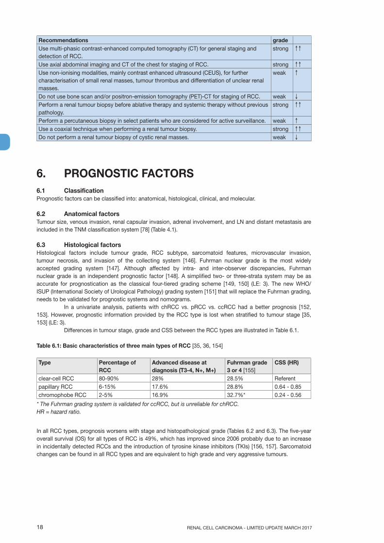
6. PROGNOSTIC FACTORS6.1 Classification Prognostic factors can be classified into: anatomical, histological, clinical, and molecular.
6.2 Anatomical factorsTumour size, venous invasion, renal capsular invasion, adrenal involvement, and LN and distant metastasis are included in the TNM classification system [78] (Table 4.1).
6.3 Histological factorsHistological factors include tumour grade, RCC subtype, sarcomatoid features, microvascular invasion, tumour necrosis, and invasion of the collecting system [146]. Fuhrman nuclear grade is the most widely accepted grading system [147]. Although affected by intra- and inter-observer discrepancies, Fuhrman nuclear grade is an independent prognostic factor [148]. A simplified two- or three-strata system may be as accurate for prognostication as the classical four-tiered grading scheme [149, 150] (LE: 3). The new WHO/ISUP (International Society of Urological Pathology) grading system [151] that will replace the Fuhrman grading, needs to be validated for prognostic systems and nomograms. In a univariate analysis, patients with chRCC vs. pRCC vs. ccRCC had a better prognosis [152, 153]. However, prognostic information provided by the RCC type is lost when stratified to tumour stage [35, 153] (LE: 3).
Differences in tumour stage, grade and CSS between the RCC types are illustrated in Table 6.1.
Table 6.1: Basic characteristics of three main types of RCC [35, 36, 154]
Type Percentage of RCC
Advanced disease at diagnosis (T3-4, N+, M+)
Fuhrman grade 3 or 4 [155]
CSS (HR)
clear-cell RCC 80-90% 28% 28.5% Referentpapillary RCC 6-15% 17.6% 28.8% 0.64 - 0.85chromophobe RCC 2-5% 16.9% 32.7%* 0.24 - 0.56
* The Fuhrman grading system is validated for ccRCC, but is unreliable for chRCC.HR = hazard ratio.
In all RCC types, prognosis worsens with stage and histopathological grade (Tables 6.2 and 6.3). The five-year overall survival (OS) for all types of RCC is 49%, which has improved since 2006 probably due to an increase in incidentally detected RCCs and the introduction of tyrosine kinase inhibitors (TKIs) [156, 157]. Sarcomatoid changes can be found in all RCC types and are equivalent to high grade and very aggressive tumours.

19RENAL CELL CARCINOMA - LIMITED UPDATE MARCH 2017
Table 6.2: Cancer-specific survival by stage and histopathological grade in RCCs - HR (95% CI) [36].
T1N0M0 ReferentT2N0M0 2.71 (2.17-3.39)T3N0M0 5.20 (4.36-6.21)T4N0M0 16.88 (12.40-22.98)N+M0 16.33 (12.89-20.73)M+ 33.23 (28.18-39.18)Grade 1 ReferentGrade 2 1.16 (0.94-1.42)Grade 3 1.97 (1.60-2.43)Grade 4 2.82 (2.08-3.31)
CI = confidential interval. HR = hazard ratio.
Long-term survival in RCC patients treated by RN or PN between 1970 and 2003; for unilateral, sporadic ccRCC, pRCC or chRCC in a cohort study [154] (Table 6.3).
Table 6.3: Cancer-specific survival of surgically treated patients by RCC type (estimated survival rate in percentage [95% CI])
Survival time 5 years (%) 10 years (%) 15 years (%) 20 years (%)clear-cell RCC 71 (69-73) 62 (60-64) 56 (53-58) 52 (49-55)papillary RCC 91 (88-94) 86 (82-89) 85 (81-89) 83 (78-88)chromophobe RCC 88 (83-94) 86 (80-92) 84 (77-91) 81 (72-90)
Two subgroups of pRCC with different outcomes have been identified [158]. Type 1 have a favourable prognosis. Type 2 are mostly high-grade tumours with a propensity for metastases (LE: 3). For more details, see Section 3.2 Histological diagnosis. Renal cell cancer with Xp 11.2 translocation has a poor prognosis [159]. Its incidence is low, but it should be systematically addressed in young patients. Renal cell cancer type classification has been confirmed by cytogenetic and genetic analyses [155, 160, 161] (LE: 2b).
6.4 Clinical factorsThese include performance status (PS), local symptoms, cachexia, anaemia, platelet count, neutrophil/lymphocyte ratio, C-reactive protein (CRP) and albumin [95, 162-166] (LE: 3).
6.5 Molecular factorsNumerous molecular markers such as carbonic anhydrase IX (CaIX), vascular endothelial growth factor (VEGF), hypoxia-inducible factor (HIF), Ki67 (proliferation), p53, p21 [167], PTEN (phosphatase and tensin homolog) (cell cycle), E-cadherin, osteopontin [168] CD44 (cell adhesion) [169, 170], CXCR4 [171], and other cell cycle and proliferative markers [64, 172] are being investigated (LE: 3). None of these markers have clearly improved the predictive accuracy of current prognostic systems, none have been externally validated, and their routine use in clinical practice is at present not recommended. Several retrospective studies and large molecular screening programmes have identified mutated genes in ccRCC with distinct clinical outcomes. The expression of the BAP1 and PBRM1 genes, situated on chromosome 3p in a region that is deleted in more than 90% of ccRCCs, have shown to be independent prognostic factors for tumour recurrence [173-175]. These published reports suggest that patients with BAP1-mutant tumours have worse outcomes compared with patients with PBRM1-mutant tumours [174]. Validated data from surgical series can predict relapse using a sixteen gene signature. This signature is likely to be adopted in clinical trials and may be helpful in the clinical setting in due time [176].
The recognition of the potential relevance of immunotherapy as an approach to RCC management is growing. Prognostic information of cytokines and blockade of immune-inhibitory molecules such as PD-L1 have shown promising therapeutic results. Emerging evidence of chromosomal alterations, through Genome-Wide Association Studies (GWAS), miRNA, SNPs and gene methylations all contribute to improving diagnostic and prognostic information. A number of studies have confirmed prognostic information based on gain of chromosomal regions 7q, 8q and 20q, and chromosomal losses of regions 9p, 9q and 14q, which are associated with poor survival. CpG-methylation-based assays also independently predict survival in ccRCC [177, 178]. An international collaboration is currently investigating GWAS loci for prognostic information.
RENAL CELL CARCINOMA - LIMITED UPDATE MARCH 201720
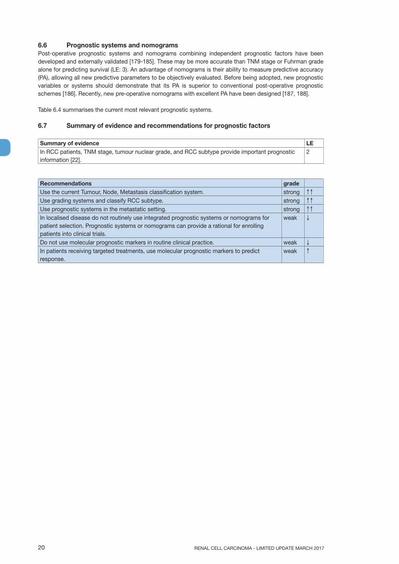
6.6 Prognostic systems and nomogramsPost-operative prognostic systems and nomograms combining independent prognostic factors have been developed and externally validated [179-185]. These may be more accurate than TNM stage or Fuhrman grade alone for predicting survival (LE: 3). An advantage of nomograms is their ability to measure predictive accuracy (PA), allowing all new predictive parameters to be objectively evaluated. Before being adopted, new prognostic variables or systems should demonstrate that its PA is superior to conventional post-operative prognostic schemes [186]. Recently, new pre-operative nomograms with excellent PA have been designed [187, 188].
Table 6.4 summarises the current most relevant prognostic systems.
6.7 Summary of evidence and recommendations for prognostic factors
Summary of evidence LEIn RCC patients, TNM stage, tumour nuclear grade, and RCC subtype provide important prognostic information [22].
2
Recommendations gradeUse the current Tumour, Node, Metastasis classification system. strong ↑↑
Use grading systems and classify RCC subtype. strong ↑↑
Use prognostic systems in the metastatic setting. strong ↑↑
In localised disease do not routinely use integrated prognostic systems or nomograms for patient selection. Prognostic systems or nomograms can provide a rational for enrolling patients into clinical trials.
weak ↓
Do not use molecular prognostic markers in routine clinical practice. weak ↓
In patients receiving targeted treatments, use molecular prognostic markers to predict response.
weak ↑

21RENAL CELL CARCINOMA - LIMITED UPDATE MARCH 2017
Tabl
e 6.
4: A
nato
mic
al, h
isto
logi
cal,
and
clin
ical
var
iabl
es in
the
com
mon
ly u
sed
prog
nost
ic m
odel
s fo
r lo
calis
ed a
nd m
etas
tatic
RC
C
Pro
gnos
tic
Mod
els
Vari
able
s
TNM
S
tage
ECO
GP
SK
arno
fsky
P
SR
CC
re
late
d sy
mpt
oms
Fuhr
man
gr
ade
Tum
our
necr
osis
Tum
our
size
Del
ay
betw
een
diag
nosi
s an
d tr
eatm
ent
LDH
Cor
rect
ed
calc
ium
Hae
mog
lobi
nN
eutr
ophi
l co
unt
Pla
tele
t co
unt
Loca
lised
R
CC
UIS
Sx
xx
SS
IGN
xx
xx
Pos
t-op
erat
ive
Kar
akie
wic
z’s
nom
ogra
m
xx
xx
Met
asta
tic
RC
CM
SK
CC
pr
ogno
stic
sy
stem
xx
xx
x
IMD
Cx
xx
xx
xH
eng’
s m
odel
xx
xx
xx
ECO
G-P
S =
Eas
tern
Coo
pera
tive
Onc
olog
y G
roup
- p
erfo
rman
ce s
tatu
s; IM
DC
= In
tern
atio
nal M
etas
tatic
Ren
al C
ance
r Dat
abas
e C
onso
rtiu
m; L
DH
= la
ctat
e de
hydr
ogen
ase;
MS
KC
C =
Mem
oria
l Slo
an K
ette
ring
Can
cer C
ente
r; P
S =
per
form
ance
sta
tus;
SS
IGN
= S
tage
Siz
e G
rade
Nec
rosi
s; U
ISS
= U
nive
rsity
of C
alifo
rnia
Los
Ang
eles
inte
grat
ed
stag
ing
syst
em.

RENAL CELL CARCINOMA - LIMITED UPDATE MARCH 201722
7. DISEASE MANAGEMENT7.1 Treatment of localised RCC7.1.1 IntroductionA SR underpins the findings of sections 7.1.2 to 7.2.4.2. The review included all relevant published literature comparing surgical management of localised RCC (T1-2N0M0) [189, 190]. Randomised or quasi-RCTs were included. However, due to the very limited number of RCTs, non-randomised studies (NRS), prospective observational studies with controls, retrospective matched-pair studies, and comparative studies from the databases of well-defined registries were also included.
7.1.2 Surgical treatment7.1.2.1 Nephron-sparing surgery vs. radical nephrectomyMultiple retrospective series as well as one prospective RCT including patients with organ-confined RCC of limited size, respectively T-stage (pT1), have demonstrated a comparable CSS for PN vs. RN [191-195]. However, trials that directly compared both approaches in terms of their oncological safety are rarely available, therefore, the data presented is based on a comparison of data available from retrospective series that have included patient cohorts of different and, in part, limited size.
In addition, PN vs. RN was demonstrated to better preserve general kidney function, thereby lowering the risk of development of metabolic or cardiovascular disorders.
When compared with a radical surgical approach, for NSS, several retrospective analyses of large databases have suggested a decreased cardiac-specific mortality [196, 197] as well as improved OS as compared to RN. However, in some series this held true only for a younger patient population and/or patients without significant comorbidity at the time of the surgical intervention [198, 199]. An analysis of the Medicare database [200] could not demonstrate an OS benefit for patients > 75 years of age when RN or PN were compared with non-surgical management. Another series that addressed this question and also included Medicare patients suggested an OS benefit in an older RCC patient population (75-80 years) when subjected to surgery rather than non-surgical management. Shuch et al. compared patients subjected to PN for RCC with a non-cancer, healthy control group via a retrospective database analysis, showing an OS benefit for the cancer cohort, [201]. These conflicting results indicate that unknown statistical confounders hamper the retrospective analysis of population-based tumour registries.
In contrast, the only prospectively randomised but prematurely closed and heavily underpowered, trial available so far did not demonstrate an inferiority of RN vs. PN in terms of OS. Taken together, the OS advantage suggested for PN vs. RN remains an unresolved issue.
It has been suggested that the more pronounced deterioration of renal function after RN negatively affects patients´ OS [98, 202]. Patients with a normal pre-operative renal function and a decreased GFR due to surgical treatment, generally present with a stable renal function longer term [203]. In contrast, adverse OS in patients with a pre-existing GFR reduction does not seem to result from further renal function impairment following surgery, but rather from other medical comorbidities causing pre-surgical CKD. However, in particular in patients with pre-existing CKD, PN is the treatment of choice to limit the risk of development of ESKD which requires haemodialysis.
Only a limited number of studies are available addressing quality of life (QoL) following PN vs. RN irrespective of the surgical approach used (open- vs. minimally invasive). Quality of life was ranked higher following PN as compared to RN, but in general, patients’ health status deteriorated following both approaches [191, 192, 194, 204-208].
In terms of the intra- and peri-operative morbidity/complications associated with PN vs. RN, there was no difference in the length of hospital stay [192, 193, 207], the number of red blood cell (RBC) units applied [192, 207, 208], or the mean intra-operative blood loss [192, 207]. Complication rates were inconsistently reported and one intervention was not favoured over another [209]. One study indicated a longer operation time for open PN [209], but this was not confirmed by others [210].
In view of the above and since oncological safety (CSS and FS) of PN has been proven to be similar for RN, PN is the treatment of choice for T1b RCC since it preserves kidney function better and in the long term limits development of metabolic as well as cardiovascular disorders. Whether decreased mortality from any cause can be attributed to PN is still unresolved, but in patients with pre-existing CKD, PN is the preferred surgical treatment option as it avoids further deterioration of kidney function, the latter being associated with a higher risk of development of ESKD and the need for haemodialysis.

23RENAL CELL CARCINOMA - LIMITED UPDATE MARCH 2017
Partial nephrectomy is unsuitable in some patients with localised RCC due to:• insufficient volume of remaining parenchyma to maintain proper organ function; • renal vein thrombosis;• unfavourable tumour location e.g. adherence to the renal vessels; • use of anticoagulants.
In these situations the curative therapy is RN including removal of the tumour-bearing kidney. Complete resection of the primary tumour by open- or laparoscopic surgery offers a reasonable chance of cure.
7.1.2.2 Associated procedures7.1.2.2.1 AdrenalectomyOne prospective NRS compared the outcomes of RN or PN with, or without, ipsilateral adrenalectomy [211]. Multivariate analysis showed that upper pole location was not predictive of adrenal involvement, but tumour size was. No difference in OS at five or ten years was seen, with, or without, adrenalectomy. Adrenalectomy was justified using criteria based on radiographic and intra-operative findings. Only 48 of 2,065 patients underwent concurrent ipsilateral adrenalectomy of which 42 were for benign lesions.
7.1.2.2.2 Lymph node dissection for clinically negative lymph nodes (cN0)The indication for lymph node dissection (LND) together with PN or RN is still controversial [212]. The clinical assessment of LN status is based on the detection of an enlargement of LNs; either by CT/MRI or the intra-operative palpability of enlarged nodes. Less than 20% of suspected metastatic nodes (cN+) are positive for metastatic disease at histopathological examination (pN+) [213]. Both CT and MRI are unsuitable for detecting malignant disease in nodes of normal shape and size [214]. For clinically positive LNs (cN+) see Section 7.2.2.
For patients with clinically negative LNs (cN0) six clinical trials have evaluated the clinical value of LND [212], the latter including one RCT [213] and five comparative studies [215-219]. Smaller retrospective studies have suggested a clinical benefit associated with a more or less extensive lymphadenectomy preferably in patients at high risk for lymphogenic spread. The number of LN metastases (< / > 4) as well as the intra– and extracapsular extension of intranodal metastasis correlated with the patients´ clinical prognosis in some studies [214, 220-222]. Better survival outcomes were seen in patients with a low number of positive LNs (< 4) and no extranodal extension. On the basis of a retrospective SEER database analysis of > 9,000 patients no effects of an extended LND on the disease-specific survival (DSS) of patients with pathologically confined negative nodes was demonstrated [223]. However, in patients with pathologically proven lymphogenic spread (pN+), an increase of ten for the number of nodes dissected resulted in a 10% absolute increase in DSS. In addition, in a larger cohort of 1,983 patients Capitano et al. demonstrated that extended LND results in a significant prolongation of CSS in patients with unfavourable prognostic features (e.g., sarcomatoid differentiation, large tumour size) [224].
Only one prospective RCT evaluating the clinical value of LND combined with surgical treatment of primary RCC has been published so far. With an incidence of only 4%, lymphatic spread appears to be very low. Recognising the latter, only a staging effect was attributed to a (super)extended LND [213]. This trial included a very high percentage of patients with pT2 tumours, which are not at increased risk for LN metastases. Additionally, only 25% of patients with pT3 tumours were subjected to a complete LND. The LN template used by the authors was also not clearly stated.
The most optimal surgical approach remains controversial. Retrospective studies suggest that an extended LND should involve the LNs surrounding the ipsilateral great vessel and the inter-aortocaval region from the crus of the diaphragm to the common iliac artery. Involvement of inter-aortocaval LNs without regional hilar involvement is reported in up to 35-45% of cases [214, 215, 225]. At least fifteen LNs should be removed [224, 226]. Sentinel LND is an investigational technique [227, 228].
7.1.2.2.3 EmbolisationBefore routine nephrectomy, tumour embolisation has no benefit [229, 230]. In patients unfit for surgery, or with non-resectable disease, embolisation can control symptoms, including visible haematuria or flank pain [231-233]. These indications will be repeated in Sections 7.2 and 7.3 with cross reference to the summary of evidence and recommendations below.
RENAL CELL CARCINOMA - LIMITED UPDATE MARCH 201724
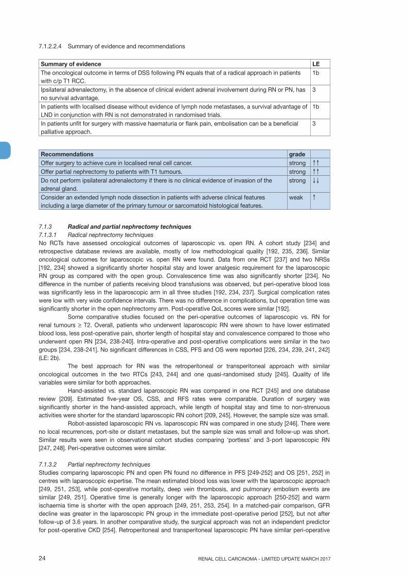
7.1.2.2.4 Summary of evidence and recommendations
Summary of evidence LEThe oncological outcome in terms of DSS following PN equals that of a radical approach in patients with c/p T1 RCC.
1b
Ipsilateral adrenalectomy, in the absence of clinical evident adrenal involvement during RN or PN, has no survival advantage.
3
In patients with localised disease without evidence of lymph node metastases, a survival advantage of LND in conjunction with RN is not demonstrated in randomised trials.
1b
In patients unfit for surgery with massive haematuria or flank pain, embolisation can be a beneficial palliative approach.
3
Recommendations gradeOffer surgery to achieve cure in localised renal cell cancer. strong ↑↑
Offer partial nephrectomy to patients with T1 tumours. strong ↑↑
Do not perform ipsilateral adrenalectomy if there is no clinical evidence of invasion of the adrenal gland.
strong ↓↓
Consider an extended lymph node dissection in patients with adverse clinical features including a large diameter of the primary tumour or sarcomatoid histological features.
weak ↑
7.1.3 Radical and partial nephrectomy techniques 7.1.3.1 Radical nephrectomy techniquesNo RCTs have assessed oncological outcomes of laparoscopic vs. open RN. A cohort study [234] and retrospective database reviews are available, mostly of low methodological quality [192, 235, 236]. Similar oncological outcomes for laparoscopic vs. open RN were found. Data from one RCT [237] and two NRSs [192, 234] showed a significantly shorter hospital stay and lower analgesic requirement for the laparoscopic RN group as compared with the open group. Convalescence time was also significantly shorter [234]. No difference in the number of patients receiving blood transfusions was observed, but peri-operative blood loss was significantly less in the laparoscopic arm in all three studies [192, 234, 237]. Surgical complication rates were low with very wide confidence intervals. There was no difference in complications, but operation time was significantly shorter in the open nephrectomy arm. Post-operative QoL scores were similar [192].
Some comparative studies focused on the peri-operative outcomes of laparoscopic vs. RN for renal tumours > T2. Overall, patients who underwent laparoscopic RN were shown to have lower estimated blood loss, less post-operative pain, shorter length of hospital stay and convalescence compared to those who underwent open RN [234, 238-240]. Intra-operative and post-operative complications were similar in the two groups [234, 238-241]. No significant differences in CSS, PFS and OS were reported [226, 234, 239, 241, 242] (LE: 2b).
The best approach for RN was the retroperitoneal or transperitoneal approach with similar oncological outcomes in the two RTCs [243, 244] and one quasi-randomised study [245]. Quality of life variables were similar for both approaches.
Hand-assisted vs. standard laparoscopic RN was compared in one RCT [245] and one database review [209]. Estimated five-year OS, CSS, and RFS rates were comparable. Duration of surgery was significantly shorter in the hand-assisted approach, while length of hospital stay and time to non-strenuous activities were shorter for the standard laparoscopic RN cohort [209, 245]. However, the sample size was small.
Robot-assisted laparoscopic RN vs. laparoscopic RN was compared in one study [246]. There were no local recurrences, port-site or distant metastases, but the sample size was small and follow-up was short. Similar results were seen in observational cohort studies comparing ‘portless’ and 3-port laparoscopic RN [247, 248]. Peri-operative outcomes were similar.
7.1.3.2 Partial nephrectomy techniquesStudies comparing laparoscopic PN and open PN found no difference in PFS [249-252] and OS [251, 252] in centres with laparoscopic expertise. The mean estimated blood loss was lower with the laparoscopic approach [249, 251, 253], while post-operative mortality, deep vein thrombosis, and pulmonary embolism events are similar [249, 251]. Operative time is generally longer with the laparoscopic approach [250-252] and warm ischaemia time is shorter with the open approach [249, 251, 253, 254]. In a matched-pair comparison, GFR decline was greater in the laparoscopic PN group in the immediate post-operative period [252], but not after follow-up of 3.6 years. In another comparative study, the surgical approach was not an independent predictor for post-operative CKD [254]. Retroperitoneal and transperitoneal laparoscopic PN have similar peri-operative

25RENAL CELL CARCINOMA - LIMITED UPDATE MARCH 2017
outcomes [255]. Simple tumour enucleation also had similar PFS and CSS rates compared to standard PN and RN in a large study [256, 257].
Hand-assisted laparoscopic PN (HALPN) is rarely performed. A recent comparative study of open vs. HALPN showed no difference in OS or RFS at intermediate-term follow-up. The authors observed a lower rate of intra-operative and all-grade post-operative 30-day complications in HALPN than in open PN patients, but there was no significant difference in high Clavien Grade complications. Glomerular filtration rate three months after operation was lower in the HALPN than in the open PN group [258].
The feasibility of off-clamp laparoscopic PN and laparo-endoscopic single-site PN has been shown in selected patients but larger studies are needed to confirm their safety and clinical role [259, 260].
At present, the oncological outcomes of robot-assisted vs. laparoscopic or open PN have been compared only in studies with short-term follow-up. One recent study prospectively compared the peri-operative outcomes of a series of robot-assisted and open PN performed by the same experienced surgeon. Robot-assisted PN was superior to open PN in terms of lower estimated blood loss and shorter hospital stay. Warm ischaemia time, operative time, immediate- early- and short-term complications, variation of creatinine levels, and pathologic margins were similar among the groups [261].
A recent meta-analysis, including a series of NSS, with variable methodological quality compared the peri-operative outcomes of robot-assisted and laparoscopic PN. The robotic group had a significantly lower rate of conversion to open surgery and to radical surgery, shorter warm ischaemia time, smaller change in estimated GFR after surgery and shorter length of stay. No significant difference was observed between the two groups regarding complications, change of serum creatinine after surgery, operative time, estimated blood loss and positive surgical margins (PSMs) [262].
7.1.3.3 Positive margins on histopathological specimens of resected tumours A positive surgical margin is encountered in about 8% of PNs [263]. Studies comparing different resection techniques (open, laparoscopic, robotic) are inconclusive [264, 265]. Most trials showed that intra-operative frozen section analysis had no influence on the risk of definite PSMs [266]. A PSM status occurs more frequently in cases in which surgery is imperative, including bilateral tumours [267, 268]. Positive surgical margins increase the risk of disease recurrence, primarily in patients with adverse pathological features (pT2a-pT3a, grade III-IV) [263, 267, 268]. The effect of margin status on long-term oncologic outcomes remains to be determined [264], but PSMs need not translate into worse CSS [267, 268]. Therefore, RN or re-resection of margins presents overtreatment in many cases, but a small percentage of patients will harbour residual malignancy [269]. Patients with PSMs should be informed that they will be subjected to a more intense surveillance (imaging) programme and are at increased risk for secondary local therapies [267, 270]. However, protection from recurrence is not ensured by negative surgical margins [271].
7.1.3.4 Summary of evidence and recommendations
Summary of evidence LELaparoscopic radical nephrectomy has lower morbidity than open surgery. 1bOncological outcomes for T1-T2a tumours are equivalent between laparoscopic and open radical nephrectomy.
2a
Partial nephrectomy can be performed, either with an open, pure laparoscopic or robot-assisted approach, based on surgeon’s expertise and skills.
2b
Partial nephrectomy is associated with a higher percentage of positive surgical margins compared with radical nephrectomy.
3
Recommendations gradeOffer laparoscopic radical nephrectomy to patients with T2 tumours and localised masses not treatable by partial nephrectomy.
strong ↑↑
Do not perform radical nephrectomy in patients with T1 tumours for whom partial nephrectomy is indicated.
strong ↓↓

RENAL CELL CARCINOMA - LIMITED UPDATE MARCH 201726
7.1.4 Therapeutic approaches as alternatives to surgery7.1.4.1 Surgical versus non-surgical treatmentPopulation-based studies compared the oncological outcomes of surgery (RN or PN) and non-surgical management for tumours < 4 cm. The analyses showed a significantly lower cancer-specific mortality in patients treated with surgery [200, 272]. However, the patients assigned to the surveillance arm were older and likely to be frailer and less suitable candidates for surgery. Other-cause mortality rates in the non-surgical group significantly exceeded that of the surgical group [272]. Analyses of older patients (> 75 years) failed to show the same benefit in cancer-specific mortality for surgical treatment [273-275].
7.1.4.2 SurveillanceElderly and comorbid patients with incidental small renal masses have a low RCC-specific mortality and significant competing-cause mortality [276, 277]. Active surveillance is defined as the initial monitoring of tumour size by serial abdominal imaging (US, CT, or MRI) with delayed intervention reserved for tumours showing clinical progression during follow-up [278]. The concept of AS differs from the concept of watchful waiting. Watchful waiting is reserved for patients whose comorbidities contraindicate any subsequent active treatment and do not require follow-up imaging, unless clinically indicated.
In the largest reported series of AS, the growth of renal tumours was low and progression to metastatic disease was reported in only a limited number of patients [279, 280].
A single-institutional comparative study evaluating patients aged > 75 years showed decreased OS for those who underwent surveillance and nephrectomy relative to NSS for clinically T1 renal tumours. However, patients selected for surveillance were older with greater comorbidity. At multi-variate analysis, management type was not associated with OS after adjusting for age, comorbidity, and other variables [276]. No statistically significant difference in OS and CSS were observed in another study of RN vs. PN vs. AS for T1a renal masses with a follow-up of 34 months [281].
The initial results of the multi-institutional Delayed Intervention and Surveillance for Small Renal Masses (DISSRM) registry were recently published. This prospective, NRS enrolled 497 patients with solid renal masses < 4 cm in size who chose AS or primary active intervention. Patients who selected AS were older, had worse ECOG scores, more comorbidities, smaller tumours, and more often multiple and bilateral lesions. Overall survival for primary intervention and AS was 98% and 96% at two years, and 92% and 75% at five years, respectively (p = 0.06). At five years, CSS was 99% and 100%, respectively (p = 0.3). Active surveillance was not predictive of OS or CSS in regression modelling with relatively short follow up [282].
Overall, both short- and intermediate-term oncological outcomes indicate that in selected patients with advanced age and/or comorbidities, AS is appropriate to initially monitor small renal masses, followed, if required, by treatment for progression [278-280, 283-286].
A multicentre study assessed QoL of patients undergoing immediate intervention vs. AS. Patients undergoing immediate intervention had higher QoL scores at baseline, specifically for physical health. The perceived benefit in physical health persisted for at least one year following intervention. Mental health, which includes domains of depression and anxiety, was not adversely affected while on AS [287].
7.1.4.3 Ablative therapies7.1.4.3.1 CryoablationCryoablation is performed using either a percutaneous or a laparoscopic-assisted approach. In comparative studies, there was no significant difference in the overall complication rates between laparoscopic and percutaneous cryoablation [288-290]. One comparative study reported similar OS, CSS, and RFS in 172 laparoscopic patients with a longer follow up compared with 123 patients treated percutaneously with a shorter follow up [289]. A shorter average length of hospital stay was found with the percutaneous technique [289, 290]. No studies are available comparing surveillance strategies to cryoablation.
A recent SR including 82 articles reported complication rates ranging between 8 and 20% with most complications being minor [291]. Although a precise definition of tumour recurrence is lacking, the authors reported a lower RFS as compared to that of PN.
7.1.4.3.2 Cryoablation versus partial nephrectomyStudies compared open, laparoscopic or robotic PN with percutaneous or laparoscopic cryoablation. Oncological outcomes were mixed, with some studies showing no difference in OS, CSS, RFS, DFS, local recurrence or progression to metastatic disease [292, 293], with some showing significant benefit for the PN techniques for some or all of these outcomes [294-297]. Not all studies reported all outcomes listed, and some were small and included benign tumours. No study showed an oncological benefit for cryoablation over PN.
Peri-operative outcomes, complication rates and other QoL measures were mixed. Some studies found the length of hospital stay was shorter and surgical blood loss was less with cryoablation [292-294], whilst also finding no differences in other peri-operative outcomes such as recovery times, complication rates

27RENAL CELL CARCINOMA - LIMITED UPDATE MARCH 2017
or post-operative serum creatinine levels. Two studies [296, 297] reported specific Clavien rates, with mostly non-significant differences, which were mixed for intra-operative vs. post-operative complications. Estimated GFRs were not significantly different in two of the studies, but in favour of cryoablation in a third [295-297]. Estimates of new CKD were also mixed, with one study in favour of cryoablation [295], another strongly in favour of PN [296], and the third showing no difference [297]. One study compared PN with ablation therapy, either cryoablation or RFA [223], and showed significantly improved DSS at both five and ten years for PN.
A recent study compared 1,057 patients treated by PN to 180 treated by RFA and 187 treated by cryoablation for a cT1 tumour and found no difference regarding RFS between the three techniques. Metastasis-free survival was superior after PN and cryoablation compared to RFA for cT1a patients. However, follow-up of patients treated by thermal ablations was shorter [198].
7.1.4.3.3 Radiofrequency ablationRadiofrequency ablation is performed laparoscopically or percutaneously. Four studies compared patients with T1a tumours treated by laparoscopic or percutaneous RFA [298-301].
Complications occurred in up to 29% of patients but were mostly minor. Complication rates were similar in patients treated laparoscopically or percutaneously. One study with a limited number of patients [300] found a higher rate of incomplete ablation in patients treated by percutaneous RFA. However, no differences in recurrence or CSS were found in the three comparative studies.
7.1.4.3.4 Radiofrequency ablation versus partial nephrectomyMost publications about RFA are retrospective cohort studies with a low number of patients and limited follow up. Three studies retrospectively compared RFA to surgery in patients with T1a tumours [302-304].
One study [303] compared T1a patients who underwent either RFA (percutaneous or laparoscopic) or PN and found no difference in OS and CSS. Another study retrospectively reviewed 105 T1a patients treated by percutaneous RFA or RN. Cancer-specific survival was 100% in both groups. Overall survival was lower in the RFA group but patients treated with surgery were younger [302].
In a monocentric study that compared 34 RFA patients to sixteen open PN patients, a higher rate of complications and transfusions was shown in the PN group. Although the tumours were larger in PN patients, progression rates were similar (0%) [304].
A recent meta-analysis [305] reported comparable complication rates and post-operative eGFRs between RFA and PN. The local tumour recurrence rate was higher in the RFA group than in the PN group (OR = 1.81) but there was no difference regarding the occurrence of distant metastasis.
7.1.4.3.5 Cryoablation versus radiofrequency ablationTwo studies compared RFA and cryoablation [306, 307]. No significant differences were reported for OS, CSS, or RFS in either study. For local RFS at five years, one study [306] reported improvement with RFA, while the other [307] reported a benefit with cryoablation. One study [306] reported no differences in Clavien complication rates between the techniques.
7.1.4.3.6 Other ablative techniquesSome studies have shown the feasibility of other ablative techniques, such as microwave ablation, laser ablation, and high-intensity focused US ablation. However, these techniques are considered experimental.
7.1.4.3.7 Summary of evidence and recommendation for therapeutic approaches as alternative to surgery
Summary of evidence LEMost population-based analyses show a significantly lower cancer-specific mortality for patients treated with surgery compared to non-surgical management.
3
In active surveillance cohorts, the growth of small renal masses is low in most cases and progression to metastatic disease is rare (1-2%).
3
Quality of the available data does not allow definitive conclusions regarding morbidity and oncological outcomes of cryoablation and radiofrequency ablation.
3
Low quality studies suggest a higher local recurrence rate for thermal ablation therapies compared to partial nephrectomy.
3

RENAL CELL CARCINOMA - LIMITED UPDATE MARCH 201728
Recommendation gradeOffer active surveillance, radiofrequency ablation and cryoablation to elderly and/or comorbid patients with small renal masses.
weak ↑
7.2 Treatment of locally advanced RCC7.2.1 IntroductionIn addition to the summary of evidence and recommendations outlined in Section 7.1 for localised RCC, certain therapeutic strategies arise in specific situations for locally advanced disease.
7.2.2 Management of clinically positive lymph nodes (cN+)In the presence of clinically positive LNs (cN+), LND is always justified [38]. However, the extent of LND remains controversial [214].
7.2.3 Management of locally advanced unresectable RCCIn patients with non-resectable disease, embolisation can control symptoms, including visible haematuria or flank pain [231-233]. The use of neoadjuvant targeted therapy to downsize tumours is experimental and cannot be recommended outside clinical trials.
7.2.4 Management of RCC with venous tumour thrombusTumour thrombus formation in the IVC in RCC patients is a significant adverse prognostic factor. Traditionally, patients with venous tumour thrombus undergo surgery to remove the kidney and tumour thrombus. Aggressive surgical resection is widely accepted as the default management option for patients with venous tumour thrombus [308-316]. However, uncertainties remain over the best approach for surgical treatment of these patients.
7.2.4.1 The evidence base for surgery in patients with venous tumour thrombus The data on whether patients with venous tumour thrombus should undergo surgery is derived from case series. In one of the largest published studies [313] a higher level of thrombus was not associated with increased tumour dissemination to LNs, perinephric fat or distant metastasis. Thus, all patients with non-metastatic disease and venous tumour thrombus, and an acceptable PS, should be considered for surgical intervention, irrespective of the extent of tumour thrombus at presentation (LE: 3). The surgical technique and approach for each case should be selected based on the extent of tumour thrombus (LE: 3).
7.2.4.2 The evidence base for different surgical strategiesA SR was undertaken which included comparison-only studies on the management of venous tumour thrombus, in non-metastatic RCC [317, 318]. Only five studies were eligible for final inclusion, with high risks of bias across all studies.
Minimal access techniques resulted in significantly shorter operating time compared with traditional median sternotomy [319, 320]. Pre-operative embolisation [321] was associated with increased operating time, blood loss, hospital stay and peri-operative mortality in patients with T3 RCC.
No significant differences in oncological and process outcomes were observed between cardiopulmonary bypass with deep hypothermic circulatory arrest or partial bypass under normothermia or single caval clamp without circulatory support [322].
No surgical method was shown to be superior for the excision of venous tumour thrombus. The surgical method was dependent on the level of tumour thrombus, and the grade of occlusion of the IVC [317, 319, 320, 322]. The relative benefits and harms of other strategies and approaches regarding access to the IVC and the role of IVC filters and bypass procedures remain uncertain.
7.2.4.3 Summary of evidence and recommendations for the management of RCC with venous tumour thrombus
Summary of evidence LEIn patients with locally advanced disease due to clinically enlarged lymph nodes, the survival benefit of lymph node dissection is unclear but lymph node dissection can add staging information.
3
Low quality data suggest that tumour thrombus excision in non-metastatic disease may be beneficial. 3Tumour embolisation or inferior vena cava filter do not appear to offer any benefits. 3
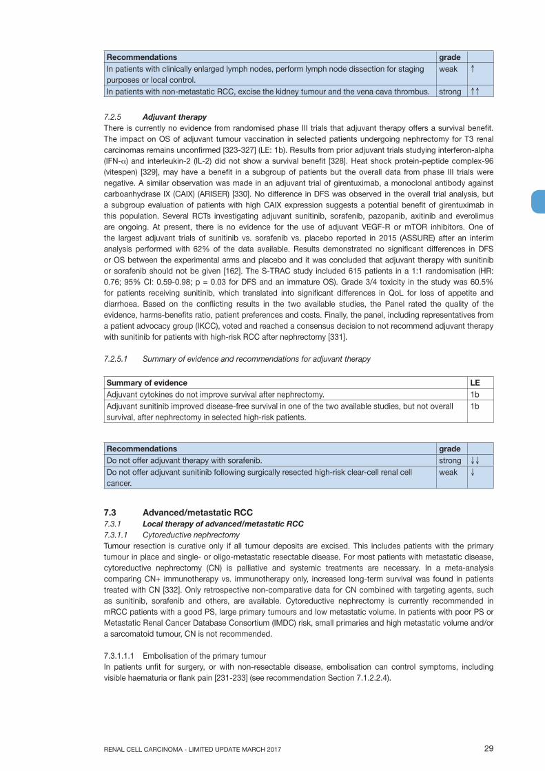
29RENAL CELL CARCINOMA - LIMITED UPDATE MARCH 2017
Recommendations gradeIn patients with clinically enlarged lymph nodes, perform lymph node dissection for staging purposes or local control.
weak ↑
In patients with non-metastatic RCC, excise the kidney tumour and the vena cava thrombus. strong ↑↑
7.2.5 Adjuvant therapyThere is currently no evidence from randomised phase III trials that adjuvant therapy offers a survival benefit. The impact on OS of adjuvant tumour vaccination in selected patients undergoing nephrectomy for T3 renal carcinomas remains unconfirmed [323-327] (LE: 1b). Results from prior adjuvant trials studying interferon-alpha (IFN-α) and interleukin-2 (IL-2) did not show a survival benefit [328]. Heat shock protein-peptide complex-96 (vitespen) [329], may have a benefit in a subgroup of patients but the overall data from phase III trials were negative. A similar observation was made in an adjuvant trial of girentuximab, a monoclonal antibody against carboanhydrase IX (CAIX) (ARISER) [330]. No difference in DFS was observed in the overall trial analysis, but a subgroup evaluation of patients with high CAIX expression suggests a potential benefit of girentuximab in this population. Several RCTs investigating adjuvant sunitinib, sorafenib, pazopanib, axitinib and everolimus are ongoing. At present, there is no evidence for the use of adjuvant VEGF-R or mTOR inhibitors. One of the largest adjuvant trials of sunitinib vs. sorafenib vs. placebo reported in 2015 (ASSURE) after an interim analysis performed with 62% of the data available. Results demonstrated no significant differences in DFS or OS between the experimental arms and placebo and it was concluded that adjuvant therapy with sunitinib or sorafenib should not be given [162]. The S-TRAC study included 615 patients in a 1:1 randomisation (HR: 0.76; 95% CI: 0.59-0.98; p = 0.03 for DFS and an immature OS). Grade 3/4 toxicity in the study was 60.5% for patients receiving sunitinib, which translated into significant differences in QoL for loss of appetite and diarrhoea. Based on the conflicting results in the two available studies, the Panel rated the quality of the evidence, harms-benefits ratio, patient preferences and costs. Finally, the panel, including representatives from a patient advocacy group (IKCC), voted and reached a consensus decision to not recommend adjuvant therapy with sunitinib for patients with high-risk RCC after nephrectomy [331].
7.2.5.1 Summary of evidence and recommendations for adjuvant therapy
Summary of evidence LEAdjuvant cytokines do not improve survival after nephrectomy. 1bAdjuvant sunitinib improved disease-free survival in one of the two available studies, but not overall survival, after nephrectomy in selected high-risk patients.
1b
Recommendations gradeDo not offer adjuvant therapy with sorafenib. strong ↓↓
Do not offer adjuvant sunitinib following surgically resected high-risk clear-cell renal cell cancer.
weak ↓
7.3 Advanced/metastatic RCC7.3.1 Local therapy of advanced/metastatic RCC7.3.1.1 Cytoreductive nephrectomyTumour resection is curative only if all tumour deposits are excised. This includes patients with the primary tumour in place and single- or oligo-metastatic resectable disease. For most patients with metastatic disease, cytoreductive nephrectomy (CN) is palliative and systemic treatments are necessary. In a meta-analysis comparing CN+ immunotherapy vs. immunotherapy only, increased long-term survival was found in patients treated with CN [332]. Only retrospective non-comparative data for CN combined with targeting agents, such as sunitinib, sorafenib and others, are available. Cytoreductive nephrectomy is currently recommended in mRCC patients with a good PS, large primary tumours and low metastatic volume. In patients with poor PS or Metastatic Renal Cancer Database Consortium (IMDC) risk, small primaries and high metastatic volume and/or a sarcomatoid tumour, CN is not recommended.
7.3.1.1.1 Embolisation of the primary tumourIn patients unfit for surgery, or with non-resectable disease, embolisation can control symptoms, including visible haematuria or flank pain [231-233] (see recommendation Section 7.1.2.2.4).

RENAL CELL CARCINOMA - LIMITED UPDATE MARCH 201730
7.3.1.1.2 Summary of evidence and recommendation for local therapy of advanced/metastatic RCC
Summary of evidence LECytoreductive nephrectomy combined with interferon-alpha improves survival in patients with metastatic RCC and good performance status.
1a
Cytoreductive nephrectomy for patients with simultaneous complete resection of a single metastasis or oligometastases may improve survival and delay systemic therapy.
3
Recommendation gradeOffer cytoreductive nephrectomy to favourable- and intermediate-risk patients with metastatic RCC.
weak ↑
7.3.2 Local therapy of metastases in mRCCA systematic review of the local treatment of metastases from RCC in any organ was undertaken [333]. Interventions included metastasectomy, various radiotherapy modalities, and no local treatment. The outcomes assessed were OS, CSS and PFS, local symptom control and adverse events. A risk-of-bias assessment was conducted [334]. Of the 2,235 studies identified only sixteen non-randomised comparative studies were included.
Eight studies reported on local therapies of RCC-metastases in various organs [335-342]. This included metastases to any single organ or multiple organs. Three studies reported on local therapies of RCC metastases in bone, including the spine [343-345], two in the brain [346, 347] and one each in the liver [348] lung [349] and pancreas [350]. Three studies [339, 341, 349] were abstracts. Data were too heterogeneous for meta-analysis. There was considerable variation in the type and distribution of systemic therapies (cytokines and VEGF-inhibitors) and in reporting the results.
7.3.2.1 Complete versus no/incomplete metastasectomyAll eight studies [335-342] on RCC metastases in various organs compared complete vs. no and/or incomplete metastasectomy. However, in one study [338], complete resections were achieved in only 45% of the metastasectomy cohort, which was compared with no metastasectomy. Non-surgical modalities were not applied. Six studies [335, 337-339, 341, 342] reported a significantly longer median OS or CSS following complete metastasectomy (the median value for OS or CSS was 40.75 months, range 23-122 months) compared with incomplete and/or no metastasectomy (the median value for OS or CSS was 14.8 months, range 8.4-55.5 months). Of the two remaining studies, one [336] showed no significant difference in CSS between complete and no metastasectomy, and one [340] reported a longer median OS for metastasectomy albeit no p-value was provided.
Three studies reported on treatment of RCC metastases to the lung [349], liver [348], and pancreas [350], respectively. The lung study reported a significantly higher median OS for metastasectomy vs. medical therapy only for both target therapy and immunotherapy. Similarly, the liver and pancreas study reported a significantly higher median OS and five-year OS for metastasectomy vs. no metastasectomy.
7.3.2.2 Local therapies for RCC bone metastasesOf the three studies identified, one compared single-dose image-guided radiotherapy (IGRT) with hypo-fractionated IGRT in patients with RCC bone metastases [345]. Single-dose IGRT (> 24 Gray) had a significantly better three-year actuarial local PFS rate, also shown by Cox regression analysis. Another study [343] compared metastasectomy/curettage and local stabilisation with no surgery of solitary RCC bone metastases in various locations. A significantly higher five-year CSS rate was observed in the intervention group.
After adjusting for prior nephrectomy, gender and age, multi-variate analysis still favoured metastasectomy/curettage and stabilisation. A third study compared the efficacy and durability of pain relief between single-dose stereotactic body radiotherapy (SBRT) and conventional radiotherapy (CRT) in patients with RCC bone metastases to the spine [344]. Pain, objective response rate (ORR), time-to-pain relief and duration of pain relief were similar.
7.3.2.3 Local therapies for RCC brain metastasesTwo studies on RCC brain metastases were included. A three-armed study [346] compared stereotactic radiosurgery (SRS) vs. whole brain radiotherapy (WBRT) vs. SRS + WBRT. Each group was further subdivided into recursive partitioning analysis (RPA) classes I to III (I favourable, II moderate and III poor patient status). Two-year OS and intracerebral control were equivalent in patients treated with SRS alone and SRS + WBRT.
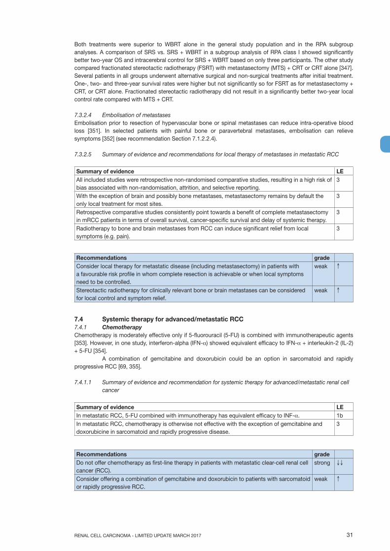
31RENAL CELL CARCINOMA - LIMITED UPDATE MARCH 2017
Both treatments were superior to WBRT alone in the general study population and in the RPA subgroup analyses. A comparison of SRS vs. SRS + WBRT in a subgroup analysis of RPA class I showed significantly better two-year OS and intracerebral control for SRS + WBRT based on only three participants. The other study compared fractionated stereotactic radiotherapy (FSRT) with metastasectomy (MTS) + CRT or CRT alone [347]. Several patients in all groups underwent alternative surgical and non-surgical treatments after initial treatment. One-, two- and three-year survival rates were higher but not significantly so for FSRT as for metastasectomy + CRT, or CRT alone. Fractionated stereotactic radiotherapy did not result in a significantly better two-year local control rate compared with MTS + CRT.
7.3.2.4 Embolisation of metastasesEmbolisation prior to resection of hypervascular bone or spinal metastases can reduce intra-operative blood loss [351]. In selected patients with painful bone or paravertebral metastases, embolisation can relieve symptoms [352] (see recommendation Section 7.1.2.2.4).
7.3.2.5 Summary of evidence and recommendations for local therapy of metastases in metastatic RCC
Summary of evidence LEAll included studies were retrospective non-randomised comparative studies, resulting in a high risk of bias associated with non-randomisation, attrition, and selective reporting.
3
With the exception of brain and possibly bone metastases, metastasectomy remains by default the only local treatment for most sites.
3
Retrospective comparative studies consistently point towards a benefit of complete metastasectomy in mRCC patients in terms of overall survival, cancer-specific survival and delay of systemic therapy.
3
Radiotherapy to bone and brain metastases from RCC can induce significant relief from local symptoms (e.g. pain).
3
Recommendations gradeConsider local therapy for metastatic disease (including metastasectomy) in patients with a favourable risk profile in whom complete resection is achievable or when local symptoms need to be controlled.
weak ↑
Stereotactic radiotherapy for clinically relevant bone or brain metastases can be considered for local control and symptom relief.
weak ↑
7.4 Systemic therapy for advanced/metastatic RCC7.4.1 ChemotherapyChemotherapy is moderately effective only if 5-fluorouracil (5-FU) is combined with immunotherapeutic agents [353]. However, in one study, interferon-alpha (IFN-α) showed equivalent efficacy to IFN-α + interleukin-2 (IL-2) + 5-FU [354]. A combination of gemcitabine and doxorubicin could be an option in sarcomatoid and rapidly progressive RCC [69, 355].
7.4.1.1 Summary of evidence and recommendation for systemic therapy for advanced/metastatic renal cell cancer
Summary of evidence LEIn metastatic RCC, 5-FU combined with immunotherapy has equivalent efficacy to INF-α. 1bIn metastatic RCC, chemotherapy is otherwise not effective with the exception of gemcitabine and doxorubicine in sarcomatoid and rapidly progressive disease.
3
Recommendations gradeDo not offer chemotherapy as first-line therapy in patients with metastatic clear-cell renal cell cancer (RCC).
strong ↓↓
Consider offering a combination of gemcitabine and doxorubicin to patients with sarcomatoid or rapidly progressive RCC.
weak ↑

RENAL CELL CARCINOMA - LIMITED UPDATE MARCH 201732
7.4.2 Immunotherapy7.4.2.1 IFN-α monotherapy and combined with bevacizumabConflicting results exist for IFN-α in clear-cell (cc) mRCC. Several studies showed that IFN-α in mRCC has a survival advantage similar to that of hormonal therapy [356]. Interferon-α resulted in a response rate of 6-15%, a 25% decrease in tumour progression risk and a modest survival benefit compared to placebo [357, 358]. However, patients with intermediate-risk disease, failed to confirm this benefit [359].
Interferon-α may only be effective in some patient subgroups, including patients with ccRCC, favourable-risk criteria, as defined by the Memorial Sloan-Kettering Cancer Center (MSKCC) and lung metastases only [356]. The moderate efficacy of immunotherapy was confirmed in a Cochrane meta-analysis [358]. Bevacizumab + IFN-α increased response rates and PFS in first-line therapy compared with IFN-α monotherapy [360]. All studies comparing targeted drugs to IFN-α monotherapy therapy showed superiority for sunitinib, bevacizumab + IFN-α, and temsirolimus [360-363]. Interferon-α has been superseded by targeted therapy in cc-mRCC.
Table 7.1: The Metastatic Renal Cancer Database Consortium (IMDC) risk model [364]
Risk factors** Cut-off point usedKarnofsky performance status < 80%Time from diagnosis to treatment < 12 monthsHaemoglobin < Lower limit of laboratory reference rangeCorrected serum calcium > 10.0 mg/dL (2.4 mmol/L)Absolute neutrophil count (neutrophilia) > upper limit of normal Platelets (thrombocytosis) > upper limit of normal
* The MSKCC (Motzer) criteria are also widely used in this setting [357].** Favourable (low) risk, no risk factors; intermediate risk, one or two risk factors; poor (high) risk, three to six risk
factors.
7.4.2.2 Interleukin-2Interleukin-2 has been used to treat mRCC since 1985, with response rates ranging from 7% to 27% [363, 365, 366]. Complete and durable responses have been achieved with high-dose bolus IL-2, however IL-2 remains the only drug to date that can cure a small percentage of RCC patients. [367]. The toxicity of IL-2 is substantially greater than that of IFN-α [358].
7.4.2.3 Vaccines and targeted immunotherapyA vaccine trial with tumour antigen 5T4 + first-line standard therapy (i.e. sunitinib, IL-2 or IFN-α) showed no survival benefit compared with placebo and first-line standard therapy [368]. Several vaccination studies are ongoing. Monoclonal antibodies against programmed death-1 (PD-1) or its ligand (PD-1L), which have efficacy and acceptable toxicity in patients with RCC [369], are currently being investigated in phase III trials.
7.4.2.4 Immune checkpoint blockadeImmune checkpoint blockade with monoclonal antibodies target and block the inhibitory T-cell receptor PD-1 or cytotoxic T-lymphocyte associated antigen 4 (CTLA-4)signalling to restore tumour specific T-cell immunity [370]. A randomised dose-ranging phase II trial of nivolumab in metastatic RCC patients revealed a high objective response rate with rapid and durable responses in heavily pre-treated patients [371]. A phase III trial of nivolumab vs. everolimus after one or two lines of VEGF-targeted therapy (CheckMate 025, NCT01668784) reported a longer OS, better QoL and fewer Grade 3 or 4 adverse events with nivolumab than with everolimus [172, 372, 373]. Nivolumab has superior OS to everolimus ([HR]: 0.73, 95% CI: 0.57-0.93, p < 0.002) in VEGF-refractory RCC with a median OS of 25 months for nivolumab and 19.6 months for everolimus (LE: 1b). Patients who had failed multiple lines of VEGF-targeted therapy were included in this trial making the results broadly applicable. The trial included 15% MSKCC poor-risk patients. There was no PFS advantage with nivolumab despite the OS advantage. A phase III trial is currently investigating the combination of nivolumab and ipilumimab vs. sunitinib in first-line treatment (CheckMate 214, NCT 02231749) [167]. Combinations of VEGF-targeted therapy and immune therapy are also being investigated and include: • Javelin Renal 101 - NCT02684006;• IMmotion151 - NCT02420821;• pembrolizumab + axitinib - NCT02133742;• lenvatinib + everolimus or pembrolizumab - NCT02811861.
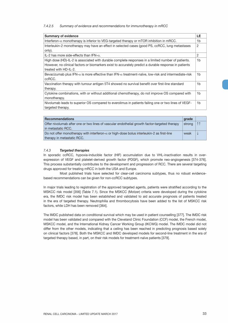
33RENAL CELL CARCINOMA - LIMITED UPDATE MARCH 2017
7.4.2.5 Summary of evidence and recommendations for immunotherapy in mRCC
Summary of evidence LEInterferon-α monotherapy is inferior to VEG-targeted therapy or mTOR inhibition in mRCC. 1bInterleukin-2 monotherapy may have an effect in selected cases (good PS, ccRCC, lung metastases only).
2
IL-2 has more side-effects than IFN-α. 2High dose (HD)-IL-2 is associated with durable complete responses in a limited number of patients. However, no clinical factors or biomarkers exist to accurately predict a durable response in patients treated with HD-IL-2.
1b
Bevacizumab plus IFN-α is more effective than IFN-α treatment-naïve, low-risk and intermediate-risk ccRCC.
1b
Vaccination therapy with tumour antigen 5T4 showed no survival benefit over first-line standard therapy.
1b
Cytokine combinations, with or without additional chemotherapy, do not improve OS compared with monotherapy.
1b
Nivolumab leads to superior OS compared to everolimus in patients failing one or two lines of VEGF-targeted therapy.
1b
Recommendations gradeOffer nivolumab after one or two lines of vascular endothelial growth factor-targeted therapy in metastatic RCC.
strong ↑↑
Do not offer monotherapy with interferon-α or high-dose bolus interleukin-2 as first-line therapy in metastatic RCC.
weak ↓
7.4.3 Targeted therapiesIn sporadic ccRCC, hypoxia-inducible factor (HIF) accumulation due to VHL-inactivation results in over-expression of VEGF and platelet-derived growth factor (PDGF), which promote neo-angiogenesis [374-376]. This process substantially contributes to the development and progression of RCC. There are several targeting drugs approved for treating mRCC in both the USA and Europe.
Most published trials have selected for clear-cell carcinoma subtypes, thus no robust evidence-based recommendations can be given for non-ccRCC subtypes.
In major trials leading to registration of the approved targeted agents, patients were stratified according to the MSKCC risk model [356] (Table 7.1). Since the MSKCC (Motzer) criteria were developed during the cytokine era, the IMDC risk model has been established and validated to aid accurate prognosis of patients treated in the era of targeted therapy. Neutrophilia and thrombocytosis have been added to the list of MSKCC risk factors, while LDH has been removed [364].
The IMDC published data on conditional survival which may be used in patient counselling [377]. The IMDC risk model has been validated and compared with the Cleveland Clinic Foundation (CCF) model, the French model, MSKCC model, and the International Kidney Cancer Working Group (IKCWG) model. The IMDC model did not differ from the other models, indicating that a ceiling has been reached in predicting prognosis based solely on clinical factors [378]. Both the MSKCC and IMDC developed models for second-line treatment in the era of targeted therapy based, in part, on their risk models for treatment-naïve patients [379].
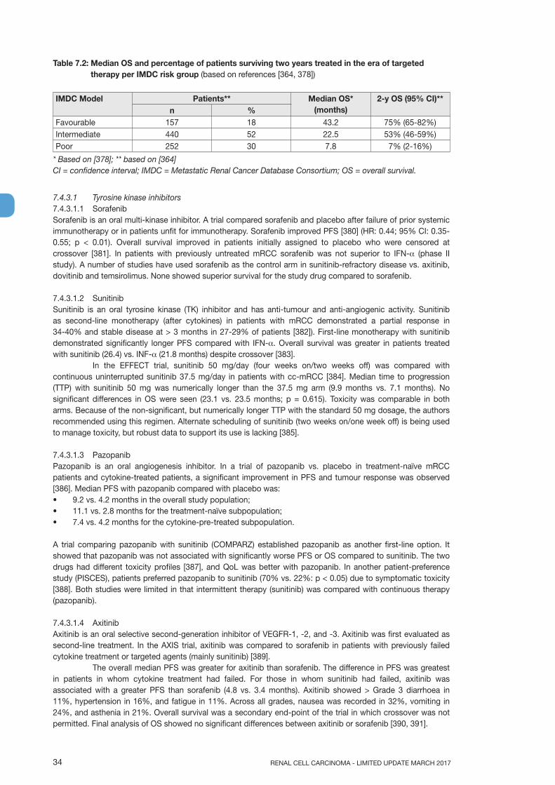
RENAL CELL CARCINOMA - LIMITED UPDATE MARCH 201734
Table 7.2: Median OS and percentage of patients surviving two years treated in the era of targeted therapy per IMDC risk group (based on references [364, 378])
IMDC Model Patients** Median OS* (months)
2-y OS (95% CI)**n %
Favourable 157 18 43.2 75% (65-82%)Intermediate 440 52 22.5 53% (46-59%)Poor 252 30 7.8 7% (2-16%)
* Based on [378]; ** based on [364]CI = confidence interval; IMDC = Metastatic Renal Cancer Database Consortium; OS = overall survival.
7.4.3.1 Tyrosine kinase inhibitors7.4.3.1.1 SorafenibSorafenib is an oral multi-kinase inhibitor. A trial compared sorafenib and placebo after failure of prior systemic immunotherapy or in patients unfit for immunotherapy. Sorafenib improved PFS [380] (HR: 0.44; 95% CI: 0.35- 0.55; p < 0.01). Overall survival improved in patients initially assigned to placebo who were censored at crossover [381]. In patients with previously untreated mRCC sorafenib was not superior to IFN-α (phase II study). A number of studies have used sorafenib as the control arm in sunitinib-refractory disease vs. axitinib, dovitinib and temsirolimus. None showed superior survival for the study drug compared to sorafenib.
7.4.3.1.2 SunitinibSunitinib is an oral tyrosine kinase (TK) inhibitor and has anti-tumour and anti-angiogenic activity. Sunitinib as second-line monotherapy (after cytokines) in patients with mRCC demonstrated a partial response in 34-40% and stable disease at > 3 months in 27-29% of patients [382]). First-line monotherapy with sunitinib demonstrated significantly longer PFS compared with IFN-α. Overall survival was greater in patients treated with sunitinib (26.4) vs. INF-α (21.8 months) despite crossover [383].
In the EFFECT trial, sunitinib 50 mg/day (four weeks on/two weeks off) was compared with continuous uninterrupted sunitinib 37.5 mg/day in patients with cc-mRCC [384]. Median time to progression (TTP) with sunitinib 50 mg was numerically longer than the 37.5 mg arm (9.9 months vs. 7.1 months). No significant differences in OS were seen (23.1 vs. 23.5 months; p = 0.615). Toxicity was comparable in both arms. Because of the non-significant, but numerically longer TTP with the standard 50 mg dosage, the authors recommended using this regimen. Alternate scheduling of sunitinib (two weeks on/one week off) is being used to manage toxicity, but robust data to support its use is lacking [385].
7.4.3.1.3 PazopanibPazopanib is an oral angiogenesis inhibitor. In a trial of pazopanib vs. placebo in treatment-naïve mRCC patients and cytokine-treated patients, a significant improvement in PFS and tumour response was observed [386]. Median PFS with pazopanib compared with placebo was:• 9.2 vs. 4.2 months in the overall study population;• 11.1 vs. 2.8 months for the treatment-naïve subpopulation;• 7.4 vs. 4.2 months for the cytokine-pre-treated subpopulation.
A trial comparing pazopanib with sunitinib (COMPARZ) established pazopanib as another first-line option. It showed that pazopanib was not associated with significantly worse PFS or OS compared to sunitinib. The two drugs had different toxicity profiles [387], and QoL was better with pazopanib. In another patient-preference study (PISCES), patients preferred pazopanib to sunitinib (70% vs. 22%: p < 0.05) due to symptomatic toxicity [388]. Both studies were limited in that intermittent therapy (sunitinib) was compared with continuous therapy (pazopanib).
7.4.3.1.4 AxitinibAxitinib is an oral selective second-generation inhibitor of VEGFR-1, -2, and -3. Axitinib was first evaluated as second-line treatment. In the AXIS trial, axitinib was compared to sorafenib in patients with previously failed cytokine treatment or targeted agents (mainly sunitinib) [389].
The overall median PFS was greater for axitinib than sorafenib. The difference in PFS was greatest in patients in whom cytokine treatment had failed. For those in whom sunitinib had failed, axitinib was associated with a greater PFS than sorafenib (4.8 vs. 3.4 months). Axitinib showed > Grade 3 diarrhoea in 11%, hypertension in 16%, and fatigue in 11%. Across all grades, nausea was recorded in 32%, vomiting in 24%, and asthenia in 21%. Overall survival was a secondary end-point of the trial in which crossover was not permitted. Final analysis of OS showed no significant differences between axitinib or sorafenib [390, 391].
35RENAL CELL CARCINOMA - LIMITED UPDATE MARCH 2017
In a randomised phase III trial of axitinib vs. sorafenib in first-line treatment-naïve cc-mRCC, a significant difference in median PFS between the treatment groups was not demonstrated [392]. As a result of this study, axitinib is not approved for first-line therapy.
7.4.3.1.5 CabozantinibCabozantinib is an oral inhibitor of TK, including MET, VEGF and AXL. Cabozantinib was investigated in a phase I study in patients resistant to VEGFR and mTOR inhibitors demonstrating objective responses and disease control [171]. Based on these results a randomised phase III trial investigated cabozantinib vs. everolimus in patients with ccRCC failing one or more VEGF-targeted therapies (METEOR) [64]. Cabozantinib delayed PFS compared to everolimus in VEGF-targeted therapy refractory disease by 42% (HR: 0.58 95% CI: 0.45-0.75) [64] (LE: 1b). The median PFS for cabozantinib was 7.4 months (95% CI: 5.6-9.1) vs. 3.8 months (95% CI: 3.7-5.4) for everolimus. The trial recruited 658 patients although PFS was assessed on the first 375 patients. The median OS was 21.4 months (95% CI: 18.7 to not estimable) with cabozantinib and 16.5 months (95% CI 14.7-18.8) with everolimus in VEGF- resistant RCC. The HR for death was 0.66 (95% CI: 0.53-0.83; p = 0.0003) [393]. Grade 3 or 4 adverse events were reported in 74% with cabozantinib and 65% with everolimus. Adverse events were managed with dose reductions; doses were reduced in 60% of the patients who received cabozantinib. Discontinuation due to toxicity was not significantly different for the two drugs. The trial included 16% MSKCC poor-risk patients.
7.4.3.1.6 LenvatinibLenvatinib is an oral multi-target TKI of VEGFR1, VEGFR2, and VEGFR3, with inhibitory activity against fibroblast growth factor receptors (FGFR1, FGFR2, FGFR3, and FGFR4), platelet growth factor receptor α (PDGFRα), re-arranged during transfection (RET), and receptor for stem cell factor (KIT). It has recently been investigated in randomised phase II study in combination with everolimus vs. lenvatinib or everolimus alone (see Section 7.4.6.1.1.5 for discussion of results).
7.4.4 Monoclonal antibody against circulating VEGF7.4.4.1 Bevacizumab monotherapy and bevacizumab + IFN-αBevacizumab is a humanised monoclonal antibody. The double-blind AVOREN study compared bevacizumab + IFN-α with INF-α monotherapy in mRCC [360]. Overall response was higher in the bevacizumab + IFN-α group. Median PFS increased from 5.4 months with IFN-α to 10.2 months with bevacizumab + IFN-α. No benefit was seen in MSKCC poor-risk patients. Median OS in this trial, which allowed crossover after progression, was not greater in the bevacizumab/IFN-α group (23.3 vs. 21.3) [394].
An open-label trial (CALGB 90206) [395, 396], of bevacizumab + IFN-α vs. IFN-α showed a higher median PFS for the combination group. Objective response rate was also higher in the combination group. Overall toxicity was greater for bevacizumab + IFN-α, with significantly more Grade 3 hypertension, anorexia, fatigue, and proteinuria.
7.4.5 mTOR inhibitors7.4.5.1 TemsirolimusTemsirolimus is a specific inhibitor of mTOR [397]. Patients with modified high-risk mRCC in the NCT00065468 trial received first-line temsirolimus or IFN-α monotherapy, or a combination of both [362]. Median OS was higher in the temsirolimus group. However, OS in the temsirolimus + IFN-α group was not significantly superior to IFN-α alone [362]. Interferon-α toxicity was marked, partly due to the high doses used. The INTORSECT trial investigated temsirolimus vs. sorafenib in patients who had previously failed sunitinib. Although no benefit in PFS was observed, a significant OS benefit for sorafenib was noted [398]. Based on these results, temsirolimus is not recommended in patients with VEFG TKI-refractory disease.
7.4.5.2 EverolimusEverolimus is an oral mTOR inhibitor, which is established in the treatment of VEGF-refractory disease. The RECORD-1 study compared everolimus + best supportive care (BSC) vs. placebo + BSC in patients with previously failed anti-VEGFR treatment (or previously intolerant of VEGF-targeted therapy) [399]. The initial data showed a median PFS of four months vs. 1.9 months for everolimus and placebo, respectively [399]. This was extended to 4.9 months in the final analysis (HR: 0.33) [400]. Subset analysis of PFS for patients receiving only one previous VEFG TKI was 5.4 months [401]. This included some patients who were intolerant rather than progressed on therapy (PFS was also 5.4 months) [402]. RECORD-1 included patients who failed multiple lines of VEGF-targeted therapy, and received everolimus in a third- and fourth-line setting [399].
The RECORD-3 randomised phase II study of sequential first-line sunitinib and second-line everolimus vs. sequential first-line everolimus and second-line sunitinib in treatment-naïve mRCC reported a higher median

RENAL CELL CARCINOMA - LIMITED UPDATE MARCH 201736
PFS for first-line treatment in the sunitinib group [403]. Primary endpoint was to assess PFS non-inferiority of first-line everolimus to first-line sunitinib. A large number of the crossover patients did not receive the planned subsequent therapy making further analysis complex and underpowered.
7.4.6 Therapeutic strategies 7.4.6.1 Therapy for treatment-naïve patients with clear-cell mRCCKey trials have established sunitinib, pazopanib and bevacizumab plus IFN-α as first-line treatment options in treatment-naïve patients with cc-mRCC and a favourable-to-intermediate risk score. The evidence for subsequent therapies after temsirolimus in poor-risk patients is unclear. It is therefore more appealing to treat poor-risk patients with sunitinib or pazopanib, both of which were tested in pivotal trials in this population.
7.4.6.1.1 Sequencing targeted therapy7.4.6.1.1.1 Following progression of disease with one or more lines of VEGF-targeted therapySeveral trials investigated therapeutic options for patients who progressed on first-line VEGF-targeted therapy, including studies which investigated options after one or more lines of VEGF-targeted therapy. RECORD-1 established VEGF TKI therapy until disease progression, followed by everolimus as one of the treatment options for patients with mRCC [399]. However, both nivolumab and cabozantinib were superior to everolimus following a similar trial design as RECORD-1 [172]. Both of these agents should be considered a new standard of care in patients of all risk categories who have failed one or more VEGF-targeted therapies (Figure 7.1).
Nivolumab should be considered for all patients in whom it is not contraindicated in the VEGF- refractory setting owing to a significant OS advantage compared to everolimus, as well as its attractive tolerability profile. Cabozantinib is the first TKI to have both a superior PFS and OS compared to everolimus. Both nivolumab and cabozantinib have different toxicity profiles.
Axitinib is superior to sorafinib in terms of PFS in sunitinib-refractory ccRCC [388]. Neither nivolumab nor cabozantinib has been tested directly against axitinib in the second-line setting. However, the OS advantage of both drugs and tolerability of nivolumab over everolimus in this setting makes them preferable to axitinib.
Tolerability is an important consideration when recommendations cannot be made for efficacy alone. Both everolimus and sorafenib have been outperformed by other agents in VEGF-refractory disease and should not be the standard of care in pure VEGF-refractory disease where superior alternatives are available. It is not currently possible to determine therapy based on baseline characteristics or biomarker expression for any of the above drugs.
Direct comparison of RECORD-1, Checkmate-25 and METEOR data with AXIS data is not advised due to differences in patient populations [389-391, 399].
INTORSECT compared temsirolimus vs. sorafenib after disease progression on sunitinib [398]. Median PFS was higher, but not significant, in the temsirolimus group. However, there was a significant difference in OS in favour of sorafenib. Neither of these agents are recommended or widely used in this setting. These data are not necessarily relevant to other mTOR inhibitors such as everolimus.
Based on difference in OS, recommendations can currently be made as to the best sequence of targeted therapy (Figure 7.1). Two major trials, testing nivolumab and cabozantinib, have changed treatment paradigms in VEGF-refractory RCC (LE: 1a). There is a strong rationale for using both drugs in sequence in the second and third line following VEGF-targeted therapy. This creates a new a standard for the majority of patients.
7.4.6.1.1.2 Treatment after progression of disease with mTOR inhibitionThere are limited data addressing this issue. In view of the efficacy of VEGF-targeted therapy in renal cancer, a switch to VEGF-targeted therapy is advised (Panel consensus in conjunction with Motzer et al. [404]).
7.4.6.1.1.3 Treatment after progression of disease with cytokinesTrials have established sorafenib, axitinib and pazopanib as therapeutic options in this setting with a median PFS of 5.5, 12.1 and 7.4 months, respectively. Based on trial data, axitinib is superior to sorafenib in patients previously treated with cytokine therapy [389-391].
7.4.6.1.1.4 Treatment after second-line targeted therapy7.4.6.1.1.4.1 Treatment after two VEGF-targeted therapiesBased on the results of the nivolumab and cabozantinib trials, a strong rationale exists for preferring both drugs as third-line treatment upon failure of two VEGF-targeted therapies [64, 172] (Figure 7.1).
37RENAL CELL CARCINOMA - LIMITED UPDATE MARCH 2017
7.4.6.1.1.4.2 Treatment after VEGFR- and mTOR inhibitionAlthough the GOLD trial failed to demonstrate superior efficacy of dovitinib over sorafenib in patients with mRCC who experienced disease progression after receiving prior VEGF- and mTOR-targeted therapies, the results suggest efficacy and safety of sorafenib in the third-line setting [404]. This sequence is not recommended when alternative superior drugs are available.
7.4.6.1.1.4.3 Combination of targeted agentsNo combinations of targeted agents are currently recommended, however, there have been a number of trials with VEFG-targeted therapy and mTOR inhibitors [405-409]. A small randomised phase II trial in which 153 patients received either lenvatinib plus everolimus (n = 51), single-agent lenvatinib (n = 52), or single-agent everolimus (n = 50) demonstrated a PFS benefit for the combination [410]. Lenvatinib plus everolimus significantly prolonged PFS compared with everolimus alone (median 14.6 months [95% CI: 5.9-20.1] vs. 5.5 months [3.5-7.1]; HR: 0.40; 95% CI: 0.24-0.68; p = 0.0005), but not compared with lenvatinib alone (7.4 months [95% CI: 5.6-10.2]; HR: 0.66; 95% CI: 0.30-1.10; p = 0·12). In a post-hoc updated analysis (data cut-off Dec 10, 2014), the difference in OS between lenvatinib plus everolimus vs. single-agent everolimus was significantly increased, median OS 25.5 months [95% CI: 16.4-NE] vs. 15.4 months [11.8-19.6]; HR: 0.51; 95% CI: 0.30-0.88; p = 0.024. Grade 3 or worse serious adverse events occurred in 23 (45%) patients allocated to lenvatinib plus everolimus, 23 (44%) allocated to single-agent lenvatinib, and 19 (38%) allocated to single-agent everolimus.
7.4.6.2 Non-clear-cell renal cancerNo phase III trials of patients with non-ccRCC have been reported. Expanded access programmes and subset analysis from RCC studies suggest the outcome of these patients with targeted therapy is poorer than for ccRCC. Targeted treatment in non-ccRCC has focused on temsirolimus, everolimus, sorafenib and sunitinib [362, 411-413].
The most common non-clear-cell subtypes are papillary type 1 and non-type 1 papillary RCCs. There are small single-arm trials for sunitinib and everolimus [413-416]. A trial of both types of pRCC treated with everolimus (RAPTOR) [416], showed a median PFS of 3.7 months per central review in the intention-to-treat population with a median OS of 21.0 months.
Another trial investigated foretinib (a dual MET/VEGFR2 inhibitor) in patients with pRCC. Toxicity was acceptable with a high relative risk in patients with germline MET mutations [417]. However, a randomised phase II trial of everolimus vs. sunitinib (ESPN) with crossover design in non-cc-mRCC including 73 patients (27 with pRCC) was stopped after a futility analysis for PFS and OS [418]. The final results presented at the 2014 annual meeting of the American Society of Clinical Oncology showed a non-significant trend favouring sunitinib (6.1 vs. 4.1 months). Based on a SR including subgroup analysis of the ESPN, RECORD-3 and another phase II trial (ASPEN) sunitinib and everolimus remain options in this population, with a preference for sunitinib [136, 419, 420]. Patients with non-cc-mRCC should be referred to a clinical trial where appropriate.
Collecting-duct cancers are resistant to systemic therapy. There is a lack of data to support specific therapy in these patients. There is limited data supporting the use of targeted therapy in other histological subtypes such as chromophobe tumours [362, 411].

RENAL CELL CARCINOMA - LIMITED UPDATE MARCH 201738
Figure 7.1: Recommendations for patients with metastatic ccRCC who have failed one or more lines of VEGF targeted therapy
1 Switch to therapies not given previously.2 Nivolumab and cabozantinib have not been given after everolimus and therefore cannot be recommended
above other agents. 3 Sorafenib has an inferior progression-free survival to axitinib.4 These drugs have shown a survival advantage in VEGF-resistant disease but not in this specific setting. 5 These drugs were given after progression in the pivotal cabozantinib or nivolumab trials [64, 172].6 Sunitinib and pazopanib can be recommended in all MSKCC risk groups. Bevacizumab/interferon (favourable-
and intermediate-risk disease) and temsirolimus (poor-risk disease) have not been widely used as first-line therapy in the pivotal VEGF-resistant trials and therefore recommendations are not possible.
Table 7.3: EAU 2017 evidence-based recommendations for systemic therapy in patients with mRCC
RCC type
MSKCC risk group [356]
First-line LE^ Second-Line after VEGF therapy*
LE^ Third-line* LE^ Later lines
LE
Clear cell*
Favourable, intermediate and poor
sunitinib pazopanib bevacizumab + IFN-α (favourable-intermediate only)
1b 1b 1b
based on OS: nivolumabcabozantinib based on PFS: axitinib sorafenib#
everolimus&
2b2b
2b 2b 2b
after VEGF therapy: nivolumab cabozantinib everolimus&
after VEGF and mTOR therapy: sorafenib
after VEGF and nivolumab: cabozantinib axitinib everolimus
2b 2b2b
1b
4 4 4
any targeted agent
4
Clear cell*
poor¶ temsirolimussunitinibpazopanib
1b2b2b
any targeted agent
4
First-line therapy Therapy for VEGF resistant disease1
pazopanib or sunitinib6
cabozantinib or nivolumab
axitinib5
or cabozantinib4
or everolimus5
or nivolumab4
or sorafenib5
axitinib or everolimus2
or sorafenib3 everolimus or sorafenib

39RENAL CELL CARCINOMA - LIMITED UPDATE MARCH 2017
Non-clear cell §
any sunitinib 1b^^ Any targeted agent
4
IFN-α = interferon alpha; LE = level of evidence; MSKCC = Memorial Sloan-Kettering Cancer Center; mTOR = mammalian target of rapamycin inhibitor; RCC = renal cell cancer; TKI= tyrosine kinase inhibitor; VEGF = vascular endothelial growth factor.* Doses: IFN-α - 9 MU three times per week subcutaneously, bevacizumab 10 mg/kg bi-weekly intravenously; sunitinib 50 mg daily orally for four weeks, followed by two weeks of rest (37.5 mg continuous dosing did not show significant differences); temsirolimus 25 mg weekly intravenously; pazopanib 800 mg daily orally. Axitinib 5 mg twice daily, to be increased to 7 mg twice daily, unless greater than Grade 2 toxicity, blood pressure higher than 150/90 mmHg, or the patient is receiving antihypertensive medication. Everolimus, 10 mg daily orally.
§ No standard treatment available. Patients should be treated in the framework of clinical trials or a decision can be made in consultation with the patient to perform treatment in line with ccRCC.
¶ Poor risk criteria in the NCT00065468 trial consisted of MSKCC [356] risk plus metastases in multiple organs. Evidence for subsequent therapies unclear, making this option less appealing.
# Sorafenib was inferior to axitinib in a RCT in terms of PFS but not OS [391].^ Level of evidence was downgraded in instances when data were obtained from subgroup analysis
within a RCT.& Everolimus was inferior in terms of OS to nivolumab and in terms of PFS to cabozantinib and should not
routinely be given where other superior agents are available. ^^ Based on a SR [420].
7.4.6.3 Summary of evidence and recommendations for systemic therapy in metastatic renal cell cancer
Summary of evidence LEVEGF and TKIs increase PFS and/or OS as both first-line and second-line treatments for clear-cell mRCC.
1b
Axitinib has proven efficacy and superiority in PFS as a second-line treatment after failure of cytokines and VEGF-targeted therapy in comparison with sorafenib.
1b
Sunitinib is more effective than IFN-α in treatment-naïve patients. 1bBevacizumab plus IFN-α is more effective than IFN-α in treatment-naïve low-risk and intermediate-risk patients.
1b
pazopanib is superior to placebo in both naïve mRCC patients and post-cytokine patients. 1bFirst line pazopanib is not inferior to sunitinib in clear-cell mRCC patients. 1bTemsirolimus monotherapy prolongs OS compared to IFN-α in poor-risk mRCC. 1bNivolumab is superior to everolimus in terms of OS and adverse events in patients failing one or two lines of VEGF-targeted therapy.
1b
Cabozantinib is superior to everolimus in terms of PFS and OS in patients failing one or more lines of VEGF-targeted therapy.
1b
Everolimus prolongs PFS in patients who have previously failed or are intolerant of VEGF-targeted therapy when compared to placebo.
1b
Sorafenib has broad activity in a spectrum of settings in ccRCC patients previously treated with cytokine or targeted therapies. It is inferior to axitinib in both sunitinib or cytokine pre-treated patients.
4
Both mTOR inhibitors (everolimus and temsirolimus) and VEFG-targeted therapies (sunitinib or sorafenib) can be used in non-clear cell RCC.
3
No combination has proven to be better than single-agent therapy, with the exception of the combination of lenvatinib plus everolimus.
1a

RENAL CELL CARCINOMA - LIMITED UPDATE MARCH 201740
Recommendations gradeOffer sunitinib or pazopanib as first-line therapy for metastatic clear-cell renal cell cancer (ccRCC).
strong ↑↑
Consider offering bevacizumab + Interferon (IFN)-α as first-line therapy for metastatic RCC in favourable and intermediate-risk ccRCC.
weak ↑
Consider offering temsirolimus as first-line treatment in poor-risk RCC patients. weak ↑
Offer cabozantinib for ccRCC after one or two lines of vascular endothelial growth factor (VEGF)-targeted therapy in metastatic RCC.
strong ↑↑
Offer nivolumab after one or two lines of VEGF-targeted therapy in metastatic RCC. strong ↑↑
Offer axitinib or everolimus to ccRCC patients who failed VEGF-targeted therapy, and when nivolumab or cabozantinib are not safe, tolerable or available.
strong ↑↑
Sequence targeted agents in treating metastatic RCC. strong ↑↑
Sunitinib can be offered as first-line therapy for non-clear cell mRCC. weak ↑
7.5 Recurrent RCC7.5.1 IntroductionLocally recurrent disease can occur after RN, PN and thermal ablation. After nephron-sparing treatment the recurrence may be intrarenal and/or regional, e.g. venous tumour thrombi or retroperitoneal LN metastases. Both are often summarised as loco-regional recurrences. Recurrence for pT1 tumours after PN are observed in 2.2% and are generally managed surgically depending on the extent of the loco-regional recurrence [421]. After thermal ablation loco-regional recurrences (intrarenal and regional) have been described in up to 12% [422]. Repeated ablation has often been recommended for intrarenal recurrences following thermal ablation. For loco-regional recurrences surgical resection is mandatory and has been described for isolated local recurrences following nephrectomy.
After nephrectomy locally recurrent disease is defined as disease recurring in the renal fossa or remnant kidney. However, metastasis in the non-removed ipsilateral adrenal or non-resected LNs makes interpretation of the true incidence of isolated recurrence in the renal fossa difficult. Treatment of adrenal metastases or LN metastases are often described in series of metastasectomy (see Section 7.3). Isolated local recurrence, however, is rare.
The largest series on the treatment of isolated recurrence was published in 2009 [423]. In 2,945 patients who underwent nephrectomy the authors identified 54 isolated local recurrences in the renal fossa. These, however, included recurrences to the ipsilateral adrenal and LNs. Exclusively retrospective non-comparative data exist which suggest that aggressive local resection offers durable local tumour control and improves survival. Adverse prognostic factors were, a positive surgical margin after resection, the size of the recurrence and sarcomatoid histologic features [423]. In cases where complete surgical removal is not feasible due to advanced tumour growth and pain, palliative treatments including radiation treatment can be considered.
7.5.2 Summary of evidence and recommendation for advanced/metastatic RCC
Summary of evidence LEIsolated recurrence in the local renal fossa is rare. 3Patients who undergo resection of local recurrences in the absence of sarcomatoid features may benefit from durable local control and improved survival.
3
Recommendation gradeOffer surgical resection of local recurrent disease, when feasible. weak ↑

41RENAL CELL CARCINOMA - LIMITED UPDATE MARCH 2017
8. FOLLOW-UP IN RCC8.1 IntroductionSurveillance after treatment for RCC allows the urologist to monitor or identify:• post-operative complications;• renal function;• local recurrence;• recurrence in the contralateral kidney;• development of metastases.
There is no consensus on surveillance after RCC treatment, and there is no evidence that early vs. later diagnosis of recurrences improves survival. However, follow-up is important to increase the available information on RCC, and should be performed by a urologist, who should record the time to recurrence or the development of metastases. Patients undergoing follow-up seem to have a longer OS when compared to patients not undergoing routine follow-up [424].
An individualised, risk-based, approach to RCC surveillance was recently proposed. The authors use competing risk models, incorporating patient age, pathologic stage, relapse location and comorbidities, to calculate when the risk of non-RCC death exceeds the risk of RCC recurrence [425]. For patients with low-stage disease but with a Charlson comorbidity index > 2, the risk of non-RCC death exceeded that of abdominal recurrence risk already one month after surgery, regardless of patient age.
Renal function is assessed by the measurement of serum creatinine and eGFR. Repeated long-term monitoring of eGFR is indicated in case of impaired renal function before, or after, surgery. Renal function [426, 427] and non-cancer survival [196, 428, 429] can be optimised by performing NSS, whenever possible, for T1 and T2 tumours [430] (LE: 3). Recurrence after PN is rare, but early diagnosis is useful, as the most effective treatment is redux surgery [431, 432]. Recurrence in the contralateral kidney is also rare and might be related to positive margins, multifocality, and grade [433] (LE: 3). Surveillance can identify local recurrences or metastases at an early stage. In metastatic disease, extended tumour growth can limit the opportunity for surgical resection, which is considered the standard therapy in cases of resectable and preferably solitary lesions. In addition, early diagnosis of tumour recurrence may enhance the efficacy of systemic treatment if the tumour burden is low.
8.2 Which investigations for which patients, and when?There is no high level evidence to support any surveillance scheme. However, intensive radiological surveillance for all patients is not necessary. The outcome after surgery for T1a low-grade tumours is almost always excellent. It is therefore reasonable to stratify the follow up, taking into account the risk of developing recurrence or metastases. Although there is no randomised evidence, large studies have examined prognostic factors with long follow-up periods [35, 434, 435] (LE: 4). One study has shown a survival benefit for patients who were followed within a structured surveillance protocol vs. patients who were not [424]:
• The sensitivity of chest radiography and US for small metastases is poor. Surveillance with these imaging modalities should not be done [436].
• In low-risk tumours, surveillance intervals should be adapted taking into account radiation exposure and benefit. To reduce radiation exposure, MRI can be used outside the thorax.
• When the risk of relapse is intermediate or high, CT of the chest and abdomen should be performed.• Surveillance should also include evaluation of renal function and cardiovascular risk factors.• Positron-emission tomography and PET-CT as well as bone scintigraphy should not be used in RCC
surveillance, due to their limited specificity and sensitivity.• The risk of acute renal failure seems to be negligible in patients with a GFR > 20 mL/min and chronic renal
impairment [437].
Controversy exists on the optimal duration of follow-up. Some argue that follow-up with imaging is not cost-effective after five years; however, late metastases are more likely to be solitary and justify more aggressive therapy with curative intent. In addition, patients with tumours that develop in the contralateral kidney can be treated with NSS if the tumours are detected early. For tumours < 4 cm, there is no difference between PN and RN with regard to recurrences during follow up [438] (LE: 3).
Several authors [182, 184, 439, 440], have designed scoring systems and nomograms to quantify the likelihood of patients developing tumour recurrences, metastases, and subsequent death. These systems have been compared and validated [441] (LE: 2). Using prognostic variables, several stage-based surveillance
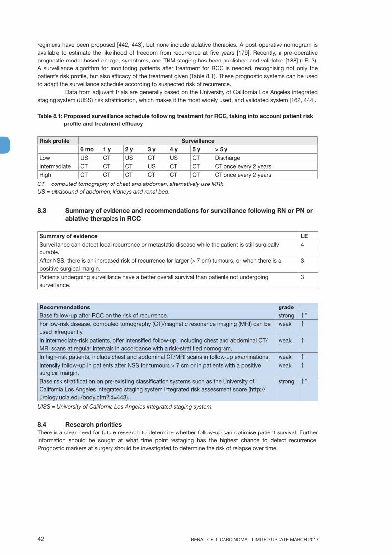
RENAL CELL CARCINOMA - LIMITED UPDATE MARCH 201742
regimens have been proposed [442, 443], but none include ablative therapies. A post-operative nomogram is available to estimate the likelihood of freedom from recurrence at five years [179]. Recently, a pre-operative prognostic model based on age, symptoms, and TNM staging has been published and validated [188] (LE: 3). A surveillance algorithm for monitoring patients after treatment for RCC is needed, recognising not only the patient’s risk profile, but also efficacy of the treatment given (Table 8.1). These prognostic systems can be used to adapt the surveillance schedule according to suspected risk of recurrence.
Data from adjuvant trials are generally based on the University of California Los Angeles integrated staging system (UISS) risk stratification, which makes it the most widely used, and validated system [162, 444].
Table 8.1: Proposed surveillance schedule following treatment for RCC, taking into account patient risk profile and treatment efficacy
Risk profile Surveillance6 mo 1 y 2 y 3 y 4 y 5 y > 5 y
Low US CT US CT US CT DischargeIntermediate CT CT CT US CT CT CT once every 2 yearsHigh CT CT CT CT CT CT CT once every 2 years
CT = computed tomography of chest and abdomen, alternatively use MRI;US = ultrasound of abdomen, kidneys and renal bed.
8.3 Summary of evidence and recommendations for surveillance following RN or PN or ablative therapies in RCC
Summary of evidence LESurveillance can detect local recurrence or metastatic disease while the patient is still surgically curable.
4
After NSS, there is an increased risk of recurrence for larger (> 7 cm) tumours, or when there is a positive surgical margin.
3
Patients undergoing surveillance have a better overall survival than patients not undergoing surveillance.
3
Recommendations gradeBase follow-up after RCC on the risk of recurrence. strong ↑↑
For low-risk disease, computed tomography (CT)/magnetic resonance imaging (MRI) can be used infrequently.
weak ↑
In intermediate-risk patients, offer intensified follow-up, including chest and abdominal CT/MRI scans at regular intervals in accordance with a risk-stratified nomogram.
weak ↑
In high-risk patients, include chest and abdominal CT/MRI scans in follow-up examinations. weak ↑
Intensify follow-up in patients after NSS for tumours > 7 cm or in patients with a positive surgical margin.
weak ↑
Base risk stratification on pre-existing classification systems such as the University of California Los Angeles integrated staging system integrated risk assessment score (http://urology.ucla.edu/body.cfm?id=443).
strong ↑↑
UISS = University of California Los Angeles integrated staging system.
8.4 Research prioritiesThere is a clear need for future research to determine whether follow-up can optimise patient survival. Further information should be sought at what time point restaging has the highest chance to detect recurrence. Prognostic markers at surgery should be investigated to determine the risk of relapse over time.

43RENAL CELL CARCINOMA - LIMITED UPDATE MARCH 2017
9. REFERENCES1. Ljungberg, B., et al. Renal cell carcinoma guideline. Eur Urol, 2007. 51: 1502.
https://www.ncbi.nlm.nih.gov/pubmed/174088502. Ljungberg, B., et al. EAU guidelines on renal cell carcinoma: 2014 update. Eur Urol, 2015. 67: 913.
https://www.ncbi.nlm.nih.gov/pubmed/256167103. Guyatt, G.H., et al. GRADE: an emerging consensus on rating quality of evidence and strength of
recommendations. BMJ, 2008. 336: 924.https://www.ncbi.nlm.nih.gov/pubmed/18436948
4. Guyatt, G.H., et al. What is “quality of evidence” and why is it important to clinicians? BMJ, 2008. 336: 995.https://www.ncbi.nlm.nih.gov/pubmed/18456631
5. Guyatt, G.H., et al. Going from evidence to recommendations. BMJ, 2008. 336: 1049.https://www.ncbi.nlm.nih.gov/pubmed/18467413
6. Vogel, T., et al. Imaging in Suspected Renal Cell Carcinoma: A Systematic Review. prior to print, 2017.
7. Marconi, L., et al. Systematic review of the best surgical treatment for clinical =T2N0M0 Renal Cell Cancer PROSPERO International prospective register of systematic reviews, 2016. CRD42016036697.https://www.ncbi.nlm.nih.gov/pubmed/26707869
8. Fernández-Pello, S., et al. A Systematic Review and Meta-analysis Comparing the Effectiveness and Adverse Effects of Different Systemic Treatments for Non-clear Cell Renal Cell Carcinoma. Eur Urol, 2017. 71: 426.https://www.ncbi.nlm.nih.gov/pubmed/27939075
9. European Network of Cancer Registries: Eurocim version 4.0. 2001: Lyon, France.10. Lindblad, P. Epidemiology of renal cell carcinoma. Scand J Surg, 2004. 93: 88.
https://www.ncbi.nlm.nih.gov/pubmed/1528555911. Ferlay, J., et al. Cancer incidence and mortality patterns in Europe: estimates for 40 countries in
2012. Eur J Cancer, 2013. 49: 1374.https://www.ncbi.nlm.nih.gov/pubmed/23485231
12. Levi, F., et al. The changing pattern of kidney cancer incidence and mortality in Europe. BJU Int, 2008. 101: 949.https://www.ncbi.nlm.nih.gov/pubmed/18241251
13. King, S.C., et al. Continued increase in incidence of renal cell carcinoma, especially in young patients and high grade disease: United States 2001 to 2010. J Urol, 2014. 191: 1665.https://www.ncbi.nlm.nih.gov/pubmed/24423441
14. Bergstrom, A., et al. Obesity and renal cell cancer--a quantitative review. Br J Cancer, 2001. 85: 984.https://www.ncbi.nlm.nih.gov/pubmed/11592770
15. Clague, J., et al. Family history and risk of renal cell carcinoma: results from a case-control study and systematic meta-analysis. Cancer Epidemiol Biomarkers Prev, 2009. 18: 801.https://www.ncbi.nlm.nih.gov/pubmed/19240244
16. Choueiri, T.K., et al. Analgesic use and the risk of kidney cancer: a meta-analysis of epidemiologic studies. Int J Cancer, 2014. 134: 384.https://www.ncbi.nlm.nih.gov/pubmed/23400756
17. Liu, B., et al. Cruciferous vegetables consumption and risk of renal cell carcinoma: a meta-analysis. Nutr Cancer, 2013. 65: 668.https://www.ncbi.nlm.nih.gov/pubmed/23859034
18. Cheungpasitporn, W., et al. The risk of kidney cancer in patients with kidney stones: a systematic review and meta-analysis. QJM, 2015. 108: 205.https://www.ncbi.nlm.nih.gov/pubmed/25208892
19. Gonzalez, H.C., et al. Chronic hepatitis C infection as a risk factor for renal cell carcinoma. Dig Dis Sci, 2015. 60: 1820.https://www.ncbi.nlm.nih.gov/pubmed/25592719
20. Macleod, L.C., et al. Risk factors for renal cell carcinoma in the VITAL study. J Urol, 2013. 190: 1657.https://www.ncbi.nlm.nih.gov/pubmed/23665301
21. Lipworth, L., et al. The epidemiology of renal cell carcinoma. J Urol, 2006. 176: 2353.https://www.ncbi.nlm.nih.gov/pubmed/17085101
22. International Agency for Research on cancer (IARC). WHO IARC monographs. 2004. 83.http://monographs.iarc.fr/ENG/Monographs/PDFs/

RENAL CELL CARCINOMA - LIMITED UPDATE MARCH 201744
23. Weikert, S., et al. Blood pressure and risk of renal cell carcinoma in the European prospective investigation into cancer and nutrition. Am J Epidemiol, 2008. 167: 438.https://www.ncbi.nlm.nih.gov/pubmed/18048375
24. Daniel, C.R., et al. Large prospective investigation of meat intake, related mutagens, and risk of renal cell carcinoma. Am J Clin Nutr, 2012. 95: 155.https://www.ncbi.nlm.nih.gov/pubmed/22170360
25. Weikert, S., et al. Fruits and vegetables and renal cell carcinoma: findings from the European prospective investigation into cancer and nutrition (EPIC). Int J Cancer, 2006. 118: 3133.https://www.ncbi.nlm.nih.gov/pubmed/16425278
26. Rohrmann, S., et al. Meat and fish consumption and the risk of renal cell carcinoma in the European prospective investigation into cancer and nutrition. Int J Cancer, 2015. 136: E423.https://www.ncbi.nlm.nih.gov/pubmed/25258006
27. Bellocco, R., et al. Alcohol drinking and risk of renal cell carcinoma: results of a meta-analysis. Ann Oncol, 2012. 23: 2235.https://www.ncbi.nlm.nih.gov/pubmed/22398178
28. Song, D.Y., et al. Alcohol intake and renal cell cancer risk: a meta-analysis. Br J Cancer, 2012. 106: 1881.https://www.ncbi.nlm.nih.gov/pubmed/22516951
29. Patard, J.J., et al. Prognostic significance of the mode of detection in renal tumours. BJU Int, 2002. 90: 358.https://www.ncbi.nlm.nih.gov/pubmed/12175389
30. Kato, M., et al. Natural history of small renal cell carcinoma: evaluation of growth rate, histological grade, cell proliferation and apoptosis. J Urol, 2004. 172: 863.https://www.ncbi.nlm.nih.gov/pubmed/15310984
31. Tsui, K.H., et al. Renal cell carcinoma: prognostic significance of incidentally detected tumors. J Urol, 2000. 163: 426.https://www.ncbi.nlm.nih.gov/pubmed/10647646
32. Moch, H., et al. The 2016 WHO Classification of Tumours of the Urinary System and Male Genital Organs-Part A: Renal, Penile, and Testicular Tumours. Eur Urol, 2016. 70: 93.https://www.ncbi.nlm.nih.gov/pubmed/26935559
33. Moch, H., et al. IARC WHO Classification of Tumours, No 8. WHO Classification of Tumours of the Urinary System and Male Genital Organs. Fourth edition. 2016, Lyon, France.http://apps.who.int/bookorders/anglais/detart1.jsp?codlan=1&codcol=70&codcch=4008
34. Brugarolas, J. Molecular genetics of clear-cell renal cell carcinoma. J Clin Oncol, 2014. 32: 1968.https://www.ncbi.nlm.nih.gov/pubmed/24821879
35. Capitanio, U., et al. A critical assessment of the prognostic value of clear cell, papillary and chromophobe histological subtypes in renal cell carcinoma: a population-based study. BJU Int, 2009. 103: 1496.https://www.ncbi.nlm.nih.gov/pubmed/19076149
36. Keegan, K.A., et al. Histopathology of surgically treated renal cell carcinoma: survival differences by subtype and stage. J Urol, 2012. 188: 391.https://www.ncbi.nlm.nih.gov/pubmed/22698625
37. Beck, S.D., et al. Effect of papillary and chromophobe cell type on disease-free survival after nephrectomy for renal cell carcinoma. Ann Surg Oncol, 2004. 11: 71.https://www.ncbi.nlm.nih.gov/pubmed/14699037
38. Tsui, K.H., et al. Prognostic indicators for renal cell carcinoma: a multivariate analysis of 643 patients using the revised 1997 TNM staging criteria. J Urol, 2000. 163: 1090.https://www.ncbi.nlm.nih.gov/pubmed/10737472
39. Linehan, W.M., et al. Comprehensive Molecular Characterization of Papillary Renal-Cell Carcinoma. N Engl J Med, 2016. 374: 135.https://www.ncbi.nlm.nih.gov/pubmed/26536169
40. Steffens, S., et al. Incidence and long-term prognosis of papillary compared to clear cell renal cell carcinoma--a multicentre study. Eur J Cancer, 2012. 48: 2347.https://www.ncbi.nlm.nih.gov/pubmed/22698386
41. Ledezma, R.A., et al. Clinically localized type 1 and 2 papillary renal cell carcinomas have similar survival outcomes following surgery. World J Urol, 2016. 34: 687.https://www.ncbi.nlm.nih.gov/pubmed/26407582
42. Urge, T., et al. Typical signs of oncocytic papillary renal cell carcinoma in everyday clinical praxis. World J Urol, 2010. 28: 513.https://www.ncbi.nlm.nih.gov/pubmed/20454896

45RENAL CELL CARCINOMA - LIMITED UPDATE MARCH 2017
43. Volpe, A., et al. Chromophobe renal cell carcinoma (RCC): oncological outcomes and prognostic factors in a large multicentre series. BJU Int, 2012. 110: 76.https://www.ncbi.nlm.nih.gov/pubmed/22044519
44. Hora, M., et al. Tumours in end-stage kidney. Transplant Proc, 2008. 40: 3354.https://www.ncbi.nlm.nih.gov/pubmed/19100388
45. Neuzillet, Y., et al. Renal cell carcinoma (RCC) in patients with end-stage renal disease exhibits many favourable clinical, pathologic, and outcome features compared with RCC in the general population. Eur Urol, 2011. 60: 366.https://www.ncbi.nlm.nih.gov/pubmed/21377780
46. Srigley, J.R., et al. Uncommon and recently described renal carcinomas. Mod Pathol, 2009. 22 Suppl 2: S2.https://www.ncbi.nlm.nih.gov/pubmed/19494850
47. Srigley, J.R., et al. The International Society of Urological Pathology (ISUP) Vancouver Classification of Renal Neoplasia. Am J Surg Pathol, 2013. 37: 1469.https://www.ncbi.nlm.nih.gov/pubmed/24025519
48. Eble JN, et al. Pathology and genetics of tumours of the urinary systemand male genital organs. World Health Organization Classification of Tumours., in Pathology and genetics of tumours of the urinary systemand male genital organs. 2004, IARC: Lyon.
49. Shuch, B., et al. Defining early-onset kidney cancer: implications for germline and somatic mutation testing and clinical management. J Clin Oncol, 2014. 32: 431.https://www.ncbi.nlm.nih.gov/pubmed/24378414
50. Pignot, G., et al. Survival analysis of 130 patients with papillary renal cell carcinoma: prognostic utility of type 1 and type 2 subclassification. Urology, 2007. 69: 230.https://www.ncbi.nlm.nih.gov/pubmed/17275070
51. Przybycin, C.G., et al. Hereditary syndromes with associated renal neoplasia: a practical guide to histologic recognition in renal tumor resection specimens. Adv Anat Pathol, 2013. 20: 245.https://www.ncbi.nlm.nih.gov/pubmed/23752087
52. Shuch, B., et al. The surgical approach to multifocal renal cancers: hereditary syndromes, ipsilateral multifocality, and bilateral tumors. Urol Clin North Am, 2012. 39: 133.https://www.ncbi.nlm.nih.gov/pubmed/22487757
53. Bratslavsky, G., et al. Salvage partial nephrectomy for hereditary renal cancer: feasibility and outcomes. J Urol, 2008. 179: 67.https://www.ncbi.nlm.nih.gov/pubmed/17997447
54. Grubb, R.L., 3rd, et al. Hereditary leiomyomatosis and renal cell cancer: a syndrome associated with an aggressive form of inherited renal cancer. J Urol, 2007. 177: 2074.https://www.ncbi.nlm.nih.gov/pubmed/17509289
55. Nielsen, S.M., et al. Von Hippel-Lindau Disease: Genetics and Role of Genetic Counseling in a Multiple Neoplasia Syndrome. J Clin Oncol, 2016. 34: 2172.https://www.ncbi.nlm.nih.gov/pubmed/27114602
56. Kauffman, E.C., et al. Molecular genetics and cellular features of TFE3 and TFEB fusion kidney cancers. Nat Rev Urol, 2014. 11: 465.https://www.ncbi.nlm.nih.gov/pubmed/25048860
57. Bhatt, J.R., et al. Natural History of Renal Angiomyolipoma (AML): Most Patients with Large AMLs >4cm Can Be Offered Active Surveillance as an Initial Management Strategy. Eur Urol, 2016. 70: 85.https://www.ncbi.nlm.nih.gov/pubmed/26873836
58. Nese, N., et al. Pure epithelioid PEComas (so-called epithelioid angiomyolipoma) of the kidney: A clinicopathologic study of 41 cases: detailed assessment of morphology and risk stratification. Am J Surg Pathol, 2011. 35: 161.https://www.ncbi.nlm.nih.gov/pubmed/21263237
59. Mues, A.C., et al. Contemporary experience in the management of angiomyolipoma. J Endourol, 2010. 24: 1883.https://www.ncbi.nlm.nih.gov/pubmed/20919915
60. Ramon, J., et al. Renal angiomyolipoma: long-term results following selective arterial embolization. Eur Urol, 2009. 55: 1155.https://www.ncbi.nlm.nih.gov/pubmed/18440125
61. Nelson, C.P., et al. Contemporary diagnosis and management of renal angiomyolipoma. J Urol, 2002. 168: 1315.https://www.ncbi.nlm.nih.gov/pubmed/12352384

RENAL CELL CARCINOMA - LIMITED UPDATE MARCH 201746
62. Ouzaid, I., et al. Active surveillance for renal angiomyolipoma: outcomes and factors predictive of delayed intervention. BJU Int, 2014. 114: 412.https://www.ncbi.nlm.nih.gov/pubmed/24325283
63. Hocquelet, A., et al. Long-term results of preventive embolization of renal angiomyolipomas: evaluation of predictive factors of volume decrease. Eur Radiol, 2014. 24: 1785.https://www.ncbi.nlm.nih.gov/pubmed/24889998
64. Choueiri, T.K., et al. Cabozantinib versus Everolimus in Advanced Renal-Cell Carcinoma. N Engl J Med, 2015. 373: 1814.https://www.ncbi.nlm.nih.gov/pubmed/Choueiri
65. Murray, T.E., et al. Transarterial Embolization of Angiomyolipoma: A Systematic Review. J Urol, 2015. 194: 635.https://www.ncbi.nlm.nih.gov/pubmed/25916674
66. Castle, S.M., et al. Radiofrequency ablation (RFA) therapy for renal angiomyolipoma (AML): an alternative to angio-embolization and nephron-sparing surgery. BJU Int, 2012. 109: 384.https://www.ncbi.nlm.nih.gov/pubmed/22176671
67. Bissler, J.J., et al. Everolimus for renal angiomyolipoma in patients with tuberous sclerosis complex or sporadic lymphangioleiomyomatosis: extension of a randomized controlled trial. Nephrol Dial Transplant, 2016. 31: 111.https://www.ncbi.nlm.nih.gov/pubmed/26156073
68. Staehler, M., et al. Nephron-sparing resection of angiomyolipoma after sirolimus pretreatment in patients with tuberous sclerosis. Int Urol Nephrol, 2012. 44: 1657.https://www.ncbi.nlm.nih.gov/pubmed/23054313
69. Roubaud, G., et al. Combination of gemcitabine and doxorubicin in rapidly progressive metastatic renal cell carcinoma and/or sarcomatoid renal cell carcinoma. Oncology, 2011. 80: 214.https://www.ncbi.nlm.nih.gov/pubmed/21720184
70. Abern, M.R., et al. Characteristics and outcomes of tumors arising from the distal nephron. Urology, 2012. 80: 140.https://www.ncbi.nlm.nih.gov/pubmed/22626576
71. Husillos, A., et al. [Collecting duct renal cell carcinoma]. Actas Urol Esp, 2011. 35: 368.https://www.ncbi.nlm.nih.gov/pubmed/21450372
72. Hora, M., et al. MiT translocation renal cell carcinomas: two subgroups of tumours with translocations involving 6p21 [t (6; 11)] and Xp11.2 [t (X;1 or X or 17)]. Springerplus, 2014. 3: 245.https://www.ncbi.nlm.nih.gov/pubmed/24877033
73. Choudhary, S., et al. Renal oncocytoma: CT features cannot reliably distinguish oncocytoma from other renal neoplasms. Clin Radiol, 2009. 64: 517.https://www.ncbi.nlm.nih.gov/pubmed/19348848
74. Bird, V.G., et al. Differentiation of oncocytoma and renal cell carcinoma in small renal masses (<4 cm): the role of 4-phase computerized tomography. World J Urol, 2011. 29: 787.https://www.ncbi.nlm.nih.gov/pubmed/20717829
75. Kurup, A.N., et al. Renal oncocytoma growth rates before intervention. BJU Int, 2012. 110: 1444.https://www.ncbi.nlm.nih.gov/pubmed/22520366
76. Kawaguchi, S., et al. Most renal oncocytomas appear to grow: observations of tumor kinetics with active surveillance. J Urol, 2011. 186: 1218.https://www.ncbi.nlm.nih.gov/pubmed/21849182
77. Richard, P.O., et al. Active Surveillance for Renal Neoplasms with Oncocytic Features is Safe. J Urol, 2016. 195: 581.https://www.ncbi.nlm.nih.gov/pubmed/26388501
78. Brierley, J.D., et al., TNM classification of malignant tumors. UICC International Union Against Cancer. 8th edn. Wiley-Blackwell, 2017. 199.http://www.uicc.org/tnm
79. Gospodarowicz, M.K., et al. The process for continuous improvement of the TNM classification. Cancer, 2004. 100: 1.https://www.ncbi.nlm.nih.gov/pubmed/14692017
80. Kim, S.P., et al. Independent validation of the 2010 American Joint Committee on Cancer TNM classification for renal cell carcinoma: results from a large, single institution cohort. J Urol, 2011. 185: 2035.https://www.ncbi.nlm.nih.gov/pubmed/21496854
81. Novara, G., et al. Validation of the 2009 TNM version in a large multi-institutional cohort of patients treated for renal cell carcinoma: are further improvements needed? Eur Urol, 2010. 58: 588.https://www.ncbi.nlm.nih.gov/pubmed/20674150

47RENAL CELL CARCINOMA - LIMITED UPDATE MARCH 2017
82. Waalkes, S., et al. Is there a need to further subclassify pT2 renal cell cancers as implemented by the revised 7th TNM version? Eur Urol, 2011. 59: 258.https://www.ncbi.nlm.nih.gov/pubmed/21030143
83. Bertini, R., et al. Renal sinus fat invasion in pT3a clear cell renal cell carcinoma affects outcomes of patients without nodal involvement or distant metastases. J Urol, 2009. 181: 2027.https://www.ncbi.nlm.nih.gov/pubmed/19286201
84. Poon, S.A., et al. Invasion of renal sinus fat is not an independent predictor of survival in pT3a renal cell carcinoma. BJU Int, 2009. 103: 1622.https://www.ncbi.nlm.nih.gov/pubmed/19154464
85. Bedke, J., et al. Perinephric and renal sinus fat infiltration in pT3a renal cell carcinoma: possible prognostic differences. BJU Int, 2009. 103: 1349.https://www.ncbi.nlm.nih.gov/pubmed/19076147
86. Heidenreich, A., et al. Preoperative imaging in renal cell cancer. World J Urol, 2004. 22: 307.https://www.ncbi.nlm.nih.gov/pubmed/15290202
87. Sheth, S., et al. Current concepts in the diagnosis and management of renal cell carcinoma: role of multidetector ct and three-dimensional CT. Radiographics, 2001. 21 Spec No: S237.https://www.ncbi.nlm.nih.gov/pubmed/11598260
88. Wittekind B.J, C. Compton CC, Sobin LH (eds). A Commentary on Uniform Use. UICC International Union against cancer. 4th edition. Wiley-Blackwell. 106.http://eu.wiley.com/WileyCDA/WileyTitle/productCd-1444332430.html
89. Klatte, T., et al. A Literature Review of Renal Surgical Anatomy and Surgical Strategies for Partial Nephrectomy. Eur Urol, 2015. 68: 980.https://www.ncbi.nlm.nih.gov/pubmed/25911061
90. Spaliviero, M., et al. An Arterial Based Complexity (ABC) Scoring System to Assess the Morbidity Profile of Partial Nephrectomy. Eur Urol, 2016. 69: 72.https://www.ncbi.nlm.nih.gov/pubmed/26298208
91. Hakky, T.S., et al. Zonal NePhRO scoring system: a superior renal tumor complexity classification model. Clin Genitourin Cancer, 2014. 12: e13.https://www.ncbi.nlm.nih.gov/pubmed/24120084
92. Jayson, M., et al. Increased incidence of serendipitously discovered renal cell carcinoma. Urology, 1998. 51: 203.https://www.ncbi.nlm.nih.gov/pubmed/9495698
93. Lee, C.T., et al. Mode of presentation of renal cell carcinoma provides prognostic information. Urol Oncol, 2002. 7: 135.https://www.ncbi.nlm.nih.gov/pubmed/12474528
94. Patard, J.J., et al. Correlation between symptom graduation, tumor characteristics and survival in renal cell carcinoma. Eur Urol, 2003. 44: 226.https://www.ncbi.nlm.nih.gov/pubmed/12875943
95. Kim, H.L., et al. Paraneoplastic signs and symptoms of renal cell carcinoma: implications for prognosis. J Urol, 2003. 170: 1742.https://www.ncbi.nlm.nih.gov/pubmed/14532767
96. Magera, J.S., Jr., et al. Association of abnormal preoperative laboratory values with survival after radical nephrectomy for clinically confined clear cell renal cell carcinoma. Urology, 2008. 71: 278.https://www.ncbi.nlm.nih.gov/pubmed/18308103
97. Uzzo, R.G., et al. Nephron sparing surgery for renal tumors: indications, techniques and outcomes. J Urol, 2001. 166: 6.https://www.ncbi.nlm.nih.gov/pubmed/11435813
98. Huang, W.C., et al. Chronic kidney disease after nephrectomy in patients with renal cortical tumours: a retrospective cohort study. Lancet Oncol, 2006. 7: 735.https://www.ncbi.nlm.nih.gov/pubmed/16945768
99. Israel, G.M., et al. How I do it: evaluating renal masses. Radiology, 2005. 236: 441.https://www.ncbi.nlm.nih.gov/pubmed/16040900
100. Fan, L., et al. Diagnostic efficacy of contrast-enhanced ultrasonography in solid renal parenchymal lesions with maximum diameters of 5 cm. J Ultrasound Med, 2008. 27: 875.https://www.ncbi.nlm.nih.gov/pubmed/18499847
101. Correas, J.M., et al. [Guidelines for contrast enhanced ultrasound (CEUS)--update 2008]. J Radiol, 2009. 90: 123.https://www.ncbi.nlm.nih.gov/pubmed/19212280

RENAL CELL CARCINOMA - LIMITED UPDATE MARCH 201748
102. Mitterberger, M., et al. Contrast-enhanced ultrasound for diagnosis of prostate cancer and kidney lesions. Eur J Radiol, 2007. 64: 231.https://www.ncbi.nlm.nih.gov/pubmed/17881175
103. Israel, G.M., et al. Pitfalls in renal mass evaluation and how to avoid them. Radiographics, 2008. 28: 1325.https://www.ncbi.nlm.nih.gov/pubmed/18794310
104. Rosenkrantz, A.B., et al. MRI features of renal oncocytoma and chromophobe renal cell carcinoma. AJR Am J Roentgenol, 2010. 195: W421.https://www.ncbi.nlm.nih.gov/pubmed/21098174
105. Hindman, N., et al. Angiomyolipoma with minimal fat: can it be differentiated from clear cell renal cell carcinoma by using standard MR techniques? Radiology, 2012. 265: 468.https://www.ncbi.nlm.nih.gov/pubmed/23012463
106. Pedrosa, I., et al. MR imaging of renal masses: correlation with findings at surgery and pathologic analysis. Radiographics, 2008. 28: 985.https://www.ncbi.nlm.nih.gov/pubmed/18635625
107. Sokhi, H.K., et al. Stage T3a renal cell carcinoma: staging accuracy of CT for sinus fat, perinephric fat or renal vein invasion. Br J Radiol, 2015. 88: 20140504.https://www.ncbi.nlm.nih.gov/pubmed/25410425
108. Gong, I.H., et al. Relationship among total kidney volume, renal function and age. J Urol, 2012. 187: 344.https://www.ncbi.nlm.nih.gov/pubmed/22099987
109. Ferda, J., et al. Assessment of the kidney tumor vascular supply by two-phase MDCT-angiography. Eur J Radiol, 2007. 62: 295.https://www.ncbi.nlm.nih.gov/pubmed/17324548
110. Shao, P., et al. Precise segmental renal artery clamping under the guidance of dual-source computed tomography angiography during laparoscopic partial nephrectomy. Eur Urol, 2012. 62: 1001.https://www.ncbi.nlm.nih.gov/pubmed/22695243
111. Janus, C.L., et al. Comparison of MRI and CT for study of renal and perirenal masses. Crit Rev Diagn Imaging, 1991. 32: 69.https://www.ncbi.nlm.nih.gov/pubmed/1863349
112. Krestin, G.P., et al. [The importance of magnetic resonance tomography in the diagnosis and staging of renal cell carcinoma]. Radiologe, 1992. 32: 121.https://www.ncbi.nlm.nih.gov/pubmed/1565792
113. Mueller-Lisse, U.G., et al. Imaging of advanced renal cell carcinoma. World J Urol, 2010. 28: 253.https://www.ncbi.nlm.nih.gov/pubmed/20458484
114. Kabala, J.E., et al. Magnetic resonance imaging in the staging of renal cell carcinoma. Br J Radiol, 1991. 64: 683.https://www.ncbi.nlm.nih.gov/pubmed/1884119
115. Putra, L.G., et al. Improved assessment of renal lesions in pregnancy with magnetic resonance imaging. Urology, 2009. 74: 535.https://www.ncbi.nlm.nih.gov/pubmed/19604560
116. Giannarini, G., et al. Potential and limitations of diffusion-weighted magnetic resonance imaging in kidney, prostate, and bladder cancer including pelvic lymph node staging: a critical analysis of the literature. Eur Urol, 2012. 61: 326.https://www.ncbi.nlm.nih.gov/pubmed/22000497
117. Park, J.W., et al. Significance of 18F-fluorodeoxyglucose positron-emission tomography/computed tomography for the postoperative surveillance of advanced renal cell carcinoma. BJU Int, 2009. 103: 615.https://www.ncbi.nlm.nih.gov/pubmed/19007371
118. Bechtold, R.E., et al. Imaging approach to staging of renal cell carcinoma. Urol Clin North Am, 1997. 24: 507.https://www.ncbi.nlm.nih.gov/pubmed/9275976
119. Miles, K.A., et al. CT staging of renal carcinoma: a prospective comparison of three dynamic computed tomography techniques. Eur J Radiol, 1991. 13: 37.https://www.ncbi.nlm.nih.gov/pubmed/1889427
120. Lim, D.J., et al. Computerized tomography in the preoperative staging for pulmonary metastases in patients with renal cell carcinoma. J Urol, 1993. 150: 1112.https://www.ncbi.nlm.nih.gov/pubmed/8371366
49RENAL CELL CARCINOMA - LIMITED UPDATE MARCH 2017
121. Marshall, M.E., et al. Low incidence of asymptomatic brain metastases in patients with renal cell carcinoma. Urology, 1990. 36: 300.https://www.ncbi.nlm.nih.gov/pubmed/2219605
122. Koga, S., et al. The diagnostic value of bone scan in patients with renal cell carcinoma. J Urol, 2001. 166: 2126.https://www.ncbi.nlm.nih.gov/pubmed/11696720
123. Henriksson, C., et al. Skeletal metastases in 102 patients evaluated before surgery for renal cell carcinoma. Scand J Urol Nephrol, 1992. 26: 363.https://www.ncbi.nlm.nih.gov/pubmed/1292074
124. Seaman, E., et al. Association of radionuclide bone scan and serum alkaline phosphatase in patients with metastatic renal cell carcinoma. Urology, 1996. 48: 692.https://www.ncbi.nlm.nih.gov/pubmed/8911510
125. Warren, K.S., et al. The Bosniak classification of renal cystic masses. BJU Int, 2005. 95: 939.https://www.ncbi.nlm.nih.gov/pubmed/15839908
126. Bosniak, M.A. The use of the Bosniak classification system for renal cysts and cystic tumors. J Urol, 1997. 157: 1852.https://www.ncbi.nlm.nih.gov/pubmed/9112545
127. Richard, P.O., et al. Renal Tumor Biopsy for Small Renal Masses: A Single-center 13-year Experience. Eur Urol, 2015. 68: 1007.https://www.ncbi.nlm.nih.gov/pubmed/25900781
128. Shannon, B.A., et al. The value of preoperative needle core biopsy for diagnosing benign lesions among small, incidentally detected renal masses. J Urol, 2008. 180: 1257.https://www.ncbi.nlm.nih.gov/pubmed/18707712
129. Maturen, K.E., et al. Renal mass core biopsy: accuracy and impact on clinical management. AJR Am J Roentgenol, 2007. 188: 563.https://www.ncbi.nlm.nih.gov/pubmed/17242269
130. Volpe, A., et al. Contemporary results of percutaneous biopsy of 100 small renal masses: a single center experience. J Urol, 2008. 180: 2333.https://www.ncbi.nlm.nih.gov/pubmed/18930274
131. Veltri, A., et al. Diagnostic accuracy and clinical impact of imaging-guided needle biopsy of renal masses. Retrospective analysis on 150 cases. Eur Radiol, 2011. 21: 393.https://www.ncbi.nlm.nih.gov/pubmed/20809129
132. Abel, E.J., et al. Percutaneous biopsy of primary tumor in metastatic renal cell carcinoma to predict high risk pathological features: comparison with nephrectomy assessment. J Urol, 2010. 184: 1877.https://www.ncbi.nlm.nih.gov/pubmed/20850148
133. Leveridge, M.J., et al. Outcomes of small renal mass needle core biopsy, nondiagnostic percutaneous biopsy, and the role of repeat biopsy. Eur Urol, 2011. 60: 578.https://www.ncbi.nlm.nih.gov/pubmed/21704449
134. Breda, A., et al. Comparison of accuracy of 14-, 18- and 20-G needles in ex-vivo renal mass biopsy: a prospective, blinded study. BJU Int, 2010. 105: 940.https://www.ncbi.nlm.nih.gov/pubmed/19888984
135. Marconi, L., et al. Systematic Review and Meta-analysis of Diagnostic Accuracy of Percutaneous Renal Tumour Biopsy. Eur Urol, 2016. 69: 660.https://www.ncbi.nlm.nih.gov/pubmed/26323946
136. Motzer, R.J., et al. Phase II randomized trial comparing sequential first-line everolimus and second-line sunitinib versus first-line sunitinib and second-line everolimus in patients with metastatic renal cell carcinoma. J Clin Oncol, 2014. 32: 2765.https://www.ncbi.nlm.nih.gov/pubmed/25049330
137. Wood, B.J., et al. Imaging guided biopsy of renal masses: indications, accuracy and impact on clinical management. J Urol, 1999. 161: 1470.https://www.ncbi.nlm.nih.gov/pubmed/10210375
138. Somani, B.K., et al. Image-guided biopsy-diagnosed renal cell carcinoma: critical appraisal of technique and long-term follow-up. Eur Urol, 2007. 51: 1289.https://www.ncbi.nlm.nih.gov/pubmed/17081679
139. Vasudevan, A., et al. Incidental renal tumours: the frequency of benign lesions and the role of preoperative core biopsy. BJU Int, 2006. 97: 946.https://www.ncbi.nlm.nih.gov/pubmed/16643475
140. Neuzillet, Y., et al. Accuracy and clinical role of fine needle percutaneous biopsy with computerized tomography guidance of small (less than 4.0 cm) renal masses. J Urol, 2004. 171: 1802.https://www.ncbi.nlm.nih.gov/pubmed/15076280

RENAL CELL CARCINOMA - LIMITED UPDATE MARCH 201750
141. Schmidbauer, J., et al. Diagnostic accuracy of computed tomography-guided percutaneous biopsy of renal masses. Eur Urol, 2008. 53: 1003.https://www.ncbi.nlm.nih.gov/pubmed/18061339
142. Wunderlich, H., et al. The accuracy of 250 fine needle biopsies of renal tumors. J Urol, 2005. 174: 44.https://www.ncbi.nlm.nih.gov/pubmed/15947574
143. Abel, E.J., et al. Multi-Quadrant Biopsy Technique Improves Diagnostic Ability in Large Heterogeneous Renal Masses. J Urol, 2015. 194: 886.https://www.ncbi.nlm.nih.gov/pubmed/25837535
144. Harisinghani, M.G., et al. Incidence of malignancy in complex cystic renal masses (Bosniak category III): should imaging-guided biopsy precede surgery? AJR Am J Roentgenol, 2003. 180: 755.https://www.ncbi.nlm.nih.gov/pubmed/12591691
145. Lang, E.K., et al. CT-guided biopsy of indeterminate renal cystic masses (Bosniak 3 and 2F): accuracy and impact on clinical management. Eur Radiol, 2002. 12: 2518.https://www.ncbi.nlm.nih.gov/pubmed/12271393
146. Sun, M., et al. Prognostic factors and predictive models in renal cell carcinoma: a contemporary review. Eur Urol, 2011. 60: 644.https://www.ncbi.nlm.nih.gov/pubmed/21741163
147. Fuhrman, S.A., et al. Prognostic significance of morphologic parameters in renal cell carcinoma. Am J Surg Pathol, 1982. 6: 655.https://www.ncbi.nlm.nih.gov/pubmed/7180965
148. Lang, H., et al. Multicenter determination of optimal interobserver agreement using the Fuhrman grading system for renal cell carcinoma: Assessment of 241 patients with > 15-year follow-up. Cancer, 2005. 103: 625.https://www.ncbi.nlm.nih.gov/pubmed/15611969
149. Rioux-Leclercq, N., et al. Prognostic ability of simplified nuclear grading of renal cell carcinoma. Cancer, 2007. 109: 868.https://www.ncbi.nlm.nih.gov/pubmed/17262800
150. Sun, M., et al. A proposal for reclassification of the Fuhrman grading system in patients with clear cell renal cell carcinoma. Eur Urol, 2009. 56: 775.https://www.ncbi.nlm.nih.gov/pubmed/19573980
151. Delahunt, B., et al. The International Society of Urological Pathology (ISUP) grading system for renal cell carcinoma and other prognostic parameters. Am J Surg Pathol, 2013. 37: 1490.https://www.ncbi.nlm.nih.gov/pubmed/24025520
152. Cheville, J.C., et al. Comparisons of outcome and prognostic features among histologic subtypes of renal cell carcinoma. Am J Surg Pathol, 2003. 27: 612.https://www.ncbi.nlm.nih.gov/pubmed/12717246
153. Patard, J.J., et al. Prognostic value of histologic subtypes in renal cell carcinoma: a multicenter experience. J Clin Oncol, 2005. 23: 2763.https://www.ncbi.nlm.nih.gov/pubmed/15837991
154. Leibovich, B.C., et al. Histological subtype is an independent predictor of outcome for patients with renal cell carcinoma. J Urol, 2010. 183: 1309.https://www.ncbi.nlm.nih.gov/pubmed/20171681
155. Linehan, W.M., et al. Genetic basis of cancer of the kidney: disease-specific approaches to therapy. Clin Cancer Res, 2004. 10: 6282S.https://www.ncbi.nlm.nih.gov/pubmed/15448018
156. Wahlgren, T., et al. Treatment and overall survival in renal cell carcinoma: a Swedish population-based study (2000-2008). Br J Cancer, 2013. 108: 1541.https://www.ncbi.nlm.nih.gov/pubmed/23531701
157. Li, P., et al. Survival among patients with advanced renal cell carcinoma in the pretargeted versus targeted therapy eras. Cancer Med, 2016. 5: 169.https://www.ncbi.nlm.nih.gov/pubmed/26645975
158. Delahunt, B., et al. Morphologic typing of papillary renal cell carcinoma: comparison of growth kinetics and patient survival in 66 cases. Hum Pathol, 2001. 32: 590.https://www.ncbi.nlm.nih.gov/pubmed/11431713
159. Klatte, T., et al. Renal cell carcinoma associated with transcription factor E3 expression and Xp11.2 translocation: incidence, characteristics, and prognosis. Am J Clin Pathol, 2012. 137: 761.https://www.ncbi.nlm.nih.gov/pubmed/22523215

51RENAL CELL CARCINOMA - LIMITED UPDATE MARCH 2017
160. Yang, X.J., et al. A molecular classification of papillary renal cell carcinoma. Cancer Res, 2005. 65: 5628.https://www.ncbi.nlm.nih.gov/pubmed/15994935
161. Furge, K.A., et al. Identification of deregulated oncogenic pathways in renal cell carcinoma: an integrated oncogenomic approach based on gene expression profiling. Oncogene, 2007. 26: 1346.https://www.ncbi.nlm.nih.gov/pubmed/17322920
162. Haas, N.B., et al. Initial results from ASSURE (E2805): Adjuvant sorafenib or sunitinib for unfavorable renal carcinoma, an ECOG-ACRIN-led, NCTN phase III trial. ASCO Meeting Abstracts, 2015. 33: 403.http://meetinglibrary.asco.org/content/141765-159
163. Bensalah, K., et al. Prognostic value of thrombocytosis in renal cell carcinoma. J Urol, 2006. 175: 859.https://www.ncbi.nlm.nih.gov/pubmed/16469566
164. Kim, H.L., et al. Cachexia-like symptoms predict a worse prognosis in localized t1 renal cell carcinoma. J Urol, 2004. 171: 1810.https://www.ncbi.nlm.nih.gov/pubmed/15076282
165. Patard, J.J., et al. Multi-institutional validation of a symptom based classification for renal cell carcinoma. J Urol, 2004. 172: 858.https://www.ncbi.nlm.nih.gov/pubmed/15310983
166. Cho, D.S., et al. Prognostic significance of modified Glasgow Prognostic Score in patients with non-metastatic clear cell renal cell carcinoma. Scand J Urol, 2016. 50: 186.https://www.ncbi.nlm.nih.gov/pubmed/26878156
167. A Phase 3, Randomized, Open-Label Study of Nivolumab Combined With Ipilimumab Versus Sunitinib Monotherapy in Subjects With Previously Untreated, Advanced or Metastatic Renal Cell Carcinoma. 2015 p. NCT02231749.https://clinicaltrials.gov/ct2/show/NCT02231749
168. Sim, S.H., et al. Prognostic utility of pre-operative circulating osteopontin, carbonic anhydrase IX and CRP in renal cell carcinoma. Br J Cancer, 2012. 107: 1131.https://www.ncbi.nlm.nih.gov/pubmed/22918393
169. Sabatino, M., et al. Serum vascular endothelial growth factor and fibronectin predict clinical response to high-dose interleukin-2 therapy. J Clin Oncol, 2009. 27: 2645.https://www.ncbi.nlm.nih.gov/pubmed/19364969
170. Li, G., et al. Serum carbonic anhydrase 9 level is associated with postoperative recurrence of conventional renal cell cancer. J Urol, 2008. 180: 510.https://www.ncbi.nlm.nih.gov/pubmed/18550116
171. Choueiri, T.K., et al. A phase I study of cabozantinib (XL184) in patients with renal cell cancer. Ann Oncol, 2014. 25: 1603.https://www.ncbi.nlm.nih.gov/pubmed/24827131
172. Motzer, R.J., et al. Nivolumab versus Everolimus in Advanced Renal-Cell Carcinoma. N Engl J Med, 2015. 373: 1803.https://www.ncbi.nlm.nih.gov/pubmed/26406148
173. Minardi, D., et al. Loss of nuclear BAP1 protein expression is a marker of poor prognosis in patients with clear cell renal cell carcinoma. Urol Oncol, 2016. 34: 338 e11.https://www.ncbi.nlm.nih.gov/pubmed/27085487
174. Kapur, P., et al. Effects on survival of BAP1 and PBRM1 mutations in sporadic clear-cell renal-cell carcinoma: a retrospective analysis with independent validation. Lancet Oncol, 2013. 14: 159.https://www.ncbi.nlm.nih.gov/pubmed/23333114
175. Joseph, R.W., et al. Clear Cell Renal Cell Carcinoma Subtypes Identified by BAP1 and PBRM1 Expression. J Urol, 2016. 195: 180.https://www.ncbi.nlm.nih.gov/pubmed/26300218
176. Rini, B., et al. A 16-gene assay to predict recurrence after surgery in localised renal cell carcinoma: development and validation studies. Lancet Oncol, 2015. 16: 676.https://www.ncbi.nlm.nih.gov/pubmed/25979595
177. Kohn, L., et al. Specific genomic aberrations predict survival, but low mutation rate in cancer hot spots, in clear cell renal cell carcinoma. Appl Immunohistochem Mol Morphol, 2015. 23: 334.https://www.ncbi.nlm.nih.gov/pubmed/24992170
178. Wei, J.H., et al. A CpG-methylation-based assay to predict survival in clear cell renal cell carcinoma. Nat Commun, 2015. 6: 8699.https://www.ncbi.nlm.nih.gov/pubmed/26515236

RENAL CELL CARCINOMA - LIMITED UPDATE MARCH 201752
179. Sorbellini, M., et al. A postoperative prognostic nomogram predicting recurrence for patients with conventional clear cell renal cell carcinoma. J Urol, 2005. 173: 48.https://www.ncbi.nlm.nih.gov/pubmed/15592023
180. Zisman, A., et al. Improved prognostication of renal cell carcinoma using an integrated staging system. J Clin Oncol, 2001. 19: 1649.https://www.ncbi.nlm.nih.gov/pubmed/11250993
181. Frank, I., et al. An outcome prediction model for patients with clear cell renal cell carcinoma treated with radical nephrectomy based on tumor stage, size, grade and necrosis: the SSIGN score. J Urol, 2002. 168: 2395.https://www.ncbi.nlm.nih.gov/pubmed/12441925
182. Leibovich, B.C., et al. Prediction of progression after radical nephrectomy for patients with clear cell renal cell carcinoma: a stratification tool for prospective clinical trials. Cancer, 2003. 97: 1663.https://www.ncbi.nlm.nih.gov/pubmed/12655523
183. Patard, J.J., et al. Use of the University of California Los Angeles integrated staging system to predict survival in renal cell carcinoma: an international multicenter study. J Clin Oncol, 2004. 22: 3316.https://www.ncbi.nlm.nih.gov/pubmed/15310775
184. Karakiewicz, P.I., et al. Multi-institutional validation of a new renal cancer-specific survival nomogram. J Clin Oncol, 2007. 25: 1316.https://www.ncbi.nlm.nih.gov/pubmed/17416852
185. Zigeuner, R., et al. External validation of the Mayo Clinic stage, size, grade, and necrosis (SSIGN) score for clear-cell renal cell carcinoma in a single European centre applying routine pathology. Eur Urol, 2010. 57: 102.https://www.ncbi.nlm.nih.gov/pubmed/18715700
186. Isbarn, H., et al. Predicting cancer-control outcomes in patients with renal cell carcinoma. Curr Opin Urol, 2009. 19: 247.https://www.ncbi.nlm.nih.gov/pubmed/19325492
187. Raj, G.V., et al. Preoperative nomogram predicting 12-year probability of metastatic renal cancer. J Urol, 2008. 179: 2146.https://www.ncbi.nlm.nih.gov/pubmed/18423735
188. Karakiewicz, P.I., et al. A preoperative prognostic model for patients treated with nephrectomy for renal cell carcinoma. Eur Urol, 2009. 55: 287.https://www.ncbi.nlm.nih.gov/pubmed/18715700
189. MacLennan, S., et al. Systematic review of perioperative and quality-of-life outcomes following surgical management of localised renal cancer. Eur Urol, 2012. 62: 1097.https://www.ncbi.nlm.nih.gov/pubmed/22841673
190. MacLennan, S., et al. Systematic review of oncological outcomes following surgical management of localised renal cancer. Eur Urol, 2012. 61: 972.https://www.ncbi.nlm.nih.gov/pubmed/22405593
191. Butler, B.P., et al. Management of small unilateral renal cell carcinomas: radical versus nephron-sparing surgery. Urology, 1995. 45: 34.https://www.ncbi.nlm.nih.gov/pubmed/7817478
192. Gratzke, C., et al. Quality of life and perioperative outcomes after retroperitoneoscopic radical nephrectomy (RN), open RN and nephron-sparing surgery in patients with renal cell carcinoma. BJU Int, 2009. 104: 470.https://www.ncbi.nlm.nih.gov/pubmed/19239445
193. D’Armiento, M., et al. Elective conservative surgery for renal carcinoma versus radical nephrectomy: a prospective study. Br J Urol, 1997. 79: 15.https://www.ncbi.nlm.nih.gov/pubmed/9043488
194. Lee JH, et al. Comparison of the surgical outcome and renal function between radical and nephron-sparing surgery for renal cell carcinomas. Korean J Urol, 2007: 671.http://www.koreascience.or.kr/article/ArticleFullRecord.jsp?cn=BNGGBM_2007_v48n7_671
195. Van Poppel, H., et al. A prospective, randomised EORTC intergroup phase 3 study comparing the oncologic outcome of elective nephron-sparing surgery and radical nephrectomy for low-stage renal cell carcinoma. Eur Urol, 2011. 59: 543.https://www.ncbi.nlm.nih.gov/pubmed/21186077
196. Huang, W.C., et al. Partial nephrectomy versus radical nephrectomy in patients with small renal tumors--is there a difference in mortality and cardiovascular outcomes? J Urol, 2009. 181: 55.https://www.ncbi.nlm.nih.gov/pubmed/19012918

53RENAL CELL CARCINOMA - LIMITED UPDATE MARCH 2017
197. Kates, M., et al. Increased risk of overall and cardiovascular mortality after radical nephrectomy for renal cell carcinoma 2 cm or less. J Urol, 2011. 186: 1247.https://www.ncbi.nlm.nih.gov/pubmed/21849201
198. Thompson, R.H., et al. Comparison of partial nephrectomy and percutaneous ablation for cT1 renal masses. Eur Urol, 2015. 67: 252.https://www.ncbi.nlm.nih.gov/pubmed/25108580
199. Sun, M., et al. Management of localized kidney cancer: calculating cancer-specific mortality and competing risks of death for surgery and nonsurgical management. Eur Urol, 2014. 65: 235.https://www.ncbi.nlm.nih.gov/pubmed/23567066
200. Sun, M., et al. Comparison of partial vs radical nephrectomy with regard to other-cause mortality in T1 renal cell carcinoma among patients aged >/=75 years with multiple comorbidities. BJU Int, 2013. 111: 67.https://www.ncbi.nlm.nih.gov/pubmed/22612472
201. Shuch, B., et al. Overall survival advantage with partial nephrectomy: a bias of observational data? Cancer, 2013. 119: 2981.https://www.ncbi.nlm.nih.gov/pubmed/23674264
202. Weight, C.J., et al. Nephrectomy induced chronic renal insufficiency is associated with increased risk of cardiovascular death and death from any cause in patients with localized cT1b renal masses. J Urol, 2010. 183: 1317.https://www.ncbi.nlm.nih.gov/pubmed/20171688
203. Scosyrev, E., et al. Renal function after nephron-sparing surgery versus radical nephrectomy: results from EORTC randomized trial 30904. Eur Urol, 2014. 65: 372.https://www.ncbi.nlm.nih.gov/pubmed/23850254
204. Antonelli, A., et al. Elective partial nephrectomy is equivalent to radical nephrectomy in patients with clinical T1 renal cell carcinoma: results of a retrospective, comparative, multi-institutional study. BJU Int, 2012. 109: 1013.https://www.ncbi.nlm.nih.gov/pubmed/21883829
205. Badalato, G.M., et al. Survival after partial and radical nephrectomy for the treatment of stage T1bN0M0 renal cell carcinoma (RCC) in the USA: a propensity scoring approach. BJU Int, 2012. 109: 1457.https://www.ncbi.nlm.nih.gov/pubmed/21933334
206. Poulakis, V., et al. Quality of life after surgery for localized renal cell carcinoma: comparison between radical nephrectomy and nephron-sparing surgery. Urology, 2003. 62: 814.https://www.ncbi.nlm.nih.gov/pubmed/14624900
207. Shekarriz, B., et al. Comparison of costs and complications of radical and partial nephrectomy for treatment of localized renal cell carcinoma. Urology, 2002. 59: 211.https://www.ncbi.nlm.nih.gov/pubmed/11834387
208. Van Poppel, H., et al. A prospective randomized EORTC intergroup phase 3 study comparing the complications of elective nephron-sparing surgery and radical nephrectomy for low-stage renal cell carcinoma. Eur Urol, 2007. 51: 1606.https://www.ncbi.nlm.nih.gov/pubmed/17140723
209. Gabr, A.H., et al. Approach and specimen handling do not influence oncological perioperative and long-term outcomes after laparoscopic radical nephrectomy. J Urol, 2009. 182: 874.https://www.ncbi.nlm.nih.gov/pubmed/19616234
210. Imamura M, et al. Systematic review of the clinical effectiveness of surgical management for localised renal cell carcinoma. University of Aberdeen, Academic Urology Unit, 2011. http://www.europeanurology.com/article/S0302-2838(12)00233-3
211. Lane, B.R., et al. Management of the adrenal gland during partial nephrectomy. J Urol, 2009. 181: 2430.https://www.ncbi.nlm.nih.gov/pubmed/19371896
212. Bekema, H.J., et al. Systematic review of adrenalectomy and lymph node dissection in locally advanced renal cell carcinoma. Eur Urol, 2013. 64: 799.https://www.ncbi.nlm.nih.gov/pubmed/23643550
213. Blom, J.H., et al. Radical nephrectomy with and without lymph-node dissection: final results of European Organization for Research and Treatment of Cancer (EORTC) randomized phase 3 trial 30881. Eur Urol, 2009. 55: 28.https://www.ncbi.nlm.nih.gov/pubmed/18848382
214. Capitanio, U., et al. Lymph node dissection in renal cell carcinoma. Eur Urol, 2011. 60: 1212.https://www.ncbi.nlm.nih.gov/pubmed/21940096

RENAL CELL CARCINOMA - LIMITED UPDATE MARCH 201754
215. Herrlinger, A., et al. What are the benefits of extended dissection of the regional renal lymph nodes in the therapy of renal cell carcinoma. J Urol, 1991. 146: 1224.https://www.ncbi.nlm.nih.gov/pubmed/1942267
216. Peters, P.C., et al. The role of lymphadenectomy in the management of renal cell carcinoma. Urol Clin North Am, 1980. 7: 705.https://www.ncbi.nlm.nih.gov/pubmed/7456182
217. Yamashita, Y. et al. The therapeutic value of lymph node dissection for renal cell carcinoma. Nishinihon J Urol, 1989: 777. [No abstract available].
218. Sullivan, L.D., et al. Surgical management of renal cell carcinoma at the Vancouver General Hospital: 20-year review. Can J Surg, 1979. 22: 427.https://www.ncbi.nlm.nih.gov/pubmed/497910
219. Siminovitch, J.P., et al. Lymphadenectomy in renal adenocarcinoma. J Urol, 1982. 127: 1090.https://www.ncbi.nlm.nih.gov/pubmed/7087013
220. Kim S, et al. The relationship of lymph node dissection with recurrence and survival for patients treated with nephrectomy for high-risk renal cell carcinoma. J Urol, 2012. 187: e233.http://www.jurology.com/article/S0022-5347(12)01011-7/abstract
221. Dimashkieh, H.H., et al. Extranodal extension in regional lymph nodes is associated with outcome in patients with renal cell carcinoma. J Urol, 2006. 176: 1978.https://www.ncbi.nlm.nih.gov/pubmed/17070225
222. Terrone, C., et al. Reassessing the current TNM lymph node staging for renal cell carcinoma. Eur Urol, 2006. 49: 324.https://www.ncbi.nlm.nih.gov/pubmed/16386352
223. Whitson, J.M., et al. Population-based comparative effectiveness of nephron-sparing surgery vs ablation for small renal masses. BJU Int, 2012. 110: 1438.https://www.ncbi.nlm.nih.gov/pubmed/22639860
224. Capitanio, U., et al. Extent of lymph node dissection at nephrectomy affects cancer-specific survival and metastatic progression in specific sub-categories of patients with renal cell carcinoma (RCC). BJU Int, 2014. 114: 210.https://www.ncbi.nlm.nih.gov/pubmed/24854206
225. Chapin, B.F., et al. The role of lymph node dissection in renal cell carcinoma. Int J Clin Oncol, 2011. 16: 186.https://www.ncbi.nlm.nih.gov/pubmed/21523561
226. Kwon, T., et al. Reassessment of renal cell carcinoma lymph node staging: analysis of patterns of progression. Urology, 2011. 77: 373.https://www.ncbi.nlm.nih.gov/pubmed/20817274
227. Bex, A., et al. Intraoperative sentinel node identification and sampling in clinically node-negative renal cell carcinoma: initial experience in 20 patients. World J Urol, 2011. 29: 793.https://www.ncbi.nlm.nih.gov/pubmed/21107845
228. Sherif, A.M., et al. Sentinel node detection in renal cell carcinoma. A feasibility study for detection of tumour-draining lymph nodes. BJU Int, 2012. 109: 1134.https://www.ncbi.nlm.nih.gov/pubmed/21883833
229. May, M., et al. Pre-operative renal arterial embolisation does not provide survival benefit in patients with radical nephrectomy for renal cell carcinoma. Br J Radiol, 2009. 82: 724.https://www.ncbi.nlm.nih.gov/pubmed/19255117
230. Subramanian, V.S., et al. Utility of preoperative renal artery embolization for management of renal tumors with inferior vena caval thrombi. Urology, 2009. 74: 154.https://www.ncbi.nlm.nih.gov/pubmed/19428069
231. Maxwell, N.J., et al. Renal artery embolisation in the palliative treatment of renal carcinoma. Br J Radiol, 2007. 80: 96.https://www.ncbi.nlm.nih.gov/pubmed/17495058
232. Hallscheidt, P., et al. [Preoperative and palliative embolization of renal cell carcinomas: follow-up of 49 patients]. Rofo, 2006. 178: 391.https://www.ncbi.nlm.nih.gov/pubmed/16612730
233. Lamb, G.W., et al. Management of renal masses in patients medically unsuitable for nephrectomy--natural history, complications, and outcome. Urology, 2004. 64: 909.https://www.ncbi.nlm.nih.gov/pubmed/15533476
234. Hemal, A.K., et al. Laparoscopic versus open radical nephrectomy for large renal tumors: a long-term prospective comparison. J Urol, 2007. 177: 862.https://www.ncbi.nlm.nih.gov/pubmed/17296361

55RENAL CELL CARCINOMA - LIMITED UPDATE MARCH 2017
235. Brewer, K., et al. Perioperative and renal function outcomes of minimally invasive partial nephrectomy for T1b and T2a kidney tumors. J Endourol, 2012. 26: 244.https://www.ncbi.nlm.nih.gov/pubmed/22192099
236. Sprenkle, P.C., et al. Comparison of open and minimally invasive partial nephrectomy for renal tumors 4-7 centimeters. Eur Urol, 2012. 61: 593.https://www.ncbi.nlm.nih.gov/pubmed/22154728
237. Peng B, et al. Retroperitoneal laparoscopic nephrectomy and open nephrectomy for radical treatment of renal cell carcinoma: A comparison of clinical outcomes. Acad J Sec Milit Med Univ, 2006: 1167.https://www.researchgate.net/publication/283136329
238. Ebbing, J., et al. Evaluation of perioperative complications in open and laparoscopic surgery for renal cell cancer with tumor thrombus involvement using the Clavien-Dindo classification. Eur J Surg Oncol, 2015. 41: 941.https://www.ncbi.nlm.nih.gov/pubmed/25817982
239. Laird, A., et al. Matched pair analysis of laparoscopic versus open radical nephrectomy for the treatment of T3 renal cell carcinoma. World J Urol, 2015. 33: 25.https://www.ncbi.nlm.nih.gov/pubmed/24647880
240. Steinberg, A.P., et al. Laparoscopic radical nephrectomy for large (greater than 7 cm, T2) renal tumors. J Urol, 2004. 172: 2172.https://www.ncbi.nlm.nih.gov/pubmed/15538225
241. Jeon, S.H., et al. Comparison of laparoscopic versus open radical nephrectomy for large renal tumors: a retrospective analysis of multi-center results. BJU Int, 2011. 107: 817.https://www.ncbi.nlm.nih.gov/pubmed/21029315
242. Hattori, R., et al. Laparoscopic radical nephrectomy for large renal-cell carcinomas. J Endourol, 2009. 23: 1523.https://www.ncbi.nlm.nih.gov/pubmed/19698022
243. Desai, M.M., et al. Prospective randomized comparison of transperitoneal versus retroperitoneal laparoscopic radical nephrectomy. J Urol, 2005. 173: 38.https://www.ncbi.nlm.nih.gov/pubmed/15592021
244. Nambirajan, T., et al. Prospective, randomized controlled study: transperitoneal laparoscopic versus retroperitoneoscopic radical nephrectomy. Urology, 2004. 64: 919.https://www.ncbi.nlm.nih.gov/pubmed/15533478
245. Nadler, R.B., et al. A prospective study of laparoscopic radical nephrectomy for T1 tumors--is transperitoneal, retroperitoneal or hand assisted the best approach? J Urol, 2006. 175: 1230.https://www.ncbi.nlm.nih.gov/pubmed/16515966
246. Hemal, A.K., et al. A prospective comparison of laparoscopic and robotic radical nephrectomy for T1-2N0M0 renal cell carcinoma. World J Urol, 2009. 27: 89.https://www.ncbi.nlm.nih.gov/pubmed/18704439
247. Soga, N., et al. Comparison of radical nephrectomy techniques in one center: minimal incision portless endoscopic surgery versus laparoscopic surgery. Int J Urol, 2008. 15: 1018.https://www.ncbi.nlm.nih.gov/pubmed/19138194
248. Park Y, et al. Laparoendoscopic single-site radical nephrectomy for localized renal cell carcinoma: comparison with conventional laparoscopic surgery. J Endourol 2009. 23: A19. [No abstract available].
249. Gill, I.S., et al. Comparison of 1,800 laparoscopic and open partial nephrectomies for single renal tumors. J Urol, 2007. 178: 41.https://www.ncbi.nlm.nih.gov/pubmed/17574056
250. Lane, B.R., et al. 7-year oncological outcomes after laparoscopic and open partial nephrectomy. J Urol, 2010. 183: 473.https://www.ncbi.nlm.nih.gov/pubmed/20006866
251. Gong, E.M., et al. Comparison of laparoscopic and open partial nephrectomy in clinical T1a renal tumors. J Endourol, 2008. 22: 953.https://www.ncbi.nlm.nih.gov/pubmed/18363510
252. Marszalek, M., et al. Laparoscopic and open partial nephrectomy: a matched-pair comparison of 200 patients. Eur Urol, 2009. 55: 1171.https://www.ncbi.nlm.nih.gov/pubmed/19232819
253. Kaneko, G., et al. The benefit of laparoscopic partial nephrectomy in high body mass index patients. Jpn J Clin Oncol, 2012. 42: 619.https://www.ncbi.nlm.nih.gov/pubmed/22561514

RENAL CELL CARCINOMA - LIMITED UPDATE MARCH 201756
254. Muramaki, M., et al. Prognostic Factors Influencing Postoperative Development of Chronic Kidney Disease in Patients with Small Renal Tumors who Underwent Partial Nephrectomy. Curr Urol, 2013. 6: 129.https://www.ncbi.nlm.nih.gov/pubmed/24917730
255. Tugcu, V., et al. Transperitoneal versus retroperitoneal laparoscopic partial nephrectomy: initial experience. Arch Ital Urol Androl, 2011. 83: 175.https://www.ncbi.nlm.nih.gov/pubmed/22670314
256. Minervini, A., et al. Simple enucleation versus radical nephrectomy in the treatment of pT1a and pT1b renal cell carcinoma. Ann Surg Oncol, 2012. 19: 694.https://www.ncbi.nlm.nih.gov/pubmed/21861225
257. Minervini, A., et al. Simple enucleation is equivalent to traditional partial nephrectomy for renal cell carcinoma: results of a nonrandomized, retrospective, comparative study. J Urol, 2011. 185: 1604.https://www.ncbi.nlm.nih.gov/pubmed/21419454
258. Nisen, H., et al. Hand-assisted laparoscopic versus open partial nephrectomy in patients with T1 renal tumor: Comparative perioperative, functional and oncological outcome. Scand J Urol, 2015: 1.https://www.ncbi.nlm.nih.gov/pubmed/26317448
259. Rais-Bahrami, S., et al. Off-clamp versus complete hilar control laparoscopic partial nephrectomy: comparison by clinical stage. BJU Int, 2012. 109: 1376.https://www.ncbi.nlm.nih.gov/pubmed/21992566
260. Bazzi, W.M., et al. Comparison of laparoendoscopic single-site and multiport laparoscopic radical and partial nephrectomy: a prospective, nonrandomized study. Urology, 2012. 80: 1039.https://www.ncbi.nlm.nih.gov/pubmed/22990064
261. Masson-Lecomte, A., et al. A prospective comparison of the pathologic and surgical outcomes obtained after elective treatment of renal cell carcinoma by open or robot-assisted partial nephrectomy. Urol Oncol, 2013. 31: 924.https://www.ncbi.nlm.nih.gov/pubmed/21906969
262. Choi, J.E., et al. Comparison of perioperative outcomes between robotic and laparoscopic partial nephrectomy: a systematic review and meta-analysis. Eur Urol, 2015. 67: 891.https://www.ncbi.nlm.nih.gov/pubmed/25572825
263. Shah, P.H., et al. Positive Surgical Margins Increase Risk of Recurrence after Partial Nephrectomy for High Risk Renal Tumors. J Urol, 2016. 196: 327.https://www.ncbi.nlm.nih.gov/pubmed/26907508
264. Tabayoyong, W., et al. Variation in Surgical Margin Status by Surgical Approach among Patients Undergoing Partial Nephrectomy for Small Renal Masses. J Urol, 2015. 194: 1548.https://www.ncbi.nlm.nih.gov/pubmed/26094808
265. Porpiglia, F., et al. Partial Nephrectomy in Clinical T1b Renal Tumors: Multicenter Comparative Study of Open, Laparoscopic and Robot-assisted Approach (the RECORd Project). Urology, 2016. 89: 45.https://www.ncbi.nlm.nih.gov/pubmed/26743388
266. Steinestel, J., et al. Positive surgical margins in nephron-sparing surgery: risk factors and therapeutic consequences. World J Surg Oncol, 2014. 12: 252.https://www.ncbi.nlm.nih.gov/pubmed/25103683
267. Bensalah, K., et al. Positive surgical margin appears to have negligible impact on survival of renal cell carcinomas treated by nephron-sparing surgery. Eur Urol, 2010. 57: 466.https://www.ncbi.nlm.nih.gov/pubmed/19359089
268. Lopez-Costea, M.A., et al. Oncological outcomes and prognostic factors after nephron-sparing surgery in renal cell carcinoma. Int Urol Nephrol, 2016. 48: 681.https://www.ncbi.nlm.nih.gov/pubmed/26861062
269. Sundaram, V., et al. Positive margin during partial nephrectomy: does cancer remain in the renal remnant? Urology, 2011. 77: 1400.https://www.ncbi.nlm.nih.gov/pubmed/21411126
270. Kim, S.P., et al. Treatment of Patients with Positive Margins after Partial Nephrectomy. J Urol, 2016. 196: 301.https://www.ncbi.nlm.nih.gov/pubmed/27188474
271. Antic, T., et al. Partial nephrectomy for renal tumors: lack of correlation between margin status and local recurrence. Am J Clin Pathol, 2015. 143: 645.https://www.ncbi.nlm.nih.gov/pubmed/25873497
272. Zini, L., et al. A population-based comparison of survival after nephrectomy vs nonsurgical management for small renal masses. BJU Int, 2009. 103: 899.https://www.ncbi.nlm.nih.gov/pubmed/19154499

57RENAL CELL CARCINOMA - LIMITED UPDATE MARCH 2017
273. Sun, M., et al. 1634 Management of localized kidney cancer: calculating cancer-specific mortality and competing-risks of death tradeoffs between surgery and acive surveillance. J Urol, 2013. 189: e672.http://www.sciencedirect.com/science/article/pii/S0022534713033764
274. Huang WC, et al. Surveillance for the management of small renal masses: outcomes in a population-based cohort. J Urol, 2013: e483.https://www.ncbi.nlm.nih.gov/pmc/articles/PMC2748741/
275. Hyams ES, et al. Partial nephrectomy vs. Non-surgical management for small renal massess: a population-based comparison of disease-specific and overall survival. J Urol, 2012. 187: E678.http://www.jurology.com/article/S0022-5347(12)01914-3/abstract
276. Lane, B.R., et al. Active treatment of localized renal tumors may not impact overall survival in patients aged 75 years or older. Cancer, 2010. 116: 3119.https://www.ncbi.nlm.nih.gov/pubmed/20564627
277. Hollingsworth, J.M., et al. Five-year survival after surgical treatment for kidney cancer: a population-based competing risk analysis. Cancer, 2007. 109: 1763.https://www.ncbi.nlm.nih.gov/pubmed/17351954
278. Volpe, A., et al. The natural history of incidentally detected small renal masses. Cancer, 2004. 100: 738.https://www.ncbi.nlm.nih.gov/pubmed/14770429
279. Jewett, M.A., et al. Active surveillance of small renal masses: progression patterns of early stage kidney cancer. Eur Urol, 2011. 60: 39.https://www.ncbi.nlm.nih.gov/pubmed/21477920
280. Smaldone, M.C., et al. Small renal masses progressing to metastases under active surveillance: a systematic review and pooled analysis. Cancer, 2012. 118: 997.https://www.ncbi.nlm.nih.gov/pubmed/21766302
281. Patel, N., et al. Active surveillance of small renal masses offers short-term oncological efficacy equivalent to radical and partial nephrectomy. BJU Int, 2012. 110: 1270.https://www.ncbi.nlm.nih.gov/pubmed/22564495
282. Pierorazio, P.M., et al. Five-year analysis of a multi-institutional prospective clinical trial of delayed intervention and surveillance for small renal masses: the DISSRM registry. Eur Urol, 2015. 68: 408.https://www.ncbi.nlm.nih.gov/pubmed/25698065
283. Abou Youssif, T., et al. Active surveillance for selected patients with renal masses: updated results with long-term follow-up. Cancer, 2007. 110: 1010.https://www.ncbi.nlm.nih.gov/pubmed/17628489
284. Abouassaly, R., et al. Active surveillance of renal masses in elderly patients. J Urol, 2008. 180: 505.https://www.ncbi.nlm.nih.gov/pubmed/18550113
285. Crispen, P.L., et al. Natural history, growth kinetics, and outcomes of untreated clinically localized renal tumors under active surveillance. Cancer, 2009. 115: 2844.https://www.ncbi.nlm.nih.gov/pubmed/19402168
286. Rosales, J.C., et al. Active surveillance for renal cortical neoplasms. J Urol, 2010. 183: 1698.https://www.ncbi.nlm.nih.gov/pubmed/20299038
287. Pierorazio P, et al. Quality of life on active surveillance for small masses versus immediate intervention: interim analysis of the DISSRM (delayed intervention and surveillance for small renal masses) registry. J Urol, 2013. 189: e259. [No abstract available].
288. Sisul, D.M., et al. RENAL nephrometry score is associated with complications after renal cryoablation: a multicenter analysis. Urology, 2013. 81: 775.https://www.ncbi.nlm.nih.gov/pubmed/23434099
289. Kim EH, et al. Outcomes of laparoscopic and percutaneous cryoablation for renal masses. J Urol, 2013. 189: e492. [No abstract available].
290. Goyal, J., et al. Single-center comparative oncologic outcomes of surgical and percutaneous cryoablation for treatment of renal tumors. J Endourol, 2012. 26: 1413.https://www.ncbi.nlm.nih.gov/pubmed/22642574
291. Zargar, H., et al. Cryoablation for Small Renal Masses: Selection Criteria, Complications, and Functional and Oncologic Results. Eur Urol, 2016. 69: 116.https://www.ncbi.nlm.nih.gov/pubmed/25819723
292. O’Malley, R.L., et al. A matched-cohort comparison of laparoscopic cryoablation and laparoscopic partial nephrectomy for treating renal masses. BJU Int, 2007. 99: 395.https://www.ncbi.nlm.nih.gov/pubmed/17092288

RENAL CELL CARCINOMA - LIMITED UPDATE MARCH 201758
293. Ko, Y.H., et al. A matched-cohort comparison of laparoscopic renal cryoablation using ultra-thin cryoprobes with open partial nephrectomy for the treatment of small renal cell carcinoma. Cancer Res Treat, 2008. 40: 184.https://www.ncbi.nlm.nih.gov/pubmed/19688128
294. Desai, M.M., et al. Laparoscopic partial nephrectomy versus laparoscopic cryoablation for the small renal tumor. Urology, 2005. 66: 23.https://www.ncbi.nlm.nih.gov/pubmed/16194703
295. Haber, G.P., et al. Tumour in solitary kidney: laparoscopic partial nephrectomy vs laparoscopic cryoablation. BJU Int, 2012. 109: 118.https://www.ncbi.nlm.nih.gov/pubmed/21895929
296. Guillotreau, J., et al. Robotic partial nephrectomy versus laparoscopic cryoablation for the small renal mass. Eur Urol, 2012. 61: 899.https://www.ncbi.nlm.nih.gov/pubmed/22264680
297. Klatte, T., et al. Perioperative, oncologic, and functional outcomes of laparoscopic renal cryoablation and open partial nephrectomy: a matched pair analysis. J Endourol, 2011. 25: 991.https://www.ncbi.nlm.nih.gov/pubmed/21568698
298. Lian, H., et al. Single-center comparison of complications in laparoscopic and percutaneous radiofrequency ablation with ultrasound guidance for renal tumors. Urology, 2012. 80: 119.https://www.ncbi.nlm.nih.gov/pubmed/22633890
299. Young, E.E., et al. Comparison of safety, renal function outcomes and efficacy of laparoscopic and percutaneous radio frequency ablation of renal masses. J Urol, 2012. 187: 1177.https://www.ncbi.nlm.nih.gov/pubmed/22357170
300. Kim, S.D., et al. Radiofrequency ablation of renal tumors: four-year follow-up results in 47 patients. Korean J Radiol, 2012. 13: 625.https://www.ncbi.nlm.nih.gov/pubmed/22977331
301. Trudeau, V., et al. Comparison of Postoperative Complications and Mortality Between Laparoscopic and Percutaneous Local Tumor Ablation for T1a Renal Cell Carcinoma: A Population-based Study. Urology, 2016. 89: 63.https://www.ncbi.nlm.nih.gov/pubmed/26514977
302. Takaki, H., et al. Midterm results of radiofrequency ablation versus nephrectomy for T1a renal cell carcinoma. Jpn J Radiol, 2010. 28: 460.https://www.ncbi.nlm.nih.gov/pubmed/20661697
303. Olweny, E.O., et al. Radiofrequency ablation versus partial nephrectomy in patients with solitary clinical T1a renal cell carcinoma: comparable oncologic outcomes at a minimum of 5 years of follow-up. Eur Urol, 2012. 61: 1156.https://www.ncbi.nlm.nih.gov/pubmed/22257424
304. Arnoux, V., et al. [Perioperative outcomes and mid-term results of radiofrequency ablation and partial nephrectomy in indications of renal tumor treatment and imperative nephron-sparing procedure]. Prog Urol, 2013. 23: 99.https://www.ncbi.nlm.nih.gov/pubmed/23352302
305. Pan, X.W., et al. Radiofrequency ablation versus partial nephrectomy for treatment of renal masses: A systematic review and meta-analysis. Kaohsiung J Med Sci, 2015. 31: 649.https://www.ncbi.nlm.nih.gov/pubmed/26709228
306. Atwell, T.D., et al. Percutaneous ablation of renal masses measuring 3.0 cm and smaller: comparative local control and complications after radiofrequency ablation and cryoablation. AJR Am J Roentgenol, 2013. 200: 461.https://www.ncbi.nlm.nih.gov/pubmed/23345372
307. Samarasekera D, et al. Percutaneous radiofrequency ablation versus percutaneous cryoablation: long-term outcomes following ablation for renal cell carcinoma. J Urol, 2013. 189: e737.http://www.jurology.com/article/S0022-5347(13)03121-2/abstract
308. Nesbitt, J.C., et al. Surgical management of renal cell carcinoma with inferior vena cava tumor thrombus. Ann Thorac Surg, 1997. 63: 1592.https://www.ncbi.nlm.nih.gov/pubmed/9205155
309. Hatcher, P.A., et al. Surgical management and prognosis of renal cell carcinoma invading the vena cava. J Urol, 1991. 145: 20.https://www.ncbi.nlm.nih.gov/pubmed/1984092
310. Neves, R.J., et al. Surgical treatment of renal cancer with vena cava extension. Br J Urol, 1987. 59: 390.https://www.ncbi.nlm.nih.gov/pubmed/3594097

59RENAL CELL CARCINOMA - LIMITED UPDATE MARCH 2017
311. Haferkamp, A., et al. Renal cell carcinoma with tumor thrombus extension into the vena cava: prospective long-term followup. J Urol, 2007. 177: 1703.https://www.ncbi.nlm.nih.gov/pubmed/17437789
312. Kirkali, Z., et al. A critical analysis of surgery for kidney cancer with vena cava invasion. Eur Urol, 2007. 52: 658.https://www.ncbi.nlm.nih.gov/pubmed/17548146
313. Moinzadeh, A., et al. Prognostic significance of tumor thrombus level in patients with renal cell carcinoma and venous tumor thrombus extension. Is all T3b the same? J Urol, 2004. 171: 598.https://www.ncbi.nlm.nih.gov/pubmed/14713768
314. Kaplan, S., et al. Surgical management of renal cell carcinoma with inferior vena cava tumor thrombus. Am J Surg, 2002. 183: 292.https://www.ncbi.nlm.nih.gov/pubmed/11943130
315. Bissada, N.K., et al. Long-term experience with management of renal cell carcinoma involving the inferior vena cava. Urology, 2003. 61: 89.https://www.ncbi.nlm.nih.gov/pubmed/12559273
316. Skinner, D.G., et al. Vena caval involvement by renal cell carcinoma. Surgical resection provides meaningful long-term survival. Ann Surg, 1989. 210: 387.https://www.ncbi.nlm.nih.gov/pubmed/2774709
317. Lardas, M., et al. Systematic Review of Surgical Management of Nonmetastatic Renal Cell Carcinoma with Vena Caval Thrombus. Eur Urol, 2016.70: 265.https://www.ncbi.nlm.nih.gov/pubmed/26707869
318. Ljungberg B., et al. Systematic Review Methodology for the European Association of Urology Guidelines for Renal Cell Carcinoma (2014 update).https://uroweb.org/wp-content/uploads/Systematic_methodology_RCC_2014_update.pdf
319. Wotkowicz, C., et al. Management of renal cell carcinoma with vena cava and atrial thrombus: minimal access vs median sternotomy with circulatory arrest. BJU Int, 2006. 98: 289.https://www.ncbi.nlm.nih.gov/pubmed/16879667
320. Faust W, et al. Minimal access versus median sternotomy for cardiopulmonary bypass in the management of renal cell carcinoma with vena caval and atrial involvement. J Urol, 2013. 189 (Suppl.): e255. [No abstract available].
321. Chan AA, et al. Impact of preoperative renal artery embolization on surgical outcomes and overall survival in patients with renal cell carcinoma and inferior vena cava thrombus. J Urol, 2011: e707. [No abstract available].
322. Orihashi, K., et al. Deep hypothermic circulatory arrest for resection of renal tumor in the inferior vena cava: beneficial or deleterious? Circ J, 2008. 72: 1175.https://www.ncbi.nlm.nih.gov/pubmed/18577831
323. Galligioni, E., et al. Adjuvant immunotherapy treatment of renal carcinoma patients with autologous tumor cells and bacillus Calmette-Guerin: five-year results of a prospective randomized study. Cancer, 1996. 77: 2560.https://www.ncbi.nlm.nih.gov/pubmed/8640706
324. Figlin, R.A., et al. Multicenter, randomized, phase III trial of CD8(+) tumor-infiltrating lymphocytes in combination with recombinant interleukin-2 in metastatic renal cell carcinoma. J Clin Oncol, 1999. 17: 2521.https://www.ncbi.nlm.nih.gov/pubmed/10561318
325. Clark, J.I., et al. Adjuvant high-dose bolus interleukin-2 for patients with high-risk renal cell carcinoma: a cytokine working group randomized trial. J Clin Oncol, 2003. 21: 3133.https://www.ncbi.nlm.nih.gov/pubmed/12810695
326. Atzpodien, J., et al. Adjuvant treatment with interleukin-2- and interferon-alpha2a-based chemoimmunotherapy in renal cell carcinoma post tumour nephrectomy: results of a prospectively randomised trial of the German Cooperative Renal Carcinoma Chemoimmunotherapy Group (DGCIN). Br J Cancer, 2005. 92: 843.https://www.ncbi.nlm.nih.gov/pubmed/15756254
327. Jocham, D., et al. Adjuvant autologous renal tumour cell vaccine and risk of tumour progression in patients with renal-cell carcinoma after radical nephrectomy: phase III, randomised controlled trial. Lancet, 2004. 363: 594.https://www.ncbi.nlm.nih.gov/pubmed/14987883
328. Janowitz, T., et al. Adjuvant therapy in renal cell carcinoma-past, present, and future. Semin Oncol, 2013. 40: 482.https://www.ncbi.nlm.nih.gov/pubmed/23972712

RENAL CELL CARCINOMA - LIMITED UPDATE MARCH 201760
329. Wood, C., et al. An adjuvant autologous therapeutic vaccine (HSPPC-96; vitespen) versus observation alone for patients at high risk of recurrence after nephrectomy for renal cell carcinoma: a multicentre, open-label, randomised phase III trial. Lancet, 2008. 372: 145.https://www.ncbi.nlm.nih.gov/pubmed/18602688
330. Chamie, K., et al. Carbonic anhydrase-IX score is a novel biomarker that predicts recurrence and survival for high-risk, nonmetastatic renal cell carcinoma: Data from the phase III ARISER clinical trial. Urol Oncol, 2015. 33: 204 e25.https://www.ncbi.nlm.nih.gov/pubmed/25823535
331. Ravaud, A., et al. Phase III trial of sunitinib (SU) vs placebo (PBO) as adjuvant treatment for high-risk renal cell carcinoma (RCC) after nephrectomy (S-TRAC). Ann Oncol 2016. 27: LBA1.https://academic.oup.com/annonc/article-abstract/doi/10.1093/annonc/mdw435.22/2800531/genitourinary-tumours-non-prostatePhase-III-trial
332. Flanigan, R.C., et al. Cytoreductive nephrectomy in patients with metastatic renal cancer: a combined analysis. J Urol, 2004. 171: 1071.https://www.ncbi.nlm.nih.gov/pubmed/14767273
333. Dabestani, S., et al. Local treatments for metastases of renal cell carcinoma: a systematic review. Lancet Oncol, 2014. 15: e549.https://www.ncbi.nlm.nih.gov/pubmed/25439697
334. Dabestani S, et al. EAU Renal Cell Carcinoma Guideline Panel. Systematic review methodology for the EAU RCC Guideline 2013 update. 2013.https://uroweb.org/wp-content/uploads/Systematic_methodology_RCC_2014_update.pdf
335. Alt, A.L., et al. Survival after complete surgical resection of multiple metastases from renal cell carcinoma. Cancer, 2011. 117: 2873.https://www.ncbi.nlm.nih.gov/pubmed/21692048
336. Brinkmann OA, et al. The role of residual tumor resection in patients with metastatic renal cell carcinoma and partial remission following immunotherapy. Eur Urol, 2007: 641.https://www.researchgate.net/publication/271563765
337. Kwak, C., et al. Metastasectomy without systemic therapy in metastatic renal cell carcinoma: comparison with conservative treatment. Urol Int, 2007. 79: 145.https://www.ncbi.nlm.nih.gov/pubmed/17851285
338. Lee, S.E., et al. Metastatectomy prior to immunochemotherapy for metastatic renal cell carcinoma. Urol Int, 2006. 76: 256.https://www.ncbi.nlm.nih.gov/pubmed/16601390
339. Petralia G, et al. Complete metastasectomy is an independent predictor of cancer-specific survival in patients with clinically metastatic renal cell carcinoma. Eur Urol Suppl 2010, 2010: 162.http://www.europeanurology.com/article/S1569-9056(10)60446-0/abstract/450
340. Russo, P., et al. Cytoreductive nephrectomy and nephrectomy/complete metastasectomy for metastatic renal cancer. ScientificWorldJournal, 2007. 7: 768.https://www.ncbi.nlm.nih.gov/pubmed/17619759
341. Staehler M, et al. Metastasectomy significantly prolongs survival in patients with metastatic renal cancer. Eur Urol Suppl 2009, 2009: 181. [No abstract available].
342. Eggener, S.E., et al. Risk score and metastasectomy independently impact prognosis of patients with recurrent renal cell carcinoma. J Urol, 2008. 180: 873.https://www.ncbi.nlm.nih.gov/pubmed/18635225
343. Fuchs, B., et al. Solitary bony metastasis from renal cell carcinoma: significance of surgical treatment. Clin Orthop Relat Res, 2005: 187.https://www.ncbi.nlm.nih.gov/pubmed/15685074
344. Hunter, G.K., et al. The efficacy of external beam radiotherapy and stereotactic body radiotherapy for painful spinal metastases from renal cell carcinoma. Pract Radiat Oncol, 2012. 2: e95.https://www.ncbi.nlm.nih.gov/pubmed/24674192
345. Zelefsky, M.J., et al. Tumor control outcomes after hypofractionated and single-dose stereotactic image-guided intensity-modulated radiotherapy for extracranial metastases from renal cell carcinoma. Int J Radiat Oncol Biol Phys, 2012. 82: 1744.https://www.ncbi.nlm.nih.gov/pubmed/21596489
346. Fokas, E., et al. Radiotherapy for brain metastases from renal cell cancer: should whole-brain radiotherapy be added to stereotactic radiosurgery?: analysis of 88 patients. Strahlenther Onkol, 2010. 186: 210.https://www.ncbi.nlm.nih.gov/pubmed/20165820
61RENAL CELL CARCINOMA - LIMITED UPDATE MARCH 2017
347. Ikushima, H., et al. Fractionated stereotactic radiotherapy of brain metastases from renal cell carcinoma. Int J Radiat Oncol Biol Phys, 2000. 48: 1389.https://www.ncbi.nlm.nih.gov/pubmed/11121638
348. Staehler, M.D., et al. Liver resection for metastatic disease prolongs survival in renal cell carcinoma: 12-year results from a retrospective comparative analysis. World J Urol, 2010. 28: 543.https://www.ncbi.nlm.nih.gov/pubmed/20440505
349. Amiraliev A, et al. Treatment strategy in patients with pulmonary metastases of renal cell cancer. Int Cardio Thor Surg, 2012: S20.https://www.ncbi.nlm.nih.gov/pubmed/
350. Zerbi, A., et al. Pancreatic metastasis from renal cell carcinoma: which patients benefit from surgical resection? Ann Surg Oncol, 2008. 15: 1161.https://www.ncbi.nlm.nih.gov/pubmed/18196343
351. Kickuth, R., et al. Interventional management of hypervascular osseous metastasis: role of embolotherapy before orthopedic tumor resection and bone stabilization. AJR Am J Roentgenol, 2008. 191: W240.https://www.ncbi.nlm.nih.gov/pubmed/19020210
352. Forauer, A.R., et al. Selective palliative transcatheter embolization of bony metastases from renal cell carcinoma. Acta Oncol, 2007. 46: 1012.https://www.ncbi.nlm.nih.gov/pubmed/17851849
353. Stadler, W.M., et al. Prognostic factors for survival with gemcitabine plus 5-fluorouracil based regimens for metastatic renal cancer. J Urol, 2003. 170: 1141.https://www.ncbi.nlm.nih.gov/pubmed/14501711
354. Gore, M.E., et al. Interferon alfa-2a versus combination therapy with interferon alfa-2a, interleukin-2, and fluorouracil in patients with untreated metastatic renal cell carcinoma (MRC RE04/EORTC GU 30012): an open-label randomised trial. Lancet, 2010. 375: 641.https://www.ncbi.nlm.nih.gov/pubmed/20153039
355. Haas, N.B., et al. A phase II trial of doxorubicin and gemcitabine in renal cell carcinoma with sarcomatoid features: ECOG 8802. Med Oncol, 2012. 29: 761.https://www.ncbi.nlm.nih.gov/pubmed/21298497
356. Interferon-alpha and survival in metastatic renal carcinoma: early results of a randomised controlled trial. Medical Research Council Renal Cancer Collaborators. Lancet, 1999. 353: 14.https://www.ncbi.nlm.nih.gov/pubmed/10023944
357. Motzer, R.J., et al. Interferon-alfa as a comparative treatment for clinical trials of new therapies against advanced renal cell carcinoma. J Clin Oncol, 2002. 20: 289.https://www.ncbi.nlm.nih.gov/pubmed/11773181
358. Coppin, C., et al. Immunotherapy for advanced renal cell cancer. Cochrane Database Syst Rev, 2005: CD001425.https://www.ncbi.nlm.nih.gov/pubmed/15674877
359. Negrier, S., et al. Medroxyprogesterone, interferon alfa-2a, interleukin 2, or combination of both cytokines in patients with metastatic renal carcinoma of intermediate prognosis: results of a randomized controlled trial. Cancer, 2007. 110: 2468.https://www.ncbi.nlm.nih.gov/pubmed/17932908
360. Escudier, B., et al. Bevacizumab plus interferon alfa-2a for treatment of metastatic renal cell carcinoma: a randomised, double-blind phase III trial. Lancet, 2007. 370: 2103.https://www.ncbi.nlm.nih.gov/pubmed/18156031
361. Motzer, R.J., et al. Sunitinib versus interferon alfa in metastatic renal-cell carcinoma. N Engl J Med, 2007. 356: 115.https://www.ncbi.nlm.nih.gov/pubmed/17215529
362. Hudes, G., et al. Temsirolimus, interferon alfa, or both for advanced renal-cell carcinoma. N Engl J Med, 2007. 356: 2271.https://www.ncbi.nlm.nih.gov/pubmed/17538086
363. Rosenberg, S.A., et al. Prospective randomized trial of high-dose interleukin-2 alone or in conjunction with lymphokine-activated killer cells for the treatment of patients with advanced cancer. J Natl Cancer Inst, 1993. 85: 622.https://www.ncbi.nlm.nih.gov/pubmed/8468720
364. Heng, D.Y., et al. Prognostic factors for overall survival in patients with metastatic renal cell carcinoma treated with vascular endothelial growth factor-targeted agents: results from a large, multicenter study. J Clin Oncol, 2009. 27: 5794.https://www.ncbi.nlm.nih.gov/pubmed/21741163

RENAL CELL CARCINOMA - LIMITED UPDATE MARCH 201762
365. Fyfe, G., et al. Results of treatment of 255 patients with metastatic renal cell carcinoma who received high-dose recombinant interleukin-2 therapy. J Clin Oncol, 1995. 13: 688.https://www.ncbi.nlm.nih.gov/pubmed/7884429
366. McDermott, D.F., et al. Randomized phase III trial of high-dose interleukin-2 versus subcutaneous interleukin-2 and interferon in patients with metastatic renal cell carcinoma. J Clin Oncol, 2005. 23: 133.https://www.ncbi.nlm.nih.gov/pubmed/15625368
367. Yang, J.C., et al. Randomized study of high-dose and low-dose interleukin-2 in patients with metastatic renal cancer. J Clin Oncol, 2003. 21: 3127.https://www.ncbi.nlm.nih.gov/pubmed/12915604
368. Amato, R.J., et al. Vaccination of metastatic renal cancer patients with MVA-5T4: a randomized, double-blind, placebo-controlled phase III study. Clin Cancer Res, 2010. 16: 5539.https://www.ncbi.nlm.nih.gov/pubmed/20881001
369. Brahmer, J.R., et al. Safety and activity of anti-PD-L1 antibody in patients with advanced cancer. N Engl J Med, 2012. 366: 2455.https://www.ncbi.nlm.nih.gov/pubmed/22658128
370. Ribas, A. Tumor immunotherapy directed at PD-1. N Engl J Med, 2012. 366: 2517.https://www.ncbi.nlm.nih.gov/pubmed/22658126
371. Motzer, R.J., et al. Nivolumab for Metastatic Renal Cell Carcinoma: Results of a Randomized Phase II Trial. J Clin Oncol, 2015. 33: 1430.https://www.ncbi.nlm.nih.gov/pubmed/25452452
372. Nivolumab Combined With Ipilimumab Versus Sunitinib in Previously Untreated Advanced or Metastatic Renal Cell Carcinoma (CheckMate 214). 2015.http://meetinglibrary.asco.org/content/145999-156
373. Study of Nivolumab (BMS-936558) vs. Everolimus in Pre-Treated Advanced or Metastatic Clear-cell Renal Cell Carcinoma (CheckMate 025). 2015. 2015.https://clinicaltrials.gov/ct2/show/NCT01668784
374. Patel, P.H., et al. Targeting von Hippel-Lindau pathway in renal cell carcinoma. Clin Cancer Res, 2006. 12: 7215.https://www.ncbi.nlm.nih.gov/pubmed/https://clinicaltrials.gov/ct2/show/NCT01668784
375. Yang, J.C., et al. A randomized trial of bevacizumab, an anti-vascular endothelial growth factor antibody, for metastatic renal cancer. N Engl J Med, 2003. 349: 427.https://www.ncbi.nlm.nih.gov/pubmed/12890841
376. Patard, J.J., et al. Understanding the importance of smart drugs in renal cell carcinoma. Eur Urol, 2006. 49: 633.https://www.ncbi.nlm.nih.gov/pubmed/16481093
377. Harshman, L.C., et al. Conditional survival of patients with metastatic renal-cell carcinoma treated with VEGF-targeted therapy: a population-based study. Lancet Oncol, 2012. 13: 927.https://www.ncbi.nlm.nih.gov/pubmed/22877847
378. Heng, D.Y., et al. External validation and comparison with other models of the International Metastatic Renal-Cell Carcinoma Database Consortium prognostic model: a population-based study. Lancet Oncol, 2013. 14: 141.https://www.ncbi.nlm.nih.gov/pubmed/23312463
379. Ko, J.J., et al. The International Metastatic Renal Cell Carcinoma Database Consortium model as a prognostic tool in patients with metastatic renal cell carcinoma previously treated with first-line targeted therapy: a population-based study. Lancet Oncol, 2015. 16: 293.https://www.ncbi.nlm.nih.gov/pubmed/25681967
380. Escudier, B., et al. Sorafenib in advanced clear-cell renal-cell carcinoma. N Engl J Med, 2007. 356: 125.https://www.ncbi.nlm.nih.gov/pubmed/17215530
381. Bellmunt, J., et al. The medical treatment of metastatic renal cell cancer in the elderly: position paper of a SIOG Taskforce. Crit Rev Oncol Hematol, 2009. 69: 64.https://www.ncbi.nlm.nih.gov/pubmed/18774306
382. Motzer, R.J., et al. Activity of SU11248, a multitargeted inhibitor of vascular endothelial growth factor receptor and platelet-derived growth factor receptor, in patients with metastatic renal cell carcinoma. J Clin Oncol, 2006. 24: 16.https://www.ncbi.nlm.nih.gov/pubmed/16330672
383. Motzer, R.J., et al. Overall survival and updated results for sunitinib compared with interferon alfa in patients with metastatic renal cell carcinoma. J Clin Oncol, 2009. 27: 3584.https://www.ncbi.nlm.nih.gov/pubmed/19487381

63RENAL CELL CARCINOMA - LIMITED UPDATE MARCH 2017
384. Motzer, R.J., et al. Randomized phase II trial of sunitinib on an intermittent versus continuous dosing schedule as first-line therapy for advanced renal cell carcinoma. J Clin Oncol, 2012. 30: 1371.https://www.ncbi.nlm.nih.gov/pubmed/22430274
385. Bracarda, S., et al. Sunitinib administered on 2/1 schedule in patients with metastatic renal cell carcinoma: the RAINBOW analysis. Ann Oncol, 2016. 27: 366.https://www.ncbi.nlm.nih.gov/pubmed/26685011
386. Sternberg, C.N., et al. Pazopanib in locally advanced or metastatic renal cell carcinoma: results of a randomized phase III trial. J Clin Oncol, 2010. 28: 1061.https://www.ncbi.nlm.nih.gov/pubmed/20100962
387. Motzer, R.J., et al. Pazopanib versus sunitinib in metastatic renal-cell carcinoma. N Engl J Med, 2013. 369: 722.https://www.ncbi.nlm.nih.gov/pubmed/23964934
388. Escudier BJ. et al. Patient preference between pazopanib (Paz) and sunitinib (Sun): Results of a randomized double-blind, placebo-controlled, cross-over study in patients with metastatic renal cell carcinoma (mRCC)—PISCES study, NCT 01064310. J Clin Oncol 2012. 30.http://meetinglibrary.asco.org/content/98799-114
389. Rini, B.I., et al. Comparative effectiveness of axitinib versus sorafenib in advanced renal cell carcinoma (AXIS): a randomised phase 3 trial. Lancet, 2011. 378: 1931.https://www.ncbi.nlm.nih.gov/pubmed/22056247
390. Dror Michaelson M., et al. Phase III AXIS trial of axitinib versus sorafenib in metastatic renal cell carcinoma: Updated results among cytokine-treated patients. J Clin Oncol 2012. J Clin Oncol 30, 2012 (suppl; abstr 4546).http://meetinglibrary.asco.org/content/94426-114
391. Motzer, R.J., et al. Axitinib versus sorafenib as second-line treatment for advanced renal cell carcinoma: overall survival analysis and updated results from a randomised phase 3 trial. Lancet Oncol, 2013. 14: 552.https://www.ncbi.nlm.nih.gov/pubmed/23598172
392. Hutson, T.E., et al. Axitinib versus sorafenib as first-line therapy in patients with metastatic renal-cell carcinoma: a randomised open-label phase 3 trial. Lancet Oncol, 2013. 14: 1287.https://www.ncbi.nlm.nih.gov/pubmed/24206640
393. Choueiri, T.K., et al. Cabozantinib versus everolimus in advanced renal cell carcinoma (METEOR): final results from a randomised, open-label, phase 3 trial. Lancet Oncol, 2016. 17: 917.https://www.ncbi.nlm.nih.gov/pubmed/27279544
394. Escudier BJ, et al. Phase III trial of bevacizumab plus interferon alfa-2a in patients with metastatic renal cell carcinoma (AVOREN): final analysis of overall survival. J Clin Oncol, 2010. 28: 2144.https://www.ncbi.nlm.nih.gov/pubmed/16860997
395. Rini, B.I., et al. Phase III trial of bevacizumab plus interferon alfa versus interferon alfa monotherapy in patients with metastatic renal cell carcinoma: final results of CALGB 90206. J Clin Oncol, 2010. 28: 2137.http://www.ncbi.nlm.nih.gov/pubmed/20368558
396. Rini, B.I., et al. Bevacizumab plus interferon alfa compared with interferon alfa monotherapy in patients with metastatic renal cell carcinoma: CALGB 90206. J Clin Oncol, 2008. 26: 5422.https://www.ncbi.nlm.nih.gov/pubmed/18936475
397. Larkin, J.M., et al. Kinase inhibitors in the treatment of renal cell carcinoma. Crit Rev Oncol Hematol, 2006. 60: 216.https://www.ncbi.nlm.nih.gov/pubmed/16860997
398. Hutson, T.E., et al. Randomized phase III trial of temsirolimus versus sorafenib as second-line therapy after sunitinib in patients with metastatic renal cell carcinoma. J Clin Oncol, 2014. 32: 760.https://www.ncbi.nlm.nih.gov/pubmed/24297950
399. Motzer, R.J., et al. Efficacy of everolimus in advanced renal cell carcinoma: a double-blind, randomised, placebo-controlled phase III trial. Lancet, 2008. 372: 449.https://www.ncbi.nlm.nih.gov/pubmed/18653228
400. Motzer, R.J., et al. Phase 3 trial of everolimus for metastatic renal cell carcinoma : final results and analysis of prognostic factors. Cancer, 2010. 116: 4256.https://www.ncbi.nlm.nih.gov/pubmed/20549832
401. Calvo, E., et al. Everolimus in metastatic renal cell carcinoma: Subgroup analysis of patients with 1 or 2 previous vascular endothelial growth factor receptor-tyrosine kinase inhibitor therapies enrolled in the phase III RECORD-1 study. Eur J Cancer, 2012. 48: 333.https://www.ncbi.nlm.nih.gov/pubmed/22441644

RENAL CELL CARCINOMA - LIMITED UPDATE MARCH 201764
402. Bracarda, S., et al. Everolimus in metastatic renal cell carcinoma patients intolerant to previous VEGFr-TKI therapy: a RECORD-1 subgroup analysis. Br J Cancer, 2012. 106: 1475.https://www.ncbi.nlm.nih.gov/pubmed/22441644
403. Motzer R.J., et al. Record-3: Phase II randomized trial comparing sequential first-line everolimus (EVE) and second-line sunitinib (SUN) versus first-line SUN and second-line EVE in patients with metastatic renal cell carcinoma (mRCC). J Clin Oncol 2013 31.http://meetinglibrary.asco.org/content/113103-132
404. Motzer R., et al. Phase 3 trial of dovitinib vs sorafenib in patients with metastatic renal cell carcinoma after 1 prior VEGF pathway–targeted and 1 prior mTOR inhibitor therapy. Eur J Cancer, 2013. 49: abstract 34. [No abstract available].
405. Bukowski, R.M., et al. Randomized phase II study of erlotinib combined with bevacizumab compared with bevacizumab alone in metastatic renal cell cancer. J Clin Oncol, 2007. 25: 4536.https://www.ncbi.nlm.nih.gov/pubmed/17876014
406. Negrier, S., et al. Temsirolimus and bevacizumab, or sunitinib, or interferon alfa and bevacizumab for patients with advanced renal cell carcinoma (TORAVA): a randomised phase 2 trial. Lancet Oncol, 2011. 12: 673.https://www.ncbi.nlm.nih.gov/pubmed/21664867
407. McDermott D.F., et al. The BEST trial (E2804): A randomized phase II study of VEGF, RAF kinase, and mTOR combination targeted therapy (CTT) with bevacizumab (bev), sorafenib (sor), and temsirolimus (tem) in advanced renal cell carcinoma (RCC). J Clin Oncol 31, 2013 (suppl 6; abstr 345), 2013. 31.http://meetinglibrary.asco.org/content/107093-134
408. Rini, B.I., et al. Randomized phase III trial of temsirolimus and bevacizumab versus interferon alfa and bevacizumab in metastatic renal cell carcinoma: INTORACT trial. J Clin Oncol, 2014. 32: 752.https://www.ncbi.nlm.nih.gov/pubmed/24297945
409. Ravaud A, et al. Randomized phase II study of first-line everolimus (EVE) + bevacizumab (BEV) versus interferon alfa-2A (IFN) + BEV in patients (pts) with metastatic renal cell carcinoma (MRCC): RECORD-2., in The Annual Meeting of the European Society for Medical Oncology 2012, ESMO: (Vienna, Austria). http://oncologypro.esmo.org/Meeting-Resources/ESMO-2012/Randomized-phase-II-study-of-first-line-everolimus-EVE-bevacizumab-BEV-versus-interferon-alfa-2a-IFN-BEV-in-patients-pts-with-metastatic-renal-cell-carcinoma-mRCC-record-2
410. Motzer, R.J., et al. Lenvatinib, everolimus, and the combination in patients with metastatic renal cell carcinoma: a randomised, phase 2, open-label, multicentre trial. Lancet Oncol, 2015. 16: 1473.https://www.ncbi.nlm.nih.gov/pubmed/26482279
411. Gore, M.E., et al. Safety and efficacy of sunitinib for metastatic renal-cell carcinoma: an expanded-access trial. Lancet Oncol, 2009. 10: 757.https://www.ncbi.nlm.nih.gov/pubmed/19615940
412. Sánchez P, et al. Non-clear cell advanced kidney cancer: is there a gold standard? Anticancer Drugs 2011. 22 S9.https://www.ncbi.nlm.nih.gov/pubmed/21173605
413. Koh, Y., et al. Phase II trial of everolimus for the treatment of nonclear-cell renal cell carcinoma. Ann Oncol, 2013. 24: 1026.https://www.ncbi.nlm.nih.gov/pubmed/23180114
414. Tannir, N.M., et al. A phase 2 trial of sunitinib in patients with advanced non-clear cell renal cell carcinoma. Eur Urol, 2012. 62: 1013.https://www.ncbi.nlm.nih.gov/pubmed/22771265
415. Ravaud A, et al. First-line sunitinib in type I and II papillary renal cell carcinoma (PRCC): SUPAP, a phase II study of the French Genito-Urinary Group (GETUG) and the Group of Early Phase trials (GEP) J. Clin Oncol, 2009. Vol 27, No 15S: 5146.http://meeting.ascopubs.org/cgi/content/short/27/15S/5146
416. Escudier BJ, et al. Open-label phase II trial of first-line everolimus monotherapy in patients with papillary metastatic renal cell carcinoma: RAPTOR final analysis. Eur J Cancer, 2016. Volume 49 Supplement 2, 2013.https://www.ncbi.nlm.nih.gov/pubmed/27680407
417. Choueiri, T.K., et al. Phase II and Biomarker Study of the Dual MET/VEGFR2 Inhibitor Foretinib in Patients With Papillary Renal Cell Carcinoma. J Clin Oncol, 2013. 31: 181.https://www.ncbi.nlm.nih.gov/pubmed/23213094
65RENAL CELL CARCINOMA - LIMITED UPDATE MARCH 2017
418. Tannir N.M., et al. Everolimus versus sunitinib prospective evaluation in metastatic non-clear cell renal cell carcinoma (The ESPN Trial): A multicenter randomized phase 2 trial. J Clin Oncol 2014. 32.http://meetinglibrary.asco.org/content/134866-144
419. Armstrong AJ, et al. Final clinical results of a randomized phase II international trial of everolimus vs. sunitinib in patients with metastatic non-clear cell renal cell carcinoma (ASPEN). J Clin Oncol, 2015. 33.http://meetinglibrary.asco.org/content/147311-156
420. Fernández-Pello, S., et al. A Systematic Review and Meta-analysis Comparing the Effectiveness and Adverse Effects of Different Systemic Treatments for Nonclear Cell Renal Cell Carcinoma. Eur Urol, 2017. 71: 426.https://www.ncbi.nlm.nih.gov/pubmed/27939075
421. Kreshover, J.E., et al. Renal cell recurrence for T1 tumors after laparoscopic partial nephrectomy. J Endourol, 2013. 27: 1468.https://www.ncbi.nlm.nih.gov/pubmed/24074156
422. Wah, T.M., et al. Radiofrequency ablation (RFA) of renal cell carcinoma (RCC): experience in 200 tumours. BJU Int, 2014. 113: 416.https://www.ncbi.nlm.nih.gov/pubmed/24053769
423. Margulis, V., et al. Predictors of oncological outcome after resection of locally recurrent renal cell carcinoma. J Urol, 2009. 181: 2044.https://www.ncbi.nlm.nih.gov/pubmed/19286220
424. Beisland, C., et al. A prospective risk-stratified follow-up programme for radically treated renal cell carcinoma patients: evaluation after eight years of clinical use. World J Urol, 2016. 34: 1087.https://www.ncbi.nlm.nih.gov/pubmed/26922650
425. Stewart-Merrill, S.B., et al. Oncologic Surveillance After Surgical Resection for Renal Cell Carcinoma: A Novel Risk-Based Approach. J Clin Oncol, 2015. 33: 4151.https://www.ncbi.nlm.nih.gov/pubmed/26351352
426. Pettus, J.A., et al. Effect of baseline glomerular filtration rate on survival in patients undergoing partial or radical nephrectomy for renal cortical tumors. Mayo Clin Proc, 2008. 83: 1101.https://www.ncbi.nlm.nih.gov/pubmed/18828969
427. Snow, D.C., et al. Rapid communication: chronic renal insufficiency after laparoscopic partial nephrectomy and radical nephrectomy for pathologic t1a lesions. J Endourol, 2008. 22: 337.https://www.ncbi.nlm.nih.gov/pubmed/18257672
428. Zini, L., et al. Radical versus partial nephrectomy: effect on overall and noncancer mortality. Cancer, 2009. 115: 1465.https://www.ncbi.nlm.nih.gov/pubmed/19195042
429. Thompson, R.H., et al. Radical nephrectomy for pT1a renal masses may be associated with decreased overall survival compared with partial nephrectomy. J Urol, 2008. 179: 468.https://www.ncbi.nlm.nih.gov/pubmed/18076931
430. Jeldres, C., et al. Partial versus radical nephrectomy in patients with adverse clinical or pathologic characteristics. Urology, 2009. 73: 1300.https://www.ncbi.nlm.nih.gov/pubmed/19376568
431. Bruno, J.J., 2nd, et al. Renal cell carcinoma local recurrences: impact of surgical treatment and concomitant metastasis on survival. BJU Int, 2006. 97: 933.https://www.ncbi.nlm.nih.gov/pubmed/16643473
432. Sandhu, S.S., et al. Surgical excision of isolated renal-bed recurrence after radical nephrectomy for renal cell carcinoma. BJU Int, 2005. 95: 522.https://www.ncbi.nlm.nih.gov/pubmed/15705072
433. Bani-Hani, A.H., et al. Associations with contralateral recurrence following nephrectomy for renal cell carcinoma using a cohort of 2,352 patients. J Urol, 2005. 173: 391.https://www.ncbi.nlm.nih.gov/pubmed/15643178
434. Lam, J.S., et al. Renal cell carcinoma 2005: new frontiers in staging, prognostication and targeted molecular therapy. J Urol, 2005. 173: 1853.https://www.ncbi.nlm.nih.gov/pubmed/15879764
435. Scoll, B.J., et al. Age, tumor size and relative survival of patients with localized renal cell carcinoma: a surveillance, epidemiology and end results analysis. J Urol, 2009. 181: 506.https://www.ncbi.nlm.nih.gov/pubmed/19084868
436. Doornweerd, B.H., et al. Chest X-ray in the follow-up of renal cell carcinoma. World J Urol, 2014. 32: 1015.https://www.ncbi.nlm.nih.gov/pubmed/24096433

RENAL CELL CARCINOMA - LIMITED UPDATE MARCH 201766
437. McDonald, J.S., et al. Frequency of acute kidney injury following intravenous contrast medium administration: a systematic review and meta-analysis. Radiology, 2013. 267: 119.https://www.ncbi.nlm.nih.gov/pubmed/23319662
438. Patard, J.J., et al. Safety and efficacy of partial nephrectomy for all T1 tumors based on an international multicenter experience. J Urol, 2004. 171: 2181.https://www.ncbi.nlm.nih.gov/pubmed/15126781
439. Kattan, M.W., et al. A postoperative prognostic nomogram for renal cell carcinoma. J Urol, 2001. 166: 63.https://www.ncbi.nlm.nih.gov/pubmed/11435824
440. Lam, J.S., et al. Postoperative surveillance protocol for patients with localized and locally advanced renal cell carcinoma based on a validated prognostic nomogram and risk group stratification system. J Urol, 2005. 174: 466.https://www.ncbi.nlm.nih.gov/pubmed/16006866
441. Cindolo, L., et al. Comparison of predictive accuracy of four prognostic models for nonmetastatic renal cell carcinoma after nephrectomy: a multicenter European study. Cancer, 2005. 104: 1362.https://www.ncbi.nlm.nih.gov/pubmed/16116599
442. Skolarikos, A., et al. A review on follow-up strategies for renal cell carcinoma after nephrectomy. Eur Urol, 2007. 51: 1490.https://www.ncbi.nlm.nih.gov/pubmed/17229521
443. Chin, A.I., et al. Surveillance strategies for renal cell carcinoma patients following nephrectomy. Rev Urol, 2006. 8: 1.https://www.ncbi.nlm.nih.gov/pubmed/16985554
444. Ravaud, A., et al. Adjuvant Sunitinib in High-Risk Renal-Cell Carcinoma after Nephrectomy. N Engl J Med, 2016. https://www.ncbi.nlm.nih.gov/pubmed/27718781
10. CONFLICT OF INTERESTAll members of the Renal Cell Cancer working group have provided disclosure statements of all relationships that they have that might be perceived as a potential source of a conflict of interest. This information is publically accessible through the European Association of Urology website: https://uroweb.org/guideline/renal-cell-carcinoma/?type=panel/. This guidelines document was developed with the financial support of the European Association of Urology. No external sources of funding and support have been involved. The EAU is a non-profit organisation and funding is limited to administrative assistance and travel and meeting expenses. No honoraria or other reimbursements have been provided.