case eport m and non-infant m , relevance of f linical
TRANSCRIPT

Medical and Surgical SciencesInternational Journal of20
20Vol. 7No 2
Case Report
95
MaltreatMent and non-Infant MaltreatMent, relevance of forensIc clInIcal anatoMy, PresentatIon of a case
Maltrato y no maltrato infantil. Relevancia de la anatomía clínica forense. Presentación de un caso
Oscar Alonso Plaza. M.D.National Institute of Legal Medicine and Forensic Sciences, Cali-Colombia. Faculty of Health Sciences, Pontificia Universidad Javeriana, Cali-Colombia
Alejandro Sandoval M.D.National Institute of Legal Medicine and Forensic Sciences, Cali-Colombia. Faculty of Health Sciences, Pontificia Universidad Javeriana, Cali-Colombia
Corresponding author at: Oscar Alonso Plaza. M.D E-mail address: [email protected]
SUMMARYIn a recently published article, Andrea Porzionato et al, they expose the relevance of Forensic Clinical Anatomy as a tool in judicial strata when there are medical-legal implications for suspected child abuse and the presence of anatomical variants and traumatic injuries that at any given time are difficult to differentiate. A case is reported where the careful dissection of a minor’s body reveals a congenital malformation of the genital-urinary tract that causes repeated urinary tract infections resulting in sepsis and death, based on this description and the context of death is determined that death is associated with child abuse from lack of medical attention.
Key Words: Forensic Clinical Anatomy; Medicolegal Analysis; Forensic Medicine; Child abuse; genitourinary malformation.
1. IntroductionSubstitute households exist in Colombia for the care of minors when their fundamental rights have
been violated and their attention is delegated to families who are in charge of their maintenance in exchange for financial aid. Colombian law stipulates that persons protected by the state as children in foster care, prison inmates and older adults in old age, when dying for any cause, medical-legal necropsy is mandatory in order to determine whether care was appropriate. We make available to the international forensic community a case report where the Forensic Clinical Anatomy (Porzionato et al., 2017) acquires a value relevant to the duality of two types of anatomical variants that can be framed in child abuse and non-abuse.
Receipt: 01/10/2020Acceptance: 16/10/2020
doi: 10.32457/ijmss.v7i2.551
96Oscar Alonso Plaza & Alejandro Sandoval
2. Case reportIt is a child under 8 years of age who was protected by the Colombian state since the mother
had house arrest, a legal figure in Colombian law where a person convicted of a crime serves the sentence at home. The deceased minor was in a foster home when she has sudden deterioration of her general condition, is taken to a local hospital where she enters without vital signs. During the external examination of the corpse, an injury to the left side wall of the chest is described as a hematoma so the authority in the legal document requesting the autopsy describes that the cause of death of the child is presumed to be violent. Prior to necropsy, the caregiver stated that the child never had a deterioration in her health and did not suffer from any illness.
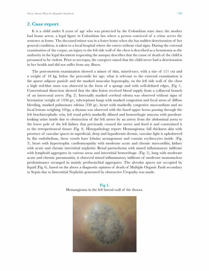
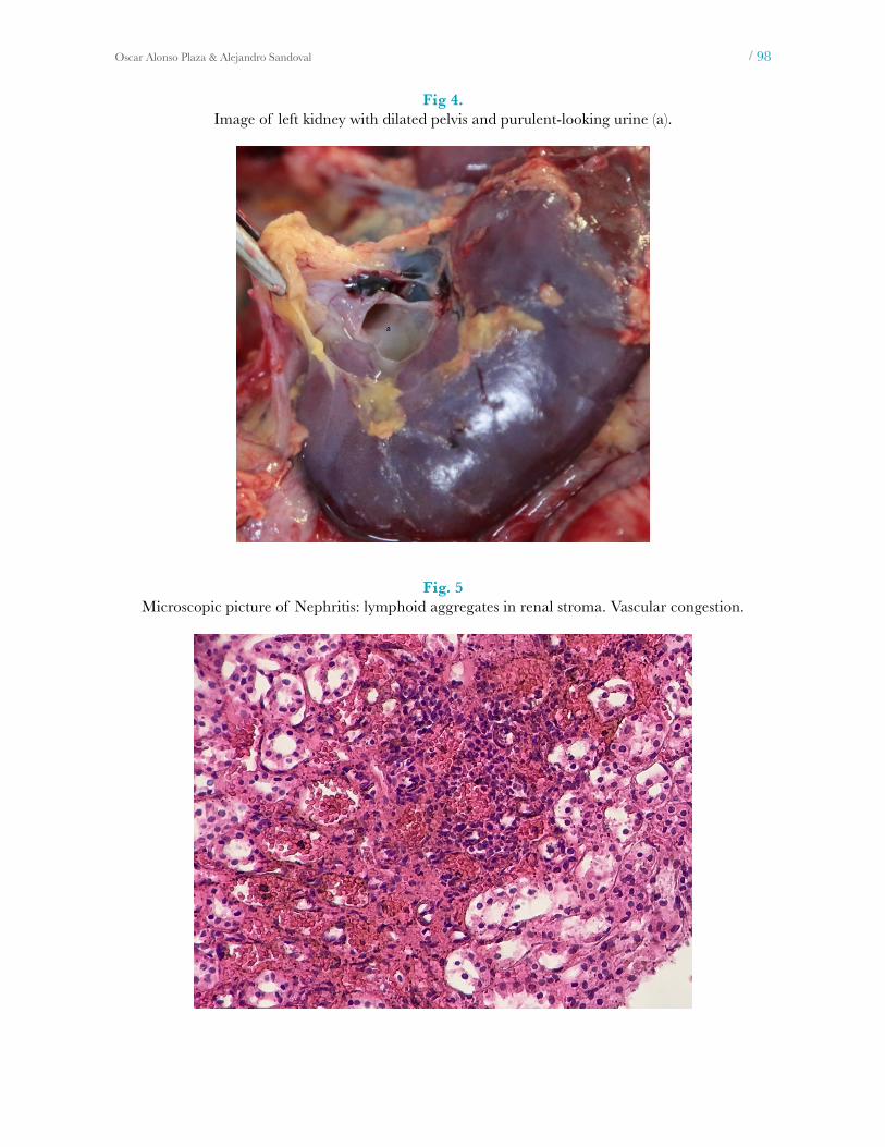
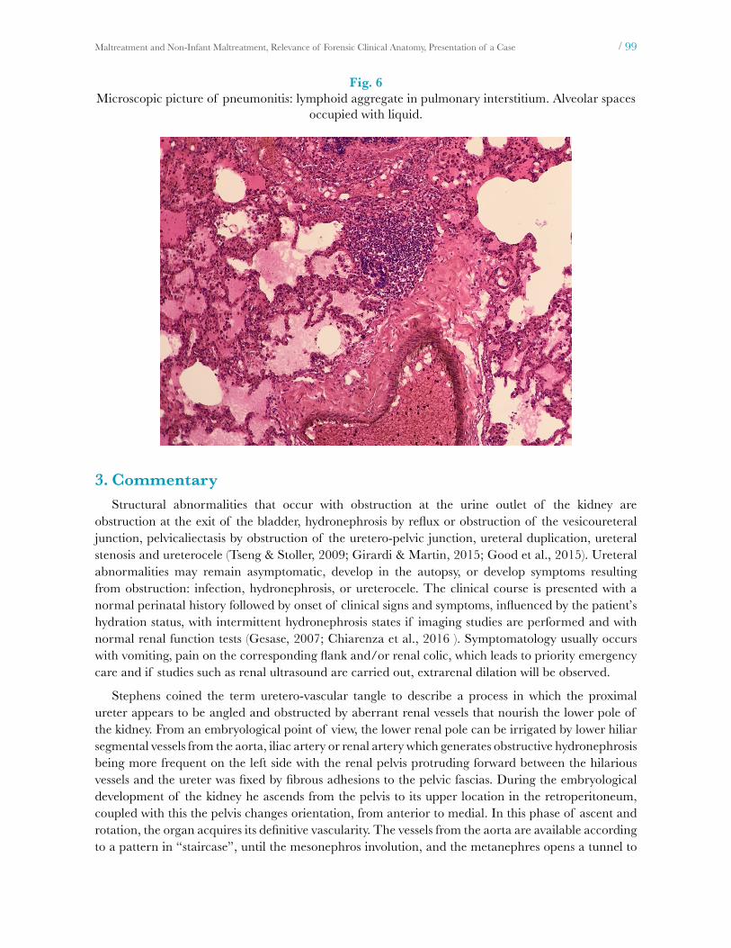
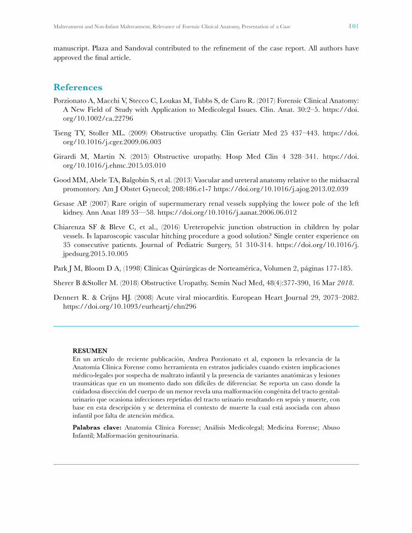
The post-mortem examination showed a minor of thin, mixed-race, with a size of 111 cm and a weight of 18 kg, below the percentile for age; what is relevant to the external examination is the sparse adipose panicle and the marked muscular hypotrophy, on the left side wall of the chest a high red-blue mass was observed in the form of a sponge and with well-defined edges, (Fig 1). Conventional dissection showed that the skin lesion received blood supply from a collateral branch of an intercostal artery (Fig. 2). Internally marked cerebral edema was observed without signs of herniation (weight of 1436 gr), subcrepitant lungs with marked congestion and focal areas of diffuse bleeding, marked pulmonary edema (330 gr), heart with markedly congestive myocardium and no focal lesions weighing 105gr, a thymus was observed with the fused upper horns passing through the left brachiocephalic vein, left renal pelvis markedly dilated and hemorrhagic mucosa with purulent-looking urine inside due to obstruction of the left ureter by an artery from the abdominal aorta to the lower pole of the left kidney that previously crossed the ureter and fixed it and constrained it to the retroperitoneal tissues (Fig. 4). Histopathology report: Hemangioma: full thickness skin with presence of vascular spaces in superficial, deep and hypodermis dermis, vascular light is upholstered by flat endothelium, these vessels have lobular arrangement and contain erythrocytes inside. (Fig. 3), heart with hypertrophic cardiomyopathy with moderate acute and chronic myocarditis, kidney with acute and chronic interstitial nephritis: Renal parenchyma with mixed inflammatory infiltrate with lymphoid aggregates in various areas and interstitial hemorrhage. (Fig: 5), lung with moderate acute and chronic pneumonitis, is observed mixed inflammatory infiltrate of moderate mononuclear predominance arranged in mainly peribronchial aggregates. The alveolar spaces are occupied by liquid (Fig. 6), based on the above a diagnostic opinion of death of Multiple Organic Fault secondary to Sepsis due to Interstitial Nephritis generated by obstructive Uropathy was made.
Fig 1.Hemangioma in the left lateral wall of the thorax.

97Maltreatment and Non-Infant Maltreatment, Relevance of Forensic Clinical Anatomy, Presentation of a Case
Fig 2.Hemangioma (a), irrigation of the hemangioma (b).
Fig. 3.Microscopic picture of the hemangioma: Proliferation of small benign-looking vessels in the
superficial and deep dermis, with a lobed arrangement and erythrocytes inside.
98Oscar Alonso Plaza & Alejandro Sandoval
Fig 4.Image of left kidney with dilated pelvis and purulent-looking urine (a).
Fig. 5Microscopic picture of Nephritis: lymphoid aggregates in renal stroma. Vascular congestion.
99Maltreatment and Non-Infant Maltreatment, Relevance of Forensic Clinical Anatomy, Presentation of a Case
Fig. 6 Microscopic picture of pneumonitis: lymphoid aggregate in pulmonary interstitium. Alveolar spaces
occupied with liquid.
3. CommentaryStructural abnormalities that occur with obstruction at the urine outlet of the kidney are
obstruction at the exit of the bladder, hydronephrosis by reflux or obstruction of the vesicoureteral junction, pelvicaliectasis by obstruction of the uretero-pelvic junction, ureteral duplication, ureteral stenosis and ureterocele (Tseng & Stoller, 2009; Girardi & Martin, 2015; Good et al., 2015). Ureteral abnormalities may remain asymptomatic, develop in the autopsy, or develop symptoms resulting from obstruction: infection, hydronephrosis, or ureterocele. The clinical course is presented with a normal perinatal history followed by onset of clinical signs and symptoms, influenced by the patient’s hydration status, with intermittent hydronephrosis states if imaging studies are performed and with normal renal function tests (Gesase, 2007; Chiarenza et al., 2016 ). Symptomatology usually occurs with vomiting, pain on the corresponding flank and/or renal colic, which leads to priority emergency care and if studies such as renal ultrasound are carried out, extrarenal dilation will be observed.
Stephens coined the term uretero-vascular tangle to describe a process in which the proximal ureter appears to be angled and obstructed by aberrant renal vessels that nourish the lower pole of the kidney. From an embryological point of view, the lower renal pole can be irrigated by lower hiliar segmental vessels from the aorta, iliac artery or renal artery which generates obstructive hydronephrosis being more frequent on the left side with the renal pelvis protruding forward between the hilarious vessels and the ureter was fixed by fibrous adhesions to the pelvic fascias. During the embryological development of the kidney he ascends from the pelvis to its upper location in the retroperitoneum, coupled with this the pelvis changes orientation, from anterior to medial. In this phase of ascent and rotation, the organ acquires its definitive vascularity. The vessels from the aorta are available according to a pattern in “staircase”, until the mesonephros involution, and the metanephres opens a tunnel to

100Oscar Alonso Plaza & Alejandro Sandoval
a high retroperitoneal location after these vessels. In this way the kidney as it ascends from its original position to its final position obtains its blood supply from the vessel successively higher and detaches from the lower one. It is then thought that any event that alters the ascent/rotation process, combined with the formation of the renal vessels, could generate an abnormal uretero-vascular relationship that causes partial obstruction (Park & Bloom, 1998).
The etiopathogenesis of pondo-statural growth retardation in kidney injury has multiple factors including the age of onset and the degree of alteration of the glomerular filtration, also an inadequate nutrition is a contributing factor since anorexia, vomiting and rejection of food causes a delay in growth, associated with this chronic anemia due to recurrent infections will produce an inadequate oxygenation of the cartilaginous dishes (Sherer & Stoller, 2018)
Myocarditis is a pathology whose form of presentation and clinical course is very variable, presenting a chronology of three distinct phases. In the first phase, the virus damages the cellular structures of the myocyte generating cardiac dilation; in the second phase there is an immune dysregulation produced by the damage to the myocytes; In the third phase, dilated cardiomyopathy occurs due to extensive myocardial injury (Dennert et al., 2008).
For this case report, there is an omission in the provision of this minor in the health service when presenting a nephropathy of chronic course derived from a congenital obstruction, with the generation of recurrent urinary tract infections with subsequent growth retardation, together with an event catastrophic as acute and chronic myocarditis and pneumonitis, situations that would have followed a different course with adequate and timely medical attention.
The Forensic Clinical Anatomy for this case is of vital importance because it is explained to the authority the duality of two injuries that appear in the minor deceased, an injury such as a hemangioma that although simulates child abuse is a pathology of normal course without affecting the its development; The structural lesion produced by obstructive uropathy generates recurrent infections that with timely and adequate care are diagnosed and corrected, that is why child abuse is supported as the omission in the provision of a vital service such as attention medical in a diagnosis pathology and affordable treatment at this stage of the XXI century.
4. Financial disclosures:None
5. Conflict of interest:None
6. Author contributionOscar Alonso Plaza and Alejandro Sandoval conceived of the study. Plaza was senior consultant at
this case report and participated in its coordination. Plaza and Sandoval contributed to the acquisition of clinical data, its analysis and interpretation and to the preparation of images. Plaza and Sandoval carried out the literature review. Plaza, Sandoval and Aguirre contributed to the preparation of the
101Maltreatment and Non-Infant Maltreatment, Relevance of Forensic Clinical Anatomy, Presentation of a Case
manuscript. Plaza and Sandoval contributed to the refinement of the case report. All authors have approved the final article.
ReferencesPorzionato A, Macchi V, Stecco C, Loukas M, Tubbs S, de Caro R. (2017) Forensic Clinical Anatomy:
A New Field of Study with Application to Medicolegal Issues. Clin. Anat. 30:2–5. https://doi.org/10.1002/ca.22796
Tseng TY, Stoller ML. (2009) Obstructive uropathy. Clin Geriatr Med 25 437–443. https://doi.org/10.1016/j.cger.2009.06.003
Girardi M, Martin N. (2015) Obstructive uropathy. Hosp Med Clin 4 328–341. https://doi.org/10.1016/j.ehmc.2015.03.010
Good MM, Abele TA, Balgobin S, et al. (2013) Vascular and ureteral anatomy relative to the midsacral promontory. Am J Obstet Gynecol; 208:486.e1-7 https://doi.org/10.1016/j.ajog.2013.02.039
Gesase AP. (2007) Rare origin of supernumerary renal vessels supplying the lower pole of the left kidney. Ann Anat 189 53—58. https://doi.org/10.1016/j.aanat.2006.06.012
Chiarenza SF & Bleve C, et al., (2016) Ureteropelvic junction obstruction in children by polar vessels. Is laparoscopic vascular hitching procedure a good solution? Single center experience on 35 consecutive patients. Journal of Pediatric Surgery, 51 310-314. https://doi.org/10.1016/j.jpedsurg.2015.10.005
Park J M, Bloom D A, (1998) Clínicas Quirúrgicas de Norteamérica, Volumen 2, páginas 177-185.
Sherer B &Stoller M. (2018) Obstructive Uropathy. Semin Nucl Med, 48(4):377-390, 16 Mar 2018.
Dennert R. & Crijns HJ. (2008) Acute viral miocarditis. European Heart Journal 29, 2073–2082. https://doi.org/10.1093/eurheartj/ehn296
RESUMENEn un artículo de reciente publicación, Andrea Porzionato et al, exponen la relevancia de la Anatomía Clínica Forense como herramienta en estratos judiciales cuando existen implicaciones médico-legales por sospecha de maltrato infantil y la presencia de variantes anatómicas y lesiones traumáticas que en un momento dado son difíciles de diferenciar. Se reporta un caso donde la cuidadosa disección del cuerpo de un menor revela una malformación congénita del tracto genital-urinario que ocasiona infecciones repetidas del tracto urinario resultando en sepsis y muerte, con base en esta descripción y se determina el contexto de muerte la cual está asociada con abuso infantil por falta de atención médica.
Palabras clave: Anatomía Clínica Forense; Análisis Medicolegal; Medicina Forense; Abuso Infantil; Malformación genitourinaria.