biochemical and structural properties of mouse kat iii abbreviations:
TRANSCRIPT

1
Biochemical and structural properties of mouse KAT III 1
Qian Han1, Howard Robinson2, Tao Cai3, Danilo A. Tagle4 and Jianyong Li1 * 2
3
1Department of Biochemistry, Virginia Tech, Blacksburg, Virginia 24061, USA 4
2Biology Department, Brookhaven National Laboratory, Upton, NY 11973, USA 5
3OIIB, NIDCR, National Institutes of Health, Bethesda, Maryland 20892-4322, USA 6
4Neurogenetics, NINDS, National Institutes of Health, Bethesda, Maryland 2089-29525, USA 7
Running Title: Mouse Kynurenine Aminotransferase III 8
*Address correspondence to 9
Jianyong Li 10
Tel: 540-231-1182; Fax: 540-231-9070; E-mail: [email protected] 11
Abbreviations: 12
KAT, kynurenine aminotransferase; αKMB, α-keto-methylthiobutyric acid; KYNA, kynurenic acid; 13
LLP, lysine-pyridoxal-5’-phosphate; mKAT III, mouse kynurenine aminotransferase III; PLP, 14
pyridoxal-5’-phosphate, PMP, pyridoxamine-5’-phosphate; RMS, root mean square; SHR, 15
spontaneously hypertensive rat 16
17
The atomic coordinates and structure factors (codes: 3e2f, 3e2y and 3e2z) have been deposited in the 18
Protein Data Bank, Research Collaboratory for Structural Bioinformatics, Rutgers University, New 19
Brunswick, NJ (http://www.rcsb.org). 20
ACCEPTED
Copyright © 2008, American Society for Microbiology and/or the Listed Authors/Institutions. All Rights Reserved.Mol. Cell. Biol. doi:10.1128/MCB.01272-08 MCB Accepts, published online ahead of print on 24 November 2008
on March 23, 2018 by guest
http://mcb.asm
.org/D
ownloaded from

2
ABSTRACT 1
Kynurenine aminotransferase III (KAT III) has been considered to be involved in the production of 2
mammalian brain kynurenic acid (KYNA) which plays an important role in protecting neurons from 3
over-stimulation by excitatory neurotransmitters. The enzyme was identified based on its high 4
sequence identity with mammalian KAT I, but its activity to kynurenine and its structural 5
characteristics have not been established. In this study, the biochemical and structural properties of 6
mouse KAT III (mKAT III) were determined. Specifically, mKAT III cDNA was amplified from a 7
mouse brain cDNA library and its recombinant protein was expressed in insect cell protein expression 8
system. We established that mKAT III is able to efficiently catalyze the transamination of kynurenine 9
to KYNA and has optimum activity at relatively basic conditions around pH 9.0 and at relatively high 10
temperature of 50 – 60 oC. In addition, mKAT III is active to a number of other amino acids. Its 11
activity to kynurenine is significantly decreased in the presence of methionine, histidine, glutamine, 12
leucine, cysteine and 3-hydroxykynurenienine. Through macromolecular crystallography we 13
determined the mKAT III crystal structure and its structures in complex with kynurenine and 14
glutamine. Structural analysis revealed the overall architecture of mKAT III, and its cofactor binding 15
site and active center residues. This is the first report concerning the biochemical characteristics and 16
crystal structures of KAT III enzymes, which provides a basis towards understanding the overall 17
physiological role of mammalian KAT III in vivo, and insight into regulating the levels of endogenous 18
KYNA through modulation of the enzyme in the mouse brain. 19
Key Words: kynurenine aminotransferase; kynurenic acid; kynurenine; crystal structure; NMDA; 20
neurodegenerative diseases; spontaneously hypertensive rat. 21
ACCEPTED
on March 23, 2018 by guest
http://mcb.asm
.org/D
ownloaded from

3
INTRODUCTION 1
Kynurenic acid (KYNA) is the only known endogenous antagonist of the N-methyl-D-aspartate 2
subtype of glutamate receptors (6, 36, 44, 54). KYNA is also an antagonist of the α7-nicotinic 3
acetylcholine receptor (1, 27, 43, 53). KYNA was recently identified as an endogenous ligand for an 4
orphan G-protein-coupled receptor (GPR35) that is predominantly expressed in immune cells (57). 5
Abnormal concentration of KYNA has been observed in patients with multiple neurodegenerative 6
diseases, including Huntington disease (5, 17), Alzheimer's disease (58), schizophrenia (11, 12, 52), 7
and acquired immunodeficiency syndrome dementia (18). These data suggest that KYNA, acting as 8
an endogenous modulator of glutamatergic and cholinergic neurotransmission, may play a role in the 9
pathogenesis of these disorders. In addition to its role as an excitatory amino acid antagonist, KYNA 10
is also involved in the control of cardiovascular function by acting at the rostral ventrolateral medulla 11
of the central nervous system (8). Spontaneously hypertensive rat (SHR), the most widely used animal 12
model for studying genetic hypertension, is associated with abnormally low KYNA levels in the area 13
of central nervous system that tunes physiological blood pressure (29, 34). 14
KYNA is produced enzymatically by irreversible transamination of kynurenine, the key intermediate 15
in the tryptophan catabolic pathway. In human, rat and mouse three proteins, designated as 16
Kynurenine aminotransferase (KAT) I, II and III, are involved in KYNA synthesis in the central 17
nervous system (16, 25, 41, 51, 60). Very recently, mitochondrial aspartate aminotransferase from rat 18
and human brain was reported to be able to catalyze the transamination of kynurenine to KYNA and 19
therefore was named KAT IV (15). Although the involvement of these enzymes in brain KYNA 20
production has been discussed, their specific contributions in brain KYNA synthesis remain to be 21
established. 22
Among the individual mammalian KATs, the KAT I and KAT III share similar genomic structures 23
and show high sequence identity (60), and may therefore have overlapping biological functions. In 24
ACCEPTED
on March 23, 2018 by guest
http://mcb.asm
.org/D
ownloaded from

4
kat-2−/− mouse brain, an increase in KAT I and KAT III expression suggests that upregulation of KAT 1
I and KAT III may compensate for the loss of KAT II (60). These compensatory changes may also 2
explain the rescue of hyperactivity and abnormal motor coordination in kat-2−/− mice (1, 59, 60). 3
These studies suggest the importance of mammalian KAT I and KAT III in maintaining KYNA level 4
in kat-2−/− mouse brain. Although many studies have dealt with the biochemical characteristics of 5
mammalian KAT I and KAT II (3, 4, 9, 16, 19, 25, 49) little is known about the biochemical activity 6
of KAT III. Likewise, the crystal structures of human KAT I (48) and its homologues, glutamine-7
phenylpyruvate aminotransferase from Thermus thermophilus HB8 (13) and KAT from a mosquito, 8
Aedes aegypti (AeKAT) (22) have been determined, as well as the crystal structure of human KAT II 9
(26, 47) and its homologue from Pyrococcus horikoshii (7). Although KAT III has been cloned in 10
mice, rats and humans as a member of the mammalian KAT family (60), its structure has not been 11
determined in any of these species. In this study, we expressed mKAT III in a eukaryotic protein 12
expression system, purified its recombinant protein, verified its activity to kynurenine and other 13
substrates, and determined its 3-dimensional structure, and binding properties with kynurenine and 14
glutamine. Our data provide the first biochemical and structural basis for the mouse KAT III and form 15
the basis for understanding its physiological functions. 16
MATERIALS AND METHODS 17
Expression and characterization of mKAT III 18
Expression of recombinant mKAT. Total mRNA from mouse brain was isolated using TRIZOL 19
reagent (Gibco-BRL) according to the manufacturer’s instructions and used as templates for reverse 20
transcription to produce their corresponding cDNA. A forward primer (5’-21
CTGCAGATGCTTTTGGCCCAGAGG-3’) containing a Pst I restriction site (underlined) and a 22
reverse primer (5’-AAGCTTTCAAGACTTCTGGCTGTTCCAA-3) containing a Hind III restriction 23
site (underlined) were used for PCR amplification of the mouse KAT III coding sequence 24
ACCEPTED
on March 23, 2018 by guest
http://mcb.asm
.org/D
ownloaded from

5
(AAQ15190). The amplified cDNA fragment was cloned into a TA cloning vector and then subcloned 1
into a baculovirus transfer vector pBlueBac4.5 (Invitrogen, Carlsbad, California) between the Pst I 2
and Hind III restriction sites. Because the signal peptide (the first 35 amino acids encoded by exon 2) 3
is cleaved when it is targeted to the mitochondria (37), it was not included in the expression construct. 4
The KAT III recombinant baculovirus transfer vector was propagated in E. coli. The cDNA insert was 5
verified in-frame and adjacent to the 3’-end of the polyhedron promoter by DNA sequencing. The 6
recombinant baculovirus transfer vector was co-transfected with linearized Bac-N-Blue™ (AcMNPV, 7
Autographa californica multiple nuclear polyhedrosis virus) viral DNA in the presence of 8
InsectinPlus™ insect cell-specific liposomes into Spodoptera frugiperda (Sf9) insect cells 9
(Invitrogen). The recombinant baculoviruses were purified by a plaque assay procedure. Blue 10
recombinant plaques were transferred to twelve-well microtiter plates and amplified in Sf9 cells. Viral 11
DNA was isolated for gene amplification analysis to determine the purity of the recombinant virus. A 12
high-titer viral stock of a pure recombinant virus was generated by amplification in a suspension 13
culture of Sf9 cells. The titer of the viral stock and the time-course at a multiplicity of infection of 6 14
were established according to the manufacturer’s instructions. Sf9 cells were cultured in spinner 15
flasks in TNM-FH medium containing 10% fetal bovine serum (Invitrogen, Carlsbad, California). 16
KAT III recombinant virus was inoculated into the culture at a cell density of 2.5 × 10⁶ cells /ml. Sf9 17
cells were harvested at day 4 post-inoculation by centrifugation (800 g for 15 min at 4 °C), and stored 18
at -80 oC until use. 19
Purification of recombinant mKAT III. Cell pellets were dissolved in 50 mM phosphate buffer, pH 20
8.0, containing 0.1% Triton X-100 and 1 mM phenylmethylsulphonyl fluoride (PMSF). After 21
sonication in cold water for 5 min, the cell lysate was centrifuged at 20,000 g for 20 min at 4 °C and 22
the supernatant was retained for mKAT III purification. Supernatant containing mKAT III was 23
dialyzed against 20 mM phosphate buffer (pH 8.0), containing 1 mM PMSF, 0.5 mM EDTA and 5 24
mM DTT, and then applied to a DEAE Sepharose column (2.5 X 20 cm). mKAT III active fractions 25
ACCEPTED
on March 23, 2018 by guest
http://mcb.asm
.org/D
ownloaded from

6
were pooled, dialyzed against 10 mM phosphate buffer (pH 7.5) containing 5 mM DTT and then 1
applied to a hydroxyapatite column (1.5 x 10 cm). KAT III active fractions were eluted using a 2
phosphate gradient (10-400 mM), its active fractions were pooled, dialyzed in 20 mM phosphate 3
buffer (pH 8.0), and then applied into a Mono-Q column (GE Health). KAT III fractions from Mono-4
Q column were concentrated using 2.0 ml membrane concentrators (with molecular weight cutoff at 5
30,000). Concentrated KAT III was analyzed by gel filtration chromatography and collected KAT III 6
peak was analyzed by SDS-PAGE to verify its purity. 7
Activity assay. KAT activity assay was based on previously described methods (20). Briefly, a 8
reaction mixture of 50 µl containing 5 mM L-kynurenine, 5 mM α-ketoglutarate, 40 µM PLP, and 2 9
µg of protein sample was prepared in 100 mM boric acid buffer (pH 9.0). The mixture was incubated 10
for 15 min at 45 °C, and the reaction was stopped by adding an equal volume of 0.8 M formic acid. 11
The reaction mixture was centrifuged at 15,000 g for 10 min and the supernatant was analyzed by 12
HPLC with UV detection at 330 nm for both kynurenine and KYNA. To test the transamination 13
activity to other amino acids, the mixture was assayed in 10 mM amino acid, 2 mM glyoxylate or 14
phenylpyruvate (for activity towards glycine), 40 µM PLP, 100 mM boric acid buffer, pH 9.0, and 2 15
µg enzyme in a total volume of 50 µl. The mixture was incubated for 15 min at 45 oC and then 16
assayed for o-phthaldialdehyde thiol (OPT)-amino acid product conjugate by HPLC with 17
electrochemical detection after their corresponding reaction mixtures were derivatized by OPT 18
reagent (21). In the kinetic study for other amino acid substrates, a different amino acid at varying 19
concentrations (0.23–15 mM) and 2 mM glyoxylate in 50 µl total volume prepared in 100 mM boric 20
acid buffer, pH 9.0 were assayed for KAT activity. The mixture was incubated for 15 min at 45 °C. 21
To determine the substrate specificity for α-keto acids, 16 individual α-keto acids (indicated in Table 22
2) were tested for their ability to function as an amino group acceptor for mKAT III. In the assays, a 23
different α-keto acid at varying concentrations (0.06–60 mM) was used in the presence of 10 mM 24
ACCEPTED
on March 23, 2018 by guest
http://mcb.asm
.org/D
ownloaded from

7
kynurenine and the rate of KYNA production was determined as described in the KAT activity assay. 1
Effect of pH and temperature on mKAT III. To determine the effect of buffer pH on mKAT III 2
activity, a buffer mixture consisting of 100 mM phosphate and 100 mM boric acid was prepared and 3
the pH of the buffer was adjusted to 6.0, 7.0, 8.0, 9.0, 10.0 and 11.0. The buffer mixture was selected 4
to maintain a relatively consistent buffer composition and ionic strength yet have sufficient buffering 5
capacity for relatively broad pH range. The reaction mixture containing 5 mM kynurenine, 2 mM 6
glyoxylate, and 2 µg mKAT III was prepared using the buffer mixture at different pHs and tested for 7
KAT activity. To determine the effect of temperature on KAT III-catalyzed kynurenine 8
transamination, the reaction mixture in 50 µl phosphate-borate buffer (pH 9.0) was incubated at 9
different temperatures ranging from 10 – 80 oC, and the product formed in the reaction mixture was 10
measured by HPLC-UV analysis. 11
Effect of other amino acids on mKAT III catalyzed kynurenine transamination. To determine the 12
effect of a competing amino acid on mKAT III catalyzed KYNA production from kynurenine, a 13
different amino acid (with a final concentration of 5 mM) was incorporated into the typical reaction 14
mixture (5 mM kynurenine, 2 mM glyoxylate, and 2 µg mKAT III in 50 µL) and the enzyme activity 15
was assayed in the same manner as described for the KAT activity assay. The assays for each amino 16
acid (indicated in Table 2) were performed in triplicate and the results were analyzed by using the 17
Student's t-test. 18
Crystal structure of mKAT III 19
mKAT III Crystallization. The crystals were grown by hanging-drop vapor diffusion methods with the 20
volume of reservoir solution at 500 µl and the drop volume at 2 µl, containing 1 µl of protein sample 21
(10 mg/ml) and 1 µl of reservoir solution. The optimized crystallization buffer consisted of 21% PEG 22
400, 10% glycerol, 150 mM CaCl2 and 100 mM HEPES, pH 7.5. The crystals of the glutamine-23
enzyme and kynurenine-enzyme complexes were obtained by soaking the mKAT III crystals in 2.5 24
ACCEPTED
on March 23, 2018 by guest
http://mcb.asm
.org/D
ownloaded from

8
mM glutamine and kynurenine, respectively, in the crystallization buffer for a week. 1
Data collection and processing. Individual mKAT III crystal was cryogenised using 15 % glycerol in 2
the crystallization buffer as a cryo-protectant solution. Diffraction data of mKAT III crystals were 3
collected at the Brookhaven National Synchrotron Light Source beam line X29A (λ = 1.0908 Å). 4
Data was collected using an ADSC Q315 CCD detector. All data were indexed and integrated using 5
HKL-2000 software; scaling and merging of diffraction data were performed using the program 6
SCALEPACK (42). The parameters of the crystals and data collection are listed in Table 1. 7
Structure determination. The structure of mKAT III was determined by the molecular replacement 8
method using chain A of the published AeKAT structure (Protein Data Bank code, 1R5E) (23) 9
without any ligands, cofactors or waters. The program Molrep (56) was employed to calculate both 10
cross-rotation and translation functions of the model. The initial model was subjected to iterative 11
cycles of crystallographic refinement with the Refmac 5.2 (40) and graphic sessions for model 12
building using the program O (31). In the first cycles of the refinement, tight restraints on non-13
crystallographic symmetry were applied, but these were gradually released and the molecules were 14
refined independently in later stages since this gave the lowest Rfree values. The cofactor and substrate 15
molecules were modeled before adding solvent molecules based on both the 2Fo - Fc and Fo - Fc 16
electron density maps when the R factor dropped to a value of around 0.24 at full resolution for the 17
structures. Solvent molecules were automatically added and refined with ARP/wARP (45) together 18
with Refmac 5.2. Superposition of structures was done using Lsqkab (32) in the CCP4 suite. Figures 19
were generated using Pymol (10). Protein and substrate interaction also was analyzed using Pymol 20
(10). 21
RESULTS 22
Enzyme purification and activity assay 23
ACCEPTED
on March 23, 2018 by guest
http://mcb.asm
.org/D
ownloaded from

9
Using DEAE-Sepharose, hydroxyapatite, Mono-Q and gel-filtration chromatography, we purified 1
mKAT III recombinant protein to more than 98% purity as determined by SDS-PAGE analyses. The 2
purified mKAT III showed high KAT activity using glyoxylate as an amino group acceptor. Among 3
the temperature points tested, mKAT III showed the highest KAT activity at 50 – 60 °C (Fig. 1A). 4
When the phosphate and borate buffer mixture, adjusted to pH 6.0 – 11.0, was used to prepare mKAT 5
III/kynurenine/glyoxylate reaction mixtures, mKAT III displayed optimum activity around pH 9.0-6
10.0 (Fig. 1B), which is close to the optimum pH reported in the literature (3, 4, 16, 49) for 7
mammalian brain KAT I but apparently different from the optimum pH (7.5-9.0) of recombinant 8
human KAT I (25). mKAT III was tested for aminotransferase activity towards 24 different amino 9
acids using glyoxylate as a primary amino group acceptor. The enzyme showed activity to a number 10
of amino acids, including some aromatic amino acids (phenylalanine, kynurenine, tryptophan, 3-11
hydroxykynurenine and tyrosine), sulfur containing amino acids (methionine and cysteine) and other 12
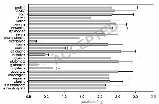
amino acids (glutamine, histidine, asparagine, serine, alanine, aminobutyrate and lysine) (Fig. 2). The 13
substrate profile of mKAT III overlaps considerably with that of human KAT I (25), but human KAT 14
I favors hydrophobic substrates and mKAT III prefers hydrophilic substrates. For example, mKAT III 15
has high transamination activity to asparagine to which hKAT I has essentially no activity (25). 16
Kinetic data provided an overview regarding the catalytic efficiency of mKAT III towards individual 17
amino acids (Table 2). It is apparent that mKAT III is efficient in catalyzing the transamination of 18
glutamine, histidine, methionine, phenylalanine, asparagine, cysteine and kynurenine. Sixteen α-keto 19
acids were tested for their potential as an amino group acceptor for mKAT III with 10 mM 20
kynurenine as the amino group donor. Thirteen of them have detectable activity. Table 2 illustrates 21
the enzyme kinetic parameters towards each α-keto acid, including Km and kcat/Km. Based on kinetic 22
analysis, glyoxylate, α-ketocaproic acid, phenylpyruvate, α-ketobutyrate, αKMB, α-ketovalerate, 23
indo-3-pyruvate, p-hydroxy-phenylpyruvate, mercaptopyruvate and oxaloacetate are good amino 24
group acceptors for mKAT III. Pyruvate, phenylpyruvate and α-ketoglutarate are poor co-substrates 25
ACCEPTED
on March 23, 2018 by guest
http://mcb.asm
.org/D
ownloaded from

10
for the enzyme. The keto acid substrate profile of mKAT III is very similar to human KAT I. 1
However, indo-3-pyruvate that strongly inhibited hKAT I activity showed no noticeable inhibition on 2
mKAT III. 3
Effects of other amino acids on mKAT III catalyzed kynurenine transamination 4
When the typical reaction mixture (5 mM kynurenine, 2 mM glyoxylate and 2 µg mKAT III) was 5
assayed in the presence of each of the other amino acids (5 mM), most of the amino acids showed no 6
noticeable effect on decreasing the rate of the mKAT III-catalyzed kynurenine transamination, but the 7
presence of cysteine, glutamine, histidine, methionine, leucine or phenylalanine greatly decreased the 8
rate of kynurenine transamination (Fig. 3). The decrease in the rate of KYNA production in the 9
mKAT III/kynurenine/α-keto acid reaction mixture in the presence of a different amino acid is 10
apparently due to competitive inhibition of the KAT III-catalyzed kynurenine transamination. In 11
addition, noticeable inhibition of 3-hydroxyknurenine to KAT III-catalyzed kynurenine 12
transamination was also observed (Fig. 3). Although asparagine, tryptophan, serine, aminobutyrate, 13
lysine served as the amino group donors for mKAT III, these amino acids did not seem to compete 14
with kynurenine to inhibit KAT activity (Fig. 3). Moreover, transamination of leucine by mKAT III 15
was not observed, but leucine effectively inhibited KAT III-catalyzed kynurenine transamination, 16
suggesting that leucine can enter and occupy the active site of KAT III. 17
Overall Structure 18
The crystal structure of mKAT III was determined by molecular replacement and refined to 2.60 Å 19
resolution. The final model contains 2 × 410 amino acid residues and yields a crystallographic R 20
value of 18.2% and an Rfree value of 23.3% with ideal geometry (Table 1). There are two protein 21
molecules in an asymmetric unit, which forms a biological homodimer. The residues of the two 22
subunits in mKAT III structure are numbered 42 – 451 for chain A, and 42* - 451* for chain B. The 23
results of the refinement are summarized in Table 1. All residues in the two subunits except for 24
ACCEPTED
on March 23, 2018 by guest
http://mcb.asm
.org/D
ownloaded from
11
Tyr312 are in favorable regions of the Ramachandran plot as defined with PROCHECK (35). 1
Although Tyr312 (A), and (B) fall within a disallowed region of the Ramachandran plot (Table 1) of 2
the final model, their excellent electron density allowed us to unambiguously assign the observed 3
conformations. 4
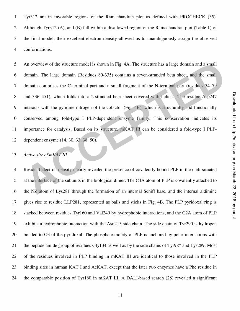
An overview of the structure model is shown in Fig. 4A. The structure has a large domain and a small 5
domain. The large domain (Residues 80-335) contains a seven-stranded beta sheet, and the small 6
domain comprises the C-terminal part and a small fragment of the N-terminal part (residues 54–79 7
and 336–451), which folds into a 2-stranded beta sheet covered with helices. The residue Asp247 8
interacts with the pyridine nitrogen of the cofactor (Fig. 4B), which is structurally and functionally 9
conserved among fold-type I PLP-dependent enzyme family. This conservation indicates its 10
importance for catalysis. Based on its structure, mKAT III can be considered a fold-type I PLP-11
dependent enzyme (14, 30, 33, 38, 50). 12
Active site of mKAT III 13
Residual electron density clearly revealed the presence of covalently bound PLP in the cleft situated 14
at the interface of the subunits in the biological dimer. The C4A atom of PLP is covalently attached to 15
the NZ atom of Lys281 through the formation of an internal Schiff base, and the internal aldimine 16
gives rise to residue LLP281, represented as balls and sticks in Fig. 4B. The PLP pyridoxal ring is 17
stacked between residues Tyr160 and Val249 by hydrophobic interactions, and the C2A atom of PLP 18
exhibits a hydrophobic interaction with the Asn215 side chain. The side chain of Tyr290 is hydrogen 19
bonded to O3 of the pyridoxal. The phosphate moiety of PLP is anchored by polar interactions with 20
the peptide amide group of residues Gly134 as well as by the side chains of Tyr98* and Lys289. Most 21
of the residues involved in PLP binding in mKAT III are identical to those involved in the PLP 22
binding sites in human KAT I and AeKAT, except that the later two enzymes have a Phe residue in 23
the comparable position of Tyr160 in mKAT III. A DALI-based search (28) revealed a significant 24
ACCEPTED
on March 23, 2018 by guest
http://mcb.asm
.org/D
ownloaded from

12
structural homology of mKAT III with human KAT I (PDB code 1w7l, r.m.s.d. 1.2 Å) (48) and 1
AeKAT (PDB code 2r5c, r.m.s.d. 1.2 Å) (23) present in the Protein Data Bank. All these 3-D 2
structures support that mKAT III is a member of KAT enzymes. 3
Substrate Recognition in mKAT III: glutamine complex 4
The crystal structure of mKAT III: glutamine complex was determined by molecular replacement 5
using above mKAT III structure as an initial model and refined to 2.26 Å resolution. The final model 6
contains 2 × 410 amino acid residues and yields a crystallographic R value of 17.5% and an Rfree value 7
of 22.1% with ideal geometry (Table 1). Inspection of the crystal structure of mKAT III: glutamine 8
complex revealed that the glutamine lies near the PLP cofactor that was changed to PMP form, but 9
glutamine and PLP did not form an external aldimine. Several residues, including Arg430, Tyr250, 10
Tyr160, Asn219, Phe373, Gln71, Gly72, Trp54, and Tyr98*, define the substrate-binding site and 11
contact with the glutamine molecule. The carboxylate of glutamine forms a salt bridge with the 12
guanidinium group of Arg430 (Fig. 5). Gln71 and Gly72 are located at the turning point of the loop 13
that dives into and partially plugs the enzyme active site, thus shielding the substrate-binding pocket 14
from the bulk solvents. 15
Substrate Recognition in mKAT III: kynurenine structure 16
The crystal structure of mKAT III: kynurenine complex was determined by molecular replacement 17
using mKAT III: glutamine structure as an initial model and refined to 2.80 Å resolution. Its final 18
model contains 2 × 410 amino acid residues and yields a crystallographic R value of 19.2% and an 19
Rfree value of 23.7% with ideal geometry (Table 1). Similar to mKAT III: glutamine complex (Fig. 5), 20
inspection of the crystal structure of mKAT III: kynurenine complex revealed that kynurenine lies 21
near the PLP cofactor in both subunits, and kynurenine and the cofactor do not form an external 22
aldimine in any subunits, but only one cofactor is changed to PMP form in one subunit (Fig. 6A) and 23
remained as an internal aldimine (LLP) form in the other subunit (Fig. 6B). Interactive residues 24
ACCEPTED
on March 23, 2018 by guest
http://mcb.asm
.org/D
ownloaded from

13
between kynurenine and protein are Arg430, Tyr160, Asn219, Phe373, Gln71, Gly72, Trp54, 1
Tyr312* and Tyr98*. The interactions are similar to those in mKAT III: glutamine structure, but the 2
phenyl ring of kynurenine has hydrophobic interactions with the phenyl rings of Tyr98* and Tyr312* 3
and the indole ring of Trp54 (Fig. 6). Upon superposition of mKAT III: glutamine structure to mKAT 4
III: kynurenine structure, we did not find any significant conformational changes of main protein 5
chain and interactive residues. 6
Conformational change as a result of the complex formation 7
Through superposition of all 6 subunits from the three mKAT III structures, we identified a major 8
main chain conformational change involving residues 51 to 65 in the four subunits of KAT III: 9
kynurenine or glutamine complexes. Upon kynurenine or glutamine binding, the N-terminal part of 10
the subunit moves towards and interacts with the substrate, resulting in a close interaction of Trp-54 11
and the substrate. This conformational change also involves some side-chain movement of Tyr-160 12
and Tyr-312*. In KAT III: substrate complexes, Tyr-160 shifts toward the substrate and forms a 13
hydrogen bond with the substrate glutamine or kynurenine, and Tyr-312* turns away from the center, 14
which provides room for the substrate, but maintains its interaction with the substrate (Fig. 8). 15
DISCUSSION 16
It has generally been accepted that KYNA functions as a non-competitive antagonist of glutamatergic 17
receptors in mammalian brain and plays a role in alleviating potential overstimulation of the receptors 18
by glutamate. Abnormal concentrations of brain KYNA have been implicated in several 19
neurodegenerative diseases. KYNA, a tryptophan metabolite, is derived from kynurenine by 20
aminotransferase-catalyzed reactions. The compound does not pass through the blood-brain-barrier 21
easily and therefore needs to be synthesized locally. Because of the close association between 22
abnormal concentration of brain KYNA and neurodegeneration, the enzymes involved in KYNA 23
production are potential targets for regulating brain KYNA. However such efforts have been hindered 24
ACCEPTED
on March 23, 2018 by guest
http://mcb.asm
.org/D
ownloaded from

14
by the lack of detailed knowledge regarding structure/function relationship of the mammalian KATs. 1
Mammalian KAT III has been named based on its sequence homology with mammalian KAT I and is 2
expressed in mammalian brains, but its biochemical and structural characteristics have never been 3
documented at the protein level. This study demonstrates that mKAT III, expressed in eukaryotic 4
protein expression system, can efficiently catalyze the transamination of kynurenine to KYNA. 5
Mouse KAT III shows activity towards a number of amino acids, including methionine, glutamine, 6
and histidine, and the presence of these amino acids decreases KAT III-catalyzed kynurenine 7
transamination (Fig. 3 & Table 2). These findings are similar to those determined for human KAT I 8
(25). An earlier report also described the inhibition of human KAT I by glutamine, phenylalanine, and 9
tryptophan (4). It seems apparent from our results that the biochemical behaviors of KAT I and KAT 10
III are highly similar; and consequently it is difficult to distinguish between KAT I and KAT III 11
activity in crude biological samples. 12
Critical residues related to KAT activity are conserved between KAT III and KAT I. By genetic 13
analysis, a missense mutation (E61G) in KAT I gene was identified in SHR, which leads to reduced 14
KAT I enzymatic activity and higher blood pressure (29, 34). Our previous study dealing with 15
structural characterization of human KAT I revealed that the evolutionarily conserved Glu27 (i.e., 16
Glu61 in full-length sequence of KAT I), positioned at the C terminus of the helix H1, is a pivotal 17
residue in fixing the helix in the observed conformations. Therefore, the substitution of Glu27 with 18
glycine of KAT I would affect the conformational change induced by substrate binding with dramatic 19
changes in it is catalytic function (48). Interestingly, the position corresponding to the Glu27 is 20
replaced with an aspartate residue in all mammalian KAT III (not shown). Structurally aspartate is 21
similar to glutamate; therefore, the high conservation of either glutamate in KAT I or aspartate in 22
KAT III suggests that this position is likely essential for enzyme catalysis. 23
Based on the similarities of genomic structure, protein structure and biochemical function between the 24
ACCEPTED
on March 23, 2018 by guest
http://mcb.asm
.org/D
ownloaded from

15
KAT I and KAT III, it is reasonable to speculate that KAT I and KAT III have overlapping biological 1
functions in vivo. The high similarity of mammalian KAT I and KAT III raises a question as to their 2
origins. Evidence of gene duplication and genetic redundancy in KAT I and KAT III are first seen in 3
the nematode Caenorhabditis elegans. Knockdown or knockout of either KAT I or KAT III in C. 4
elegans did not result in any observable phenotypes in growth, body movements, mating, and brood 5
size (60), which suggests functional compensation between KAT I and KAT III in nematodes. 6
Although mammalian KAT I and KAT III share highly overlapping biochemical properties, there also 7
are noticeable differences between these two enzymes. The availability of highly purified 8
recombinant human KAT I and mKAT III enabled us to critically compare the biochemical 9
characteristics of these two enzymes. In a previous study, we determined that human KAT I was able 10
to efficiently catalyze kynurenine to KNYA under physiological conditions, but it did not show any 11
activity towards 3-hydroxykynurenine (3-HK) (25) and differs from other reports (16, 40, 49) 12
concerning mammalian KAT I. Comparison of the pH profiles between human KAT I and mKAT III 13
clearly indicates that mKAT III is more active at basic conditions than that of human KAT I. 14
Sequence comparison revealed that KAT III from human and mouse contains more basic residues and 15
has higher isoelectric point than KAT I, which might be related to their differences in pH preferences. 16
For example, mouse KAT III has 34 lysine residues, but human KAT I only shows 24 lysine residues. 17
Presence of 3-HK has no effect on hKAT I-catalyzed kynurenine transamination (25), but noticeably 18
decreased mKAT III-catalyzed kynurenine transamination (Fig. 3), suggesting that 3-HK is a 19
substrate of mKAT III. Based on these results, it seems apparent that the biochemical characteristics 20
of mKAT III are similar to those described for mammalian brain KAT I (16, 49). 21
There are different optimum pH values for KAT I in literature. The optimum pH 9.5~10 for KAT I 22
activity were seen in isolated human brain (3, 4, 16, 49) and placenta (39), whereas the optimal low 23
pH 6.5-9 for “KAT I” activity were reported in the insect cell-expressed human KAT I (pH 7.5 - 9.0) 24
ACCEPTED
on March 23, 2018 by guest
http://mcb.asm
.org/D
ownloaded from

16
(25), rat liver (pH 6.5) (55), and heart (pH 8 - 9) (2). Since KAT I is a highly conserved protein, it is 1
unlikely that the isolated KAT I, when tested in vitro, have different optimum pH. Therefore, we 2
argue that the isolated “KAT I” with high optimal pH could potentially be KAT III. 3
High alkaline and thermostable enzymes in vitro have been observed in many other cases, such as 4
alkaline phosphatase (pH 9.5). The pH values in most of the living cell structures, on the other hand, 5
are near neutrality, i.e. for the cytoplasm it is pH 6.9 and for the nucleus it is pH 7.6. It is possible that 6
the pH required for optimum activity in vivo might not be the same as that in vitro. Enzyme activity is 7
usually determined using substrate at saturating concentrations. It has been indicated that if the 8
substrate concentration was diluted to physiological concentration, the pH optimum of the enzyme 9
might be lowered to physiological values (46). Consequently, one has to be conscientious when 10
judging the physiological function of an enzyme based on its in vitro optimum pH conditions. To 11
fully understand the physiological functions of mammalian KAT I and KAT III, it is necessary to 12
perform extensive studies of both enzymes. 13
The 3-D structure of mKAT III in comparison with human KAT I and AeKAT provides a structural 14
basis explaining the substrate specificity of these three enzymes. We previously compared the 15
substrate specificity of AeKAT with hKAT I and observed that AeKAT prefers hydrophilic substrates 16
while human KAT I favors hydrophobic amino group donors (Han et al., 2004). Based on substrate 17
specificity, it seems that mKAT III prefers hydrophilic substrate even more strongly than AeKAT 18
(Fig. 2 & Table 2) (24). A superposition of human KAT I and AeKAT: glutamine complex onto 19
mKAT III: glutamine complex shows that the three proteins have similar architecture (Fig. 7A). There 20
are 11 residues involved in glutamine binding for the AeKAT-glutamine complex (23) and the 21
superposition of these enzymes determined that 9 of 11 substrate binding residues are identical in 22
these three enzymes. The counterparts of Tyr160 in mKAT III is Phe125 in human KAT I and Tyr135 23
in AeKAT, and the counterparts of Tyr312 in mKAT III is Phe278 in human KAT I and Tyr286 in 24
ACCEPTED
on March 23, 2018 by guest
http://mcb.asm
.org/D
ownloaded from

17
AeKAT (Fig. 7B). Tyrosine is less hydrophobic than phenylalanine and the hydrophilic hydroxyl 1
group could interact with polar group of the amino acid donors, which likely explains the increasing 2
preference of AeKAT and mKAT III to hydrophilic amino group donors as compared with human 3
KAT I. 4
Leucine is one of the preferred substrates of human KAT I, and it inhibits human KAT I-catalyzed 5
kynurenine transamination at high concentrations (25). This amino acid is effective in decreasing 6
mKAT III-catalyzed kynurenine transamination, but surprisingly mKAT III displayed essentially no 7
activity to leucine. Based on its effect on decreasing the mKAT-catalyzed kynurenine transamination, 8
leucine apparently is interactive with the enzyme, but the substrate might not be able to position 9
properly at the active site, thereby unable to serve as a substrate. Leucine is one of the most 10
hydrophobic amino acids (next only to phenylalanine). It is possible that both Tyr160 and Tyr312 in 11
mKAT III hinders proper positioning of leucine at its active site. However in order to clearly 12
understand the interaction of the mKAT III with leucine, it is necessary to have the structure of 13
mKAT III: leucine complex determined. Such structural data should be useful for rational design of 14
inhibitors against KAT III. In addition, the ability and inability to catalyze the transamination of 15
leucine by KAT I and KAT III, respectively, could serve as a criterion to distinguish KAT III from 16
KAT I activity. 17
In summary, this study established that mKAT III was able to catalyze the kynurenine to KYNA 18
pathway. The 3-D structures of mKAT III and comparison of its complexes with the substrates to 19
human KAT I and AeKAT provide insight into its preferred substrate binding and catalysis. 20
Moreover, similarity in biochemical characteristics of mKAT III to those reported for mammalian 21
brain KAT I in the literature poses a possibility that the reported brain KAT I at high pH may actually 22
be KAT III. 23
24
ACCEPTED
on March 23, 2018 by guest
http://mcb.asm
.org/D
ownloaded from

18
ACKNOWLEDGEMENTS 1
This work was carried out in part at the National Synchrotron Light Source, Brookhaven National
Laboratory, supported in part by the Intramural Research Program of the institutes of NIDCR and
NINDS at NIH and partly by the College of Agriculture and Life Sciences, Virginia Tech. The
authors would like to acknowledge the support of the Virginia Tech Department of Biological
Sciences for the use of their X-ray facility and are grateful to Dr Nancy Vogelaar, Chris Ceccarelli
(Oxford Diffraction) and Dr Florian Schubot for engaging us in helpful discussions.
REFERENCES
1. Alkondon, M., E. F. Pereira, P. Yu, E. Z. Arruda, L. E. Almeida, P. Guidetti, W. P.
Fawcett, M. T. Sapko, W. R. Randall, R. Schwarcz, D. A. Tagle, and E. X. Albuquerque.
2004. Targeted deletion of the kynurenine aminotransferase ii gene reveals a critical role of
endogenous kynurenic acid in the regulation of synaptic transmission via alpha7 nicotinic
receptors in the hippocampus. J Neurosci 24:4635-48.
2. Baran, H., G. Amann, B. Lubec, and G. Lubec. 1997. Kynurenic acid and kynurenine
aminotransferase in heart. Pediatr Res 41:404-10.
3. Baran, H., N. Cairns, B. Lubec, and G. Lubec. 1996. Increased kynurenic acid levels and
decreased brain kynurenine aminotransferase I in patients with Down syndrome. Life Sci
58:1891-9.
4. Baran, H., E. Okuno, R. Kido, and R. Schwarcz. 1994. Purification and characterization of
kynurenine aminotransferase I from human brain. J Neurochem 62:730-8.
5. Beal, M. F., W. R. Matson, K. J. Swartz, P. H. Gamache, and E. D. Bird. 1990.
Kynurenine pathway measurements in Huntington's disease striatum: evidence for reduced
formation of kynurenic acid. J Neurochem 55:1327-39.
6. Birch, P. J., C. J. Grossman, and A. G. Hayes. 1988. Kynurenic acid antagonises responses
ACCEPTED
on March 23, 2018 by guest
http://mcb.asm
.org/D
ownloaded from

19
to NMDA via an action at the strychnine-insensitive glycine receptor. Eur J Pharmacol
154:85-7.
7. Chon, H., H. Matsumura, Y. Koga, K. Takano, and S. Kanaya. 2005. Crystal structure of a
human kynurenine aminotransferase II homologue from Pyrococcus horikoshii OT3 at 2.20 A
resolution. Proteins 61:685-8.
8. Colombari, E., M. A. Sato, S. L. Cravo, C. T. Bergamaschi, R. R. Campos, Jr., and O. U.
Lopes. 2001. Role of the medulla oblongata in hypertension. Hypertension 38:549-54.
9. Cooper, A. J., J. T. Pinto, B. F. Krasnikov, Z. V. Niatsetskaya, Q. Han, J. Li, D. Vauzour,
and J. P. Spencer. 2008. Substrate specificity of human glutamine transaminase K as an
aminotransferase and as a cysteine S-conjugate beta-lyase. Arch Biochem Biophys 474:72-81.
10. DeLano, W. L. 2002. The PyMOL Molecular Graphics System The PyMOL Molecular
Graphics System. Delano Scientifc, San Carlos, CA, USA. .
11. Erhardt, S., K. Blennow, C. Nordin, E. Skogh, L. H. Lindstrom, and G. Engberg. 2001.
Kynurenic acid levels are elevated in the cerebrospinal fluid of patients with schizophrenia.
Neurosci Lett 313:96-8.
12. Erhardt, S., L. Schwieler, L. Nilsson, K. Linderholm, and G. Engberg. 2007. The
kynurenic acid hypothesis of schizophrenia. Physiol Behav 92:203-209.
13. Goto, M., R. Omi, I. Miyahara, A. Hosono, H. Mizuguchi, H. Hayashi, H. Kagamiyama,
and K. Hirotsu. 2004. Crystal Structures of Glutamine:Phenylpyruvate Aminotransferase
from Thermus thermophilus HB8: INDUCED FIT AND SUBSTRATE RECOGNITION. J
Biol Chem 279:16518-25.
14. Grishin, N. V., M. A. Phillips, and E. J. Goldsmith. 1995. Modeling of the spatial structure
of eukaryotic ornithine decarboxylases. Protein Sci 4:1291-304.
15. Guidetti, P., L. Amori, M. T. Sapko, E. Okuno, and R. Schwarcz. 2007. Mitochondrial
aspartate aminotransferase: a third kynurenate-producing enzyme in the mammalian brain. J
ACCEPTED
on March 23, 2018 by guest
http://mcb.asm
.org/D
ownloaded from

20
Neurochem 102:103-11.
16. Guidetti, P., E. Okuno, and R. Schwarcz. 1997. Characterization of rat brain kynurenine
aminotransferases I and II. J Neurosci Res 50:457-65.
17. Guidetti, P., P. H. Reddy, D. A. Tagle, and R. Schwarcz. 2000. Early kynurenergic
impairment in Huntington's disease and in a transgenic animal model. Neurosci Lett 283:233-
5.
18. Guillemin, G. J., S. J. Kerr, and B. J. Brew. 2005. Involvement of quinolinic acid in AIDS
dementia complex. Neurotox Res 7:103-23.
19. Han, Q., T. Cai, D. A. Tagle, H. Robinson, and J. Li. 2008. Substrate specificity and
structure of human aminoadipate aminotransferase/kynurenine aminotransferase II. Biosci
Rep. 28:205-15.
20. Han, Q., J. Fang, and J. Li. 2002. 3-Hydroxykynurenine transaminase identity with alanine
glyoxylate transaminase. A probable detoxification protein in Aedes aegypti. J Biol Chem
277:15781-7.
21. Han, Q., J. Fang, and J. Li. 2001. Kynurenine aminotransferase and glutamine transaminase
K of Escherichia coli: identity with aspartate aminotransferase. Biochem J 360:617-23.
22. Han, Q., Y. G. Gao, H. Robinson, H. Ding, S. Wilson, and J. Li. 2005. Crystal structures of
Aedes aegypti kynurenine aminotransferase. Febs J 272:2198-206.
23. Han, Q., Y. G. Gao, H. Robinson, and J. Li. 2008. Structural insight into the mechanism of
substrate specificity of Aedes kynurenine aminotransferase. Biochemistry 47:1622-30.
24. Han, Q., and J. Li. 2004. Cysteine and keto acids modulate mosquito kynurenine
aminotransferase catalyzed kynurenic acid production. FEBS Lett 577:381-385.
25. Han, Q., J. Li, and J. Li. 2004. pH dependence, substrate specificity and inhibition of human
kynurenine aminotransferase I. Eur J Biochem 271:4804-4814.
26. Han, Q., H. Robinson, and J. Li. 2008. Crystal structure of human kynurenine
ACCEPTED
on March 23, 2018 by guest
http://mcb.asm
.org/D
ownloaded from

21
aminotransferase II. J Biol Chem 283:3567-73.
27. Hilmas, C., E. F. Pereira, M. Alkondon, A. Rassoulpour, R. Schwarcz, and E. X.
Albuquerque. 2001. The brain metabolite kynurenic acid inhibits alpha7 nicotinic receptor
activity and increases non-alpha7 nicotinic receptor expression: physiopathological
implications. J Neurosci 21:7463-73.
28. Holm, L., and C. Sander. 1993. Protein Structure Comparison by Alignment of Distance
Matrices. J. Mol. Biol. 233:123-138.
29. Ito, S., K. Komatsu, K. Tsukamoto, and A. F. Sved. 2000. Excitatory amino acids in the
rostral ventrolateral medulla support blood pressure in spontaneously hypertensive rats.
Hypertension 35:413-7.
30. Jansonius, J. N. 1998. Structure, evolution and action of vitamin B6-dependent enzymes.
Curr Opin Struct Biol 8:759-69.
31. Jones, T. A., J. Y. Zou, S. W. Cowan, and M. Kjeldgaard. 1991. Improved methods for
building protein models in electron density maps and the location of errors in these models.
Acta Crystallogr A 47 ( Pt 2):110-9.
32. Kabsch, W. 1976. Crystal Physics, Diffraction, Theoretical and General Crystallography.
Acta Cryst. A32:922-923.
33. Kack, H., J. Sandmark, K. Gibson, G. Schneider, and Y. Lindqvist. 1999. Crystal
structure of diaminopelargonic acid synthase: evolutionary relationships between pyridoxal-5'-
phosphate-dependent enzymes. J Mol Biol 291:857-76.
34. Kwok, J. B., R. Kapoor, T. Gotoda, Y. Iwamoto, Y. Iizuka, N. Yamada, K. E. Isaacs, V.
V. Kushwaha, W. B. Church, P. R. Schofield, and V. Kapoor. 2002. A missense mutation
in kynurenine aminotransferase-1 in spontaneously hypertensive rats. J Biol Chem 277:35779-
82.
35. Laskowski, R. A., M. W. Macarthur, D. S. Moss, and J. M. Thornton. 1993. Procheck - a
ACCEPTED
on March 23, 2018 by guest
http://mcb.asm
.org/D
ownloaded from

22
Program to Check the Stereochemical Quality of Protein Structures. J of Appl Crystallogr
26:283-291.
36. Leeson, P. D., and L. L. Iversen. 1994. The glycine site on the NMDA receptor: structure-
activity relationships and therapeutic potential. J Med Chem 37:4053-67.
37. Malherbe, P., D. Alberati-Giani, C. Kohler, and A. M. Cesura. 1995. Identification of a
mitochondrial form of kynurenine aminotransferase/glutamine transaminase K from rat brain.
FEBS Lett 367:141-4.
38. Mehta, P. K., T. I. Hale, and P. Christen. 1993. Aminotransferases: demonstration of
homology and division into evolutionary subgroups. Eur J Biochem 214:549-61.
39. Milart, P., E. M. Urbanska, W. A. Turski, T. Paszkowski, and R. Sikorski. 2001.
Kynurenine aminotransferase I activity in human placenta. Placenta 22:259-61.
40. Murshudov, G. N., A. A. Vagin, and E. J. Dodson. 1997. Refinement of macromolecular
structures by the maximum-likelihood method. Acta Crystallogr D Biol Crystallogr 53:240-
55.
41. Okuno, E., M. Nakamura, and R. Schwarcz. 1991. Two kynurenine aminotransferases in
human brain. Brain Res 542:307-12.
42. Otwinowski, Z., and W. Minor. 1997. Processing of X-ray Diffraction Data Collected in
Oscillation Mode. Methods in Enzymology 276:307-326.
43. Pereira, E. F., C. Hilmas, M. D. Santos, M. Alkondon, A. Maelicke, and E. X.
Albuquerque. 2002. Unconventional ligands and modulators of nicotinic receptors. J
Neurobiol 53:479-500.
44. Perkins, M. N., and T. W. Stone. 1982. An iontophoretic investigation of the actions of
convulsant kynurenines and their interaction with the endogenous excitant quinolinic acid.
Brain Res 247:184-7.
45. Perrakis, A., T. K. Sixma, K. S. Wilson, and V. S. Lamzin. 1997. wARP: improvement and
ACCEPTED
on March 23, 2018 by guest
http://mcb.asm
.org/D
ownloaded from

23
extension of crystallographic phases by weighted averaging of multiple refined dummy atomic
models. Acta Cryst D 53:448-455.
46. Ross, M. H., J. O. Ely, and J. G. Archer. 1951. Alkaline phosphatase activity and pH
optima. J Biol Chem 192:561-8.
47. Rossi, F., S. Garavaglia, V. Montalbano, M. A. Walsh, and M. Rizzi. 2008. Crystal
Structure of Human Kynurenine Aminotransferase II, a Drug Target for the Treatment of
Schizophrenia. J Biol Chem 283:3559-66.
48. Rossi, F., Q. Han, J. Li, J. Li, and M. Rizzi. 2004. Crystal structure of human kynurenine
aminotransferase I. J Biol Chem 279:50214-50220.
49. Schmidt, W., P. Guidetti, E. Okuno, and R. Schwarcz. 1993. Characterization of human
brain kynurenine aminotransferases using [3H]kynurenine as a substrate. Neuroscience
55:177-84.
50. Schneider, G., H. Kack, and Y. Lindqvist. 2000. The manifold of vitamin B6 dependent
enzymes. Structure 8:R1-6.
51. Schwarcz, R., and R. Pellicciari. 2002. Manipulation of brain kynurenines: glial targets,
neuronal effects, and clinical opportunities. J Pharmacol Exp Ther 303:1-10.
52. Schwarcz, R., A. Rassoulpour, H. Q. Wu, D. Medoff, C. A. Tamminga, and R. C.
Roberts. 2001. Increased cortical kynurenate content in schizophrenia. Biol Psychiatry
50:521-30.
53. Stone, T. W. 2007. Kynurenic acid blocks nicotinic synaptic transmission to hippocampal
interneurons in young rats. Eur J Neurosci 25:2656-65.
54. Stone, T. W., and M. N. Perkins. 1984. Actions of excitatory amino acids and kynurenic acid
in the primate hippocampus: a preliminary study. Neurosci Lett 52:335-40.
55. Takeuchi, F., H. Otsuka, and Y. Shibata. 1983. Purification, characterization and
identification of rat liver mitochondrial kynurenine aminotransferase with alpha-aminoadipate
ACCEPTED
on March 23, 2018 by guest
http://mcb.asm
.org/D
ownloaded from

24
aminotransferase. Biochim Biophys Acta 743:323-30.
56. Vagin, A., and A. Teplyakov. 1997. MOLREP: an automated program for molecular
replacement. J Appl Cryst 30:1022-1025.
57. Wang, J., N. Simonavicius, X. Wu, G. Swaminath, J. Reagan, H. Tian, and L. Ling. 2006.
Kynurenic acid as a ligand for orphan G protein-coupled receptor GPR35. J Biol Chem
281:22021-8.
58. Widner, B., F. Leblhuber, J. Walli, G. P. Tilz, U. Demel, and D. Fuchs. 2000. Tryptophan
degradation and immune activation in Alzheimer's disease. J Neural Transm 107:343-53.
59. Yu, P., N. A. Di Prospero, M. T. Sapko, T. Cai, A. Chen, M. Melendez-Ferro, F. Du, W.
O. Whetsell, Jr., P. Guidetti, R. Schwarcz, and D. A. Tagle. 2004. Biochemical and
phenotypic abnormalities in kynurenine aminotransferase II-deficient mice. Mol Cell Biol
24:6919-30.
60. Yu, P., Z. Li, L. Zhang, D. A. Tagle, and T. Cai. 2006. Characterization of kynurenine
aminotransferase III, a novel member of a phylogenetically conserved KAT family. Gene
365:111-8.
ACCEPTED
on March 23, 2018 by guest
http://mcb.asm
.org/D
ownloaded from

25
Table 1. Data collection and refinement statistics of mouse kynurenine aminotransferase III crystals
Crystal Data mKAT III mKAT III: GLN mKAT III: KYN
Space Group P43212 Unit Cell a = b (Å) 91.8 91.6 91.5 c (Å) 233.6 232.5 233.5 α = β = γ (°) 90.0 90.0 90.0 Data collection X-ray source BNLa-X29 Wavelength (Å) 1.0809 Resolution (Å) b 2.60 (2.69-2.60) 2.26 (2.34-2.26) 2.80 (2.90-2.80) Total number of reflections
302,180 465,071 287,431
Number of unique reflections
32, 023 47,345 25,229
R-merge b 0.15 (0.52) 0.09 (0.43) 0.12 (0.39) I/s b 8.9 (1.1) 12.6 (1.1) 11.2 (1.6) Redundancy b 10.4 (2.0) 10.3 (2.1) 11.9 (3.7) Completeness (%)b 91.1 (57.4) 95.4 (64.5) 95.6 (67.3) Refinement statistics R-work (%) 19.6 17.5 19.2 R-free (%) 24.6 22.1 23.7 RMS Bond lengths (Å) 0.025 0.020 0.024 RMS Bond angles (°) 2.211 1.830 2.205 No. of ligand or cofactor molecules c
2 LLP, 10 GOL 2 PMP, 2 GLN, 4 GOL,
1 PMP, 1 LLP, 2 KYN, 4 GOL
No. of water molecules 140 411 83 Average B overall (Å2) 30.5 27.8 22.6 Ramachandran plot statistics (%) Most favored 87.6 91.1 86.6 Allowed 12.0 8.5 13.0 Generously allowed 0.1 0.1 0.1 Disallowed 0.3 0.3 0.3 a Brookhaven National Laboratory
b The values in parentheses are for the highest resolution shell
c LLP, lysine-pyridoxal-5’-phosphate; GOL, glycerol; PMP, pyridoxamine-5’-phosphate;
KYN, kynurenine.
ACCEPTED
on March 23, 2018 by guest
http://mcb.asm
.org/D
ownloaded from

26
Table 2. Kinetic parameters of mKAT III towards amino acids and αααα-keto acids. The activities
were measured as described in Experimental procedures. The Km and kcat for keto acids were derived
by using varying concentrations of individual keto acids in the presence of 10 mM of kynurenine; for
amino acids the data were derived by using varying concentrations of individual amino acids in the
presence of 2 mM glyoxylate. The parameters were calculated by fitting the Michaelis–Menten
equation to the experimental data using the enzyme kinetics module.
Km kcat kcat /Km
mM min-1 min-1mM-1
Amino acid substrates
glutamine 0.7 ± 0.2 136.0 ± 14.0 194.2
histidine 0.7 ± 0.4 120.0 ± 20.0 171.4
methionine 0.9 ± 0.7 146.0 ± 50.0 162.2
phenylalanine 1.1 ± 0.4 162.0 ± 20.0 147.2
asparagine 1.4 ± 0.4 176.0 ± 18.0 125.7
cysteine 0.7 ± 0.4 78.0 ± 12.0 111.4
kynurenine 1.5 ± 0.5 138.0 ± 18.0 92.0
serine 3.0 ± 0.7 130.0 ± 10.0 43.3
tryptophan 7.1 ± 4.2 213.9 ± 60.0 30.1
tyrosine 2.7 ± 0.9 62.0 ± 8.0 23.0
alanine 6.2 ± 2.5 92.0 ± 21.9 14.8
Keto acid substrates
glyoxylate 0.4 ± 0.2 162.8 ± 35.4 407.0
α-ketocaproic acid 0.5 ± 0.2 155.3 ± 16.2 310.6
phenylpyruvate 0.6 ± 0.3 152.0 ± 27.3 253.3
α-ketobutyrate 1.0 ± 0.1 175.5 ± 7.4 175.5
αKMB 0.4 ± 0.2 60.9 ± 11.9 152.3
α-ketovalerate 1.2 ± 0.3 162.2 ± 11.5 135.27
indo-3-pyruvate 0.5 ± 0.5 62.0 ± 13.0 124.0
hydroxy-phenylpyruvate 1.0 ± 0.1 88.9 ± 5.0 88.9
mercaptopyruvate 2.4 ± 0.5 196.5 ± 13.6 81.9
oxaloacetate 4.9 ± 1.2 220.7 ± 27.6 45.0
pyruvate 10.6 ± 3.8 112.3 ± 16.9 10.6
α-ketoisocaproic acid 5.3 ± 1.4 44.4 ± 6.3 8.4
α-ketoglutarate 8.1 ± 12.8 22.2 ± 11.7 2.7
ACCEPTED
on March 23, 2018 by guest
http://mcb.asm
.org/D
ownloaded from

27
FIGURE LEGENDS
FIGURE 1
Effect of pH and temperature on enzyme activity. The activities of recombinant mKAT III at different
temperatures (A) and pH values (B).
FIGURE 2
Transamination activity of mKAT III towards different amino acids with glyoxylate as an amino
group acceptor. Purified recombinant mKAT III was incubated with each of the 24 amino acids at
10 mM in the presence of 2 mm glyoxylate or phenylpyruvate (for activity towards glycine). The
activity was quantified by the amount of glycine or phenylalanine produced in the reaction mixture.
3-HK, 3-hydroxy-kynurenine, Kyn: kynurenine.
FIGURE 3
Effect of various amino acids on mKAT III KAT activity. The activities were quantified by the
amount of KYNA produced in the reaction mixtures. The rate of KYNA production in the mKAT III
and different reaction mixture are shown. The bars labeled with star (** P < 0.01, * P<0.05) are
significantly different from the control. Kyn: kynurenine, 3-HK, 3-hydroxy-kynurenine, GABA, γ-
aminobutyric acid.
FIGURE 4
A, Stereo schematic representation of the structure of mKAT III. Small domain (pink), Large domain
(blue) and N terminal arm (green) are labeled. B, Cofactor binding site. LLP and the protein residues
within 4 Å distance of PLP cofactor are shown as sticks. The omit map calculated without the LLP is
shown as an Fo - Fc electron density map contoured at the 2.8 sigma level.
ACCEPTED
on March 23, 2018 by guest
http://mcb.asm
.org/D
ownloaded from

28
FIGURE 5
The PMP and the protein residues within 4 Å of GLN are shown in stereo. The omit map calculated
without the PMP, and GLN is shown as an Fo - Fc electron density map contoured at the 2.8 sigma
level.
FIGURE 6
The PMP (A), LLP (B) and the protein residues within 4 Å of KYN are shown in stereo. The omit
map calculated without the PMP, LLP and KYN is shown as an Fo - Fc electron density map
contoured at the 1.5 sigma level.
FIGURE 7
A. Stereo cartoon representation of mKAT III structure (pink), human KAT I (hKAT I, blue)
superimposed onto mKAT III and AeKAT (green) superimposed onto mKAT III. B. The protein
residues involved in glutamine substrate binding in AeKAT: glutamine structure and in mKAT III:
glutamine structure and the comparative residues in human KAT I are shown as sticks. Only the
different residues are labeled and colored differently. The residues and ligand from AeKAT are
colored in green, those from human KAT I are colored in blue, and those from mKAT III are colored
in pink.
FIGURE 8
Superposition of two subunits of mKAT III structure (pink), two subunits of mKAT III: kynurenine
complex structure (blue) and two subunits of mKAT III: glutamine complex structure (green). The N-
terminal residues 49 – 67 are shown in ribbon and the other residues are depicted as sticks.
ACCEPTED
on March 23, 2018 by guest
http://mcb.asm
.org/D
ownloaded from