atresia esofagica toracoscopia
TRANSCRIPT
7/21/2019 Atresia Esofagica Toracoscopia
http://slidepdf.com/reader/full/atresia-esofagica-toracoscopia 1/7
R E V I E W A R T I C L E
Thoracoscopic repair of esophageal atresia andtracheo-esophageal fistula in neonates: the current state
of the art
Steven Rothenberg
Accepted: 12 August 2014 / Published online: 29 August 2014
Springer-Verlag Berlin Heidelberg 2014
Abstract The first thoracoscopic esophageal atresia with
tracheo-esophageal fistula (EATEF) repair was performedin March of 2000. This report evaluates the results and
evolution of the technique over the last decade. Thoraco-
scopic esophageal atresia repair has proven to be an
effective and safe technique. Initial experience resulted in a
higher stricture rate but this improved with experience and
changes in technique over the last decade. The outcomes
are similar to or superior to that of an open thoracotomy
and avoid the musculoskeletal morbidity associated with
that technique.
Keywords Tracheo-esophageal fistula Esophageal
atresia
Thoracoscopy
Introduction
Esophageal atresia (EA) with or without a tracheo-esoph-
ageal (TEF) fistula is one of the rarer congenital anomalies
occurring in one in 3,000 births. Traditionally these
patients have presented shortly after birth because of an
inability to pass an oro-gastric tube, respiratory distress, or
an inability to tolerate feedings. The condition maybe
associated with other major congenital anomalies (VATER
syndrome), or may be an isolated defect [1–3]. Improve-
ments in maternal-fetal ultrasound have resulted in prenatal
diagnosis in a number of cases. This allows the surgeon to
plan for delivery and eventual surgery. Patients with a
tracheo-esophageal fistula require relatively emergent sur-
gical intervention to prevent aspiration of gastric acid and
over distension of the intestines. Those with pure atresia
can be dealt with in a more leisurely fashion as long as theinfants’ oral secretions are controlled by continuous or
intermittent suction.
Advancements in technique and instrumentation in
pediatric endoscopic surgery have allowed significantly
more complex and delicate procedures to be performed,
even in small neonates. Over the last 20 years, the number
and breadth of minimally invasive surgical (MIS) proce-
dures performed in infants have increased dramatically
including the repair of esophageal atresia. [4–8]. In 1999, a
stepping stone was laid when a successful thoracoscopic
repair of a pure esophageal atresia was completed in a
2-month-old male [9]. In 2000, we reported on the first
successful repair of an esophageal atresia with tracheo-
esophageal fistula (EATEF) in a newborn using a com-
pletely thoracoscopic approach [10] and 2 years later
reported on the first significant series [11]. These mile-
stones allowed for a more widespread adoption of these
techniques so that numerous pediatric surgical units around
the world are now performing minimally invasive EATEF
repair. There are now reports from over the world reporting
excellent results using a thoracoscopic approach for this
disease. This paper will review the development and
technical advances of this technique as well as review the
author’s personal experience.
Technique
The technique has been previously well described with
mild variations between centers. A brief description will be
given here for clarity. General endotracheal anesthesia is
administered and an attempt is made to maintain low peak
pressures until the fistula is ligated to prevent over
S. Rothenberg (&)
Rocky Mountain Hospital for Children, Denver, CO, USA
e-mail: [email protected]
1 3
Pediatr Surg Int (2014) 30:979–985
DOI 10.1007/s00383-014-3586-7
7/21/2019 Atresia Esofagica Toracoscopia
http://slidepdf.com/reader/full/atresia-esofagica-toracoscopia 2/7
distension of the abdomen. Single lung ventilation is not
attempted, instead a low flow, low pressure of CO2
(4 mmHg, 1 L/min) is used to collapse the right lung and
create space.
Positioning
Once the endotracheal tube is secured, the patient is placedin a modified prone position with the right side elevated
approximately 308. If there is a right-sided arch then a left
sided approach is used. This positioning gives the surgeon
access to the area between the anterior and posterior axil-
lary line for trocar placement, while allowing gravity to
retract the lung away from the posterior mediastinum. This
arrangement gives excellent exposure of the fistula and
esophageal segments without the need of an extra trocar for
a lung retractor. The surgeon and the assistant stand in
front of the patient and the monitor is placed behind the
patient. This allows the surgeon and the assistant to work in
line with the camera towards the point of dissection. Theassistant should not be placed on the opposite side of the
table as this will place him at a complete paradox with the
telescope. The scrub nurse can be on either side of the
patient depending on the room layout. Because of the fine
manipulation necessary the surgeon and the assistant
should position themselves so that they are in the most
ergonomic and comfortable position.
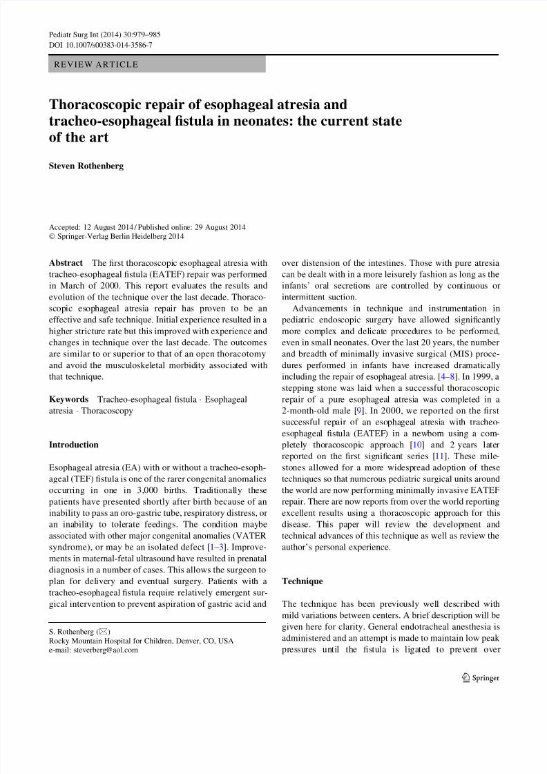
Port placement
Port placement is extremely important because of the small
chest cavity and the intricate nature of the dissection and
reconstruction. The procedure can be performed with three
ports but occasionally a fourth port is necessary to retract
the lung.
The initial port (3–5 mm) is placed in the fifth inter-
costal space posterior to the tip of the scapula. This is the
camera port and gives the surgeon excellent visualization
of the posterior mediastinum in the area of the fistula and
eventual anastomosis. A 308 lens is used to allow the
surgeon to ‘‘look down’’ on his instruments and avoid
‘‘instrument dueling’’.
The two instrument ports are placed to achieve a ninety
degree angle at the presumed site of the anastomosis. The
first port is placed in the mid-axillary line one to 2 inter-
spaces above the camera port. This upper port is 5 mm to
allow for the introduction of a clip applier and suture. A
3 mm port can be used if the surgeon wishes to suture
ligate the fistula and passes the sutures through the chest
wall. The lower port is 3 mm in size and is placed one or
two intercostal spaces below, and slightly posterior to the
camera port (Fig. 1). Ideally the instrument tips will
approximate a right angle (908) at the level of the fistula.
This positioning will facilitate suturing the anastomosis. A
fourth port can be placed either higher or lower in the
thoracic cavity to help retract the lung, but this has not been
necessary in the majority of cases. The operation then
follows the same pattern as for the open procedure.
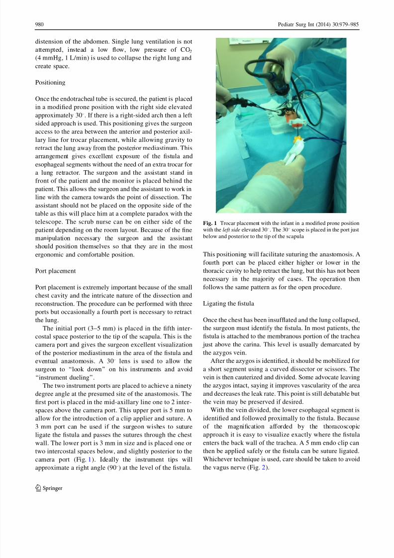
Ligating the fistula
Once the chest has been insufflated and the lung collapsed,
the surgeon must identify the fistula. In most patients, the
fistula is attached to the membranous portion of the trachea
just above the carina. This level is usually demarcated by
the azygos vein.
After the azygos is identified, it should be mobilized for
a short segment using a curved dissector or scissors. The
vein is then cauterized and divided. Some advocate leaving
the azygos intact, saying it improves vascularity of the area
and decreases the leak rate. This point is still debatable but
the vein may be preserved if desired.
With the vein divided, the lower esophageal segment is
identified and followed proximally to the fistula. Because
of the magnification afforded by the thoracoscopic
approach it is easy to visualize exactly where the fistula
enters the back wall of the trachea. A 5 mm endo clip can
then be applied safely or the fistula can be suture ligated.
Whichever technique is used, care should be taken to avoid
the vagus nerve (Fig. 2).
Fig. 1 Trocar placement with the infant in a modified prone position
with the left side elevated 308. The 308 scope is placed in the port just
below and posterior to the tip of the scapula
980 Pediatr Surg Int (2014) 30:979–985
1 3
7/21/2019 Atresia Esofagica Toracoscopia
http://slidepdf.com/reader/full/atresia-esofagica-toracoscopia 3/7
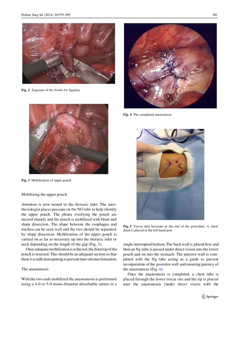
Mobilizing the upper pouch
Attention is now turned to the thoracic inlet. The anes-
thesiologist places pressure on the NG tube to help identify
the upper pouch. The pleura overlying the pouch are
incised sharply and the pouch is mobilized with blunt and
sharp dissection. The plane between the esophagus and
trachea can be seen well and the two should be separated
by sharp dissection. Mobilization of the upper pouch is
carried on as far as necessary up into the thoracic inlet or
neck depending on the length of the gap (Fig. 3).
Once adequate mobilization is achieved, the distal tip of the
pouch is resected. This should be an adequate section so that
there is a sufficientopening to prevent later stricture formation.
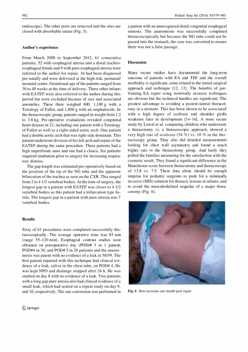
The anastomosis
With the two ends mobilized the anastomosis is performed
using a 4-0 or 5-0 mono-filament absorbable suture in a
single interrupted fashion. The back wall is placed first and
then an Ng tube is passed under direct vision into the lower
pouch and on into the stomach. The anterior wall is com-
pleted with the Ng tube acting as a guide to prevent
incorporation of the posterior wall and ensuring patency of
the anastomosis (Fig. 4).
Once the anastomosis is completed, a chest tube is
placed through the lower trocar site and the tip is placed
near the anastomosis (under direct vision with the
Fig. 2 Exposure of the fistula for ligation
Fig. 3 Mobilization of upper pouch
Fig. 4 The completed anastomosis
Fig. 5 Trocar skin incisions at the end of the procedure. A chest
drain is placed in the left hand port
Pediatr Surg Int (2014) 30:979–985 981
1 3
7/21/2019 Atresia Esofagica Toracoscopia
http://slidepdf.com/reader/full/atresia-esofagica-toracoscopia 4/7
endoscope). The other ports are removed and the sites are
closed with absorbable suture (Fig. 5).
Author’s experience
From March 2000 to September 2012, 61 consecutive
patients, 52 with esophageal atresia and a distal tracheo-
esophageal fistula and 9 with pure esophageal atresia were
referred to the author for repair. 16 had been diagnosed
pre-natally and were delivered at the high risk, perinatal/
neonatal center. Gestational age of the patients ranged from
30 to 40 weeks at the time of delivery. Three other infants
with EATEF were also referred to the author during this
period but were excluded because of size and associated
anomalies. These three weighed 800, 1,100 g with a
Tetralogy of Fallot, and 1,800 g with an omphalocele. In
the thoracoscopic group, patients ranged in weight from 1.2
to 3.8 kg. Pre-operative evaluations revealed congenital
heart disease in 22, including one patient with a Tetralogy
of Fallot as well as a right-sided aortic arch. One patient
had a double aortic arch that was right-side dominant. This
patient underwent division of his left arch and repair of the
EATEF during the same procedure. Three patients had a
high imperforate anus and one had a cloaca. Six patients
required intubation prior to surgery for increasing respira-
tory distress.
The gap length was estimated pre-operatively based on
the position of the tip of the NG tube and the apparent
bifurcation of the trachea as seen on the CXR. This ranged
from 2 to 4 1/2 vertebral bodies. At the time of surgery, the
longest gap in a patient with EATEF was closer to 4 1/2
vertebral bodies as this patient had a trifurcation type fis-
tula. The longest gap in a patient with pure atresia was 7
vertebral bodies.
Results
Sixty of 61 procedures were completed successfully tho-
racoscopically. The average operative time was 85 min
(range 55–120 min). Esophageal contrast studies were
obtained on postoperative day (POD)# 3 in 1 patient,POD#4 in 30, and POD# 5 in 28 patients and the anasto-
mosis was patent with no evidence of a leak in 58/59. The
first patient repaired with this technique had clinical evi-
dence of a leak, saliva in the chest tube, on POD# 4. He
was kept NPO and drainage stopped after 24 h. He was
studied on day 8 with no evidence of a leak. Two patients
with a long gap pure atresia also had clinical evidence of a
small leak, which had sealed on a repeat study on day 9,
and 10, respectively. The one conversion was performed in
a patient with an unrecognized distal congenital esophageal
stenosis. The anastomosis was successfully completed
thoracoscopically but because the NG tube could not be
passed into the stomach, the case was converted to ensure
there was not a false passage.
Discussion
Many recent studies have documented the long-term
outcome of patients with EA and TEF and the overall
morbidity is significant, some related to the initial surgical
approach and technique [12, 13]. The benefits of per-
forming EA repair using minimally invasive techniques
are obvious but the technical hurdles are significant. The
greatest advantage is avoiding a postero-lateral thoracot-
omy in a neonate. This has been shown to be associated
with a high degree of scoliosis and shoulder girdle
weakness later in development [14–16]. A more recent
study by Lawal et al. comparing children who underwenta thoracotomy vs. a thoracoscopic approach, showed a
very high rate of scoliosis (54 %) vs. 10 % in the tho-
racoscopy group. They also did detailed measurements
looking for chest wall asymmetry and found a much
higher rate in the thoracotomy group. And lastly they
polled the families measuring for the satisfaction with the
cosmetic result. They found a significant difference in the
Manchester score between thoracotomy and thoracoscopy
of 13.8 vs. 7.5. These data alone should be enough
impetus for pediatric surgeons to push for a minimally
invasive (MIS) solution for thoracic lesions in infants, and
to avoid the musculoskeletal sequelae of a major thora-
cotomy (Fig. 6).
Fig. 6 Skin incisions one month post repair
982 Pediatr Surg Int (2014) 30:979–985
1 3
7/21/2019 Atresia Esofagica Toracoscopia
http://slidepdf.com/reader/full/atresia-esofagica-toracoscopia 5/7
The benefit of the improved cosmetic result of the tho-
racoscopic approach is actually something pediatric sur-
geons have valued and sought after for years. Bianchi and
others have advocated muscle sparing and/or lateral tho-
racotomies, with various skin incisions to improve on this
problem but these incisions can be difficult to develop,
offer more limited access, and still require spreading of the
rib interspace [17]. Despite an improvement over a stan-dard thoracotomy incision, there are still significant issues
associated with these various techniques. The cosmetic
result is not comparable to that of a thoracoscopic
approach, and may not eliminate the risk of scoliosis.
An unanticipated benefit of the thoracoscopic approach
is the superior visualization of the anatomy and especially
the fistula. Since the fistula is visualized perpendicular to
its insertion to the membranous trachea, the exact site for
ligation can be identified easily, thereby minimizing the
residual pouch attached to the trachea. The use of the 5 mm
titanium clips has proven to be simple and effective with no
evidence of tracheal leak or recurrent fistula. Others chooseto suture ligate the fistula and this technique has been
successful as well.
A recognized advantage after performing the first cases
thoracoscopically was that the dissection and anastomosis
were basically carried out in situ. Because the separation of
the fistula and the upper pouch from the trachea was per-
formed under direct magnified vision from a lateral
approach there was little manipulation or force applied to
the trachea itself. This may help diminish the degree of
tracheomalacia that these children have post-operatively.
Also the plane between the upper pouch and trachea was
more obvious making injury to the membranous wall of the
trachea less likely.
The improved visualization also helps with the mobili-
zation. It is easy to see and dissect well up into the thoracic
inlet allowing greater length to be obtained on the upper
pouch in cases of a long gap. The distal end can also be
visualized and mobilized more easily, especially in cases of
pure EA. This has been borne out in our ability to anas-
tomose primarily patients with gaps as long as seven ver-
tebral bodies. By performing the anastomosis in situ there
may be less tension on the esophageal ends allowing longer
gaps to be brought together without tearing.
The major technical hurdle in this operation is the
suturing of the anastomosis. The placement of the sutures
and knot tying are technically demanding Also as opposed
to the open technique where the entire posterior row of
sutures can be placed and then brought together to disperse
the tension along multiple points during knot tying, this
method places all the tension on one suture at a time. So far
this has not been a significant problem but it could prove to
be. For this procedure to become more widely accepted it
maybe necessary to develop a mechanical anastomotic
device or self- knotting suture. The rate of anastomotic
narrowing requiring at least one dilatation in our series was
20 % but was initially almost 50 %, a figure much higher
than our open experience. This may have been secondary
to inadequate approximation of the mucosal ends or an
insufficient opening being made in the upper pouch. We
have modified the technique to eliminate both of these
problems as well as switching to a monofilament absorb-able suture. These changes seem to have resolved the
problem in only 2 of the last 24 patients (10 %) requiring
dilatation in the last half of the series [18].
There are now multiple studies from institutions all over
the world comparing open vs. thoracoscopic EA-TEF
repair. Lugo et al. [19] found the results and outcomes of a
thoracoscopic approach comparable to that of the open. Al
Tokhais et al. [20] also found comparable outcomes in a
multi-institutional study. A recent survey, performed by
IPEG (International Pediatric Endosurgical Group), of its
members showed wide adoption of the thoracoscopic
approach. 170 surgeons responded from 31 countries andover half stated that a thoracoscopic approach was their
first choice [21]. This shows that the technique is not just
limited to ‘‘the experts’’ and that many centers which
practice advanced MIS have been able to adopt and master
the technique.
Perhaps the most compelling paper is a recent review by
Borruto et al. [22] in which they reviewed 5 large series
that compared open vs. thoracoscopic repair and performed
a meta-analysis. They showed no statistically significant
difference in the complications or outcomes between tho-
racoscopic vs. the open repair. (Table 1) This would mean
the major difference is the avoidance of a major thora-
cotomy, the advantages of which have already been
discussed.
There has been concern about the physiologic stresses
during a thoracoscopic repair because of the CO2 insuf-
flation, and one lung ventilation. Some are concerned that
there may be significant hypercapnia, acidosis, and cerebral
hypoperfusion. A recent study from Bishay et al. [23] from
London looked patients undergoing thoracoscopic CDH
and TEF repair vs. those undergoing a thoracotomy. In the
CDH group they found significant evidence of hypercapnia
and acidosis in the thoracoscopic group. They found sim-
ilar findings in the TEF group but these did not reach
statistical significance. However, other studies including
two recent reports at the EUPSA and IPEG meetings by
Stolwijk from Utrecht showed no such problem [24, 25].
They measured for hypercapnia, acidosis, and cerebral
perfusion and found no significant difference between the
open and thoracoscopic group. They did use average
pressure of 5 mmHg which is significantly less than that of
the London group and their operative times were less and
perhaps this explains the results of the other study. In any
Pediatr Surg Int (2014) 30:979–985 983
1 3

7/21/2019 Atresia Esofagica Toracoscopia
http://slidepdf.com/reader/full/atresia-esofagica-toracoscopia 6/7
event many other series including our own have not shown
clinical problems of significant hypercapnia or acidosis in
these patients.
Clearly the technical and physiologic hurdles to
accomplish this type of repair are many and it will require
continued advances before this surgery becomes com-
monplace. However, the ability to perform this complex
reconstruction without a thoracotomy lays further ground
work in minimizing surgical morbidity in even the smallestpediatric patients.
References
1. Ladd WE (1944) The surgical treatment of esophageal atresia and
tracheo-esophageal fistulas. N Engl J Med 230:625–637
2. Koop CE, Hamilton JP (1965) Atresia of the esophagus:
increased survival with staged procedures in the poor-risk infant.
Ann Surg 162:389–401
3. Engum SA, Grosfeld JL, West KA et al (1995) Analysis of
morbidity and mortality in 227 cases of esophageal atresia and/ortracheo-esophageal fistula over two decades. Arch Surg
130:502–509
4. Rothenberg SS, Chang HT, Bealer JF (1998) Experience with
minimally invasive surgery in Infants. Am J Surg 176:654–658
5. Takao Fujimoto, Segewa O, Ezaki S, Miyano T (1999) Laparo-
scopic surgery for newborn infants. Surg Endosc 13:773–777
6. Rothenberg SS, Chang JHT, Toews WH, Washington RL (1995)
Thoracoscopic closure of patent ductus arteriosus: a less trau-
matic and more cost-effective technique. J Pediatr Surg
30:1057–1060
7. Rothenberg SS, Chang JHT, Bealer J (2004) First decades
experience with minimally invasive surgery in neonates. Pediatr
Endosurg Innovative Tech. 8:88–94
8. Ponsky TA, Rothenberg SS (2008) Minimally invasive surgery in
infants less than 5 kg: experience of 649 cases. Surg Endosc
22:2214–2219
9. Lobe TE, Rothenberg SS, Waldschmidt J, Stroeder L (1999)
Thoracoscopic repair of esophageal atresia in an infant: a surgical
first. Pediatr Endosurg Innovative Tech 3:141–148
10. Rothenberg SS (2000) Thoracoscopic repair of a tracheo-esoph-
ageal in a neonate. Pediatr Endosurg Innovative Tech 4:150–15611. Rothenberg SS (2002) Thoracoscopic repair of tracheo-esopha-
geal fistula and esophageal atresia in newborns. J Pediatr Surg
37:869–872
12. Little DC, Rescorla FJ, Grosfeld JL et al (2003) J Pediatr Surg
6:852–856
13. Konkin DE, Ohal WA, Webber EM et al (2003) J Pediatr Surg
12:1726–1729
14. Freeman NV, Walkden J (1969) Previously unreported shoulder
deformity following right lateral thoracotomy for esophageal
atresia. J Pediatr Surg 4:627–636
15. Vaiquez JJ, Murcia J, DiezPardo JA (1995) Morbid musculo-
skeletal sequelae of thoracotomy for tracheo-esophageal fistula.
J Pediatr Surg 20:511–514
16. Durning RP, Scoles PV, Fox OD (1980) Scoliosis after thora-
cotomy in tracheo-esophageal fistula patients. J Bone Joint SurgAm 62:1156–1158
17. Bianchi A, Sowende O, Aliza NK et al (1998) Aesthetics and
lateral thoracotomy in the neonate. J Pediatr Surg 12:1798–1800
18. Rothenberg SS (2012) Thoracoscopic repair of esophageal atresia
and tracheo-esophageal fistula in neonates: evolution of a tech-
nique. J Laparoendosc Adv Surg Tech A 22(2):195–199
19. Lugo B, Malhotra A, Guner Y, Nguyen T, Ford H, Nguyen N
(2008) Thoracoscopic versus open repair of tracheoesopphageal
fistula and esophageal atresia. J Laproendosc Adv Surg Tech
18(5):753–756
Table 1 Comparative studies of open vs thoracoscopic TEF showing no significant difference in outcomes or complications
Author Type of study Level of
evidence
No. of thoracoscopies
vs.
no. of thoracotomies
Endpoints Results thoracoscopy
vs.
thoracotomy (%)
Borruto et al. [17] 2012 Meta-analysis 3a 69 vs. 97 leakage rate No differences
Stricture rate No differences
Szavay et al. [14] 2011 Retrospective comparativestudy 3b 25 vs. 32 Leakage rate 4 vs. 3Stricture rate 0 vs. 0
Operating rate Longer
pCO2max value Higher
Ventilation time No differences
Complication time No differences
Allal et al. [21] 2009 Retrospective comparative
study
3b 14 vs. 14(?3)’ Leakage rate 14 vs. 19
Stricture rate 14 vs. 50
Operating time Longer
Complication
rate
No differences
Al Tokhais et al. [20]
2008
Retrospective comparative
study
3b 23 vs. 22 Leakage rate 17 vs. 14
Stricture rate 8 vs. 29Operating time No differences
984 Pediatr Surg Int (2014) 30:979–985
1 3
7/21/2019 Atresia Esofagica Toracoscopia
http://slidepdf.com/reader/full/atresia-esofagica-toracoscopia 7/7
20. Al Tokhais T, Zamakhshary M, Aldekhayel S, Mandora H, Sayed
S, AlHarbi K, Alqahtani A (2008) Thoracoscopic repair of tra-
cheo-esophageal fistulas; a case controlled matched study.
J Pediatric Surg. 43(5):805–809
21. Lai D, Miyano G, Juang D, Sharp N, St Peter S (2013) Current
patterns of practice and technique in the repair of esophageal
atresia and tracheo-esophageal fistula: an IPEG survey. J Lapro-
endosc Adv Surg Tech (7):635–638
22. Borruto F, Impellizzeri P, Monatatto A et al (2012) Thoracoscopy
versus thoracotomy for esophageal atresia and tracheo-esopha-
geal fistula: review of the literature and meta-analysis. Eur J
Pediatr Surg 22(6):415–419
23. Bishay M, Giacomello L, Retrosi G et al (2013) Hypercapnia and
acidosis during open and thoracoscopic repair of congenital
diaphragmatic hernia and esophageal atresia : results of a pilot
randomized controlled trial. Ann Surg 25(6):895–900
24. Stolwijk L, Tytgat S, Keunen K, et al The Effects of CO2
insufflation of cerebral oxygenation in neonates during thoraco-
scopic repair of esophageal atresia. Presented at EUPSA annual
congress, Dublin June 2014
25. Stolwijk L, Tytgat S, Keunen, et al The effects of CO2-insuffla-
tion with 5 and 10 mmHg during thoracoscopy on cerebral
oxygenation and hemodynamics in PIGLETS. Presented IPEG,
Edinburgh July 2014
Pediatr Surg Int (2014) 30:979–985 985
1 3