af5d3c923d929b8
TRANSCRIPT

Molecular Biology of the CellFifth Edition
Molecular Biology of the CellFifth Edition
Chapter 15Mechanisms of Cell
Communication
Chapter 15Mechanisms of Cell
Communication
Copyright © Garland Science 2008
Alberts • Johnson • Lewis • Raff • Roberts • WalterAlberts • Johnson • Lewis • Raff • Roberts • Walter

Figure 15-1 Molecular Biology of the Cell (© Garland Science 2008)
Communication between cells is mediated by extracellular signals

Figure 15-2 Molecular Biology of the Cell (© Garland Science 2008)
Cells respond to the extracellular environment: example: Saccharomyces cerevisiae

Figure 15-3a Molecular Biology of the Cell (© Garland Science 2008)

Figure 15-3b Molecular Biology of the Cell (© Garland Science 2008)

Figure 15-4 Molecular Biology of the Cell (© Garland Science 2008)

Figure 15-5a Molecular Biology of the Cell (© Garland Science 2008)
Endocrine signaling : - slow- Act at very low concentration

Figure 15-5b Molecular Biology of the Cell (© Garland Science 2008)
Synaptic signaling - Fast and precise- High local concentration
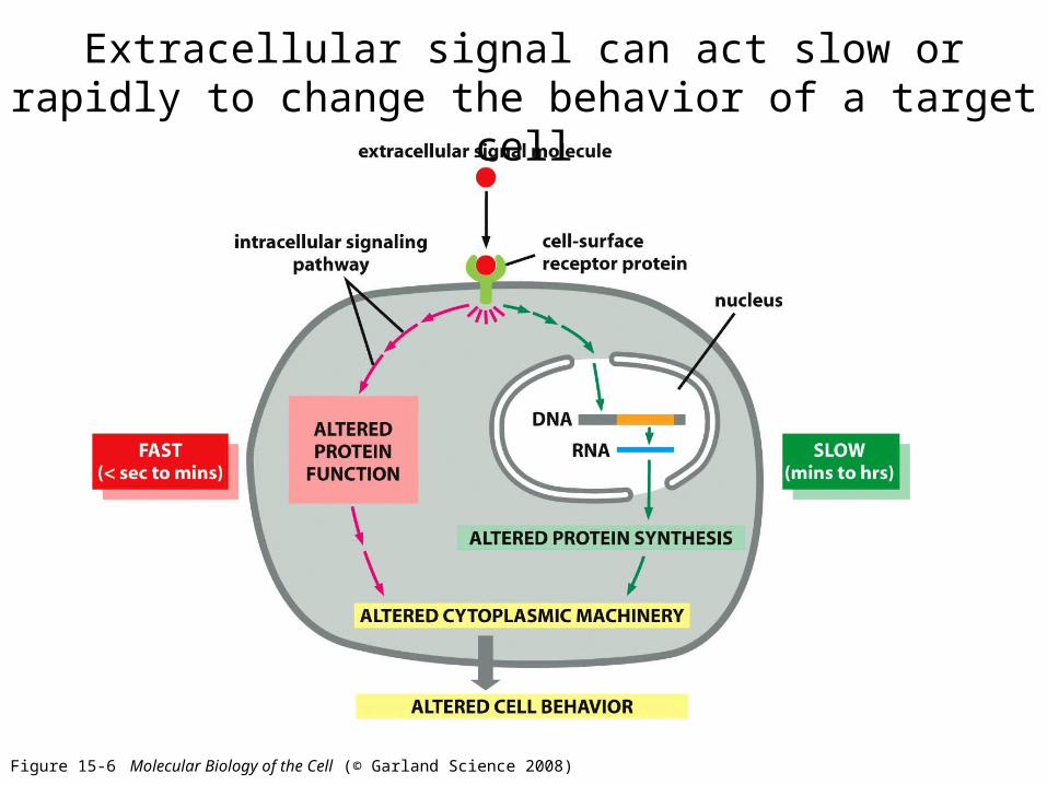
Figure 15-6 Molecular Biology of the Cell (© Garland Science 2008)
Extracellular signal can act slow or rapidly to change the behavior of a target cell

Figure 15-7 Molecular Biology of the Cell (© Garland Science 2008)
Gap junctions allow neighboring cells to share signaling information
Allow small molecules to pass freely between cells, like Calcium, Cyclic AMP (2nd messenger)

Figure 15-8 Molecular Biology of the Cell (© Garland Science 2008)
Cells are programmed to respond to specific combinations of extracellular signal molecules

Figure 15-9 Molecular Biology of the Cell (© Garland Science 2008)
Different types of cells usually respond differently to the same extracellular signal molecules
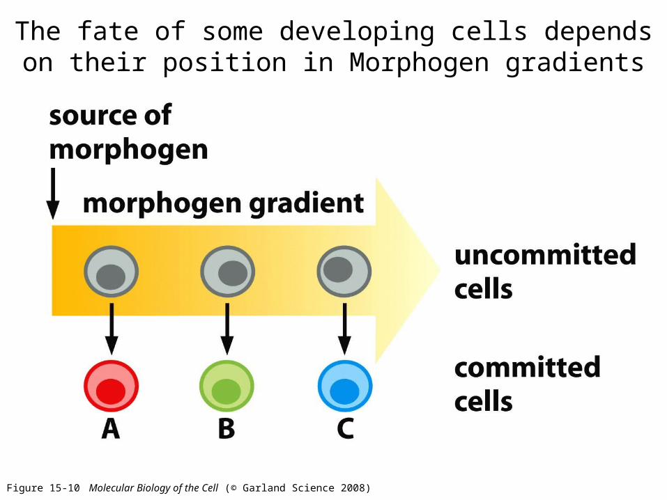
Figure 15-10 Molecular Biology of the Cell (© Garland Science 2008)
The fate of some developing cells depends on their position in Morphogen gradients

Figure 15-11 Molecular Biology of the Cell (© Garland Science 2008)
The cell can alter the concentration of an intracellular molecule quickly if the lifetime of the molecule is short

Figure 15-12b Molecular Biology of the Cell (© Garland Science 2008)
Nitric oxide acts as a signaling molecule, it relaxes smooth muscles
Nitroglycerides used on patients with angina – reduces the work load of the heart.

Figure 15-13 Molecular Biology of the Cell (© Garland Science 2008)
Small hydrophobic molecules that diffuse directly across the plasma membrane
These bind to intracellular receptors to regulate gene expression

Figure 15-14a Molecular Biology of the Cell (© Garland Science 2008)
THE NUCLEAR RECEPTORS SUPERFAMILY
Steroid hormones (cortisol) made of cholesterol, cortisol is made in the adrenal cortex, influences metabolism.Steroid sex hormones made by testes and ovaries – reponsible for secondary sex characteristicsVitamin D – made in the skin, regulates calcium metabolism.* The nuclear receptor (with ligand) then binds to DNA to regulate transcription

Figure 15-14b Molecular Biology of the Cell (© Garland Science 2008)
Ligand binding alters the conformation of the receptor

Figure 15-14c Molecular Biology of the Cell (© Garland Science 2008)
Ligand binding alters the conformation of the receptor: inhibitor dissociates and coactivator binds
* In some cases ligand binding inhibits transcription

Figure 15-15 Molecular Biology of the Cell (© Garland Science 2008)
The transcription response takes place in multiple steps: primary and secondary responses

THE THREE LARGEST CLASS OF CELL-SURFACE RECEPTORS
Most extracellular signals do not enter the cell like the hydrophobic ones, but they bind to specific receptors at the plasma membrane
Ion-channel-coupledG-protein-coupled Enzyme-coupled
These receptors act as signal transducers
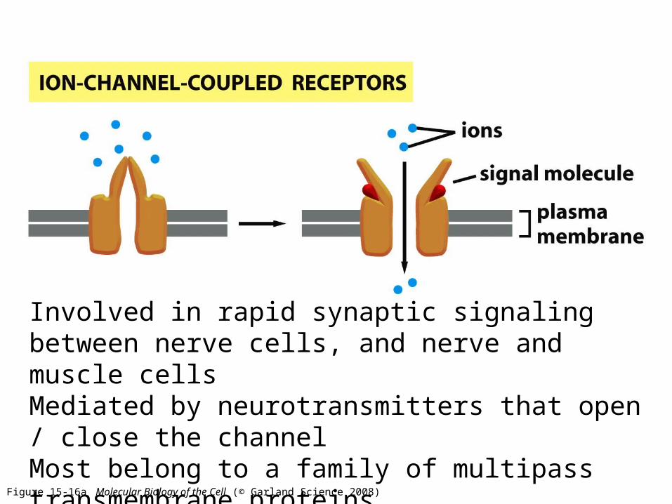
Figure 15-16a Molecular Biology of the Cell (© Garland Science 2008)
Involved in rapid synaptic signaling between nerve cells, and nerve and muscle cells Mediated by neurotransmitters that open / close the channel Most belong to a family of multipass transmembrane proteins

Figure 15-16b Molecular Biology of the Cell (© Garland Science 2008)
A trimeric G protein (GTP binding) mediates the interaction between the activated receptor and this target protein. All belong to a family of multipass transmembrane proteins

Figure 15-16c Molecular Biology of the Cell (© Garland Science 2008)
Function as enzymes or associate with enzymes that they activate.Are usually single pass transmembrane proteins, ligand binding site outside the cell and catalytic (enzyme-binding) site inside the cell.Majority are protein kinases or associate with protein kinases.

Figure 15-17 Molecular Biology of the Cell (© Garland Science 2008)
Most activated cell-surface receptors relay signals via small molecules and a network of intracellular signaling proteins:
called second messengers
Examples of small messengers:
In cytosol:-Cyclic AMP : cAMP-Calcium ion
Fat soluble:-Diacylglycerol
Large intracellular signaling proteins

Figure 15-18 Molecular Biology of the Cell (© Garland Science 2008)
Many intracellular signaling proteins function as switches that are activated by phosphorylation or GTP binding
Ser/thre kinases are majorityTyrosine kinases

Figure 15-19 Molecular Biology of the Cell (© Garland Science 2008)
Small monomeric GTPases regulated by GTP/GDP binding

Figure 15-20 Molecular Biology of the Cell (© Garland Science 2008)
Signal integration: two pathways cause phosphorylation of one target at different sites
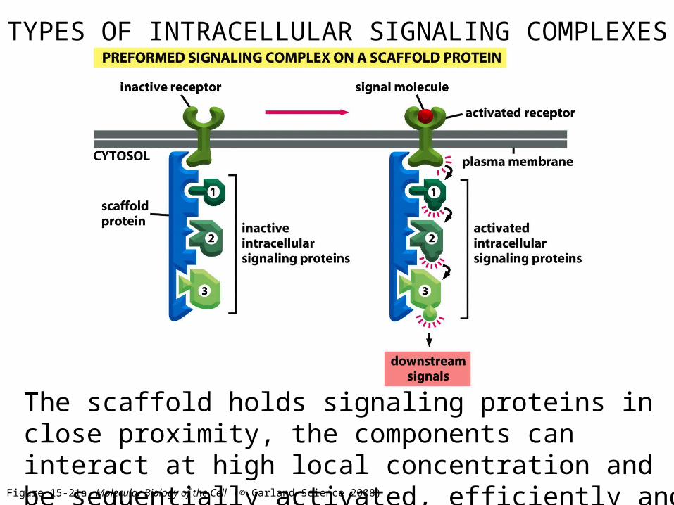
Figure 15-21a Molecular Biology of the Cell (© Garland Science 2008)
TYPES OF INTRACELLULAR SIGNALING COMPLEXES
The scaffold holds signaling proteins in close proximity, the components can interact at high local concentration and be sequentially activated, efficiently and selectively

Figure 15-21b Molecular Biology of the Cell (© Garland Science 2008)
TYPES OF INTRACELLULAR SIGNALING COMPLEXES
Transient assembly of complexes, due to phosphorylation (that is reversible)

Figure 15-21c Molecular Biology of the Cell (© Garland Science 2008)
TYPES OF INTRACELLULAR SIGNALING COMPLEXES
Receptor activation leads to the production of modified phospholipids (phosphinositides), which recruit specific intracellular signaling proteins.

Figure 15-22 Molecular Biology of the Cell (© Garland Science 2008)
Induced proximity via interaction domains, used to relay signals from protein to protein

Figure 15-23 Molecular Biology of the Cell (© Garland Science 2008)
When a cell responds to extracellular signals, it can be a smooth graded or a switch like response

Figure 15-24b, c Molecular Biology of the Cell (© Garland Science 2008)
Looking at a whole population, the response may appear smooth while each cell is having an all or non response
It is important to look at individual cells to detect all-or-none responses

Figure 15-25 Molecular Biology of the Cell (© Garland Science 2008)
Example:
Adrenaline binding to a G-protein-coupled cell-surface receptor increases the intracellular concentration of cyclic AMP which in turn activates enzymes that promote glycogen breakdown and inhibit enzymes that promote glycogen synthesis.

Figure 15-26 Molecular Biology of the Cell (© Garland Science 2008)
Intracellular signaling incorporate feedback loops

Figure 15-27 Molecular Biology of the Cell (© Garland Science 2008)
Positive feedback mechanism giving switch-like behavior
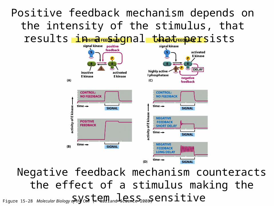
Figure 15-28 Molecular Biology of the Cell (© Garland Science 2008)
Positive feedback mechanism depends on the intensity of the stimulus, that results in a signal that persists
Negative feedback mechanism counteracts the effect of a stimulus making the system less sensitive

Figure 15-29 Molecular Biology of the Cell (© Garland Science 2008)
Target cells can become adapted (desensitized) to an extracellular signal molecule

Figure 15-30 Molecular Biology of the Cell (© Garland Science 2008)
G-protein coupled receptors, the largest class of cell surface receptors
- More than 700 GPCRs in humans- Sigh, smell and taste use these receptors- One signal can activate many GPCRs

Figure 15-32 Molecular Biology of the Cell (© Garland Science 2008)
Trimeric G-proteins relay signals from GPCRs
-Various types of G-proteins, each one specific for a particular set of GPCRs, and particular set of target proteins in the membrane- They all have similar structures and operate similarly.- Gproteins have three subunits: alpha, beta and gamma.-Alpha-GDP unstimulated-Alpha-GTP stimulated (has intrinsic GTPase); also regulators of G-protein signaling act as GTPases

Figure 15-33 Molecular Biology of the Cell (© Garland Science 2008)
Some G-proteins regulate the production of Cyclic AMPCyclic AMP acts as a second messenger

Figure 15-34 Molecular Biology of the Cell (© Garland Science 2008)
Cyclic AMP is synthesized from ATP by a plasma membrane bound enzyme adenylyl cyclase and is quickly
destroyed by cAMP phosphodiesterase Different G-proteins cause different effect of cAMP:-Stimulatory G-protein (Gs) activates adenylyl cyclase- Inhibitory G-protein (Gi) inhibits adenylyl cyclase
Ex: cholera toxin is an enzyme that catalyzes transfer of ADP ribose from NAD+ to Gs. Now Gs can no longer hydrolyze the GTP and is always ON, making adenylyl cyclase active always, more cAMP causes more Cl- to be in the gut (and hence more water)

Table 15-1 Molecular Biology of the Cell (© Garland Science 2008)
Individuals with genetic defects in Gs alpha show decrease response to some hormones (so they have metabolic abnormalities, abnormal bone development and are mentally retarded).
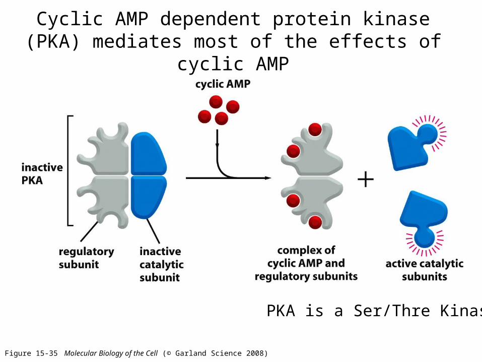
Figure 15-35 Molecular Biology of the Cell (© Garland Science 2008)
Cyclic AMP dependent protein kinase (PKA) mediates most of the effects of cyclic AMP
PKA is a Ser/Thre Kinase
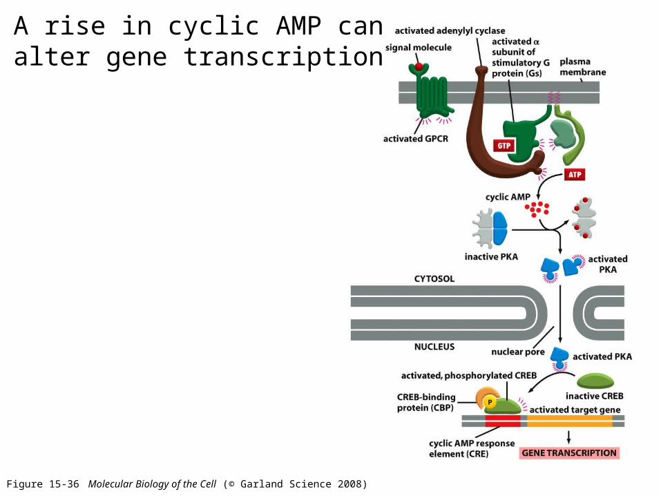
Figure 15-36 Molecular Biology of the Cell (© Garland Science 2008)
A rise in cyclic AMP can alter gene transcription

Table 15-2 Molecular Biology of the Cell (© Garland Science 2008)
Many GPCRs exert their effects mainly via G proteins that activate the plasma membrane - bound enzyme phospholipase C-(PLC)

Figure 15-38 Molecular Biology of the Cell (© Garland Science 2008)
Phospholipase C-(PLC) works on PIP2 to make DAG and IP3

Figure 15-39 Molecular Biology of the Cell (© Garland Science 2008)
IP3 diffuses in the cytoplasm and binds to the ER causing Calcium release in the cytosol

Figure 15-40 Molecular Biology of the Cell (© Garland Science 2008)
Calcium functions as an intracellular mediator, for example during fertilization it initiates embryonic development
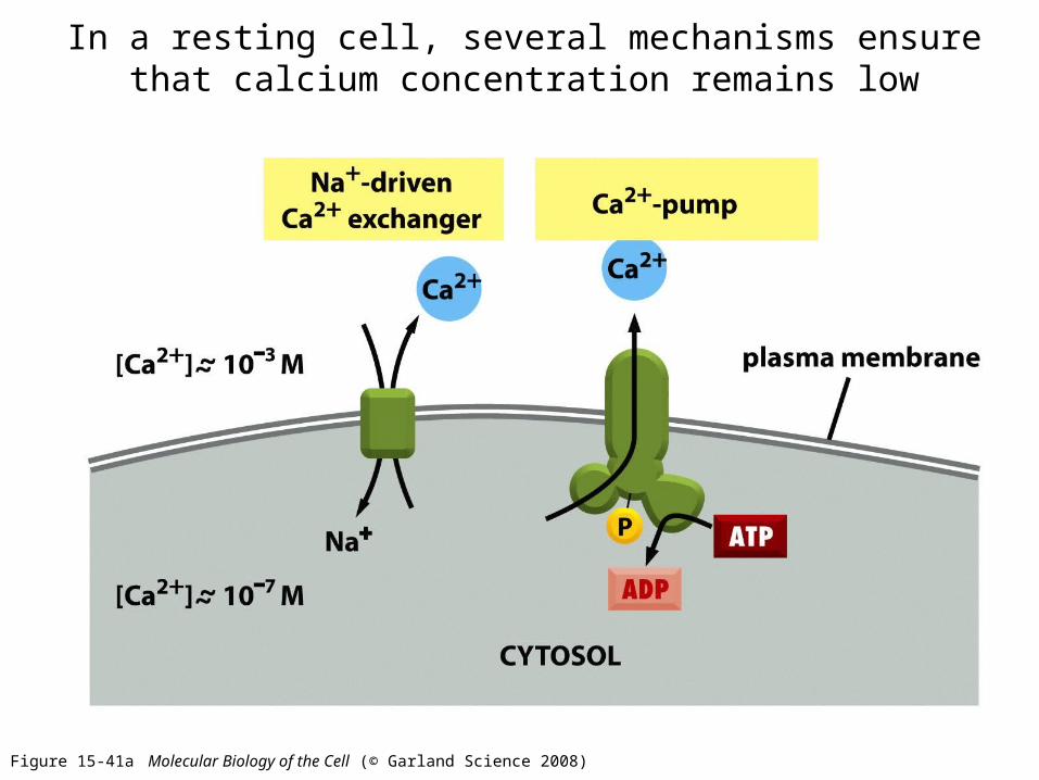
Figure 15-41a Molecular Biology of the Cell (© Garland Science 2008)
In a resting cell, several mechanisms ensure that calcium concentration remains low

Figure 15-41b Molecular Biology of the Cell (© Garland Science 2008)
In a resting cell, several mechanisms ensure that calcium concentration remains low

Figure 15-43 Molecular Biology of the Cell (© Garland Science 2008)
Various Ca2+-binding proteins help to relay the cytosolic Ca2+ signal: Calmodulin, when bound to Calcium changes conformation
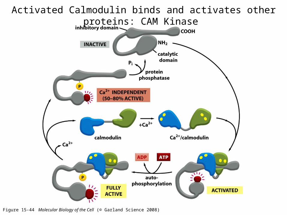
Figure 15-44 Molecular Biology of the Cell (© Garland Science 2008)
Activated Calmodulin binds and activates other proteins: CAM Kinase

Table 15-3 Molecular Biology of the Cell (© Garland Science 2008)
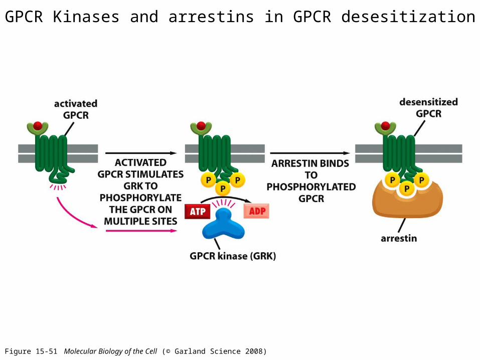
Figure 15-51 Molecular Biology of the Cell (© Garland Science 2008)
GPCR Kinases and arrestins in GPCR desesitization

Figure 15-52 Molecular Biology of the Cell (© Garland Science 2008)
Receptor Tyrosine kinases

Table 15-4 Molecular Biology of the Cell (© Garland Science 2008)

Figure 15-53a Molecular Biology of the Cell (© Garland Science 2008)

Figure 15-53b Molecular Biology of the Cell (© Garland Science 2008)
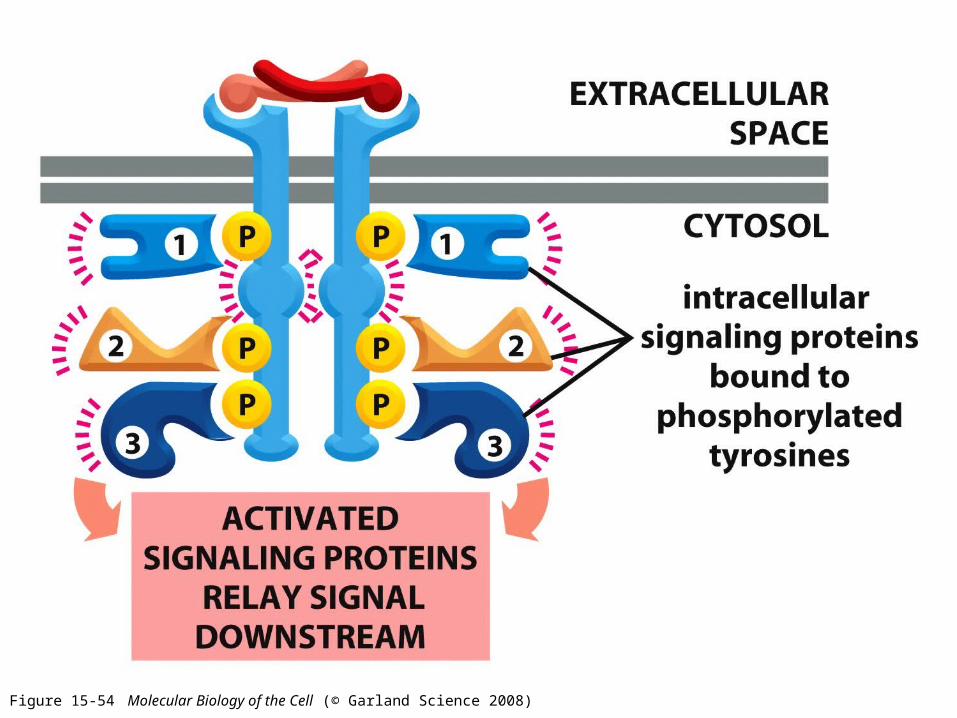
Figure 15-54 Molecular Biology of the Cell (© Garland Science 2008)

Figure 15-55a Molecular Biology of the Cell (© Garland Science 2008)

Table 15-5 Molecular Biology of the Cell (© Garland Science 2008)
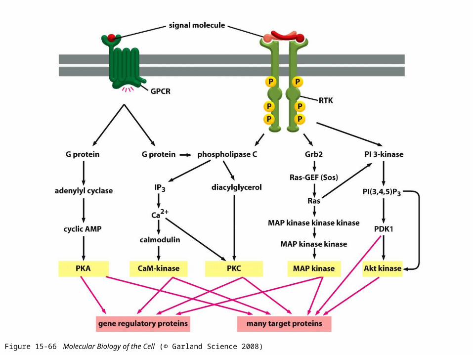
Figure 15-66 Molecular Biology of the Cell (© Garland Science 2008)