urban aerosol particulate matter promotes necrosis and
TRANSCRIPT
antioxidants
Article
Urban Aerosol Particulate Matter Promotes Necrosis andAutophagy via Reactive Oxygen Species-Mediated CellularDisorders that are Accompanied by Cell Cycle Arrest in RetinalPigment Epithelial Cells
Hyesook Lee 1,2,* , Da Hye Kim 1,2, Jeong-Hwan Kim 3, Seh-Kwang Park 3,4, Ji-Won Jeong 5, Mi-Young Kim 3,Seok-Ho Hong 6, Kyoung Seob Song 7 , Gi-Young Kim 8 , Jin Won Hyun 9 and Yung Hyun Choi 1,2,*
�����������������
Citation: Lee, H.; Kim, D.H.; Kim,
J.-H.; Park, S.-K.; Jeong, J.-W.; Kim,
M.-Y.; Hong, S.-H.; Song, K.S.; Kim,
G.-Y.; Hyun, J.W.; et al. Urban
Aerosol Particulate Matter Promotes
Necrosis and Autophagy via Reactive
Oxygen Species-Mediated Cellular
Disorders that are Accompanied by
Cell Cycle Arrest in Retinal Pigment
Epithelial Cells. Antioxidants 2021, 10,
149. https://doi.org/10.3390/
antiox10020149
Received: 17 December 2020
Accepted: 15 January 2021
Published: 20 January 2021
Publisher’s Note: MDPI stays neutral
with regard to jurisdictional claims in
published maps and institutional affil-
iations.
Copyright: © 2021 by the authors.
Licensee MDPI, Basel, Switzerland.
This article is an open access article
distributed under the terms and
conditions of the Creative Commons
Attribution (CC BY) license (https://
creativecommons.org/licenses/by/
4.0/).
1 Anti-Aging Research Center, Dong-eui University, Busan 47340, Korea; [email protected] Department of Biochemistry, Dong-eui University College of Korean Medicine, Busan 47227, Korea3 Research and Development Department, BGN CARE Co., Ltd., Busan 47195, Korea;
[email protected] (J.-H.K.); [email protected] (M.-Y.K.)4 BGN Eye Clinic, Seoul 05551, Korea; [email protected] BGN Eye Clinic, Busan 47195, Korea; [email protected] Department of Internal Medicine, School of Medicine, Kangwon National University, Chuncheon 24341,
Korea; [email protected] Department of Cell Biology and Biophysics, Kosin University College of Medicine, Busan 49267, Korea;
[email protected] Department of Marine Life Sciences, Jeju National University, Jeju 63243, Korea; [email protected] Jeju National University School of Medicine and Jeju Research Center for Natural Medicine, Jeju 63243, Korea;
[email protected]* Correspondence: [email protected] (H.L.); [email protected] (Y.H.C.); Tel.: +82-51-890-3315 (H.L.);
+82-51-890-3319 (Y.H.C.)
Abstract: Urban particulate matter (UPM) is recognized as a grave public health problem worldwide.Although a few studies have linked UPM to ocular surface diseases, few studies have reported onretinal dysfunction. Thus, the aim of the present study was to evaluate the influence of UPM onthe retina and identify the main mechanism of UPM toxicity. In this study, we found that UPMsignificantly induced cytotoxicity with morphological changes in ARPE-19 human retinal pigmentepithelial (RPE) cells and increased necrosis and autophagy but not apoptosis. Furthermore, UPMsignificantly increased G2/M arrest and simultaneously induced alterations in cell cycle regulators.In addition, DNA damage and mitochondrial dysfunction were remarkably enhanced by UPM.However, the pretreatment with the potent reactive oxygen species (ROS) scavenger N-acetyl-L-cysteine (NAC) effectively suppressed UPM-mediated cytotoxicity, necrosis, autophagy, and cellcycle arrest. Moreover, NAC markedly restored UPM-induced DNA damage and mitochondrialdysfunction. Meanwhile, UPM increased the expression of mitophagy-regulated proteins, but NAChad no effect on mitophagy. Taken together, although further studies are needed to identify the roleof mitophagy in UPM-induced RPE injury, the present study provides the first evidence that ROS-mediated cellular damage through necrosis and autophagy is one of the mechanisms of UPM-inducedretinal disorders.
Keywords: mitophagy; necrosis; reactive oxygen species (ROS); retinal pigment epithelial (RPE) cells;urban aerosol particulate matter (UPM)
1. Introduction
Across Asia, urban air pollution has become part of the daily existence of citizens, andit is recognized as a grave public health problem [1,2]. Accumulated evidence has demon-strated that a high proportion of air pollutants adversely affect health and socioeconomic
Antioxidants 2021, 10, 149. https://doi.org/10.3390/antiox10020149 https://www.mdpi.com/journal/antioxidants
Antioxidants 2021, 10, 149 2 of 16
perspectives, such as an increase in medical costs and a reduction in productivity, for citi-zens living in urban areas of Europe [3]. Recently, the World Health Organization (WHO)cited that air pollutants are a major environmental cause of premature mortality [4]. Amongurban air pollutants, urban particulate matter (UPM) contains various toxic substances,such as metals, biological contaminants, inorganic elements, and elemental and organcarbon [5]. Significantly, fine particulate matter (PM2.5) can remain in the atmosphere forover several hours to weeks, deeply penetrating into the major organs through breathingand skin [6,7]. In this regard, the latest epidemiological and toxicological studies havedemonstrated that UPM results in negative biological effects on the respiratory tract [8],cardiovascular systems [9], immune systems [10], nervous systems [11], and skin [12].
Importantly, there has been increasing interest in the harmful effect of UPM on visualsystems, which is an organ directly exposed to urban air pollutants [13–15]. In addition,some nonclinical studies have demonstrated that exposure to UPM leads to the apoptosisand inflammation of the corneal and conjunctival epithelium in vitro and in vivo [16,17].More notably, recent studies have shown that the inner part of the eye may also be affectedby urban air pollutants, because air pollution leads to systemic disease [18,19]. A fewepidemiological studies have reported that urban air pollutants induce retinal disordersand result in increasing prevalence rates of retinal disorders including age-related maculardegeneration (AMD), diabetic retinopathy, and sclerosis of the retina [19–22]. One animalstudy demonstrated that PM induces edema in retina [23]. More recently, Tien et al. [24]published results showing that perfluorooctanoic acid contained in PM induces oxidativestress and inflammation in the retinal pigment epithelium (RPE) and acts as a risk factor forAMD. In 2020, another study suggested that glial fibrillary acidic protein (GFAP), a hallmarkof glial activation in accordance with neural damage, is markedly expressed by long-termexposure to UPM [16]. Although a few epidemiological studies have reported that UPMcauses retinal dysfunction and increases the prevalence of retinal disorders, studies ofthe underlying mechanism are extremely limited. In this regard, our previous reportdemonstrated that diesel PM promotes migration and cellular dysfunction of RPE throughreactive oxygen species (ROS) production, consequently inducing retinal dysfunction [25].Our previous report is the first to establish that epithelial–mesenchymal transition (EMT) isthe pathological mechanism of retinal exposure to PM. Nevertheless, the main pathologicalmechanism of retinal dysfunction exposure to UPM is still unknown. Therefore, to evaluatethe influence of UPM on the retina, we investigated the cytotoxic effect of UPM in ARPE-19 human retinal pigment epithelial cells and identified the main mechanism of UPMcytotoxicity.
2. Materials and Methods2.1. UPM Preparation
Urban aerosols (certified reference material No. 28) were obtained from the NationalInstitute for Environmental Studies (NIES, Ibaraki, Japan) [26]. UPM was dissolved in aculture medium.
2.2. Cell Culture and Treatment
The human RPE cell line ARPE-19 was obtained from the American Type CultureCollection (Manassas, MD, USA). The cells were maintained in Dulbecco’s modified Eagle’smedium: nutrient mixture F-12 (Invitrogen-Gibco, Carlsbad, CA, USA) supplemented with10% fetal bovine serum at 37 ◦C in a 5% CO2 incubator. When the cells were approximately80% confluent, the cells were pretreated with or without 5 mM N-acetylcysteine (NAC,Sigma-Aldrich Chemical Co., St. Louis, MO, USA) or 200 µM necrostatin-1 (Sigma-AldrichChemical Co.). After 1 h, the cells were incubated with a desired concentration of UPM for24 h.

Antioxidants 2021, 10, 149 3 of 16
2.3. Cytotoxicity Assay
Cytotoxicity was assessed using the CCK-8 assay kit (Abcam Inc., Cambridge, UK) toevaluate cytotoxicity according to the manufacturer’s instructions. The CCK-8 formazanproducts were measured at 460 nm using a microplate spectrophotometer (VERSA Max,Molecular Device Co., Sunnyvale, CA, USA).
2.4. Observation of Cellular and Nuclear Morphology
The cellular morphology was observed under an inverted microscope (Carl Zeiss,Oberkochen, Germany). As described previously [27], nuclear morphology was acquiredusing a fluorescence microscope (Carl Zeiss) by 4,6-diamidino-2-phenylindole (DAPI;Sigma-Aldrich Chemical Co.) staining.
2.5. Flow Cytometric Analysis for Cell Death and Cell Cycle Distribution
To estimate the mode of cell death, the cells were harvested and stained with FITCAnnexin V Apoptosis Detection Kit (BD Biosciences, San Diego, CA, USA) for 20 min,according to the manufacturer’s protocol. The fluorescence intensity was detected usingflow cytometry (BD Biosciences), and the annexin V-negative cells and annexin V-positivecells were considered to be necrotic and apoptotic, respectively [28]. To quantify the phasedistribution of the cell cycle, the cells were stained with 40 µg/mL propidium iodine (PI;BD Biosciences) for 30 min and subjected to flow cytometry as previously described [29].
2.6. Analysis of Caspases Activities and Autophagy
Caspase-3, -8, and -9 enzyme-linked immunosorbent assay (ELISA) kits were pur-chased from R&D Systems Inc. (Minneapolis, MN, USA), and the activities were deter-mined according to the manufacturer’s instruction. Autophagic vacuoles were analyzedusing a Cyto-ID autophagy detection kit (Enzo Life Sciences Inc., FGA, NY, USA). DAPI wasused to counterstain the nuclei. Stained cells were observed on a fluorescence microscope.
2.7. Western Blot Analysis
Total protein and mitochondrial protein were extracted by the PRO-PREP ProteinExtraction Solution (Intron Biotechnology, Gyeonggi-do, Korea) and a mitochondria iso-lation kit (Thermo Fisher Scientific, Waltham, MA, USA), respectively. As previouslydescribed [30], equal proteins were separated by SDS-PAGE and transferred to a PVDFmembrane, and subsequently, the membranes were probed specific primary antibodies(Supplementary Table S1). After incubation for overnight, the corresponding secondaryantibodies were added and incubated. The protein expression was detected by a Fusion FXImage system (Vilber Lourmat, Torcy, France).
2.8. Intracellular ROS Detection
Cells were stained with 5,6-carboxy-2’,7’-dichlorodihydrofluorescein diacetate (DCF-DA; Invitrogen-Gibco) to assess intracellular ROS levels, and images were visualized by afluorescence microscope [25].
2.9. Mitochondrial Membrane Potential (MMP, ∆Ψm) Analysis
Cells were stained with 5,5’,6,6’-tetrachloro-1,1’,3,3’-tetraethylimidacarbocyanine io-dide (JC-1; Invitrogen-Gibco) to investigate the MMP (∆Ψm), and fluorescence images werecaptured by fluorescence microscope [29].
2.10. Immunofluorescence
To observe the expression of γH2AX, immunofluorescence staining was performedas described previously [31]. Information on the antibodies used was provided in Supple-mentary Table S1.
DAPI was used to counterstain the nuclei, and stained cells were observed by fluores-cence microscope.

Antioxidants 2021, 10, 149 4 of 16
2.11. Statistical Analysis
The data are expressed as the mean ± SD. GraphPad Prism 5.03 (GraphPad SoftwareInc., La Jolla, CA, USA) software was used for statistical analyses. Significant differenceswere analyzed using the analysis of variance (ANOVA) followed by Tukey’s test. Probabil-ity values of p < 0.05 were considered to indicate significant.
3. Results3.1. UPM Has Cytotoxicity in ARPE-19 Cells
To evaluate the effect of UPM on ARPE-19 cell viability, we performed a CCK-8 assay.Figure 1A shows that UPM significantly induced cytotoxicity in a dose-dependent manner.At 200 µg/mL UPM, the treated cells exhibited an approximately 76% viability comparedwith the untreated cells. Morphologically, we observed a marked accumulation of UPMaround the cells that showed cell swelling, including an enlarged and flattened shapeand the formation of cytoplasmic blebs (Figure 1B). Meanwhile, the DAPI staining resultsshowed that although partial chromatin condensation was observed in UPM-stimulatedcells, typical apoptotic DNA chromatin condensation was not exhibited (Figure 1C).
Antioxidants 2021, 10, x FOR PEER REVIEW 4 of 16
To observe the expression of γH2AX, immunofluorescence staining was performed as described previously [31]. Information on the antibodies used was provided in Supple-mentary Table S1.
DAPI was used to counterstain the nuclei, and stained cells were observed by fluo-rescence microscope.
2.11. Statistical Analysis The data are expressed as the mean ± SD. GraphPad Prism 5.03 (GraphPad Software
Inc., La Jolla, CA, USA) software was used for statistical analyses. Significant differences were analyzed using the analysis of variance (ANOVA) followed by Tukey’s test. Proba-bility values of p < 0.05 were considered to indicate significant.
3. Results 3.1. UPM Has Cytotoxicity in ARPE-19 Cells
To evaluate the effect of UPM on ARPE-19 cell viability, we performed a CCK-8 as-say. Figure 1A shows that UPM significantly induced cytotoxicity in a dose-dependent manner. At 200 μg/mL UPM, the treated cells exhibited an approximately 76% viability compared with the untreated cells. Morphologically, we observed a marked accumulation of UPM around the cells that showed cell swelling, including an enlarged and flattened shape and the formation of cytoplasmic blebs (Figure 1B). Meanwhile, the DAPI staining results showed that although partial chromatin condensation was observed in UPM-stim-ulated cells, typical apoptotic DNA chromatin condensation was not exhibited (Figure 1C).
Figure 1. Urban particulate matter (UPM) induces slight cytotoxicity in ARPE-19 cells. ARPE-19 cells were incubated with UPM for 24 h. (A) Cytotoxicity was measured by a CCK-8 assay. Data are expressed as the mean ± SD (n = 6). * p < 0.05, ** p < 0.01, and *** p < 0.001 when compared to untreated cells. (B) Morphological cellular changes were observed under an inverted microscope. (C) DAPI staining was performed to visualize the morphological changes in the nucleus and was pictured under a fluorescence microscope. Scale bar: 75 μm.
3.2. UPM Induces Necrosis and Autophagy but Not Apoptosis in ARPE-19 Cells To examine the cause of UPM-induced cytotoxicity with the lack of apoptotic chro-
matin condensation, we performed annexin V/PI staining, which is implicated in the iden-tification of cell death modes. The results showed that the annexin V−/PI+ cell population, which was considered to be necrotic, was significantly increased by exposure to 200 μg/mL UPM (Figure 2A,B). However, the annexin V+/PI+ cell population that was consid-ered to be apoptotic was not affected following UPM treatment. Meanwhile, apoptotic cell
Figure 1. Urban particulate matter (UPM) induces slight cytotoxicity in ARPE-19 cells. ARPE-19cells were incubated with UPM for 24 h. (A) Cytotoxicity was measured by a CCK-8 assay. Dataare expressed as the mean ± SD (n = 6). * p < 0.05, ** p < 0.01, and *** p < 0.001 when compared tountreated cells. (B) Morphological cellular changes were observed under an inverted microscope.(C) DAPI staining was performed to visualize the morphological changes in the nucleus and waspictured under a fluorescence microscope. Scale bar: 75 µm.
3.2. UPM Induces Necrosis and Autophagy but Not Apoptosis in ARPE-19 Cells
To examine the cause of UPM-induced cytotoxicity with the lack of apoptotic chro-matin condensation, we performed annexin V/PI staining, which is implicated in theidentification of cell death modes. The results showed that the annexin V−/PI+ cell pop-ulation, which was considered to be necrotic, was significantly increased by exposureto 200 µg/mL UPM (Figure 2A,B). However, the annexin V+/PI+ cell population thatwas considered to be apoptotic was not affected following UPM treatment. Meanwhile,apoptotic cell population was greatly increased in response to 300 µM H2O2 that was usedas a positive control for apoptosis. In this respect, we considered that UPM-mediated cyto-toxicity was involved in necrotic cell death but not apoptosis. To reconfirm whether UPMwas involved in apoptosis in ARPE-19 cells, the expression of apoptosis-regulatory proteinswas evaluated using Western blot analysis. Figure 2C shows that UPM did not affect theexpression of death receptors and Bcl2 families and cleavage of poly (ADP-ribose) poly-merase (PARP). Moreover, we confirmed that the expression of Bad was also unchanged byUPM exposure in mitochondrial fraction (Figure 2D). In addition, UPM did not affect both
Antioxidants 2021, 10, 149 5 of 16
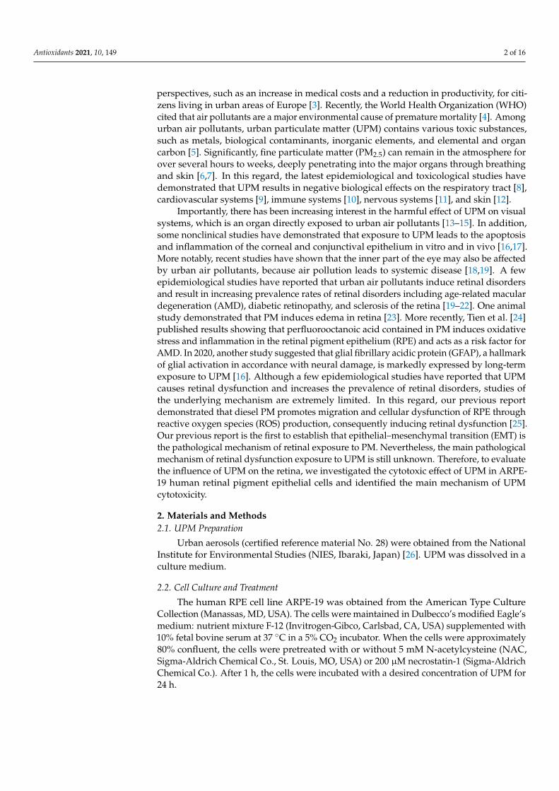
the expression of pro-caspases and activity of caspases (Figure 2C,E). Furthermore, with thepretreatment with necrostatin-1, an inhibitor of necrosis, approximately 50% suppressedthe increase in UPM-induced necrotic cells (Figure 2F,G). This result suggested that UPM in-duced a cytotoxic effect that partially resulted from necrosis. Thus, we further investigatedwhether autophagy, another mode of cell death, is involved in UPM-mediated cytotoxicity.As a result, we found that Cyto-ID-stained cells were enhanced following exposure to UPM(Figure 2H). Based on these results, we considered that UPM induced necrotic and au-tophagic cell death, but no apoptosis in ARPE-19 cells.
Antioxidants 2021, 10, x FOR PEER REVIEW 5 of 16
population was greatly increased in response to 300 μM H2O2 that was used as a positive control for apoptosis. In this respect, we considered that UPM-mediated cytotoxicity was involved in necrotic cell death but not apoptosis. To reconfirm whether UPM was in-volved in apoptosis in ARPE-19 cells, the expression of apoptosis-regulatory proteins was evaluated using Western blot analysis. Figure 2C shows that UPM did not affect the ex-pression of death receptors and Bcl2 families and cleavage of poly (ADP-ribose) polymer-ase (PARP). Moreover, we confirmed that the expression of Bad was also unchanged by UPM exposure in mitochondrial fraction (Figure 2D). In addition, UPM did not affect both the expression of pro-caspases and activity of caspases (Figure 2C,E). Furthermore, with the pretreatment with necrostatin-1, an inhibitor of necrosis, approximately 50% sup-pressed the increase in UPM-induced necrotic cells (Figure 2F,G). This result suggested that UPM induced a cytotoxic effect that partially resulted from necrosis. Thus, we further investigated whether autophagy, another mode of cell death, is involved in UPM-medi-ated cytotoxicity. As a result, we found that Cyto-ID-stained cells were enhanced follow-ing exposure to UPM (Figure 2H). Based on these results, we considered that UPM in-duced necrotic and autophagic cell death, but no apoptosis in ARPE-19 cells.
Figure 2. UPM induces necrosis and autophagy without apoptosis in ARPE-19 cells. (A–E) The cells were treated with the indicated concentration of UPM for 24 h. (A) The representative histograms of the cells stained with annexin V/propidium iodine (PI) and analyzed using a flow cytometer with necrotic cells identified as annexin V−/PI+ cells (upper left quadrant) and apoptotic cells defined as annexin V-positive cells (lower left quadrant). (B) The percentages of necrotic cells. (C) The expres-sion of apoptosis-regulatory proteins. (D) The expression of Bad in mitochondrial fraction. (E) Rel-ative activities of caspase-3, -8, and -9. (F) The representative histograms of ARPE-19 cells pretreated
Figure 2. UPM induces necrosis and autophagy without apoptosis in ARPE-19 cells. (A–E) The cellswere treated with the indicated concentration of UPM for 24 h. (A) The representative histograms ofthe cells stained with annexin V/propidium iodine (PI) and analyzed using a flow cytometer withnecrotic cells identified as annexin V−/PI+ cells (upper left quadrant) and apoptotic cells definedas annexin V-positive cells (lower left quadrant). (B) The percentages of necrotic cells. (C) Theexpression of apoptosis-regulatory proteins. (D) The expression of Bad in mitochondrial fraction.(E) Relative activities of caspase-3, -8, and -9. (F) The representative histograms of ARPE-19 cellspretreated with 200 µM necrostatin-1 (Nec-1) for 1 h and incubated with 200 µg/mL UPM for 24 hand were stained with annexin V/PI. (G) The percentages of necrotic cells. Data are expressed asthe mean ± SD (n = 5). ** p < 0.01 and *** p < 0.001 when compared to untreated cells. ### p < 0.001compared to 200 µg/mL UPM-treated cells. (H) Cyto-ID-stained cells (autophagic cells, green) andDAPI-stained cells (nuclei, blue) were captured under fluorescence microscopy. Scale bar: 75 µm.
Antioxidants 2021, 10, 149 6 of 16
3.3. UPM Promotes Cell Cycle Arrest at the G2/M Phase in ARPE-19 Cells
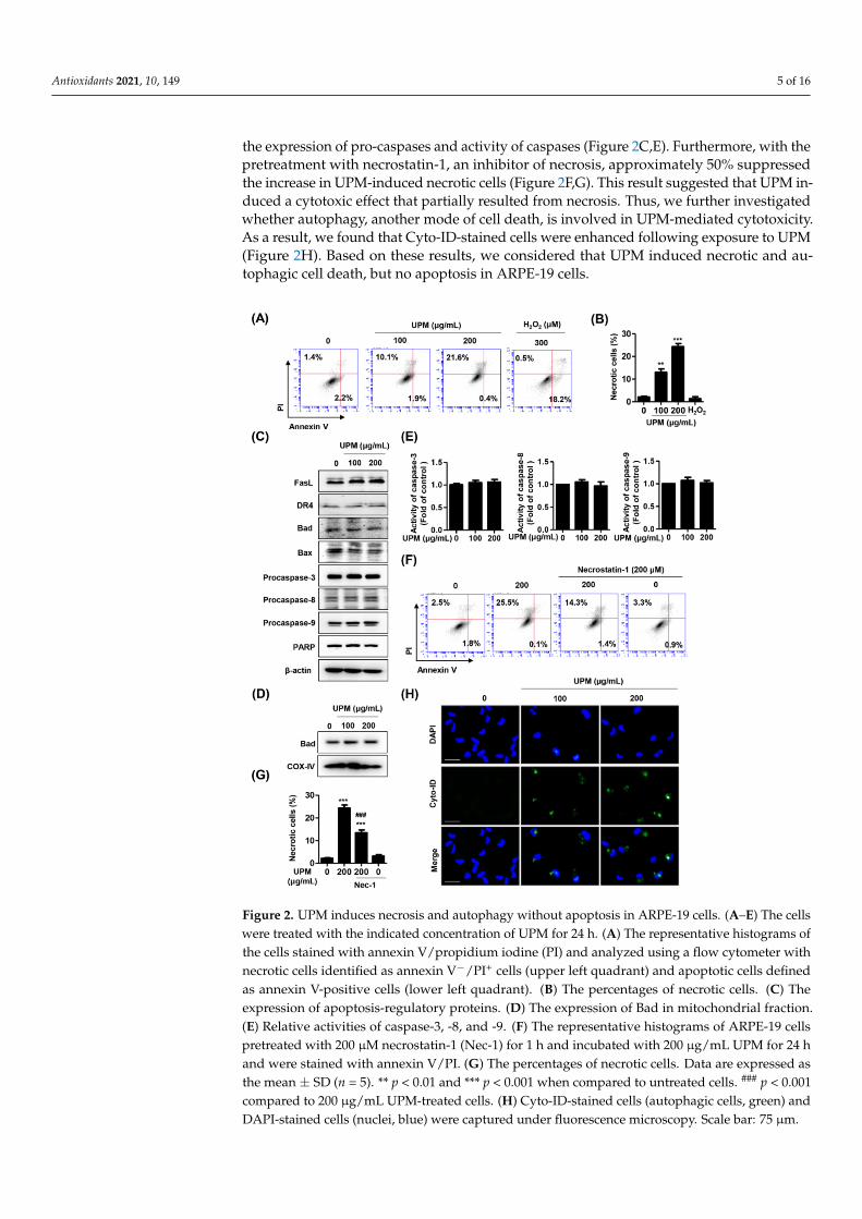
Next, to evaluate whether UPM-induced cytotoxicity accompanied by necrosis andautophagy was due to a change in the cell cycle, alterations in cell cycle distribution wereassessed. The flow cytometric analysis using PI staining indicated that UPM graduallyinduced a dose-dependent effect on G2/M phase arrest (Figure 3A,B and SupplementaryFigure S1). At 200 µg/mL UPM exposure, the cells showed that the G2/M populationincreased from approximately 33% to 54% compared to the control. Additionally, at themolecular level, it was found that the expression of p21 was increased under stimulationwith UPM, while the expression levels of p16, p27, and p53 were not changed (Figure 3C).Furthermore, other cell cycle regulators, including cyclin B1, cyclin D1, cyclin-dependentkinase (Cdk) 1, Cdk 2, and Cdk 6, were decreased. Meanwhile, UPM-mediated G2/M cellcycle arrest was partially attenuated under necrostatin-1 treatment (Figure 3D,E).
Antioxidants 2021, 10, x FOR PEER REVIEW 6 of 16
with 200 μM necrostatin-1 (Nec-1) for 1 h and incubated with 200 μg/mL UPM for 24 h and were stained with annexin V/PI. (G) The percentages of necrotic cells. Data are expressed as the mean ± SD (n = 5). ** p < 0.01 and *** p < 0.001 when compared to untreated cells. ### p < 0.001 compared to 200 μg/mL UPM-treated cells. (H) Cyto-ID-stained cells (autophagic cells, green) and DAPI-stained cells (nuclei, blue) were captured under fluorescence microscopy. Scale bar: 75 μm.
3.3. UPM Promotes Cell Cycle Arrest at the G2/M Phase in ARPE-19 Cells Next, to evaluate whether UPM-induced cytotoxicity accompanied by necrosis and
autophagy was due to a change in the cell cycle, alterations in cell cycle distribution were assessed. The flow cytometric analysis using PI staining indicated that UPM gradually induced a dose-dependent effect on G2/M phase arrest (Figure 3A,B and Supplementary Figure S1). At 200 μg/mL UPM exposure, the cells showed that the G2/M population in-creased from approximately 33% to 54% compared to the control. Additionally, at the mo-lecular level, it was found that the expression of p21 was increased under stimulation with UPM, while the expression levels of p16, p27, and p53 were not changed (Figure 3C). Fur-thermore, other cell cycle regulators, including cyclin B1, cyclin D1, cyclin-dependent ki-nase (Cdk) 1, Cdk 2, and Cdk 6, were decreased. Meanwhile, UPM-mediated G2/M cell cycle arrest was partially attenuated under necrostatin-1 treatment (Figure 3D,E).
Figure 3. UPM mediates G2/M cell cycle arrest in ARPE-19 cells. (A) The representative histograms of ARPE-19 cells incubated with PI for flow cytometry analysis. (B) The average percentages of ARPE-19 cells incubated with PI in each phase of the cell, except for the cells at the sub-G1 phase. (C) The expression of cell cycle-regulatory proteins. (D) The representative histograms of ARPE-19 cells pretreated with 200 μM necrostatin-1 for 1 h and then treated with 200 μg/mL UPM for 24 h. The cells were stained with PI. (E) The average percentages of ARPE-19 cells pretreated with 200 μM necrostatin-1 for 1 h and treated with 200 μg/mL UPM for 24 h in each phase of the cell cycle, except for the cells at the sub-G1 phase.
3.4. UPM Disrupts DNA and Mitochondria in ARPE-19 Cells
Figure 3. UPM mediates G2/M cell cycle arrest in ARPE-19 cells. (A) The representative histogramsof ARPE-19 cells incubated with PI for flow cytometry analysis. (B) The average percentages ofARPE-19 cells incubated with PI in each phase of the cell, except for the cells at the sub-G1 phase.(C) The expression of cell cycle-regulatory proteins. (D) The representative histograms of ARPE-19cells pretreated with 200 µM necrostatin-1 for 1 h and then treated with 200 µg/mL UPM for 24 h.The cells were stained with PI. (E) The average percentages of ARPE-19 cells pretreated with 200 µMnecrostatin-1 for 1 h and treated with 200 µg/mL UPM for 24 h in each phase of the cell cycle, exceptfor the cells at the sub-G1 phase.

Antioxidants 2021, 10, 149 7 of 16
3.4. UPM Disrupts DNA and Mitochondria in ARPE-19 Cells
To identify whether ROS were involved in UPM-mediated cytotoxicity, we prefer-entially observed alterations in intracellular ROS levels and subsequently assessed theirinfluence on organelles. As a result of DCF-DA staining, intracellular ROS levels weremarkedly enhanced by UPM exposure (Figure 4A and Supplementary Figure S1). More-over, the fluorescence intensity of γH2AX, a DNA damage marker, was substantiallyincreased in UPM-stimulated cells (Figure 4B). In addition, the exposure to UPM in adose-dependent manner induced the loss of MMP (∆Ψm), which was accompanied byboth the upregulation of the expression of JC-1 monomers and the downregulation of theexpression of JC-1 aggregates (Figure 4C,D and Supplementary Figure S1). Furthermore,UPM promoted the expression of mitophagy regulators, including PTEN-induced kinase 1(PINK1), Parkin, and light chain 3 (LC3) I/II (Figure 4E).
Antioxidants 2021, 10, x FOR PEER REVIEW 7 of 16
To identify whether ROS were involved in UPM-mediated cytotoxicity, we preferen-tially observed alterations in intracellular ROS levels and subsequently assessed their in-fluence on organelles. As a result of DCF-DA staining, intracellular ROS levels were mark-edly enhanced by UPM exposure (Figure 4A and Supplementary Figure S1). Moreover, the fluorescence intensity of γH2AX, a DNA damage marker, was substantially increased in UPM-stimulated cells (Figure 4B). In addition, the exposure to UPM in a dose-depend-ent manner induced the loss of MMP (∆Ψm), which was accompanied by both the upreg-ulation of the expression of JC-1 monomers and the downregulation of the expression of JC-1 aggregates (Figure 4C,D and Supplementary Figure S1). Furthermore, UPM pro-moted the expression of mitophagy regulators, including PTEN-induced kinase 1 (PINK1), Parkin, and light chain 3 (LC3) I/II (Figure 4E).
Figure 4. UPM triggers DNA and mitochondrial disorders in ARPE-19 cells. (A) Fluorescence images of ARPE-19 cells stained with 10 μM DCF-DA. Intracellular reactive oxygen species (ROS) generation was identified as a DCF-DA intensity that was observed under a fluorescence microscope. Scale bar: 200 μm. (B) Fluorescence images of the cells immunostained with γH2AX antibody (green) and visualized using a fluorescence microscope. DAPI was used to counterstain the nuclei (blue). Scale bar: 75 μm. (C) Fluorescence images of the cells stained with 10 μM JC-1 with the monomer showing green fluorescence defining low MMP (∆Ψm) and aggregates showing red fluorescence characterizing high MMP (∆Ψm). Scale bar: 75 μm. (D) Quantification of JC-1 red and green intensity. Data are expressed as the mean ± SD (n = 5). *** p < 0.001 when compared to untreated cells. (E) The expression of mitophagy-regulated proteins.
3.5. The Excess ROS Induced by UPM Triggers Necrosis, Autophagy, and Cell Cycle Arrest in ARPE-19 Cells
Based on the above results showing that UPM promoted intracellular ROS genera-tion and organelle dysfunction, we investigated the role of ROS in UPM-induced necrosis and autophagy. The pretreatment with NAC, a potential ROS scavenger, significantly re-covered UPM-induced cytotoxicity and morphological changes (Figure 5A,B). Addition-ally, Figure 5C,D shows that NAC greatly suppressed the increase in necrotic cells by UPM. Furthermore, NAC markedly suppressed the expression of Cyto-ID-positive cell population (Figure 5E). Likewise, the UPM-induced cell cycle arrest at the G2/M phase
Figure 4. UPM triggers DNA and mitochondrial disorders in ARPE-19 cells. (A) Fluorescence images of ARPE-19 cellsstained with 10 µM DCF-DA. Intracellular reactive oxygen species (ROS) generation was identified as a DCF-DA intensitythat was observed under a fluorescence microscope. Scale bar: 200 µm. (B) Fluorescence images of the cells immunostainedwith γH2AX antibody (green) and visualized using a fluorescence microscope. DAPI was used to counterstain the nuclei(blue). Scale bar: 75 µm. (C) Fluorescence images of the cells stained with 10 µM JC-1 with the monomer showing greenfluorescence defining low MMP (∆Ψm) and aggregates showing red fluorescence characterizing high MMP (∆Ψm). Scalebar: 75 µm. (D) Quantification of JC-1 red and green intensity. Data are expressed as the mean ± SD (n = 5). *** p < 0.001when compared to untreated cells. (E) The expression of mitophagy-regulated proteins.

Antioxidants 2021, 10, 149 8 of 16
3.5. The Excess ROS Induced by UPM Triggers Necrosis, Autophagy, and Cell Cycle Arrest inARPE-19 Cells
Based on the above results showing that UPM promoted intracellular ROS generationand organelle dysfunction, we investigated the role of ROS in UPM-induced necrosis andautophagy. The pretreatment with NAC, a potential ROS scavenger, significantly recov-ered UPM-induced cytotoxicity and morphological changes (Figure 5A,B). Additionally,Figure 5C,D shows that NAC greatly suppressed the increase in necrotic cells by UPM.Furthermore, NAC markedly suppressed the expression of Cyto-ID-positive cell popula-tion (Figure 5E). Likewise, the UPM-induced cell cycle arrest at the G2/M phase was alsorestored under NAC treatment (Figure 5F,G). These results suggested that UPM promotedintracellular ROS generation, which is involved in necrosis, autophagy, and cell cycle arrest.
Antioxidants 2021, 10, x FOR PEER REVIEW 8 of 16
was also restored under NAC treatment (Figure 5F,G). These results suggested that UPM promoted intracellular ROS generation, which is involved in necrosis, autophagy, and cell cycle arrest.
Figure 5. Suppression of ROS attenuates UPM-induced necrosis, auophagy, and cell cycle arrest in AREP-19 cells. The cells were pretreated with 5 mM NAC for 1 h and then treated with 200 μg/mL UPM for 24 h. (A) Cell viability measured by a CCK-8 assay. Data are expressed as the mean ± SD (n = 4). * p < 0.05 and *** p < 0.001 when compared to untreated cells. # p < 0.05 and ###p < 0.001 compared to 200 μg/mL UPM-treated cells. (B) The shape of the cell and the morphological changes in the nucleus. Scale bar: 75 μm. (C) Representative histograms of cell death mode. (F) Cell cycle profiles derived from flow cytometry analysis. (D) The percentages of necrotic cells. Data are ex-pressed as the mean ± SD (n = 5). *** p < 0.001 when compared to untreated cells. *** p < 0.001 com-pared to 200 μg/mL UPM-treated cells. (E) Cyto-ID-stained cells (autophagic cells, green) and DAPI-stained cells (nuclei, blue) captured under fluorescence microscopy. Scale bar: 75 μm. (G) The aver-age percentages of cells in each phase of the cell cycle.
3.6. ROS Directly Regulate UPM-Induced DNA and Mitochondrial Damage in ARPE-19 Cells Next, we verified whether UPM-induced ROS triggered organelle dysfunction, be-
cause UPM results in necrosis and autophagy through ROS generation. As shown in Fig-ure 6A, NAC markedly attenuated intracellular ROS generation and MMP (∆Ψm) loss fol-lowing UPM exposure. The quantified results also showed that inhibition of ROS by NAC significantly suppressed the intensity of JC-1 monomers and DCF-DA (Figure 6B,C). Fur-thermore, we observed that NAC markedly inhibited the fluorescence expression of γH2AX in UPM-stimulated cells (Figure 6D). However, the upregulation of mitophagy regulators by UPM was not changed by NAC treatment (Figure 6E).
Figure 5. Suppression of ROS attenuates UPM-induced necrosis, auophagy, and cell cycle arrest in AREP-19 cells. The cellswere pretreated with 5 mM NAC for 1 h and then treated with 200 µg/mL UPM for 24 h. (A) Cell viability measured by aCCK-8 assay. Data are expressed as the mean ± SD (n = 4). * p < 0.05 and *** p < 0.001 when compared to untreated cells.# p < 0.05 and ### p < 0.001 compared to 200 µg/mL UPM-treated cells. (B) The shape of the cell and the morphologicalchanges in the nucleus. Scale bar: 75 µm. (C) Representative histograms of cell death mode. (F) Cell cycle profilesderived from flow cytometry analysis. (D) The percentages of necrotic cells. Data are expressed as the mean ± SD (n = 5).*** p < 0.001 when compared to untreated cells. *** p < 0.001 compared to 200 µg/mL UPM-treated cells. (E) Cyto-ID-stainedcells (autophagic cells, green) and DAPI-stained cells (nuclei, blue) captured under fluorescence microscopy. Scale bar:75 µm. (G) The average percentages of cells in each phase of the cell cycle.

Antioxidants 2021, 10, 149 9 of 16
3.6. ROS Directly Regulate UPM-Induced DNA and Mitochondrial Damage in ARPE-19 Cells
Next, we verified whether UPM-induced ROS triggered organelle dysfunction, be-cause UPM results in necrosis and autophagy through ROS generation. As shown inFigure 6A, NAC markedly attenuated intracellular ROS generation and MMP (∆Ψm) lossfollowing UPM exposure. The quantified results also showed that inhibition of ROS byNAC significantly suppressed the intensity of JC-1 monomers and DCF-DA (Figure 6B,C).Furthermore, we observed that NAC markedly inhibited the fluorescence expression ofγH2AX in UPM-stimulated cells (Figure 6D). However, the upregulation of mitophagyregulators by UPM was not changed by NAC treatment (Figure 6E).
Antioxidants 2021, 10, x FOR PEER REVIEW 9 of 16
Figure 6. Inhibition of ROS suppresses UPM-induced DNA and mitochondrial damage in AREP-19 cells. (A) Representative fluorescence images of DCF-DA-stained cells. Scale bar: 200 μm. (B) The fluorescence expression of γH2AX (green). DAPI was used to counterstain the nuclei (blue). Scale bar: 75 μm. (C) Representative images of JC-1 fluorescence staining. Monomers showing green flu-orescence define low MMP, and aggregates showing red fluorescence characterize high MMP. Scale bar: 25 μm. (D) Quantification of JC-1 red and green intensities. Data are expressed as the mean ± SD (n = 4). * p < 0.05 and *** p < 0.001 when compared to untreated cells. ###p < 0.001 compared to 200 μg/mL UPM-treated cells. (E) The expression of mitophagy-regulated proteins.
4. Discussion The retina, the back segment of the eye, functions in vision control and composed of
10 layers that consist of 8 specific cells [32]. In particular, the RPE, the outer layer of the retina, plays an essential role in the vision process by exchanging nutrients, metabolic end products, and signal molecules with neighboring cells [32]. Therefore, RPE cells are critical for retinal homeostasis, and the loss and dysfunction of RPE cells are causative of AMD, which is the leading cause of blindness in the elderly [33]. Several genetic and environ-mental risk factors for AMD lead to inflammation, anatomical changes, and dysfunction in the retina, which can eventually lead to loss of vision [34]. Recently, it has been reported that air pollutants are considered one of the environmental risk factors for AMD and are involved in ROS overload by oxidative stress, which can lead to RPE cell death [19,24].
Figure 6. Inhibition of ROS suppresses UPM-induced DNA and mitochondrial damage in AREP-19cells. (A) Representative fluorescence images of DCF-DA-stained cells. Scale bar: 200 µm. (B) Thefluorescence expression of γH2AX (green). DAPI was used to counterstain the nuclei (blue). Scalebar: 75 µm. (C) Representative images of JC-1 fluorescence staining. Monomers showing greenfluorescence define low MMP, and aggregates showing red fluorescence characterize high MMP.Scale bar: 25 µm. (D) Quantification of JC-1 red and green intensities. Data are expressed as the mean± SD (n = 4). * p < 0.05 and *** p < 0.001 when compared to untreated cells. ### p < 0.001 compared to200 µg/mL UPM-treated cells. (E) The expression of mitophagy-regulated proteins.
Antioxidants 2021, 10, 149 10 of 16
4. Discussion
The retina, the back segment of the eye, functions in vision control and composedof 10 layers that consist of 8 specific cells [32]. In particular, the RPE, the outer layer ofthe retina, plays an essential role in the vision process by exchanging nutrients, metabolicend products, and signal molecules with neighboring cells [32]. Therefore, RPE cells arecritical for retinal homeostasis, and the loss and dysfunction of RPE cells are causativeof AMD, which is the leading cause of blindness in the elderly [33]. Several geneticand environmental risk factors for AMD lead to inflammation, anatomical changes, anddysfunction in the retina, which can eventually lead to loss of vision [34]. Recently, it hasbeen reported that air pollutants are considered one of the environmental risk factors forAMD and are involved in ROS overload by oxidative stress, which can lead to RPE celldeath [19,24].
Apoptosis, necrosis, and autophagy are three major types of cell death in response tooxidative stress [35]. Apoptosis can be executed by the extrinsic and intrinsic pathways,both of which activate effector caspases, leading to the cleavage or degradation of cellularsubstrates, including PARP and histones, ultimately leading to apoptotic cell death [36,37].Morphologically, apoptosis is characterized by cytoplasmic shrinkage, chromatin conden-sation and fragmentation, and the maintenance of the plasma membrane [36]. In contrast,necrosis is an unregulated form of cell death and is mediated by receptor-interacting pro-tein kinases (RIPKs) that are involved in necrosome formation, subsequently leading tomitochondrial fission and cell death [38]. During necrosis, mitochondrial fragmentationand mitochondrial permeability transition pore (mPTP) opening occur, which lead to MMP(∆Ψm) loss, ATP depletion and ROS release, although the signaling pathways prefacingthese events are unknown [36,39]. The morphological features of necrosis include cellswelling, an increase in cell volume, the formation of cytoplasmic vacuoles and blebs,and the eventual disruption of the cell membrane [36]. Autophagy is a predominant cellsurvival process of homeostasis that involves degradation of damaged organelles throughformation of autophagosome [40]. Accumulated evidences suggested that autophagy isimpaired in the aged retina, which contributes to the RPE dysfunction [41,42]. Althoughthe current paradigm is that apoptosis is a major mechanism for RPE cell death by oxida-tive stress, the role of apoptosis in the RPE death mechanism in retinal disorders is stillcontroversial [36]. In this regard, we investigated which cell death mode is involved in RPEdysfunction following UPM exposure and identified the cell death mechanism. Our resultsshowed that UPM induced cell death with morphological changes by necrosis, but typicalapoptotic DNA chromatin condensation did not occur (Figure 1). Furthermore, the find-ings from the flow cytometric analysis and immunoblots indicated that UPM mediatedcell death caused by necrosis and did not affect the expression of apoptotic regulators(Figure 2A–E). Additionally, based on the result showing that necrostatin-1, a RIP1 kinaseinhibitor, markedly restored UPM-mediated necrotic cell death (Figure 2F,G), our findingsdemonstrated that UPM-induced cytotoxicity is involved in necrotic cell death in the RPE.This finding supports the previous results of Hanus et al. [43] showing that during RPEcell death upon oxidative stress induced by pro-oxidants, cardinal features of necrosiswere observed but apoptosis features were not observed. The results also suggested thatnecrostatin-1 and the silencing of RIPK3 largely prevented oxidative stress-induced RPEdeath, which established that necrosis is a major type of RPE cell death in response tooxidative stress [43]. More recently, Kang et al. [16] demonstrated that long-term expo-sure to UPM led to anatomical changes in the retina whereas apoptosis did not occur.Although necrostain-1 significantly inhibited annexin V−/PI+ cells in the present study,approximately 50% suppressed the increase in UPM-induced necrotic cells by necrostain-1. This result suggested that UPM induces a cytotoxic effect that partially results fromnecrosis. We should keep in mind, however, that necrostain-1 could also partially influenceapoptosis [44]. Importantly, we observed a dose dependence increased the expression ofCyto-ID, a specific dye for autophagic compartments [45], in UPM-treated ARPE-19 cells(Figure 2H). Several reports demonstrated that autophagy plays an essential role in
Antioxidants 2021, 10, 149 11 of 16
bronchial cells and animals upon UPM exposure [46,47]. In a ocular system, a few studiesreported that autophagy is involved in PM-mediated pathogenic mechanisms in cornealepithelial cells [48,49]. However, the role of autophagy on retina in response to UPMexposure has remained elusive. Based on these knowledge, our finding that UPM inducedautophagy in ARPE-19 cells provides the first evidence of the effect of UPM on the posteriorpart of the eye. Taken together, our findings that UPM-induced RPE cell death is mainlycaused by necrosis and autophagy can provide the explanation of a novel mechanism forAMD pathogenesis upon exposure to urban air pollution.
In response to stressful conditions, cells are able to block the cell cycle transientlyor irreversibly [50]. In fact, the cellular response to DNA damage causes either cell cyclearrest or apoptosis [51]. Cell cycle progression is regulated by CDKs, and its activity iscoordinated by binding of their essential regulatory subunits, cyclins [52]. Numerousstudies have demonstrated that UPM may inhibit cell growth by cell death or reductionof proliferation, which has been linked to arrest in various steps of the cell cycle [53–56].Zhang et al. [56] provided the evidence that PM2.5 induces cell cycle arrest in G1 phasein alveolar epithelial cells through the downregulation of cyclin E, A, and D1 and theupregulation of p21. Furthermore, Wu et al. [55] suggested that UPM leads to G2/Mcell cycle arrest, DNA damage, and cell death in bronchial epithelial cells. In addition,Gualtieri et al. [53] reported that UPM causes mitotic arrest, resulting in cell death inbronchial epithelial cells. The same research team published the result that UPM inducesa delay in G2 phase and augments ROS formation, which causes damage to the DNAand spindle apparatus [54]. Although accumulated evidence has demonstrated that UPMinduces cell cycle arrest with cell death in various cell types, the cellular responses of RPEafter exposure to UPM on cell cycle regulation are not known. In the present study, wefound that UPM induced G2/M cell cycle arrest, along with the overexpression of γH2AXin the nucleus (Figure 3A,B and Figure 4B,C). Furthermore, our findings showed that UPMwas attributed to modulation of cell cycle regulators, including the upregulation of p21and the downregulation of cyclin B/D and CDK1/2/6, while the expression levels of p16,p27, and p53 were not altered (Figure 3C). p21 is known to act as a tumor suppressor andinhibits the activity of the cyclin/CDK complex, which in turn leads to cell cycle arrest [57].The cell cycle arrest at the G2/M phase indicates that damage to intracellular DNA isdifficult to repair [58]. One of the responses to DNA damage is the expression of γH2AX,which is an early sign of DNA damage induced by replication stalling [51]. The formationof γH2AX foci takes place immediately after the generation of DNA breaks, as well asreplication stalling and single-stranded DNA breaks [51]. In bronchial epithelial cells, UPMpromotes the breakage of DNA stands and triggers γH2AX activation in response to DNAdamage, leading to cell death and mitotic arrest [53]. According to Fernandez-Capetilloet al., H2AX-null cells were defective in cell cycle progression at the G2/M phase afterexposure to ionizing radiation [51]. Fragkos et al. [59] demonstrated that adeno-associatedvirus-induced DNA damage leads to γH2AX generation and subsequently results in cellcycle arrest by upregulating p21. On the other hand, in H2AX-deficient cells, the responseto DNA damage causes apoptosis via p21 degradation, suggesting that H2AX is requiredfor p21-induced cell cycle arrest [59]. Taken together, our results established that DNAdamage by UPM-mediated cellular stress induces the expression of γH2AX and subse-quently inhibits cell cycle progression through the upregulation of p21 in RPE cells. In ad-dition, a RIP1 kinase inhibitor markedly restored UPM-induced cell arrest at G2/M phase(Figure 3D,E), suggesting that UPM-induced cell cycle arrest is involved in necrosis inRPE cells.
Accumulated evidence has consistently shown that ROS induced by oxidative stressare crucial mediators of UPM toxicity [60]. UPM contains redox-active components, suchas heterocyclic polycyclic aromatic hydrocarbons (PAHs), nitro-PAHs, and various metals,which can lead to intracellular ROS generation by catalyzing Fenton’s reaction [61,62].Given that mitochondria are a major cellular source of ROS, growing evidence sug-gests that it is imperative to understand the effect of PM on mitochondrial structure and
Antioxidants 2021, 10, 149 12 of 16
function [63,64]. In this regard, it has been reported that oxidative stress and mitochondrialdysfunction occur in various cell types upon exposure to UPM [64,65]. Miao et al. [64]suggested that PM induces endothelial toxicity through alterations in mitochondrial mor-phology and function, including MMP (∆Ψm) loss and mPTP opening. The findings fromBhargava et al. [65] showed that PM exposure promotes ROS generation and resultsin mitochondrial membrane depolarization and alteration of mitochondrial respiratorychain enzyme activity in peripheral blood lymphocytes. Among the studies on the eye,Cui et al. [66] suggested that PM causes a delay in corneal epithelium wound healing andROS formation might play a critical role in this process. Recently, Somayajulu et al. [67]established that PM2.5 triggers ROS, which results in increased mRNA levels of oxidativestress and inflammation in mouse and human corneal epithelial cells. Although a few stud-ies have suggested that PM triggers ROS generation in corneal and conjunctival epithelium,no studies on ROS and mitochondrial function upon PM have been reported. Furthermore,no studies have focused on the role of ROS in the retina, except for our previously publishedreport, in which we demonstrated that diesel PM2.5 promotes intracellular ROS generation,which causes mitochondrial dysfunction, including suppression of mitochondrial activityand loss of MMP (∆Ψm), subsequently leading to retinal dysfunction [25]. In the presentstudy, we found that UPM promoted intracellular ROS generation and concurrently in-duced MMP (∆Ψm) loss in RPE cells (Figure 4A,D,E). Notably, blocking ROS generation byNAC partially reversed UPM-mediated cytotoxicity, necrosis, and G2/M cell cycle arrest,as well as almost totally suppressed UPM-induced autophagy, DNA damage, and MMP(∆Ψm) loss (Figures 5 and 6). These results demonstrated that UPM-induced RPE cell deathis caused by necrosis and autophagy mechanisms that involve DNA damage-triggered cellcycle arrest and mitochondrial damage-triggered MMP (∆Ψm) loss and that are partiallyregulated by ROS generation.
Mitophagy is a homeostatic process for the selective degradation of dysfunctional anddamaged mitochondria [68]. Under pathological conditions, active PINK1 accumulateson the outer mitochondrial membrane (OMM) to promote Parkin recruitment, and sub-sequently, Parkin triggers the polyubiquitination of several OMM proteins [69]. In turn,several adaptor molecules bind to polyubiquitinated proteins and initiate the formationof autophagosomes by binding with LC3 [69]. Recently, it was established that PM2.5induces mitochondrial dynamics in liver fibroblasts and may trigger mitophagy via thePINK1/Parkin signaling pathway by ROS generation [70]. Another study reported that theambient pollutant acrolein results in mitochondrial DNA damage through mitochondrialfission and mitophagy in lung epithelial cells and fibroblasts [71]. According to Wanget al.’s finding, the dysfunction of mitochondrial degradation by mitophagy can causethe activation of the mitochondrial-dependent apoptosis pathway [71]. In the presentstudy, we found that UPM promoted the expression of mitophagy regulators, includingPINK1, Parkin, and LC3 I/II, but this upregulation by UPM was not affected by NAC(Figures 4F and 6E). Based on this result, UPM induced mitochondrial dysfunction via intra-cellular ROS generation in the RPE, while mitophagy was regulated by a ROS-independentpathway. Although further studies on the regulatory mechanisms of ROS and mitophagy inUPM-exposed RPE are required, our findings provide the first evidence that UPM inducesmitochondrial dysfunction and leads to mitophagy in RPE.
5. Conclusions
Overall, our results showed that UPM promotes RPE cell death, involving necrosisand autophagy but not apoptosis. Furthermore, exposure to UPM leads to cell cyclearrest at the G2/M phase, which may be caused by the formation of γH2AX foci andsubsequent p21 upregulation. In addition, UPM causes mitochondrial dysfunction withMMP (∆Ψm) loss and upregulates mitophagy. These UPM-mediated cellular alterations areregulated by ROS production, except for mitophagy. Although further studies are neededto identify the role of mitophagy in UPM-induced RPE injury, the present study providesthe first evidence that ROS-mediated cellular damage with necrosis and autophagy may be

Antioxidants 2021, 10, 149 13 of 16
the critical mechanism of UPM-induced retinal disorders (Figure 7). Taken together, ourfindings suggested that targeting oxidative stress-induced necrosis and autophagy in theRPE may be a viable approach to preventing urban air pollutant-induced retinal disorders,including AMD.
Antioxidants 2021, 10, x FOR PEER REVIEW 13 of 16
Figure 7. UPM promotes necrosis and autophagy via ROS-mediated cellular disorders that are ac-companied by cell cycle arrest and mitophagy in ARPE-19 cells. UPM induces cytotoxicity, which is due to autophagic and necrotic cell death with cell cycle arrest at the G2/M phase. Furthermore, UPM markedly enhances DNA damage with the overexpression of γH2AX. In addition, UPM no-tably increases mitochondrial dysfunction with MMP (∆Ψm) loss and induces mitophagy. This UPM-mediated cellular damage is attributed to intracellular ROS production but not mitophagy. In conclusion, ROS-mediated cellular damage with autophagy and necrosis may be the critical mech-anisms underlying UPM-induced retinal disorders.
Supplementary Materials: The following are available online at www.mdpi.com/xxx/s1, Figure S1: Low concentration of UPM induced cellular dysfunction in ARPE-19 cells., Table S1: Primary and secondary antibodies used for immunoblotting and immunofluorescence.
Author Contributions: Conceptualization, H.L., J.-H.K., and Y.H.C.; methodology, D.H.K. and J.-H.K.; Investigation, H.L., D.H.K., S.-K.P., J.-W.J., K.S.S., and J.W.H.; data curation, S.-K.P., J.-W.J., M.-Y.K., S.-H.H., and G.-Y.K.; formal analysis, J.-W.J., M.-Y.K., J.W.H., G.-Y.K., and Y.H.C.; writing of the original draft preparation, H.L., S.-H.H., J.W.H., and Y.H.C.; writing of review and editing, H.L., J.-H.K., S.-K.P., G.-Y.K., and Y.H.C.; supervision, Y.H.C.; project administration, H.L. All authors have read and agreed to the published version of the manuscript.
Funding: This research was funded by the Basic Science Research Program through the National Research Foundation of Korea (grant number: 2019R1C1C1008623).
Institutional Review Board Statement: Not applicable.
Informed Consent Statement: Not applicable.
Data Availability Statement: The data presented in this study are available within the article and its supplementary material. Other data that support the findings of this study are available upon request from the corresponding authors.
Conflicts of Interest: The authors declare no conflicts of interest.
References 1. Wang, Q.; Li, J.; Yang, J.; Chen, Y.; Li, Y.; Li, S.; Xie, C.; Chen, C.; Wang, L.; Wang, L.; et al. Seasonal characterization of aerosol
composition and sources in a polluted city in Central China. Chemosphere 2020, 258, 127310.
Figure 7. UPM promotes necrosis and autophagy via ROS-mediated cellular disorders that areaccompanied by cell cycle arrest and mitophagy in ARPE-19 cells. UPM induces cytotoxicity, whichis due to autophagic and necrotic cell death with cell cycle arrest at the G2/M phase. Furthermore,UPM markedly enhances DNA damage with the overexpression of γH2AX. In addition, UPMnotably increases mitochondrial dysfunction with MMP (∆Ψm) loss and induces mitophagy. ThisUPM-mediated cellular damage is attributed to intracellular ROS production but not mitophagy.In conclusion, ROS-mediated cellular damage with autophagy and necrosis may be the criticalmechanisms underlying UPM-induced retinal disorders.
Supplementary Materials: The following are available online at https://www.mdpi.com/2076-3921/10/2/149/s1, Figure S1: Low concentration of UPM induced cellular dysfunction in ARPE-19 cells.,Table S1: Primary and secondary antibodies used for immunoblotting and immunofluorescence.
Author Contributions: Conceptualization, H.L., J.-H.K., and Y.H.C.; methodology, D.H.K. and J.-H.K.; Investigation, H.L., D.H.K., S.-K.P., J.-W.J., K.S.S., and J.W.H.; data curation, S.-K.P., J.-W.J.,M.-Y.K., S.-H.H., and G.-Y.K.; formal analysis, J.-W.J., M.-Y.K., J.W.H., G.-Y.K., and Y.H.C.; writing ofthe original draft preparation, H.L., S.-H.H., J.W.H., and Y.H.C.; writing of review and editing, H.L.,J.-H.K., S.-K.P., G.-Y.K., and Y.H.C.; supervision, Y.H.C.; project administration, H.L. All authors haveread and agreed to the published version of the manuscript.
Funding: This research was funded by the Basic Science Research Program through the NationalResearch Foundation of Korea (grant number: 2019R1C1C1008623).
Institutional Review Board Statement: Not applicable.
Informed Consent Statement: Not applicable.

Antioxidants 2021, 10, 149 14 of 16
Data Availability Statement: The data presented in this study are available within the article andits supplementary material. Other data that support the findings of this study are available uponrequest from the corresponding authors.
Conflicts of Interest: The authors declare no conflict of interest.
References1. Wang, Q.; Li, J.; Yang, J.; Chen, Y.; Li, Y.; Li, S.; Xie, C.; Chen, C.; Wang, L.; Wang, L.; et al. Seasonal characterization of aerosol
composition and sources in a polluted city in Central China. Chemosphere 2020, 258, 127310. [CrossRef]2. Ngarambe, J.; Joen, S.J.; Han, C.H.; Yun, G.Y. Exploring the relationship between particulate matter, CO, SO2, NO2, O3 and urban
heat island in Seoul, Korea. J. Hazard. Mater. 2021, 403, 123615. [CrossRef]3. Kelly, F.J. Urban air quality and health: Two steps forward, one step back. Eur. Respir. J. 2019, 53, 1900280. [CrossRef]4. Bakolis, I.; Hammoud, R.; Stewart, R.; Beevers, S.; Dajnak, D.; MacCrimmon, S.; Broadbent, M.; Pritchard, M.; Shiode, N.; Fecht, D.;
et al. Mental health consequences of urban air pollution: Prospective population-based longitudinal survey. Soc. PsychiatryPsychiatr. Epidemiol. 2020, 1–13. [CrossRef]
5. Crobeddu, B.; Aragao-Santiago, L.; Bui, L.C.; Boland, S.; Squiban, B.A. Oxidative potential of particulate matter 2.5 as predictiveindicator of cellular stress. Environ. Pollut. 2017, 230, 125–133. [CrossRef]
6. Atkinson, R.W.; Fuller, G.W.; Anderson, H.R.; Harrison, R.M.; Armstrong, B. Urban ambient particle metrics and health:A time-series analysis. Epidemiology 2010, 21, 501–511. [CrossRef]
7. Ritz, B.; Hoffmann, B.; Peters, A. The effects of fine dust, ozone, and nitrogen dioxide on health. Dtsch. Arztebl. Int. 2019, 51–52,881–886. [CrossRef]
8. Mu, G.; Zhou, M.; Wang, B.; Cao, L.; Yang, S.; Qiu, W.; Nie, X.; Ye, Z.; Zhou, Y.; Chen, W. Personal PM2.5 exposure and lungfunction: Potential mediating role of systematic inflammation and oxidative damage in urban adults from the general population.Sci. Total Environ. 2020, 755, 142522. [CrossRef]
9. Lu, Y.; Lin, S.; Fatmi, Z.; Malashock, D.; Hussain, M.M.; Siddique, A.; Carpenter, D.O.; Lin, Z.; Khwaja, H.A. Assessing the associ-ation between fine particulate matter (PM2.5) constituents and cardiovascular diseases in a mega-city of Pakistan. Environ. Pollut.2019, 252, 1412–1422. [CrossRef]
10. Shahbaz, M.A.; Martikainen, M.V.; Rönkkö, T.J.; Komppula, M.; Jalava, P.I.; Roponen, M. Urban air PM modifies differentlyimmune defense responses against bacterial and viral infections in vitro. Environ. Res. 2020, 192, 110244. [CrossRef]
11. Chen, X.; Guo, J.; Huang, Y.; Liu, S.; Huang, Y.; Zhang, Z.; Zhang, F.; Lu, Z.; Li, F.; Zheng, J.C.; et al. Urban airborne PM2.5-activated microglia mediate neurotoxicity through glutaminase-containing extracellular vesicles in olfactory bulb. Environ. Pollut.2020, 264, 114716. [CrossRef]
12. Kitakaze, T.; Yoshioka, Y.; Furuyashiki, T.; Ashida, H. Enzymatically synthesized glycogen protects inflammation induced byurban particulate matter in normal human epidermal keratinocytes. J. Clin. Biochem. Nutr. 2020, 67, 29–35. [CrossRef]
13. Gutiérrez, A.M.; Giuliani, D.; Porta, A.A.; Andrinolo, D. Relationship between ocular surface alterations and concentrations ofaerial particulate matter. J. Ophthalmic Vis. Res. 2019, 14, 419–427. [CrossRef]
14. Fu, Q.; Mo, Z.; Lyu, D.; Zhang, L.; Qin, Z.; Tang, Q.; Yin, H.; Xu, P.; Wu, L.; Lou, X.; et al. Air pollution and outpatient visits forconjunctivitis: A case-crossover study in Hangzhou, China. Environ. Pollut. 2017, 231, 1344–1350. [CrossRef]
15. Mo, Z.; Fu, Q.; Lyu, D.; Zhang, L.; Qin, Z.; Tang, Q.; Yin, H.; Xu, P.; Wu, L.; Wang, X.; et al. Impacts of air pollution on dry eyedisease among residents in Hangzhou, China: A case-crossover study. Environ. Pollut. 2019, 246, 183–189. [CrossRef]
16. Kang, W.S.; Choi, H.; Jang, G.; Lee, K.H.; Kim, E.; Kim, K.J.; Jeong, G.Y.; Kim, J.S.; Na, C.S.; Kim, S. Long-term exposure to urbanparticulate matter on the ocular surface and the incidence of deleterious changes in the cornea, conjunctiva and retina in rats.Int. J. Mol. Sci. 2020, 21, 4976. [CrossRef]
17. Lee, T.G.; Hyun, S.W.; Jo, K.; Park, B.; Lee, I.S.; Song, S.J.; Kim, C.S. Achyranthis radix extract improves urban particulatematter-induced dry eye disease. Int. J. Environ. Res. Public Health 2019, 16, 3229. [CrossRef]
18. Block, M.L.; Calderón-Garcidueñas, L. Air pollution: Mechanisms of neuroinflammation and CNS disease. Trends Neurosci. 2009,32, 506–516. [CrossRef]
19. Chang, K.H.; Hsu, P.Y.; Lin, C.J.; Lin, C.L.; Juo, S.H.; Liang, C.L. Traffic-related air pollutants increase the risk for age-relatedmacular degeneration. J. Investig. Med. 2019, 67, 1076–1081. [CrossRef]
20. Louwies, T.; Panis, L.I.; Kicinski, M.; De Boever, P.; Nawrot, T.S. Retinal microvascular responses to short-term changes inparticulate air pollution in healthy adults. Environ. Health Perspect. 2013, 121, 1011–1016. [CrossRef]
21. Pan, S.C.; Huang, C.C.; Chin, W.S.; Chen, B.Y.; Chan, C.C.; Guo, Y.L. Association between air pollution exposure and diabeticretinopathy among diabetics. Environ. Res. 2020, 181, 108960. [CrossRef]
22. Provost, E.B.; Panis, L.I.; Saenen, N.D.; Kicinski, M.; Louwies, T.; Vrijens, K.; De Boever, P.; Nawrot, T.S. Recent versus chronicfine particulate air pollution exposure as determinant of the retinal microvasculature in school children. Environ. Res. 2017, 159,103–110. [CrossRef]
23. Kim, S.; Park, H.; Park, H.; Joung, B.; Kim, E. The acute respiratory exposure by intratracheal instillation of Sprague-Dawley ratswith diesel particulate matter induces retinal thickening. Cutan. Ocul. Toxicol. 2016, 5, 275–280. [CrossRef]

Antioxidants 2021, 10, 149 15 of 16
24. Tien, P.T.; Lin, H.J.; Tsai, Y.Y.; Lim, Y.P.; Chen, C.S.; Chang, C.Y.; Lin, C.J.; Chen, J.J.; Wu, S.M.; Huang, Y.J.; et al. Perfluorooctanoicacid in indoor particulate matter triggers oxidative stress and inflammation in corneal and retinal cells. Sci. Rep. 2020, 10, 15702.[CrossRef]
25. Lee, H.; Hwang-Bo, H.; Ji, S.Y.; Kim, M.Y.; Kim, S.Y.; Park, C.; Hong, S.H.; Kim, G.Y.; Song, K.S.; Hyun, J.W.; et al. Diesel particulatematter2.5 promotes epithelial-mesenchymal transition of human retinal pigment epithelial cells via generation of reactive oxygenspecies. Environ. Pollut. 2020, 262, 114301. [CrossRef]
26. Mori, I.; Sun, Z.; Ukachi, M.; Nagano, K.; McLeod, C.W.; Cox, A.G.; Nishikawa, M. Development and certification of the newNIES CRM 28: Urban aerosols for the determination of multielements. Anal. Bioanal. Chem. 2008, 391, 1997–2003. [CrossRef]
27. Choi, Y.H. Trans-cinnamaldehyde protects C2C12 myoblasts from DNA damage, mitochondrial dysfunction and apoptosiscaused by oxidative stress through inhibiting ROS production. Genes Genom. 2020. [CrossRef]
28. Zhang, N.; Li, F.; Gao, J.; Zhang, S.; Wang, Q. Osteopontin accelerates the development and metastasis of bladder cancer viaactivating JAK1/STAT1 pathway. Genes Genom. 2020, 42, 467–475. [CrossRef]
29. Hwangbo, H.; Kim, S.Y.; Lee, H.; Park, S.H.; Hong, S.H.; Park, C.; Kim, G.Y.; Leem, S.H.; Hyun, J.W.; Cheong, J.; et al. Auranofinenhances sulforaphane-mediated apoptosis in hepatocellular carcinoma Hep3B cells through inactivation of the PI3K/Aktsignaling pathway. Biomol. Ther. 2020, 28, 443–455. [CrossRef]
30. Park, S.; Kim, M.; Hong, Y.; Lee, H.; Tran, Q.; Kim, C.; Kwon, S.H.; Park, J.; Park, J.; Kim, S.H. Myristoylated TMEM39AS41,a cell-permeable peptide, causes lung cancer cell death. Toxicol. Res. 2020, 36, 123–130. [CrossRef]
31. Park, C.; Cha, H.J.; Lee, H.; Hwang-Bo, H.; Ji, S.Y.; Kim, M.Y.; Hong, S.H.; Jeong, J.W.; Han, M.H.; Choi, S.H.; et al. Induction ofG2/M cell cycle arrest and apoptosis by genistein in human bladder cancer T24 cells through inhibition of the ROS-dependentPI3k/Akt signal transduction pathway. Antioxidants 2019, 8, 327. [CrossRef] [PubMed]
32. Willermain, F.; Scifo, L.; Weber, C.; Caspers, L.; Perret, J.; Delporte, C. Potential interplay between hyperosmolarity andinflammation on retinal pigmented epithelium in pathogenesis of diabetic retinopathy. Int. J. Mol. Sci. 2018, 19, 1056. [CrossRef][PubMed]
33. Golestaneh, N.; Chu, Y.; Xiao, Y.Y.; Stoleru, G.L.; Theos, A.C. Dysfunctional autophagy in RPE, a contributing factor in age-relatedmacular degeneration. Cell Death Dis. 2018, 8, e2537. [CrossRef] [PubMed]
34. Lambert, N.G.; ElShelmani, H.; Singh, M.K.; Mansergh, F.C.; Wride, M.A.; Padilla, M.; Keegan, D.; Hogg, R.E.; Ambati, B.K.Risk factors and biomarkers of age-related macular degeneration. Prog. Retin. Eye Res. 2016, 54, 64–102. [CrossRef] [PubMed]
35. D’Arcy, M.S. Cell death: A review of the major forms of apoptosis, necrosis and autophagy. Cell Biol. Int. 2019, 43, 582–592.[CrossRef] [PubMed]
36. Hanus, J.; Anderson, C.; Wang, S. RPE necroptosis in response to oxidative stress and in AMD. Ageing Res. Rev. 2015, 24, 286–298.[CrossRef]
37. Hanus, J.; Kalinowska-Herok, M.; Widlak, P. The major apoptotic endonuclease DFF40/CAD is a deoxyribose-specific anddouble-strand-specific enzyme. Apoptosis 2008, 13, 377–382. [CrossRef]
38. Christofferson, D.E.; Yuan, J. Necroptosis as an alternative form of programmed cell death. Curr. Opin. Cell Biol. 2010, 22, 263–268.[CrossRef]
39. Declercq, W.; Vanden Berghe, T.; Vandenabeele, P. RIP kinases at the crossroads of cell death and survival. Cell 2009, 138, 229–232.[CrossRef]
40. Chan, C.M.; Huang, D.Y.; Sekar, P.; Hsu, S.H.; Lin, W.W. Reactive oxygen species-dependent mitochondrial dynamics andautophagy confer protective effects in retinal pigment epithelial cells against sodium iodate-induced cell death. J. Biomed. Sci.2019, 26, 40. [CrossRef]
41. Blasiak, J.; Pawlowska, E.; Szczepanska, J.; Kaarniranta, K. Interplay between autophagy and the ubiquitin-proteasome systemand its role in the pathogenesis of age-related macular degeneration. Int. J. Mol. Sci. 2019, 20, 210. [CrossRef] [PubMed]
42. Keeling, E.; Lotery, A.J.; Tumbarello, D.A.; Ratnayaka, J.A. Impaired cargo clearance in the retinal pigment epithelium (RPE)underlies irreversible blinding diseases. Cells 2018, 7, 16. [CrossRef] [PubMed]
43. Hanus, J.; Zhang, H.; Wang, Z.; Liu, Q.; Zhou, Q.; Wang, S. Induction of necrotic cell death by oxidative stress in retinal pigmentepithelial cells. Cell Death Dis. 2013, 4, e965. [CrossRef] [PubMed]
44. Vandenabeele, P.; Grootjans, S.; Callewaert, N.; Takahashi, N. Necrostatin-1 blocks both RIPK1 and IDO: Consequences for thestudy of cell death in experimental disease models. Cell Death Differ. 2013, 20, 185–187. [CrossRef]
45. Guo, S.; Liang, Y.; Murphy, S.F.; Huang, A.; Shen, H.; Kelly, D.F.; Sobrado, P.; Sheng, Z. A rapid and high content assay thatmeasures cyto-ID-stained autophagic compartments and estimates autophagy flux with potential clinical applications. Autophagy2015, 11, 560–572. [CrossRef]
46. Chen, Z.H.; Wu, Y.F.; Wang, P.L.; Wu, Y.P.; Li, Z.Y.; Zhao, Y.; Zhou, J.S.; Zhu, C.; Cao, C.; Mao, Y.Y.; et al. Autophagy is essentialfor ultrafine particle-induced inflammation and mucus hyperproduction in airway epithelium. Autophagy 2016, 12, 297–311.[CrossRef]
47. Deng, X.; Zhang, F.; Rui, W.; Long, F.; Wang, L.; Feng, Z.; Chen, D.; Ding, W. PM2.5-induced oxidative stress triggers autophagyin human lung epithelial A549 cells. Toxicol. Vitr. 2013, 27, 1762–1770. [CrossRef]
48. Fu, Q.; Lyu, D.; Zhang, L.; Qin, Z.; Tang, Q.; Yin, H.; Lou, X.; Chen, Z.; Yao, K. Airborne particulate matter (PM2.5) triggersautophagy in human corneal epithelial cell line. Environ. Pollut. 2017, 227, 314–322. [CrossRef]
Antioxidants 2021, 10, 149 16 of 16
49. Lyu, D.; Chen, Z.; Almansoob, S.; Chen, H.; Ye, Y.; Song, F.; Zhang, L.; Qin, Z.; Tang, Q.; Yin, H.; et al. Transcriptomic profiling ofhuman corneal epithelial cells exposed to airborne fine particulate matter (PM2.5). Ocul. Surf. 2020, 18, 554–564. [CrossRef]
50. Pearce, A.K.; Humphrey, T.C. Integrating stress-response and cell-cycle checkpoint pathways. Trends Cell Biol. 2001, 11, 426–433.[CrossRef]
51. Fernandez-Capetillo, O.; Lee, A.; Nussenzweig, M.; Nussenzweig, A. H2AX: The histone guardian of the genome. DNA Repair2004, 3, 959–967. [CrossRef] [PubMed]
52. Jorgensen, P.; Tyers, M. How cells coordinate growth and division. Curr. Biol. 2004, 14, 1014–1027. [CrossRef] [PubMed]53. Gualtieri, M.; Ovrevik, J.; Mollerup, S.; Asare, N.; Longhin, E.; Dahlman, H.J.; Camatini, M.; Holme, J.A. Airborne urban particles
(Milan winter-PM2.5) cause mitotic arrest and cell death: Effects on DNA, mitochondria, AhR binding and spindle organization.Mutat. Res. 2011, 713, 18–31. [CrossRef] [PubMed]
54. Longhin, E.; Holme, J.A.; Gutzkow, K.B.; Arlt, V.M.; Kucab, J.E.; Camatini, M.; Gualtieri, M. Cell cycle alterations induced byurban PM2.5 in bronchial epithelial cells: Characterization of the process and possible mechanisms involved. Part. Fibre. Toxicol.2013, 10, 63. [CrossRef] [PubMed]
55. Wu, J.; Shi, Y.; Asweto, C.O.; Feng, L.; Yang, X.; Zhang, Y.; Hu, H.; Duan, J.; Sun, Z. Fine particle matters induce DNA damageand G2/M cell cycle arrest in human bronchial epithelial BEAS-2B cells. Environ. Sci. Pollut. Res. Int. 2017, 24, 25071–25081.[CrossRef]
56. Zhang, J.; Ghio, A.J.; Gao, M.; Wei, K.; Rosen, G.D.; Upadhyay, D. Ambient particulate matter induces alveolar epithelial cell cyclearrest: Role of G1 cyclins. FEBS Lett. 2007, 581, 5315–5320. [CrossRef]
57. Al Bitar, S.; Gali-Muhtasib, H. The role of the cyclin dependent kinase inhibitor p21cip1/waf1 in targeting cancer: Molecu-lar mechanisms and novel therapeutics. Cancers 2019, 11, 1475. [CrossRef]
58. Lezaja, A.; Altmeyer, M. Inherited DNA lesions determine G1 duration in the next cell cycle. Cell Cycle 2018, 17, 24–32. [CrossRef]59. Fragkos, M.; Jurvansuu, J.; Beard, P. H2AX is required for cell cycle arrest via the p53/p21 pathway. Mol. Cell Biol. 2009, 29,
2828–2840. [CrossRef]60. Leni, Z.; Künzi, L.; Geiser, M. Air pollution causing oxidative stress. Curr. Opin. Toxicol. 2020, 20–21, 1–8. [CrossRef]61. Lodovici, M.; Bigagli, E. Oxidative stress and air pollution exposure. J. Toxicol. 2011, 2011, 487074. [CrossRef] [PubMed]62. Park, C.G.; Cho, H.K.; Shin, H.J.; Park, K.H.; Lim, H.B. Comparison of mutagenic activities of various ultra-fine particles.
Toxicol. Res. 2018, 34, 163–172. [CrossRef] [PubMed]63. Leclercq, B.; Kluza, J.; Antherieu, S.; Sotty, J.; Alleman, L.Y.; Perdrix, E.; Garçon, G. Air pollution-derived PM2.5 impairs
mitochondrial function in healthy and chronic obstructive pulmonary diseased human bronchial epithelial cells. Environ. Pollut.2018, 243, 1434–1449. [CrossRef] [PubMed]
64. Miao, X.; Li, W.; Niu, B.; Li, J.; Sun, J.; Qin, M.; Zhou, Z. Mitochondrial dysfunction in endothelial cells induced by airborne fineparticulate matter (<2.5 µm). J. Appl. Toxicol. 2019, 39, 1424–1432. [CrossRef]
65. Bhargava, A.; Tamrakar, S.; Aglawe, A.; Lad, H.; Srivastava, R.K.; Mishra, D.K.; Tiwari, R.; Chaudhury, K.; Goryacheva, I.Y.;Mishra, P.K. Ultrafine particulate matter impairs mitochondrial redox homeostasis and activates phosphatidylinositol 3-kinasemediated DNA damage responses in lymphocytes. Environ. Pollut. 2018, 234, 406–419. [CrossRef]
66. Cui, Y.H.; Hu, Z.X.; Gao, Z.X.; Song, X.L.; Feng, Q.Y.; Yang, G.; Li, Z.J.; Pan, H.W. Airborne particulate matter impairs cornealepithelial cells migration via disturbing FAK/RhoA signaling pathway and cytoskeleton organization. Nanotoxicology 2018, 12,312–324. [CrossRef]
67. Somayajulu, M.; Ekanayaka, S.; McClellan, S.A.; Bessert, D.; Pitchaikannu, A.; Zhang, K.; Hazlett, L.D. Airborne particulatesaffect corneal homeostasis and immunity. Invest. Ophthalmol. Vis. Sci. 2020, 61, 23. [CrossRef]
68. Sachdeva, K.; Do, D.C.; Zhang, Y.; Hu, X.; Chen, J.; Gao, P. Environmental exposures and asthma development: Autophagy, mi-tophagy, and cellular senescence. Front. Immunol. 2019, 10, 2787. [CrossRef]
69. Palikaras, K.; Lionaki, E.; Tavernarakis, N. Mechanisms of mitophagy in cellular homeostasis, physiology and pathology.Nat. Cell Biol. 2018, 20, 1013–1022. [CrossRef]
70. Qiu, Y.N.; Wang, G.H.; Zhou, F.; Hao, J.J.; Tian, L.; Guan, L.F.; Geng, X.K.; Ding, Y.C.; Wu, H.W.; Zhang, K.Z. PM2.5 induces liverfibrosis via triggering ROS-mediated mitophagy. Ecotoxicol. Environ. Saf. 2019, 167, 178–187. [CrossRef]
71. Wang, H.T.; Lin, J.H.; Yang, C.H.; Haung, C.H.; Weng, C.W.; Maan-Yuh Lin, A.; Lo, Y.L.; Chen, W.S.; Tang, M.S. Acrolein inducesmtDNA damages, mitochondrial fission and mitophagy in human lung cells. Oncotarget 2017, 8, 70406–70421. [CrossRef][PubMed]