liver - necrosis - national toxicology programliver – necrosis recommendation: necrosis should not...
TRANSCRIPT
Liver – Necrosis
1
Liver – Necrosis
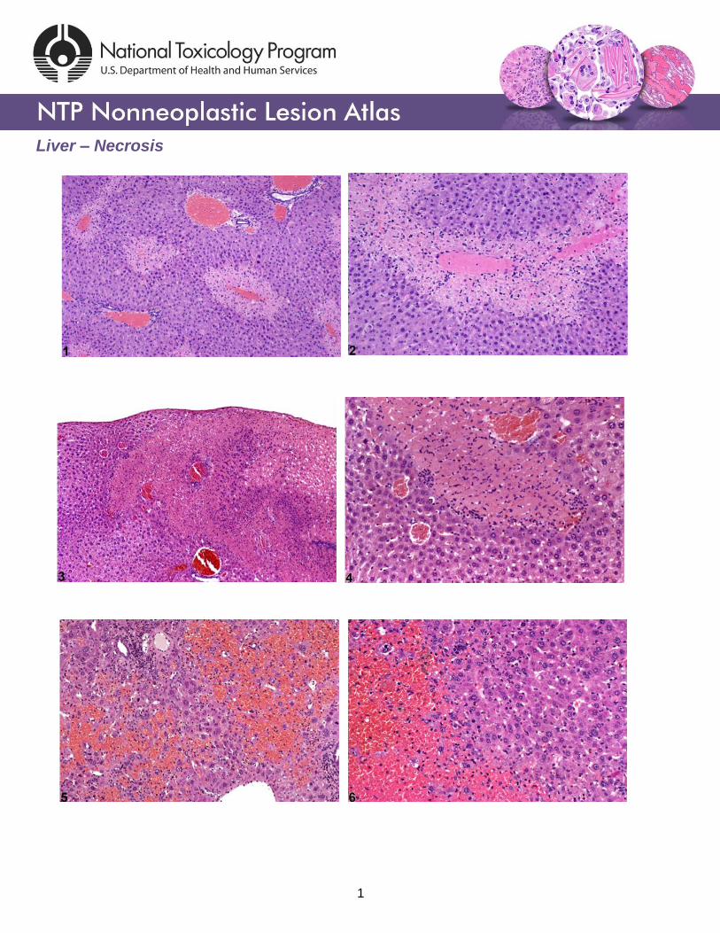
Figure Legend: Figure 1 Necrosis–sharply demarcated centrilobular necrosis in a male
B6C3F1 mouse from a subchronic study. Figure 2 Necrosis–sharply demarcated centrilobular
necrosis in a male B6C3F1 mouse from a subchronic study. Figure 3 Necrosis–patchy areas of
coagulation necrosis in a female Swiss Webster mouse from a subchronic study. Figure 4
Necrosis–patchy areas of coagulation necrosis in a female Swiss Webster mouse from a
subchronic study (higher magnification of Figure 3). Figure 5 Necrosis–hemorrhagic necrosis in
a Swiss CD-1 mouse from a chronic study. Figure 6 Necrosis–hemorrhagic necrosis in a Swiss
CD-1 mouse from a chronic study. Figure 7 Necrosis–focal necrosis with fatty change in a male
B6C3F1 mouse from a chronic study. Figure 8 Necrosis–focal necrosis with fatty change in a
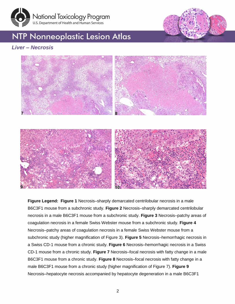
male B6C3F1 mouse from a chronic study (higher magnification of Figure 7). Figure 9
Necrosis–hepatocyte necrosis accompanied by hepatocyte degeneration in a male B6C3F1
2
Liver – Necrosis
mouse from a subchronic study. Figure 10 Necrosis–hepatocyte necrosis accompanied by
hepatocyte degeneration in a male B6C3F1 mouse from a subchronic study (higher
magnification of Figure 9).
Comment: The extent, pattern, and morphologic features of hepatocellular necrosis depend on
the degree of metabolic activation of hepatotoxic xenobiotics, host response to the toxicant,
dose and duration of xenobiotic exposure, and timing of liver sample evaluation after dosing.
Classical coagulation necrosis is typically caused by ischemia or infarction, and tissue
architecture is somewhat maintained because lysosomal enzymes responsible for proteolysis
are denatured. Another form of necrosis, liquefaction necrosis, may result in cellular dissolution
and loss of cytologic architecture. Changes that may accompany necrosis include hemorrhage,
fatty change, cytoplasmic vacuolization, cytologic degeneration, and inflammatory cell
infiltration.
Figure 1 and Figure 2 represent sharply demarcated centrilobular necrosis with loss of
hepatocyte cytologic detail. Figure 3 and Figure 4 represent irregular patchy areas of
coagulation necrosis with early infiltration of inflammatory cells. There is no distinctive lobular
pattern to this necrosis. Figure 5 and Figure 6 represent an example of necrosis characterized
by loss of hepatocytes and replacement with erythrocytes. This is an example of hemorrhagic
necrosis. Figure 7 and Figure 8 represent focal necrosis associated with fatty change. The
patches of necrosis (see Figure 8, bottom center) are hypereosinophilic, and the fatty change is
centrilobular with extension well into midlobular areas.
Figure 9 and Figure 10 represent a diagnosis of hepatocyte necrosis accompanied by
hepatocyte degeneration, microvesicular fatty change, nuclear pyknosis, and inflammation.
“Degeneration” is a term often used to indicate reversible cell or tissue damage and is
considered to be a precursor to necrosis. Because the presence of the inflammatory response is
extensive, an additional diagnosis of inflammation (cellular infiltrate) to capture the ongoing
process with more fidelity may be appropriate.
3
Liver – Necrosis
Recommendation: Necrosis should not be subclassified based on type, with the exception of
single-cell necrosis. For a given xenobiotic, dose and animal variability in response can
influence whether hepatocellular necrosis is panlobular or centrilobular and whether it is focal or
occurs in extensive irregular patches. If the fundamental process is the same, the lesion(s)
should be recorded simply as necrosis and assigned a severity grade. The pattern and other
features of the hepatocellular necrosis should be described in the pathology narrative. Splitting
out diagnoses too finely will result in complicated incidence tables and may compromise
appropriate interpretation of any induced toxicity. When accompanying changes such as fatty
change or inflammation are sufficiently extensive, separate diagnoses may be warranted with
severity grading and discussion in the pathology narrative. Since degeneration is considered
part of the continuum of changes involved in the necrotic process, it should not be diagnosed
separately when present with necrosis. However, degeneration without necrosis may occur at
exposure levels below doses that cause necrosis and thus may warrant a separate diagnosis. In
some cases, hepatocellular necrosis can result in cavernous, blood-filled spaces within the
hepatic parenchyma. These blood-filled spaces should not be diagnosed as hemorrhage
because they are secondary to necrosis.
References:
Greaves P. 2007. Histopathology of Preclinical Toxicity Studies: Interpretation and Relevance in Drug Safety Evaluation, 3rd ed. Elsevier, Amsterdam. Abstract: http://www.sciencedirect.com/science/book/9780444527714
Eustis SL, Boorman GA, Harada T, Popp JA. 1990. Liver. In: Pathology of the Fischer Rat (Boorman GA, Eustis SL, Elwell MR, Montgomery CA, MacKenzie WF, eds). Academic Press, San Diego, 71–94. Abstract: http://www.ncbi.nlm.nih.gov/nlmcatalog/9002563
Evans JG, Lake BG. 1998. The digestive system II. Hepatobiliary system. In: Target Organ Pathology (Turton J, Hooson J, eds). Taylor and Francis, London, 61–98. Abstract: http://www.amazon.com/Target-Organ-Pathology-Basic-Text/dp/0748401571
Harada T, Enomoto A, Boorman GA, Maronpot RR. 1999. Liver and gallbladder. In: Pathology of the Mouse: Reference and Atlas (Maronpot RR, Boorman GA, Gaul BW, eds). Cache River Press, Vienna, IL, 119–183. Abstract: http://www.cacheriverpress.com/books/pathmouse.htm
4
Liver – Necrosis
References:
Hardisty JF, Brix AE. 2005. Comparative hepatic toxicity: Prechronic/chronic liver toxicity in rodents. Toxicol Pathol 33:35–40. Full-Text: http://tpx.sagepub.com/content/33/1/35.full.pdf
Haschek WM, Rousseaux CG, Wallig MA. 2010. Fundamentals of Toxicologic Pathology, 2nd ed. Academic Press, San Diego, 691. Abstract: http://www.sciencedirect.com/science/book/9780123704696
National Toxicology Program. 1993. NTP TR-394. Toxicology and Carcinogenesis Studies of Acetaminophen (CAS No. 103-90-2) in F344 Rats and B6C3F1 Mice (Feed Studies). NTP, Research Triangle Park, NC. Full-Text: http://ntp.niehs.nih.gov/ntp/htdocs/LT_rpts/tr394.pdf
National Toxicology Program. 1998. NTP TR-468. Toxicology and Carcinogenesis Studies of Oxazepam (CAS No. 604-75-1) in Swiss-Webster and B6C3F1 Mice (Feed Studies). NTP, Research Triangle Park, NC. Full-Text: http://ntp.niehs.nih.gov/ntp/htdocs/LT_rpts/tr468.pdf
National Toxicology Program. 2012. NTP TR-571. Toxicology and Carcinogenesis Studies of Kava Kava Extract (CAS No. 9000-38-8) in F344/N Rats and B6C3F1 Mice (Gavage Studies). NTP, Research Triangle Park, NC. Full-Text: http://ntp.niehs.nih.gov/ntp/htdocs/LT_rpts/tr571.pdf
Thoolen B, Maronpot RR, Harada T, Nyska A, Rousseaux C, Nolte T, Malarkey D, Kaufmann W, Kutter K, Deschl U, Nakae D, Gregson R, Winlove M, Brix A, Singl B, Belpoggi F, Ward JM. 2010. Hepatobiliary lesion nomenclature and diagnostic criteria for lesions in rats and mice (INHAND). Toxicol Pathol 38:5S–81S. Full-Text: http://tpx.sagepub.com/content/38/7_suppl/5S.full
Author:
Robert R. Maronpot, DVM, MS, MPH, DACVP, DABT, FIATP Senior Pathologist Experimental Pathology Laboratories, Inc. Research Triangle Park, NC
5