the oncogenic serine/threonine kinase pim-1 · pdf filethe pim-1 oncogene encodes a...
TRANSCRIPT
The oncogenic serine/threonine kinase Pim-1 phosphorylates and
inhibits the activity of C-TAK1: a novel role for
Pim-1 at the G2/M cell cycle checkpoint
Malte Bachmann, Hanjo Hennemann1, Pei Xiang Xing2,
Ingrid Hoffmann3 and Tarik Möröy*
Institut für Zellbiologie (Tumorforschung), IFZ, Universitätsklinikum Essen,
Virchowstrasse 173, D-45122 Essen, 2Austin Research Institute, Cancer Immunotherapy
Laboratory, Heidelberg, Victoria 3084, Australia. 3Deutsches Krebsforschungszentrum,
DKFZ, Im Neuenheimer Feld 280, 69120 Heidelberg, Germany
1present address: Caesar, Centre of Advanced European Studies and Research, Ludwig-
Erhard-Allee 2, D-53175 Bonn,
*corresponding author:
Institut für Zellbiologie (Tumorforschung), I F Z
Virchowstrasse 173, D-45122 Essen, Germany,
Tel.: 49 (201) 723 – 3380, Fax: 49 (201) 723 – 5904
Email: [email protected]
JBC Papers in Press. Published on August 19, 2004 as Manuscript M404440200
Copyright 2004 by The American Society for Biochemistry and Molecular Biology, Inc.
by guest on May 6, 2018
http://ww
w.jbc.org/
Dow
nloaded from
2
Abstract
The Pim-1 oncogene encodes a serine-threonine kinase which relays signals from
cytokine receptors and contributes to the formation of lymphoid tumors when expressed at
high levels. Here we show that the protein kinase C-TAK1 (Cdc twenty-five C associated
kinase 1) is a binding partner and a substrate of Pim-1. A physical interaction of Pim-1
and C-TAK1 could be shown biochemically and in yeast two hybrid assays. Immuno-
fluorescence experiments suggested that Pim-1/C-TAK1 complexes are predominantly
cytoplasmic. When transiently transfected, Pim-1 is also found in the nucleus and can
recruit C-TAK1 to this compartment. Both Pim-1 and C-TAK1 undergo auto-
phosphorylation but only Pim-1 is able to phosphorylate C-TAK1 but not vice versa.
Mass spectrometry analysis of C-TAK1 suggested that the sites of auto-phosphorylation
and Pim-1-mediated phosphorylation are distinct and not overlapping. Phosphorylation by
Pim-1 decreases C-TAK1 kinase activity significantly, in particular its ability to
phosphorylate and inactivate Cdc25C, a protein which actively promotes cell cycle
progression at the G2/M phase. Hence, our findings directly suggest a novel role for
Pim-1 as a positive regulator at the G2/M transition of the cell cycle.
Keywords: Pim-1, C-TAK1, cell cycle, G2/M checkpoint
Running title: Pim-1 phosphorylates C-TAK1
by guest on May 6, 2018
http://ww
w.jbc.org/
Dow
nloaded from
3
Introduction
The pim-1 gene encodes a 33kD cytoplasmic serine/threonine kinase (1, 2) and was first
discovered as a locus frequently activated by proviral insertion in Moloney Murine
Leukemia Virus (MoMuLV) induced T-cell lymphomas (3-5). The expression pattern of
Pim-1 is widespread and the protein is found in a series of tumors and tissues but highest
expression levels are found in cells of the hematopoietic and lymphoid system. Evidence
that pim-1 is directly implicated in the tumorigenic process was provided by the analysis
of Eµ pim-1 transgenic animals (6).
Mice carrying a homozygous deletion of pim-1 generated by gene targeting show a very
subtle phenotype (7, 8) probably because Pim-1 is active in several redundant signaling
pathways or because other Pim family members such as Pim-2 or Pim-3 can rescue a loss
of Pim-1 (9-11). Experiments with IL-3 dependent cells suggested that Pim-1 is mediating
gp130 mediated cell proliferation (12) and that the pim-1 gene is a direct target of the
latent STAT transcription factors (13) in particular STAT3 but also STAT5. Therefore,
Pim-1 is considered to be an effector of many cytokine signaling pathways in particular of
those that initiate signaling through STAT3 and STAT5 as for instance interleukin (IL)-2,
-3, -6, -7 and prolactin (12, 14-19). More recent data implicated Pim-1 in the regulation of
Socs-1 which is a negative regulator of the Jak/STAT pathway and suggested that Pim-1
can also modulate cytokine signaling pathways in addition to its role as a direct effector
kinase (20).
A putative substrate target sequence of Pim-1 has been identified by using a chemically
synthesized peptide library (21). However, since Pim-1 is able to phosphorylate itself but
does not contain such a recognition sequence (22, 23) it is very likely that other sites exist
that can be phosphorylated by Pim-1. Efforts to shed more light on the function of Pim-1
have resulted in the identification of several interaction partners and substrates including
by guest on May 6, 2018
http://ww
w.jbc.org/
Dow
nloaded from
4
p100, which is an activator of the c-Myb transcription factor (24) and NFAT (25)
suggesting that Pim-1 can affect the regulation of transcription in the nucleus.
Furthermore, the G1 specific phosphatase Cdc25A was found to be a substrate of Pim-1
and it could be demonstrated that it can be activated through phosphorylation by Pim-1
(26). Cdc25A activates the kinase activity of G1 specific cyclin/CDK complexes by
removing inhibitory phosphate groups and is a positive regulator of cell cycle progression
in the G1 phase. The identification of Cdc25A as a Pim-1 substrate was therefore the first
direct proof that Pim-1 activity is linked to cell cycle progression. Later, other experiments
indicated that the CKI (cycline kinase inhibitor) p21Waf is inactivated through Pim-1
phosphorylation (27) or suggested a synergistic role for Pim-1 and Myc in cell cycle
progression dependent of STAT3 (12).
In addition to a role in promoting cell cycle progression, Pim-1 also has been linked to the
regulation of programmed cell death and an anti-apoptotic effect of Pim-1 has been
demonstrated in several independent experimental systems (12, 28-33). A direct effect of
Pim-1 on particular constituents of the known apoptotic signaling pathways could however
not be shown and the question how Pim-1 regulates apoptosis remains open. A number of
other substrates of Pim-1 have been found among them are HP-1, a heterochromatin
binding protein with a role in gene silencing (34) and PAP1, a novel protein with a putative
function in transcription repression and the regulation of mRNA splicing (35). Other
Pim-1 interacting proteins such as TRAF2/SNX6 or Socs-1 belong to the group of
adapter proteins and are involved in STAT or TNF Receptor signal transduction pathways
(20, 36).
We wished to further elucidate how the Pim-1 kinase connects signal transduction pathways
initiated by cytokines and the Jak/STAT pathway to the cell cycle machinery and to describe
how Pim-1 translates this signal into a proliferative response. To this end, we aimed to find so
far unknown substrates of Pim-1 that have a direct role in the regulation of cell cycle
by guest on May 6, 2018
http://ww
w.jbc.org/
Dow
nloaded from
5
progression. Using a novel yeast interaction cloning system we identify the kinase C-TAK1
(Cdc twenty-five C associated kinase 1) as a Pim-1 interaction partner and substrate, and also
demonstrate that phosphorylation by Pim-1 significantly decreases C-TAK1 activity
suggesting that Pim-1 is involved in the regulation of cell cycle progression at the G2/M
transition by affecting the activity of Cdc25C through C-TAK1 in vitro and in vivo (37).
by guest on May 6, 2018
http://ww
w.jbc.org/
Dow
nloaded from
6
Material and Methods
Ras Recruitment Screening (RRS) and mutational analysis in yeast
All yeast plasmids used in this study were derived from the galactose-inducible Yes2
(Invitrogen) and the constitutive ADNS vector. DNA-fragments for Pim-1-K67M and
c-Jun fused in frame to a Ha-Ras sequence (constitutively active form) were generated by
polymerase chain reaction (PCR). Yes2 derived plasmids: human c-Fos fused to Src
myristoylation signals (M-Fos). ADNS derived plasmids: Human Pim-1-K67M (amino
acids 1-313; ADNS-Pim-1-K67M-5'Ras), and human c-Jun leucine zipper (amino acids
249-331; Jun-Z-Ras) were fused with Ha-Ras sequences. The cDNA library used in this
study has been described before (38, 39). Ras Recruitment System (RRS) library
screening with Pim-1-K67M bait was performed essentially as described (38, 40). For the
ß-galactosidase assay the Pim-1-K67M cDNA was subcloned into a BamHI/ EcoRI cut
pLexA vector in frame with the LexA binding domain (41). The sequence encoding the
C-TAK1-Y6131 fragment was amplified by PCR and inserted in frame with the VP16
transactivation domain into pVP16 (42). The VP16-Gfi-VII plasmid was used as a
negative control (41). The LexA-Pim-1-K67M and either the VP16-C-TAK1-Y6131 or
VP16-Gfi-VII plasmids were introduced into the yeast strain L40 (42). The assay was
performed as described previously (41). Expression of proteins was tested by western
blotting using either anti VP16 (Santa Cruz, 1-21) or anti LexA (Santa Cruz, 2-12)
antibodies.
Antibodies
The following primary antibodies were used for western blotting: anti Cdc25C,
Biodiagnostics (BM-025C-100A) or Santa Cruz (C-20; H6); anti C-TAK1; anti Pim-1,
Santa Cruz (19F7) and anti FlagM2 (Sigma). As secondary antibodies, peroxidase-
conjugated donkey anti-rabbit IgG or peroxidase-conjugated donkey anti-mouse IgG
(Dianova) were used. For immuno-precipitations a LexA (Santa Cruz; 2-12) and an anti
by guest on May 6, 2018
http://ww
w.jbc.org/
Dow
nloaded from

7
Pim-1 monoclonal antibody P9 (unpublished data) were used. Anti C-TAK1 antibodies
was produced by immunization of rabbit using C-TAK1-1-165, which was purified on
GST-C-TAK1-1_165 affinity column after expressed in bacteria and removed from GST
by thrombin digestion.
Kinase assay
To assay Pim-1 and C-TAK1 kinase activities, the respective GST-proteins were purified,
mixed (40 µl bead-slurry) and resuspended in 50 µl kinase buffer (Pim-1: 20 mM PIPES,
pH=7.0, 5 mM MnCl2, 7 mM ß-mercaptoethanol; C-TAK1: 50 mM Tris-HCl, pH=7.4, 10
mM MgCl2; both: 10 µm ATP and 10 µCi of [ 3 2P]ATP. Flag-tagged Pim-1 or C-TAK1
proteins immuno-precipitated from transfected Cos7 cells also served as kinases.
Reactions were incubated at 30 °C for 30 min. (Pim-1) or at 20 °C for 30 min. (C-TAK1),
boiled in SDS-sample buffer, resolved on a SDS-gel and subsequently analyzed by x-ray
film exposure.
Mass spectrometry
GST-C-TAK1-wt, C-TAK1-N183A and Pim-1wt proteins were purified. Kinase assays
were performed as described but were done with non-radioactively labeled ATP.
Phosphorylated or non-phosphorylated proteins were cut out of the gel and digested either
with the proteases trypsin, chymotrypsin or glu-C. To detect phosphorylation fragments
were analyzed by a mass spectrometer.
C-TAK1 inactivation assay
Two consecutive kinase assays were performed. In the first kinase assay, purified
GST-Pim-1-wt or GST-Pim-1-K67M proteins were eluted from the GSH-agarose beads
and used as kinases. The purified substrate (C-TAK1) remained coupled to the GSH-
agarose beads. After the first kinase assay C-TAK1-GSH-agarose beads were precipitated,
washed once with C-TAK1 kinase buffer to eliminate soluble Pim-1 protein and were used
by guest on May 6, 2018
http://ww
w.jbc.org/
Dow
nloaded from
8
in the second kinase assay this time as a kinase. Purified GST-Cdc25C protein served as a
substrate in the second kinase assay. The samples were boiled in SDS-sample buffer,
resolved on a SDS-gel and subsequently analyzed by x-ray film exposure.
Cell cycle analysis
The cell cycle phase distribution of 293 cells was examined by flow cytometry using
FACScan and Cell Quest software (Beckton Dickinson). 1 x 10⁶ cells were transfected
with pBB14 (GFP; 43, 44) and Pim-1 or C-TAK1 constructs. 24 h after transfection 293
cells were treated with 10 µg/ml bleomycin for 24 h and harvested. The cells were washed
with PBS and fixed in PBS/ ethanol for 1 h. After centrifugation cells were stained with
propidium iodide (20 µg/ml) for 30 min and analyzed.
by guest on May 6, 2018
http://ww
w.jbc.org/
Dow
nloaded from
9
Results
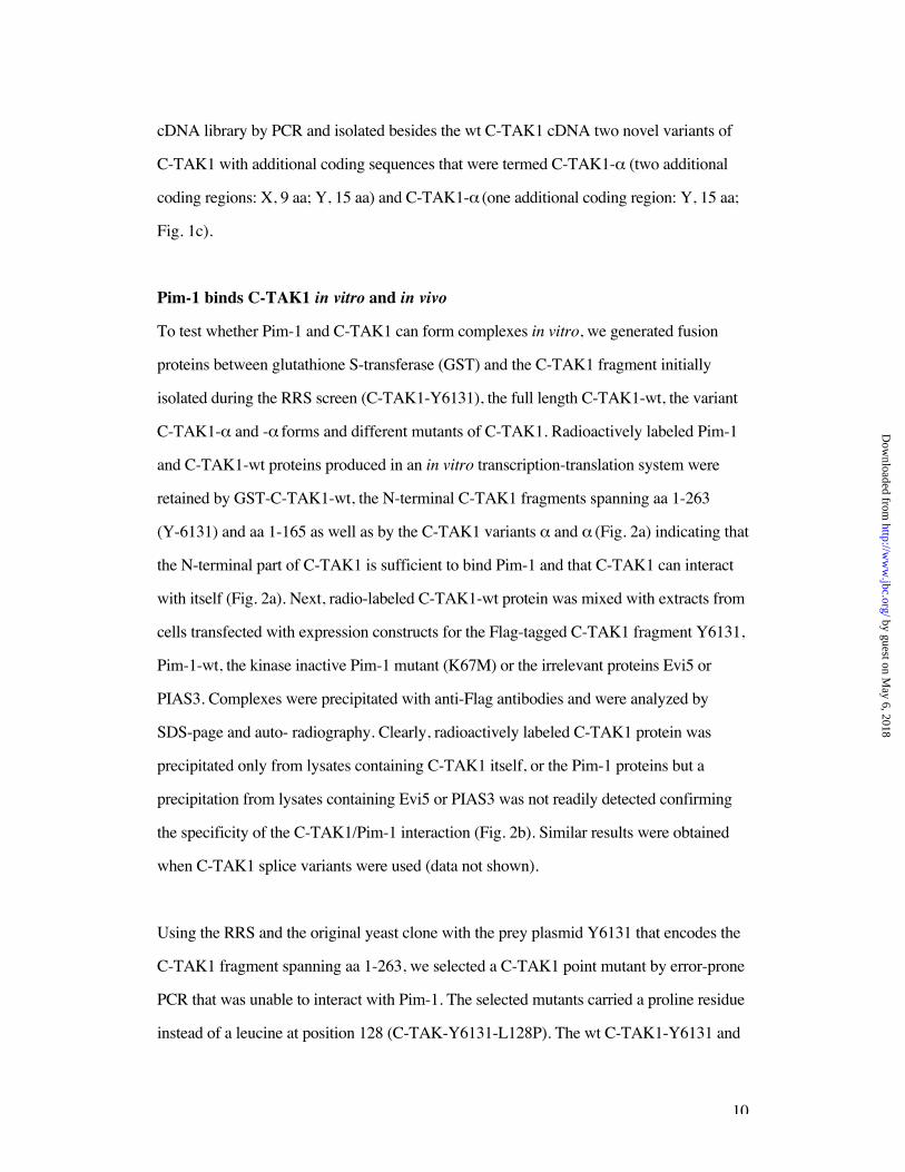
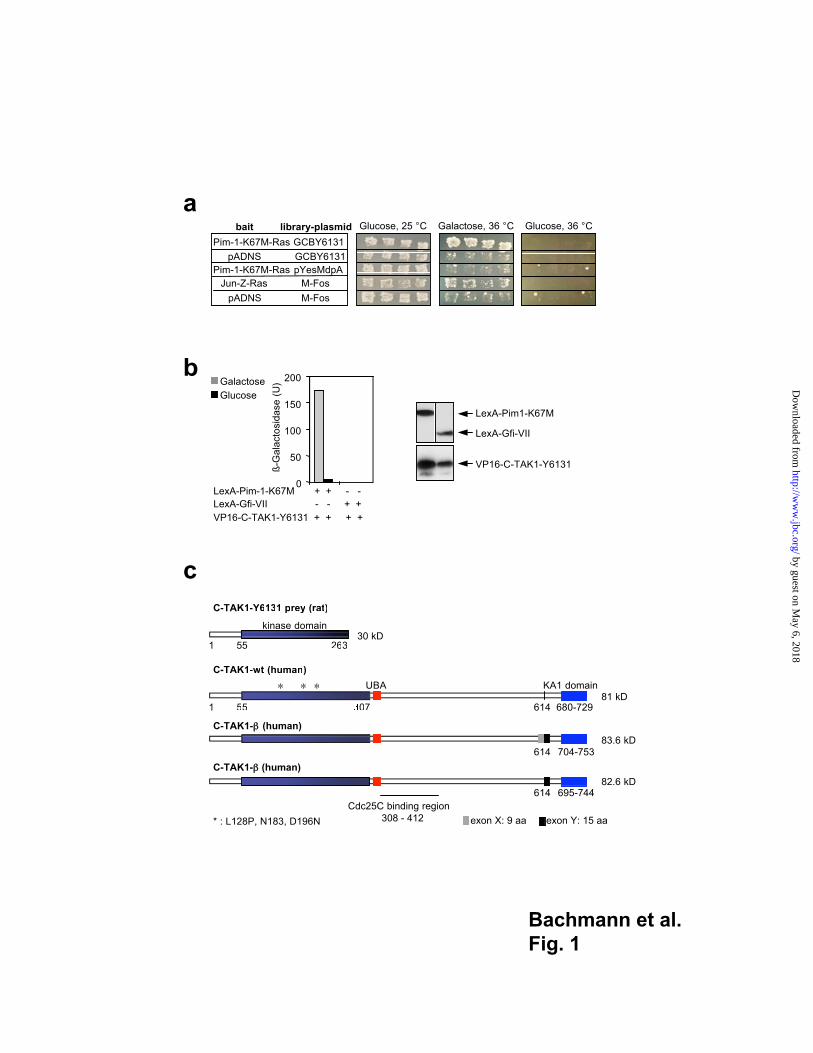
Pim-1 interacts with C-TAK1 in yeast
To identify proteins that interact with the Pim-1 kinase, we used a yeast interaction cloning
strategy based on the "Ras Recruitment System" (RRS, 40) We constructed a "bait"
plasmid able to express the kinase-inactive 33 kD form of Pim-1 (Pim-1-K67M) in fusion
with Ha-Ras in the vector pADNS-Ras(mut) (40) and introduced this along with a mGAP
expression plasmid (45) into the yeast mutant strain cdc25-2 which contains a temperature
sensitive allele of the GTPase exchange factor (GEF) CDC25. This GEF activates the
endogenous yeast Ras pathway under permissive conditions (25 °C), but is inactivated by
a shift to the restrictive temperature of 36 °C. After introduction of DNA from the
GC library (38) 417 initial clones were obtained, 13 of which showed library plasmid
dependency on galactose/glucose medium. Four of 11 clones that passed a bait specificity
test (Fig. 1a) contained DNA sequences coding for C-TAK1 (37). One of the clones
(Y6131, see Fig. 1) which contained the longest C-TAK1 sequence covering amino acid
positions 1-263 was used in the subsequent experiments.
To verify a potential interaction between Pim-1 and C-TAK1, we fused the obtained
C-TAK1 sequence to sequences encoding the herpes simplex virus VP16 transactivation
domain in a galactose-inducible vector. This construct was cotransfected with plasmids
encoding fusion proteins between the DNA binding domain of LexA and the kinase
inactive mutant of Pim-1. As a control, we used a construct encoding a fusion protein
between LexA and a stretch of the zinc finger transcription factor Gfi1 (Fig. 1b, 41).
Western blot analysis of extracts from transformed yeast cells demonstrated that the
expression constructs were functional (Fig. 1b). In the presence of galactose, a high
-galactosidase activity was obtained only with the constructs expressing LexA-Pim-1 and
C-TAK1-VP16 fusion proteins (Fig. 1b) supporting an interaction between Pim-1 and
C-TAK1. To obtain the human full length C-TAK1 clone, we screened a human spleen
by guest on May 6, 2018
http://ww
w.jbc.org/
Dow
nloaded from
10
cDNA library by PCR and isolated besides the wt C-TAK1 cDNA two novel variants of
C-TAK1 with additional coding sequences that were termed C-TAK1- (two additional
coding regions: X, 9 aa; Y, 15 aa) and C-TAK1- (one additional coding region: Y, 15 aa;
Fig. 1c).
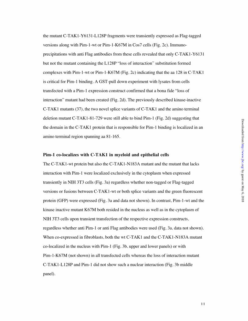
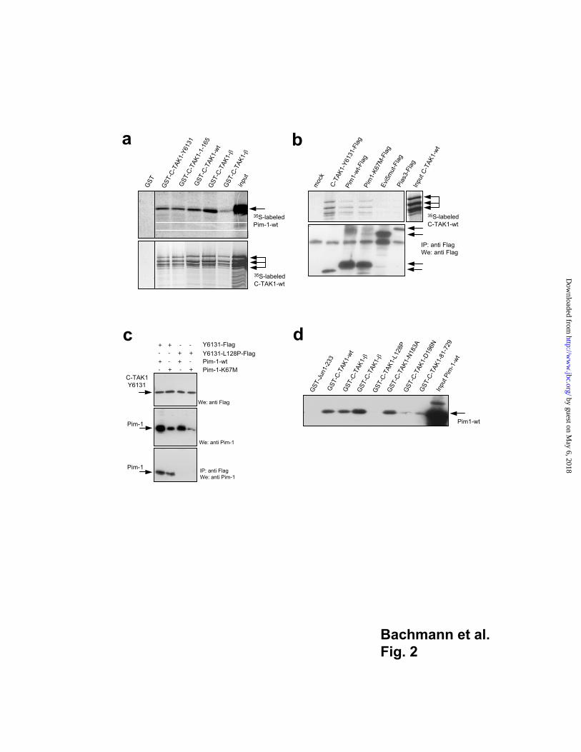
Pim-1 binds C-TAK1 in vitro and in vivo
To test whether Pim-1 and C-TAK1 can form complexes in vitro, we generated fusion
proteins between glutathione S-transferase (GST) and the C-TAK1 fragment initially
isolated during the RRS screen (C-TAK1-Y6131), the full length C-TAK1-wt, the variant
C-TAK1- and - forms and different mutants of C-TAK1. Radioactively labeled Pim-1
and C-TAK1-wt proteins produced in an in vitro transcription-translation system were
retained by GST-C-TAK1-wt, the N-terminal C-TAK1 fragments spanning aa 1-263
(Y-6131) and aa 1-165 as well as by the C-TAK1 variants and (Fig. 2a) indicating that
the N-terminal part of C-TAK1 is sufficient to bind Pim-1 and that C-TAK1 can interact
with itself (Fig. 2a). Next, radio-labeled C-TAK1-wt protein was mixed with extracts from
cells transfected with expression constructs for the Flag-tagged C-TAK1 fragment Y6131,
Pim-1-wt, the kinase inactive Pim-1 mutant (K67M) or the irrelevant proteins Evi5 or
PIAS3. Complexes were precipitated with anti-Flag antibodies and were analyzed by
SDS-page and auto- radiography. Clearly, radioactively labeled C-TAK1 protein was
precipitated only from lysates containing C-TAK1 itself, or the Pim-1 proteins but a
precipitation from lysates containing Evi5 or PIAS3 was not readily detected confirming
the specificity of the C-TAK1/Pim-1 interaction (Fig. 2b). Similar results were obtained
when C-TAK1 splice variants were used (data not shown).
Using the RRS and the original yeast clone with the prey plasmid Y6131 that encodes the
C-TAK1 fragment spanning aa 1-263, we selected a C-TAK1 point mutant by error-prone
PCR that was unable to interact with Pim-1. The selected mutants carried a proline residue
instead of a leucine at position 128 (C-TAK-Y6131-L128P). The wt C-TAK1-Y6131 and
by guest on May 6, 2018
http://ww
w.jbc.org/
Dow
nloaded from
11
the mutant C-TAK1-Y6131-L128P fragments were transiently expressed as Flag-tagged
versions along with Pim-1-wt or Pim-1-K67M in Cos7 cells (Fig. 2c). Immuno-
precipitations with anti Flag antibodies from these cells revealed that only C-TAK1-Y6131
but not the mutant containing the L128P “loss of interaction” substitution formed
complexes with Pim-1-wt or Pim-1-K67M (Fig. 2c) indicating that the aa 128 in C-TAK1
is critical for Pim-1 binding. A GST-pull down experiment with lysates from cells
transfected with a Pim-1 expression construct confirmed that a bona fide “loss of
interaction” mutant had been created (Fig. 2d). The previously described kinase-inactive
C-TAK1 mutants (37), the two novel splice variants of C-TAK1 and the amino terminal
deletion mutant C-TAK1-81-729 were still able to bind Pim-1 (Fig. 2d) suggesting that
the domain in the C-TAK1 protein that is responsible for Pim-1 binding is localized in an
amino terminal region spanning aa 81-165.
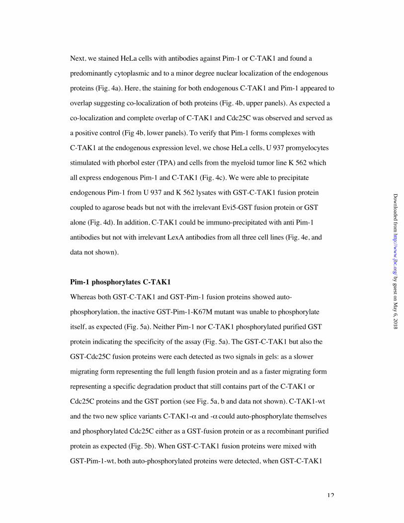
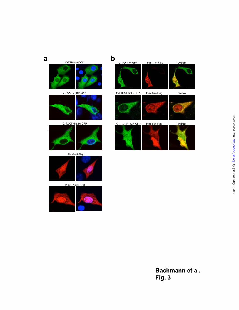
Pim-1 co-localizes with C-TAK1 in myeloid and epithelial cells
The C-TAK1-wt protein but also the C-TAK1-N183A mutant and the mutant that lacks
interaction with Pim-1 were localized exclusively in the cytoplasm when expressed
transiently in NIH 3T3 cells (Fig. 3a) regardless whether non-tagged or Flag-tagged
versions or fusions between C-TAK1-wt or both splice variants and the green fluorescent
protein (GFP) were expressed (Fig. 3a and data not shown). In contrast, Pim-1-wt and the
kinase inactive mutant K67M both resided in the nucleus as well as in the cytoplasm of
NIH 3T3 cells upon transient transfection of the respective expression constructs,
regardless whether anti Pim-1 or anti Flag antibodies were used (Fig. 3a, data not shown).
When co-expressed in fibroblasts, both the wt C-TAK1 and the C-TAK1-N183A mutant
co-localized in the nucleus with Pim-1 (Fig. 3b, upper and lower panels) or with
Pim-1-K67M (not shown) in all transfected cells whereas the loss of interaction mutant
C-TAK1-L128P and Pim-1 did not show such a nuclear interaction (Fig. 3b middle
panel).
by guest on May 6, 2018
http://ww
w.jbc.org/
Dow
nloaded from
12
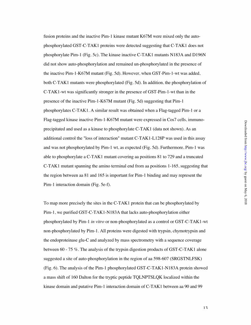
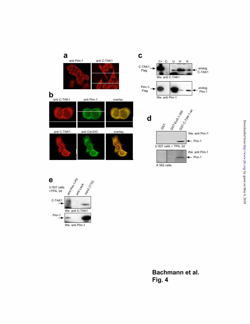
Next, we stained HeLa cells with antibodies against Pim-1 or C-TAK1 and found a
predominantly cytoplasmic and to a minor degree nuclear localization of the endogenous
proteins (Fig. 4a). Here, the staining for both endogenous C-TAK1 and Pim-1 appeared to
overlap suggesting co-localization of both proteins (Fig. 4b, upper panels). As expected a
co-localization and complete overlap of C-TAK1 and Cdc25C was observed and served as
a positive control (Fig 4b, lower panels). To verify that Pim-1 forms complexes with
C-TAK1 at the endogenous expression level, we chose HeLa cells, U 937 promyelocytes
stimulated with phorbol ester (TPA) and cells from the myeloid tumor line K 562 which
all express endogenous Pim-1 and C-TAK1 (Fig. 4c). We were able to precipitate
endogenous Pim-1 from U 937 and K 562 lysates with GST-C-TAK1 fusion protein
coupled to agarose beads but not with the irrelevant Evi5-GST fusion protein or GST
alone (Fig. 4d). In addition, C-TAK1 could be immuno-precipitated with anti Pim-1
antibodies but not with irrelevant LexA antibodies from all three cell lines (Fig. 4e, and
data not shown).
Pim-1 phosphorylates C-TAK1
Whereas both GST-C-TAK1 and GST-Pim-1 fusion proteins showed auto-
phosphorylation, the inactive GST-Pim-1-K67M mutant was unable to phosphorylate
itself, as expected (Fig. 5a). Neither Pim-1 nor C-TAK1 phosphorylated purified GST
protein indicating the specificity of the assay (Fig. 5a). The GST-C-TAK1 but also the
GST-Cdc25C fusion proteins were each detected as two signals in gels: as a slower
migrating form representing the full length fusion protein and as a faster migrating form
representing a specific degradation product that still contains part of the C-TAK1 or
Cdc25C proteins and the GST portion (see Fig. 5a, b and data not shown). C-TAK1-wt
and the two new splice variants C-TAK1- and - could auto-phosphorylate themselves
and phosphorylated Cdc25C either as a GST-fusion protein or as a recombinant purified
protein as expected (Fig. 5b). When GST-C-TAK1 fusion proteins were mixed with
GST-Pim-1-wt, both auto-phosphorylated proteins were detected, when GST-C-TAK1
by guest on May 6, 2018
http://ww
w.jbc.org/
Dow
nloaded from
13
fusion proteins and the inactive Pim-1 kinase mutant K67M were mixed only the auto-
phosphorylated GST-C-TAK1 proteins were detected suggesting that C-TAK1 does not
phosphorylate Pim-1 (Fig. 5c). The kinase inactive C-TAK1 mutants N183A and D196N
did not show auto-phosphorylation and remained un-phosphorylated in the presence of
the inactive Pim-1-K67M mutant (Fig. 5d). However, when GST-Pim-1-wt was added,
both C-TAK1 mutants were phosphorylated (Fig. 5d). In addition, the phosphorylation of
C-TAK1-wt was significantly stronger in the presence of GST-Pim-1-wt than in the
presence of the inactive Pim-1-K67M mutant (Fig. 5d) suggesting that Pim-1
phosphorylates C-TAK1. A similar result was obtained when a Flag-tagged Pim-1 or a
Flag-tagged kinase inactive Pim-1-K67M mutant were expressed in Cos7 cells, immuno-
precipitated and used as a kinase to phosphorylate C-TAK1 (data not shown). As an
additional control the "loss of interaction" mutant C-TAK1-L128P was used in this assay
and was not phosphorylated by Pim-1 wt, as expected (Fig. 5d). Furthermore, Pim-1 was
able to phosphorylate a C-TAK1 mutant covering aa positions 81 to 729 and a truncated
C-TAK1 mutant spanning the amino terminal end from aa positions 1-165, suggesting that
the region between aa 81 and 165 is important for Pim-1 binding and may represent the
Pim-1 interaction domain (Fig. 5e-f).
To map more precisely the sites in the C-TAK1 protein that can be phosphorylated by
Pim-1, we purified GST-C-TAK1-N183A that lacks auto-phosphorylation either
phosphorylated by Pim-1 in vitro or non-phosphorylated as a control or GST-C-TAK1-wt
non-phosphorylated by Pim-1. All proteins were digested with trypsin, chymotrypsin and
the endoproteinase glu-C and analyzed by mass spectrometry with a sequence coverage
between 60 - 75 %. The analysis of the trypsin digestion products of GST-C-TAK1 alone
suggested a site of auto-phosphorylation in the region of aa 598-607 (SRGSTNLFSK)
(Fig. 6). The analysis of the Pim-1 phosphorylated GST-C-TAK1-N183A protein showed
a mass shift of 160 Dalton for the tryptic peptide TQLNPTSLQK localized within the
kinase domain and putative Pim-1 interaction domain of C-TAK1 between aa 90 and 99
by guest on May 6, 2018
http://ww
w.jbc.org/
Dow
nloaded from
14
and suggested the existence of at least two phosphorylation sites at threonine 90 or 95 or
at serine 96 (Fig. 6a). Several C-TAK mutants were generated where the serine and both
threonine residues were replaced by alanin or glycine in the inactive C-TAK1-N183A
form. All mutants were found to interact with Pim-1(data not shown) and to be
phosphorylated by Pim-1 (Fig. 6b), suggesting that C-TAK1 contains additional Pim-1
phosphorylation sites that were undetectable by mass spectrometry.
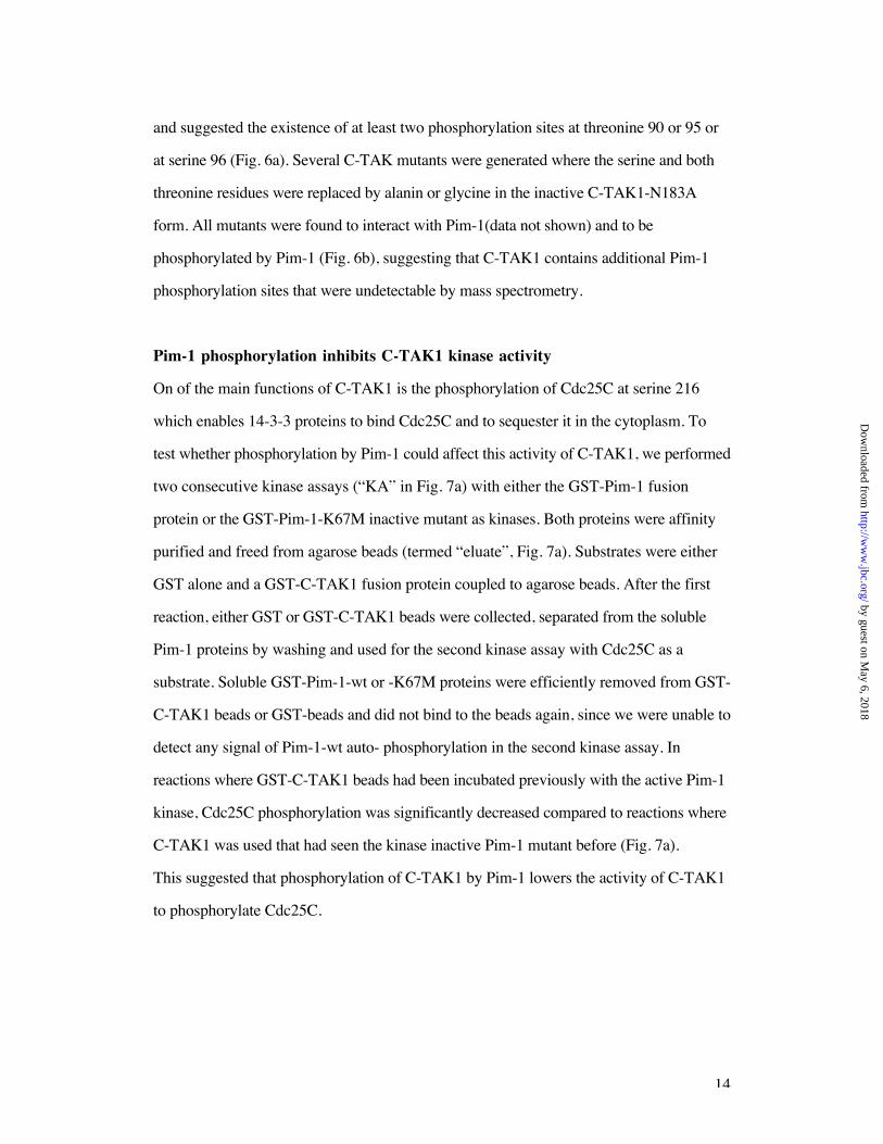
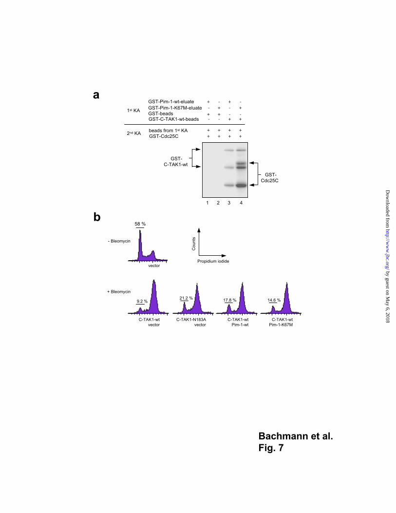
Pim-1 phosphorylation inhibits C-TAK1 kinase activity
On of the main functions of C-TAK1 is the phosphorylation of Cdc25C at serine 216
which enables 14-3-3 proteins to bind Cdc25C and to sequester it in the cytoplasm. To
test whether phosphorylation by Pim-1 could affect this activity of C-TAK1, we performed
two consecutive kinase assays (“KA” in Fig. 7a) with either the GST-Pim-1 fusion
protein or the GST-Pim-1-K67M inactive mutant as kinases. Both proteins were affinity
purified and freed from agarose beads (termed “eluate”, Fig. 7a). Substrates were either
GST alone and a GST-C-TAK1 fusion protein coupled to agarose beads. After the first
reaction, either GST or GST-C-TAK1 beads were collected, separated from the soluble
Pim-1 proteins by washing and used for the second kinase assay with Cdc25C as a
substrate. Soluble GST-Pim-1-wt or -K67M proteins were efficiently removed from GST-
C-TAK1 beads or GST-beads and did not bind to the beads again, since we were unable to
detect any signal of Pim-1-wt auto- phosphorylation in the second kinase assay. In
reactions where GST-C-TAK1 beads had been incubated previously with the active Pim-1
kinase, Cdc25C phosphorylation was significantly decreased compared to reactions where
C-TAK1 was used that had seen the kinase inactive Pim-1 mutant before (Fig. 7a).
This suggested that phosphorylation of C-TAK1 by Pim-1 lowers the activity of C-TAK1
to phosphorylate Cdc25C.
by guest on May 6, 2018
http://ww
w.jbc.org/
Dow
nloaded from
15
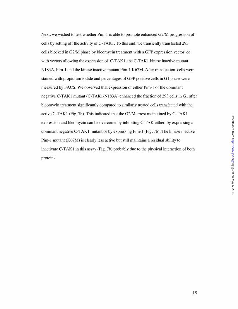
Next, we wished to test whether Pim-1 is able to promote enhanced G2/M progression of
cells by setting off the activity of C-TAK1. To this end, we transiently transfected 293
cells blocked in G2/M phase by bleomycin treatment with a GFP expression vector or
with vectors allowing the expression of C-TAK1, the C-TAK1 kinase inactive mutant
N183A, Pim-1 and the kinase inactive mutant Pim-1 K67M. After transfection, cells were
stained with propidium iodide and percentages of GFP positive cells in G1 phase were
measured by FACS. We observed that expression of either Pim-1 or the dominant
negative C-TAK1 mutant (C-TAK1-N183A) enhanced the fraction of 293 cells in G1 after
bleomycin treatment significantly compared to similarly treated cells transfected with the
active C-TAK1 (Fig. 7b). This indicated that the G2/M arrest maintained by C-TAK1
expression and bleomycin can be overcome by inhibiting C-TAK either by expressing a
dominant negative C-TAK1 mutant or by expressing Pim-1 (Fig. 7b). The kinase inactive
Pim-1 mutant (K67M) is clearly less active but still maintains a residual ability to
inactivate C-TAK1 in this assay (Fig. 7b) probably due to the physical interaction of both
proteins.
by guest on May 6, 2018
http://ww
w.jbc.org/
Dow
nloaded from
16
Discussion
Pim-1 plays a prominent role in transmitting signals from cytokine receptors and in the
malignant transformation of lymphoid cells and it has been demonstrated that Pim-1 is
associated with enhanced cell proliferation (12). We have identified C-TAK1 - a
serine/threonine kinase and a cell cycle regulator active at the G2/M transition as a novel
Pim-1 substrate. In vitro synthesized proteins as well as endogenous C-TAK1 and Pim-1
were shown to bind to each other by and to co-localize in different cell types supporting a
physical interaction between Pim-1 and C-TAK1 proteins. As a consequence of this
interaction C-TAK1 is phosphorylated by Pim-1 but, vice versa, Pim-1 is not a substrate
for the C-TAK1 kinase and the phosphorylation of C-TAK1 by Pim-1 leads to a
significant decrease of C-TAK1 kinase activity.
Pim-1 is an upstream negative regulator of C-TAK1
C-TAK1 is constitutively expressed in all cell cycle phases (37) in contrast to the DNA
damage activated kinases Chk1 or Chk2/Cds1 which - similar to C-TAK1 - inactivate
Cdc25C by phosphorylation at serine 216 which leads to 14-3-3 dependent sequestering
of the protein to the cytoplasm (46-48). Recently, the phosphatase PP1 was identified as
an antagonist of C-TAK1 since it is able to de-phosphorylate Cdc25C on serine 216
resulting in a de-repression of its activity (49). Our experimental findings described here
suggest that Pim-1 is a regulator of C-TAK1 activity since it phosphorylates C-TAK1
which causes a significantly reduced activity of C-TAK1 with regard to its ability to
phosphorylate Cdc25C at serine 216. Our studies with C-TAK1 mutants and mass
spectrometry data suggested that phosphorylation by Pim-1 occurs at several sites within
the C-TAK1 protein. It is therefore difficult to assign the loss of C-TAK1 activity after
Pim-1 phosphorylation to a specific single amino acid. Nevertheless, since the activity of
C-TAK1 appears to depend directly on Pim-1 phosphorylation, Pim-1 can be considered
as an upstream negative regulator of C-TAK1.
by guest on May 6, 2018
http://ww
w.jbc.org/
Dow
nloaded from
17
The physical interaction between Pim-1 and C-TAK1 is independent of phosphorylation
of C-TAK1 by Pim-1 since the kinase inactive mutant of Pim-1 (K67M) is still able to
bind C-TAK1 and to recruit it into the nucleus like the wt Pim-1 protein. Recent
observations in normal lymphoid cells suggest that Pim-1 can reside in the nucleus as well
as in the cytoplasm and that a nuclear localization is required for the protein to exert its
biological function (50). Other reports also indicate that endogenous Pim-1 can reside in
both nuclear and cytoplasmic compartments (27, 33, 51) and the interaction of Pim-1 with
the transcription factors or co-factors NF-AT, p100 or c-Myb that have been reported have
to occur in the nucleus. It is therefore likely that Pim-1 resides in both cellular
compartments. We had indeed found a predominantly cytoplasmic localization of
endogenous Pim-1 in HeLa cells or in cells of the lines K 562 or U 937, but upon
transient transfection, we observed that Pim-1 is both cytoplasmic and nuclear.
Considering that Pim-1 has both nuclear and cytoplasmic interaction partners it is
conceivable that Pim-1 is active and finds substrates in both cellular compartments or can
shuttle between cytoplasm and nucleus depending on the type or status of the cells or on
the actual expression level. Evidence for such a behavior comes from experiments with
U 937 cells where Pim-1 becomes nuclear after treatment and activation of the cells with
phorbol ester (27). Still, the significance of a recruitment of C-TAK1 to the nucleus by
Pim-1 as observed in transfected cells remains to be clarified.
Regulation of the G2/M transition of the cell cycle
Phosphorylation at serine 216 by DNA damage kinases Chk1/Cds1 or C-TAK1
inactivates Cdc25C and removes it as an active player from the stage of the G2/M
transition since it is no longer available for the polo like kinase 1 (Plk1) that
phosphorylates Cdc25C at the N-terminus to render it fully active as a phosphatase (52,
53). Inactivation of Cdc25C by serine 216 phosphorylation then leads to an arrest at the
G2/M transition. The aa serine 216 is located in the neighborhood of the NES-sequence
(nuclear export sequences, aa 190-199) and the NLS (nuclear localization signal; aa
by guest on May 6, 2018
http://ww
w.jbc.org/
Dow
nloaded from

18
240-244; 54) of Cdc25C. After phosphorylation of Cdc25C at serine 216 by DNA
damage kinases or C-TAK1 and the subsequent binding of 14-3-3 proteins, the
NLS-sequence is no longer accessible and Cdc25C remains in the cytoplasm. In its first
activation step, Cdc25C is de-phosphorylated at serine 216 by PP1 (49). Moreover, it is
known that Cdc25C is phosphorylated during prophase by Plks at the N-terminus and is
then transported to the nucleus or is retained there (55, 56). It is conceivable that Pim-1
acts to ensure full activation of Cdc25C and prevents its re-phosphorylation by C-TAK1
by phosphorylating and inactivating C-TAK1. The inactivation of C-TAK1 would confer
the role to Pim-1 as a C-TAK1 antagonist and indirect activator of Cdc25C to Pim-1; a
role similar to Plks. Such a role of Pim-1 is further supported by our findings with
transfected cells that are blocked in G2/M phase. Here, Pim-1 was able to set off the effect
of C-TAK1 and enhanced the percentages of cells in G1 phase. Since the kinase inactive
mutant of Pim-1 (K67M) was noticeably less active in this assay, this result is consistent
with a role of Pim-1 in promoting cell cycle progression by inactivating C-TAK1 via
direct phosphorylation. However, the kinase inactive Pim-1 mutant clearly had a residual
activity and could still increase the amount of the G1-phase cells albeit at a lower degree
than active Pim-1. This indicates that the inhibitory effect of Pim-1 on C-TAK1 activity is
not only attributable to the phosphorylation of C-TAK1 by Pim-1 but may also be caused
by the mere physical interaction of both proteins since the kinase inactive Pim-1 K67M
mutant is still able to bind C-TAK1.
Pim-1 as a positive regulator of cell cycle progression at the G2/M transition
After N-terminal hyper-phosphorylation by Plk-1, the active Cdc25C phosphatase is able
to activate the MPF-complex through de-phosphorylation of one of its components, Cdk1.
Subsequently, the active MPF complex translocates into the nucleus after phosphorylation
of its other component, cyclin B1, and initiates the transcription of M-Phase specific
genes leading to the start of prophase. Our data presented here suggest that the Pim-1
kinase may play a role in this process since it can modulate the activity of C-TAK1 in this
by guest on May 6, 2018
http://ww
w.jbc.org/
Dow
nloaded from
19
signaling pathway through binding and phosphorylation. Pim-1 decreases the kinase
activity of C-TAK1 which could relieve newly synthesized Cdc25C from the restriction
represented by a phosphorylated serine 216 or inactivated Cdc25C is de-phosphorylated
by PP1. Thus, Cdc25C would be available for Plks to be phosphorylated at the N-terminal
region which enables it to stimulate the activity of MPF. In this model, Pim-1 would act at
a critical point of the G2/M transition phase by blocking a reaction that is inhibitory for
the activity of Cdc25C, namely its phosphorylation on serine 216 by C-TAK1 (Fig. 8).
According to our findings it is not unlikely that this may occur in the cytoplasm as well as
in the nucleus.
A first indication that Pim-1 may indeed have a regulatory role at the G2/M transition was
obtained when NuMA (Nuclear mitotic apparatus protein) was identified as a Pim-1
substrate (51). NuMA is located in the spindle poles during mitosis and it was shown that
Pim-1 has a role in promoting the formation of a complex between NuMA, HP-1 , a
heterochromatin binding protein, dynein and dynactin which is necessary for progression
through mitosis. These findings also support a role of Pim-1 as a positive cell cycle
regulatory kinase active at the G2/M phase transition. Such a role of Pim-1 could explain
how signals from cytokines that initiate the activation of STAT3/STAT5 and the up
regulation of their target genes, among them the gene for Pim-1, result in the support of
cell proliferation but also offers an explanation for the oncogenic activity of Pim-1 and its
potential to malignantly transform lymphoid cells.
by guest on May 6, 2018
http://ww
w.jbc.org/
Dow
nloaded from
20
Acknowledgements
We are indebted to M. Karin for the GST-Jun1-233 plasmid, L.W. Enquist for the
pBB14 plasmid and Denise Pargmann for the Evi5 plasmids. We thank Angelika Warda
for technical assistance. This work was supported by a grant from the Deutsche
Forschungsgemeinschaft, DFG (Mo 435-16/1, 16/2) and the Fonds der Chemischen
Industrie.
by guest on May 6, 2018
http://ww
w.jbc.org/
Dow
nloaded from
21
References
1. Padma, R., and Nagarajan, L. (1991) Cancer Res. 51, 2486-2489. 2. Saris, C. J., Domen, J., and Berns, A. (1991) EMBO J. 10, 655-664. 3. Cuypers, H. T., Selten, G., Quint, W., Zijlstra, M., Maandag, E. R., Boelens, W., van
Wezenbeek, P., Melief, C., and Berns, A. (1984) Cell 37, 141-150. 4. Mucenski, M. L., Gilbert, D. J., Taylor, B. A., Jenkins, N. A., Copeland. N. G.
(1987) Oncogene Res. 2, 33-48.
5. Selten, G., Cuypers, H. T., and Berns, A. (1985) EMBO J. 4, 1793-1798. 6. van Lohuizen, M., Verbeek, S., Krimpenfort, P., Domen, J., Saris, C., Radaszkiewicz,
T., and Berns, A. (1989) Cell 56, 673-682. 7. Domen, J., van der Lugt, N. M., Laird, P. W., Saris, C. J., and Berns, A. (1993)
Leukemia 7 Suppl 2, 108-112. 8. Laird, P. W., van der Lugt, N. M., Clarke, A., Domen, J., Linders, K., McWhir, J.,
Berns, A., Hooper, M. (1993) Nucleic Acids Res 21(20), 4750-4755 9. Allen, J. D., Verhoeven, E., Domen, J., van der Valk, M., and Berns, A. (1997)
Oncogene 15, 1133-1141.10. Eichmann, A., Yuan, L., Breant, C., Alitalo, K., and Koskinen, P. J. (2000) Oncogene
19, 1215-1224.11. van der Lugt, N. M., Domen, J., Verhoeven, E., Linders, K., van der Gulden, H., Allen,
J., and Berns, A. (1995) EMBO J. 14, 2536-2544.12. Shirogane, T., Fukada, T., Muller, J. M., Shima, D. T., Hibi, M., and Hirano, T.
(1999) Immunity 11, 709-719.13. Borg, K. E., Zhang, M., Hegge, D., Stephen, R. L., Buckley, D. J., Magnuson, N. S.,
and Buckley, A. R. (1999) Endocrinology 140, 5659-5668.14. Buckley, A. R., Buckley, D. J., Leff, M. A., Hoover, D. S., and Magnuson, N. S.
(1995) Endocrinology 136, 5252-5259.15. Jaster, R., Tschirch, E., Bittorf, T., and Brock, J. (1999) Cell Signal. 11, 331-335.16. Jaster, R., Tschirch, E., Bittorf, T., and Brock, J. (1999) Cell Signal. 11, 769-775.17. Krumenacker, J. S., Buckley, D. J., Leff, M. A., McCormack, J. T., de Jong, G., Gout,
P. W., Reed, J. C., Miyashita, T., Magnuson, N. S., Buckley, A. R. (1998)Endocrine 9(2), 163-170.
18. Matikainen, S., Sareneva, T., Ronni, T., Lehtonen, A., Koskinen, P. J., and Julkunen, I.(1999) Blood 93, 1980-1991.
19. Narimatsu, M., Maeda, H., Itoh, S., Atsumi, T., Ohtani, T., Nishida, K., Itoh, M.,Kamimura, D., Park, S. J., Mizuno, K., Miyazaki, J., Hibi, M., Ishihara, K.,Nakajima, K., and Hirano, T. (2001) Mol. Cell. Biol. 21, 6615-6625.
20. Chen, X. P., Losman, J. A., Cowan, S., Donahue, E., Fay, S., Vuong, B. Q., Nawijn,M. C., Capece, D., Cohan, V. L., and Rothman, P. (2002) Proc. Natl. Acad. Sci.U. S. A. 99, 2175-2180.
21. Friedmann, M., Nissen, M. S., Hoover, D. S., Reeves, R., and Magnuson, N. S.(1992) Arch. Biochem. Biophys. 298, 594-601.
22. Palaty, C. K., Clark-Lewis, I., Leung, D., and Pelech, S. L. (1997) Biochem. Cell.Biol. 75, 153-162
23. Palaty, C. K., Kalmar, G., Tai, G., Oh, S., Amankawa, L., Affolter, M., Aebersold, R.,and Pelech, S. L. (1997) J. Biol. Chem. 272, 10514-10521.
24. Leverson, J. D., Koskinen, P. J., Orrico, F. C., Rainio, E. M., Jalkanen, K. J., Dash, A.B., Eisenman, R. N., and Ness, S. A. (1998) Mol. Cell. 2, 417-425.
25. Rainio, E. M., Sandholm, J., and Koskinen, P. J. (2002) J. Immunol. 168, 1524-1527.
26. Mochizuki, T., Kitanaka, C., Noguchi, K., Muramatsu, T., Asai, A., and Kuchino, Y.(1999) J. Biol. Chem. 274, 18659-18666.
27. Wang, Z., Bhattacharya, N., Mixter, P. F., Wei, W., Sedivy, J., Magnuson, N. S.(2002) Biochim. Biophys. Acta. 1593, 45-55.
28. Lilly, M., and Kraft, A. (1997) Cancer Res. 57, 5348-5355.
by guest on May 6, 2018
http://ww
w.jbc.org/
Dow
nloaded from
22
29. Lilly, M., Sandholm, J., Cooper, J. J., Koskinen, P. J., and Kraft, A. (1999)Oncogene 18, 4022-4031.
30. Möröy, T., Grzeschiczek, A., Petzold, S., Hartmann, K. U. (1993) Proc. Natl. Acad.Sci. U. S. A. 90, 10734-10738.
31. Nosaka, T., Kawashima, T., Misawa, K., Ikuta, K., Mui, A. L., and Kitamura, T.(1999) EMBO J. 18, 4754-4765.
32. Pircher, T. J., Zhao, S., Geiger, J. N., Joneja, B., and Wojchowski, D. M. (2000)Oncogene 19, 3684-3692.
33. Wang, Z., Bhattacharya, N., Meyer, M. K., Seimiya, H., Tsuruo, T., Tonani, J. A., andMagnuson, N. S. (2001) Arch. Biochem. Biophys. 390, 9-18.
34. Koike, N., Maita, H., Taira, T., Ariga, H., and Iguchi-Ariga, S. M. (2000) FEBS Lett.467, 17-21.
35. Maita, H., Harada, Y., Nagakubo, D., Kitaura, H., Ikeda, M., Tamai, K., Takahashi, K.,Ariga, H., and Iguchi-Ariga, S. M. (2000) Eur. J. Biochem. 267, 5168-5178.
36. Ishibashi, Y., Maita, H., Yano, M., Koike, N., Tamai, K., Ariga, H., and Iguchi-Ariga,S. M. (2001) FEBS Lett 506, 33-38.
37. Peng, C. Y., Graves, P. R., Ogg, S., Thoma, R. S., Byrnes, M. J., 3rd, Wu, Z.,Stephenson, M. T., and Piwnica-Worms, H. (1998) Cell Growth Differ. 9, 197-208.
38. Aronheim, A., Zandi, E., Hennemann, H., Elledge, S. J., and Karin, M. (1997) Mol.Cell. Biol. 17, 3094-3102.
39. Hennemann, H., Vassen, L., Geisen, G, Eilers, M. and Möröy T. (2003) J. Biol.Chem. 278 (31), 28799-28811.
40. Broder, Y. C., Katz, S., and Aronheim, A. (1998) Curr. Biol. 8, 1121-1124.41. Rödel, B., Tavassoli, K., Karsunky, H., Schmidt, T., Bachmann, M., Schaper, F.,
Heinrich, P., Shuai, K., Elsasser, H. P., and Möröy, T. (2000) EMBO J. 19, 5845-5855.
42. Hollenberg, S. M., Sternglanz, R., Cheng, P. F., Weintraub, H. (1995) Mol. Cell. Biol. 15, 3813-3822.43. Brideau, A. D., Banfield, B. W., and Enquist, L. W. (1998) J. Virology 72, 4560-
4570.44. Kalejta, R. F., Brideau, A. D., Banfield, B. W., and Beavis, A.J. (1999) J. Exp. Cell
Res. 248, 322-328.45. Aronheim, A. (1997) Nucleic Acids Res 25, 3373-3374.46. Blasina, A., de Weyer, I. V., Laus, M. C., Luyten, W. H., Parker, A. E., and
McGowan, C. H. (1999) Curr. Biol. 9, 1-10.47. Furnari, B., Blasina, A., Boddy, M. N., McGowan, C. H., and Russell, P. (1999) Mol.
Biol. Cell 10, 833-845.48. Sanchez, Y., Wong, C., Thoma, R. S., Richman, R., Wu, Z., Piwnica-Worms, H., and
Elledge, S. J. (1997) Science 277, 1497-1501.49. Margolis, S. S., Walsh, S., Weiser, D. C., Yoshida, M., Shenolikar, S., Kornbluth, S.
(2003) EMBO J. 22, 5734-5745.50. Ionov Y, Le X, Tunquist BJ, Sweetenham J, Sachs T, Ryder J, Johnson T, Lilly MB,
Kraft AS. (2003) Anticancer Res. 23 (1A), 167-178.51. Bhattacharya, N., Wang, Z., Davitt, C., McKenzie, I. F., Xing, P. X., and Magnuson,
N. S. (2002) Chromosoma 111, 80-95.52. Qian, Y. W., Erikson, E., Taieb, F. E., and Maller, J. L. (2001) Mol. Biol. Cell. 12,
1791-1799.53. Roshak, A.K., Capper, E.A., Imburgia, C., Fornwald, J., Scott, G., Marshall, L.A.
(2000) Cell Signal. 12, 405-411.54. Graves, P. R., Lovly, C. M., Uy, G. L., and Piwnica-Worms, H. (2001) Oncogene 20,
1839-1851.55. Takizawa, C. G., and Morgan, D. O. (2000) Curr. Opin. Cell. Biol. 12, 658-665.56. Toyoshima-Morimoto, F., Taniguchi, E., and Nishida, E. (2002) EMBO Rep. 3, 341-
348.
by guest on May 6, 2018
http://ww
w.jbc.org/
Dow
nloaded from
23
Figure Legends
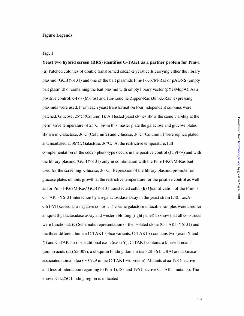
Fig. 1
Yeast two hybrid screen (RRS) identifies C-TAK1 as a partner protein for Pim-1
(a) Patched colonies of double transformed cdc25-2 yeast cells carrying either the library
plasmid (GCBY6131) and one of the bait plasmids Pim-1-K67M-Ras or pADNS (empty
bait plasmid) or containing the bait plasmid with empty library vector (pYesMdpA). As a
positive control, c-Fos (M-Fos) and Jun-Leucine Zipper-Ras (Jun-Z-Ras) expressing
plasmids were used. From each yeast transformation four independent colonies were
patched. Glucose, 25°C (Column 1): All tested yeast clones show the same viability at the
permissive temperature of 25°C. From this master plate the galactose and glucose plates
shown in Galactose, 36 C (Column 2) and Glucose, 36 C (Column 3) were replica plated
and incubated at 36°C. Galactose, 36°C: At the restrictive temperature, full
complementation of the cdc25 phenotype occurs in the positive control (Jun/Fos) and with
the library plasmid (GCBY6131) only in combination with the Pim-1-K67M-Ras bait
used for the screening. Glucose, 36°C: Repression of the library plasmid promoter on
glucose plates inhibits growth at the restrictive temperature for the positive control as well
as for Pim-1-K67M-Ras/ GCBY6131 transfected cells. (b) Quantification of the Pim-1/
C-TAK1-Y6131 interaction by a -galactosidase assay in the yeast strain L40. LexA-
Gfi1-VII served as a negative control. The same galactose inducible samples were used for
a liquid ß-galactosidase assay and western blotting (right panel) to show that all constructs
were functional. (c) Schematic representation of the isolated clone (C-TAK1-Y6131) and
the three different human C-TAK1 splice variants. C-TAK1- contains two (exon X and
Y) and C-TAK1- one additional exon (exon Y). C-TAK1 contains a kinase domain
(amino acids (aa) 55-307), a ubiquitin binding domain (aa 328-364, UBA) and a kinase
associated domain (aa 680-729 in the C-TAK1-wt protein). Mutants at aa 128 (inactive
and loss of interaction regarding to Pim-1),183 and 196 (inactive C-TAK1-mutants). The
known Cdc25C binding region is indicated.
by guest on May 6, 2018
http://ww
w.jbc.org/
Dow
nloaded from
24
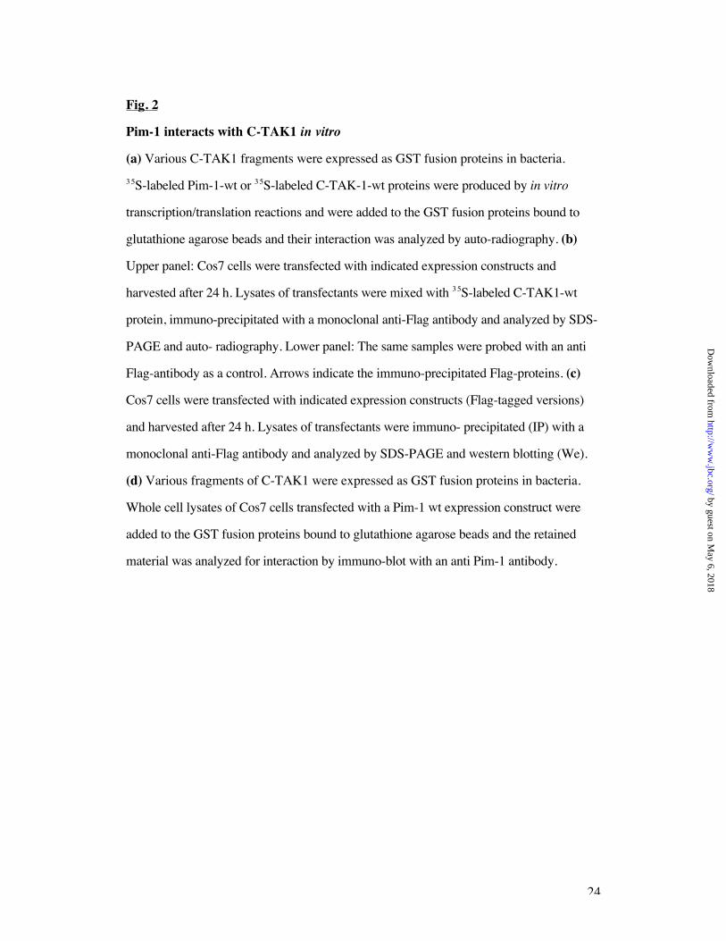
Fig. 2
Pim-1 interacts with C-TAK1 in vitro
(a) Various C-TAK1 fragments were expressed as GST fusion proteins in bacteria.
3 5S-labeled Pim-1-wt or 3 5S-labeled C-TAK-1-wt proteins were produced by in vitro
transcription/translation reactions and were added to the GST fusion proteins bound to
glutathione agarose beads and their interaction was analyzed by auto-radiography. (b)
Upper panel: Cos7 cells were transfected with indicated expression constructs and
harvested after 24 h. Lysates of transfectants were mixed with 3 5S-labeled C-TAK1-wt
protein, immuno-precipitated with a monoclonal anti-Flag antibody and analyzed by SDS-
PAGE and auto- radiography. Lower panel: The same samples were probed with an anti
Flag-antibody as a control. Arrows indicate the immuno-precipitated Flag-proteins. (c)
Cos7 cells were transfected with indicated expression constructs (Flag-tagged versions)
and harvested after 24 h. Lysates of transfectants were immuno- precipitated (IP) with a
monoclonal anti-Flag antibody and analyzed by SDS-PAGE and western blotting (We).
(d) Various fragments of C-TAK1 were expressed as GST fusion proteins in bacteria.
Whole cell lysates of Cos7 cells transfected with a Pim-1 wt expression construct were
added to the GST fusion proteins bound to glutathione agarose beads and the retained
material was analyzed for interaction by immuno-blot with an anti Pim-1 antibody.
by guest on May 6, 2018
http://ww
w.jbc.org/
Dow
nloaded from
25
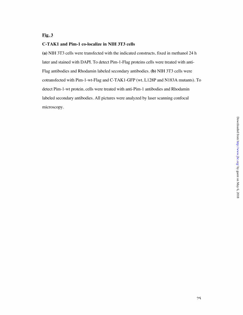
Fig. 3
C-TAK1 and Pim-1 co-localize in NIH 3T3 cells
(a) NIH 3T3 cells were transfected with the indicated constructs, fixed in methanol 24 h
later and stained with DAPI. To detect Pim-1-Flag proteins cells were treated with anti-
Flag antibodies and Rhodamin labeled secondary antibodies. (b) NIH 3T3 cells were
cotransfected with Pim-1-wt-Flag and C-TAK1-GFP (wt, L128P and N183A mutants). To
detect Pim-1-wt protein, cells were treated with anti-Pim-1 antibodies and Rhodamin
labeled secondary antibodies. All pictures were analyzed by laser scanning confocal
microscopy.
by guest on May 6, 2018
http://ww
w.jbc.org/
Dow
nloaded from
26
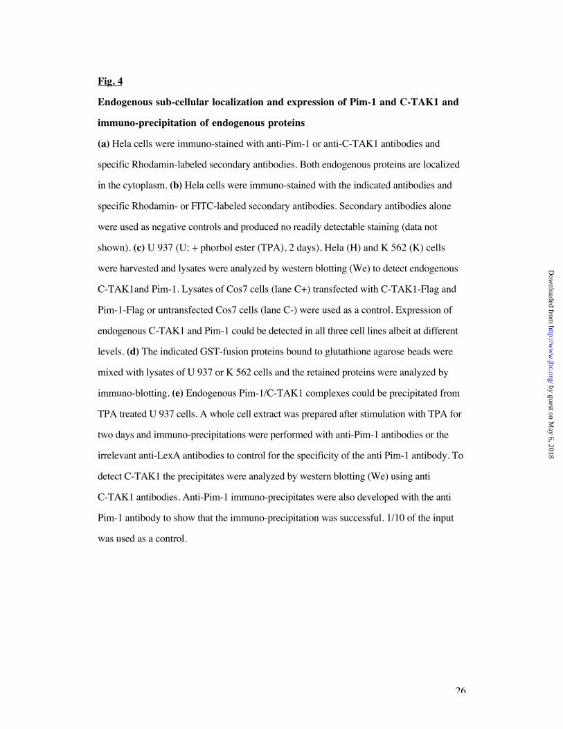
Fig. 4
Endogenous sub-cellular localization and expression of Pim-1 and C-TAK1 and
immuno-precipitation of endogenous proteins
(a) Hela cells were immuno-stained with anti-Pim-1 or anti-C-TAK1 antibodies and
specific Rhodamin-labeled secondary antibodies. Both endogenous proteins are localized
in the cytoplasm. (b) Hela cells were immuno-stained with the indicated antibodies and
specific Rhodamin- or FITC-labeled secondary antibodies. Secondary antibodies alone
were used as negative controls and produced no readily detectable staining (data not
shown). (c) U 937 (U; + phorbol ester (TPA), 2 days), Hela (H) and K 562 (K) cells
were harvested and lysates were analyzed by western blotting (We) to detect endogenous
C-TAK1and Pim-1. Lysates of Cos7 cells (lane C+) transfected with C-TAK1-Flag and
Pim-1-Flag or untransfected Cos7 cells (lane C-) were used as a control. Expression of
endogenous C-TAK1 and Pim-1 could be detected in all three cell lines albeit at different
levels. (d) The indicated GST-fusion proteins bound to glutathione agarose beads were
mixed with lysates of U 937 or K 562 cells and the retained proteins were analyzed by
immuno-blotting. (e) Endogenous Pim-1/C-TAK1 complexes could be precipitated from
TPA treated U 937 cells. A whole cell extract was prepared after stimulation with TPA for
two days and immuno-precipitations were performed with anti-Pim-1 antibodies or the
irrelevant anti-LexA antibodies to control for the specificity of the anti Pim-1 antibody. To
detect C-TAK1 the precipitates were analyzed by western blotting (We) using anti
C-TAK1 antibodies. Anti-Pim-1 immuno-precipitates were also developed with the anti
Pim-1 antibody to show that the immuno-precipitation was successful. 1/10 of the input
was used as a control.
by guest on May 6, 2018
http://ww
w.jbc.org/
Dow
nloaded from
27
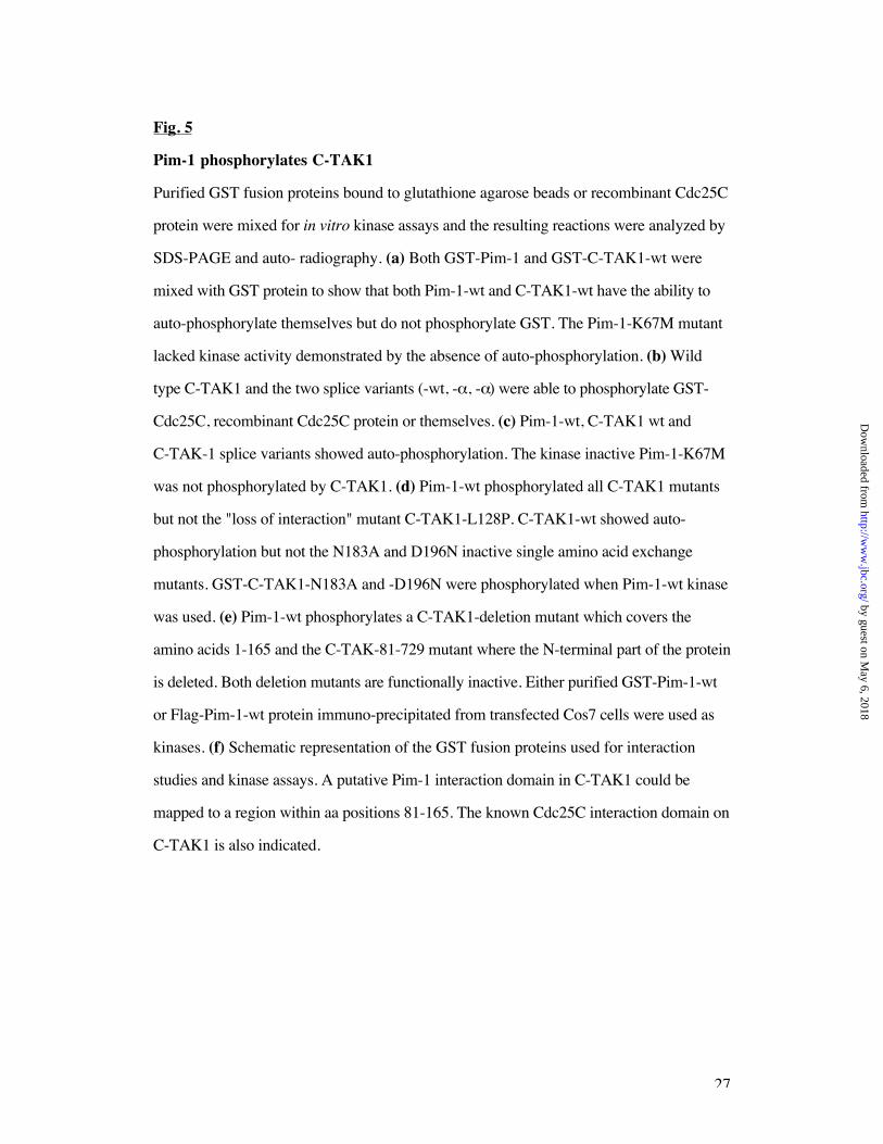
Fig. 5
Pim-1 phosphorylates C-TAK1
Purified GST fusion proteins bound to glutathione agarose beads or recombinant Cdc25C
protein were mixed for in vitro kinase assays and the resulting reactions were analyzed by
SDS-PAGE and auto- radiography. (a) Both GST-Pim-1 and GST-C-TAK1-wt were
mixed with GST protein to show that both Pim-1-wt and C-TAK1-wt have the ability to
auto-phosphorylate themselves but do not phosphorylate GST. The Pim-1-K67M mutant
lacked kinase activity demonstrated by the absence of auto-phosphorylation. (b) Wild
type C-TAK1 and the two splice variants (-wt, - , - ) were able to phosphorylate GST-
Cdc25C, recombinant Cdc25C protein or themselves. (c) Pim-1-wt, C-TAK1 wt and
C-TAK-1 splice variants showed auto-phosphorylation. The kinase inactive Pim-1-K67M
was not phosphorylated by C-TAK1. (d) Pim-1-wt phosphorylated all C-TAK1 mutants
but not the "loss of interaction" mutant C-TAK1-L128P. C-TAK1-wt showed auto-
phosphorylation but not the N183A and D196N inactive single amino acid exchange
mutants. GST-C-TAK1-N183A and -D196N were phosphorylated when Pim-1-wt kinase
was used. (e) Pim-1-wt phosphorylates a C-TAK1-deletion mutant which covers the
amino acids 1-165 and the C-TAK-81-729 mutant where the N-terminal part of the protein
is deleted. Both deletion mutants are functionally inactive. Either purified GST-Pim-1-wt
or Flag-Pim-1-wt protein immuno-precipitated from transfected Cos7 cells were used as
kinases. (f) Schematic representation of the GST fusion proteins used for interaction
studies and kinase assays. A putative Pim-1 interaction domain in C-TAK1 could be
mapped to a region within aa positions 81-165. The known Cdc25C interaction domain on
C-TAK1 is also indicated.
by guest on May 6, 2018
http://ww
w.jbc.org/
Dow
nloaded from
28
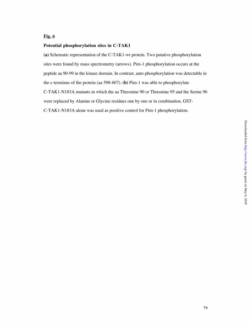
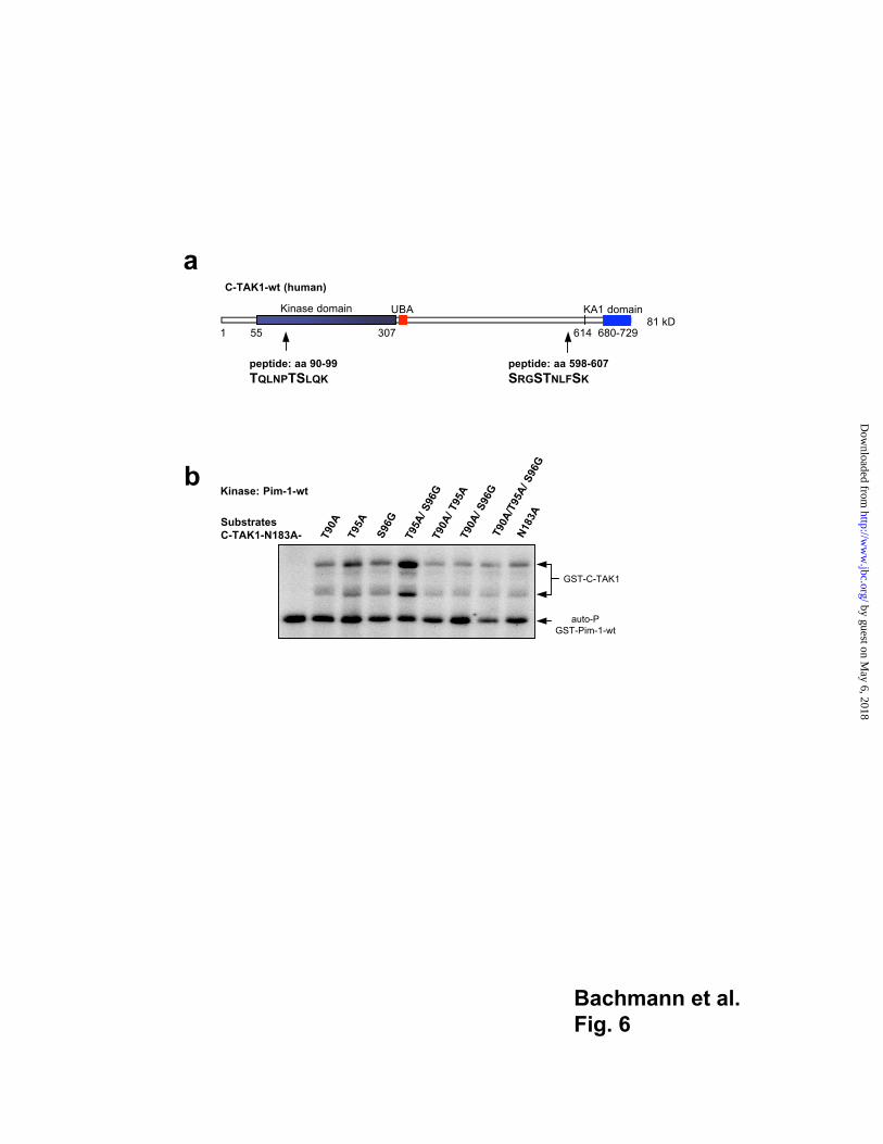
Fig. 6
Potential phosphorylation sites in C-TAK1
(a) Schematic representation of the C-TAK1-wt protein. Two putative phosphorylation
sites were found by mass spectrometry (arrows). Pim-1 phosphorylation occurs at the
peptide aa 90-99 in the kinase domain. In contrast, auto-phosphorylation was detectable in
the c-terminus of the protein (aa 598-607). (b) Pim-1 was able to phosphorylate
C-TAK1-N183A mutants in which the aa Threonine 90 or Threonine 95 and the Serine 96
were replaced by Alanine or Glycine residues one by one or in combination. GST-
C-TAK1-N183A alone was used as positive control for Pim-1 phosphorylation.
by guest on May 6, 2018
http://ww
w.jbc.org/
Dow
nloaded from
29
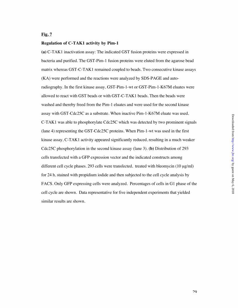
Fig. 7
Regulation of C-TAK1 activity by Pim-1
(a) C-TAK1 inactivation assay: The indicated GST fusion proteins were expressed in
bacteria and purified. The GST-Pim-1 fusion proteins were eluted from the agarose bead
matrix whereas GST-C-TAK1 remained coupled to beads. Two consecutive kinase assays
(KA) were performed and the reactions were analyzed by SDS-PAGE and auto-
radiography. In the first kinase assay, GST-Pim-1-wt or GST-Pim-1-K67M eluates were
allowed to react with GST beads or with GST-C-TAK1 beads. Then the beads were
washed and thereby freed from the Pim-1 eluates and were used for the second kinase
assay with GST-Cdc25C as a substrate. When inactive Pim-1-K67M eluate was used,
C-TAK1 was able to phosphorylate Cdc25C which was detected by two prominent signals
(lane 4) representing the GST-Cdc25C proteins. When Pim-1-wt was used in the first
kinase assay, C-TAK1 activity appeared significantly reduced, resulting in a much weaker
Cdc25C phosphorylation in the second kinase assay (lane 3). (b) Distribution of 293
cells transfected with a GFP expression vector and the indicated constructs among
different cell cycle phases. 293 cells were transfected, treated with bleomycin (10 µg/ml)
for 24 h, stained with propidium iodide and then subjected to the cell cycle analysis by
FACS. Only GFP expressing cells were analyzed. Percentages of cells in G1 phase of the
cell cycle are shown. Data representative for five independent experiments that yielded
similar results are shown.
by guest on May 6, 2018
http://ww
w.jbc.org/
Dow
nloaded from
30
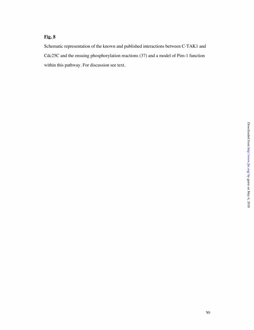
Fig. 8
Schematic representation of the known and published interactions between C-TAK1 and
Cdc25C and the ensuing phosphorylation reactions (37) and a model of Pim-1 function
within this pathway. For discussion see text.
by guest on May 6, 2018
http://ww
w.jbc.org/
Dow
nloaded from
Bachmann et al.
Fig. 1
C-TAK1-wt (human)
Cdc25C binding region
308 - 412
C-TAK1-Y6131 prey (rat)
263
kinase domain
1 55
81 kDUBA KA1 domain
1 55 680-72907 614
30 kD
83.6 kD
614
82.6 kD614
exon X: 9 aa exon Y: 15 aa
C-TAK1- (human)
C-TAK1- (human)
704-753
695-744
c
Galactose, 36 °C Glucose, 36 °Cbait library-plasmid Glucose, 25 °C
Pim-1-K67M-Ras GCBY6131
pADNS GCBY6131
Pim-1-K67M-Ras pYesMdpA
Jun-Z-Ras M-Fos
pADNS M-Fos
a
0
50
100
150
200Galactose
Glucose
ß-Galactosidase(U)
LexA-Pim-1-K67M + + - -
LexA-Gfi-VII - - + +
VP16-C-TAK1-Y6131 + + + +
LexA-Pim1-K67M
LexA-Gfi-VII
VP16-C-TAK1-Y6131
b
* * *
* : L128P, N183, D196N
by guest on May 6, 2018
http://ww
w.jbc.org/
Dow
nloaded from
Bachmann et al.
Fig. 2
a b
mock
C-TAK1-Y6131-Flag
Pim1-wt-Flag
Pim1-K67M-Flag
Evi5mut-Flag
Pias3-Flag
InputC-TAK1-wt
35S-labeled
C-TAK1-wt
GST-C-TAK1-
d
GST-Jun1-233
GST-C-TAK1-wt
GST-C-TAK1-
GST-C-TAK1-N183A
GST-C-TAK1-D196N
GST-C-TAK1-L128P
GST-C-TAK1-81-729
InputPim-1-wt
Pim1-wt
C-TAK1
Y6131
We: anti Flag
IP: anti Flag
We: anti Pim-1
Pim-1
We: anti Pim-1
Pim-1
+ + - -
- - + +
+ - + -
- + - +
Y6131-Flag
Y6131-L128P-Flag
Pim-1-wt
Pim-1-K67M
c
GST
GST-C-TAK1-1-165
GST-C-TAK1-wt
GST-C-TAK1-
GST-C-TAK1-
input
35S-labeled
Pim-1-wt
35S-labeled
C-TAK1-wt
GST-C-TAK1-Y6131
IP: anti Flag
We: anti Flag
by guest on May 6, 2018
http://ww
w.jbc.org/
Dow
nloaded from
C-TAK1-wt-GFP
C-TAK1-L128P-GFP
Pim-1-wt-Flag
Pim-1-K67M-Flag
Bachmann et al.
Fig. 3
aC-TAK1-wt-GFP Pim-1-wt-Flag overlay
C-TAK1-L128P-GFP Pim-1-wt-Flag overlay
b
C-TAK1-N183A-GFP C-TAK1-N183A-GFP Pim-1-wt-Flag overlay
by guest on May 6, 2018
http://ww
w.jbc.org/
Dow
nloaded from
Bachmann et al.
Fig. 4
anti Pim-1 anti C-TAK1
a
banti Pim-1anti C-TAK1 overlay
anti Cdc25C overlayanti C-TAK1
d
U 937 cells + TPA, 2d
K 562 cells
GST
GST-Evi5-1-300
GST-C-TAK-1-wt
We: anti Pim-1
Pim-1
Pim-1
We: anti Pim-1
U 937 cells
+TPA, 2d
antiPim-1-P9
antiLexA
input(1/10)
We: anti Pim-1
We: anti C-TAK1
C-TAK1
Pim-1
e
cC+ C- U H K
Pim-1-
Flag
We: anti Pim-1
endog.
Pim-1
We: anti C-TAK1
C-TAK1-
Flagendog.
C-TAK1
by guest on May 6, 2018
http://ww
w.jbc.org/
Dow
nloaded from

Bachmann et al.
Fig. 5
a
auto-P
GST-Pim-1
auto-P GST-
C-TAK1-wt
30 kD
Kinases:
GST-Pim-1-wt
GST-Pim-1-K67M
GST-C-TAK1-wt
b
Cdc25C
rec.protein
auto-P
GST-
C-TAK1
Kinases:
Substrates:
GST-Cdc25C
Cdc25C-rec.protein
GST-
Cdc25C
c
GST-C-TAK1- wt wt
Substrates: GST-
Pim-1-wt
GST-
Pim-1-K67M
GST-
Pim-1
GST-
C-TAK1
Kinases:
GST-C-TAK1- wt wt wt+
+
-
-
+
-
-
- -
+
+ + +
--
- -
- -
Cdc25C interaction domain (308-412)
Pim-1 interaction domain (81-165)
UBAKA1 domain
55 680-729307 614
* *
kinase domain
C-TAK1-
1-16516555
C-TAK1-
81-72981 680-729307
72 kD
18 kD
C-TAK1-wt/ /
N183A/ D196N 81 kD ++ ++
Interaction
with Pim-1
Substrate of
Pim-1-wt
55 680-729307
*C-TAK1-L128P
81 kD
/
- - -
++ ++ -
++ ++ -
1
1
1
f C-TAK1auto-P
only wt
Substrate: GST
d
GST-Pim-1
GST-
C-TAK1
Kinases:
GST-Pim-1-wt
GST-Pim-1-K67M
+
+
-
-
+
+
-
-
+
+
-
-
+
+
-
-
GST-C-TAK1-wt
GST-C-TAK1-L128P
GST-C-TAK1-N183A
GST-C-TAK1-D196N +
-
-
-
+
-
-
-
+
-
-
-
+
-
-
-
+
-
-
-
+
-
-
-
+
-
-
- +
-
-
-
Substrates:
e
Substrates:GST-Pim-1
GST-
C-TAK1
GST-C-TAK-1
1-165
Flag-Pim-1
- -
GST-C-TAK1-1-165
GST-C-TAK1-81-729
GST-C-TAK1-N183A
++
+ +
+
- - -
- -
- -
-
Kinases:
GST-Pim-1-wt
Flag-Pim-1-wt +
- -
-
++ -
---
by guest on May 6, 2018
http://ww
w.jbc.org/
Dow
nloaded from
Bachmann et al.
Fig. 6
b
Substrates
C-TAK1-N183A- T90A
T95A
S96G
T95A/S96G
T90A/T95A
T90A/S96G
N183A
a
T90A/T95A/S96G
GST-C-TAK1
auto-P
GST-Pim-1-wt
Kinase: Pim-1-wt
C-TAK1-wt (human)
81 kDUBA KA1 domain
1 55 680-729307 614
peptide: aa 90-99
TQLNPTSLQKpeptide: aa 598-607
SRGSTNLFSK
Kinase domain
by guest on May 6, 2018
http://ww
w.jbc.org/
Dow
nloaded from
Bachmann et al.
Fig. 7
a
GST-
Cdc25C
GST-
C-TAK1-wt
GST-Pim-1-wt-eluate
GST-Pim-1-K67M-eluate
GST-C-TAK1-wt-beads
GST-Cdc25C
1st KA
2nd KA
GST-beads
beads from 1st KA
1 2 3 4
+
-+
-
+
- +
-
+
++
+
+
+ +
+
-
++
-
-
+ +
-
b
C-TAK1-wt
vector
9.2 %
C-TAK1-wt
Pim-1-wt
17.8 %
C-TAK1-wt
Pim-1-K67M
14.6 %
vector
58 %
Counts
Propidium iodide
C-TAK1-N183A
vector
21.2 %
+ Bleomycin
- Bleomycin
by guest on May 6, 2018
http://ww
w.jbc.org/
Dow
nloaded from
Bachmann et al.
Fig. 8
active
C-TAK1
MPF
G2
M
Cdc25C Cdc25C
P
P
P
cdk1
Cyclin B1
Pim-1
by guest on May 6, 2018
http://ww
w.jbc.org/
Dow
nloaded from
Malte Bachmann, Hanjo Hennemann, Pei Xiang Xing, Ingrid Hoffmann and Tarik Moroyof C-TAK1: a novel role for Pim-1 at the G2/M cell cycle checkpoint
The oncogenic serine/threonine kinase Pim-1 phosphorylates and inhibits the activity
published online August 19, 2004J. Biol. Chem.
10.1074/jbc.M404440200Access the most updated version of this article at doi:
Alerts:
When a correction for this article is posted•
When this article is cited•
to choose from all of JBC's e-mail alertsClick here
by guest on May 6, 2018
http://ww
w.jbc.org/
Dow
nloaded from