a novel mitochondrial matrix serine/threonine …genesdev.cshlp.org/content/21/7/784.full.pdfa novel...
TRANSCRIPT

A novel mitochondrial matrixserine/threonine protein phosphataseregulates the mitochondria permeabilitytransition pore and is essentialfor cellular survival and developmentGang Lu,1,4 Shuxun Ren,1,4 Paavo Korge,3 Jayoung Choi,5 Yuan Dong,5 James Weiss,2,3
Carla Koehler,4,6 Jau-nian Chen,4,5 and Yibin Wang1,2,3,4,7
1Department of Anesthesiology, University of California at Los Angeles, Los Angeles, California 90095, USA; 2Departmentof Physiology, University of California at Los Angeles, Los Angeles, California 90095, USA; 3Department of Medicine,University of California at Los Angeles, Los Angeles, California 90095, USA; 4Molecular Biology Institute, University ofCalifornia at Los Angeles, Los Angeles, California 90095, USA; 5Department of Molecular, Cellular, and DevelopmentalBiology, University of California at Los Angeles, Los Angeles, California 90095, USA; 6Department of Chemistry andBiochemistry, University of California at Los Angeles, Los Angeles, California 90095, USA
Mitochondria play a central role in the regulation of programmed cell death signaling. Here, we report thefinding of a mitochondrial matrix-targeted protein phosphatase 2C family member (PP2Cm) that regulatesmitochondrial membrane permeability transition pore (MPTP) opening and is essential for cell survival,embryonic development, and cardiac function. PP2Cm is highly conserved among vertebrates, with thehighest expression levels detected in the heart and brain. Small hairpin RNA (shRNA)-mediated knockdown ofPP2Cm resulted in cell death associated with loss of mitochondrial membrane potential in cultured cardiacmycoytes and an induction of hepatocyte apoptosis in vivo. PP2Cm-deficient mitochondria showed elevatedsusceptibility to calcium-induced MPTP opening, whereas mitochondrial oxidative phosphorylation activitieswere not affected. Finally, inactivation of PP2Cm in developing zebrafish embryos caused abnormal cardiacand neural development as well as heart failure associated with induced apoptosis. These data suggest thatPP2Cm is a novel mitochondrial protein phosphatase that has a critical function in cell death and survival,and may play a role in regulating the MPTP opening.
[Keywords: Mitochondrial permeability transition pore; protein phosphatase; cell death; heart failure;developmental defects; zebrafish]
Supplemental material is available at http://www.genesdev.org.
Received October 3, 2006; revised version accepted February 9, 2007.
It is well established that mitochondria play a centralrole in the intrinsic apoptosis pathway where internaland external death signals lead to the release of cyto-chrome c and apoptosis-inducing factor (AIF), as well asother proapoptotic molecules through Bcl-2 familymembers (Bax and Bak) located on the mitochondriaouter membrane. In addition, mitochondria-mediatedapoptosis can also be triggered by the opening of themitochondria permeability transition pore (MPTP).Complexes of the voltage-dependent anion channel(VDAC), the adenine nucleotide translocator (ANT), and
cyclophilin D (CypD) are already implicated to have cy-closporine A (CsA)-dependent MPTP activity in the mi-tochondrial inner membrane. The loss of inner mem-brane potential due to the MPTP opening triggers mito-chondrial swelling and outer membrane rupture, whichin turn leads to the release of proapoptotic factors fromthe intermembrane space (IMS). However, the underly-ing molecular mechanisms and the signaling moleculesinvolved in the regulation of MPTP opening under stressconditions are largely unknown.
In addition to being an essential organelle for cellularmetabolism and survival, the mitochondrion has beenrecognized as a site where diverse signaling pathwaysconverge and integrate (Ravagnan et al. 2002; Newmeyerand Ferguson-Miller 2003; Horbinski and Chu 2005). In-deed, a number of Ser/Thr protein kinases, including
7Corresponding author.E-MAIL [email protected]; FAX (310) 206-5097.Article published online ahead of print. Article and publication date areonline at http://www.genesdev.org/cgi/doi/10.1101/gad.1499107.
784 GENES & DEVELOPMENT 21:784–796 © 2007 by Cold Spring Harbor Laboratory Press ISSN 0890-9369/07; www.genesdev.org
Cold Spring Harbor Laboratory Press on February 28, 2020 - Published by genesdev.cshlp.orgDownloaded from
PKA, PKB/AKT, PKC, and JNK, have been found to belocated in mitochondria. In addition, a number of an-choring proteins for various protein kinases are alsoidentified in mitochondria, including AKAP, PICK,Grb10, and Sab (Huang et al. 1999; Nantel et al. 1999;Alto et al. 2002; Wiltshire et al. 2002; Wang et al. 2003).PKA activity targeted in the mitochondrial inner mem-brane and matrix is shown to activate Complex I of therespiratory chain in the bovine heart (Scacco et al. 2000;Technikova-Dobrova et al. 2001; Chen et al. 2004). Inaddition, PKB/AKT activity leads to phosphorylation ofthe mitochondrial ATP synthase � subunit and glycogensynthase kinase 3� (Gsk3�), which in turn, inhibits py-ruvate dehydrogenase activity (Bijur and Jope 2003).Likewise, several PKC isoforms are detected in mito-chondria with different target proteins. For instance,PKC� promotes cardioprotection against ischemia/reper-fusion injury through phosphorylation of a VDAC,which prevents the opening of the MPTP (Baines et al.2003). Further, both PKC� and PKC� have been impli-cated in mitochondrial KATP channel regulation in isch-emic preconditioning (Cohen et al. 2000; Chen et al.2001). Finally, ERK and JNK kinases are targeted to mi-tochondria to phosphorylate apoptotic proteins, includ-ing Bcl-xl, Bcl-2, and BAD, and regulate apoptosis (Denget al. 2000; Kang et al. 2003; Schroeter et al. 2003; Bri-chese et al. 2004). In short, Ser/Thr protein kinase tar-geting to mitochondria has significant impact on pro- oranti-apoptotic factors, respiration, and ionic channel ac-tivity, illustrating the important role of mitochondriaprotein phosphorylation in cellular signaling. Proteinphosphorylation is a dynamic process involving a balanc-ing act of kinases and phosphatases (Shenolikar 1994).Therefore, mitochondria protein phosphorylation willalso most likely be regulated by mitochondria-targetedprotein phosphatases. Although Protein Phosphatase1(PP1) and PP2A have been found in mitochondria (Ru-volo et al. 2002; Dagda et al. 2003; Brichese et al. 2004;Tamura et al. 2004), their specific contribution to themitochondrial function remains unclear. Recently, anovel tyrosine protein phosphatase, PTPMT1, was re-ported to be located specifically in the mitochondria ma-trix and to play an important role in both ATP produc-tion and insulin secretion (Pagliarini et al. 2005), furthersupporting the notion that protein phosphorylation anddephosphorylation in mitochondria are an importantmechanism of cell signaling. However, the molecularmechanism regulating protein phosphorylation in mito-chondria, particularly in the mitochondria matrix, hasnot been investigated.
In this report, we identified a novel Ser/Thr proteinphosphatase, named PP2Cm, that is targeted exclusivelyto the mitochondria matrix. PP2Cm is highly conservedfrom fish to mammals and contains a Ser/Thr phospha-tase domain commonly shared by all PP2C family mem-bers. It is targeted to the mitochondria matrix via a mi-tochondrial targeting sequence at its N-terminal end,making it the first Ser/Thr protein phosphatase so faridentified in this mitochondrial compartment. Fromboth in vitro and in vivo studies, we demonstrate that
PP2Cm is an essential protein for cellular survival andMPTP regulation. Loss of PP2Cm expression leads to celldeath and MPTP opening in response to calcium over-load without measurable impact on mitochondrial res-piration. PP2Cm expression is diminished in hypertro-phic and failing hearts, and suppression of PP2Cm ex-pression in zebrafish causes developmental defects in thecentral nerves system and heart. All of this evidence sug-gests that PP2Cm is a modulator of MPTP function andplays a critical role in cell death regulation and normaldevelopment and physiology in heart and other systems.This finding reveals a new signaling component inMPTP regulation, and suggests a previously uncharacter-ized mechanism in apoptosis regulation involving themitochondrial matrix protein phosphorylation/dephos-phorylation.
Results
PP2Cm is an exclusively mitochondria targetedSer/Thr phosphatase
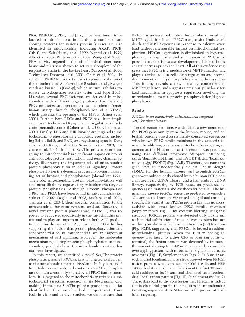
Through genome mining, we identified a new member ofthe PP2C gene family from the human, mouse, and ze-brafish genome based on its highly conserved sequenceswith known PP2C family members in the catalytic do-main. In addition, a putative mitochondria targeting se-quence at the N-terminal of the protein was predictedusing two different programs: Mitoprot (http://ihg.gsf.de/ihg/mitoprot.html) and iPSORT (http://hc.ims.u-tokyo.ac.jp/iPSORT) (Fig. 1A,B). Therefore, we name thegene PP2C in Mitochondria (PP2Cm). The full-lengthcDNAs for the human, mouse, and zebrafish PP2Cmgene were subsequently cloned from a human EST clone,a mouse heart cDNA library, and a fish embryo cDNAlibrary, respectively, by PCR based on predicted se-quences (see Materials and Methods for details). The hu-man and mouse PP2Cm genes are predicted to encode a372-amino-acid protein. We raised a polyclonal antibodyspecifically against the PP2Cm protein that has no cross-activity with other known PP2C family members(Supplementary Fig. 1). By Western blotting using thisantibody, PP2Cm protein was detected only in the mi-tochondrial subfraction of mouse liver extracts but notin the cytosolic or endoplasmic reticulum (ER) fractions(Fig. 1C,D), suggesting that PP2Cm is indeed a residentmitochondrial protein. When the PP2Cm coding se-quence was fused to either GFP or Flag tag at its C-terminal, the fusion protein was detected by immuno-fluorescent staining for GFP or Flag tag with a completeoverlapping pattern with mitotracker signals in culturedmyocytes (Fig. 1E; Supplementary Figs. 2, 3). Similar mi-tochondrial localization was also observed when PP2Cmfusion protein was expressed in COS-1 cells and HEK293 cells (data not shown). Deletion of the first 30 aminoacid residues at its N-terminal abolished its mitochon-drial localization pattern (Fig. 1E; Supplementary Fig. 2).These data lead to the conclusion that PP2Cm is indeeda mitochondrial protein that requires its mitochondriatargeting sequence at its N terminus for proper intracel-lular targeting.
Cell death regulation by PP2Cm
GENES & DEVELOPMENT 785
Cold Spring Harbor Laboratory Press on February 28, 2020 - Published by genesdev.cshlp.orgDownloaded from
PP2Cm exists as a soluble protein in the mitochondriamatrix
Because the suborganellar localization of PP2Cm may beimportant for its function within the mitochondrion, weutilized hypotonic lysis (Koehler et al. 1998) and carbon-ate extraction (Fujiki et al. 1982) techniques to deter-mine if PP2Cm was localized to the mitochondrial ma-trix, inner membrane, or IMS. Purified mouse liver mi-tochondria were treated with 0.1 M Na2CO3 (pH 11), andmembrane proteins were recovered in the pellet aftercentrifugation. PP2Cm was recovered in the superna-tant, indicating it is a soluble protein like TIMM13 andnot an integral membrane protein like TOMM40 (Fig.2A). Mitochondria were also subjected to hypotonic lysisin buffers containing decreased concentration of the os-moticum sucrose (Fig. 2B). At 200 mM sucrose, the outer
membrane begins to rupture, but the inner membraneremains intact, even when the sucrose concentration isdecreased to 25 mM. Based on protease protection,PP2Cm is localized in the matrix like Hsp60 and not inthe IMS like TIMM13. Thus, PP2Cm is a soluble proteinlocalized in the mitochondrial matrix.
PP2Cm is a bona fide Ser/Thr phosphatase withenriched expression in heart and brain, but reducedexpression in failing heart
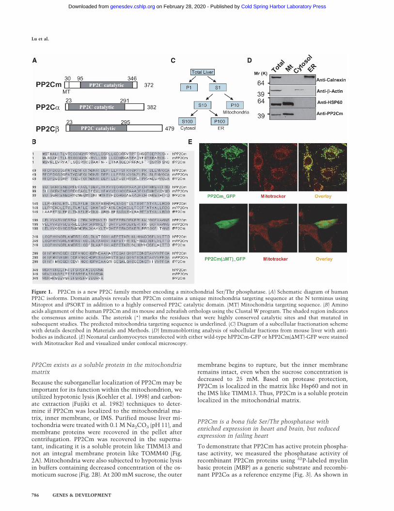
To demonstrate that PP2Cm has active protein phospha-tase activity, we measured the phosphatase activity ofrecombinant PP2Cm proteins using 32P-labeled myelinbasic protein (MBP) as a generic substrate and recombi-nant PP2C� as a reference enzyme (Fig. 3). As shown in
Figure 1. PP2Cm is a new PP2C family member encoding a mitochondrial Ser/Thr phosphatase. (A) Schematic diagram of humanPP2C isoforms. Domain analysis reveals that PP2Cm contains a unique mitochondria targeting sequence at the N terminus usingMitoprot and iPSORT in addition to a highly conserved PP2C catalytic domain. (MT) Mitochondria targeting sequence. (B) Aminoacids alignment of the human PP2Cm and its mouse and zebrafish orthologs using the Clustal W program. The shaded region indicatesthe consensus amino acids. The asterisk (*) marks the residues that were highly conserved catalytic sites and that mutated insubsequent studies. The predicted mitochondria targeting sequence is underlined. (C) Diagram of a subcellular fractionation schemewith details described in Materials and Methods. (D) Immunoblotting analysis of subcellular fractions from mouse liver with anti-bodies as indicated. (E) Neonatal cardiomyocytes transfected with either wild-type hPP2Cm-GFP or hPP2Cm(�MT)-GFP were stainedwith Mitotracker Red and visualized under confocal microscopy.
Lu et al.
786 GENES & DEVELOPMENT
Cold Spring Harbor Laboratory Press on February 28, 2020 - Published by genesdev.cshlp.orgDownloaded from

Figure 3B, wild-type PP2Cm recombinant proteinshowed significant protein phosphatase activity in an ex-pected Mn2+-dependent manner, whereas proteins with aspecific mutation in highly conserved aspartate at posi-tion 298 (D298A), known to be essential for catalyticactivity of other PP2C isoforms, showed no detectablephosphatase activity (Fig. 3D). However, PP2Cm onlydephosphorylated <10% of the total phosphorylated sitesin MBP substrate in contrast to >70% efficiency carriedout by PP2C� (Fig. 3C). The specific activity of PP2Cmwas also significantly lower than PP2C� under the sameassay conditions (Fig. 3D). All these data indicate thatPP2Cm is a bona fide Ser/Thr phosphatase with differentsubstrate selectivity and specific activity compared withPP2C�. By Northern blot, mouse PP2Cm transcript wasdetected as a single species at ∼5.9 kb in size, with thehighest level detected in the adult heart, brain, and dia-phragm, and lower levels in the liver, lung, kidney, skel-etal muscle, and thymus (Fig. 3E). Interestingly, thesetissues all have high metabolic activities and extensivemitochondrial contents. Western blot analysis usingPP2Cm-specific antibody showed that the PP2Cm pro-tein is also enriched in the heart, brain, liver, and thy-mus, with lower expression detected in other tissues(Supplementary Fig. 5). To determine the expression pro-file of PP2Cm in the diseased heart, we created pressureoverload by trans-aortic constriction (TAC) in adultmouse hearts, which has been described to induce car-diac hypertrophy and a transition to heart failure (Taki-moto et al. 2005). Cardiac hypertrophy with preservedfunction was observed within 1 wk post-TAC, while de-compensated heart failure was apparent after 3 wk post-TAC (Takimoto et al. 2005; S. Ren and Y. Wang, un-publ.). The expression of PP2Cm at both the mRNA andprotein levels was significantly reduced in hypertrophiedhearts, and was further reduced in the decompensated
failing heart (Fig. 3F,G). In contrast, other mitochondriaproteins, including Rhodanease and TOMM40, were notchanged compared with the control. These data suggestthat PP2Cm expression is highly associated with mito-chondria content and metabolic status, and its selectiveloss of expression correlates with the progression ofpathological remodeling in diseased hearts.
PP2Cm is essential for myocyte survivaland maintaining mitochondria membrane potentialin vitro
To investigate the functional significance of PP2Cm, en-dogenous PP2Cm was inhibited in cultured neonatal ratventricular myocytes using small hairpin RNA (shRNA)delivered via adenovirus vectors. As shown in Figure 4A,all three shRNAs targeting different segments of ratPP2Cm mRNA effectively reduced endogenous PP2Cmprotein expression 3 d post-transfection. PP2Cm inacti-vation led to significant and selective induction of stress-activated mitogen-acitvated protein (MAP) kinases,c-Jun N-terminal Kinase (JNK), and p38 kinase, whileMAP kinase ERK was not affected (Fig. 4A). In addi-tion, inactivation of PP2Cm also resulted in marked in-duction of the atrial natriuretic factor (ANF), a well-established marker gene for myocyte stress response(Fig. 4B). Seven days post-shRNA-mediated knockdownof PP2Cm, a significant induction of cell death was ob-served compared with control cells (Fig. 4C). Inductionof cell death was associated with dissipated mitochon-drial membrane potential (��) as measured by a signifi-cant reduction in a red/green fluorescence ratio of theJC-1 signal in PP2Cm-deficient myocytes (Reers et al.1995) (Fig. 4C,D). Inactivation of PP2Cm in HeLa cellsresulted in loss of cell proliferation and induced celldeath similar to that observed in cardiomyocytes (data
Figure 2. PP2Cm is a soluble mitochondria ma-trix protein. (A) Isolated mouse liver mitochon-dria fraction was subjected to sodium carbonateextraction. After centrifugation, pellet (P) and su-pernatant (S) are analyzed by immunoblotting us-ing anti-TOMM40, an outer membrane protein(OM), and anti-TIMM13, a mitochondria IMSprotein, as indicated. (B) Isolated mouse liver mi-tochondria were incubated in hypotonic sucrosebuffers as indicated to disrupt the outer mem-brane in the absence (top panel) and presence(bottom panel) of soybean trypsin. After centrifu-gation, total (T), pellet (P), and supernatant (S)fractions were analyzed by immunoblotting withanti-Hsp60, a matrix protein, or anti-TIMM13antibodies as indicated.
Cell death regulation by PP2Cm
GENES & DEVELOPMENT 787
Cold Spring Harbor Laboratory Press on February 28, 2020 - Published by genesdev.cshlp.orgDownloaded from
not shown). These in vitro analyses suggest that PP2Cmis essential for cell survival, and plays an important rolein maintaining mitochondrial membrane potential.
Loss of PP2Cm leads to hepatic injury and hepatocyteapoptosis in vivo
To further investigate the in vivo function of PP2Cm, weemployed shRNA to specifically inactivate the endog-enous PP2Cm in mouse liver using the recombinant ad-enovirus vector administered through intravenous injec-tion. It has already been established that systemic deliv-ery of adenovirus vectors in mice leads to effectivetargeting to the liver (Wang et al. 1996). Indeed, applica-tion of Adv-PP2Cm shRNA significantly reduced endog-enous PP2Cm expression at both mRNA and protein lev-els in the mouse liver while administration of a controlAdv-luciferase shRNA vector had no detectable impact
on PP2Cm expression (Fig. 5A,C). The mice receivingAdv-PP2Cm shRNA showed significantly elevated hepa-tocyte apoptosis measured by TUNEL staining (Fig.5B,C), and a marked induction of �-fetal protein (AFP)gene (Fig. 5D), a marker for hepatic injury and pathology.Two interferon response genes (MxA and OSA1�) werealso modestly elevated; however, their inductions wereobserved in both Luciferase-shRNA and PP2Cm-shRNA-treated tissues. These data indicate that, similar to itsrole in cultured myocytes, endogenous PP2Cm is essen-tial for normal survival and function of hepatocytes invivo.
Loss of PP2Cm sensitizes mitochondria to Ca-inducedpermeability transition
To reveal the underlying mechanisms involved inPP2Cm-mediated mitochondria regulation, we first de-
Figure 3. PP2Cm is bona fide Ser/Thr protein phosphatase with enriched expression in the heart and brain. (A) Coomassie-stainedSDS-PAGE gel of GST recombinant proteins as indicated at the top. (B) Phosphatase activity measured from purified recombinanthuman wild-type PP2Cm-GST fusion protein for its Mn2+ dependency using 32P-labeled MBP as substrate. (C) One microgram of32P-labeled MBP was incubated overnight with 0.5 µg of purified wild-type PP2C�, PP2Cm, and PP2Cm-D298A mutant as labeled. Thedephosphorylation efficiency of the recombinant phosphatases was expressed as the ratio of dephosphorylated versus total 32P-labeledMBP at the end of the reaction. Values are mean ± SD of three independent experiments. (#) p < 0.01. (D) Specific phosphatase activitiesof PP2Ca and PP2Cm toward MBP were calculated based on V0 measured from the linear phase of the dephosphorylation reaction. Theresults were mean ± SD of three independent experiments. (#) p < 0.01 versus PP2Cm. (E) Radiograph of Northern blot analysis for thePP2Cm mRNA level in different tissues of the adult mouse (top panel) and a fluorescent signal of ethidium bromide-stained 18Sribosomal RNA used as an equal loading control (bottom panel). (F) PP2Cm mRNA levels in hypertrophy or failing mouse hearts werequantified by real-time quantitative RT–PCR after normalization against GAPDH. At least three animals of each group were exam-ined. Shown are mean ± SD. The cardiac hypertrophic or heart failure was established by transaortic constriction for 1 or 3 wk,respectively, as described in Materials and Methods. (G) PP2Cm and other mitochondria protein levels in hypertrophy or failing heartswere determined by immunostaining using the anti-PP2Cm antibody, anti-Rhodanese, and anti-TOMM40, as indicated. The �-actinlevel was used for equal loading.
Lu et al.
788 GENES & DEVELOPMENT
Cold Spring Harbor Laboratory Press on February 28, 2020 - Published by genesdev.cshlp.orgDownloaded from

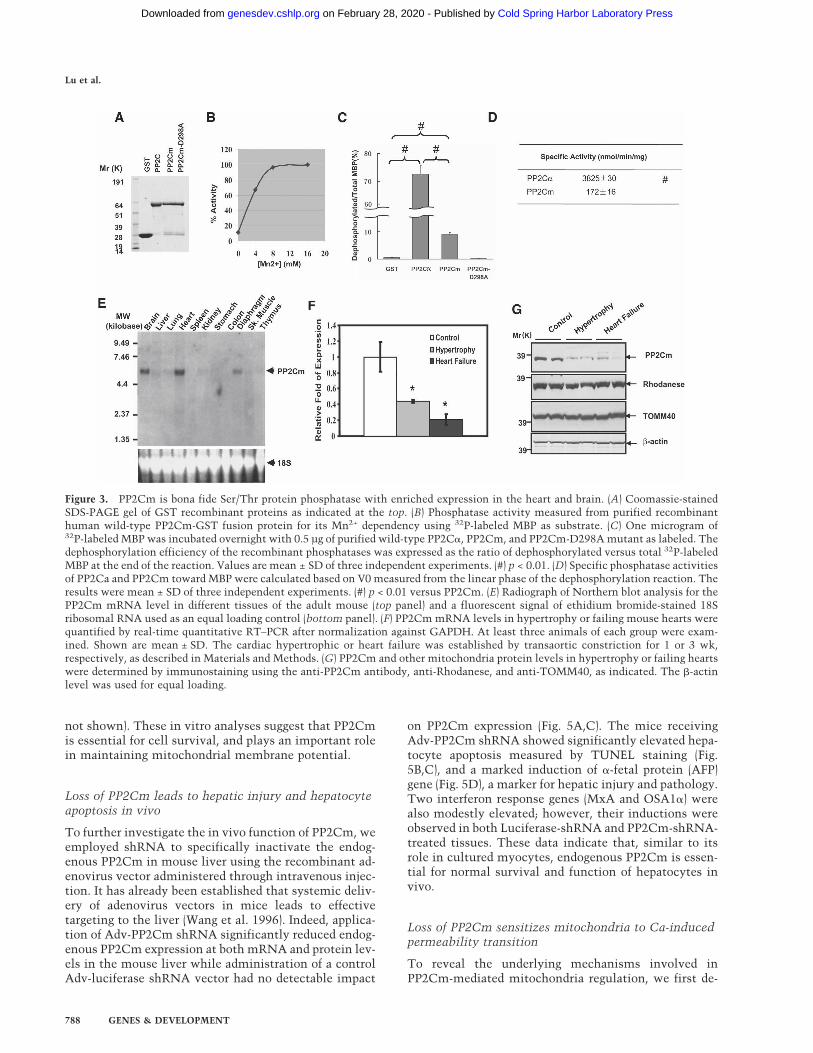
termined the effect of PP2Cm inactivation on oxidativephosphorylation and ATP production in isolated mito-chondria. We measured oxygen consumption, ��, andthe mitochondrial matrix volume simultaneously afterenergizing the mitochondria with Complex I substratesand adding ADP in multiple pulses (Fig. 6A,B). From thisexperiment, we found that PP2Cm inactivation did notaffect �� recovery, oxygen consumption, or the mito-chondrial matrix volume after repeated ADP challenges,suggesting that PP2Cm deficiency does not have a sig-nificant impact on oxidative phosphorylation (Fig. 6A,B).In contrast, in the presence of consecutive 2-µM Ca2+
pulses, PP2Cm-deficient mitochondria quickly lost ��and displayed matrix swelling, the key features of mito-chondrial permeability transition, whereas control mito-
chondria tolerated challenges with Ca2+ pulses muchbetter (Fig. 6C,D). Importantly, the loss of mitochondrialmembrane potential and increased matrix volume werereversed by CsA and EGTA (Fig. 6C), confirming thatPP2Cm deficiency sensitized the opening of MPTP.These data suggest that PP2Cm is an important regulatorof MPTP opening, and the loss of PP2Cm can promotecell death by triggering the mitochondrial permeabilitytransition.
PP2Cm is essential to normal development of brainand digestive system of zebrafish
Zebrafish PP2Cm is highly homologous to mammaliancounterparts based on sequence alignment (Fig. 1), and
Figure 4. Loss of PP2Cm in vitro promotes cell death and mitochondria membrane potential dissipation. (A) Neonatal cardiomyo-cytes were transfected with Adv-GFP or Adv-shRNAs targeting three different regions of PP2Cm coding sequence for 48 h at amultiplicity of infection (MOI) of 50. The silencing efficiency was confirmed by immunoblotting with a PP2Cm polyclonal antibody.The MAP kinases activation was examined with total and anti-phospho-JNK, anti-phospho-p38, and anti-phospho-ERK antibodies asindicated. (B) mRNA levels of PP2Cm and ANF by real-time quantitative RT–PCR relative to GAPDH. Neonatal rat ventricularmyocytes were transfected with either Adv-GFP or Adv-PP2Cm-shRNA3 using either 50 or 100 MOI as labeled. Total RNAs wereisolated 3 d post-transfection. (C, panels a,e) Representative images of cells observed in cardiomyocytes 7 d post-Adv-PP2Cm-shRNA3transfection compared with controls. Cells were stained with JC-1 fluorescent dye and imaged under identical exposure parametersusing Rhodamin (red; panels b,f) and FITC (green; panels c,g) filters on a Zeiss confocal microscope as described in Materials andMethods. The merged images are shown in panels d and h. (D) The average ratios of red versus green fluorescent signal intensities(shown as relative pixel intensity) were measured from eight control and eight PP2C-shRNA3-treated cells from digitally recordedimages using the Metamorph program.
Cell death regulation by PP2Cm
GENES & DEVELOPMENT 789
Cold Spring Harbor Laboratory Press on February 28, 2020 - Published by genesdev.cshlp.orgDownloaded from

shows similar mitochondria-targeted intracellular local-ization when it was expressed in mammalian cells(Supplementary Fig. 5). In developing zebrafish embryos,PP2Cm expression was highly expressed in the CNS,heart, and other tissues at 24 h post-fertilization (hpf) asrevealed by whole-mount in situ hybridization using anantisense PP2Cm cDNA probe (Fig. 7A). In adult ze-brafish, PP2Cm transcript was detected in the brain,heart, skeletal muscle, and liver by RT–PCR (Fig. 7B). Toinvestigate whether PP2Cm in zebrafish has a conservedfunction in regulating cell survival, we employed twotypes of morpholinos, ATG-MO and SP-MO, to suppressPP2Cm expression in the developing zebrafish embryo(Fig. 7C). The ATG-MO targets the start codon ofPP2Cm to inhibit translation initiation. The SP-MO tar-gets the splicing donor of PP2Cm intron 4, which shouldresult in a truncated PP2Cm (1–236) mutant protein dueto premature translation termination (SupplementaryFig. 6). To ensure target specificity of PP2Cm morpholi-nos, we used a modified morpholino (AM-MO) with a5-base-pair (bp) mismatch to PP2Cm ATG-MO as a nega-tive control for the ATG-MO. In addition, we coinjected
PP2Cm mRNA with SP-MO to demonstrate the speci-ficity of the SP-MO effects. Suppression of PP2Cm ex-pression by SP-MO in the developing embryos resultedin a marked induction of apoptosis and developmentaldefect that was significantly mitigated by coinjection ofwild-type zebrafish PP2Cm mRNA (Fig. 7D–I; Supple-mentary Fig. 8). Based on embryo size and the presence ofbrain cell death, 58.4% (59/101) of the embryos coin-jected with SP-MO and PP2Cm mRNA were completelyrescued, 22.3% (23/101) were partially rescued, while theremaining 18.8% (19/101) showed no obvious differencesfrom SP-MO morphants without coinjection of PP2CmmRNA. Similarly, ATG-MO resulted in abnormal devel-opment associated with significant induction in apopto-sis as measured from acridine orange (SupplementaryFig. 7) staining and TUNEL staining (data not shown)compared with untreated embryos (data not shown) orembryos injected with the control AM-MO. Suppressionof PP2Cm expression by either SP-MO or ATG-MO re-duced brain size (Fig. 7J,K,M), and reduced and delayed aCNS marker gene Pax2 expression in the mid/hindbrainboundary, otic vesicle, and spinal cord in 24 and 48 hpf
Figure 5. Loss of PP2Cm in vivo induces hepatocyte apoptosis and liver injury. (A) Western blot analysis of PP2Cm expression in totalliver lysates from wild-type mice or mice treated with adenovirus expressing either luciferase control shRNA or PP2Cm-specificshRNA3 for 7 d. (B, top panel) Mouse livers treated as described above were fixed and assessed for apoptisis by TUNEL staining.(Middle panel) The nuclei were counterstained with DAPI. (Bottom panel) Corresponding images were overlaid to distinguish falsepositive signals. DNase I-treated liver sections served as a positive control. (C) The percentage of apoptotic cells was tallied andpresented as mean ± SD (n = 4). (D) Hepatic gene profiles of mouse livers treated as above were examined with quantitative RT–PCRby normalizing against GAPDH. (*) p < 0.05, PP2Cm shRNA versus control; (#) p < 0.05, luciferase shRNA versus control, Student’st-test.
Lu et al.
790 GENES & DEVELOPMENT
Cold Spring Harbor Laboratory Press on February 28, 2020 - Published by genesdev.cshlp.orgDownloaded from
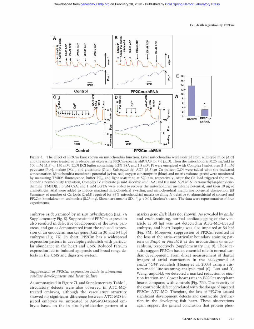
embryos as determined by in situ hybridization (Fig. 7J;Supplementary Fig. 8). Suppression of PP2Cm expressionalso resulted in defective development of the liver, pan-creas, and gut as demonstrated from the reduced expres-sion of an endoderm marker gene fkd2 in 30 and 54 hpfembryos (Fig. 7K). In short, PP2Cm has a widespreadexpression pattern in developing zebrafish with particu-lar abundance in the heart and CNS. Reduced PP2Cmexpression led to induced apoptosis and broad range de-fects in the CNS and digestive system.
Suppression of PP2Cm expression leads to abnormalcardiac development and heart failure
As summarized in Figure 7L and Supplementary Table 1,circulatory defects were also observed in ATG-MO-treated embryos, although the vasculature structureshowed no significant difference between ATG-MO-in-jected embryos vs. untreated or AM-MO-treated em-bryos based on the in situ hybridization pattern of a
marker gene flick (data not shown). As revealed by amhcand vmhc staining, normal cardiac jogging of the ven-tricle at 30 hpf was not detected in ATG-MO-treatedembryos, and heart looping was also impaired at 54 hpf(Fig. 7M). Moreover, suppression of PP2Cm resulted inthe loss of the atria–ventricular boundary staining pat-tern of Bmp4 or Notch1B at the myocardium or endo-cardium, respectively (Supplementary Fig. 9). These re-sults suggest PP2Cm has an essential role in normal car-diac development. From direct measurement of digitalimages of atrial contraction in the background ofcmlc2�GFP zebrafish (Huang et al. 2003) using a cus-tom-made line-scanning analysis tool (Q. Luo and Y.Wang, unpubl.), we detected a marked reduction of ejec-tion fraction and slower heart rates in PP2Cm morphanthearts compared with controls (Fig. 7N). The severity ofthe contractile defect correlated with the dosage of injectedPP2Cm ATG-MO. Therefore, the loss of PP2Cm causedsignificant development defects and contractile dysfunc-tion in the developing fish heart. These observationsagain support the general conclusion that protein phos-
Figure 6. The effect of PP2Cm knockdown on mitochondria function. Liver mitochondria were isolated from wild-type mice (A,C)and the mice were treated with adenovirus expressing PP2Cm-specific shRNA3 for 7 d (B,D). Then the mitochondria (0.25 mg/mL) in100 mM (A,B) or 150 mM (C,D) KCl buffer containing 0.2% BSA and 2.5 mM Pi were energized with Complex I substrates (1.6 mMpyvuvate [Pyv], malate [Mal], and glutamate [Glu]). Subsequently, ADP (A,B) or Ca pulses (C,D) were added with the indicatedconcentration. Mitochondria membrane potential (��m, red), oxygen consumption (blue), and matrix volume (green) were monitoredby measuring TMRM fluorescence, buffer PO2, and light scattering at 520 nm, respectively. After the Ca load triggered the mito-chondria permeability transition, Complex IV substrate (2 mM ascorbic acid [AA] and 0.2 mM N,N,N�,N�-tetramethyl-p-phenylene-diamine [TMPD]), 1.5 µM CsA, and 1 mM EGTA were added to recover the mitochondrial membrane potential, and then 10 µg ofalamethicin (Ala) were added to induce maximal mitochondrial swelling and mitochondrial membrane potential dissipation. (E)Summary of number of Ca loads (2 µM) required for 95% mitochondrial matrix swelling N (relative to alamethicin) of control andPP2Cm knockdown mitochondria (0.25 mg). Shown are mean ± SD. (*) p < 0.01, Student’s t-test. The data were representative of fourexperiments.
Cell death regulation by PP2Cm
GENES & DEVELOPMENT 791
Cold Spring Harbor Laboratory Press on February 28, 2020 - Published by genesdev.cshlp.orgDownloaded from
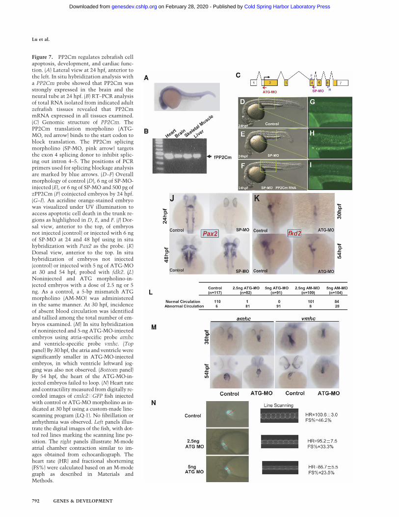
Figure 7. PP2Cm regulates zebrafish cellapoptosis, development, and cardiac func-tion. (A) Lateral view at 24 hpf, anterior tothe left. In situ hybridization analysis witha PP2Cm probe showed that PP2Cm wasstrongly expressed in the brain and theneural tube at 24 hpf. (B) RT–PCR analysisof total RNA isolated from indicated adultzefrafish tissues revealed that PP2CmmRNA expressed in all tissues examined.(C) Genomic structure of PP2Cm. ThePP2Cm translation morpholino (ATG-MO, red arrow) binds to the start codon toblock translation. The PP2Cm splicingmorpholino (SP-MO, pink arrow) targetsthe exon 4 splicing donor to inhibit splic-ing out intron 4–5. The positions of PCRprimers used for splicing blockage analysisare marked by blue arrows. (D–F) Overallmorphology of control (D), 6 ng of SP-MO-injected (E), or 6 ng of SP-MO and 500 pg ofzPP2Cm (F) coinjected embryos by 24 hpf.(G–I). An acridine orange-stained embryowas visualized under UV illumination toaccess apoptotic cell death in the trunk re-gions as highlighted in D, E, and F. (J) Dor-sal view, anterior to the top, of embryosnot injected (control) or injected with 6 ngof SP-MO at 24 and 48 hpf using in situhybridization with Pax2 as the probe. (K)Dorsal view, anterior to the top. In situhybridization of embryos not injected(control) or injected with 5 ng of ATG-MOat 30 and 54 hpf, probed with fdk2. (L)Noninjected and ATG morpholino-in-jected embryos with a dose of 2.5 ng or 5ng. As a control, a 5-bp mismatch ATGmorpholino (AM-MO) was administeredin the same manner. At 30 hpf, incidenceof absent blood circulation was identifiedand tallied among the total number of em-bryos examined. (M) In situ hybridizationof noninjected and 5-ng ATG-MO-injectedembryos using atria-specific probe amhcand ventricle-specific probe vmhc. (Toppanel) By 30 hpf, the atria and ventricle weresignificantly smaller in ATG-MO-injectedembryos, in which ventricle leftward jog-ging was also not observed. (Bottom panel)By 54 hpf, the heart of the ATG-MO-in-jected embryos failed to loop. (N) Heart rateand contractility measured from digitally re-corded images of cmlc2�GFP fish injectedwith control or ATG-MO morpholino as in-dicated at 30 hpf using a custom-made line-scanning program (LQ-1). No fibrillation orarrhythmia was observed. Left panels illus-trate the digital images of the fish, with dot-ted red lines marking the scanning line po-sition. The right panels illustrate M-modeatrial chamber contraction similar to im-ages obtained from echocardiograph. Theheart rate (HR) and fractional shorterning(FS%) were calculated based on an M-modegraph as described in Materials andMethods.
Lu et al.
792 GENES & DEVELOPMENT
Cold Spring Harbor Laboratory Press on February 28, 2020 - Published by genesdev.cshlp.orgDownloaded from
phorylation in the mitochondrial matrix regulated byPP2Cm is a conserved mechanism that regulates cellsurvival. PP2Cm-mediated mitochondrial signalingplays an important role in normal CNS and cardiac de-velopment and function.
Discussion
In this report, we identified and characterized a newmember of the Ser/Thr protein phosphatase from thePP2C family, PP2Cm. From both biochemical and cellu-lar studies, we demonstrate that PP2Cm is a mitochon-dria-targeted protein phosphatase exclusively located inthe mitochondrial matrix soluble fraction. To ourknowledge, this is the first ser/thr protein phosphataseidentified that displays such an exclusive intracellularlocalization. Specific down-regulation of PP2Cm in vitroand in vivo promotes cell death and stress-signaling ac-tivation associated with loss of the �� and MPTP open-ing. In contrast, mitochondrial respiration and oxidativephosphorylation remain unaffected based on the in vitroassay. In developing zebrafish, suppressed expression ofthe PP2Cm gene leads to brain and cardiac defects asso-ciated with impaired cardiac function and elevated apop-tosis. From all these data, we conclude that PP2Cm is amitochondrially targeted protein phosphatase that playsan essential function in MPTP regulation and serves asan indispensable component of a conserved regulatorymechanism for cellular survival.
It is well established that mitochondria are not onlyimportant for energy metabolism but also for intracellu-lar signal integration regulating cell survival and pro-grammed cell death (Crow et al. 2004; Kim et al. 2006).One critical step in the mitochondrially mediated intrin-sic programmed cell death pathway involves the releaseof proapoptotic factors (cytochrome c and AIFs) from theIMS. The release of these proapoptotic proteins can becaused either by the formation of nonselective mega-channels in the outer membrane or from outer mem-brane rupture due to mitochondrial matrix swelling trig-gered by the MPTP opening (Lemasters et al. 2002). It iswell established that outer membrane megachannels in-volving Bid and Bak proteins are regulated by Bcl-2 fam-ily members in response to a variety of pro- or anti-apo-ptotic signaling. Although calcium, inorganic phosphate,reactive oxygen species (ROS), and other stimuli areknown to induce MPT opening, the molecular compo-nents of the MPTP and their regulatory mechanisms arestill unclear (Lemasters et al. 2002). It has been specu-lated that the MPTP can be formed by protein complexesinvolving ANT in the inner membrane, VDAC in theouter membrane, and the CsA-binding protein CypD inthe matrix (Honda et al. 2005). However, it is not clearhow these triggers can induce MPTP opening and celldeath under stress conditions. Recent evidence suggestthat PKC� promotes cardioprotection against ischemia/reperfusion injury through phosphorylation of VDACand prevents the opening of the MPTP (Baines et al.2003). However, specific post-translational modificationof the mitochondrial matrix proteins involved in the
MPTP regulation and cell death has not yet been re-ported. Our report provides the first molecular evidenceto suggest that protein phosphorylation/dephosphoryla-tion in the mitochondria matrix can have a major impacton MPTP regulation and cell death. However, it is notclear which of the known components of the MPTP aresubjected to protein phosphorylation and targeted byPP2Cm. It is also not entirely clear whether PP2Cm ex-erts its effect on MPTP opening by direct dephosphory-lation of any of the MPTP protein components or byindirect modulation of other triggers, such as calciumhomeostasis, pH, ROS production, etc. The fact that res-piration and oxidative phosphorylation in PP2Cm-defi-cient mitochondria is not impaired would suggest thatthe effect of PP2Cm on the MPT opening is not second-ary to the loss of ATP production. Although our accu-mulated data are consistent with an effect on MPTPregulation, other effects on energy production or otheressential processes cannot be excluded and need furtherinvestigation. Therefore, detailed analysis of PP2Cmregulation on the coupling efficiency of each respirationcomplex, matrix calcium regulation, and matrix proteinphosphorylation in general will be important in futurestudies. Our data from in vitro assays using recombinantproteins indicate that PP2Cm has a relatively high se-lectivity and low specific activity for generic substratescompared with PP2C�. However, our resulst could bemisleading as PP2Cm may require specific pH/salt con-ditions for optimal enzymatic activity. Clearly, identifi-cation of the molecular targets of PP2Cm in the mito-chondrial matrix is critical for achieving a better under-standing of this novel member of the PP2C family atmolecular, biochemical, and functional levels. In this re-gard, current efforts leading to the identification ofPP2Cm substrates and their dynamic phosphorylationpattern under physiological/pathological conditionsshould bring important new insights to the regulatorymechanism in mitochondria function and cell death.
Mitochondrial function, including MPTP regulation,is important to normal cellular function and develop-ment. Consistent with this notion and our observationin cultured cells, loss of PP2Cm expression in the liverand developing fish embryos led to increased apoptosis.Conversely, PP2C expression was diminished in themouse model of cardiac hypertrophy and heart failure.Indeed, loss of PP2Cm expression in the mouse liver re-sulted in hepatic injury, while knockdown of PP2Cm inzebrafish lead to abnormal development in the heart,CNS, and liver. These results provide clear evidence thatmitochondrial matrix phosphorylation is a critical sig-naling mechanism for cell death regulation during nor-mal development. It is not clear whether abnormal de-velopment in the heart, brain, and liver are the conse-quences of induced cell death or induced stress signalingor both. In addition, it is not clear whether PP2Cm ex-pression is affected in neuronal or hepatic diseases suchas observed in the heart. Nevertheless, we can speculatethat PP2Cm may also play an important role in CNS andother organ function development. More rigorous inves-tigation with targeted genetic manipulation in model
Cell death regulation by PP2Cm
GENES & DEVELOPMENT 793
Cold Spring Harbor Laboratory Press on February 28, 2020 - Published by genesdev.cshlp.orgDownloaded from
systems will be needed to fully reveal the functional roleof this molecule in development and diseases.
Materials and methods
Molecular cloning of PP2Cm cDNAs
Human PP2Cm(NM_152542) were identified by a BLASTsearch with the catalytic domain of human PP2C� (AF070670).Then, full-length human PP2Cm was amplified from a humanEST clone (BG713950) by PCR using the following primers:sense, 5�-TAAAAGATCTGCCACCATGTCAACAGCTGCCTTA-3�; antisense, 5�-TAAACTCGAGTCAGGCCCATCGTCCACTGGAGGC-3�. The full-length coding sequence of mousePP2Cm was amplified from a mouse heart cDNA library byPCR using the following primers: sense, 5�-TAAAGCGGCCGCCACCATGTTATCAGCGGCCTT-3�; antisense, 5�-TAAACTCGAGTCAGGCCCATCTCCCACTGGA-3�, based on theGeneBank nucleotide sequence NM_175523. A full-length ze-brafish PP2Cm sequence was amplified from an embryoniccDNA library by PCR using the following primers: sense, 5�-TAAAGAATTCGCCACCATGTCAGTGGCTCTCCTGGT-3�;antisense, 5�-TAAAGCGGCCGCTTGTTTGTTCAGGCGAAGCG-3�, based on the zebrafish ENSEMBL gene sequence ENSDARG00000010655. Details of the cloning procedure are described in theSupplemental Material.
RNA interference (RNAi)
To achieve efficient knockdown of endogenous PP2Cm, syn-thetic 64-nucleotide (nt) oligonucleotide pairs targeting mouseand rat PP2Cm were annealed and ligated into a modified ver-sion of pSUPER vector (Oligoengine), pShuttle-pSUPER, whichwas constructed by subcloning the H1-RNA expression cassetteinto pShuttle (Adeasy, Stratagene) (a kind gift from J. Han,Scripps, La Jolla, CA). Then a recombinant adenovirus express-ing PP2Cm shRNA was generated according to the manufactur-er’s instructions. As a control, a recombinant adenoviral shRNAvector targeting firefly luciferase was constructed as describedabove. The targeting sequences were listed as follows: PP2CmshRNA1, 5�-TCTGGGATAACCGCATTGA-3�; PP2Cm shRNA2,5�-GAAGCTGACCACTGACCAT-3�; PP2Cm shRNA3, 5�-GAAGCTGACCACTGACCAT-3�; Luciferase shRNA, 5�-CTGACGCGGAATACTTCGA-3�.
Antibodies
Polyclonal antibody (Abgent) against mouse PP2Cm were gen-erated by immunizing rabbits with synthetic peptide corre-sponding to PP2Cm (47–65). The antibody was further affinity-purified using Protein G column. A polyclonal antibody against�-actin (Sc-1616) and Calnexin (SC-11397), and anti-Flag M2(Sigma) were purchased from the indicated suppliers. A poly-clonal antibody against HSP60, TOMM40, TIMM13, and Rho-danese were kind gifts from Carla Koehler.
Protein phosphatase assays
Protein phosphatase activity was determined by 32P releasefrom the MBP, phosphorylated by the catalytic subunit of cyclicAMP-dependent protein kinase (PKA) with details described inthe Supplemental Material.
RNA analysis
Total RNA was extracted from multiple mouse tissues usingTrizol (Invitrogen) according to the manufacturer’s protocols.
Northern blot analysis was performed with the use of a 32P-labeled full-length PP2Cm cDNA probe as described previously(Petrich et al. 2002). Quantitative real-time RT–PCR was per-formed as described in detail in the Supplemental Material.
Zebrafish strains and functional analysis
Zebrafish colonies were maintained as previously described(Westerfield 1995). The developmental stages of wild-type fishstrain AB and cmlc2�GFP transgenic fishes were determined bymorphological features of fish raised at 28.5°C (Kimmel et al.1995). Digital images of fish were recorded at 30 frames persecond under UV illumination and analyzed using the LQ-1program, a custom-made software program to perform line-scanning analysis of the fish atrial contraction. The heart ratewas calculated from cycle numbers per minute. Fractionalshortening (FS%) was calculated from Dd − Ds/Dd × 100%,where Dd is the diastolic chamber diameter and Ds is the sys-tolic chamber diameter.
Morpholino injections
Morpholino antisense oligonucleotides (Gene Tools) PP2CmATG(5�-ACATGCTCAGTGGCAGAAAACCACA-3�, complemen-tary to the translation initiation region), PP2CmAM (5�-ACATcCTCAcTGcCAcAAAAcCACA-3�, a 5-bp mismatch toPP2CmATG as a control), and PP2CmSP (5�-CGCTGGAAATTGCTCACCTCTCTTT-3�, complementary to the exon 4 splic-ing donor region) were synthesized. Embryos were injected withMorpholino oligos at the one- to four-cell stage, followed bydigital imaging analysis and whole-mount in situ hybridizationat 30 hpf and 54 hpf. At 24 hpf, total RNA isolated from em-bryos injected with PP2CmSP MO was subjected to RT–PCRanalysis to determine the splicing inhibition effect using twoprimers flanking the exon 4 splicing donor (For, 5�-CCTCTGTGTTGAGTGCAGGA-3�; Rev, 5�-AGCCTGCTCAGATATGCGTT-3�).
In situ hybridization
Embryos for in situ hybridization were raised in embryo me-dium supplemented with 0.2 mM 1-phenyl-2-thiourea to main-tain optical transparency (Westerfield 1995). Whole-mount insitu hybridization was performed as described previously (Chenand Fishman 1996). The antisense RNA probes used in thisstudy were PP2Cm, Pax2, vmhc, amhc, Bmp4, Notch1B, fkd2,and flick.
Acridine orange staining
Live embryos were stained with 5 µg/mL acridine orange in E3embryo buffer for 20 min, washed with E3 three times, and thenobserved by a Zeiss fluorescence microscope.
TUNEL staining
Mouse liver fixed with 10% formalin was embedded in O.C.T.tissue freezing medium and then snap-frozen in 2-methylbutanebath cooled with liquid nitrogen. Eight-micron liver cryosec-tions were examined to determine the apoptosis index usingApopTag TUNEL staining kit (Chemicon) according to themanufacturer’s instructions. Sections incubated with 10 U/mLdeoxyribonuclease I (Sigma) for 10 min served as positive con-trols.
Lu et al.
794 GENES & DEVELOPMENT
Cold Spring Harbor Laboratory Press on February 28, 2020 - Published by genesdev.cshlp.orgDownloaded from
Mitochondrial assays
All mitochondrial assays were carried out as described previ-ously (Korge et al. 2005) with details provided in the Supple-mental Material. In brief, mitochondrial ��m was estimatedfrom Tetramethylrhodamine methyl ester (TMRM) fluores-cence at 580 nm. Mitochondrial matrix volume change wasmonitored by recording 90° light scattering at 520 nm. Oxygenconsumption was recorded by measuring PO2 in the buffer via afiberoptic oxygen sensor. For ��m measurement in culturedcardiomyocytes, cells were stained with cationic fluorescentdye 5,5�,6,6�-tetrachloro-1,1�,3,3�-tetraethylbenzimidazolylcar-bocyanine iodide (JC-1; Molecular Probes), and examined undera laser-scanning confocal microscope (Olympus Fluoview). Thegreen fluorescence emission of JC-1 monomers and the red fluo-rescence of JC-1 aggregates were recorded and quantified byMetaMorph (Universal Imaging Corp.), and the relative ratio ofaggregates/monomers (red/green) fluorescence was taken as ameasurement of mitochondrial membrane potential (��m).
Acknowledgments
We thank Haiying Pu and Jing Gao for their excellent technicalassistance, and Mr. Qing Luo for developing the LQ-1 program.We also thank Dr. Peipei Ping and Dr. Enrico Stefani for insight-ful discussions. This work is supported in part by LaubischFoundation at UCLA, grants from the National Institutes ofHealth (HL062311 and HL080111), and an Established Investi-gator Award (to Y.W.), and a Predoctoral Fellowship (to G.L.)from the American Heart Association.
References
Alto, N.M., Soderling, J., and Scott, J.D. 2002. Rab32 is an A-kinase anchoring protein and participates in mitochondrialdynamics. J. Cell Biol. 158: 659–668.
Baines, C.P., Song, C.X., Zheng, Y.T., Wang, G.W., Zhang, J.,Wang, O.L., Guo, Y., Bolli, R., Cardwell, E.M., and Ping, P.2003. Protein kinase C� interacts with and inhibits the per-meability transition pore in cardiac mitochondria. Circ. Res.92: 873–880.
Bijur, G.N. and Jope, R.S. 2003. Rapid accumulation of Akt inmitochondria following phosphatidylinositol 3-kinase acti-vation. J. Neurochem. 87: 1427–1435.
Brichese, L., Cazettes, G., and Valette, A. 2004. JNK is associ-ated with Bcl-2 and PP1 in mitochondria: Paclitaxel inducesits activation and its association with the phosphorylatedform of Bcl-2. Cell Cycle 3: 1312–1319.
Chen, J.N. and Fishman, M.C. 1996. Zebrafish tinman homologdemarcates the heart field and initiates myocardial differen-tiation. Development 122: 3809–3816.
Chen, L., Hahn, H., Wu, G., Chen, C.H., Liron, T., Schechtman,D., Cavallaro, G., Banci, L., Guo, Y., Bolli, R., et al. 2001.Opposing cardioprotective actions and parallel hypertrophiceffects of � PKC and � PKC. Proc. Natl. Acad. Sci. 98: 11114–11119.
Chen, R., Fearnley, I.M., Peak-Chew, S.Y., and Walker, J.E.2004. The phosphorylation of subunits of complex I frombovine heart mitochondria. J. Biol. Chem. 279:26036–26045.
Cohen, M.V., Baines, C.P., and Downey, J.M. 2000. Ischemicpreconditioning: From adenosine receptor to KATP channel.Annu. Rev. Physiol. 62: 79–109.
Crow, M.T., Mani, K., Nam, Y.J., and Kitsis, R.N. 2004. Themitochondrial death pathway and cardiac myocyte apopto-
sis. Circ. Res. 95: 957–970.Dagda, R.K., Zaucha, J.A., Wadzinski, B.E., and Strack, S. 2003.
A developmentally regulated, neuron-specific splice variantof the variable subunit B� targets protein phosphatase 2A tomitochondria and modulates apoptosis. J. Biol. Chem. 278:24976–24985.
Deng, X., Ruvolo, P., Carr, B., and May Jr., W.S. 2000. Survivalfunction of ERK1/2 as IL-3-activated, staurosporine-resistantBcl2 kinases. Proc. Natl. Acad. Sci. 97: 1578–1583.
Fujiki, Y., Hubbard, A.L., Fowler, S., and Lazarow, P.B. 1982.Isolation of intracellular membranes by means of sodiumcarbonate treatment: Application to endoplasmic reticulum.J. Cell Biol. 93: 97–102.
Honda, H.M., Korge, P., and Weiss, J.N. 2005. Mitochondria andischemia/reperfusion injury. Ann. N. Y. Acad. Sci. 1047:248–258.
Horbinski, C. and Chu, C.T. 2005. Kinase signaling cascades inthe mitochondrion: A matter of life or death. Free Radic.Biol. Med. 38: 2–11.
Huang, L.J., Wang, L., Ma, Y., Durick, K., Perkins, G., Deerinck,T.J., Ellisman, M.H., and Taylor, S.S. 1999. NH2-terminaltargeting motifs direct dual specificity A-kinase-anchoringprotein 1 (D-AKAP1) to either mitochondria or endoplasmicreticulum. J. Cell Biol. 145: 951–959.
Huang, C.J., Tu, C.T., Hsiao, C.D., Hsieh, F.J., and Tsai, H.J.2003. Germ-line transmission of a myocardium-specific GFPtransgene reveals critical regulatory elements in the cardiacmyosin light chain 2 promoter of zebrafish. Dev. Dyn. 228:30–40.
Kang, B.P., Urbonas, A., Baddoo, A., Baskin, S., Malhotra, A.,and Meggs, L.G. 2003. IGF-1 inhibits the mitochondrialapoptosis program in mesangial cells exposed to high glu-cose. Am. J. Physiol. Renal Physiol. 285: F1013–F1024.
Kim, R., Emi, M., and Tanabe, K. 2006. Role of mitochondria asthe gardens of cell death. Cancer Chemother. Pharmacol.57: 545–553.
Kimmel, C.B., Ballard, W.W., Kimmel, S.R., Ullmann, B., andSchilling, T.F. 1995. Stages of embryonic development of thezebrafish. Dev. Dyn. 203: 253–310.
Koehler, C.M., Merchant, S., Oppliger, W., Schmid, K., Jarosch,E., Dolfini, L., Junne, T., Schatz, G., and Tokatlidis, K. 1998.Tim9p, an essential partner subunit of Tim10p for the im-port of mitochondrial carrier proteins. EMBO J. 17: 6477–6486.
Korge, P., Honda, H.M., and Weiss, J.N. 2005. K+-dependentregulation of matrix volume improves mitochondrial func-tion under conditions mimicking ischemia–reperfusion.Am. J. Physiol. Heart Circ. Physiol. 289: H66–H77.
Lemasters, J.J., Qian, T., He, L., Kim, J.S., Elmore, S.P., Cascio,W.E., and Brenner, D.A. 2002. Role of mitochondrial innermembrane permeabilization in necrotic cell death, apopto-sis, and autophagy. Antioxid. Redox Signal. 4: 769–781.
Nantel, A., Huber, M., and Thomas, D.Y. 1999. Localization ofendogenous Grb10 to the mitochondria and its interactionwith the mitochondrial-associated Raf-1 pool. J. Biol. Chem.274: 35719–35724.
Newmeyer, D.D. and Ferguson-Miller, S. 2003. Mitochondria:Releasing power for life and unleashing the machineries ofdeath. Cell 112: 481–490.
Pagliarini, D.J., Wiley, S.E., Kimple, M.E., Dixon, J.R., Kelly, P.,Worby, C.A., Casey, P.J., and Dixon, J.E. 2005. Involvementof a mitochondrial phosphatase in the regulation of ATPproduction and insulin secretion in pancreatic � cells. Mol.Cell 19: 197–207.
Petrich, B.G., Gong, X., Lerner, D.L., Wang, X., Brown, J.H.,Saffitz, J.E., and Wang, Y. 2002. c-Jun N-terminal kinase ac-
Cell death regulation by PP2Cm
GENES & DEVELOPMENT 795
Cold Spring Harbor Laboratory Press on February 28, 2020 - Published by genesdev.cshlp.orgDownloaded from
tivation mediates downregulation of connexin43 in cardio-myocytes. Circ. Res. 91: 640–647.
Ravagnan, L., Roumier, T., and Kroemer, G. 2002. Mitochon-dria, the killer organelles and their weapons. J. Cell. Physiol.192: 131–137.
Reers, M., Smiley, S.T., Mottola-Hartshorn, C., Chen, A., Lin,M., and Chen, L.B. 1995. Mitochondrial membrane potentialmonitored by JC-1 dye. Methods Enzymol. 260: 406–417.
Ruvolo, P.P., Clark, W., Mumby, M., Gao, F., and May, W.S.2002. A functional role for the B56 �-subunit of protein phos-phatase 2A in ceramide-mediated regulation of Bcl2 phos-phorylation status and function. J. Biol. Chem. 277: 22847–22852.
Scacco, S., Vergari, R., Scarpulla, R.C., Technikova-Dobrova, Z.,Sardanelli, A., Lambo, R., Lorusso, V., and Papa, S. 2000.cAMP-dependent phosphorylation of the nuclear encoded18-kDa (IP) subunit of respiratory complex I and activationof the complex in serum-starved mouse fibroblast cultures. J.Biol. Chem. 275: 17578–17582.
Schroeter, H., Boyd, C.S., Ahmed, R., Spencer, J.P., Duncan,R.F., Rice-Evans, C., and Cadenas, E. 2003. c-Jun N-terminalkinase (JNK)-mediated modulation of brain mitochondriafunction: New target proteins for JNK signalling in mito-chondrion-dependent apoptosis. Biochem. J. 372: 359–369.
Shenolikar, S. 1994. Protein serine/threonine phosphatases—New avenues for cell regulation. Annu. Rev. Cell Biol. 10:55–86.
Takimoto, E., Champion, H.C., Li, M., Belardi, D., Ren, S., Rod-riguez, E.R., Bedja, D., Gabrielson, K.L., Wang, Y., and Kass,D.A. 2005. Chronic inhibition of cyclic GMP phosphodies-terase 5A prevents and reverses cardiac hypertrophy. Nat.Med. 11: 214–222.
Tamura, Y., Simizu, S., and Osada, H. 2004. The phosphoryla-tion status and anti-apoptotic activity of Bcl-2 are regulatedby ERK and protein phosphatase 2A on the mitochondria.FEBS Lett. 569: 249–255.
Technikova-Dobrova, Z., Sardanelli, A.M., Speranza, F., Scacco,S., Signorile, A., Lorusso, V., and Papa, S. 2001. Cyclic adeno-sine monophosphate-dependent phosphorylation of mam-malian mitochondrial proteins: Enzyme and substrate char-acterization and functional role. Biochemistry 40: 13941–13947.
Wang, Y., Krushel, L.A., and Edelman, G.M. 1996. TargetedDNA recombination in vivo using an adenovirus carryingthe cre recombinase gene. Proc. Natl. Acad. Sci. 93: 3932–3936.
Wang, W.L., Yeh, S.F., Chang, Y.I., Hsiao, S.F., Lian, W.N., Lin,C.H., Huang, C.Y., and Lin, W.J. 2003. PICK1, an anchoringprotein that specifically targets protein kinase C� to mito-chondria selectively upon serum stimulation in NIH 3T3cells. J. Biol. Chem. 278: 37705–37712.
Westerfield, M. 1995. The zebrafish book, a guide for the labo-ratory use of zebrafish (Danio rerio). University of OregonPress, Eugene, OR.
Wiltshire, C., Matsushita, M., Tsukada, S., Gillespie, D.A., andMay, G.H. 2002. A new c-Jun N-terminal kinase (JNK)-in-teracting protein, Sab (SH3BP5), associates with mitochon-dria. Biochem. J. 367: 577–585.
Lu et al.
796 GENES & DEVELOPMENT
Cold Spring Harbor Laboratory Press on February 28, 2020 - Published by genesdev.cshlp.orgDownloaded from
10.1101/gad.1499107Access the most recent version at doi: originally published online March 20, 200721:2007, Genes Dev.
Gang Lu, Shuxun Ren, Paavo Korge, et al. essential for cellular survival and developmentregulates the mitochondria permeability transition pore and is A novel mitochondrial matrix serine/threonine protein phosphatase
Material
Supplemental
http://genesdev.cshlp.org/content/suppl/2007/03/19/gad.1499107.DC1
References
http://genesdev.cshlp.org/content/21/7/784.full.html#ref-list-1
This article cites 38 articles, 17 of which can be accessed free at:
License
ServiceEmail Alerting
click here.right corner of the article or
Receive free email alerts when new articles cite this article - sign up in the box at the top
Copyright © 2007, Cold Spring Harbor Laboratory Press
Cold Spring Harbor Laboratory Press on February 28, 2020 - Published by genesdev.cshlp.orgDownloaded from