o-sulfonation of serine and threonine - stanford...
TRANSCRIPT
O-Sulfonation of Serine and ThreonineMASS SPECTROMETRIC DETECTION AND CHARACTERIZATION OF A NEW POSTTRANSLATIONALMODIFICATION IN DIVERSE PROTEINS THROUGHOUT THE EUKARYOTES*
K. F. Medzihradszky,a Z. Darula,a,b E. Perlson,c M. Fainzilber,c R. J. Chalkley,a
H. Ball,d,e D. Greenbaum,f M. Bogyo,f,g D. R. Tyson,h R. A. Bradshaw,h
and A. L. Burlingamea,i
Protein sulfonation on serine and threonine residues isdescribed for the first time. This post-translational modi-fication is shown to occur in proteins isolated from organ-isms representing a broad span of eukaryote evolution,including the invertebrate mollusk Lymnaea stagnalis, theunicellular malaria parasite Plasmodium falciparum, andhumans. Detection and structural characterization of thisnovel post-translational modification was carried out us-ing liquid chromatography coupled to electrospray tan-dem mass spectrometry on proteins including a neuronalintermediate filament and a myosin light chain from thesnail, a cathepsin-C-like enzyme from the parasite, andthe cytoplasmic domain of the human orphan receptortyrosine kinase Ror-2. These findings suggest that sulfon-ation of serine and threonine may be involved in multiplefunctions including protein assembly and signal transduc-tion. Molecular & Cellular Proteomics 3:429–443, 2004.
Sulfonation occurs as a common enzymatic modification ofendogenous substances including proteins, carbohydrates,catecholamines, and estrogenic steroids as well as xenobioticchemicals (1). Sulfonation refers to the transfer of the sulfon-ate group (SO3
�1) from 3�-phosphoadenosine-5�-phospho-sulfate (PAPS),1 the only known sulfonate donor (2), and canoccur through several types of linkages, such as esters andanhydrides (O-sulfonation), amides (N-sulfonation), and thio-esters (S-sulfonation), of which O-sulfonation is the most
prominent (3). The transfer of SO3�1 to a hydroxyl or phenolic
acceptor (O-sulfonation) generates a sulfono-derivative, andthis reaction has commonly been referred to as sulfationrather than the more accurate O-sulfonation.
The majority of cellular sulfonation is of the O type andoccurs primarily on polysaccharides, steroids, catechol-amines, and thyroid hormones (1). These reactions are cata-lyzed by the soluble cytosolic sulfotransferases and appear toalter their bioactivity. For example, estrogen, testosterone,and thyroid hormones (T3 and T4) can interact with theirrespective receptors to regulate transcription, whereas theirsulfate-containing moieties cannot. Furthermore, the half-lifeof these compounds in blood is significantly shorter than thatof their conjugated counterparts, suggesting that sulfonationmaintains these compounds in an inactive state ready forrapid deployment by the removal of the sulfonyl group.
While the cytosolic sulfotransferases conjugate cell-perme-able or intracellular compounds, the membrane-bound Golgi-associated sulfotransferases are primarily responsible for sul-fonation of extracellular proteins via a co- or post-translationalmechanism. The membrane-bound sulfotransferases are re-sponsible for the sulfonation of various glycosaminoglycans,such as heparin and heparan sulfate. Additionally, these en-zymes catalyze the direct sulfonation of proteins on the O4
position of tyrosine residues (4). It is one of the last modifica-tions to occur during protein transiting the trans-Golgi andthus has been found almost exclusively on secreted andplasma membrane proteins of all metazoan species exam-ined. In addition, there is a large body of evidence that thismodification is present usually at the interface of interactingproteins and hence is known to modulate extracellular pro-tein-protein interactions. In humans, protein tyrosine sulfon-ation has been implicated in proteins of the vasculature andhemostasis. Examples include the mediation of inflammatoryleukocyte adhesion, chemokine receptors, and modulation ofthe blood coagulation cascade (5). Significantly, only tyrosineresidues have been described as sites for O-sulfonation withinproteins, and O-sulfonation of proteins has not previouslybeen shown to occur within the cytosol. Several tyrosyl pro-tein sulfotransferases (6, 7) and arylsulfatases (8) present inthe trans-Golgi have been described, but unlike tyrosinephosphorylation/dephosphorylation (9) there is no evidence ofdynamic regulation of tyrosine sulfonation (4, 5). Until now,
From the aDepartment of Pharmaceutical Chemistry and MassSpectrometry Facility, University of California, San Francisco, CA94143; cDepartment of Biological Chemistry, Weizmann Institute ofScience, Rehovot 76100, Israel; dInstitute for Neuroscience and fDe-partment of Biochemistry and Biophysics, University of California,San Francisco, CA 94143; and hDepartment of Physiology and Bio-physics, University of California, Irvine, CA 92697
Received, December 23, 2003, and in revised form, January 28,2004
Published, MCP Papers in Press, January 29, 2004, DOI10.1074/mcp.M300140-MCP200
1 The abbreviations used are: PAPS, 3�-phosphoadenosine-5�-phosphosulfate; CID, collision-induced dissociation; ECD, electroncapture dissociation; ESI, electrospray ionization; MS, mass spec-trometry; LC, liquid chromatography; HPLC, high-performance LC;PEB, protein extraction buffer; FT, Fourier transform; QqoaTOF, qua-drupole selection, quadrupole collision cell orthogonal accelerationtime-of-flight.
Research
© 2004 by The American Society for Biochemistry and Molecular Biology, Inc. Molecular & Cellular Proteomics 3.5 429This paper is available on line at http://www.mcponline.org
only widespread modification of tyrosine has been observed(10, 11).
In this report, we describe the discovery and structuralcharacterization of O-sulfonation of both serine and threonineresidues in proteins of diverse class and function isolatedfrom eukaryotes spanning the range from a unicellular para-site to humans. These include a neuronal intermediate fila-ment protein and a myosin light chain from the snail (Lymnaeastagnalis), a cathepsin-C-like protein from the protazoan ma-laria parasite (Plasmodium falciparum), and cytoplasmic con-structs of the human orphan receptor tyrosine kinase, Ror2.The presence of this new post-translational protein modifica-tion was detected and characterized by on-line high-perfor-mance liquid chromatography (HPLC) tandem electrospraymass spectrometry from proteins isolated by SDS-PAGE.
MATERIALS AND METHODS
Protein Isolation—Proteins isolated from L. stagnalis nerve axo-plasm were subjected to two-dimensional PAGE screening. Differen-tially expressed protein spots were visualized by a mass spectromet-ric-compatible silver stain, excised, digested in-gel with trypsin(donatello.ucsf.edu/ingel.html), and analyzed by liquid chromatogra-phy (LC) collision-induced dissociation (CID) mass spectrometry (MS)(see below). Tryptic peptide sequences were deduced from interpre-tation of the CID spectra measured (12).
Proteins isolated from P. falciparum extracts were obtained byaffinity isolation of total cellular extracts treated with the generalcysteine protease activity-based probe, DCG-04. This probe co-valently modifies papain family cysteine proteases and allows theirdirect isolation by virtue of a biotin tag on the probe. The detailedprotocol for the purification is outlined elsewhere (13, 14). Isolatedproteins were separated on SDS-PAGE gels followed by excision andin-gel digestion prior to analysis by tandem mass spectrometry. Theprotease found to contain a site for O-sulfonation was identified bysequence analysis using tandem mass spectrometry and was sub-sequently found to match a sequence in the P. falciparum database(locus IDMal12P1.457) that has high homology to the human cathep-sin-C protease.
Peptide Synthesis—The peptide LAGLQDEIGSLR was synthesizedusing optimized 0.25-mmol-scale Fastmoc chemistry (15) on an Ap-plied Biosystems 433 automated peptide synthesizer (Applied Bio-systems, Foster City, CA) on Rink amide methylbenzhydrylaminecopoly-(styrene-divinylbenzene) resin (Novabiochem, La Jolla, CA)with a substitution value of 0.65 mmol/g and N�-Fmoc-protectedamino acids (Novabiochem). After acidiolytic deprotection (15) of sidechain-protecting groups and cleavage of the peptide from the resin,the crude peptide was purified by reversed-phase HPLC on a semi-preparative C18 column. Sulfonation of Ser (16) was achieved bydissolving 20 �mol of peptide in 1 ml of trifluoroacetic acid andreacted with 50 �l of chlorosulfonic acid (ClSO3H) at room tempera-ture for 20 min. The reaction was terminated by adding 200 �l of H2O.The sulfonated peptide was purified by reversed-phase HPLC, andthe final product was characterized by electrospray ionization (ESI)CID MS.
Ror2 Vector Construction—The transmembrane and cytoplasmicdomains of human Ror2 were amplified by RT-PCR from total RNAisolated from human SH-SY5Y cells. The cDNA, encompassing res-idues 427–943 (17) with an XhoI site in place of the stop codon, wassubcloned into pcDNA6-Myc/His-A (Invitrogen, San Diego, CA) toadd a carboxyl-terminal Myc/His tag resulting in the plasmid termedpc6-Ror2cytoMH. To target the Ror2 construct to the inner surface of
the membrane, the chicken c-Src myristylation signal (MGSSKSKP-KDPSQRRR) was added to the amino terminus starting at residue 432using the unique SgrAI site within the myristylation sequence, creat-ing the pc6-myrRor2cytoMH vector. Residues 749–943 were deletedfrom the construct by generating a PCR fragment with an XhoI siteafter residue 748 to create the pc6-myrRor2�MH vector. No uninten-tional mutations were detected in any of the constructs.
Protein Expression and Purification—Human embryonic kidney293T cells were cultured in Dulbecco’s modified Eagle’s mediumcontaining 10% fetal bovine serum and 1% penicillin/streptomycinsolution (Invitrogen). Approximately 800,000 cells were seeded per60-mm dish and transiently transfected using LipofectAMINE™ 2000(Invitrogen) according to the manufacturer’s recommended protocol.Cells were incubated for 48 h prior to harvesting. Cells were washedtwice with ice-cold phosphate-buffered saline and scraped into 750�l of the same solution. Cells were pelleted by centrifugation at�5,000 � g for 1 min, and supernatant was removed by aspiration.Cells were lysed in protein extraction buffer (PEB) (50 mM Tris-HCl, pH7.4; 150 mM NaCl; 1 mM EGTA; 1% (v/v) Nonidet P-40; 0.25% (w/v)sodium deoxycholate; 2% protease inhibitor mixture (P-8340, Sigma,St. Louis, MO); 50 mM NaF; 1 mM sodium pyrophosphate; 1 mM
Na3VO4) and cleared by centrifugation at �14,000 � g for 10 min at4 °C. Myc/His-tagged proteins were immunoprecipitated by incubat-ing 1 mg of each sample (diluted in PEB to 500 �l) with 15 �l ofagarose-conjugated anti-myc antibody (clone 9E10; Santa Cruz Bio-technology, Santa Cruz, CA) overnight at 4 °C. Agarose beads werewashed twice with PEB and once in kinase buffer (20 mM HEPES, pH7.55; 10 mM MnCl2; 10 mM dithiothreitol; 1 mM Na3VO4). Beads werethen incubated in kinase buffer containing 100 �M ATP (20 �l reactionvolume) for 30 min at 30 °C. Reactions were stopped by the additionof 5 �l of 6� SDS-PAGE loading buffer (300 mM Tris-HCl, pH 6.8;30% (v/v) glycerol; 10% (w/v) SDS; 6 mM dithiothreitol; 0.12% (w/v)bromphenol blue).
Beta-elimination Conditions—�-elimination of H2SO4 from syn-thetic sulfopeptides was accomplished using 25 mM Ba(OH)2, em-ploying a 1-h incubation at 37 °C. Equimolar (NH4)2SO4 was thenadded in order to precipitate the excess Ba2�. The precipitate waspelleted by centrifugation, and the supernatant was used for furtheranalysis.
Tandem Mass Spectrometry—The digests and synthetic peptideswere analyzed by nano HPLC-ESI-QqoaTOF-MS using an UltimateHPLC system equipped with a FAMOS autosampler and a C18 Pep-Map 75-�m � 150-mm column (Dionex-LC-Packings, San Francisco,CA). Solvent A was 0.1% formic acid in water, and solvent B was0.1% formic acid in acetonitrile, at a flow rate of �350 nl/min. Ap-proximately 1/10 of each digest (1 �l) was injected at 5% B, then theorganic content of the mobile phase was increased linearly to 50%over 30 min. The column effluent was directed to a QSTAR Pulsartandem mass spectrometer (Applied Biosystems/MDS Sciex,Toronto, CA). During the elution of the peptides 1-s MS acquisitionswere followed by 5-s CID experiments for computer-selected precur-sor ions in information-dependent acquisition mode. The collisionenergy was set according to the mass value and charge state of theprecursor ion. The CID mass spectra were interpreted manually.
Accurate Mass Measurement—The tryptic digest of another mod-ified L. stagnalis protein was analyzed by capillary HPLC-ESI-Fouriertransform (FT) MS using a Surveyor HPLC pump interfaced to anLTQ-FT mass spectrometer (both from Thermo-Finnegan, San Jose,CA). Separation was performed using a 150-�m � 10-cm C18 col-umn (Micro Tech Scientific, Sunnyvale, CA). Solvent A was 0.1%formic acid, and solvent B was 0.1% formic acid in acetonitrile, andthe gradient was 2% B for the first 5 min, then a gradient to 40% Bover the next 40 min, followed by a gradient up to 90% B over the next10 min at a flow rate of 800 nl/min. Spraying was from an uncoated
O-Sulfonation of Serine and Threonine
430 Molecular & Cellular Proteomics 3.5
15-�m ID spraying needle (New Objective, Woburn, MA). All MS datawas acquired in the ion cyclotron cell with ion injection amounts into thecyclotron optimized by monitoring ion counts in the linear trap prior toinjection into the cyclotron.
Bioinformatic Tools—Because the L. stagnalis genome has notbeen sequenced, protein class and function assessment was carriedout with the aid of a variety of bioinformatics including databasehomology search engines (18) such as MS-Pattern (prospector.ucsf.edu) and MS-Blast (dove.embl-heidelberg.de/Blast2/msblast.html).
RESULTS
As part of an ongoing effort to identify proteins involved innerve regeneration, two-dimensional PAGE analyses of neu-ronal axoplasm from the freshwater snail L. stagnalis werecarried out (19). Differentially expressed proteins were sub-jected to in-gel tryptic digestion followed by reversed-phasechromatographic separation and mass spectral analysis of
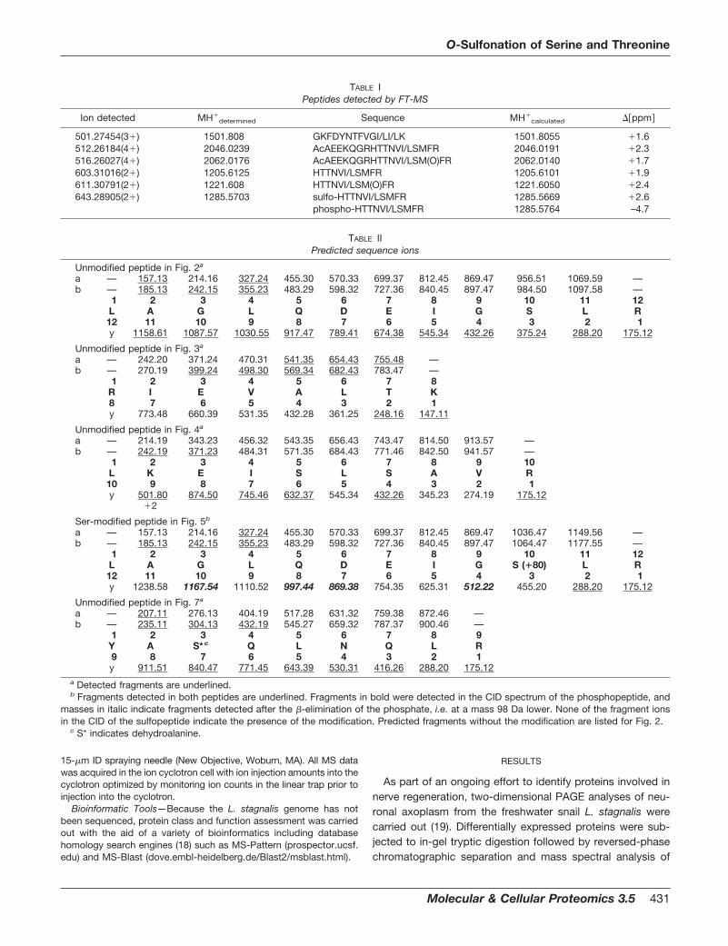
TABLE IPeptides detected by FT-MS
Ion detected MH�determined Sequence MH�
calculated ��ppm�
501.27454(3�) 1501.808 GKFDYNTFVGI/LI/LK 1501.8055 �1.6512.26184(4�) 2046.0239 AcAEEKQGRHTTNVI/LSMFR 2046.0191 �2.3516.26027(4�) 2062.0176 AcAEEKQGRHTTNVI/LSM(O)FR 2062.0140 �1.7603.31016(2�) 1205.6125 HTTNVI/LSMFR 1205.6101 �1.9611.30791(2�) 1221.608 HTTNVI/LSM(O)FR 1221.6050 �2.4643.28905(2�) 1285.5703 sulfo-HTTNVI/LSMFR 1285.5669 �2.6
phospho-HTTNVI/LSMFR 1285.5764 –4.7
TABLE IIPredicted sequence ions
Unmodified peptide in Fig. 2a
a — 157.13 214.16 327.24 455.30 570.33 699.37 812.45 869.47 956.51 1069.59 —b — 185.13 242.15 355.23 483.29 598.32 727.36 840.45 897.47 984.50 1097.58 —
1 2 3 4 5 6 7 8 9 10 11 12L A G L Q D E I G S L R12 11 10 9 8 7 6 5 4 3 2 1y 1158.61 1087.57 1030.55 917.47 789.41 674.38 545.34 432.26 375.24 288.20 175.12
Unmodified peptide in Fig. 3a
a — 242.20 371.24 470.31 541.35 654.43 755.48 —b — 270.19 399.24 498.30 569.34 682.43 783.47 —
1 2 3 4 5 6 7 8R I E V A L T K8 7 6 5 4 3 2 1y 773.48 660.39 531.35 432.28 361.25 248.16 147.11
Unmodified peptide in Fig. 4a
a — 214.19 343.23 456.32 543.35 656.43 743.47 814.50 913.57 —b — 242.19 371.23 484.31 571.35 684.43 771.46 842.50 941.57 —
1 2 3 4 5 6 7 8 9 10L K E I S L S A V R10 9 8 7 6 5 4 3 2 1y 501.80 874.50 745.46 632.37 545.34 432.26 345.23 274.19 175.12
�2
Ser-modified peptide in Fig. 5b
a — 157.13 214.16 327.24 455.30 570.33 699.37 812.45 869.47 1036.47 1149.56 —b — 185.13 242.15 355.23 483.29 598.32 727.36 840.45 897.47 1064.47 1177.55 —
1 2 3 4 5 6 7 8 9 10 11 12L A G L Q D E I G S (�80) L R12 11 10 9 8 7 6 5 4 3 2 1y 1238.58 1167.54 1110.52 997.44 869.38 754.35 625.31 512.22 455.20 288.20 175.12
Unmodified peptide in Fig. 7a
a — 207.11 276.13 404.19 517.28 631.32 759.38 872.46 —b — 235.11 304.13 432.19 545.27 659.32 787.37 900.46 —
1 2 3 4 5 6 7 8 9Y A S* c Q L N Q L R9 8 7 6 5 4 3 2 1y 911.51 840.47 771.45 643.39 530.31 416.26 288.20 175.12
a Detected fragments are underlined.b Fragments detected in both peptides are underlined. Fragments in bold were detected in the CID spectrum of the phosphopeptide, and
masses in italic indicate fragments detected after the �-elimination of the phosphate, i.e. at a mass 98 Da lower. None of the fragment ionsin the CID of the sulfopeptide indicate the presence of the modification. Predicted fragments without the modification are listed for Fig. 2.
c S* indicates dehydroalanine.
O-Sulfonation of Serine and Threonine
Molecular & Cellular Proteomics 3.5 431
the digest mixture. Peptide sequences were deduced by denovo interpretation of their CID mass spectra and used toquery genomic databases to assign tentative homologies insequenced genomes where possible. This effort revealed anumber of protein spots with extensive homology to interme-diate filament proteins from other mollusks, including Helix(20) and Aplysia (21).
During the course of carrying out de novo sequencing ofthese particular proteins, a number of digest componentswere discovered from analysis of an LC-ESI-CID-MS experi-ment that displayed identical mass values for their entire CIDsequence ion series (viz. identical fragmentation patterns), buteluted with significantly different chromatographic retentiontimes (� 1–5 min) and possessed different protonatedmolecular masses. In fact, all of the later-eluting componentsof these identical fragmentation pattern pairs displayed an80-Da increment in their measured molecular mass. Carefulanalysis of these LC-CID-MS spectra revealed seven suchtryptic peptides in one particular digest of a protein homolo-gous to intermediate filament proteins: 47SSISPGVYQQLSSS-
GITDFK66, 131KVIDELASSK140, 147LAGLQDEIGSLR158,159ELIVTYESQAK169, 304YASQLNQLR312, 340NAAYAELAT-R349, and 428TLVEQAIGTQSK439. (Note that the Ile/Leu as-signments and sequence positions indicated here are basedon cDNA sequence information obtained later). In anotherdigest, the modified peptide 8HTTNV[I/L]SMFR17 was ob-served. This protein was identified as myosin light chain andthe sequence positions are assigned based on the observa-tion of the N-terminal peptide in the digest (see Table I).
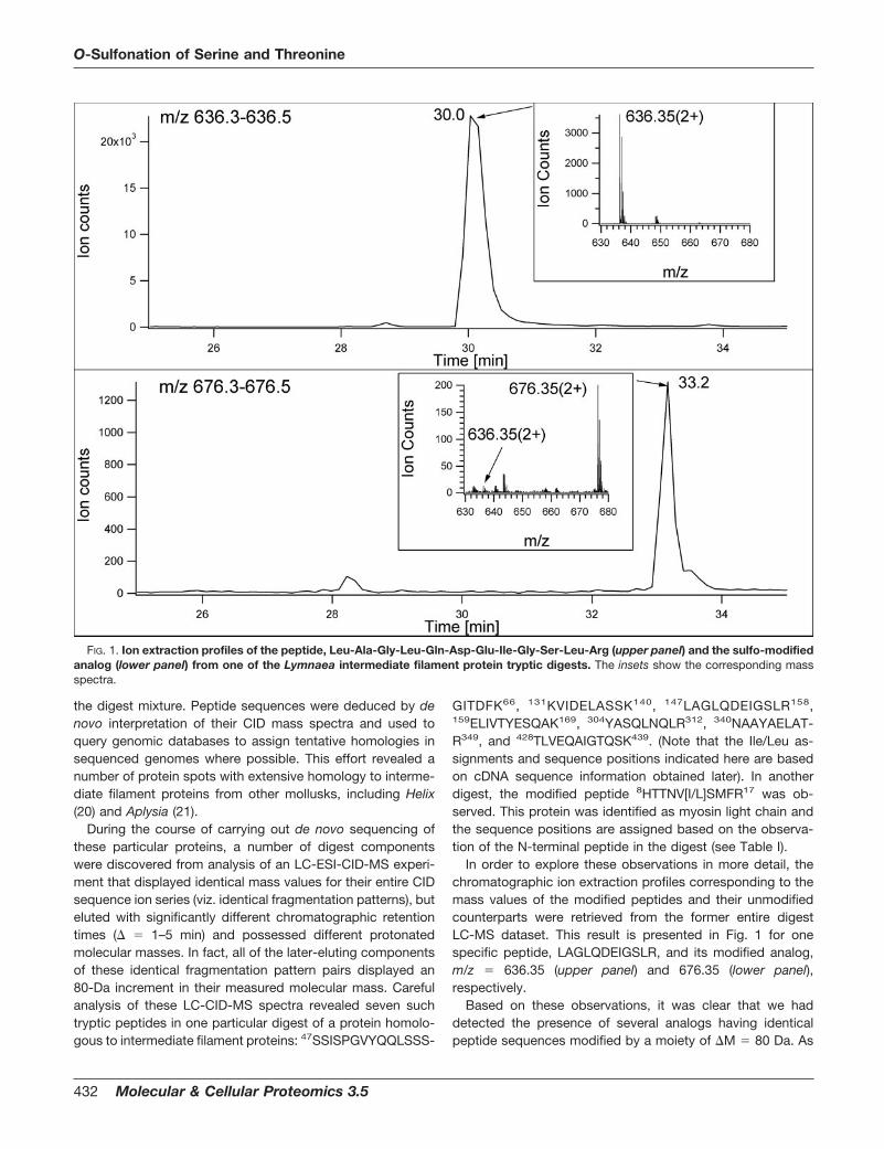
In order to explore these observations in more detail, thechromatographic ion extraction profiles corresponding to themass values of the modified peptides and their unmodifiedcounterparts were retrieved from the former entire digestLC-MS dataset. This result is presented in Fig. 1 for onespecific peptide, LAGLQDEIGSLR, and its modified analog,m/z 636.35 (upper panel) and 676.35 (lower panel),respectively.
Based on these observations, it was clear that we haddetected the presence of several analogs having identicalpeptide sequences modified by a moiety of �M 80 Da. As
FIG. 1. Ion extraction profiles of the peptide, Leu-Ala-Gly-Leu-Gln-Asp-Glu-Ile-Gly-Ser-Leu-Arg (upper panel) and the sulfo-modifiedanalog (lower panel) from one of the Lymnaea intermediate filament protein tryptic digests. The insets show the corresponding massspectra.
O-Sulfonation of Serine and Threonine
432 Molecular & Cellular Proteomics 3.5
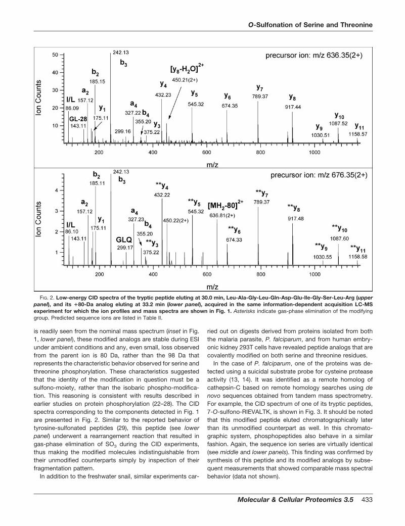
is readily seen from the nominal mass spectrum (inset in Fig.1, lower panel), these modified analogs are stable during ESIunder ambient conditions and any, even small, loss observedfrom the parent ion is 80 Da, rather than the 98 Da thatrepresents the characteristic behavior observed for serine andthreonine phosphorylation. These characteristics suggestedthat the identity of the modification in question must be asulfono-moiety, rather than the isobaric phospho-modifica-tion. This reasoning is consistent with results described inearlier studies on protein phosphorylation (22–28). The CIDspectra corresponding to the components detected in Fig. 1are presented in Fig. 2. Similar to the reported behavior oftyrosine-sulfonated peptides (29), this peptide (see lowerpanel) underwent a rearrangement reaction that resulted ingas-phase elimination of SO3 during the CID experiments,thus making the modified molecules indistinguishable fromtheir unmodified counterparts simply by inspection of theirfragmentation pattern.
In addition to the freshwater snail, similar experiments car-
ried out on digests derived from proteins isolated from boththe malaria parasite, P. falciparum, and from human embry-onic kidney 293T cells have revealed peptide analogs that arecovalently modified on both serine and threonine residues.
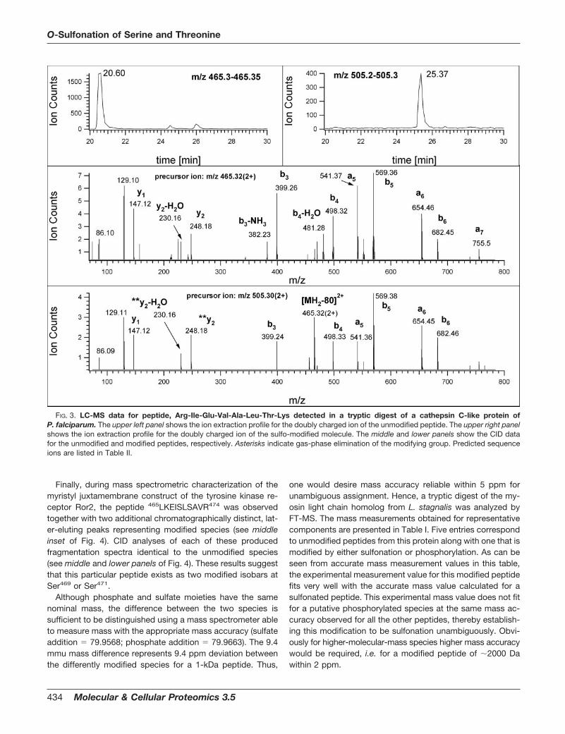
In the case of P. falciparum, one of the proteins was de-tected using a suicidal substrate probe for cysteine proteaseactivity (13, 14). It was identified as a remote homolog ofcathepsin-C based on remote homology searches using denovo sequences obtained from tandem mass spectrometry.For example, the CID spectrum of one of its tryptic peptides,7-O-sulfono-RIEVALTK, is shown in Fig. 3. It should be notedthat this modified peptide eluted chromatographically laterthan its unmodified counterpart as well. In this chromato-graphic system, phosphopeptides also behave in a similarfashion. Again, the sequence ion series are virtually identical(see middle and lower panels). This finding was confirmed bysynthesis of this peptide and its modified analogs by subse-quent measurements that showed comparable mass spectralbehavior (data not shown).
FIG. 2. Low-energy CID spectra of the tryptic peptide eluting at 30.0 min, Leu-Ala-Gly-Leu-Gln-Asp-Glu-Ile-Gly-Ser-Leu-Arg (upperpanel), and its �80-Da analog eluting at 33.2 min (lower panel), acquired in the same information-dependent acquisition LC-MSexperiment for which the ion profiles and mass spectra are shown in Fig. 1. Asterisks indicate gas-phase elimination of the modifyinggroup. Predicted sequence ions are listed in Table II.
O-Sulfonation of Serine and Threonine
Molecular & Cellular Proteomics 3.5 433
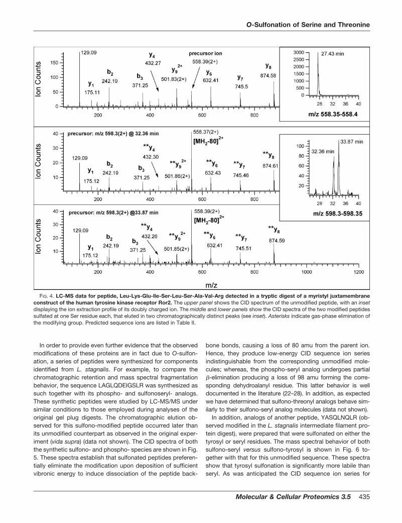
Finally, during mass spectrometric characterization of themyristyl juxtamembrane construct of the tyrosine kinase re-ceptor Ror2, the peptide 465LKEISLSAVR474 was observedtogether with two additional chromatographically distinct, lat-er-eluting peaks representing modified species (see middleinset of Fig. 4). CID analyses of each of these producedfragmentation spectra identical to the unmodified species(see middle and lower panels of Fig. 4). These results suggestthat this particular peptide exists as two modified isobars atSer469 or Ser471.
Although phosphate and sulfate moieties have the samenominal mass, the difference between the two species issufficient to be distinguished using a mass spectrometer ableto measure mass with the appropriate mass accuracy (sulfateaddition 79.9568; phosphate addition 79.9663). The 9.4mmu mass difference represents 9.4 ppm deviation betweenthe differently modified species for a 1-kDa peptide. Thus,
one would desire mass accuracy reliable within 5 ppm forunambiguous assignment. Hence, a tryptic digest of the my-osin light chain homolog from L. stagnalis was analyzed byFT-MS. The mass measurements obtained for representativecomponents are presented in Table I. Five entries correspondto unmodified peptides from this protein along with one that ismodified by either sulfonation or phosphorylation. As can beseen from accurate mass measurement values in this table,the experimental measurement value for this modified peptidefits very well with the accurate mass value calculated for asulfonated peptide. This experimental mass value does not fitfor a putative phosphorylated species at the same mass ac-curacy observed for all the other peptides, thereby establish-ing this modification to be sulfonation unambiguously. Obvi-ously for higher-molecular-mass species higher mass accuracywould be required, i.e. for a modified peptide of �2000 Dawithin 2 ppm.
FIG. 3. LC-MS data for peptide, Arg-Ile-Glu-Val-Ala-Leu-Thr-Lys detected in a tryptic digest of a cathepsin C-like protein ofP. falciparum. The upper left panel shows the ion extraction profile for the doubly charged ion of the unmodified peptide. The upper right panelshows the ion extraction profile for the doubly charged ion of the sulfo-modified molecule. The middle and lower panels show the CID datafor the unmodified and modified peptides, respectively. Asterisks indicate gas-phase elimination of the modifying group. Predicted sequenceions are listed in Table II.
O-Sulfonation of Serine and Threonine
434 Molecular & Cellular Proteomics 3.5
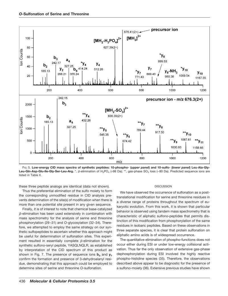
In order to provide even further evidence that the observedmodifications of these proteins are in fact due to O-sulfon-ation, a series of peptides were synthesized for componentsidentified from L. stagnalis. For example, to compare thechromatographic retention and mass spectral fragmentationbehavior, the sequence LAGLQDEIGSLR was synthesized assuch together with its phospho- and sulfonoseryl- analogs.These synthetic peptides were studied by LC-MS/MS undersimilar conditions to those employed during analyses of theoriginal gel plug digests. The chromatographic elution ob-served for this sulfono-modified peptide occurred later thanits unmodified counterpart as observed in the original exper-iment (vida supra) (data not shown). The CID spectra of boththe synthetic sulfono- and phospho- species are shown in Fig.5. These spectra establish that sulfonated peptides preferen-tially eliminate the modification upon deposition of sufficientvibronic energy to induce dissociation of the peptide back-
bone bonds, causing a loss of 80 amu from the parent ion.Hence, they produce low-energy CID sequence ion seriesindistinguishable from the corresponding unmodified mole-cules; whereas, the phospho-seryl analog undergoes partial�-elimination producing a loss of 98 amu forming the corre-sponding dehydroalanyl residue. This latter behavior is welldocumented in the literature (22–28). In addition, as expectedwe have determined that sulfono-threonyl analogs behave sim-ilarly to their sulfono-seryl analog molecules (data not shown).
In addition, analogs of another peptide, YASQLNQLR (ob-served modified in the L. stagnalis intermediate filament pro-tein digest), were prepared that were sulfonated on either thetyrosyl or seryl residues. The mass spectral behavior of bothsulfono-seryl versus sulfono-tyrosyl is shown in Fig. 6 to-gether with that for this unmodified sequence. These spectrashow that tyrosyl sulfonation is significantly more labile thanseryl. As was anticipated the CID sequence ion series for
FIG. 4. LC-MS data for peptide, Leu-Lys-Glu-Ile-Ser-Leu-Ser-Ala-Val-Arg detected in a tryptic digest of a myristyl juxtamembraneconstruct of the human tyrosine kinase receptor Ror2. The upper panel shows the CID spectrum of the unmodified peptide, with an insetdisplaying the ion extraction profile of its doubly charged ion. The middle and lower panels show the CID spectra of the two modified peptidessulfated at one Ser residue each, that eluted in two chromatographically distinct peaks (see inset). Asterisks indicate gas-phase elimination ofthe modifying group. Predicted sequence ions are listed in Table II.
O-Sulfonation of Serine and Threonine
Molecular & Cellular Proteomics 3.5 435
these three peptide analogs are identical (data not shown).Thus the preferential elimination of the sulfo moiety to form
the corresponding unmodified residue in CID analysis pre-vents determination of the site(s) of modification when there ismore than one potential site present in any given sequence.
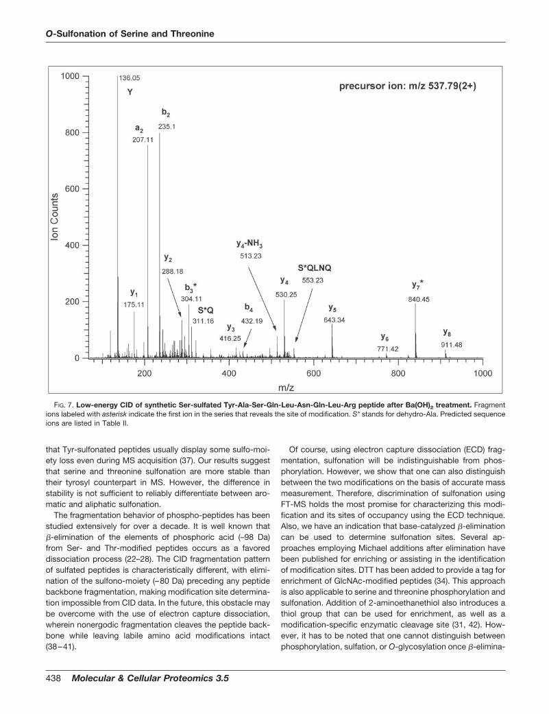
Finally, it is of interest to note that chemical base-catalyzed�-elimination has been used extensively in combination withmass spectrometry for the analysis of serine and threoninephosphorylation (29–31) and O-glycosylation (32–34). There-fore, we attempted to employ the same strategy on our syn-thetic sulfopeptides to ascertain whether this approach mightbe useful for determination of sulfonation sites. This experi-ment resulted in essentially complete �-elimination for thesynthetic sulfono-seryl peptide, YASQLNQLR, as establishedby interpretation of the CID spectrum of this product asshown in Fig. 7. The presence of sequence ions b3 and y7
confirm the formation and presence of 3-dehydroalanyl resi-due, demonstrating that this approach could be employed todetermine sites of serine and threonine O-sulfonation.
DISCUSSION
We have observed the occurrence of sulfonation as a post-translational modification for serine and threonine residues ina diverse range of proteins throughout the spectrum of eu-karyotic evolution. From this work, it is shown that particularbehavior is observed using tandem mass spectrometry that ischaracteristic of aliphatic sulfono-peptides that permits dis-tinction of this modification from phosphorylation of the sameresidues in isobaric peptides. Based on these observations inthree separate species, it is clear that protein sulfonation onaliphatic amino acids is of widespread occurrence.
The quantitative elimination of phospho-functions does notoccur either during ESI or under low-energy collisional acti-vation. Thus far the only observation of extensive gas-phasedephosphorylation during ESI involved the highly reactivephospho-histidine species (35). Therefore, the observationsdescribed above appear to be diagnostic for the presence ofa sulfono-moiety (36). Extensive previous studies have shown
FIG. 5. Low-energy CID mass spectra of synthetic peptides: 10-phospho- (upper panel) and 10-sulfo- (lower panel) Leu-Ala-Gly-Leu-Gln-Asp-Glu-Ile-Gly-Ser-Leu-Arg. *, �-elimination of H3PO4 (–98 Da); **, gas-phase SO3 loss (–80 Da). Predicted sequence ions arelisted in Table II.
O-Sulfonation of Serine and Threonine
436 Molecular & Cellular Proteomics 3.5
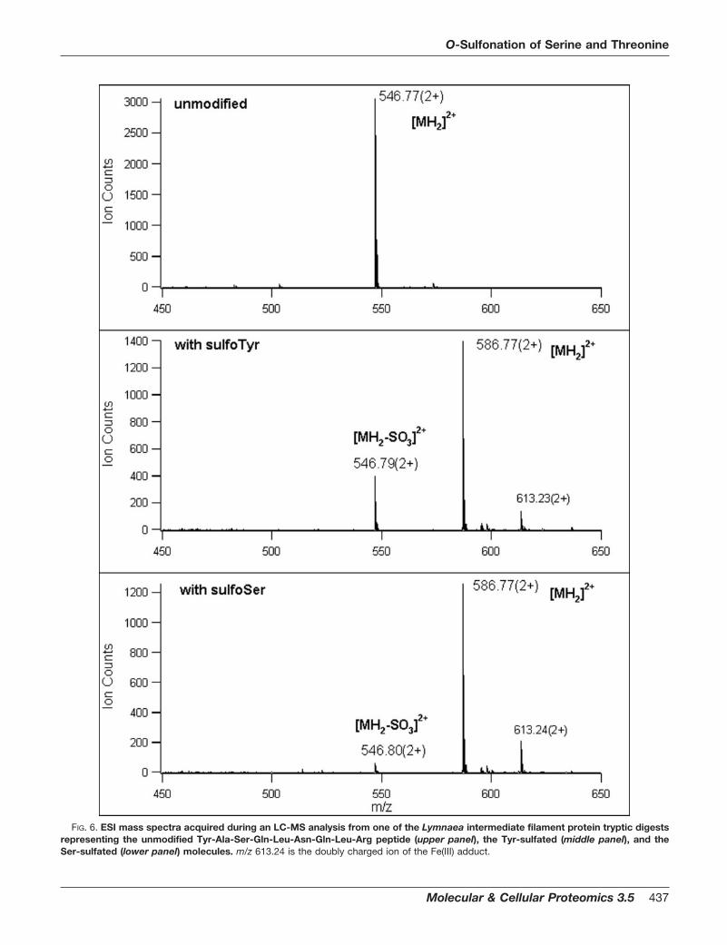
FIG. 6. ESI mass spectra acquired during an LC-MS analysis from one of the Lymnaea intermediate filament protein tryptic digestsrepresenting the unmodified Tyr-Ala-Ser-Gln-Leu-Asn-Gln-Leu-Arg peptide (upper panel), the Tyr-sulfated (middle panel), and theSer-sulfated (lower panel) molecules. m/z 613.24 is the doubly charged ion of the Fe(III) adduct.
O-Sulfonation of Serine and Threonine
Molecular & Cellular Proteomics 3.5 437
that Tyr-sulfonated peptides usually display some sulfo-moi-ety loss even during MS acquisition (37). Our results suggestthat serine and threonine sulfonation are more stable thantheir tyrosyl counterpart in MS. However, the difference instability is not sufficient to reliably differentiate between aro-matic and aliphatic sulfonation.
The fragmentation behavior of phospho-peptides has beenstudied extensively for over a decade. It is well known that�-elimination of the elements of phosphoric acid (–98 Da)from Ser- and Thr-modified peptides occurs as a favoreddissociation process (22–28). The CID fragmentation patternof sulfated peptides is characteristically different, with elimi-nation of the sulfono-moiety (–80 Da) preceding any peptidebackbone fragmentation, making modification site determina-tion impossible from CID data. In the future, this obstacle maybe overcome with the use of electron capture dissociation,wherein nonergodic fragmentation cleaves the peptide back-bone while leaving labile amino acid modifications intact(38–41).
Of course, using electron capture dissociation (ECD) frag-mentation, sulfonation will be indistinguishable from phos-phorylation. However, we show that one can also distinguishbetween the two modifications on the basis of accurate massmeasurement. Therefore, discrimination of sulfonation usingFT-MS holds the most promise for characterizing this modi-fication and its sites of occupancy using the ECD technique.Also, we have an indication that base-catalyzed �-eliminationcan be used to determine sulfonation sites. Several ap-proaches employing Michael additions after elimination havebeen published for enriching or assisting in the identificationof modification sites. DTT has been added to provide a tag forenrichment of GlcNAc-modified peptides (34). This approachis also applicable to serine and threonine phosphorylation andsulfonation. Addition of 2-aminoethanethiol also introduces athiol group that can be used for enrichment, as well as amodification-specific enzymatic cleavage site (31, 42). How-ever, it has to be noted that one cannot distinguish betweenphosphorylation, sulfation, or O-glycosylation once �-elimina-
FIG. 7. Low-energy CID of synthetic Ser-sulfated Tyr-Ala-Ser-Gln-Leu-Asn-Gln-Leu-Arg peptide after Ba(OH)2 treatment. Fragmentions labeled with asterisk indicate the first ion in the series that reveals the site of modification. S* stands for dehydro-Ala. Predicted sequenceions are listed in Table II.
O-Sulfonation of Serine and Threonine
438 Molecular & Cellular Proteomics 3.5
tion is performed. In addition, it has been reported that a smallpercentage of unmodified Ser residues also display water lossunder commonly used �-elimination conditions (43). Thus,this approach would be most appropriate for assigning theexact site(s) of modification once the presence of sulfationhas been established.
Another potential discriminatory method that could be usedto differentiate between phosphorylation and sulfonation is byperforming fragmentation of the species in negative ion massspectrometry. Phosphorylation produces a characteristicnegative ion at m/z 79 (25), whereas the sulfo-group forms anequivalent negative ion fragment at m/z 80 (28).
The identification of serine/threonine sulfonations in threeproteins from very different organisms suggests that thismodification is widespread and may occur ubiquitously in alleukaryotes. Moreover, the three modified proteins we reportare targeted to distinct cell compartments, cytoplasm (Lym-naea intermediate filament), location unknown (Plasmodiumcathepsin-like), and plasma membrane (human Ror2), sug-gesting that serine/threonine sulfonation occurs both in theendoplasmic reticulum continuum and the cytoplasm.
There are a number of sulfotransferases known, and theycan be divided into two main groups: a membrane-boundclass that is found in the Golgi and is involved in the modifi-cation of proteoglycans and polysaccharides (including theenzymes involved in tyrosine derivatization) and a solublecytoplasmic class that modifies small molecules, such asestrogen (1). PAPS is the sulfate group donor utilized by thesesulfotransferases for all sulfations described thus far (2, 4, 5).Therefore, although we do not yet know which enzyme(s) mightsulfate serine/threonine in proteins, one or more of the alreadyknown sulfotransferases is the most likely candidate. We can-not, however, rule out that a different as yet unidentified enzymeis involved and conceivably a different donor compound. Similarconsiderations apply to any sulfatases, assuming that thesemodifications are physiologically reversible.
Finally, we should note that this study does not identify afunction for any of the observed modifications. O-Sulfonation oftyrosine is thought to aid in protein-protein interactions (4), anda similar function could be true for the aliphatic modifications.
Acknowledgments—We are indebted to Vlad Zabrouskov (Thermo-Finnigan) for making the FT-MS measurements.
* Financial support was provided by National Institutes of HealthNational Center for Research Resources Grant RR 01614 (to A. L. B.),the USA-Israel Binational Science Foundation (to M. F. and A. L. B.),and by the Eotvos Scholarship of the Hungarian Scholarship Board (toZ. D.). The costs of publication of this article were defrayed in part bythe payment of page charges. This article must therefore be herebymarked “advertisement” in accordance with 18 U.S.C. Section 1734solely to indicate this fact.
b Current address: Proteomics Research Group, Biological Re-search Center of the Hungarian Academy of Sciences, H-6726,Szeged, Temesvari krt. 62, Hungary.
e Current address: Department of Biochemistry, SouthwesternMedical Center, University of Texas, Dallas, TX 75390-9050.
g Current address: Department of Pathology, Stanford UniversityMedical School, Stanford, CA 94305-5324.
i To whom correspondence should be addressed: Department ofPharmaceutical Chemistry, University of California, 513 ParnassusAvenue, San Francisco, CA 94143-0446. Tel.: 415-476-5641; Fax:415-476-0688; E-mail: [email protected].
REFERENCES
1. Strott, C. A. (2002) Sulfonation and molecular action. Endocr. Rev. 23,703–732
2. Robbins, P., and Lippmann, F. (1956) Identification of enzymatically activesulfate as adenosine-3�-phosphate-5�-phospho-sulfate. J. Am. Chem.Soc. 78, 2652–2653
3. Huxtable, R. J. (1986) in Biochemistry of Sulfur, Plenum, New York.4. Moore, K. L. (2003) The biology and enzymology of protein tyrosine O-
sulfation. J. Biol. Chem. 278, 24243–242465. Kehoe, J. W., and Bertozzi, C. R. (2000) Tyrosine sulfation: A modulator of
extracellular protein-protein interactions. Chem. Biol. 7, R57–616. Beisswanger, R., Corbeil, D., Vannier, C., Thiele, C., Dohrmann, U., Kellner,
R., Ashman, K., Niehrs, C., and Huttner, W. B. (1998) Existence of distincttyrosylprotein sulfotransferase genes: Molecular characterization of ty-rosylprotein sulfotransferase-2. Proc. Natl. Acad. Sci. U. S. A. 95,11134–11139
7. Kakuta, Y., Pedersen, L. G., Pedersen, L. C., and Negishi, M. (1998)Conserved structural motifs in the sulfotransferase family. Trends Bio-chem. Sci. 23, 129–130
8. Parenti, G., Meroni, G., and Ballabio, A. (1997) The sulfatase gene family.Curr. Opin. Genet. Dev. 7, 386–391
9. Cohen, P. (2002) The origins of protein phosphorylation. Nat. Cell Biol. 4,E127–130
10. Huttner, W. B. (1984) Determination and occurrence of tyrosine O-sulfate inproteins. Methods Enzymol. 107, 200–223
11. Krishna, R., and Wold, F. (1998) Post-translation modifications, in ProteinsAnalysis and Design (Angeletti, R. H., ed) pp. 121–206, Academic Press,San Diego
12. Medzihradszky, K. F., and Burlingame, A. L. (1994) The advantages andversatility of a high energy collision-induced dissociation based strategyfor the sequence and structural determination of proteins. Methods 6,284–303
13. Greenbaum, D., Medzihradszky, K. F., Burlingame, A., and Bogyo, M.(2000) Epoxide electrophiles as activity-dependent cysteine proteaseprofiling and discovery tools. Chem. Biol. 7, 569–581
14. Greenbaum, D. C., Baruch, A., Grainger, M., Bozdeck, Z., Medzihradszky,K. F., Engel, J., DeRisi, J., Holder, A. A., and Bogyo, M. (2002) A role forthe protease falcipain 1 in host cell invasion by the human malariaparasite. Science 298, 2002–2006
15. Ball, H. L., and Mascagni, P. (1996) Chemical synthesis and purification ofproteins: A methodology. Int. J. Pept. Protein Res. 48, 31–47
16. Previero, A., Cavadore, J. C., Torreilles, J., and Coletti-Previero, M. A.(1979) Specific sulfonation of tyrosine, tryptophan and hydroxy-aminoacids in peptides. Biochim. Biophys. Acta 581, 276–282
17. Masiakowski, P., and Carroll, R. D. (1992) A novel family of cell surfacereceptors with tyrosine kinase-like domain. J. Biol. Chem. 267,26181–26190
18. Huang, L., Jacob, R. J., Pegg, S. C., Baldwin, M. A., Wang, C. C., Burl-ingame, A. L., and Babbitt, P. C. (2001) Functional assignment of the 20S proteasome from Trypanosoma brucei using mass spectrometry andnew bioinformatics approaches. J. Biol. Chem. 276, 28327–28339
19. Perlson, E., Hanz, S., Medzihradszky, K. F., Burlingame, A. L., and Fainzil-ber, M. (2004) From snails to sciatic nerve: Retrograde injury signalingfrom axon to soma in lesioned neurons. J. Neurobiol. 58, 287–294
20. Dodemont, H., Riemer, D., and Weber, K. (1990) Structure of an inverte-brate gene encoding cytoplasmic intermediate filament (IF) proteins:Implications for the origin and the diversification of IF proteins. EMBO J.9, 4083–4094
21. Sweatt, J. D., Kennedy, T. E., Wager-Smith, K., Gawinowicz, M. A., Barzilai,A., Karl, K. A., and Kandel, E. R. (1989) Development of a database ofamino acid sequences for proteins identified and isolated on two-dimen-sional polyacrylamide gels. Electrophoresis 10, 152–157
22. Gibson, B. W., Falick, A. M., Burlingame, A. L., Nadasdi, L., Nguyen, A., and
O-Sulfonation of Serine and Threonine
Molecular & Cellular Proteomics 3.5 439
Kenyon, G. L. (1987) Liquid secondary ionization mass-spectrometriccharacterization of 2 synthetic phosphotyrosine-containing peptides.J. Am. Chem. Soc. 109, 5343–5348
23. Payne, D. M., Rossomando, A. J., Martino, P., Erickson, A. K., Her, J. H.,Shabanowitz, J., Hunt, D. F., Weber, M. J., and Sturgill, T. W. (1991)Identification of the regulatory phosphorylation sites in pp42/mitogen-activated protein kinase (MAP kinase). EMBO J. 10, 885–892
24. Tsutakawa, S. E., Medzihradszky, K. F., Flint, A. J., Burlingame, A. L., andKoshland, D. E., Jr. (1995) Determination of in vivo phosphorylation sitesin protein kinase C. J. Biol. Chem. 270, 26807–26812
25. Carr, S. A., Huddleston, M. J., and Annan, R. S. (1996) Selective detectionand sequencing of phosphopeptides at the femtomole level by massspectrometry. Anal. Biochem. 239, 180–192
26. de Carvalho, M. G., McCormack, A. L., Olson, E., Ghomashchi, F., Gelb,M. H., Yates, J. R., 3rd, and Leslie, C. C. (1996) Identification of phos-phorylation sites of human 85-kDa cytosolic phospholipase A2 ex-pressed in insect cells and present in human monocytes. J. Biol. Chem.271, 6987–6997
27. Neville, D. C. A., Rozanas, C. R., Price, E. M., Gruis, D. B., Verkman, A. S.,and Townsend, R. R. (1997) Evidence for phosphorylation of serine 753in CFTR using a novel metal-ion affinity resin and matrix-assisted laserdesorption mass spectrometry. Protein Sci. 6, 2436–2445
28. Bean, M. F., Annan, R. S., Hemling, M. E., Mentzer, M., Huddleston, M. J.,and Carr, S. A. (1995) LC-MS methods for selective detection of post-translational modifications in proteins: Glycosylation, phosphorylation,sulfation and acylation, in Techniques in Protein Chemistry (Crabb, J. W.,ed) pp. 107–116, Academic, San Diego
29. Oda, Y., Nagasu, T., and Chait, B. T. (2001) Enrichment analysis of phos-phorylated proteins as a tool for probing the phosphoproteome. Nat.Biotechnol. 19, 379–382
30. Goshe, M. B., Conrads, T. P., Panisko, E. A., Angell, N. H., Veenstra, T. D.,and Smith, R. D. (2001) Phosphoprotein isotope-coded affinity tag ap-proach for isolating and quantitating phosphopeptides in proteome-wideanalyses. Anal. Chem. 73, 2578–2586
31. Thompson, A. J., Hart, S. R., Franz, C., Barnouin, K., Ridley, A., andCramer, R. (2003) Characterization of protein phosphorylation by massspectrometry using immobilized metal ion affinity chromatography withon-resin �-elimination and Michael addition. Anal. Chem. 75, 3232–3243
32. Rademaker, G. J., Pergantis, S. A., Blok-Tip, L., Langridge, J. I., Kleen, A.,and Thomas-Oates, J. E. (1998) Mass spectrometric determination of thesites of O-glycan attachment with low picomolar sensitivity. Anal. Bio-chem. 257, 149–160
33. Greis, K. D., Hayes, B. K., Comer, F. I., Kirk, M., Barnes, S., Lowary, T. L.,
and Hart, G. W. (1996) Selective detection and site-analysis of O-Glc-NAc-modified glycopeptides by �-elimination and tandem electrospraymass spectrometry. Anal. Biochem. 234, 38–49
34. Wells, L., Vosseller, K., Cole, R. N., Cronshaw, J. M., Matunis, M. J., andHart, G. W. (2002) Mapping sites of O-GlcNAc modification using affinitytags for serine and threonine post-translational modifications. Mol. Cell.Proteomics 1, 791–804
35. Medzihradszky, K. F., Senderowicz, L., Wang, P., and Turck, C. W. (1997)Synthesis and characterization of histidine-phosphorylated peptides.Protein Sci. 6, 1405–1411
36. Nemeth-Cawley, J. F., Karnik, S., and Rouse, J. C. (2001) Analysis ofsulfated peptides using positive electrospray ionization tandem massspectrometry. J. Mass Spectrom. 36, 1301–1311
37. Wolfender, J. L., Chu, F., Ball, H., Wolfender, F., Fainzilber, M., Baldwin,M. A., and Burlingame, A. L. (1999) Identification of tyrosine sulfation inConus pennaceus conotoxins �-PnIA and �-PnIB: Further investigationof labile sulfo- and phosphopeptides by electrospray, matrix-assistedlaser desorption/ionization (MALDI) and atmospheric pressure MALDImass spectrometry. J. Mass Spectrom. 34, 447–454
38. Kelleher, N. L., Zubarev, R. A., Bush, K., Furie, B., Furie, B. C., McLafferty,F. W., and Walsh, C. T. (1999) Localization of labile posttranslationalmodifications by electron capture dissociation: The case of �-carboxy-glutamic acid. Anal. Chem. 71, 4250–4253
39. Mirgorodskaya, E., Roepstorff, P., and Zubarev, R. A. (1999) Localization ofO-glycosylation sites in peptides by electron capture dissociation in aFourier transform mass spectrometer. Anal. Chem. 71, 4431–4436
40. Stensballe, A., Jensen, O. N., Olsen, J. V., Haselmann, K. F., and Zubarev,R. A. (2000) Electron capture dissociation of singly and multiply phos-phorylated peptides. Rapid Commun. Mass Spectrom. 14, 1793–1800
41. Shi, S. D., Hemling, M. E., Carr, S. A., Horn, D. M., Lindh, I., and McLafferty,F. W. (2001) Phosphopeptide/phosphoprotein mapping by electron cap-ture dissociation mass spectrometry. Anal. Chem. 73, 19–22
42. Rusnak, F., Zhou, J., and Hathaway, G. M. (2003) Identification of phos-phorylated and glycosylated sites in peptides by chemically targetedproteolysis. J. Biomol. Tech. 13, 228–237
43. McLachlin, D. T, and Chait B. T. (2003) Improved �-elimination-basedaffinity purification strategy for enrichment of phosphopeptides. Anal.Chem. 75, 6826–6836
44. Perlson, E., Medzihradszky, K. F., Darula, Z., Munno, D. W., Syed, N. I.,Burlingame, A. L., and Fainzilber, M. (2004) Differential proteomics re-veals multiple components in retrogradely transported axoplasm afternerve injury. Mol. Cell. Proteomics 3, 510–520
O-Sulfonation of Serine and Threonine
440 Molecular & Cellular Proteomics 3.5