the head and neck. upper respiratory tract pg 583
Post on 22-Dec-2015
218 views
TRANSCRIPT
The Head and Neck

Upper Respiratory Tract
Pg 583

The Nose
• Only external part of respiratory tract
• Made of nasal bones, connective tissue and hyaline cartilage
• External Nares = Nostrils: – openings into nasal cavity
• Internal Nares = Choanae:– openings between nasal
cavity and nasopharynx

• Vestibule –space just inside external nares
• Skin lining contains:– Sebaceous glands -
greasy secretion collect dirt, lubricate, kill bacteria
– Sweat glands -acidic, slows growth of bacteria
– Hair follicles –trap smaller particles of dirt and dust
• Vibrissae – nose hairs filtering larger particles from air
Entrance to the Nasal Cavity

Nasal Cavity
• Part of respiratory passage• Boundaries
– Roof = ethmoid bone (cribiform plate)– Floor = hard palate & soft palate
• Hard palate = maxilla (palatine process) + palatine (horizontal plate)• Soft palate = skeletal muscle ending in uvula
– Lateral walls = nasal bones, superior+ middle nasal conchae (ethmoid bone), inferior nasal conchae, maxilla, palatine bone
– Nasal Septum = divides cavity into 2• Vomer & Perpendicular Plate of Ethmoid • Cartilage

Nasal Cavity
Pg 584

Nasal Cavity
• Functions– Airway of respiratory tract– Moisten and Warm air– Filter air– Resonating chamber for speech– Houses olfactory receptors
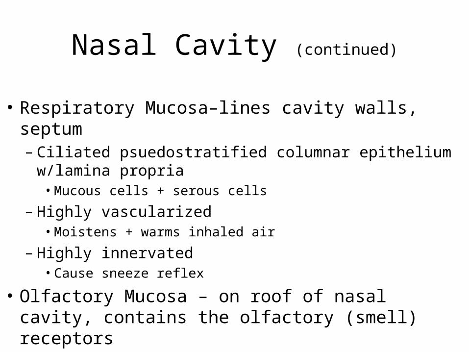
Nasal Cavity (continued)
• Respiratory Mucosa–lines cavity walls, septum– Ciliated psuedostratified columnar epithelium
w/lamina propria• Mucous cells + serous cells
– Highly vascularized• Moistens + warms inhaled air
– Highly innervated• Cause sneeze reflex
• Olfactory Mucosa – on roof of nasal cavity, contains the olfactory (smell) receptors
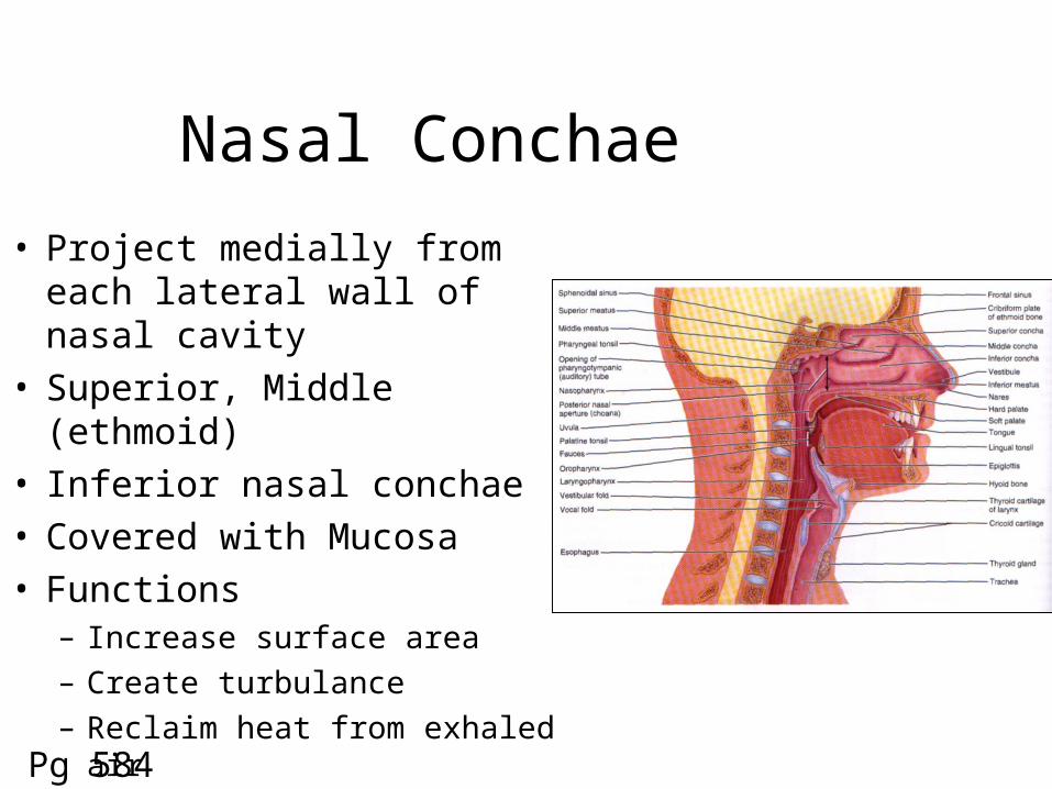
Nasal Conchae
• Project medially from each lateral wall of nasal cavity
• Superior, Middle (ethmoid)• Inferior nasal conchae• Covered with Mucosa• Functions
– Increase surface area– Create turbulance– Reclaim heat from exhaled air
Pg 584

Paranasal Sinuses• Air-filled sacs surrounding nasal cavity• Extensions of nasal cavity, continuous with it• Same respiratory mucosal lining• In Frontal, Ethmoid, Sphenoid, Maxillae bones• Function: Warm + Filter air, Lightens skull
Pg 16
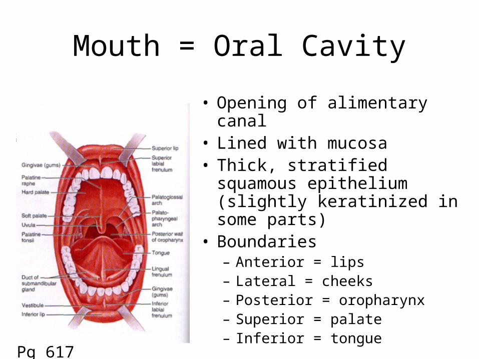
Mouth = Oral Cavity
• Opening of alimentary canal• Lined with mucosa• Thick, stratified squamous
epithelium (slightly keratinized in some parts)
• Boundaries– Anterior = lips– Lateral = cheeks– Posterior = oropharynx– Superior = palate– Inferior = tongue
Pg 617
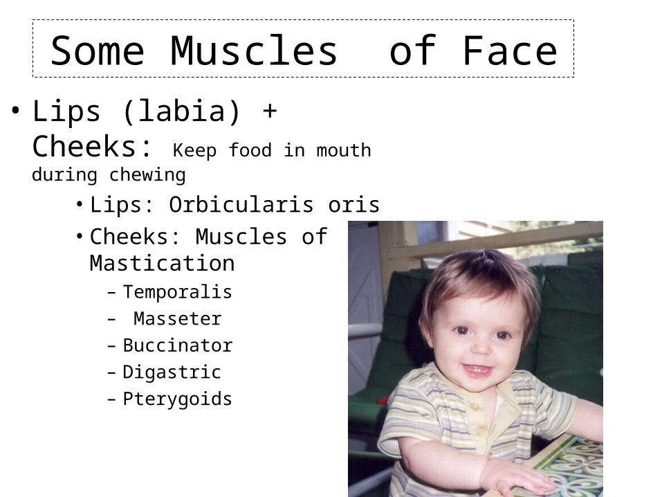
Some Muscles of Face• Lips (labia) + Cheeks: Keep
food in mouth during chewing
• Lips: Orbicularis oris• Cheeks: Muscles of Mastication
– Temporalis – Masseter – Buccinator – Digastric – Pterygoids
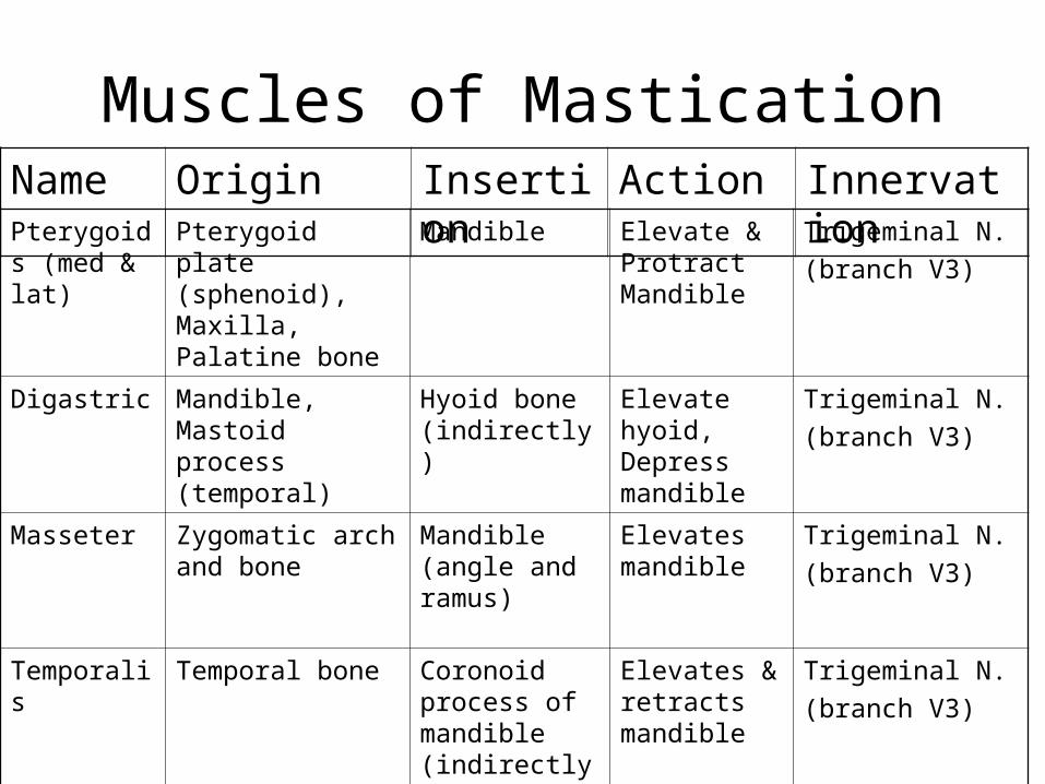
Muscles of Mastication
Pterygoids (med & lat)
Pterygoid plate (sphenoid), Maxilla, Palatine bone
Mandible Elevate & Protract Mandible
Trigeminal N.
(branch V3)
Digastric Mandible, Mastoid process (temporal)
Hyoid bone (indirectly)
Elevate hyoid, Depress mandible
Trigeminal N.
(branch V3)
Masseter Zygomatic arch and bone
Mandible (angle and ramus)
Elevates mandible
Trigeminal N.
(branch V3)
Temporalis Temporal bone Coronoid process of mandible (indirectly)
Elevates & retracts mandible
Trigeminal N.
(branch V3)
Name Origin Insertion Action Innervation
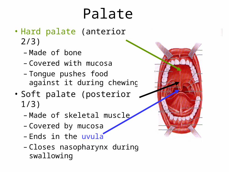
Palate• Hard palate (anterior 2/3)
– Made of bone– Covered with mucosa– Tongue pushes food
against it during chewing
• Soft palate (posterior 1/3)– Made of skeletal muscle– Covered by mucosa– Ends in the uvula– Closes nasopharynx during
swallowing
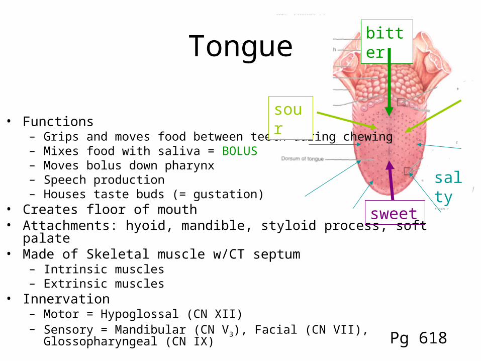
Tongue
• Functions– Grips and moves food between teeth during chewing– Mixes food with saliva = BOLUS– Moves bolus down pharynx– Speech production– Houses taste buds (= gustation)
• Creates floor of mouth• Attachments: hyoid, mandible, styloid process, soft palate• Made of Skeletal muscle w/CT septum
– Intrinsic muscles – Extrinsic muscles
• Innervation– Motor = Hypoglossal (CN XII) – Sensory = Mandibular (CN V3), Facial (CN VII), Glossopharyngeal (CN IX)
salty
bitter
sour
sweet
Pg 618

Salivary Glands
• Exocrine, tubuloalveolar glands• Produce Saliva
– H2O, Ions, Mucus, Enzymes
• Internal salivary glands are small– In mucosa of tongue, lips, palate, cheeks– Keep areas wet
• External salivary glands are large, have ducts– External to mouth– Provide saliva when necessary or anticipated– 2 Submandibular, 2 Sublingual, 2 Parotid glands
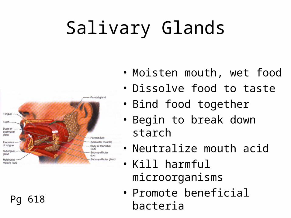
Salivary Glands
• Moisten mouth, wet food• Dissolve food to taste• Bind food together• Begin to break down starch• Neutralize mouth acid• Kill harmful microorganisms• Promote beneficial bacteria
Pg 618

Tiny Tabitha’s Teeth
The story of teeth from eruption to edentate

Tiny Tabitha: Age 4 days
• Number of Erupted Teeth = 0• Ultimate Goal = 32 Teeth
– Incisors (8): rip, cut– Canines (4): tear and pierce– Premolars (8): grinding– Molars (12): grinding
• Estimated Time of Completion = 15-25 years
• Currently jaws covered by gingiva (gum): is oral mucosa = lots of drooling

Tabitha’s First Tooth
• Deciduous (Milk) = 20– 8 Incisors = 6-10 months– 4 Canines = 16-20 months– 4 1st Molars = 12-16 months– 4 2nd Molars = 20-24 months
• Dental Formula: describes number, kind & position of teeth in ½ of the mouth
2:1:0:2
2:1:0:2 X 2 = 20
Incisors
caninesmolars
premolars
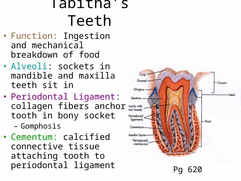
Tabitha’s Teeth
• Function: Ingestion and mechanical breakdown of food
• Alveoli: sockets in mandible and maxilla teeth sit in
• Periodontal Ligament: collagen fibers anchor tooth in bony socket– Gomphosis
• Cementum: calcified connective tissue attaching tooth to periodontal ligament
Pg 620
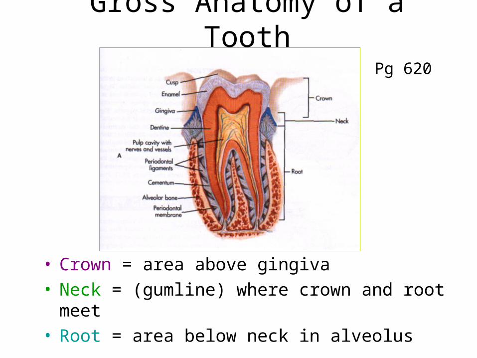
Gross Anatomy of a Tooth
• Crown = area above gingiva• Neck = (gumline) where crown and root meet• Root = area below neck in alveolus
Pg 620
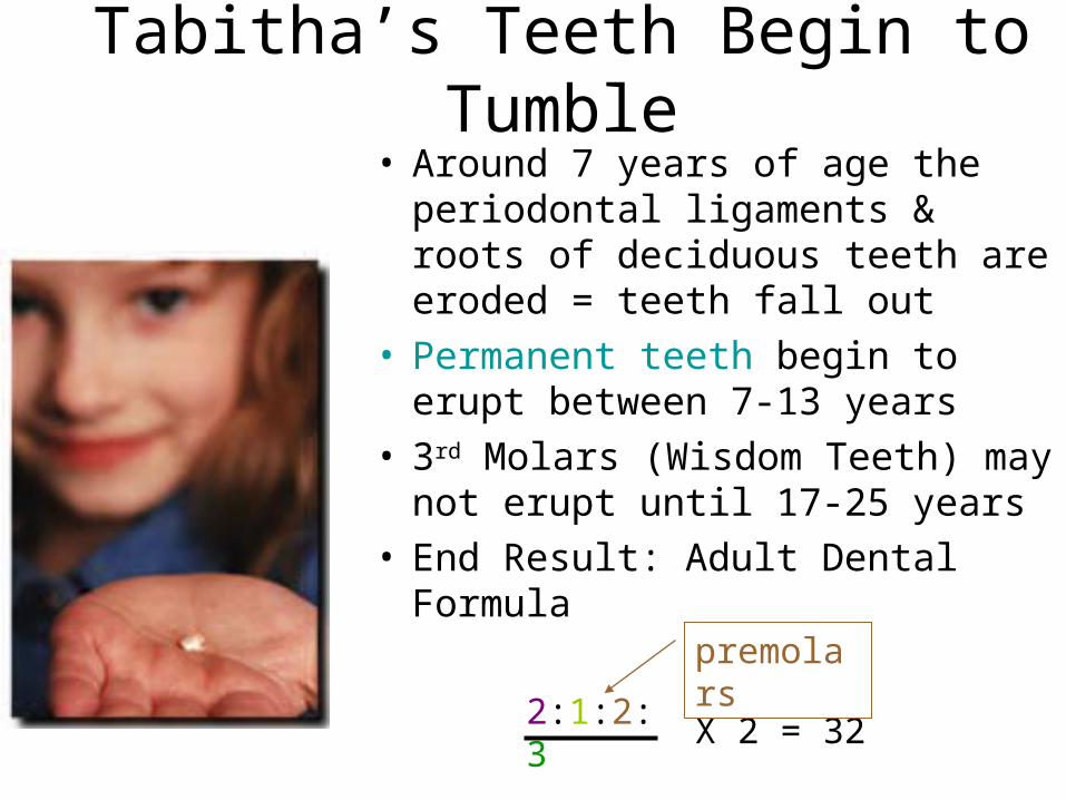
Tabitha’s Teeth Begin to Tumble
• Around 7 years of age the periodontal ligaments & roots of deciduous teeth are eroded = teeth fall out
• Permanent teeth begin to erupt between 7-13 years
• 3rd Molars (Wisdom Teeth) may not erupt until 17-25 years
• End Result: Adult Dental Formula
2:1:2:3
2:1:2:3X 2 = 32
premolars

Tabitha’s Tooth Troubles• College --> too much junkfood
= CAVITIES (caries)• Bacteria erodes through the
outer enamel covering of tooth– Avascular, Acellular– Mostly calcium salts– Not renewed or replaced– Hardest substance in body
• In severe cases it erodes the deeper dentin of tooth– Made of minerals & collagen– Is maintained during life– Harder than bone– Bulk of tooth Pg 620
Tabitha’s Tooth Trauma!
Pg 620
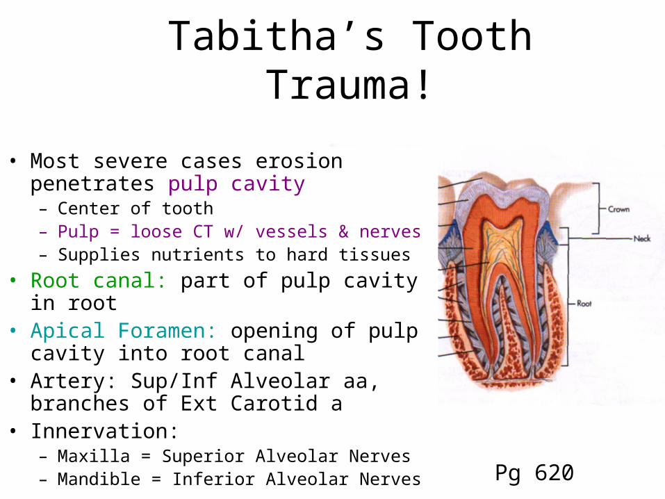
• Most severe cases erosion penetrates pulp cavity– Center of tooth– Pulp = loose CT w/ vessels & nerves– Supplies nutrients to hard tissues
• Root canal: part of pulp cavity in root• Apical Foramen: opening of pulp cavity
into root canal • Artery: Sup/Inf Alveolar aa, branches of
Ext Carotid a• Innervation:
– Maxilla = Superior Alveolar Nerves– Mandible = Inferior Alveolar Nerves
Tabitha’s Teeth: the later years
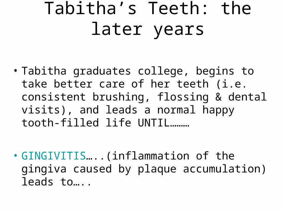
• Tabitha graduates college, begins to take better care of her teeth (i.e. consistent brushing, flossing & dental visits), and leads a normal happy tooth-filled life UNTIL………
• GINGIVITIS…..(inflammation of the gingiva caused by plaque accumulation) leads to…..
Toodaloo Tabitha’s Teeth
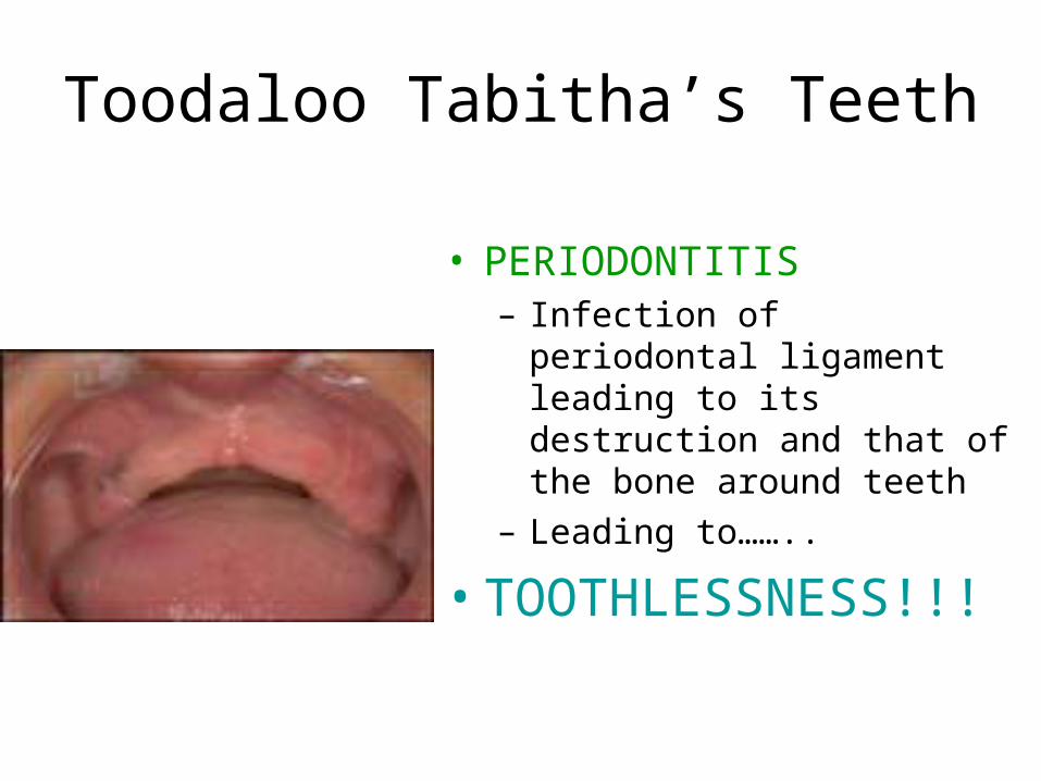
• PERIODONTITIS– Infection of periodontal
ligament leading to its destruction and that of the bone around teeth
– Leading to……..
• TOOTHLESSNESS!!!
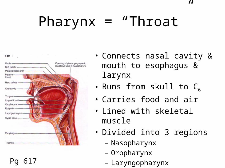
Pharynx = “Throat”
• Connects nasal cavity & mouth to esophagus & larynx
• Runs from skull to C6
• Carries food and air• Lined with skeletal muscle• Divided into 3 regions
– Nasopharynx– Oropharynx– Laryngopharynx
Pg 617
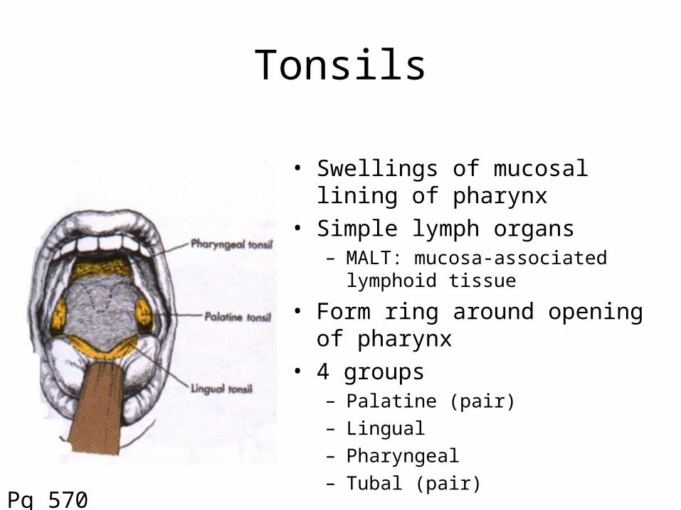
Tonsils
• Swellings of mucosal lining of pharynx
• Simple lymph organs– MALT: mucosa-associated lymphoid
tissue
• Form ring around opening of pharynx
• 4 groups– Palatine (pair)– Lingual– Pharyngeal– Tubal (pair)
Pg 570

Regions of the Pharynx
Nasopharynx
Oropharynx
Laryngopharynx
Pg 584
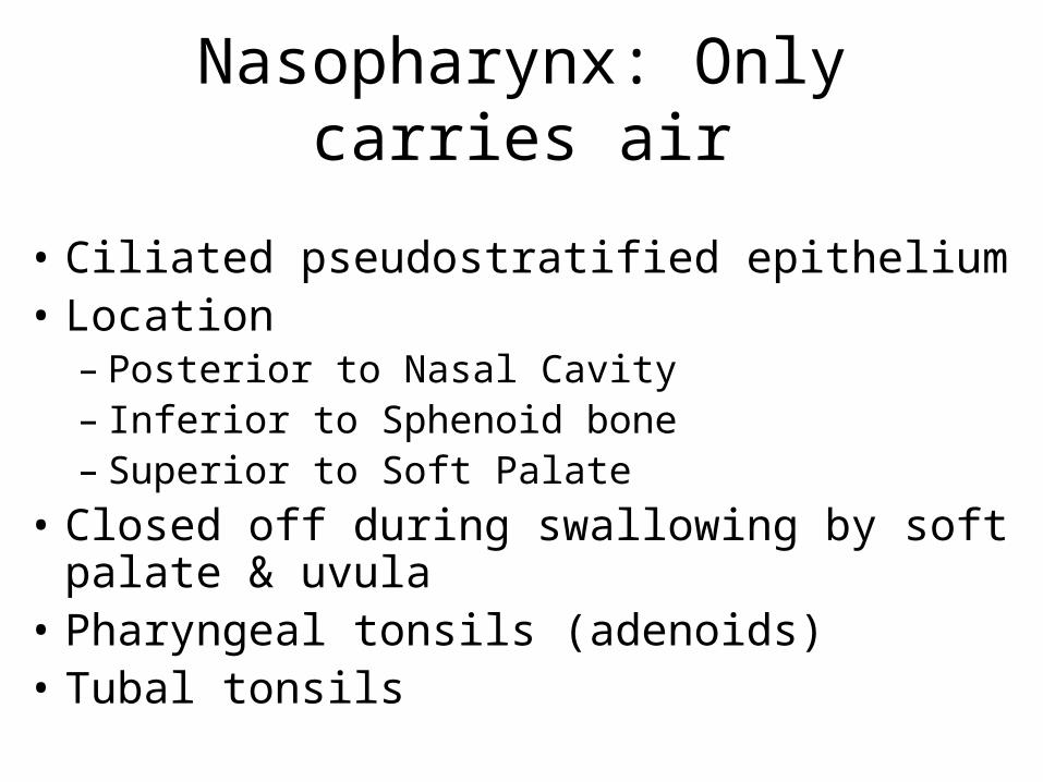
Nasopharynx: Only carries air
• Ciliated pseudostratified epithelium• Location
– Posterior to Nasal Cavity– Inferior to Sphenoid bone– Superior to Soft Palate
• Closed off during swallowing by soft palate & uvula
• Pharyngeal tonsils (adenoids)• Tubal tonsils

Oropharynx: Carries Food & Air
• Thick, protruding stratified squamosal epithelium due to great friction
• Location– Posterior to Oral Cavity– Runs from Soft Palate to Epiglottis
• Palatine tonsils
• Lingual tonsils
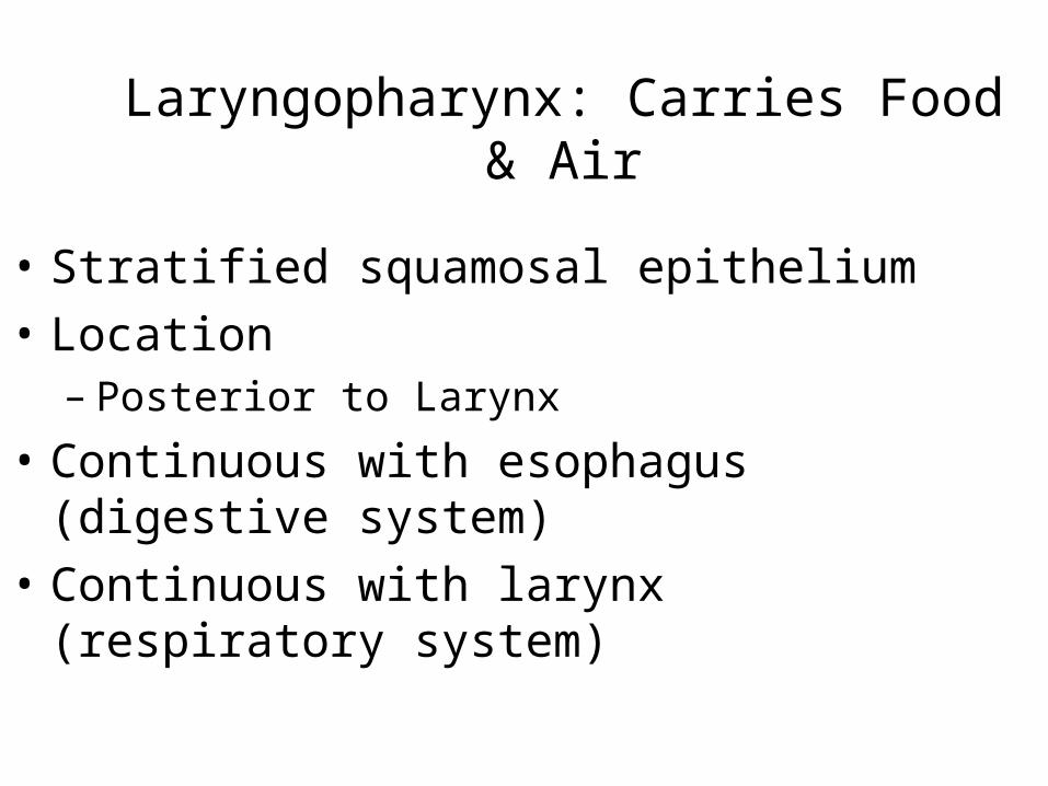
Laryngopharynx: Carries Food & Air
• Stratified squamosal epithelium
• Location– Posterior to Larynx
• Continuous with esophagus (digestive system)
• Continuous with larynx (respiratory system)

Moving the Bolus
• Swallowing– Voluntarily initiated (pharynx)– Suprahyoid, Infrahyoid,
Pharyngeal constrictors
• Peristalsis = propulsion– Involuntary– Alternate waves of contraction
and relaxation of muscles in organ walls (e.g. esophagus)
– Squeezes food from one organ to next
– Some mixingPg 611

Hyoid Bone
• Only bone not directly articulated with other bones
• Attaches via ligaments to temporal bone, larynx
• Components– Body– Pair of Greater Horns– Pair of Lesser Horns
• Functions– Moveable base for tongue– Attachment for sternohyoid, thyrohyoid– Superior attachment for larynxPg 163
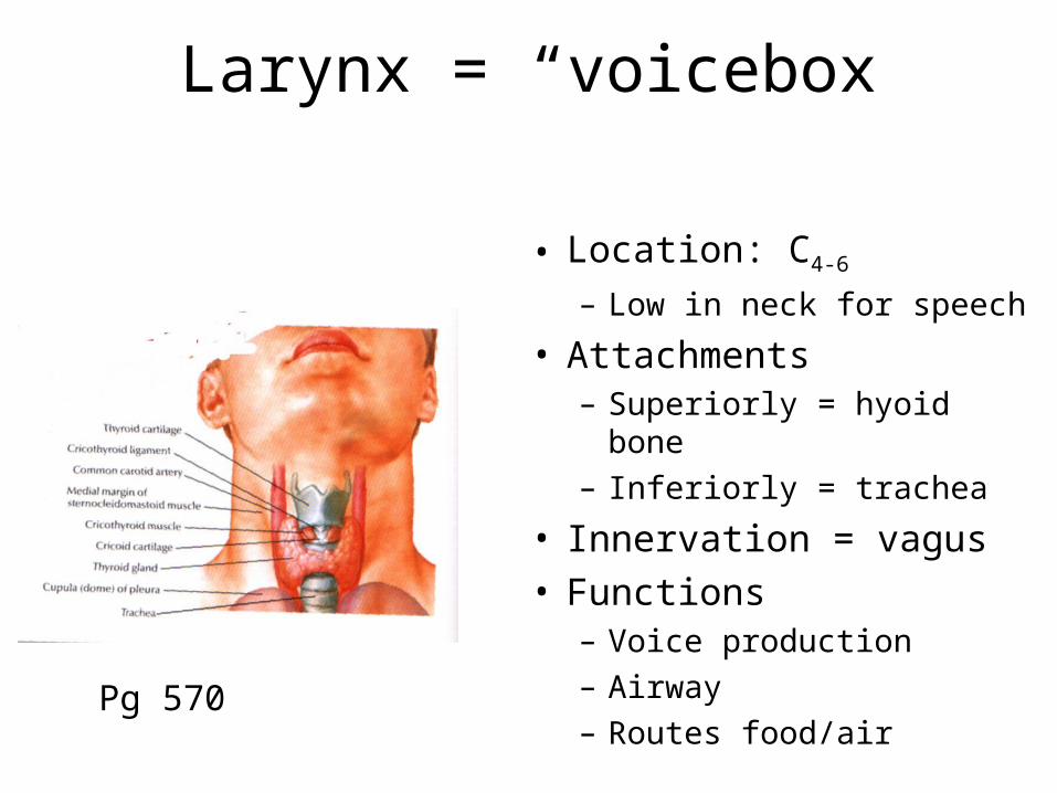
Larynx = “voicebox”
• Location: C4-6
– Low in neck for speech
• Attachments– Superiorly = hyoid bone– Inferiorly = trachea
• Innervation = vagus• Functions
– Voice production– Airway– Routes food/air
Pg 570

Laryngeal Cartilages
• 9 Cartilages connected by ligaments & membranes– 1 Epiglottis– 1 Thyroid– 1 Cricoid– 2 Arytenoid– 2 Corniculate– 2 Cuneiform
• Superior part = stratified squamosal epithelium• Below vocal cords= ciliated pseudostratified columnar

Laryngeal Cartilages
• Epiglottis– Elastic cartilage; Mucosa covering– Projects upward from anterior wall of laryngeal inlet to level
of base of tongue
• Thyroid Cartilage– Large, shield shaped, made of 2 plates– Laryngeal prominence
• Cricoid Cartilage– Shaped like signet ring– Between thyroid cartilage and trachea

Laryngeal Cartilages
Pg 587
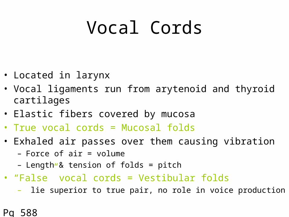
Vocal Cords
• Located in larynx• Vocal ligaments run from arytenoid and thyroid cartilages• Elastic fibers covered by mucosa• True vocal cords = Mucosal folds• Exhaled air passes over them causing vibration
– Force of air = volume– Length & tension of folds = pitch
• “False” vocal cords = Vestibular folds– lie superior to true pair, no role in voice production
Pg 588

The Vocal CordsThyroid cartilage
Aretynoid cartilage
True Vocal Folds
False Vocal Folds
True Vocal Folds

Thyroid Gland
• Location:– Along trachea, just inferior to larynx– “Butterfly” shape
• Endocrine Gland– Thyroid hormone (TH): increases basal metabolic rate– Calcitonin: depresses excessive levels of Ca2+ in blood
• Blood Supply: – Superior thyroid arteries (branches of ext. carotids)– Inferior thyroid arteries (branches of subclavians)
Pg 718
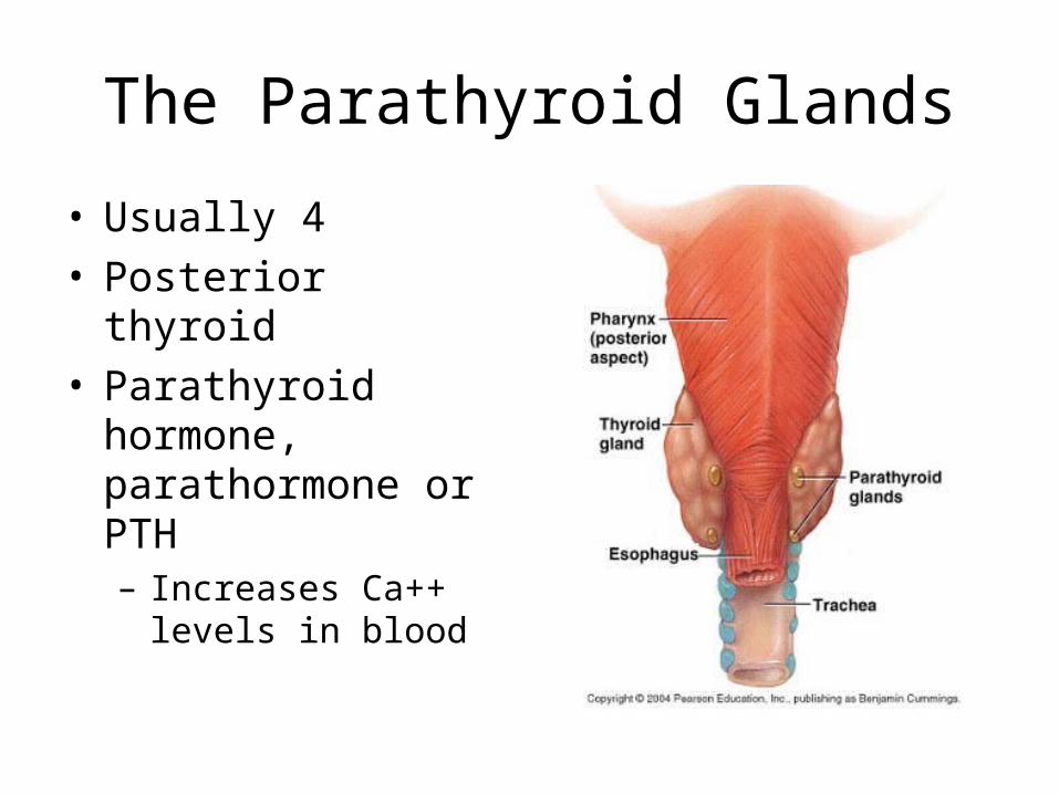
The Parathyroid Glands
• Usually 4• Posterior thyroid• Parathyroid hormone,
parathormone or PTH– Increases Ca++ levels
in blood

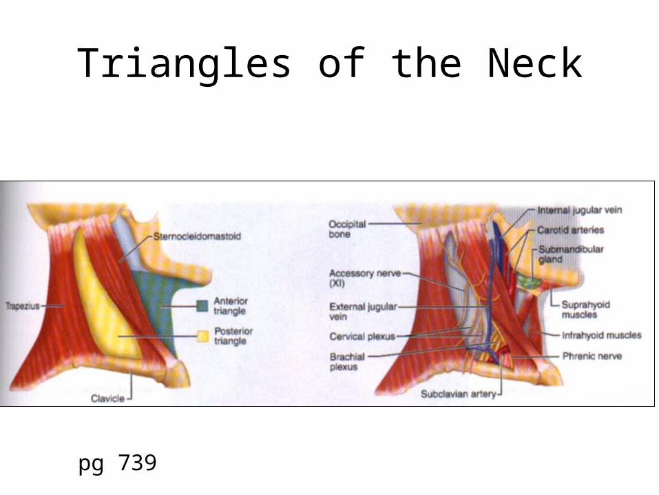
Triangles of the Neck
Each half of the neck can be divided into 2 triangles:
1 Anterior and 1 Posterior
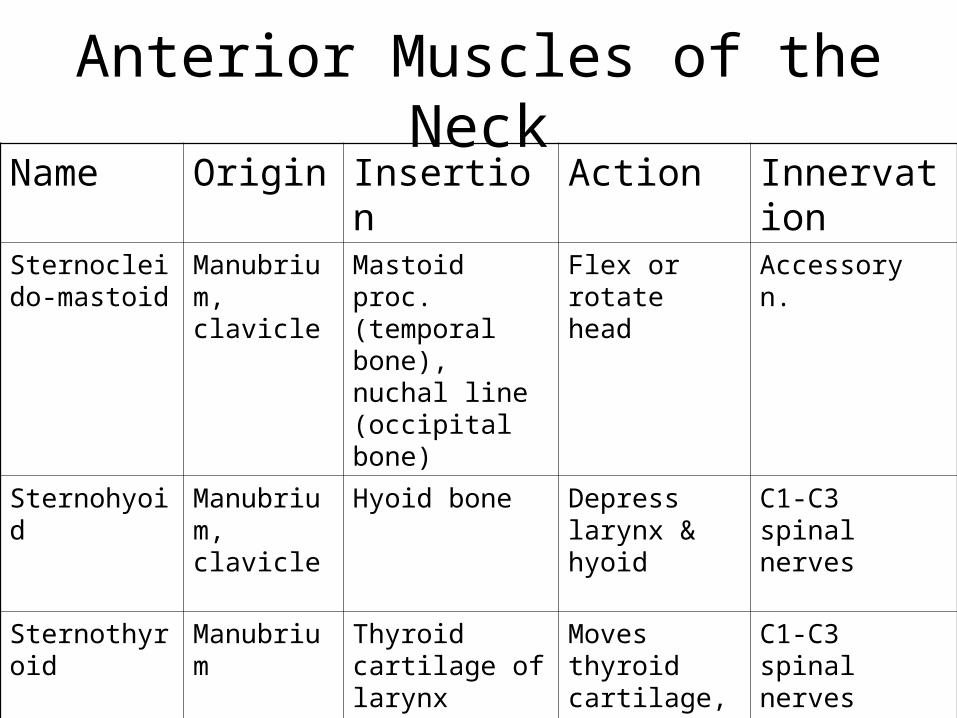
Anterior Muscles of the NeckName Origin Insertion Action Innervation
Sternocleido-mastoid
Manubrium, clavicle
Mastoid proc. (temporal bone), nuchal line (occipital bone)
Flex or rotate head
Accessory n.
Sternohyoid Manubrium, clavicle
Hyoid bone Depress larynx & hyoid
C1-C3 spinal nerves
Sternothyroid Manubrium Thyroid cartilage of larynx
Moves thyroid cartilage, larynx, hyoid inferiorly
C1-C3 spinal nerves

Anterior Triangle of the Neck
• Boundaries– Superior = Inferior margin of Mandible– Anterior = Midline of Neck– Posterior = Sternocleidomastoid muscle
• Contents– Muscles: Suprahyoid, Infrahyoid– Artery: Carotid– Vein: Internal Jugular, External Jugular– Nerve: Accessory– Glands: Submandibular
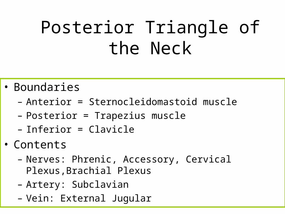
Posterior Triangle of the Neck
• Boundaries– Anterior = Sternocleidomastoid muscle– Posterior = Trapezius muscle– Inferior = Clavicle
• Contents– Nerves: Phrenic, Accessory, Cervical Plexus,Brachial Plexus– Artery: Subclavian– Vein: External Jugular
Triangles of the Neck
pg 739