tema 9 : estructura general de las plantas vasculares...
TRANSCRIPT

Tema 9: Estructura general de las plantas vasculares.
La Raíz. Estructura primaria y desarrollo.
Funciones:
• Fijación
• Absorción
• Almacenamiento
• Conducción (transporte)
Sistemas de raíces: Raíz axonomorfa
Raíz fasciculada
Origen y crecimiento de tejidos primarios.
Bibliografía: ver temas anteriores.
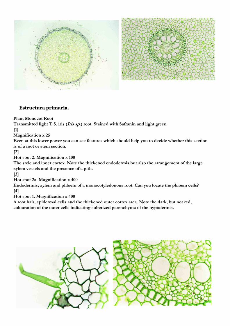
Plant Monocot Root
Transmitted light T.S. iris (Iris sp.) root. Stained with Safranin and light green
[1]
Magnification x 25
Even at this lower power you can see features which should help you to decide whether this section
is of a root or stem section.
[2]
Hot spot 2. Magnification x 100
The stele and inner cortex. Note the thickened endodermis but also the arrangement of the large
xylem vessels and the presence of a pith.
[3]
Hot spot 2a. Magnification x 400
Endodermis, xylem and phloem of a monocotyledonous root. Can you locate the phloem cells?
[4]
Hot spot 1. Magnification x 400
A root hair, epidermal cells and the thickened outer cortex area. Note the dark, but not red,
colouration of the outer cells indicating suberized parenchyma of the hypodermis.
Estructura primaria.

Plant Herbaceous Dicot Roots
Transmitted light T.S. young buttercup (Ranunculus sp.) root. Stained with Safranin and light
green.
[1] x 25. Living cells are stained green by the light green stain. The main constituents of the walls
of living cells are cellulose, hemicellulose and pectin.
Remember that red staining indicates the presence of lignin as well as celluloses, hemicelluloses
and pectins in the cell wall and this in its turn indicates that these cells are dead cells.
[2] Hot spot 3. x 200. The Stele.
Remember red stained cell walls are thickened with lignin and such cells are dead. They no longer
have organelles. In a young tissue the overall layout of the vascular bundles is critical in deciding
what organ (root or stem) you are looking at.
[3]. Hot spot 4. x 400. More detail of the cells of the stele: xylem, phloem, pericycle and cambium.
With careful observation here you will be able to recognise cambium cells wherever they might
occur in other sections. An advantage of being able to recognise cambial cells is that if you can
find cambium in a vascular bundle
you know that phloem (which is sometimes difficult to recognise on positive features) must lie on
the opposite side of the cambium to the more easily recognised xylem.
[4]. Hot spot 2. x 400. The endodermis; a boundary layer between the cortex and stele.
You will find there is a layer inside the cortex where many, but not all cells have lignin thickening
in the radial walls. In some cells the thickening is u-shaped having progressed to the internal cell
wall.
[5]. Hot spot 1. x 400. Epidermis and cortex.
The critical characteristics are location, cell shape, how the cells fit together and the thickness of
the cell walls.

Raiz dicot Raiz monocot

El vástago. Estructura primaria y desarrollo.

Ramificación del tallo.

Tallo de girasol. Tejidos vasculares en haces separados.Tallo de dedalera (Digitalis), haz vascular en cilindro.
Estructura primaria del tallo.
Dicotiledóneas
Tallo de maíz. Tejidos vasculares en cordones dispersos.
Monocotiledóneas

Crecimiento secundario.
