retinoblastoma in children materi deteksi... · 2018-01-29 · prompt referral to an ocular...
TRANSCRIPT

Early detection of Retinoblastoma
in children
Max Mantik

Introduction
• The most common primary intraocular malignancy of childhood
• 10 to 15 % of cancers that occur within the first year of life
• Typical as Leukocoria in a child < 2 years
• Untreated retinoblastoma is a deadly disease
• Metastatic spread is typically diagnosed within the first 12 months
• Survival in the contemporary era is >95 %
• The prognosis for eye salvage is far lower and depends on the stage of disease at diagnosis
2
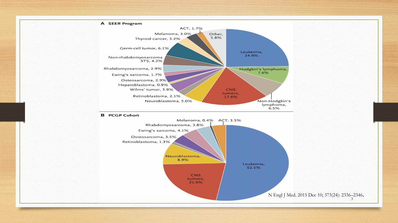
3N Engl J Med. 2015 Dec 10; 373(24): 2336–2346.

4
EARLY DETECTION

Leukocoria
5

Prompt referral to an ocular
oncologist and appropriate
management by a multidisciplinary
team are necessary to optimize visual outcome and survival
6

7
Primary care practitioner
Pediatric ophthalmologist
Pediatric oncologist
Radiation oncologist
Clinical geneticist
Retina specialist
Ocular oncologist
Neuroradiologist
Craniofacial plastic specialist
Nurse specialist
Pharmacist
Child life specialist
Clinical social worker
Low-vision specialist
Nutritionist
The multidisciplinary team

TERMINOLOGY
• Heritable retinoblastoma – Heritable (hereditary, familial, or
germline) retinoblastoma is associated with germline mutations
(ie, mutations that occur in reproductive cells [sperm and eggs]) in
the retinoblastoma (RB1) gene
• Nonheritable retinoblastoma – Nonheritable (nonhereditary,
nonfamilial, sporadic, or somatic) retinoblastoma results from
somatic mutations (ie, mutations that occur in nonreproductive
cells) in the RB1 gene
8

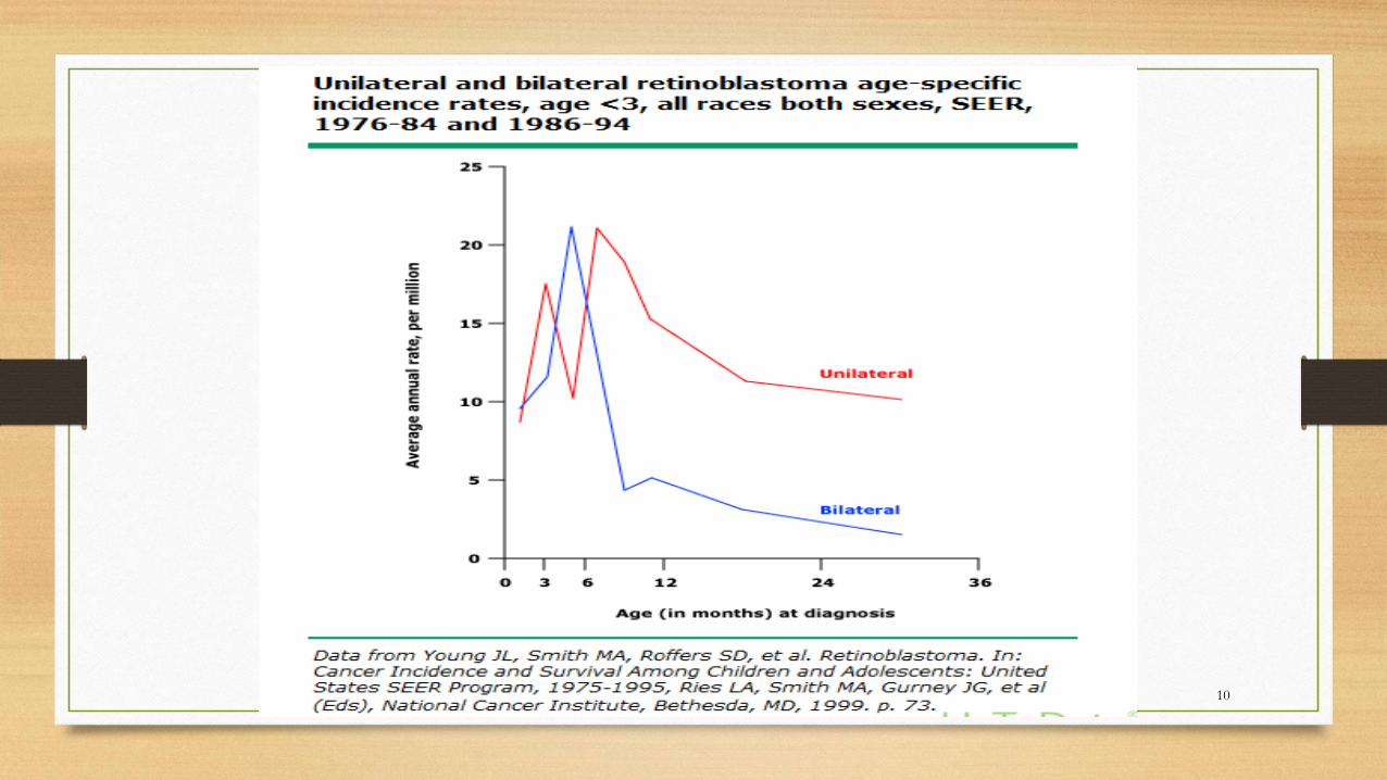
Incidence
• 1 in 15,000 to 1 in 16,600 live births
• 13 % of cancer in the first year of life
• The median age at diagnosis is 18 to 20 months;
• An average of:
• 12 months for children with bilateral disease
• 24 months for children with unilateral disease
• 95 % < 5 years.
• Boys and girls are the same9
10

Genetic predisposition
• Retinoblastoma occurs in heritable and nonheritable forms
• Germline mutations in the retinoblastoma (RB1) gene ± 40 % of
cases, predominantly in bilateral disease
• Nonheritable retinoblastoma incur new somatic mutations
• <10 % of retinoblastoma patients have a positive family history
11

PATHOGENESIS
• Mutational inactivation of both alleles of the retinoblastoma (RB1)
gene
• Chromosome 13q and encodes a nuclear protein (Rb) that acts as
a tumor suppressor
• The Rb protein restricts the cell's ability to progress from the G1
phase to the S phase of the cell cycle
12

PATHOGENESIS……
• A germline mutation at the RB1 locus (most common) or deletion of
chromosome 13q (containing the RB1 gene locus) is present in all cells of
the body
• A second "hit," occurring later in development (the second hit of the
Knudson two-hit hypothesis), affects the remaining RB1 allele within
retinal cells a typical for the heritable form
• A "two-hit" model has been proposed to explain the different clinical
features of heritable and nonheritable cases of retinoblastoma
13

14
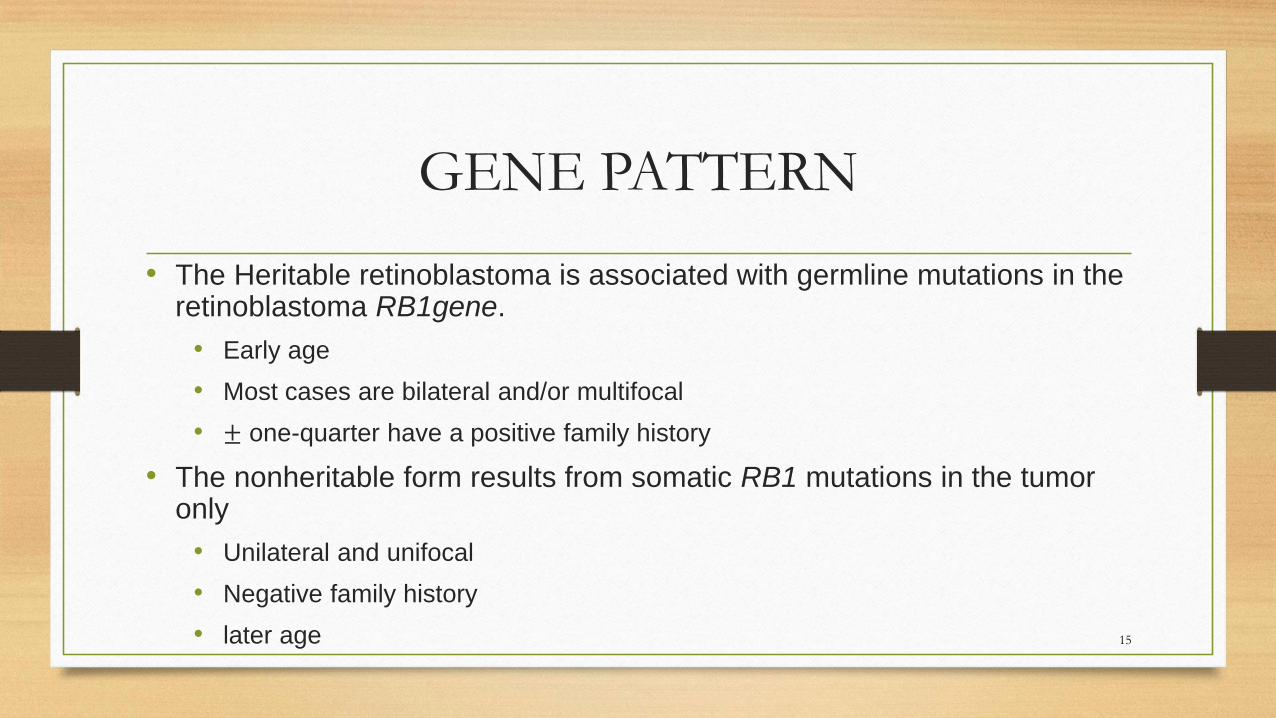
GENE PATTERN
• The Heritable retinoblastoma is associated with germline mutations in the retinoblastoma RB1gene.
• Early age
• Most cases are bilateral and/or multifocal
• ± one-quarter have a positive family history
• The nonheritable form results from somatic RB1 mutations in the tumoronly
• Unilateral and unifocal
• Negative family history
• later age 15

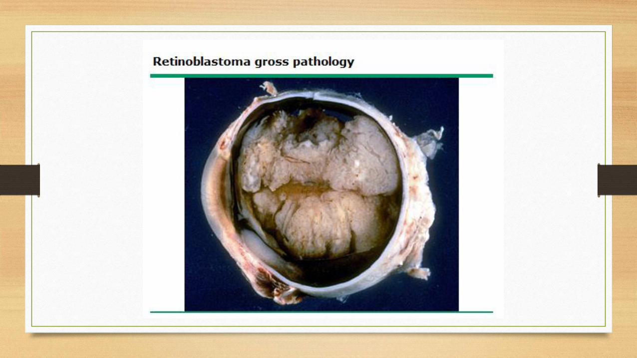
Morphologic features
• Solitary or multifocal, well-circumscribed, translucent intraretinal mass
• In advance more pink in color, with dilated feeding blood vessels
• Growth pattern
• Exophytic lead to subretinal seeds
• Endophytic the anterior chamber and layer behind the cornea, causing a
pseudo-hypopyon
• Diffuse infiltrating retinoblastoma relatively flat (very little vertical growth)
and grows intraretinally, mimicking retinitis
16

The routes of metastatic spread
• The most common are the optic nerve to the central nervous
system
• The choroid into the sclera and into the orbit
• The subarachnoid space to the contralateral optic nerve
• The cerebrospinal fluid to the CNS;
• Hematogenous dissemination to the lung, bone, liver, or brain;
and lymphatic dissemination
17

Second malignancies
Heritable retinoblastoma approximately 30 % at 40 years after
the original treatment
In contrast, nonheritable retinoblastoma less common 2 %
PNET tumors
18

19

CAUSES OF LEUKOCORIA
• Retinoblastoma (18 to 62 %)
• Cataract (60 %)
• Persistent fetal vasculature (31 % of those referred for
retinoblastoma)
• Coats disease (29 % of those referred for retinoblastoma)
20
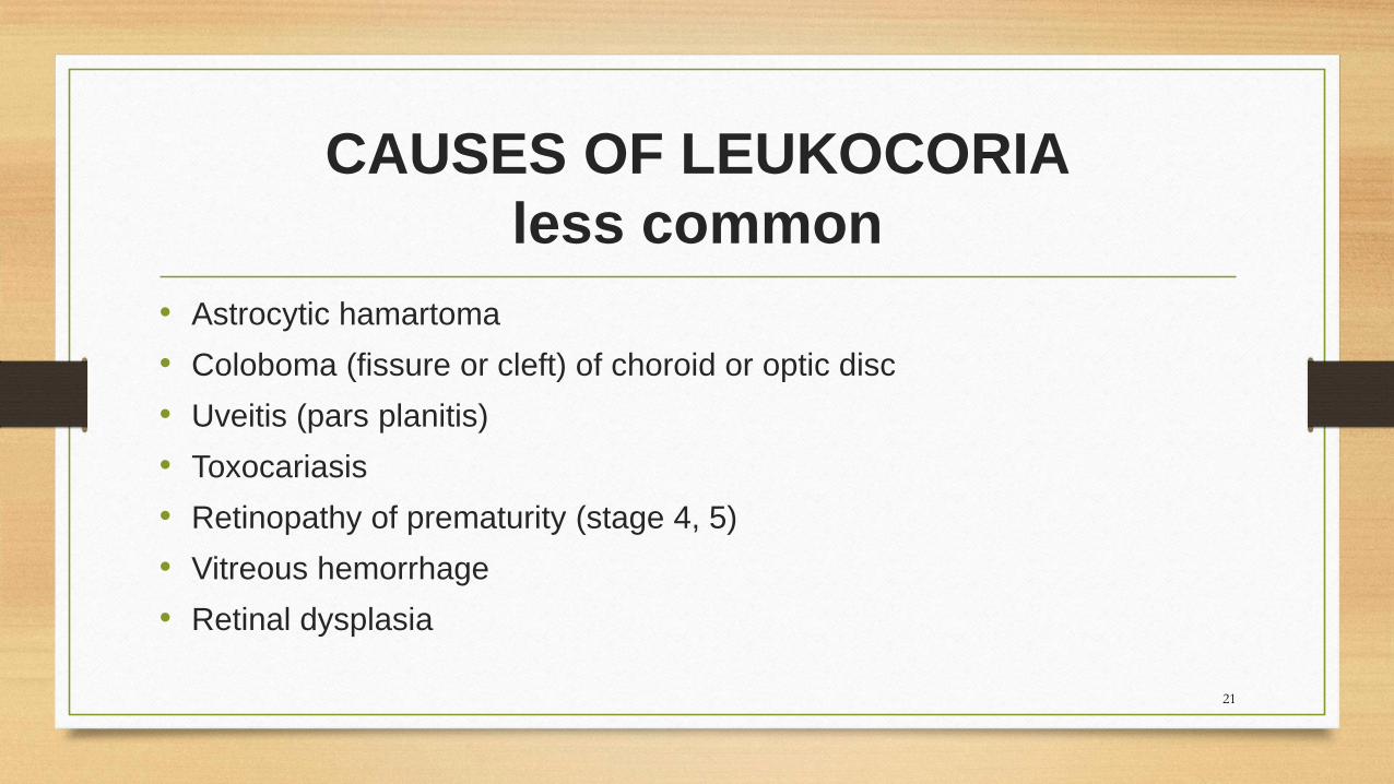
CAUSES OF LEUKOCORIA
less common
• Astrocytic hamartoma
• Coloboma (fissure or cleft) of choroid or optic disc
• Uveitis (pars planitis)
• Toxocariasis
• Retinopathy of prematurity (stage 4, 5)
• Vitreous hemorrhage
• Retinal dysplasia
21


SCREENING CHILDREN AT RISK
• A positive family history
• A personal of family history of 13q deletion or retinoblastoma [RB1]
gene mosaicism
• Should be evaluated by an ophthalmologist shortly after birth
• Every 1 to 2 months during the first 2 years of life
24
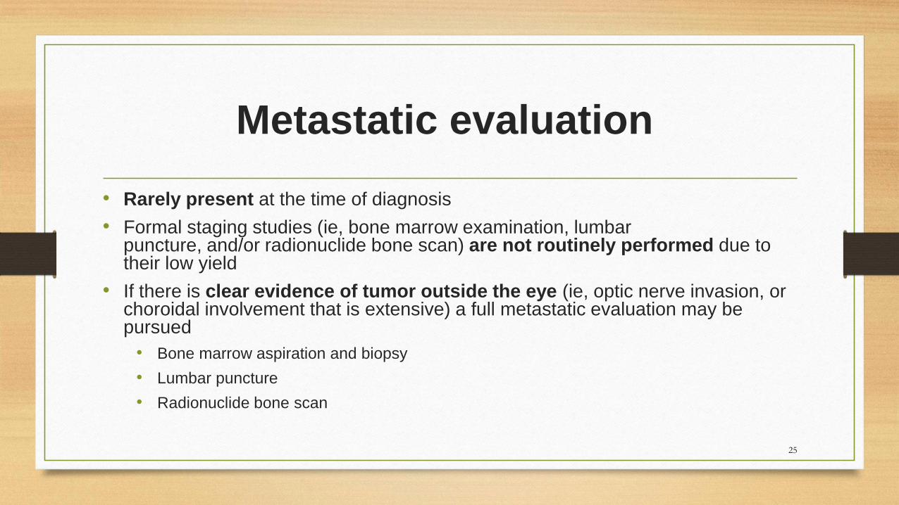
Metastatic evaluation
• Rarely present at the time of diagnosis
• Formal staging studies (ie, bone marrow examination, lumbar puncture, and/or radionuclide bone scan) are not routinely performed due to their low yield
• If there is clear evidence of tumor outside the eye (ie, optic nerve invasion, or choroidal involvement that is extensive) a full metastatic evaluation may be pursued
• Bone marrow aspiration and biopsy
• Lumbar puncture
• Radionuclide bone scan
25

Genetic testing and counseling
• Are important aspects in the management of patients with
retinoblastoma in order to estimate the risk of disease in family
members and future offspring
• Understand the genetic consequences of each form of
retinoblastoma (relates to secondary cancers in children with heritable retinoblastoma)
26

Opthalmol Clin North Am 2005; 18:41


SUMMARY
• The most common primary intraocular malignancy of childhood and accounts for 10 to 15 percent of cancers within the first year of life
• Retinoblastoma occurs in heritable (approximately 40 % of cases) and nonheritable(approximately 60 % of cases) forms
• Should undergo clinical screening and/or genetic testing for retinoblastoma
• The evaluation by an ocular oncologist and includes Complete physical examination, Ophthalmologic examination, ultrasonography, MRI of the brain and orbits
• Metastatic disease is rarely present at the time of diagnosis, and formal staging studies are not routinely performed
• Molecular genetic testing is suggested for all affected patients
31

32

33

34

35

36

Family history
• Are presumed to have heritable retinoblastoma
• 50 % risk of passing the mutation on to their offspring
• Patients with an RB1 germline mutation have a 90 percent chance
of the mutation manifesting with retinoblastoma
37


Approach to the child with leukocoria
39

41


43