nit-occlud® pda
TRANSCRIPT

GA034/Rev02_130904
Nit-Occlud® PDA
en Instructions for Use For USA only!
www.pfmmedical.com

2 GA034/Rev02_130904
Explanation of the symbols on label and packaging
Reference number
Lot number
Serial number
Read Instructions for Use carefully
Protect from direct sunlight
Store in a dry place
Expiry Date
For single use only
Sterilized by Ethylene Oxide
Do not resterilise
22
STERILIZE
REF
LOT
SN
Do not use if packaging is damaged
“MR conditional” = safe use of MR diagnostics under certain conditions

GA034/Rev02_130904 3
Manufacturer
Does not contain rubber latex components
Diethylhexylphthalate (DEHP) free
LATEX
DEHP
Caution: Federal law (USA) restricts this device to sale by or on the order of a physician

4 GA034/Rev02_130904
English
Instructions for Use Nit-Occlud® PDA
Nit-Occlud® PDA is a system for transcatheter occlusion of Patent Ductus Arteriosus (PDA) with spiral coils. The system consists of the fol-lowing parts:• Nit-Occlud®PDA
The spiral coil is mounted in a straightened fashion on a flexible delivery system including a disposable handle to which it is connected by means of a patented detachment mechanism. Nit-Occlud® PDA coils are available as Flexible and Medium type. The flexible and medium types are pre-loaded into the transportation sheath. For insertion the transportation sheath must be connected to the implantation catheter.
The Nit-Occlud® PDA coil has a cone in cone configuration which results from the fact that the proximal windings of the coil are wound in the reverse direction (see Figure 2).
• ImplantationcatheterThe implantation catheter is equipped with a marker ring at its distal tip for better orientation during fluoroscopy.
Product description
Figure 1: Nit-Occlud® PDA
The Nit-Occlud® PDA coil is a permanently implanted prothesis indicated for percutaneous, transcatheter closure of small to moderate size patent ductus arteriosus with a minimum angiographic diameter less than 4 mm.
Indication for Use
Medical conditions that exclude implantation of a Nit-Occlud® PDA coil include:• Endocarditis,endarteritisoractiveinfectionatthetimeoftheimplantation• Patientswithabodyweight<5kg• Pulmonaryhypertension(calculatedPVRgreaterthan5WoodUnits)• ThrombusinabloodvesselthroughwhichaccesstothePDAmustbeobtained• Thrombusinthevicinityoftheimplantationsite.
Contraindications
Figure 2: Nit-Occlud® PDA spiral coils (D=Distal diameter, P=Proximal diameter, Lc=Length configurated).

GA034/Rev02_130904 5
English
These instructions for use and the information on the packaging should be read carefully before each use. The PDA coil system should be used only by physicians trained in interventional occlusion techniques.
WA R N I N G S Do not use the product if the packaging has been opened, or is damaged, if you are not sure that it is sterile, or if the
expiry date has passed. Each product is packed separately, and is delivered in an EO-sterilised and non-pyrogenic condition. It is intended
for single use only. Do not reuse, reprocess or resterilize. Reuse, reprocessing or resterilization of single-use devices may result in degraded performance or a loss of functionality. Reuse of single-use devices may result in exposure to pathogens such as viruses, bacteria, fungi, or prions.
The product must be stored in dry conditions. Do not expose the packaged products to direct sunlight. Retrieval devices should be available during implant procedures for interventional retrieval of the coil if required. Care must be taken not to damage the coil or to dislodge it from the delivery system while unpacking or inserting it
into the implantation catheter. Since the delivery system has ferromagnetic properties, implantation must not be carried out in an MR environ-
ment. The coil should not be removed from the delivery system. It should not be used with another delivery system since
this may alter characteristics of configuration and detachability. A detached coil should not be remounted on the core wire of the delivery system. The configured coil should not be pulled through heart valves or ventricular chambers. The implantation catether is not suitable for application of contrast medium. It must not be connected to high pres-
sure injectors. The Nit-Occlud® PDA coil consists of a nickel-titanium alloy, which is generally considered safe. In non-clinical test-
ing, nickel has been shown to be released from the device in very small amounts. Patients who are allergic to nickel may have an allergic reaction to this device, especially those with a history of metal allergies. Certain allergic reac-tions can be serious; patients should be instructed to seek medical assistance immediately if they suspect they are experiencing an allergic reaction. Symptoms may include difficulty in breathing or swelling of the face or throat. While data are currently limited, it is possible that some patients may develop an allergy to nickel if this device is implanted.
This product contains chemicals known to the State of California to cause cancer, birth defects, or reproductive harm.
NOTE: Federal Law (USA) restricts this device to use by a physician.
Product IdentificationEach product label has peel-off labels, to allow the product to be identified precisely. These can be used for the patient file and the patient ID card.
MRI CompatibilityThe Nit-Occlud® PDA coil was determined to be MR-conditional according to the terminology specified in the American Society for Testing andMaterials(ASTM)International,Designation:F2503-05.StandardPracticeforMarkingMedicalDevicesandOtherItemsforSafetyintheMagneticResonanceEnvironment.ASTMInternational,100BarrHarborDrive,POBoxC700,WestConshohocken,Pennsylvania,2005.
Non-clinical testing demonstrated that the Nit-Occlud® PDA coil is MR conditional. A patient with this device can be scanned safely im-mediately after placement under the following conditions: • Staticmagneticfieldof3Teslaorless• Maximumspatialgradientmagneticfieldof720Gauss/cmorless• Themaximumwhole-bodyaveragedspecificabsorptionrate(SAR)shallbe limitedto2.0W/kg(normaloperatingmodeonly)for15
minutes of scanning.
General Information and Warnings
6 GA034/Rev02_130904
English
• Airembolism• Allergicreactiontodrug/contrast• Apnea• Arrhythmiarequiringmedicaltreatmentorpacing• ArteriovenousFistula• BacterialEndocarditis• Bloodlossrequiringtransfusion• ChestPain• Damagetothetricuspidorpulmonaryvalves• Death• Embolizationoftheoccluder,requiringpercutaneousorsurgical
intervention• Endarteritis• Falseaneurysmofthefemoralartery• Fever• Headache/migraine
• Heartfailure• Hemolysisafterimplantationoftheoccluder• Hypertension• Hypotensionorshock• Infection• Myocardialinfarction• Occluderfractureordamage• Perforationoftheheartorbloodvessels• Stenosisoftheleftpulmonaryarteryor
descending thoracic aorta• Stroke/TIA• Thromboembolism(cerebralorpulmornary)• ValvularRegurgitation• Vesseldamageatthesiteofgroinpuncture
(loss of pulse, hematoma etc.).
• AnangiogrammustbeperformedpriortoimplantationformeasuringlengthanddiameterofthePDA.• Theimplantationcathetermustbeflushedwithheparinizedsalinesolutionpriortointroductionandduringtheprocedure,especially
after angiography. • Thepfmmedicalimplantationcatheterisspecificallydesignedforthedeliverysystem.Othercathetersshouldnotbeusedtoimplantthe
device.• Contrastmediashouldnotbeinjectedthroughtheimplantationcatheter.• Thecoilshouldnotbepulledbackintotheimplantationcatheterusingstrongforce.• Administrationof50unitsofheparinperkgbodyweightisrecommendedafterfemoralsheathsareplaced.• Antibioticcoveragebefore(1dose)andafterimplantation(2doses)isrecommendedinordertopreventinfectionduringtheimplant
procedure. Antibiotic prophylaxis should be performed to prevent infective endocarditis during first 6 months after coil implantation.• AsuitablelateralaortogramshouldbeperformedformeasurementofPDAdimensions(seeFig.PDAMeasurements):
Potential Adverse Events
Precautionary Measures
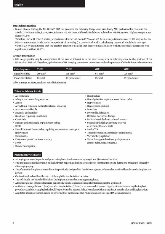
MRI-Related Heating• Innon-clinicaltesting,theNit-Occlud®PDAcoilproducedthefollowingtemperatureriseduringMRIperformedfor15-mininthe3-Tesla(3-Tesla/128-MHz,Excite,HDx,Software14X.M5,GeneralElectricHealthcare,Milwaukee,WI)MRsystem:Highesttemperaturechange +1.6°C.
Therefore, the MRI-related heating experiments for the Nit-Occlud® PDA coil at 3-Tesla using a transmit/receive RF body coil at an MRsystemreportedwholebodyaveragedSARof2.9-W/kg(i.e.,associatedwithacalorimetrymeasuredwholebodyaveragedvalueof2.7-W/kg)indicatedthatthegreatestamountofheatingthatoccurredinassociationwiththesespecificconditionswasequal to or less than +1.6°C.
Artifact Information• MR image qualitymay be compromised if the area of interest is in the exact same area or relatively close to the position of the
Nit-Occlud® PDA coil. Therefore, optimization of MR imaging parameters to compensate for the presence of this device may be necessary.
Pulse Sequence T1-SE T1-SE GRE GRE
SignalVoidSize 369 mm2 118 mm2 647 mm2 739 mm2
Plane Orientation Parallel Perpendicular Parallel Perpendicular
Table 1: Image artifacts, results of non-clinical testing

GA034/Rev02_130904 7
English
Coil SelectionAccording to the measurements, the ductus type and the following recommendations, an appropriate coil should be selected:• ThedistalcoildiameterDshouldbenomorethan2mmlargerthanD2.• ThedistalcoildiameterDshouldbeatleast3to4mmlargerthanD1.• LengthoftheconfiguredcoilLc(seeproductlabel)shouldbenotlongerthanL3.
Whendefiningtheparamatersofyourmeasurements,pleaseconsiderthattheanatomyofthePDAmaydifferamongpatients.Accordingto Krichenko et al. there are five different types of PDAs (see device performance by PDA anatomy in clinical study section below).
Table 2: Selection of the Nit-Occlud® PDA (according to angiographic PDA dimensions)
Coil Implantation Sequence
Step: 01• UnpacktheNit-Occlud®PDAconsistingofcoilwithdisposablehandleandimplantationcatheterundersterileconditions.
WA R N I N G Do not pull on the delivery system. If the coil is withdrawn into the Y connector, there is a danger that the system
can no longer be loaded. Check all screw connections. Some screw joints may have been loosened by the sterilization process.
• FlushthesystemcarefullythroughthesideaccessoftheYconnectorwithheparinizedsalinesolution,andensurethatthereisnoairremaining anywhere in the system.
• Checkthecoilpositioninsidethetransparenttransportationsheath.Thecoilshouldbeinsidethesheath.Whenitisinthisposition,it is essential not to pull on the delivery system. If the coil is not positioned inside the transportation sheath, or shows visible signs of damage, it must be replaced with a new coil.
• D1=Narrowestdiameter• D2=AorticAmpulladiameter• L3=PDAlength
• D=Distaldiameter• P=Proximaldiameter• Lc=Lengthconfigurated
Directions for Use
D1 D2 Device
1mm ≤3mm 4x4
1mm 4mm 5x4
1mm ≥5mm 6x5
2mm ≤5mm 6x5
2mm 6-7mm 7x6
2mm ≥8mm 9x6
D1 D2 Device
3mm ≤7mm 7x6
3mm 8-9mm 9x6
3mm ≥9mm 9x6 or 11x6
<4mm 9mm 11x6
<4mm 10-11mm 11x6
<4mm ≥12mm 11x6
Figure 3: PDA Measurements
Figure 4: Types of PDAs According to: Krichenko et. al. 1989, AmJ Cardiol; 63:877-880
Conical Type A Short Type B Tubular Type C Complex Type D Elongated Type E
8 GA034/Rev02_130904
English
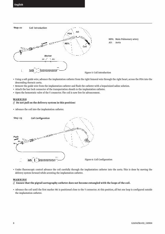
• Usingasoftguidewire,advancetheimplantationcatheterfromtherightfemoralveinthroughtherightheart,acrossthePDAintothedescending thoracic aorta.
• Removetheguidewirefromtheimplantationcatheterandflushthecatheterwithaheparinizedsalinesolution.• Attachtheluerlockconnectorofthetransportationsheathtotheimplantationcatheter.• OpenthehemostaticvalveoftheYconnector.Thecoilisnowfreeforadvancement.
WA R N I N G Do not pull on the delivery system in this position!
• Advancethecoilintotheimplantationcatheter.
• Under fluoroscopiccontroladvance thecoilcarefully throughthe implantationcatheter into theaorta.This isdonebymovingthedelivery system forward while arresting the implantation catheter.
WA R N I N G Ensure that the pigtail aortography catheter does not become entangled with the loops of the coil.
• AdvancethecoiluntilthefirstmarkerM1ispositionedclosetotheYconnector.Atthisposition,allbutoneloopisconfiguredoutsidethe implantation catheter.
Figure5:CoilIntroduction
Figure 6: Coil Configuration
MPA: Main Pulmonary arteryAO: Aorta

GA034/Rev02_130904 9
English
•Retracttheentiresystem(implantationcatheter,deliverysystem)underfluoroscopiccontroluntiltheconfiguredcoilispositionedintheampullaoftheductus(closehemostaticvalveofYconnectororfiximplantationcatheteragainstdeliverysystem).
NOTE: For longer ductus types, coil configuration inside the ductus ampulla is recommended. Here, 2–3 windings of the coil must first be configured in the aorta. Then the entire system is pulled into the ductus ampulla for further configuration of the coil.
• OpenthehemostaticvalveoftheYconnector.• Configurethelast1or2loopsonthepulmonarysideoftheductusbysimultaneouslypullingbacktheimplantationcatheter(with
your left hand) and pushing the delivery system (with your right hand). Advance the delivery system until the second marker M2 is closetotheYconnector.Atthispositionthecoilisoutsidethecatheter.
• Performanaortogramtoconfirmthatthecoilisinthecorrectposition.NOTE: If the position or size of the coil is not satisfactory, it should be repositioned or exchanged at this point.
Repositioning• Torepositionthecoil,pullitbackintotheimplantationcatheterbypullingthedeliverysystem.
WA R N I N G Close the gap between delivery system and coil before you retrieve the coil into the implantation catheter.
• Todoso,holdthehandlewithonehandandmovethedeliverysystemgentlyforwardwhileholdingitbetween2fingersofthesamehand. This movement closes the gap between coil and delivery system and should be done under fluoroscopic control. Once the gap is closed the coil can be pulled smoothly into the implantation catheter.
Figure 7: Coil Positioning
Figure8:FinalCoilAdjustment

10 GA034/Rev02_130904
English
• Whenthecoilisproperlypositioned,itshouldbereleased.Therotationscrewshouldliedirectlyagainstthepusherball.Ifthereisanygap between the two, it must be closed.
WA R N I N G Final release should only be performed if the coil is properly positioned in the PDA. Otherwise, the coil must either
be retrieved and repositioned or replaced by an appropriate substitute. Before the coil is finally released, proper po-sition of the coil should be confirmed by angiography.
• Removethesafetyclipfromthehandle.• Turn the rotation screwunder fluoroscopyclockwiseuntil thecoil is released.Note thatdependingon thecoil typebetween8–15 rotationsareneededtoreleasethecoil.Youwillfeelanincreaseinresistanceimmediatelypriortorelease.
• Removethedeliverysystemandimplantationcatheter.• Performafinalaortogramabout10minuteslatertodocumentpositionofthecoilandPDAocclusion.• Removetheaortographycatheter.
WA R N I N G Ensure that the catheter does not touch the coil.
WA R N I N G If a strong resistance is encountered while pulling the delivery system into the catheter, do not pull the system very
hard because you risk a premature release of the coil.
• Torepositiontheimplantationcatheter,thecoilshouldbepulledbackintothetransportationsheath,carefullyandundervisualcontrol,untilthetipofthecoilisinlinewiththemarkeratthedistalendoftransportationsheath.FixthecoilpositionbyclosingtheYconnector.
WA R N I N G If the coil is pulled back too far, there is the risk that it may not be possible to reload it into the delivery system.
• ThenflushtheimplantationcatheterwithheparinizedsalinesolutionandrepeattheprocedurefromStep02.
Figure 9: Coil Release

GA034/Rev02_130904 11
English
Complications may be avoided or ameliorated by the following:• Useofvenousaccessforimplants.• Flushingallcomponentswithheparinizedsaline.• Keepingdeliverysystemandcatheterstraight,avoidingloopsandcurvesonthecatheterizationtable.• Coilrecaptureinthepulmonaryarteryoraorta,avoidingpullingtheexposedcoilacrossheartvalvesorthroughtherightventricle.
Failure of detachment:Complications may arise if the coil is not released successfully. “Sticking” may occur if positioning of the coil is very time-consuming and/or if the delivery system does not protrude far enough from the end of the implantation catheter. The distal part of the delivery system must be placed outside of the implantation catheter immediately before release. If, however, the coil “sticks”, the device must be retrieved and exchanged. As done with all interventional instruments, prior to implantation of the exchanged device the catheter should be flushed thoroughly to prevent coagulation, thus avoiding elevated friction or “sticking” of the system.
Device Embolization/ Premature releaseThe coil may embolize into the pulmonary artery if the aortic cone is too small or the coil fit is too loose. This can be prevented by accurate measurement of the PDA dimensions and choice of an appropriate coil for the PDA. Correct calibration of the angiographic measurement is a very important factor.In case of coil embolization, interventional retrieval should be performed using a snare or a bioptome.In the event that the coil embolizes and interventional retrieval is unsuccessful, surgical retrieval should be considered.Protrusion/ ObstructionProtrusion of the windings into the aorta and/ or into the pulmonary artery may cause blood flow disturbances or vessel stenosis. This is avoidable by choosing an appropriate coil with a configured length (Lc) equal to or less than the PDA length (L3). The recoil force of the coil tends to return the device to its original configuration whenever possible and will retract the windings into the ampulla and/ or against the vessel wall. Pulmonary artery protrusion may be avoided by correct coil positioning during implant.
Late complicationsDelayed complications such as migration or protrusion with a significant blood flow disturbance may require surgical removal of the coil.
Study descriptionA prospective, non-randomized, multi-center, single-arm Study and a continuing access study were performed using the same protocols at 15centersintheUnitedStatesofAmericatoassessthesafetyandeffectivenessoftheFlexandMediumNit-Occlud®PDAcoilforocclusionof Patent Ductus Arteriosus (PDA) with minimum angiographic diameter of less than 4 mm. The primary effectiveness endpoints were echocardiographic and clinical closure rates at 12 months. The primary safety endpoint was the serious adverse event rate at 12 months. TheendpointrateswerecomparedtoanObjectivePerformanceCriteriaasfollows:
• Echocardiographicclosure(absenceofdetectableresidualPDAflowonechocardiogram)greaterthan85%at12months• Clinicalclosure(absenceofheartmurmur)greaterthan95%at12months• Seriousadverseeventrateoflessthan1%at12months
The following criteria were considered for their inclusion:
Technical Complications and how to avoid them
Clinical Studies
Table 3: Inclusion/Exclusion Criteria
Inclusion criteria Exclusion criteria
• PDAwith4mmorsmallerminimumdiameterbycolorDoppler
• Patientweight≥5Kg,age6monthsto21years (Patients older than 21 years may have device implanted and be included in a study registry.)
• Previoustreatmentbysurgeryor Nit-Occlud device with residual PDA noted at least 6 months after the procedure
• Associatedcardiacanomaliesrequiringsurgery• Knownbleedingorbloodclottingdisorders• Ongoingfebrileillness• Pregnancy• Pulmonaryhypertension/increasedpulmonaryvascularresistance(>5WoodUnits)
• Knownhypersensitivitytocontrastmedium

12 GA034/Rev02_130904
English
Study resultsAtotalof378patientswereenrolledand357patientswereevaluatedforsafetyandeffectiveness.Thepatient´smeanagewas4.26years(range0.5to21.9years);themeanweightwas18.1kg(range4.7to109.0kg),atotalof68.1%oftheenrolledpatientswerefemale.Ofthe357evaluable patients, 347 had successful implantation of the device (technical success). Principal safety and effectiveness results are presented in Table 4 below:
Differing Technical Failure Rates were observed based on Angiographic Classification of the PDA on the lateral aortogram and are summarized in the Table 6 below.
Table 4: Principal Safety and Effectiveness Results
RefertoTable5belowforproceduralandfluoroscopytimesbydevicesizeandtype.
Table5:ProcedureandFluoroscopyTimesbyNit-OccludDevice
Table 6: Technical Failure rate by Angiographic Classification (See Figure above)
OPC RatesNit-Occlud
PatientsPercent
95%LowerBound
95%UpperBound
Technical Success at Implantation 95%2 347/357 97.2% 95.6%
Clinical Closure at 12 Month Follow-up 95%1 308/314 98.1% 96.7%
Echocardiographic Closure at 12 Month Follow-Up 85%1 299/309 96.8% 95.0%
Mortality at 12 Months 0%1 0 0.0% 0.95%
Serious Adverse Events at 12 Months 1%1 0 0% 0.95%
Total Device and Procedure Related Adverse Events at 12 Months
6%15/316* 4.7% 7.21%
14/316** 4.4% 6.84%
Composite Success at 12 Months 80%3 294/309 95.1% 93.0%1ObjectivePerformanceCriteria(OPC)specifiedbytheMultiorganizationAdvisoryPanelto(FDA)Appendix(XII)2 Inferred from technical success rate of Gianturco coil technical success cited in Multiorganization Advisory Panel to FDA report
(Appendix XII)3 Defined in IDE protocol but not defined by the Multiorganization Advisory Panel report*Numeratorisnumberofevents;denominatorisnumberwith12mosfu+2withAEbefore12months**Numeratorisnumberofperson;denominatorisnumberwith12mosfu+2withAEbefore12months
Catalog #Device SizeDistal x Proximal Diameter
Device TypeNumber of Implants
Mean Procedure Duration [min.]
Median Procedure Duration [min.]
Mean Fluoroscopy Time [min.]
Median Fluoroscopy Time [min.]
145044 4 x 4 mm Flex 38 68.6 66.0 17.2 14.0
145054 5x4mm Flex 27 77.8 72.0 19.6 17.0
145065 6x5mm Flex 57 91.5 82.0 19.8 18.5
145076 7 x 6 mm Medium 110 83.3 73.5 17.0 15.0
145096 9 x 6 mm Medium 97 92.0 79.0 18.8 16.0
145116 11 x 6 mm Medium 26 93.0 85.0 25.5 23.5
Classification N(%ofTotal) Technical Failure Rate n/N(%)
Conical(A) 267(74.8%) 4/267 (1.5%)
Short(B) 17(4.8%) 3/17 (17.6%)
Tubular(C 5(1.4%) 1/5 (20%)
Complex(D) 18(5.0%) 1/18 (11.1%)
Elongated(E) 50(14.0%) 1/50 (2%)
TOTAL 357(100%) 10/357 (2.8%)

GA034/Rev02_130904 13
English
Study Adverse events were defined as follows:
Serious Adverse Events:• Proceduralordevicerelatedeventswhichwerelife-threatening,requiredsurgerytocorrect,resultedinhospitalizationorprolonged
hospital stay, caused long-term disability, or resulted in genetic damage or birth defect.
MajorAdverseEvents:• Proceduralordevicerelatedeventswhichwerenotlife-threatening,requiredinterventional(catheterbased)and/ormedicaltreatment
to correct up to one year follow-up evaluation but were resolved without surgical intervention.
Minor Adverse Events:• Proceduralordevicerelatedeventswhichwerenotlife-threatening,andwereresolvedwithoutinterventionorwithabriefspecific
non-surgical intervention up to one year follow-up evaluation.
The combined studies safety results were the following:• Mortalityat12months:0.0%(0/314)• SeriousAdverseEventsat12months(devicerelated):0.0%(0/314)• SeriousAdverseEventsat12months(procedurerelated):0.0%(0/314)• TotalAEs(Serious,Major,andMinor)at12monthsorlastfollowup(relatedtotheprocedureorthedevice):4.7%(15/316*)
The15AdverseEventsarefurtherdescribedinTable8below:
Table 7: Adverse EventsProceduralsuccess,effectivenessandsafetyresultswerecomparabletoorbetterthanpredefined0bjectiveperformancecriteria.*
DSMBAdjudication Category No. of Events
MajorDeviceRelated Device embolizationDevice Retrieval/RemovalObstruction of descending aorta
221
Minor Device Related Possible Thrombus 1
MajorProcedureRelated Decreased Pulse in Right FootReaction to anesthesia
12
Minor Procedure Related Reaction to anesthesiaVascularaccesssitecomplicationOther Adverse EventNauseaFever
11211
After use, medical products and accessories pose a potential biological hazard. For this reason, the products and their accessories should be handled and disposed of in accordance with recognised medical procedure, and in compliance with the relevant legal regulations and local ordinances.
pfm medical warrants that this medical device is free from defects in both materials and workmanship. The above warranties are in lieu of all other warranties, either expressed or implied, including any warranty of merchantability or fitness for a particular purpose. Suitability for use of the medical device for any surgical procedure shall be determined by the user. pfm medical shall not be liable for incidental or consequential damages of any kind.
Disposal after Use
Warranty
*Patientswith12monthfollowupandthosewithanadverseeventatanytime*MultiorganizationAdvisoryPaneltoFDAforPediatricCardiovascularDevices.
Proposed Standards for Clinical Evaluation of Patent Ductus Arteriosus Occlusion Devices. CatheterCardiovascInterv2000;51:293-296.t.

14 GA034/Rev02_130904
English

GA034/Rev02_130904 15
English

GA034/Rev02_130904
pfm medical agWankelstraße6050996Köln,GermanyT +49 (0)2236 9641-0, F +49 (0)2236 [email protected]