necrotic enteritis potential in a model system using clostridium perfringens isolated from field...
TRANSCRIPT

BioOne sees sustainable scholarly publishing as an inherently collaborative enterprise connecting authors, nonprofit publishers, academic institutions, researchlibraries, and research funders in the common goal of maximizing access to critical research.
Necrotic Enteritis Potential in a Model System Using Clostridium perfringensIsolated from Field OutbreaksAuthor(s): G. Chalmers, H. L. Bruce, D. L. Toole, D. A. Barnum, and P. BoerlinSource: Avian Diseases, 51(4):834-839. 2007.Published By: American Association of Avian PathologistsDOI: http://dx.doi.org/10.1637/7959-022807-REGR.1URL: http://www.bioone.org/doi/full/10.1637/7959-022807-REGR.1
BioOne (www.bioone.org) is a nonprofit, online aggregation of core research in the biological, ecological, andenvironmental sciences. BioOne provides a sustainable online platform for over 170 journals and books publishedby nonprofit societies, associations, museums, institutions, and presses.
Your use of this PDF, the BioOne Web site, and all posted and associated content indicates your acceptance ofBioOne’s Terms of Use, available at www.bioone.org/page/terms_of_use.
Usage of BioOne content is strictly limited to personal, educational, and non-commercial use. Commercialinquiries or rights and permissions requests should be directed to the individual publisher as copyright holder.

Necrotic Enteritis Potential in a Model System Using Clostridium perfringens Isolatedfrom Field Outbreaks
G. Chalmers,A H. L. Bruce,B D. L. Toole,B D. A. Barnum,A and P. BoerlinAC
ADepartment of Pathobiology, Ontario Veterinary College, University of Guelph, Guelph, Ontario, N1G 2W1, CanadaBMaple Leaf Foods Agresearch, 473-Sixth Concession Road, R.R.#3, Burford, Ontario, N0E 1A0, Canada
Received 28 February 2007; Accepted and published ahead of print 27 April 2007
SUMMARY. Necrotic enteritis is an enteric disease of avian species caused by the anaerobic bacterium Clostridium perfringens.The disease is regularly controlled in the broiler chicken industry with antimicrobials in feed but is reemerging in areas such asEurope where there is a ban on antimicrobials as growth promoters. To study prospective therapies, researchers must be able toreproduce this disease in a controlled environment, but this is not always possible because of differences in the pathogenicity of C.perfringens strains. Our objective was to test the potential of five isolates (SNECP43, 44, 47, 49, and 50), taken from field cases ofnecrotic enteritis, at recreating the disease in a controlled challenge experiment. SNECP43 and 50 were derived from a commonclone, with SNECP50 passed in vivo and SNECP43 subcultured in vitro. Four hundred birds were divided into 16 pens, with threepens each receiving one of five treatments, with one control pen. Day-old birds were raised on a high wheat-based diet to promotenecrotic enteritis development and were challenged with between 3.4 3 109 and 3.2 3 1011 colony-forming units (cfu) of C.perfringens in feed for a period of 24 hr starting on day 13 of the challenge experiment. Lesion scores were assessed on two birds perpen sacrificed on day 17 and on any dead birds during the 25-day study. Growth performance was assessed up to 25 days, andmortality recorded throughout. Only SNECP50 produced necrotic enteritis mortalities significantly different (P # 0.05) from thecontrol. The five isolates were also typed using pulsed-field gel electrophoresis to assess their genetic relatedness. All epidemiologicallyunrelated isolates were deemed genetically unrelated, whereas SNECP43 and 50 differed by only a single minor band. Toxin typewas assessed using polymerase chain reaction (PCR), which was also used for the detection of the gene encoding the b2-toxin.
RESUMEN. Potencial de enteritis necrotica en un modelo utilizando Clostridium perfringens aislado de brotes de campo.La enteritis necrotica es una enfermedad enterica de las especies aviares causada por la bacteria anaerobica Clostridium perfringens.
En la industria del pollo de engorde la enfermedad se controla regularmente con antimicrobianos en el alimento, pero laenfermedad esta emergiendo en areas como Europa donde existe un veto en la utilizacion de antimicrobianos como promotores delcrecimiento. Para estudiar posibles terapias, los investigadores deben ser capaces de reproducir la enfermedad en un ambientecontrolado, sin embargo, debido a las diferencias en la patogenicidad de las cepas de C. perfringens, esto no es siempre posible.Nuestro objetivo fue estudiar el potencial de cinco aislamientos (SNECP43, 44, 47, 49 y 50) tomados de casos de enteritisnecrotica en el campo para recrear la enfermedad en un experimento de desafıo controlado. Los aislados SNECP 43 y 50 fueronderivados de un clon comun (SNCCP50 con pasajes in vivo y SNECP43 subcultivado in vitro). Se dividieron 400 aves en 16corrales, con tres corrales recibiendo uno de cinco tratamientos y un corral control. Aves de un dıa de edad se criaron con una dietacon alto contenido de trigo para promover el desarrollo de enteritis necrotica y se desafiaron comenzando el dıa 13 del experimento conentre 3.4 3 109 y 3.2 3 1011 unidades formadoras de colonia de C. perfringens en el alimento durante las 24 horas del dıa. Seevaluaron las lesiones en dos aves por corral, sacrificadas el dıa 17 y en cualquier ave muerta durante los 25 dıas del estudio. La tasa decrecimiento se evaluo hasta los 25 dıas y se registro la mortalidad a lo largo del experimento. Solo el aislamiento SNCCP50 produjomortalidades por enteritis necrotica significativamente diferentes al control (P # 0.05). Los cinco aislamientos fueron a su veztipificados utilizando electroforesis en gel de campo pulsante para evaluar su asociacion genetica. Todos los aislamientos norelacionados epidemiologicamente resultaron no relacionados geneticamente, mientras que los aislamientos SNECP43 y 50 difirieronsolo en una pequena banda. El tipo de toxina se evaluo utilizando la prueba de reaccion en cadena por la polimerasa, que tambien fueutilizada para la deteccion del gen que codifica para la toxina b2.
Key words: Clostridium perfringens, broiler chicken, necrotic enteritis, pulsed-field gel electrophoresis, disease model
Abbreviations: CFU 5 colony forming units; MLF 5 Maple Leaf Foods; OD600 5 optical density at 600 nm; PCR 5 polymer-ase chain reaction; PFGE 5 pulsed-field gel electrophoresis; TE 5 10 mM Tris, 100 mM EDTA buffer
Clostridium perfringens is a gram-positive, spore-forming, anaer-obic bacillus that is found in soil and in the intestines of humans andanimals (26). It is considered to be the causative agent of necroticenteritis in birds, a disease that is characterized by reduced growthperformance, decreased feed efficiency, and depression in its mildform and by anorexia, severe morbidity, and significant mortality atits worst (2,26). Because of its effect on growth performance andmortality, necrotic enteritis is a disease of economic significance tothe broiler chicken industry (1). Factors that predispose commercialflocks to an outbreak of necrotic enteritis appear to be high protein,
high fiber, or wheat-based diets and infection with coccidia (3,30).Control of necrotic enteritis has historically consisted of minimizingpredisposing factors and application of antimicrobial agents in birdfeed or water at levels shown to be preventive (5,9,13,14,18,23,24).Recently, inclusion of antimicrobial agents in animal feeds has comeunder considerable scrutiny, with many countries legislating againstthe practice (6). As a result, outbreaks of necrotic enteritis havebecome more frequent under antibiotic-free conditions than whenantibiotics were fed at subtherapeutic levels, and it has again becomean important disease in the poultry industry (29). Much research hasbeen devoted to alternative, nondrug preventive therapies, whichhave required model methods to reliably reproduce the disease(16,20,21,30). How representative an isolated strain of C. perfringensCCorresponding author. E-mail: [email protected].
AVIAN DISEASES 51:834–839, 2007
834

used in a disease model is of field strains that cause significant diseaseand mortality in the field has yet to be examined. The objective ofthis study was to identify the potential of C. perfringens strainsisolated from commercial flocks raised without antibiotics to causenecrotic enteritis under typical Canadian commercial broiler flockconditions and to investigate any genetic differences and similaritiesbetween these strains using pulsed-field gel electrophoresis (PFGE).Toxin typing of the C. perfringens strains was performed bypolymerase chain reaction (PCR), including the b2-toxin gene cpb2(10). Clostridium perfringens plasmids are known to carry many ofthe significant toxin genes (22), and so, plasmid preparations ofSNECP43 and 50 were also compared.
MATERIALS AND METHODS
C. perfringens isolates. Five isolates of C. perfringens (SNECP43, 44,47, 49, and 50) were recovered from the surfaces of the intestinalmucosa of birds that died of necrotic enteritis during four differentoutbreaks in commercial antibiotic-free flocks in the southwesternOntario area between 2001 and 2005. They had been isolated frombirds with intestinal lesions typical of necrotic enteritis and withsymptoms ranging from minimal to significant mortality.
SNECP44, 47, and 49 isolates were cultured from epidemiologicallyunrelated cases of necrotic enteritis at three different farms in 2005.SNECP43 and 50 were derived from an isolate of a field case in 2001.The original isolate was passaged several times in vitro and kept frozen at260 C as SNECP43; this isolate is identical to the isolate known asCP4 described in a study by Thompson et al. (28). CP4 has also beenrecently used in an immunology study to test the recognition of C.perfringens antigens (19). SNECP50 was isolated from a bird successfullychallenged with the original strain in 2004. The other three isolates werechosen for their association with typical necrotic enteritis lesions shownin their respective field cases and were intended to give a highprobability of effectively recreating the disease. All strains weresubcultured and frozen at 220 C in a cooked meat medium (Difco,Becton Dickinson, Sparks, MD) until the start of the project.
Positive controls AHL155 and AHL156 for PCR toxin typing, cpeand cpb2 detection, were obtained from the Animal Health Laboratoryat the University of Guelph.
Experimental design. The study used a completely randomizeddesign with a one-way comparison of five treatments. Sixteen pens of 25birds each were used. Three pens were administered one of the followingstrains: SNECP43, SNECP44, SNECP47, SNECP49, or SNECP50.One pen was not treated and served as the control. Statisticalcomparisons were made between the 15 treatment pens and a singlecontrol pen using key variables for comparison, i.e., cumulativemortality by day, cumulative mortality due to necrotic enteritis byday, and average lesion score on day 17.
Study system. A total of 400 male day-old broiler chickens wereassigned to treatment, with 25 chicks placed in each pen on day 0. Birdswere vaccinated for Marek’s disease and bronchitis (Mildvac-MH,Intervet Canada Ltd., Whitby, Canada) at the hatchery. Birds that diedor were culled before noon on day 5 were replaced with extra birds, andthe total number of birds purchased from a commercial hatcheryallowed for replacement of early mortality.
The poultry research facility at Maple Leaf Foods (MLF) Agresearchin Burford, Ontario, Canada, was used to conduct the study. The 16pens, each providing about 13.7 m2 of floor space, were randomlyassigned by block to treatment groups. Birds were randomly assigned topens by block, such that each hatchery box contributed anapproximately equal number of birds to each treatment within eachblock.
Each pen had a concrete floor and nylon-mesh partitions supportedby a polyvinyl chloride (PVC) frame. A solid 12-inch-high plastic barrierat bird level separated adjacent pens. A welded-wire fence with 2.54-cm2
openings was located on top of all barriers. Each pen was permanentlyidentified with a number and provided about 0.55 m2/bird. Five natural-
gas heaters that were equally spaced and positioned to warm incoming airat the north wall of the building heated the barn, and fans located on thesouth-facing wall of the building exhausted air. Each pen contained fournipple-type drinkers that provided clean drinking water ad libitum. Dryfeed was provided ad libitum in tube-type feeders (1/pen) of 20-kgcapacity. The barn was cleaned, washed, and disinfected before placementof new chopped-straw bedding. The lighting program, barn temperature,litter type, and other management practices were typical of commercialbroiler chicken producers in the local geographic area. Birds that weremoribund and unable to reach feed and water were culled andeuthanatized by asphyxiation with carbon dioxide gas. This protocolwas approved by the MLF Agresearch Animal Care Committee.
One feed, MLF Agresearch Standard nonmedicated starter chick feedin crumble form, was fed through the entire study (formulationconfidential). The diet contained 25% wheat to encourage necroticenteritis development and was based on corn–soybean meal. The dietwas formulated to commercial nutritional standards for chicks from 0 to25 days of age.
C. perfringens challenge. Frozen cultures of C. perfringens werethawed and 0.5 ml was used to inoculate 43 ml of reduced thioglycolatemedia (Difco) and incubated overnight at 37 C. Each vial ofthioglycolate was then added to 430 ml cooked-meat medium (Difco)and, again, incubated overnight under the same conditions. Finally,13 liters of thioglycolate + 1% starch (Difco) was inoculated with theentire cooked-meat media preparation and held at 37 C for 8 hruntil added to the feed. Final concentrations in colony-forming units(CFU) per milliliter at the time of their addition to feed weredetermined with serial dilutions on blood agar plates. Mean CFUs ofbacteria consumed were calculated as total inoculum consumed per pendivided by the number of birds in each pen, multiplied by the bacterialconcentration values determined from the serial dilutions, resulting inthe following per-treatment: SNECP43, 3.4 3 109; SNECP44, 6.9 31010; SNECP47, 3.2 3 1011; SNECP49, 1.3 3 1011; and SNECP50,1.8 3 1010. Feed was withdrawn from all birds for approximately 8 hrbefore the first introduction of challenge. Inoculum was administered tobirds via feed ad libitum in trough-type, disposable feeders commencingthe afternoon of day 13 and ending the afternoon of day 14. The feederin the control pen was not removed during the challenge period. Whenadministration of the challenge was completed for all pens assigned tothe challenge, the disposable feeders were removed, and regular feedreturned to the challenged pens. Inoculated feed remaining was weighedto ascertain amount of challenge feed consumed.
Growth performance, mortalities, and lesion scoring. Pen averagebody weights were recorded at the beginning and end of the experimenton days 0 and 25, respectively. Feed consumption was recorded fromday 0 to 25 as well, and feed conversion derived by dividing average feedconsumed per pen by average weight gain per pen.
All birds that died after noon on day 5 were tagged with a uniquenumber that was recorded, along with date, bird weight and pennumber. Necropsies were performed to diagnose cause of death in allmortalities. Blood and clinical analyses were not performed but wouldbe of interest to complement these data in future investigations. Anybirds that were diagnosed with necrotic enteritis were scored grossly fornecrotic enteritis (4,24) and coccidiosis (15). Lesion scoring wasperformed as follows:
0 5 Small intestine was grossly normal.
1 5 Small intestine wall was grossly thinner than normal and
broke or tore easily under mild tension but had no gross evidence
of mucosal necrosis or other abnormalities.
25 One or more focal round or oval areas of acute full thickness
mucosal necrosis of the small intestine. These foci varied in
diameter from approximately 1 to 5 mm. The surface of these
lesions was generally raised above the surrounding tissue and
consisted of grey or white necrotic debris. Alternatively, if the
superficial necrotic material had been removed, the lesions were
slightly depressed and were grey or white.
Necrotic enteritis model system 835

3 5 Irregularly shaped confluent areas of full thickness mucosal
necrosis of the small intestine .5 mm in diameter but affecting
,25% of the small intestine surface area. The surface of these
lesions was generally raised above the surrounding tissue and
consisted of orange/brown necrotic debris. In some cases, where
portions of the superficial necrotic material had been removed,
the lesions were slightly depressed from surrounding tissue.
4 5 Large confluent areas of full thickness mucosal necrosis of
the small intestine affecting 25% or more of the small intestinal
surface area and involving the entire internal circumference of
the affected small bowel. The surface of these lesions was
generally raised above the surrounding tissue and consisted of
orange/brown necrotic debris.
For all birds that died or were sacrificed postchallenge, the intestinewas opened and examined for lesions and were scored if present. Aportion of the unopened intestine next to the intestinal tissue containinglesions was removed and placed in a separate preweighed and labeledsterile plastic sample bag. The air was removed from each sterile samplebag before being chilled at about 4 C until cultured anaerobically toverify the presence of C. perfringens.
C. perfringens culture. Unopened intestine was removed from thesterile plastic container, opened, and the intestinal contents removed. Anarea with necrosis was selected, sampled with a sterile loop, and thesample cultured anaerobically on blood agar media and incubated at37 C for 18 hr; the sterile loop sample was streaked on the blood agarplate into four successive quadrants, sterilizing the loop betweenquadrants.
Identification and quantification of C. perfringens. A sample fromthe same necrotic area as that cultured on blood agar was streaked ontoa microscopic slide and stained using Gram stain. The number of gram-positive rods (magnification 10003) were counted in a singlemicroscopic field and graded as 0, no gram-positive rods; 1, 1–10 gram-positive rods; 2, 11–20 gram-positive rods; 3, 21–30 gram-positive rods; 4, .30 gram-positive rods.
Colonies on blood agar plates were identified as C. perfringens if theyexhibited double-zone hemolysis (11). The number of colonies meetingthese criteria were counted within each quadrant, and the density wasscored as 4+, 1 or more colonies in the fourth quadrant; 3+, 0 coloniesin the fourth quadrant, 1–10 in the third quadrant; 2+, 0 colonies in thethird and fourth quadrants, 1–10 colonies in the second quadrant; 1+,0 colonies in the second, third, and fourth quadrants, 1–10 in the firstquadrant; 0, no colonies in any quadrant.
Pulsed-field gel electrophoresis of isolates. SNECP43, 44, 47, 49,and 50 strains were grown overnight on blood agar plates, at 37 C underanaerobic conditions. A homogenous suspension of bacteria wasembedded in 0.8% SeaKem Gold Agarose (Cambrex Bio ScienceRockland, Rockland, ME) 1-mm-thick plugs to obtain a final opticaldensity at 600 nm (OD600) of 1.25. Plugs were incubated in 10 mMTris, 100 mM EDTA (TE) buffer with 50 mg/ml lysozyme (RocheApplied Science, Mannheim, Germany) with gentle shaking at 37 C for5 hr. They were then rinsed for 15 min in TE buffer and subsequentlyincubated overnight in EDTA 0.5 M, 1% sarkosyl (Fisher Scientific,Fair Lawn, NJ), 2 mg/ml proteinase K (Roche Applied Science), pH 8.0at 50 C with gentle shaking. They were then rinsed five times each for30 mins in 10 mM Tris, 1 mM EDTA, pH 8.0 to remove traces ofproteinase K.
One plug per isolate was equilibrated in 200 ml restriction buffer atroom temperature for 20 min and was then incubated at roomtemperature for 5 hr in 200 ml fresh digestion buffer containing100 U of the restriction enzyme SmaI (New England BioLabs, Ipswich,MA) following the manufacturer’s recommendations. Electrophoresiswas performed in a 1% SeaKem Gold Agarose gel with TE buffer. Gelswere run in 0.53 Tris-borate-EDTA (Fisher Scientific) containing200 mM thiourea (Fisher Scientific) at 14 C for 19 hr. Pulse timesstarted at 4 sec and ended at 38 sec with linear ramping and a field of6 V/cm and an angle of 120u in a Bio-Rad (Hercules, CA) CHEF-IIIelectrophoresis unit. Gels were stained with ethidium bromide (Fisher
Scientific) in Tris–borate–EDTA buffer and analyzed using BioNu-merics software v4.0 (Applied Maths, Austin, TX). Band matching wasperformed using a 0.5% position tolerance, and cluster analysis wasperformed using the Dice similarity coefficient and unweighted pairgroup method with arithmetic mean.
PCR and plasmids. Each C. perfringens isolate was toxin typedaccording to Yoo and collaborators (31). In addition, detection of thecpb2 gene was performed according to Herholz and collaborators (12).Plasmid preps of SNECP43 and SNECP50 were obtained usinga QIAGEN Plasmid Mini Kit (QIAGEN Inc., Valencia, CA) witha supplementary initial lysozyme treatment (50 mg/ml) for 3 hr withshaking at 37 C. Plasmid preps were compared visually using a 0.6%UltraPure agarose gel (Invitrogen, Carlsbad, CA).
Statistical analysis. Body weight, lesion scores, and mortality datawere analyzed using one-way analysis of variance with C. perfringensstrain as the sole source of variation using the statistical analysis packageJMP Version 5.1.1 (SAS Institute, Cary, NC). Feed conversion (kg feed/kg gain) was analyzed with and without body weight at 25 days includedas a covariant in the analysis of variance. Mortality data weretransformed in Excel (Microsoft Corporation, Redmond, WA) usingarcsin (mortality)0.5 as recommended by Steel and Torrie (27) forproportions over a wide range of values. Means within sources ofvariation that were significant (P # 0.05) were examined for differencesusing Student t-tests. Linear relationships between measurements weredetermined using Pearson correlations.
RESULTS
Growth performance. Mean body weights between the treat-ments were not significantly different at day 0 but were at day 25 (P5 0.013) (Table 1). The effect of treatment on feed conversion from0 to 25 days was close to significant (P 5 0.066), with the controlbirds and treatment 5 (SNECP50) tending to require more feed perkg gain than the treatment 1 birds (SNECP43) (Table 1). Inclusionof body weight at 25 days as a covariant in the analysis of variancereduced the significance of this trend but did not eliminate itcompletely (P 5 0.16).
Mortality and lesion scores. Culture and smear scores formortalities from treatments SNECP43, 44, 47, and control were allzero. SNECP49 mortalities had an average culture and smear scoreof 1.3, whereas SNECP50 had average culture and smear scores of3.6. Birds that received SNECP50 (treatment 5) had a greaterpercentage of mortality due to necrotic enteritis than all othertreatments (P 5 0.0005), which did not differ from each other(Table 2). Lesion scores of the necrotic enteritis mortalities revealed
Table 1. Effect of C. perfringens strain on least-square mean bodyweights (kg) and feed conversion (kg feed/kg gain) of broiler cockerelsshown with standard error of the mean (SEM) and the P value ofthe effect.
TreatmentNumber of
pens
WeightFeed conversion
day 0–25Day 0 Day 25
0 (control) 1 0.040 1.05ab 1.721 (SNECP43) 3 0.047 1.07a 1.562 (SNECP44) 3 0.043 1.00bc 1.583 (SNECP47) 3 0.050 1.05a 1.604 (SNECP49) 3 0.040 1.04ab 1.605 (SNECP50) 3 0.047 0.97c 1.67SEM for group 0 0.004 0.03 0.04SEM for groups
1–5 0.003 0.02 0.02P value 0.164 0.013 0.066
a–cMeans within the same column that have different letters aresignificantly different according to Student t-test at P # 0.05
836 G. Chalmers et al.

that birds that died after receiving SNECP50 had a mean lesionscore of approximately 3.5. Mortalities for the SNECP43, 44, 47treatments and the control had mean lesion scores of zero, whereasSNECP49 had an average of 1.3 (because of a single lesion score of4). Sacrificed birds receiving SNECP50 (treatment 5) had greatermean lesion scores than those that received inoculums SNECP43,47, and 49 but similar to those of the control and SNECP44 birds(Table 2). Treatment 1 with isolate SNECP43 did not producelesions or mortalities significantly different from that found in theunchallenged birds (control). SNECP44, 47, and 49 also did notproduce lesions significantly different from the control birds.
Correlations. Pearson correlations between body weights, feedconversion, mortality, and lesion scores indicated that increasedlesion scores were related to reduced body weight at 25 days andincreased mortality, that feed conversion increased as mortality andlesion scores increased, and that total mortality was positivelycorrelated with lesion score and necrotic enteritis mortality(Table 3).
Identification and quantification of C. perfringens. All necroticenteritis mortalities had culture and smear scores equal to or greaterthan 3, indicating that large amounts of C. perfringens were presentin the intestine of the birds that died.
PFGE typing. The PFGE profiles of SNECP44, 47, and 49differed significantly from each other by at least eight bands (Fig. 1)and were not considered closely genetically related. SNECP43 and50 differed by only a single minor band and are, therefore,considered genetically related. Three repeated restriction enzymedigests of these two isolates were performed using three differentplug preparations, and this single low-intensity band difference wasconsistently visible in all replicates.
PCR and plasmids. All 5 isolates were identified as toxin type Aby PCR analysis. The b2-toxin gene (cpb2) was not detected in anyof the isolates. Lastly, there was no discernable difference in plasmid
profile between SNECP43 and 50 after agarose gel electrophoresis(data not shown).
DISCUSSION
Two treatments, the control and SNECP50, tended to haveincreased feed conversion ratios, indicating that the birds with thesetreatments consumed more feed relative to each kg weight gainedthan birds in the other treatments (P 5 0.066). An increase in thefeed conversion ratio was expected in broiler chickens with necroticenteritis because inflammation of the intestine during the infectionslows transfer of nutrients across the gut lining and reduces growth.This was supported by the correlations observed between feedconversion, body weight at 25 days, lesion score, and mortality(Table 3). The trend for the control and SNECP50-treated birds tohave increased mean feed conversion ratios was not because of bodyweight because it remained even after the inclusion of body weight at25 days as a covariant in the analysis (P 5 0.16). The increased feedconversion ratio of the birds administered strain SNECP50 waslikely indicative of the increased virulence of the strain. However, theincreased feed conversion ratio of the control birds could possiblyreflect a natural necrotic enteritis infection, as one of the controlbirds exhibited ileal necrotic enteritis lesions, or could also be due toother etiological factors within the group. Unfortunately, C.perfringens isolates from these birds were not available for PFGEtyping to confirm this hypothesis.
Mean lesion scores for birds sacrificed from the SNECP43, 47,and 49 strain treatments were the lowest at 0.33 and weresignificantly lower than the mean lesion score of birds sacrificedfrom SNECP50 (Table 2); however, the mean lesion score ofsacrificed birds from SNECP50 was not different from that of thecontrols because one of the two control birds exhibited necroticlesions from a natural infection. Having only two samples in thecontrol group, one of which had a natural infection of necroticenteritis, skewed the mean lesion score for the control birds upwards.The lesion scores of the necrotic enteritis mortalities was the bestindicator of strain virulence and revealed that birds that died afterreceiving SNECP50 had a mean lesion score of about 3.5.
Treatment with SNECP50 provided the highest number ofnecrotic enteritis mortalities, 33.7% (Table 2). This isolate provedmost capable of reproducing the disease in a challenge experiment,whereas SNECP43, 44, and 47 produced no mortalities clearlyattributable to necrotic enteritis. The difference in lethality betweenthe SNECP43 and 50 clones is highly significant and displays thestrong effect that in vitro subculturing vs. in vivo passage had onattenuating the virulence of this particular strain. It remains unclearin necrotic enteritis outbreaks what the most important factors arefor inducing disease, but differences in the genetic regulation ofmany genes including potential virulence factors may play a role; thishas been recently supported by Keyburn and collaborators (17) whofound that the alpha toxin is not necessarily the most important
Table 2. Effect of C .perfringens strain on mean lesion score ofsacrificed birds and mortality [% adjusted with arcsin (percentmortality)0.5] of broiler cockerels shown with the standard error of themean (SEM) and the P value of the effect.
TreatmentNumberof pens
Lesionscore
Necrotic enteritismortalities (%)
Total mortality(%)
0 (control) 1 1.00ab 0a 0a
1 (SNECP43) 3 0.33b 0a 7.7a
2 (SNECP44) 3 1.17ab 0a 3.8a
3 (SNECP47) 3 0.33b 0a 3.8a
4 (SNECP49) 3 0.33b 3.8a 7.7a
5 (SNECP50) 3 1.83a 33.7b 35.8b
SEM for group 0 0.48 6.5 7.4SEM for groups
1 to 5 0.26 3.8 4.3P value 0.017 0.0005 0.002
a,bMeans that have different superscripts within the same column aresignificantly different according to Student t-test at P # 0.05.
Table 3. Correlation coefficients for correlations between growth performance parameters, lesion score, and mortality (P values of correlationsin parentheses).
Variable BW0 BW25 FC LS NEM
BW0 Body weight (kg) 0 daysBW25 Body weight (kg) 25 days 20.047 (0.864)FC Feed conversion (kg feed/kg gain) 20.249 (0.352) 20.38 (0.147)LS Lesion score 20.089 (0.744) 20.627 (0.009) 0.336 (0.204)NEM NE mortality % 20.013 (0.962) 20.532 (0.034) 0.581 (0.018) 0.643 (0.007)TM Total mortality % 0.177 (0.513) 20.458 (0.074) 0.402 (0.123) 0.65 (0.006) 0.926 (,0.0001)
Necrotic enteritis model system 837
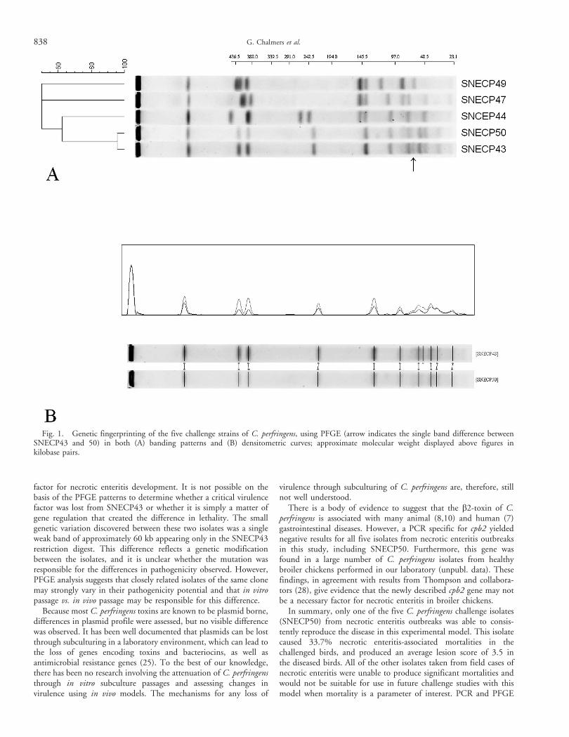
factor for necrotic enteritis development. It is not possible on thebasis of the PFGE patterns to determine whether a critical virulencefactor was lost from SNECP43 or whether it is simply a matter ofgene regulation that created the difference in lethality. The smallgenetic variation discovered between these two isolates was a singleweak band of approximately 60 kb appearing only in the SNECP43restriction digest. This difference reflects a genetic modificationbetween the isolates, and it is unclear whether the mutation wasresponsible for the differences in pathogenicity observed. However,PFGE analysis suggests that closely related isolates of the same clonemay strongly vary in their pathogenicity potential and that in vitropassage vs. in vivo passage may be responsible for this difference.
Because most C. perfringens toxins are known to be plasmid borne,differences in plasmid profile were assessed, but no visible differencewas observed. It has been well documented that plasmids can be lostthrough subculturing in a laboratory environment, which can lead tothe loss of genes encoding toxins and bacteriocins, as well asantimicrobial resistance genes (25). To the best of our knowledge,there has been no research involving the attenuation of C. perfringensthrough in vitro subculture passages and assessing changes invirulence using in vivo models. The mechanisms for any loss of
virulence through subculturing of C. perfringens are, therefore, stillnot well understood.
There is a body of evidence to suggest that the b2-toxin of C.perfringens is associated with many animal (8,10) and human (7)gastrointestinal diseases. However, a PCR specific for cpb2 yieldednegative results for all five isolates from necrotic enteritis outbreaksin this study, including SNECP50. Furthermore, this gene wasfound in a large number of C. perfringens isolates from healthybroiler chickens performed in our laboratory (unpubl. data). Thesefindings, in agreement with results from Thompson and collabora-tors (28), give evidence that the newly described cpb2 gene may notbe a necessary factor for necrotic enteritis in broiler chickens.
In summary, only one of the five C. perfringens challenge isolates(SNECP50) from necrotic enteritis outbreaks was able to consis-tently reproduce the disease in this experimental model. This isolatecaused 33.7% necrotic enteritis-associated mortalities in thechallenged birds, and produced an average lesion score of 3.5 inthe diseased birds. All of the other isolates taken from field cases ofnecrotic enteritis were unable to produce significant mortalities andwould not be suitable for use in future challenge studies with thismodel when mortality is a parameter of interest. PCR and PFGE
Fig. 1. Genetic fingerprinting of the five challenge strains of C. perfringens, using PFGE (arrow indicates the single band difference betweenSNECP43 and 50) in both (A) banding patterns and (B) densitometric curves; approximate molecular weight displayed above figures inkilobase pairs.
838 G. Chalmers et al.

analysis helped to further characterize these isolates and revealed theabsence of the cpb2 toxin gene as well, thus questioning itsimportance in necrotic enteritis development. The difference invirulence between SNECP43 and SNECP50 demonstrates thata high degree of genetic relatedness does not necessarily implyanalogous virulence in a challenge model. Thus, because of thecurrent absence of other specific virulence attributes, the virulencelevel of C. perfringens strains should not be predicted based ongenetic relatedness to known virulent strains but only upon testing inan adequate in vivo challenge model.
REFERENCES
1. Agriculture and Agri-Food Canada. Poultry marketplace—poultry ata glance. Agriculture and Agri-Food Canada, Ottawa, Ontario. p. 1. 2005.
2. Al-Sheikhly, F., and R. B. Truscott. The interaction of Clostridiumperfringens and its toxins in the production of necrotic enteritis of chickens.Avian Dis. 21:256–263. 1977.
3. Branton, S. L., B. D. Lott, J. W. Deaton, W. R. Maslin, F. W.Austin, L. M. Pote, R. W. Keirs, M. A. Latour, and E. J. Day. The effect ofadded complex carbohydrates or added dietary fiber on necrotic enteritislesions in broiler chickens. Poult. Sci. 76:24–28. 1997.
4. Brennan, J., G. Moore, S. E. Poe, A. Zimmermann, G. Vessie, D. A.Barnum, and J. Wilson. Efficacy of in-feed tylosin phosphate for thetreatment of necrotic enteritis in broiler chickens. Poult. Sci. 80:1451–1454.2001.
5. Brennan, J., J. Skinner, D. A. Barnum, and J. Wilson. The efficacy ofbacitracin methylene disalicylate when fed in combination with narasin inthe management of necrotic enteritis in broiler chickens. Poult. Sci.82:360–363. 2003.
6. Casewell, M., C. Friis, E. Marco, P. McMullin, and I. Phillips. TheEuropean ban on growth-promoting antibiotics and emerging consequencesfor human and animal health. J. Antimicrob. Chemother. 52:159–161.2003.
7. Fisher, D. J., K. Miyamoto, B. Harrison, S. Akimoto, M. R. Sarker,and B. A. McClane. Association of beta2 toxin production with Clostridiumperfringens type A human gastrointestinal disease isolates carrying a plasmidenterotoxin gene. Mol. Microbiol. 56:747–762. 2005.
8. Garmory, H. S., N. Chanter, N. P. French, D. Bueschel, J. G.Songer, and R. W. Titball. Occurrence of Clostridium perfringens beta2-toxinamongst animals, determined using genotyping and subtyping PCR assays.Epidemiol. Infect. 124:61–67. 2000.
9. George, B. A., C. L. Quarles, and D. J. Fagerberg. Virginiamycineffects on controlling necrotic enteritis infection in chickens. Poult. Sci.61:447–450. 1982.
10. Gibert, M., C. Jolivet-Reynaud, and M. R. Popoff. Beta2 toxin,a novel toxin produced by Clostridium perfringens. Gene 203:65–73. 1997.
11. Hansen, M. V., and L. P. Elliott. New presumptive identification testfor Clostridium perfringens: reverse CAMP test. J. Clin. Microbiol.12:617–619. 1980.
12. Herholz, C., R. Miserez, J. Nicolet, J. Frey, M. Popoff, M. Gibert, H.Gerber, and R. Straub. Prevalence of beta2-toxigenic Clostridium perfringensin horses with intestinal disorders. J. Clin. Microbiol. 37:358–361. 1999.
13. Hofacre, C. L., T. Beacorn, S. Collett, and G. Mathis. Usingcompetitive exclusion, mannan–oligosaccharide and other intestinal prod-ucts to control necrotic enteritis. J Appl. Poult. Res. 12:60. 2003.
14. Jackson, M. E., D. M. Anderson, H. Y. Hsiao, G. F. Mathis, and D.W. Fodge. Beneficial effect of beta-mannanase feed enzyme on performanceof chicks challenged with Eimeria sp. and Clostridium perfringens. AvianDis. 47:759–763. 2003.
15. Johnson, J., and W. M. Reid. Anticoccidial drugs: lesion scoringtechniques in battery and floor-pen experiments with chickens. Exp.Parasitol. 28:30–36. 1970.
16. Kaldhusdal, M., M. Hofshagen, A. Lovland, H. Langstrand, and K.Redhead. Necrotic enteritis challenge models with broiler chickens raised onlitter: evaluation of preconditions, Clostridium perfringens strains andoutcome variables. FEMS Immunol. Med. Microbiol. 24:337–343. 1999.
17. Keyburn, A. L., S. A. Sheedy, M. E. Ford, M. M. Williamson, M. M.Awad, J. I. Rood, and R. J. Moore. Alpha-toxin of Clostridium perfringens isnot an essential virulence factor in necrotic enteritis in chickens. Infect.Immun. 74:6496–6500. 2006.
18. Kondo, F. In vitro lecithinase activity and sensitivity to 22antimicrobial agents of Clostridium perfringens isolated from necroticenteritis of broiler chickens. Res. Vet. Sci. 45:337–340. 1988.
19. Kulkarni, R. R., V. R. Parreira, S. Sharif, and J. F. Prescott.Clostridium perfringens antigens recognized by broiler chickens immune tonecrotic enteritis. Clin. Vaccine Immunol. 13:1358–1362. 2006.
20. McReynolds, J. L., J. A. Byrd, R. C. Anderson, R. W. Moore, T. S.Edrington, K. J. Genovese, T. L. Poole, L. F. Kubena, and D. J. Nisbet.Evaluation of immunosuppresants and dietary mechanisms in an experi-mental disease model for necrotic enteritis. Poult. Sci. 83:1948–1952. 2004.
21. Olkowski, A. A., C. Wojnarowicz, M. Chirino-Trejo, and M. D.Drew. Responses of broiler chickens orally challenged with Clostridiumperfringens isolated from field cases of necrotic enteritis. Res. Vet. Sci.81:99–108. 2006.
22. Petit, L., M. Gibert, and M. R. Popoff. Clostridium perfringens:toxinotype and genotype. Trends Microbiol. 7:104–110. 1999.
23. Prescott, J. F. The prevention of experimentally induced necroticenteritis in chickens by avoparcin. Avian Dis. 23:1072–1074. 1979.
24. Prescott, J. F., R. Sivendra, and D. A. Barnum. The use of bacitracinin the prevention and treatment of experimentally-induced necrotic enteritisin the chicken. Can. Vet. J. 19:181–183. 1978.
25. Rood, J. I., and S. T. Cole. Molecular genetics and pathogenesis ofClostridium perfringens. Microbiol. Rev. 55:621–648. 1991.
26. Songer, J. G. Clostridial enteric diseases of domestic animals. Clin.Microbiol. Rev. 9:216–234. 1996.
27. Steel, R. G. D., and J. H. Torrie. Principles and procedures ofstatistics, second edition. McGraw-Hill Book Company, New York, NewYork. p. 236. 1980.
28. Thompson, D. R., V. R. Parreira, R. R. Kulkarni, and J. F. Prescott.Live attenuated vaccine-based control of necrotic enteritis of broilerchickens. Vet. Microbiol. 113:25–34. 2005.
29. Van Immerseel, F., J. De Buck, F. Pasmans, G. Huyghebaert, F.Haesebrouck, and R. Ducatelle. Clostridium perfringens in poultry: an emergingthreat for animal and public health. Avian Pathol. 33:537–549. 2004.
30. Williams, R. B., R. N. Marshall, R. M. La Ragione, and J. Catchpole.A new method for the experimental production of necrotic enteritis and itsuse for studies on the relationships between necrotic enteritis, coccidiosis andanticoccidial vaccination of chickens. Parasitol. Res. 90:19–26. 2003.
31. Yoo, H. S., S. U. Lee, K. Y. Park, and Y. H. Park. Molecular typingand epidemiological survey of prevalence of Clostridium perfringens types bymultiplex PCR. J. Clin. Microbiol. 35:228–232. 1997.
ACKNOWLEDGMENTS
We thank Kevin Thompson and Shane Stankov of Maple Leaf FoodsAgresearch farm for their excellent technical assistance and Dr. ElizabethBlack and Dr. Jeff Wilson for their expertise in lesion scoring andpoultry pathology. Thanks to Patricia Bell-Rogers at the Animal HealthLab for the control isolates for PCR. G. Chalmers was supportedfinancially throughout the project by the Poultry Industry Council ofCanada.
Necrotic enteritis model system 839