libro analgesic regional
TRANSCRIPT

Peripheral Nerve Blockade for Major Orthopedic Surgery
Mayo Clinic Analgesic Pathway
FrontMatter4.0.qxd 9/20/05 1:45 PM Page 1

Peripheral Nerve Blockade for Major Orthopedic Surgery
Robert L. Lennon, D.O.Supplemental Consultant
Department of AnesthesiologyMayo Clinic
Associate Professor of AnesthesiologyMayo Clinic College of Medicine
Rochester, Minnesota
Terese T. Horlocker, M.D.Consultant
Department of AnesthesiologyMayo Clinic
Professor of Anesthesiology and of OrthopedicsMayo Clinic College of Medicine
Rochester, Minnesota
Mayo Clinic Analgesic Pathway
MAYO CLINIC SCIENTIFIC PRESS
TAYLOR & FRANCIS GROUP
FrontMatter4.0.qxd 9/20/05 1:45 PM Page 3
ISBN 0849395720
The triple-shield Mayo logo and the words MAYO, MAYO CLINIC, and MAYO CLINICSCIENTIFIC PRESS are marks of Mayo Foundation for Medical Education and Research.
©2006 by Mayo Foundation for Medical Education and Research.
All rights reserved. This book is protected by copyright. No part of it may be reproduced, storedin a retrieval system, or transmitted, in any form or by any means—electronic, mechanical, photo-copying, recording, or otherwise—without the prior written consent of the copyright holder, exceptfor brief quotations embodied in critical articles and reviews. Inquiries should be addressed toScientific Publications, Plummer 10, Mayo Clinic, 200 First Street SW, Rochester, MN 55905.
For order inquiries, contact Taylor & Francis Group, 6000 Broken Sound Parkway NW, Suite #300,Boca Raton, FL 33487.
www.taylorandfrancis.com
Library of Congress Cataloging-in-Publication Data
Lennon, Robert L.Mayo clinic analgesic pathway : peripheral nerve blockade for major orthopedic surgery / RobertL. Lennon, Terese T. Horlocker.
p. ; cm.Includes bibliographical references and index.ISBN 0-8493-9572-0 (alk. paper)1. Anesthesia in orthopedics. 2. Nerve block. 3. Postoperative pain--Treatment. I. Title: Analgesicpathway. II. Horlocker, Terese T. III. Mayo Clinic. IV. Title.[DNLM: 1. Nerve Block--methods. 2. Lower Extremity--surgery. 3. Orthopedic Procedures. 4.Pain, Postoperative--therapy. WO 375 L567m 2006]
RD751.L56 2006617.9'6747--dc22 2005053401
Care has been taken to confirm the accuracy of the information presented and to describe generallyaccepted practices. However, the authors and publisher are not responsible for errors or omissionsor for any consequences from application of the information in this book and make no warranty, expressor implied, with respect to the contents of the publication. This book should not be relied on apartfrom the advice of a qualified health care provider.
The authors and publisher have exerted efforts to ensure that drug selection and dosage set forthin this text are in accordance with current recommendations and practice at the time of publica-tion. However, in view of ongoing research, changes in government regulations, and the constantflow of information relating to drug therapy and drug reactions, the reader is urged to check thepackage insert for each drug for any change in indications and dosage and for added warnings andprecautions. This is particularly important when the recommended agent is a new or infrequentlyemployed drug.
Some drugs and medical devices presented in this publication have Food and DrugAdministration (FDA) clearance for limited use in restricted research settings. It is the responsi-bility of the health care providers to ascertain the FDA status of each drug or device planned for usein their clinical practice.
FrontMatter4.0.qxd 9/20/05 1:45 PM Page 4

TABLE OF CONTENTS
Section I: Principles of Lower Extremity Peripheral Nerve Block . . . 11. Neural Anatomy . . . . . . . . . . . . . . . . . . . . . . . . . . . . . . . . . . . . . . . . . . . . . . . 32. Dermatomes and Osteotomes . . . . . . . . . . . . . . . . . . . . . . . . . . . . . . . . . . . . 113. Preoperative Assessment and Monitoring . . . . . . . . . . . . . . . . . . . . . . . . . . . . 154. Techniques and Equipment for Neural Localization . . . . . . . . . . . . . . . . . . . . 195. Selection of Local Anesthetic and Adjuvants. . . . . . . . . . . . . . . . . . . . . . . . . . 256. Neurologic Complications . . . . . . . . . . . . . . . . . . . . . . . . . . . . . . . . . . . . . . . 31
Section II: Lumbar Plexus Block . . . . . . . . . . . . . . . . . . . . . . . . . . 337. Psoas Compartment Approach . . . . . . . . . . . . . . . . . . . . . . . . . . . . . . . . . . . . 358. Fascia Iliaca Approach . . . . . . . . . . . . . . . . . . . . . . . . . . . . . . . . . . . . . . . . . . 439. Femoral Nerve Block . . . . . . . . . . . . . . . . . . . . . . . . . . . . . . . . . . . . . . . . . . . 47
10. Lateral Femoral Cutaneous Nerve Block. . . . . . . . . . . . . . . . . . . . . . . . . . . . . 5111. Obturator Nerve Block . . . . . . . . . . . . . . . . . . . . . . . . . . . . . . . . . . . . . . . . . 5512. Saphenous Nerve Blocks . . . . . . . . . . . . . . . . . . . . . . . . . . . . . . . . . . . . . . . . 59
Section III: Sciatic Nerve Block . . . . . . . . . . . . . . . . . . . . . . . . . . . 6513. Classic Posterior Approach of Labat . . . . . . . . . . . . . . . . . . . . . . . . . . . . . . . . 6714. Parasacral Approach. . . . . . . . . . . . . . . . . . . . . . . . . . . . . . . . . . . . . . . . . . . . 7115. Subgluteal Approach . . . . . . . . . . . . . . . . . . . . . . . . . . . . . . . . . . . . . . . . . . . 7516. Anterior Approach . . . . . . . . . . . . . . . . . . . . . . . . . . . . . . . . . . . . . . . . . . . . . 7917. Lateral Popliteal Approach . . . . . . . . . . . . . . . . . . . . . . . . . . . . . . . . . . . . . . . 8318. Posterior Popliteal Approach . . . . . . . . . . . . . . . . . . . . . . . . . . . . . . . . . . . . . 8719. Ankle Block . . . . . . . . . . . . . . . . . . . . . . . . . . . . . . . . . . . . . . . . . . . . . . . . . . 91
Section IV: Mayo Clinic Analgesic Pathway . . . . . . . . . . . . . . . . . 10120. Mayo Clinic Total Joint Anesthesia and Analgesic Pathway . . . . . . . . . . . . . 10321. Regional Anesthesia and Analgesia in the Patient
Receiving Thromboprophylaxis . . . . . . . . . . . . . . . . . . . . . . . . . . . . . . . . . . 11322. Management of Inpatient Peripheral Nerve Catheters . . . . . . . . . . . . . . . . . 11723. Management of Ambulatory Peripheral Nerve Catheters . . . . . . . . . . . . . . . 12124. Nursing Management of Peripheral Nerve Catheters . . . . . . . . . . . . . . . . . . 12525. Future Directions. . . . . . . . . . . . . . . . . . . . . . . . . . . . . . . . . . . . . . . . . . . . . 127
Index . . . . . . . . . . . . . . . . . . . . . . . . . . . . . . . . . . . . . . . . . . . . . .129
v
FrontMatter4.0.qxd 9/20/05 1:45 PM Page 5
FOREWORD
espite the explosion of new techniques and technologies, the single most important change in my practice in the past several years has been the introductionof perioperative regional block protocols. The entire perioperative experience forpatients having hip and knee arthroplasty has been improved because of this multidisciplinary approach. Undoubtedly, this approach will be shown to lead to significantly lower narcotic use, a more benign postoperative course with fewer medical complications, lower overall hospital costs, and higher patient satisfaction. These results will lead to the expectation, by patients and physicians,that these block protocols are included in the standard of care. I am indebted tomy anesthesia colleagues for the hard work that is required each and every day tomake these protocols work for patients. As a surgeon, I undoubtedly receive farmore of the credit and gratitude from my patients than deserved.
Arlen D. Hanssen, M.D.Consultant, Department of Orthopedic Surgery, Mayo Clinic
Professor of Orthopedics, Mayo Clinic College of MedicineRochester, Minnesota
vii
D
FrontMatter4.0.qxd 9/20/05 1:45 PM Page 7
PREFACE
“Regional anesthesia has come to stay.” These words by surgeon William J. Mayo,M.D., opened the foreword to Regional Anesthesia: Its Technic and Clinical Application,by Gaston Labat, M.D. Published in 1922, Labat’s text popularized regional anesthesiain the United States by describing techniques already familiar to European surgeons andanesthesiologists. Importantly, Labat described the use of infiltration and peripheral,plexus, and splanchnic blockade (using cocaine and procaine) for head and neck,intrathoracic, intra-abdominal, and extremity surgery. The techniques of peripheralneural blockade were developed early in the history of anesthesia, and over time neuraxialand general anesthesia, with their improved safety, supplanted their use.
Recently, the introduction of long-acting local anesthetics and adjuvants, therefinement of imaging methods to facilitate neural localization, and innovations in equipment technology, including stimulating needles, catheters, and portableinfusion devices, have increased the success rate and popularity of peripheral blockade. Undoubtedly, peripheral nerve blocks represent a new era in regionalanesthesia and analgesia. Competence in these techniques is crucial to future practice models. However, adequate training and proficiency affect utilization. A nationwide survey reported that 98% of anesthesiologists perform peripheraltechniques but most perform fewer than five per month (although the majoritypredict increased use in the future). Likewise, despite improvements in needle andcatheter technology and neural localization, these blocks often remain underutilizedand challenging. Studies evaluating proficiency in technical skills have noted thatregional anesthetic procedures are significantly more difficult to learn than the basic manual skills necessary for general anesthetic procedures, such as intubationand arterial cannulation. Also, the majority of resident training programs do notprovide formal instruction in peripheral blockade.
In 2003, a multidisciplinary group of surgeons, anesthesiologists, nurses, pharmacists, and physical therapists implemented the Mayo Clinic total joint analgesic pathway, a multimodal approach that utilized peripheral regional techniques and oral analgesics (no long-acting or intravenous opioids were administered). The results were truly remarkable. With the use of strict dismissalcriteria, 95% of patients undergoing total knee arthroplasty and 80% of patients
ix
FrontMatter4.0.qxd 9/20/05 1:45 PM Page 9
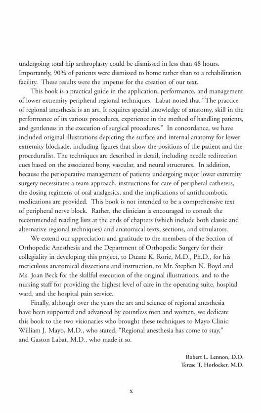
undergoing total hip arthroplasty could be dismissed in less than 48 hours.Importantly, 90% of patients were dismissed to home rather than to a rehabilitationfacility. These results were the impetus for the creation of our text.
This book is a practical guide in the application, performance, and managementof lower extremity peripheral regional techniques. Labat noted that “The practiceof regional anesthesia is an art. It requires special knowledge of anatomy, skill in theperformance of its various procedures, experience in the method of handling patients,and gentleness in the execution of surgical procedures.” In concordance, we haveincluded original illustrations depicting the surface and internal anatomy for lowerextremity blockade, including figures that show the positions of the patient and theproceduralist. The techniques are described in detail, including needle redirectioncues based on the associated bony, vascular, and neural structures. In addition,because the perioperative management of patients undergoing major lower extremitysurgery necessitates a team approach, instructions for care of peripheral catheters,the dosing regimens of oral analgesics, and the implications of antithromboticmedications are provided. This book is not intended to be a comprehensive text of peripheral nerve block. Rather, the clinician is encouraged to consult the recommended reading lists at the ends of chapters (which include both classic andalternative regional techniques) and anatomical texts, sections, and simulators.
We extend our appreciation and gratitude to the members of the Section ofOrthopedic Anesthesia and the Department of Orthopedic Surgery for their collegiality in developing this project, to Duane K. Rorie, M.D., Ph.D., for hismeticulous anatomical dissections and instruction, to Mr. Stephen N. Boyd andMs. Joan Beck for the skillful execution of the original illustrations, and to thenursing staff for providing the highest level of care in the operating suite, hospitalward, and the hospital pain service.
Finally, although over the years the art and science of regional anesthesia have been supported and advanced by countless men and women, we dedicate this book to the two visionaries who brought these techniques to Mayo Clinic:William J. Mayo, M.D., who stated, “Regional anesthesia has come to stay,” and Gaston Labat, M.D., who made it so.
Robert L. Lennon, D.O.Terese T. Horlocker, M.D.
x
FrontMatter4.0.qxd 9/20/05 1:45 PM Page 10
Recommended ReadingHadzic A, Vloka JD, Kuroda MM, Koorn R, Birnbach DJ. The practice of
peripheral nerve blocks in the United States: a national survey. Reg AnesthPain Med. 1998;23:241-6.
Konrad C, Schupfer G, Wietlisbach M, Gerber H. Learning manual skills in anesthesiology: Is there a recommended number of cases for anesthetic proce-dures? Anesth Analg. 1998;86:635-9.
Labat G. Regional anesthesia: its technic and clinical application. Philadelphia:WB Saunders Company; 1922.
Pagnano MW, Trousdale RT, Hanssen AD, Lewallen DG, Hebl JR, Kopp SL, et al.A comprehensive regional anesthesia protocol markedly improves patient careand facilitates early discharge after total knee and total hip arthroplasty. AbstractNo. SE043. Read at the 2005 annual meeting of the American Academy ofOrthopaedic Surgeons, Washington, DC, February 23 to 27, 2005. Abstractavailable from http://www.aaos.org.
xi
FrontMatter4.0.qxd 9/20/05 1:45 PM Page 11

REGIONAL ANESTHESIA AND MAYO CLINIC
A BRIEF HISTORY
Both William J. Mayo, M.D., and Charles H. Mayo, M.D., used local infiltrationanesthesia from the inception of Saint Marys Hospital in September 1889. ByJanuary 1901, local anesthesia was used in about 7% of the surgical cases at thehospital. In 1920, Dr. Charles H. Mayo visited with a surgical colleague, VictorPauchet, M.D., in France. Pauchet was a master of regional anesthetic blocks andhad taught these techniques to his student Gaston Labat, M.D. Mayo recruitedLabat to come to Rochester, Minnesota, and teach regional anesthesia to the surgeons and to write a book describing regional anesthesia.
Labat began his work at Mayo Clinic on October 1, 1920. His book, which was richly illustrated by Mayo Clinic artists, was largely a translation ofVictor Pauchet’s L’Anesthesie Regionale and included a new section on the regionalanesthetist as a specialist. Before he left Mayo Clinic in 1921, Labat taughtWilliam Meeker, M.D., his techniques of regional anesthesia. Meeker likewisetaught John Lundy, M.D., those same techniques, and in 1924 Lundy was appointed Chair, Section on Regional Anesthesia. By 1931, approximately 30% of all anesthetics given at Mayo Clinic involved a regional technique.
The Labat tradition of regional anesthesia spread across the United States.Labat’s book Regional Anesthesia: Its Technic and Clinical Application was a medicalbestseller, having several printings and two editions before World War II. Lundytaught Labat’s approaches to Ralph Waters, M.D., the founder of the first academicdepartment of anesthesiology, at the University of Wisconsin, Madison. Waterssubsequently taught Emery Rovenstine, M.D., who also was mentored in regionalanesthesia by Hippolyte Wirtheim, M.D., Labat’s partner at Bellevue Hospital inNew York City. The American Society of Regional Anesthesia (1924-1942) wasfounded around Labat and was critical to the formation of the American Board ofAnesthesiology (ABA) in 1938. Many questions on the first written examination of the ABA were directly related to the performance of percutaneous regional anesthesia. Thus, the tradition begun in Rochester, Minnesota, spread first to theEast Coast and eventually across the nation. Today, this tradition of percutaneousregional anesthesia is alive, well, and innovatively applied in the operating rooms atMayo Clinic.
xiii
FrontMatter4.0.qxd 9/20/05 1:45 PM Page 13

xiv
Representative illustrations from Regional Anesthesia: Its Technic and Clinical Application,by Gaston Labat, M.D., published in 1922.
By permission of Mayo Foundation for Medical Education and Research.
FrontMatter4.0.qxd 9/20/05 1:45 PM Page 14

Recommended ReadingBacon DR. Gaston Labat, John Lundy, Emery Rovenstine, and the Mayo Clinic:
the spread of regional anesthesia in America between the World Wars. J ClinAnesth. 2002;14:315-20.
Brown DL, Winnie AP. Biography of Louis Gaston Labat, M.D. Reg Anesth.1992;17:249-62.
Cote AV, Vachon CA, Horlocker TT, Bacon DR. From Victor Pauchet to GastonLabat: the transformation of regional anesthesia from a surgeon’s practice to thephysician anesthesiologist. Anesth Analg. 2003;96:1193-200.
Kopp SL, Horlocker TT, Bacon DR. The contribution of John Lundy in thedevelopment of peripheral and neuraxial nerve blocks at the Mayo Clinic:1925-1940. Reg Anesth Pain Med. 2002;27:322-6.
Douglas R. Bacon, M.D., M.A.Consultant, Division of Methodist North Anesthesia, Mayo Clinic
Professor of Anesthesiology and of History of MedicineMayo Clinic College of Medicine
Rochester, Minnesota
xv
FrontMatter4.0.qxd 9/20/05 1:45 PM Page 15
MEMBERS OF THE SECTION
OF ORTHOPEDIC ANESTHESIA
James R. Hebl, M.D., Section Head
David E. Byer, M.D.
John A. Dilger, M.D.
Edward D. Frie, M.D.
Terese T. Horlocker, M.D.*
Sandra J. Kopp, M.D.
Robert L. Lennon, D.O.*
Steven R. Rettke, M.D.*
Duane K. Rorie, M.D., Ph.D.*†
Kenneth P. Scott, M.D.
Rungson Sittipong, M.D.†
Laurence C. Torsher, M.D.
Jack L. Wilson, M.D.
_________*Previous Section Head
†Emeritus Member
xvi
FrontMatter4.0.qxd 9/20/05 1:45 PM Page 16

PRINCIPLES OF
LOWER EXTREMITY PERIPHERAL
NERVE BLOCK
SECTION I
Section_I_4.0.qxd 9/20/05 1:46 PM Page 1

This page intentionally left blank

NEURAL ANATOMY
3
Chapter 1
The nerve supply to the lower extremity is derived from the lumbar and sacral plexuses. The lumbosacral plexus arises from at least eight spinal nerve roots, each of which contains anterior and posterior divisions that innervate the original ventraland dorsal portions of the limb. With the exception of a small cutaneous portion of the buttock (which is supplied by upper lumbar and lower sacral segmentalnerves), the innervation of the lower extremity is entirely through the branches of the lumbosacral plexus.
Sensory and motor innervation of the anterior and medial aspects of the thigh are from the lumbar plexus. The sacral plexus provides sensory and motor innervation of the buttock, the posterior aspect of the thigh, and the leg and foot, except for the medial area innervated by the saphenous branch of the femoral nerve.
Lumbar Plexus Anatomy (L1 Through L5)The lumbar plexus is formed most commonly from the anterior rami of the firstfour lumbar nerves, frequently including a branch from T12 and occasionally abranch from L5. The plexus lies on the posterior body wall between the psoasmajor and quadratus lumborum muscles, in the so-called psoas compartment. The L2 through L4 components of the plexus primarily innervate the anterior and medial aspects of the thigh. The anterior divisions of L2 through L4 form the obturator nerve, the posterior divisions of the same components form thefemoral nerve, and the posterior divisions of L2 and L3 form the lateral femoralcutaneous nerve.
Section_I_4.0.qxd 9/20/05 1:46 PM Page 3
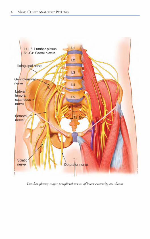
4 MAYO CLINIC ANALGESIC PATHWAY
Lumbar plexus; major peripheral nerves of lower extremity are shown.
L1-L5: Lumbar plexusS1-S4: Sacral plexus
Ilioinguinal nerve
Genitofemoralnerve
Lateralfemoralcutaneousnerve
Femoralnerve
Sciaticnerve Obturator nerve
L1
L2
L3
L4
L5
S1-S4
Section_I_4.0.qxd 9/20/05 1:46 PM Page 4
Section I PRINCIPLES OF LOWER EXTREMITY PERIPHERAL NERVE BLOCK 5
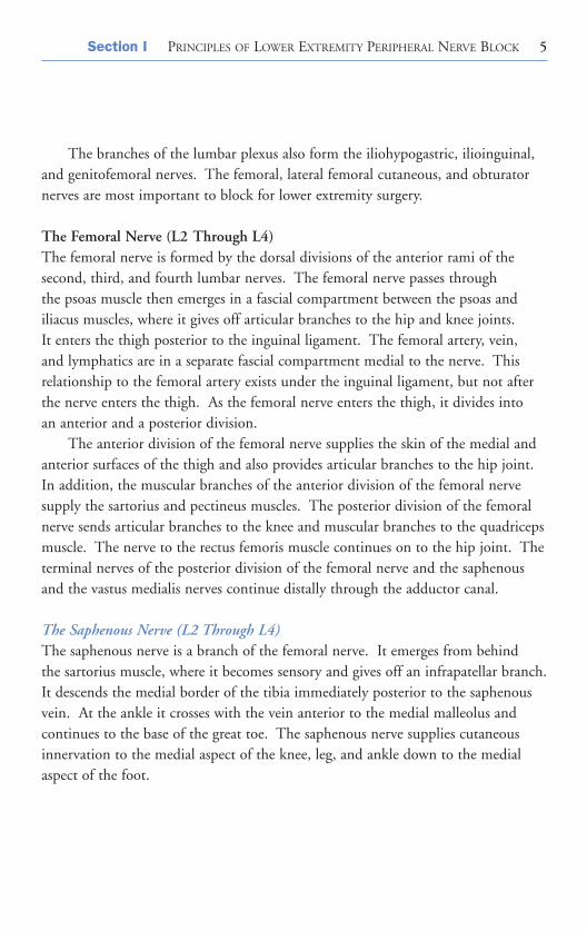
The branches of the lumbar plexus also form the iliohypogastric, ilioinguinal,and genitofemoral nerves. The femoral, lateral femoral cutaneous, and obturatornerves are most important to block for lower extremity surgery.
The Femoral Nerve (L2 Through L4)The femoral nerve is formed by the dorsal divisions of the anterior rami of the second, third, and fourth lumbar nerves. The femoral nerve passes through the psoas muscle then emerges in a fascial compartment between the psoas and iliacus muscles, where it gives off articular branches to the hip and knee joints. It enters the thigh posterior to the inguinal ligament. The femoral artery, vein, and lymphatics are in a separate fascial compartment medial to the nerve. Thisrelationship to the femoral artery exists under the inguinal ligament, but not afterthe nerve enters the thigh. As the femoral nerve enters the thigh, it divides into an anterior and a posterior division.
The anterior division of the femoral nerve supplies the skin of the medial andanterior surfaces of the thigh and also provides articular branches to the hip joint.In addition, the muscular branches of the anterior division of the femoral nervesupply the sartorius and pectineus muscles. The posterior division of the femoralnerve sends articular branches to the knee and muscular branches to the quadricepsmuscle. The nerve to the rectus femoris muscle continues on to the hip joint. Theterminal nerves of the posterior division of the femoral nerve and the saphenousand the vastus medialis nerves continue distally through the adductor canal.
The Saphenous Nerve (L2 Through L4)The saphenous nerve is a branch of the femoral nerve. It emerges from behind the sartorius muscle, where it becomes sensory and gives off an infrapatellar branch.It descends the medial border of the tibia immediately posterior to the saphenousvein. At the ankle it crosses with the vein anterior to the medial malleolus andcontinues to the base of the great toe. The saphenous nerve supplies cutaneousinnervation to the medial aspect of the knee, leg, and ankle down to the medialaspect of the foot.
Section_I_4.0.qxd 9/20/05 1:46 PM Page 5
6 MAYO CLINIC ANALGESIC PATHWAY
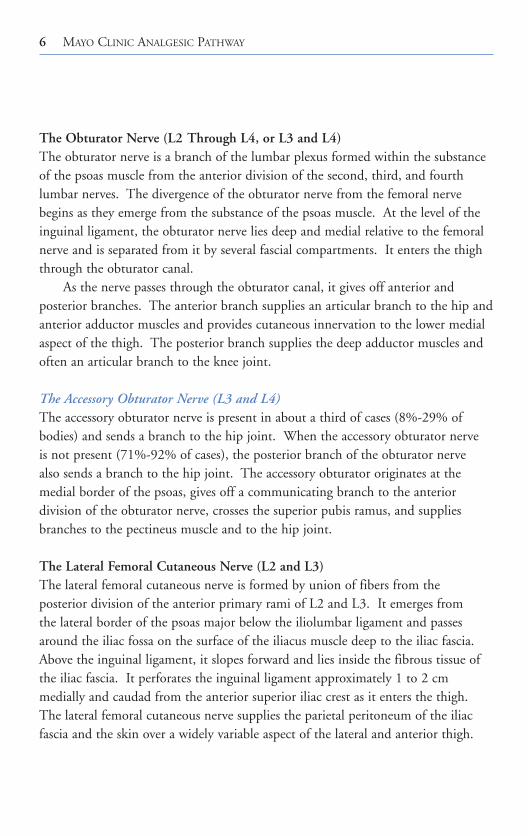
The Obturator Nerve (L2 Through L4, or L3 and L4)The obturator nerve is a branch of the lumbar plexus formed within the substanceof the psoas muscle from the anterior division of the second, third, and fourth lumbar nerves. The divergence of the obturator nerve from the femoral nervebegins as they emerge from the substance of the psoas muscle. At the level of theinguinal ligament, the obturator nerve lies deep and medial relative to the femoralnerve and is separated from it by several fascial compartments. It enters the thighthrough the obturator canal.
As the nerve passes through the obturator canal, it gives off anterior and posterior branches. The anterior branch supplies an articular branch to the hip andanterior adductor muscles and provides cutaneous innervation to the lower medialaspect of the thigh. The posterior branch supplies the deep adductor muscles andoften an articular branch to the knee joint.
The Accessory Obturator Nerve (L3 and L4)The accessory obturator nerve is present in about a third of cases (8%-29% of bodies) and sends a branch to the hip joint. When the accessory obturator nerve is not present (71%-92% of cases), the posterior branch of the obturator nerve also sends a branch to the hip joint. The accessory obturator originates at themedial border of the psoas, gives off a communicating branch to the anterior division of the obturator nerve, crosses the superior pubis ramus, and suppliesbranches to the pectineus muscle and to the hip joint.
The Lateral Femoral Cutaneous Nerve (L2 and L3)The lateral femoral cutaneous nerve is formed by union of fibers from the posterior division of the anterior primary rami of L2 and L3. It emerges from the lateral border of the psoas major below the iliolumbar ligament and passesaround the iliac fossa on the surface of the iliacus muscle deep to the iliac fascia.Above the inguinal ligament, it slopes forward and lies inside the fibrous tissue ofthe iliac fascia. It perforates the inguinal ligament approximately 1 to 2 cm medially and caudad from the anterior superior iliac crest as it enters the thigh.The lateral femoral cutaneous nerve supplies the parietal peritoneum of the iliacfascia and the skin over a widely variable aspect of the lateral and anterior thigh.
Section_I_4.0.qxd 9/20/05 1:46 PM Page 6

Section I PRINCIPLES OF LOWER EXTREMITY PERIPHERAL NERVE BLOCK 7
Sacral Plexus Anatomy (L4 and L5, S1 Through S3)The sacral plexus is formed within the pelvis by the merger of the ventral rami of L4 and L5 and S1-3 or S1-4. These nerves pass together through the pelvis and the greater sciatic foramen. The sacral plexus provides motor and sensoryinnervation to portions of the entire lower extremity including the hip, knee, and ankle. Its most important components are the posterior cutaneous and the sciatic nerves and their terminal branches.
The Posterior Femoral Cutaneous Nerve (S1 Through S3)The posterior femoral cutaneous nerve is a purely sensory nerve derived from the anterior rami of S1 through S3. It travels with the sciatic nerve out of thepelvis and into the upper aspect of the thigh. It emerges from the lower edge ofthe gluteus maximus to lie in midline subcutaneous tissue and continues down the posterior aspect of the thigh and the leg, giving off femoral and sural branches(sensory branches to the back of the thigh and the calf ). It becomes superficial inthe midline near the popliteal fossa, where its terminal branches often anastomosewith the sural nerve. The terminal branches of the posterior femoral cutaneousnerve may provide cutaneous innervation as distal as the heel.
The Sciatic Nerve (L4 and L5, S1 Through S3)The lumbosacral trunk (L4-L5) and anterior divisions of the sacral plexus (S1-S3)merge to form the tibial nerve, and the posterior divisions merge to form the common peroneal nerve. These two large mixed nerves of the sacral plexus are initially bound together by connective tissue to form the sciatic nerve. At this level, the tibial component is medial and anterior, and the common peroneal component is lateral and slightly posterior.
The sciatic nerve exits the pelvis by way of the greater sciatic notch below the piriformis muscle. At this level, the superior gluteal artery is immediately superior and medial to the sciatic nerve. As it enters the thigh and descends toward the popliteal fossa, it is posterior to the lesser trochanter of the femur onthe posterior surface of the adductor magnus muscle within the posterior medialthigh compartment deep to the biceps femoris. At the upper aspect of the poplitealfossa, the sciatic nerve lies posterior and lateral to the popliteal vessels. Here thenerve usually divides into its terminal component nerves, the tibial and commonperoneal nerves. The tibial and peroneal components provide complete sensory
Section_I_4.0.qxd 9/20/05 1:46 PM Page 7
8 MAYO CLINIC ANALGESIC PATHWAY
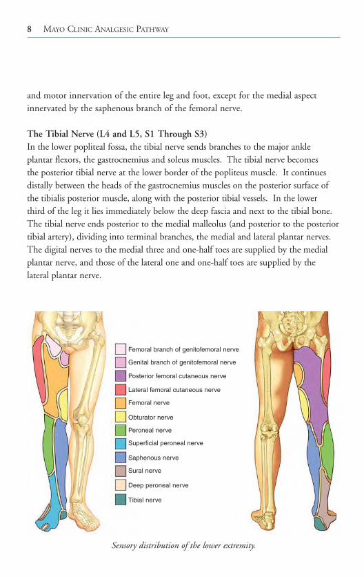
and motor innervation of the entire leg and foot, except for the medial aspectinnervated by the saphenous branch of the femoral nerve.
The Tibial Nerve (L4 and L5, S1 Through S3)In the lower popliteal fossa, the tibial nerve sends branches to the major ankle plantar flexors, the gastrocnemius and soleus muscles. The tibial nerve becomes the posterior tibial nerve at the lower border of the popliteus muscle. It continuesdistally between the heads of the gastrocnemius muscles on the posterior surface ofthe tibialis posterior muscle, along with the posterior tibial vessels. In the lower third of the leg it lies immediately below the deep fascia and next to the tibial bone.The tibial nerve ends posterior to the medial malleolus (and posterior to the posteriortibial artery), dividing into terminal branches, the medial and lateral plantar nerves.The digital nerves to the medial three and one-half toes are supplied by the medialplantar nerve, and those of the lateral one and one-half toes are supplied by the lateral plantar nerve.
Sensory distribution of the lower extremity.
Femoral branch of genitofemoral nerve
Genital branch of genitofemoral nerve
Posterior femoral cutaneous nerve
Lateral femoral cutaneous nerve
Femoral nerve
Obturator nerve
Peroneal nerve
Superficial peroneal nerve
Saphenous nerve
Sural nerve
Deep peroneal nerve
Tibial nerve
Section_I_4.0.qxd 9/20/05 1:46 PM Page 8
Section I PRINCIPLES OF LOWER EXTREMITY PERIPHERAL NERVE BLOCK 9
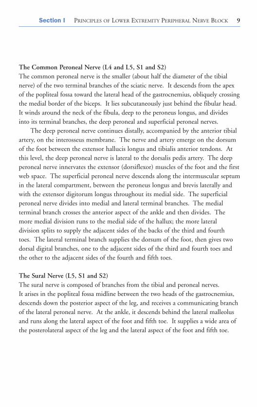
The Common Peroneal Nerve (L4 and L5, S1 and S2)The common peroneal nerve is the smaller (about half the diameter of the tibialnerve) of the two terminal branches of the sciatic nerve. It descends from the apexof the popliteal fossa toward the lateral head of the gastrocnemius, obliquely crossingthe medial border of the biceps. It lies subcutaneously just behind the fibular head.It winds around the neck of the fibula, deep to the peroneus longus, and dividesinto its terminal branches, the deep peroneal and superficial peroneal nerves.
The deep peroneal nerve continues distally, accompanied by the anterior tibialartery, on the interosseus membrane. The nerve and artery emerge on the dorsumof the foot between the extensor hallucis longus and tibialis anterior tendons. Atthis level, the deep peroneal nerve is lateral to the dorsalis pedis artery. The deepperoneal nerve innervates the extensor (dorsiflexor) muscles of the foot and the firstweb space. The superficial peroneal nerve descends along the intermuscular septumin the lateral compartment, between the peroneus longus and brevis laterally andwith the extensor digitorum longus throughout its medial side. The superficialperoneal nerve divides into medial and lateral terminal branches. The medial terminal branch crosses the anterior aspect of the ankle and then divides. Themore medial division runs to the medial side of the hallux; the more lateral division splits to supply the adjacent sides of the backs of the third and fourth toes. The lateral terminal branch supplies the dorsum of the foot, then gives twodorsal digital branches, one to the adjacent sides of the third and fourth toes andthe other to the adjacent sides of the fourth and fifth toes.
The Sural Nerve (L5, S1 and S2)The sural nerve is composed of branches from the tibial and peroneal nerves. It arises in the popliteal fossa midline between the two heads of the gastrocnemius,descends down the posterior aspect of the leg, and receives a communicating branchof the lateral peroneal nerve. At the ankle, it descends behind the lateral malleolusand runs along the lateral aspect of the foot and fifth toe. It supplies a wide area ofthe posterolateral aspect of the leg and the lateral aspect of the foot and fifth toe.
Section_I_4.0.qxd 9/20/05 1:46 PM Page 9

10 MAYO CLINIC ANALGESIC PATHWAY
Recommended ReadingAnderson JE, Grant JCB, editors. Grant’s atlas of anatomy. 8th ed. Baltimore:
Williams & Wilkins; 1983.Basmajian JV, Slonecker CE. Grant’s method of anatomy: a clinical
problem-solving approach. 11th ed. Baltimore: Williams & Wilkins; 1989.Enneking FK, Chan V, Greger J, Hadzic A, Lang SA, Horlocker TT.
Lower-extremity peripheral nerve blockade: essentials of our current understanding. Reg Anesth Pain Med. 2005;30:4-35.
Gray H, Williams PL, editors. Gray’s anatomy. 37th ed. Edinburgh: C Livingstone; 1989.
Rosse C, Gaddum-Rosse P, editors. Hollinshead’s textbook of anatomy. 5th ed.Philadelphia: Lippincott-Raven Publishers; 1997.
The visible human project. United States National Library of Medicine. National Institutes of Health [cited 2005 Jul 26]. Available from:www.nlm.nih.gov/research/visible/applications.html
Woodburne RT, Burkel WE. Essentials of human anatomy. 9th ed. New York:Oxford University Press; 1994.
Section_I_4.0.qxd 9/20/05 1:46 PM Page 10

DERMATOMES AND OSTEOTOMES
11
Chapter 2
When peripheral techniques are selected for a specific surgical procedure, it is paramount to consider not only the neurotomes but also the osteotomes and dermatomes. For example, the dermatomal supply of the hip joint typically is from L4 to as low as S2, whereas the bony structures of the hip joint do not follow the same segmental pattern and are supplied from L3 to S1. However, when neurotomes are considered, the obturator and femoral nerves, which originate from L2-L4, supply articular branches to the hip joint. Thus, the entirelumbar and sacral plexuses must both be blocked to ensure adequate coverage of the neurotomes of the hip. The same considerations hold for knee and anklesurgery. The importance of understanding the limitations of each of these blocks is essential to successful application.
The clinician also must bear in mind that there is not only extensive overlapbetween consecutive neurotomes, dermatomes, and osteotomes but also variabilityamong subjects. As a result, the innervation of a specific site or segmental levelcannot be determined with certainty. These principles may explain an incompleteor failed anesthesia, even in the presence of a successful block, in a given individual.
Recommended ReadingAnderson JE, editor. Grant’s atlas of anatomy. Baltimore: Williams & Wilkins; 1993.Enneking FK, Chan V, Greger J, Hadzic A, Lang SA, Horlocker TT.
Lower-extremity peripheral nerve blockade: essentials of our current understanding. Reg Anesth Pain Med. 2005;30:4-35.
Rosse C, Gaddum-Rosse P, editors. Hollinshead’s textbook of anatomy. 5th ed.Philadelphia: Lippincott-Raven Publishers; 1997.
Section_I_4.0.qxd 9/20/05 1:46 PM Page 11

12 MAYO CLINIC ANALGESIC PATHWAY
Dermatomes and osteotomes of the lumbosacral plexus.
-L1
-L2
-L3
-L4
-L5
-S1
-S2
-S3
-S4
-S5
-C0
Section_I_4.0.qxd 9/20/05 1:46 PM Page 12
Section I PRINCIPLES OF LOWER EXTREMITY PERIPHERAL NERVE BLOCK 13
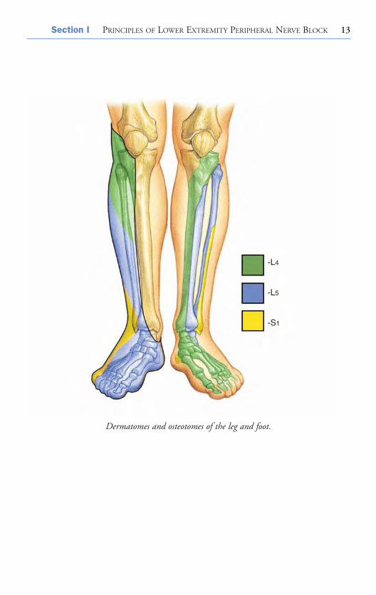
Dermatomes and osteotomes of the leg and foot.
-L4
-L5
-S1
Section_I_4.0.qxd 9/20/05 1:46 PM Page 13

This page intentionally left blank

15
PREOPERATIVE ASSESSMENTAND MONITORING
Chapter 3
Preoperative ExaminationDuring the preoperative assessment, the patient is evaluated for preexisting medical problems, allergies, previous anesthetic complications, potential airway difficulties, and considerations relating to intraoperative positioning. Overall,patients undergoing major orthopedic procedures on the lower extremity are considered at intermediate risk for cardiac complications perioperatively. However, it is often difficult to assess exercise tolerance or a recent progression of cardiac symptoms because of the limitations in mobility induced by the underlying orthopedic condition. As a result, pharmacologic functional testing,based on clinical history, may be warranted. Perioperative cardiac morbidity may be decreased by the initiation of β-adrenergic blockade.
The patient’s medications should be reviewed and the patient specificallyinstructed on which medications to continue to use until the time of surgery.Specifically, use of antihypertensive medications should not be discontinued because of the risk of perioperative cardiac events. Likewise, patients who require chronic opioid medications should be allowed to maintain their dosing regimen. Corticosteroid-dependent patients require corticosteroid replacementperioperatively. Finally, the patient should be queried regarding the use of anymedications that affect hemostasis; many patients will have been instructed by their surgeon to begin thromboprophylaxis with aspirin or warfarin preoperatively.
The patient should undergo a focused physical examination. Patients should be assessed for limitation in mouth opening or neck extension, adequacy of thyromental distance (measured from the lower border of the mandible to the thyroid notch), and state of dentition. The heart and lungs should be auscultated. In addition, the site of proposed injection for regional anesthetic
Section_I_4.0.qxd 9/20/05 1:46 PM Page 15
16 MAYO CLINIC ANALGESIC PATHWAY
should be assessed for evidence of infection and anatomical abnormalities or limitations. A brief neurologic examination, with documentation of any existing deficits, is crucial. The patient also should be evaluated for any potentialpositioning difficulties (during block performance or intraoperatively) related to arthritic involvement of other joints or body habitus. Hemoglobin and creatinine values are determined for all patients, and other laboratory testing and imaging are done as indicated by preoperative medical conditions.
Ideally, the patient should undergo a preoperative educational session in which the surgical procedure, anesthetic and analgesic options, and the postoperative rehabilitative plan are described.
Additional questions that arise can be answered on the operative day.
Sedation and MonitoringA sedative is administered during performance of the block and during the surgical procedure to decrease apprehension and anxiety, to provide analgesia for pain associated with the regional anesthetic and positioning, and to decrease awareness of perioperative events. In addition, the administration of benzodiazepines and hypnotics increases the seizure threshold in the presence of increasing blood levels of local anesthetic. It is imperative that patients remain conscious and cooperative during performance of regional blockade in order to provide feedback regarding painful needle or catheter placement or injection. An additional benefit of maintaining patient alertness is the patient’sability to describe subtle motor responses during neural stimulation at a currentbelow that required for visualization by the proceduralist; lower stimulating currents are perceived as more comfortable.
Patients undergoing peripheral blockade should be monitored to allow detection of intravascular injection (heart rate and blood pressure) and adequate oxygenation (pulse oximeter). Because levels of local anesthetic peak at approximately 60 minutes after injection following lower extremity peripheralblock, patients should be appropriately monitored for signs and symptoms of toxicity for this duration. Resuscitation equipment and medications also should be readily available. Before the patient is transferred to the operating suite, thedegree of sensory and motor block should be assessed. If there is evidence of anincomplete block, the postoperative analgesic medications may require adjustment.Likewise, patients must be immediately assessed on arrival in the recovery roomand supplemental analgesics administered early to avoid escalating discomfort.
Section_I_4.0.qxd 9/20/05 1:46 PM Page 16

Section I PRINCIPLES OF LOWER EXTREMITY PERIPHERAL NERVE BLOCK 17
Recommended ReadingEagle KA, Brundage BH, Chaitman BR, Ewy GA, Fleisher LA, Hertzer NR, et al,
Committee on Perioperative Cardiovascular Evaluation for Noncardiac Surgery.Guidelines for perioperative cardiovascular evaluation for noncardiac surgery:report of the American College of Cardiology/American Heart Association TaskForce on Practice Guidelines. Circulation. 1996;93:1278-317.
Hadzic A, Vloka JD, Claudio RE, Hadzic N, Thys DM, Santos AC. Electricalnerve localization: effects of cutaneous electrode placement and duration of thestimulus on motor response. Anesthesiology. 2004;100:1526-30.
The preoperative block area.
Section_I_4.0.qxd 9/20/05 1:47 PM Page 17

This page intentionally left blank
19
TECHNIQUES AND EQUIPMENTFOR NEURAL LOCALIZATION
Chapter 4
Historically, lower extremity peripheral blocks were performed with loss of resistance (psoas compartment, fascia iliaca), paresthesia (femoral, sciatic, popliteal),or field infiltration (femoral, lateral femoral cutaneous, ankle) techniques. Duringthe past several decades, electrical nerve stimulation has become the standardmethod of identifying neural structures. Although there are few studies comparingthe efficacy and complications of neural localization with elicitation of a paresthesiawith those of a motor response, in general the two techniques seem comparable.Nonetheless, nerve stimulation has become the primary method of neural localization with lower extremity regional techniques.
Nerve StimulatorsThe desirable qualities of a nerve stimulator include constant current output(despite varying resistances of the patient’s body, cables and connections, andground lead), a digital display, variable linear output (the current changes in proportion to the movement of the dial), a short pulse width to deliver a precisecurrent or charge to the nerve, and indicators of power or circuit failure. The optimal current with which to begin nerve localization without discomfort and the current associated with “successful” needle placement are unknown. A volunteer study reported that during femoral block, muscle contractions were painful with a stimulating current greater than 1.6 mA. In addition, after elicitation of a paresthesia, the minimal current needed to produce a motor response was less than 0.5 mA in 80% of cases, a suggestion that this may be a reasonable “final” current intensity to seek. However, this may vary between block techniques and in the presence of preexisting neurologic conditions.
Section_I_4.0.qxd 9/20/05 1:47 PM Page 19
20 MAYO CLINIC ANALGESIC PATHWAY
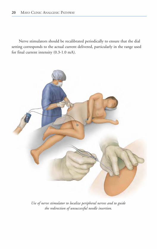
Nerve stimulators should be recalibrated periodically to ensure that the dial setting corresponds to the actual current delivered, particularly in the range usedfor final current intensity (0.3-1.0 mA).
Use of nerve stimulator to localize peripheral nerves and to guidethe redirection of unsuccessful needle insertion.
Section_I_4.0.qxd 9/20/05 1:47 PM Page 20

Section I PRINCIPLES OF LOWER EXTREMITY PERIPHERAL NERVE BLOCK 21
Stimulating NeedlesBoth uninsulated and insulated needles may be used to identify nerves with electrical stimulation. However, uninsulated needles disperse the current throughout the entire needle shaft and bevel and require a greater current to elicit a motor response. In addition, the needle tip (and site of local anestheticinjection) is likely to not be the area of greatest current density and neural stimulation. Indeed, the needle tip actually may have bypassed the nerve despiteongoing motor stimulation. For these reasons, insulated needles are recommendedif electrical stimulation is used to localize nerves. Insulated needles allow a concentrated stimulating current. Needles with a coated or insulated bevel havethe highest current concentration and allow for more precise needle placement with less stimulating current to elicit a motor response.
The Mayo Clinic customized nerve block tray.
Section_I_4.0.qxd 9/20/05 1:47 PM Page 21
22 MAYO CLINIC ANALGESIC PATHWAY
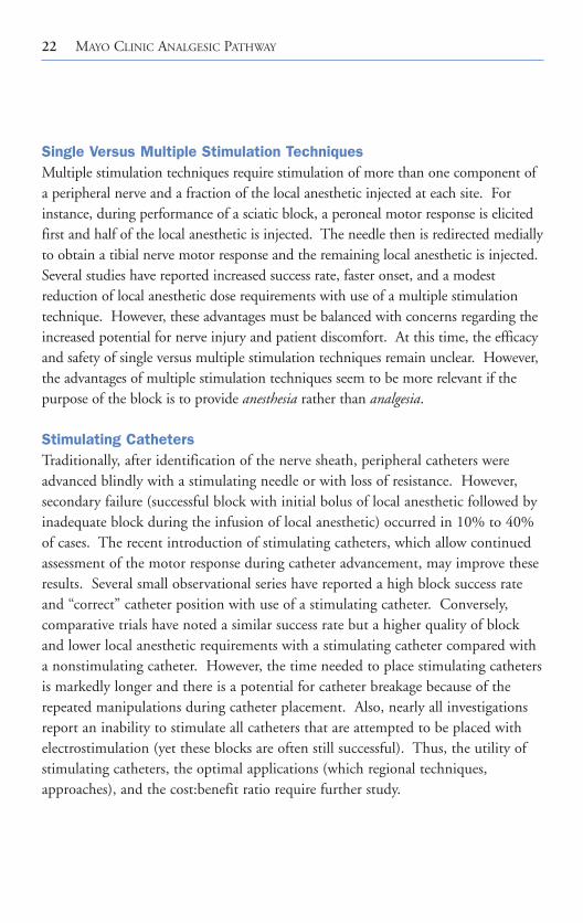
Single Versus Multiple Stimulation TechniquesMultiple stimulation techniques require stimulation of more than one component ofa peripheral nerve and a fraction of the local anesthetic injected at each site. Forinstance, during performance of a sciatic block, a peroneal motor response is elicitedfirst and half of the local anesthetic is injected. The needle then is redirected mediallyto obtain a tibial nerve motor response and the remaining local anesthetic is injected.Several studies have reported increased success rate, faster onset, and a modest reduction of local anesthetic dose requirements with use of a multiple stimulationtechnique. However, these advantages must be balanced with concerns regarding theincreased potential for nerve injury and patient discomfort. At this time, the efficacyand safety of single versus multiple stimulation techniques remain unclear. However,the advantages of multiple stimulation techniques seem to be more relevant if thepurpose of the block is to provide anesthesia rather than analgesia.
Stimulating CathetersTraditionally, after identification of the nerve sheath, peripheral catheters wereadvanced blindly with a stimulating needle or with loss of resistance. However, secondary failure (successful block with initial bolus of local anesthetic followed byinadequate block during the infusion of local anesthetic) occurred in 10% to 40%of cases. The recent introduction of stimulating catheters, which allow continuedassessment of the motor response during catheter advancement, may improve theseresults. Several small observational series have reported a high block success rateand “correct” catheter position with use of a stimulating catheter. Conversely, comparative trials have noted a similar success rate but a higher quality of blockand lower local anesthetic requirements with a stimulating catheter compared witha nonstimulating catheter. However, the time needed to place stimulating cathetersis markedly longer and there is a potential for catheter breakage because of therepeated manipulations during catheter placement. Also, nearly all investigationsreport an inability to stimulate all catheters that are attempted to be placed withelectrostimulation (yet these blocks are often still successful). Thus, the utility ofstimulating catheters, the optimal applications (which regional techniques,approaches), and the cost:benefit ratio require further study.
Section_I_4.0.qxd 9/20/05 1:47 PM Page 22

Section I PRINCIPLES OF LOWER EXTREMITY PERIPHERAL NERVE BLOCK 23
Imaging MethodsThe introduction of high-resolution portable devices has facilitated the use of ultrasonography in the operating suite; numerous studies have evaluated its efficacyfor brachial plexus techniques. Lower extremity applications involving the lumbarplexus and femoral and sciatic nerves have been described recently. However, as thedepth to the structure being imaged increases, lower scanning frequencies, whichare associated with lower resolution, are required. Thus, the existing probes are not well suited for lower extremity (compared with brachial plexus) techniques.
Using ultrasound guidance, the proceduralist is able to visualize the neuralstructures, needle advancement, and the distribution of local anesthetic duringinjection. Ultrasonography probably allows a smaller dose of local anesthetic and improves onset time compared with conventional approaches. However, the success rate with ultrasound localization is comparable to that with multiplestimulation techniques, and no data suggest that ultrasonography will reduce therisk of neurologic complications. Also, ultrasonography is unsuccessful for theidentification of neural structures in some patients. The presence of obesity orspinal deformities, conditions in which needle guidance would be most helpful,makes ultrasonography difficult. Additional information is needed to establish therole of ultrasound guidance in the performance of regional anesthetic techniques.
Recommended ReadingChoyce A, Chan VW, Middleton WJ, Knight PR, Peng P, McCartney CJ. What is
the relationship between paresthesia and nerve stimulation for axillary brachialplexus block? Reg Anesth Pain Med. 2001;26:100-4.
Cuvillon P, Ripart J, Jeannes P, Mahamat A, Boisson C, L’Hermite J, et al.Comparison of the parasacral approach and the posterior approach, with single- and double-injection techniques, to block the sciatic nerve.Anesthesiology. 2003;98:1436-41.
Davies MJ, McGlade DP. One hundred sciatic nerve blocks: a comparison oflocalisation techniques. Anaesth Intensive Care. 1993;21:76-8.
Fanelli G, Casati A, Garancini P, Torri G, Study Group on Regional Anesthesia.Nerve stimulator and multiple injection technique for upper and lower limbblockade: failure rate, patient acceptance, and neurologic complications.Anesth Analg. 1999;88:847-52.
Section_I_4.0.qxd 9/20/05 1:47 PM Page 23

Hadzic A, Vloka JD, Claudio RE, Hadzic N, Thys DM, Santos AC. Electricalnerve localization: effects of cutaneous electrode placement and duration of thestimulus on motor response. Anesthesiology. 2004;100:1526-30.
Kirchmair L, Entner T, Kapral S, Mitterschiffthaler G. Ultrasound guidance for the psoas compartment block: an imaging study. Anesth Analg.2002;94:706-10.
Marhofer P, Greher M, Kapral S. Ultrasound guidance in regional anaesthesia. Br J Anaesth. 2005 Jan;94:7-17. Epub 2004 Jul 26.
Marhofer P, Schrogendorfer K, Koinig H, Kapral S, Weinstabl C, Mayer N.Ultrasonographic guidance improves sensory block and onset time of three-in-one blocks. Anesth Analg. 1997;85:854-7.
Salinas FV, Neal JM, Sueda LA, Kopacz DJ, Liu SS. Prospective comparison ofcontinuous femoral nerve block with nonstimulating catheter placement versusstimulating catheter-guided perineural placement in volunteers. Reg AnesthPain Med. 2004;29:212-20.
24 MAYO CLINIC ANALGESIC PATHWAY
Section_I_4.0.qxd 9/20/05 1:47 PM Page 24
25
SELECTION OFLOCAL ANESTHETIC AND ADJUVANTS
Chapter 5
Local Anesthetic SolutionThe choice of local anesthetic and the addition of adjuvants for lower extremityperipheral nerve block are dependent on the anticipated duration of operation, the need for prolonged analgesia, and the timing of ambulation and weight bearing postoperatively. Prolonged blockade for 24 hours (or longer) may occurwith long-acting agents such as bupivacaine, levobupivacaine, or ropivacaine.Although this feature may result in excellent postoperative pain relief for the inpatient, it may be undesirable or a cause for concern in the ambulatory patientbecause of the potential for falls with a partially insensate or weak lower extremity.A medium-acting agent may be more appropriate in the outpatient setting fororthopedic procedures associated with minimal to moderate postoperative pain. In general, equipotent concentrations of the long-acting amides have a similar onset and quality of block. However, bupivacaine may have a slightly longer duration than levobupivacaine or ropivacaine. Likewise, higher concentrations are more likely to be associated with profound sensory and motor block, whereasinfusions of 0.1% to 0.2% bupivacaine or ropivacaine often allow complete weightbearing without notable motor deficits. Recent investigations have suggested thatincreasing the local anesthetic concentration alters the character (i.e., degree of sensory or motor block) but not the duration.
The lowest effective dose and concentration should be used to minimize localanesthetic systemic and neural toxicity. The recommendations for maximal dosesof local anesthetics were established by the manufacturers (Table 1). Maximaldoses based on patient weight (with the exception of the pediatric population) arenot evidence-based. Recommendations for 24-hour doses of local anesthetics also
Section_I_4.0.qxd 9/20/05 1:47 PM Page 25

26 MAYO CLINIC ANALGESIC PATHWAY
have been established without controlled studies. In essence, the safe dose of a local anesthetic should be individualized according to site of injection, patient age, and the presence of medical conditions that affect local anestheticpharmacology and toxicity (Table 2). Because of the potential for accumulation of local anesthetic, these considerations are believed to be most critical when large doses of local anesthetics are injected or in association with repeated blocks or continuous infusions.
Table 1. Recommended Maximal Doses of Local Anesthetics
Local anesthetic Maximal dose, mg
2-Chloroprocaine 800
With epinephrine 1,000
Lidocaine 300
With epinephrine 500
Mepivacaine 400
With epinephrine 550
Bupivacaine 175
With epinephrine 225
400/24 h
Levobupivacaine 150
With epinephrine 150
400/24 h
Ropivacaine 225
With epinephrine 225
800/24 h
Modified from Rosenberg PH, Veering BT, Urmey WF. Maximum recommended doses of local anesthetics: a multifactorial concept. Reg Anesth Pain Med. 2004;29:564-75. Used with permission.
Section_I_4.0.qxd 9/20/05 1:47 PM Page 26

Section I PRINCIPLES OF LOWER EXTREMITY PERIPHERAL NERVE BLOCK 27
Adjuvants
EpinephrineEpinephrine decreases local anesthetic uptake and plasma levels, improves the quality of block, and increases the duration of postoperative analgesia during lowerextremity peripheral blockade. Epinephrine also allows for the early detection ofintravascular injection. Importantly, concentrations of epinephrine ranging from1.7 to 5 μg/mL (1:600,000-1:200,000 dilution) reduce the uptake and prolong the blockade of medium-duration local anesthetics to a similar extent. However,concentrations of 1.7 to 2.5 μg/mL have little effect on nerve blood flow, whichtheoretically may reduce the risk of nerve injury in patients with a preexistingangiopathy or neuropathy. In addition, larger doses of epinephrine injected systemically may cause undesirable side effects in patients with known cardiac disease. Concerns regarding neural or cardiac ischemia must be balanced with the need to detect intravascular injection. In general, because of the high doses of local anesthetics administered during lower extremity peripheral block, the benefits of adding epinephrine outweigh the risks.
Commercially prepared solutions with epinephrine have a lower pH than those in which it is freshly added, resulting in a higher percentage of ionized drugmolecules. These ionized molecules do not readily cross the neural membrane,delaying the onset of local anesthetic action after injection. Epinephrine should
Table 2. Patient-Related Factors Affecting Local Anesthetic Pharmacology
Factor Modification of dose
Age
Newborn (<4 mo) Reduce 15%
Older than 70 years Reduce 10%-20%
Renal dysfunction Reduce 10%-20%, including continuous infusions
Hepatic dysfunction Reduce 10%-20%, more with continuous infusions
Heart failure Reduce 10%-20% during continuous infusions
Pregnancy Reduce concentration due to increased sensitivity
to local anesthetics
Section_I_4.0.qxd 9/20/05 1:47 PM Page 27

28 MAYO CLINIC ANALGESIC PATHWAY
not be added for ankle block. The addition of epinephrine to local anesthetics with intrinsic vasoconstrictive properties, such as ropivacaine, may not increaseblock duration but would still facilitate detection of intravascular injection.
ClonidineClonidine is an α2-adrenergic agent with analgesic properties. The effect is mostlikely peripherally mediated and dose-dependent. Clonidine consistently prolongsthe time to first analgesia when added to intermediate-acting agents during brachialplexus blockade. Side effects such as hypotension, bradycardia, and sedation do not occur with a dose less than 1.5 μg/kg or a maximal dose of 150 μg. Althoughthe efficacy of clonidine as an adjuvant for lower extremity single injection and continuous techniques is less defined, most studies report a modest (20%) prolongation of the block duration when clonidine is added to long-acting localanesthetic solutions.
OpioidsAlthough opioids, including morphine, sufentanil, and fentanyl, are often added tolumbar plexus infusions, no convincing data suggest that block onset, quality, orduration is improved when opioids are added to the local anesthetic solution.
Systemic Local Anesthetic ToxicityBecause of the relatively large doses of local anesthetic injected and the proximity ofneedle or catheter insertion to vascular structures and highly vascularized musclebeds, the potential for systemic local anesthetic toxicity would seem to be very highfor lower extremity peripheral nerve blocks. The few cases of systemic toxicityrequiring resuscitation occurred shortly after injection, a suggestion that accidentalintravascular injection, rather than systemic absorption, is the mechanism. Theseevents also were associated with proximal lumbosacral techniques, such as psoas orsciatic block. Prevention and treatment of local anesthetic toxicity are dependenton the injection of an appropriate volume and concentration of local anesthetic, the use of a vasoconstrictor adjuvant, slow injection with frequent aspiration, and increased vigilance for the early detection of toxic reactions. Local anestheticlevels peak at approximately 60 minutes after injection following lower extremityperipheral block. Thus, patients should be appropriately monitored for signs andsymptoms of increasing blood levels for this duration. Resuscitation equipmentand medications also should be readily available.
Section_I_4.0.qxd 9/20/05 1:47 PM Page 28
Section I PRINCIPLES OF LOWER EXTREMITY PERIPHERAL NERVE BLOCK 29
Treatment of local anesthetic toxic reactions is similar to the management ofother medical emergencies and focuses on ensuring adequate airway, breathing, andcirculation. An airway should be established and 100% oxygen administered.Hypoxia and hypercarbia must be avoided. If convulsions occur, a small amount ofa short-acting barbiturate (thiopental, 50-100 mg) or propofol typically terminatesthe seizure without causing cardiovascular compromise. A muscle relaxant may beneeded to secure the airway; although the tonic-clonic motion is inhibited, seizureactivity may still persist. Although most toxic reactions are limited to the centralnervous system, cardiovascular collapse with refractory ventricular fibrillation may occur, especially with bupivacaine. Sustained cardiopulmonary resuscitationwith repeated cardioversion and high doses of epinephrine may be required for circulatory support.
Recommended ReadingAuroy Y, Benhamou D, Bargues L, Ecoffey C, Falissard B, Mercier FJ, et al, the
SOS Regional Anesthesia Hotline Service. Major complications of regionalanesthesia in France. Anesthesiology. 2002;97:1274-80. Erratum in:Anesthesiology. 2003;98:595.
Enneking FK, Chan V, Greger J, Hadzic A, Lang SA, Horlocker TT. Lower-extremity peripheral nerve blockade: essentials of our current understanding. Reg Anesth Pain Med. 2005;30:4-35.
Neal JM. Effects of epinephrine in local anesthetics on the central and peripheralnervous systems: neurotoxicity and neural blood flow. Reg Anesth Pain Med.2003;28:124-34.
Neal JM, Hebl JR, Gerancher JC, Hogan QH. Brachial plexus anesthesia: essentials of our current understanding. Reg Anesth Pain Med. 2002;27:402-28. Erratum in: Reg Anesth Pain Med. 2002;27:625.
Rosenberg PH, Veering BT, Urmey WF. Maximum recommended doses of localanesthetics: a multifactorial concept. Reg Anesth Pain Med. 2004;29:564-75.
Section_I_4.0.qxd 9/20/05 1:47 PM Page 29

This page intentionally left blank

31
NEUROLOGIC COMPLICATIONS
Chapter 6
Nerve injury is a recognized complication of peripheral regional techniques. In aseries involving more than 100,000 regional anesthetic procedures, the frequency ofneurologic complications after peripheral blockade was lower than that associatedwith neuraxial techniques, and the complications were associated with pain on needle placement or injection of local anesthetic. Risk factors contributing to neurologic deficit after regional anesthesia include neural ischemia, traumatic injuryto the nerves during needle or catheter placement, infection, and choice of localanesthetic solution. However, postoperative neurologic injury due to pressure fromimproper patient positioning, tightly applied casts or surgical dressings, and surgicaltrauma often are attributed to the regional anesthetic. Patient factors such as bodyhabitus or a preexisting neurologic dysfunction also may contribute.
Although needle gauge, type (short or long bevel), and bevel configuration may influence the degree of nerve injury after peripheral nerve block, the findingsare conflicting and there are no confirmatory human studies. Theoretically, localization of neural structures with a nerve stimulator would allow a high successrate without increasing the risk of neurologic complications, but this suppositionhas not been established. Indeed, serious neurologic injury has been reported after uneventful brachial plexus block with a nerve stimulator technique. Likewise, prolonged exposure or high dose or high concentrations of local anesthetic solutions also may result in permanent neurologic deficits. In laboratorymodels, the addition of epinephrine increases the neurotoxicity of local anestheticsolutions and also decreases nerve blood flow. However, the clinical relevance of these findings in humans remains unclear. Finally, nerve damage caused by traumatic needle placement, local anesthetic neurotoxicity, and neural ischemiaduring the performance of a regional anesthetic procedure may worsen neurologicoutcome in the presence of an additional patient factor or surgical injury.
Section_I_4.0.qxd 9/20/05 1:47 PM Page 31

32 MAYO CLINIC ANALGESIC PATHWAY
Prevention of neurologic complications begins during the preoperative visitwith a careful evaluation of the patient’s medical history and appropriate discussionof the risks and benefits of the available anesthetic techniques. All preoperativeneurologic deficits must be documented to allow early diagnosis of new or worsening neurologic dysfunction postoperatively. Postoperative sensory or motor deficits also must be distinguished from residual (prolonged) local anestheticeffect. Imaging techniques, such as computed tomography and magnetic resonanceimaging, are useful for identifying infectious processes and expanding hematomas.Although most neurologic complications resolve completely within several days orweeks, significant neural injuries necessitate neurologic consultation to documentthe degree of involvement and coordinate further work-up. Neurophysiologic testing, such as nerve conduction studies, evoked potentials, and electromyography,is often useful for establishing a diagnosis and prognosis.
Recommended ReadingAuroy Y, Narchi P, Messiah A, Litt L, Rouvier B, Samii K. Serious complications
related to regional anesthesia: results of a prospective survey in France.Anesthesiology. 1997;87:479-86.
Benumof JL. Permanent loss of cervical spinal cord function associated with interscalene block performed under general anesthesia. Anesthesiology.2000;93:1541-4.
Cheney FW, Domino KB, Caplan RA, Posner KL. Nerve injury associated withanesthesia: a closed claims analysis. Anesthesiology. 1999;90:1062-9.
Fanelli G, Casati A, Garancini P, Torri G, Study Group on Regional Anesthesia.Nerve stimulator and multiple injection technique or upper and lower limbblockade: failure rate, patient acceptance, and neurologic complications.Anesth Analg. 1999;88:847-52.
Neal JM. Effects of epinephrine in local anesthetics on the central and peripheralnervous systems: neurotoxicity and neural blood flow. Reg Anesth Pain Med.2003;28:124-34.
Rice AS, McMahon SB. Peripheral nerve injury caused by injection needles usedin regional anaesthesia: influence of bevel configuration, studied in a rat model.Br J Anaesth. 1992;69:433-8.
Selander D, Edshage S, Wolff T. Paresthesiae or no paresthesiae? Nerve lesionsafter axillary blocks. Acta Anaesthesiol Scand. 1979;23:27-33.
Section_I_4.0.qxd 9/20/05 1:47 PM Page 32

Section_II.qxd 8/11/05 2:19 PM Page 33
LUMBAR PLEXUS BLOCK
SECTION II
Section_II.qxd 8/11/05 2:19 PM Page 33

This page intentionally left blank

PSOAS COMPARTMENT APPROACH
35
Chapter 7
Clinical ApplicationsThis technique offers a single injection rather than three separate needle insertionsfor anesthesia of the entire lumbar plexus. Psoas compartment block is used toprovide anesthesia for repair of hip fracture and minor thigh and knee proceduresand for postoperative analgesia in patients undergoing major knee and hip surgery.When combined with a sciatic block, the technique provides complete unilaterallower extremity anesthesia.
Patient PositionThe patient is placed in the lateral position; the hips are flexed and the operativeextremity is nondependent, similar to the position for an intrathecal injection. The shoulders and hips are perpendicular to the horizontal plane.
LandmarksThere are several variations in the needle insertion site. We prefer those ofCapdevila in order to optimize localization of the L4 transverse process and toreduce the likelihood of excessive needle depth. A vertical line is drawn to connectthe iliac crests (intercristal line). A horizontal line is drawn connecting the spinousprocesses in the midline. The posterior superior iliac spine is palpated, and a line is drawn parallel to the spinous processes and originating at the posterior superioriliac spine. The distance between the posterior superior iliac spine and midline isdivided into thirds. The needle insertion site is 1 cm cephalad to the lateral thirdand medial two-thirds of the vertical line drawn between the spinous processes and the parallel line to the spinal column passing through the posterior superior iliac spine.
Section_II.qxd 8/11/05 2:19 PM Page 35

36 MAYO CLINIC ANALGESIC PATHWAY
TechniqueA 21-gauge 10-cm (4-inch) insulated needle is advanced perpendicular to the skinentry site until contact is obtained with the transverse process of L4. The needle is redirected caudad and advanced under the transverse process until quadricepsfemoris muscle twitches are elicited. The distance from the L4 transverse process to the lumbar plexus is 2 cm in adults, regardless of sex and habitus. Thirty milliliters of solution is slowly and carefully injected incrementally with frequentaspirations for cerebrospinal fluid or blood. The vigilant proceduralist is acutelyaware that negative aspiration does not preclude intravascular injection.
Lumbar plexus block: psoas compartment approach.
Section_II.qxd 8/11/05 2:19 PM Page 36

Section II LUMBAR PLEXUS BLOCK 37
Landmarks for the psoas compartment block. Needle entry is marked 1 cm cephalad to the intercristal line, two-thirds the distance from the midline to the
posterior superior iliac spine line. The cross is the site of needle insertion.
Iliac crest Posteriorsuperioriliac spine
Continuous psoas block is performed by advancing a 20-gauge catheterthrough an 18-gauge insulated needle 5 to 6 cm into the psoas compartment after the appropriate motor response is elicited.
Evoked Motor ResponsesThe lumbar plexus is identified by elicitation of the quadriceps motor response.Quadriceps contraction is confirmed by rhythmic patellar elevation. Otherresponses associated with stimulation of the component of the lumbar plexus, such as hip adduction or abduction, also may be used.
Needle Redirection CuesAfter contact with the L4 transverse process, the needle is withdrawn to the skinand “walked off ” the transverse process by directing the needle slightly mediallyand caudad to enter the psoas compartment. If bony contact is not made, the
Section_II.qxd 8/11/05 2:19 PM Page 37

38 MAYO CLINIC ANALGESIC PATHWAY
needle is directed first caudad, then cephalad. If the anticipated motor responsestill has not been achieved, the needle is redirected slightly medially, keeping inmind that neuraxial spread is more likely with medial needle placement.
In some instances, motor responses are not elicited but the patient reports light “electrical shocks” down the leg. The nerve roots may have already split intoanterior motor fibers and posterior sensory fibers. Only stimulation of the sensoryfibers is acceptable and results in successful lumbar plexus block. A needle positionthat is too deep may directly stimulate the psoas major, producing hip flexion. Ofimportance, the normal kidney may extend down to L3; thus, if the L4 transverseprocess is contacted, it is prudent to redirect the needle caudad not cephalad.
Psoas compartment block: neuroskeletal relationships. The lumbar plexus is identified between the transverse processes of L4 and L5. Dural sleeves extend
3 to 5 cm laterally. The cross is the site of needle insertion.
Section_II.qxd 8/11/05 2:19 PM Page 38
Section II LUMBAR PLEXUS BLOCK 39
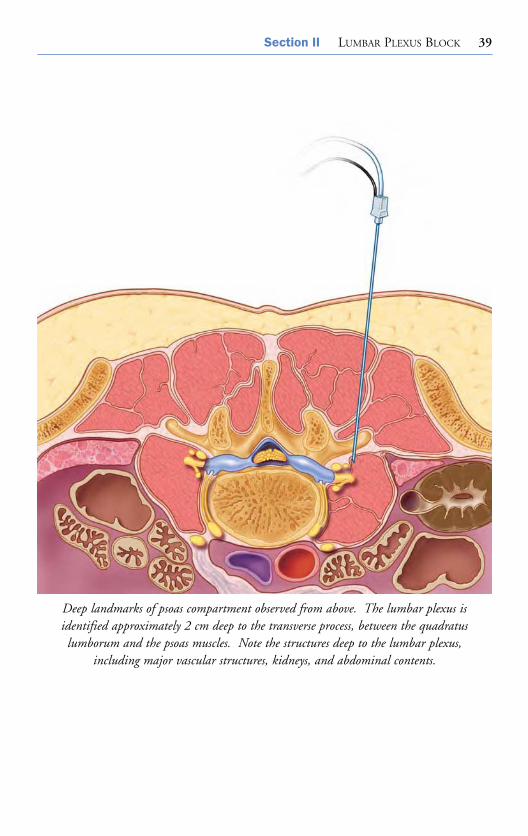
Deep landmarks of psoas compartment observed from above. The lumbar plexus is identified approximately 2 cm deep to the transverse process, between the quadratus lumborum and the psoas muscles. Note the structures deep to the lumbar plexus,
including major vascular structures, kidneys, and abdominal contents.
Section_II.qxd 8/11/05 2:19 PM Page 39

40 MAYO CLINIC ANALGESIC PATHWAY
Psoas compartment block: needle redirection. The needle is redirected to pass cephalad to the L4 transverse process or caudad to the L5 transverse process.
Neural localization with ultrasonography has been described in a cadaver study.The feasibility in human subjects remains undetermined.
Side Effects and ComplicationsThe deep needle placement with the posterior (psoas compartment) approachincreases the risk of possible epidural, subarachnoid, or intravascular injection.Epidural or sacral block is reported in approximately 25% of patients. Significanthemorrhagic complications in anticoagulated patients have been reported. Thedeep and noncompressible nature of the psoas compartment must be consideredwhen selecting an approach to lumbar plexus block; fascia iliaca or femoral techniques may be more appropriate in the therapeutically anticoagulated patient.Peripheral nerve damage is also a potential risk with this technique.
A side effect of the paravertebral approach to the lumbar plexus is the development of a sympathetic block. However, the unilateral sympathectomy is usually of little consequence.
Section_II.qxd 8/11/05 2:19 PM Page 40
Section II LUMBAR PLEXUS BLOCK 41
Recommended ReadingAwad IT, Duggan EM. Posterior lumbar plexus block: anatomy, approaches, and
techniques. Reg Anesth Pain Med. 2005;30:143-9.Capdevila X, Coimbra C, Choquet O. Approaches to the lumbar plexus: success,
risks, and outcome. Reg Anesth Pain Med. 2005;30:150-62.Capdevila X, Macaire P, Dadure C, Choquet O, Biboulet P, Ryckwaert Y, et al.
Continuous psoas compartment block for postoperative analgesia after total hiparthroplasty: new landmarks, technical guidelines, and clinical evaluation.Anesth Analg. 2002;94:1606-13.
Chayen D, Nathan H, Chayen M. The psoas compartment block. Anesthesiology.1976;45:95-9.
Kaloul I, Guay J, Cote C, Fallaha M. The posterior lumbar plexus (psoas compartment) block and the three-in-one femoral nerve block provide similarpostoperative analgesia after total knee replacement. Can J Anaesth.2004;51:45-51. Erratum in: Can J Anaesth. 2005;52:119.
Kirchmair L, Entner T, Kapral S, Mitterschiffthaler AG. Ultrasound guidance for thepsoas compartment block: an imaging study. Anesth Analg. 2002;94:706-10.
Parkinson SK, Mueller JB, Little WL, Bailey SL. Extent of blockade with variousapproaches to the lumbar plexus. Anesth Analg. 1989;68:243-8.
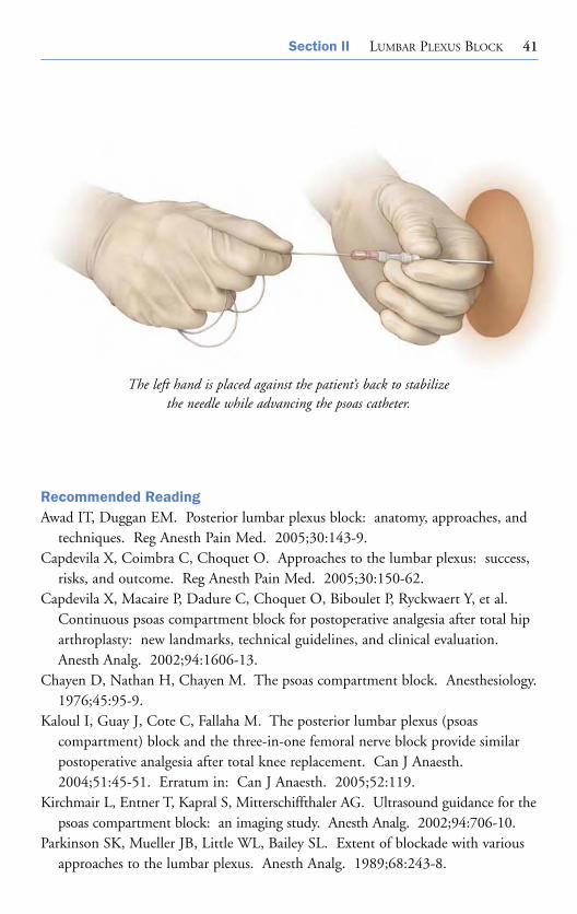
The left hand is placed against the patient’s back to stabilize the needle while advancing the psoas catheter.
Section_II.qxd 8/11/05 2:19 PM Page 41

42 MAYO CLINIC ANALGESIC PATHWAY
Stevens RD, Van Gessel E, Flory N, Fournier R, Gamulin Z. Lumbar plexus blockreduces pain and blood loss associated with total hip arthroplasty.Anesthesiology. 2000;93:115-21.
Weller RS, Gerancher JC, Crews JC, Wade KL. Extensive retroperitonealhematoma without neurologic deficit in two patients who underwent lumbarplexus block and were later anticoagulated. Anesthesiology. 2003;98:581-5.
Section_II.qxd 8/11/05 2:19 PM Page 42

FASCIA ILIACA APPROACH
43
Chapter 8
Clinical ApplicationsIndications for single injection and continuous fascia iliaca block include analgesiafor hip arthroplasty, femoral shaft fractures, and total knee arthroplasty. The supinepositioning of the patient with this approach is an advantage for patients in whomthe psoas compartment posterior approach may be technically difficult (previouslumbar surgical fusion with hardware stabilization or severe scoliosis). The fasciailiaca approach also may be applied when a previously placed psoas catheter hasbecome nonfunctional (positive blood aspiration or misplaced catheter).
Patient PositionThe patient is positioned supine, and the anesthesiologist stands on the side to be blocked.
LandmarksThis approach is based on the bony relationship of the anterior superior iliac spineand the pubic tubercle. The anterior superior iliac spine and the pubic tubercle are palpated, and the overlying skin is marked. A line is drawn joining these twopoints, and it is divided into thirds. A perpendicular line is drawn at the junctionbetween the outer one-third and the inner two-thirds. The needle insertion site is1 to 2 cm below the intersection of these two lines.
TechniqueA 22-gauge 8.75-cm (3.5-inch) pencil-tipped or short-bevel spinal needle is inserted perpendicular to the skin. An initial fascial “pop” is felt as the needlepenetrates the fascia lata. A second fascial “pop” is noted after penetration of
Section_II.qxd 8/11/05 2:19 PM Page 43

44 MAYO CLINIC ANALGESIC PATHWAY
Landmarks for the fascia iliaca block. Needle entry is marked 1 to 2 cm below the junctionbetween the outer one-third and the inner two-thirds of the line connecting the anterior
superior iliac spine and the pubic tubercle. The cross is the site of needle insertion.
the fascia iliaca. The fascia iliaca approach is a volume block; on removal of thestylet, 30 to 40 mL of solution (with intermittent aspiration) is injected. For continuous techniques, a 17-gauge Tuohy needle is used. The catheter is advanced 10 to 15 cm beyond the needle tip.
Evoked Motor ResponsesNo motor response is sought. This is a transfascial compartment block. Theessential end point is a two-“pop” (as the needle passes through the fascia lata
Section_II.qxd 8/11/05 2:19 PM Page 44
Section II LUMBAR PLEXUS BLOCK 45
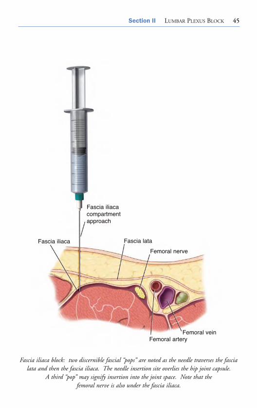
Fascia iliaca
Fascia iliacacompartmentapproach
Fascia lata
Femoral nerve
Femoral veinFemoral artery
Fascia iliaca block: two discernible fascial “pops” are noted as the needle traverses the fascialata and then the fascia iliaca. The needle insertion site overlies the hip joint capsule.
A third “pop” may signify insertion into the joint space. Note that the femoral nerve is also under the fascia iliaca.
Section_II.qxd 8/11/05 2:19 PM Page 45

46 MAYO CLINIC ANALGESIC PATHWAY
and fascia iliaca) sensation. Alternatively, stimulation with an insulated needlemay be used to facilitate needle or catheter placement. Contraction of the quadriceps is sought.
Needle Redirection CuesUse of a blunt needle (e.g., the Arrow 18-gauge 6.35-cm bullet-tip) exaggerates thefascial “pop” and is useful in mastery of the technique.
Side Effects and ComplicationsThese include hematoma formation, infection, and transient femoral neuropathy;rarely, catheter knotting occurs.
Recommended ReadingAwad IT, Duggan EM. Posterior lumbar plexus block: anatomy, approaches, and
techniques. Reg Anesth Pain Med. 2005;30:143-9.Capdevila X, Coimbra C, Choquet O. Approaches to the lumbar plexus: success,
risks, and outcome. Reg Anesth Pain Med. 2005;30:150-62.Dalens B, Tanguy A, Vanneuville G. Lumbar plexus block in children:
a comparison of two procedures in 50 patients. Anesth Analg. 1988;67:750-8.Dalens B, Vanneuville G, Tanguy A. Comparison of the fascia iliaca compartment
block with the 3-in-1 block in children. Anesth Analg. 1989;69:705-13.Erratum in: Anesth Analg. 1990;70:474.
Ganapathy S, Wasserman RA, Watson JT, Bennett J, Armstrong KP, Stockall CA,et al. Modified continuous femoral three-in-one block for postoperative painafter total knee arthroplasty. Anesth Analg. 1999;89:1197-202.
Morau D, Lopez S, Biboulet P, Bernard N, Amar J, Capdevila X. Comparison of continuous 3-in-1 and fascia iliaca compartment blocks for postoperativeanalgesia: feasibility, catheter migration, distribution of sensory block, and analgesic efficacy. Reg Anesth Pain Med. 2003;28:309-14.
Section_II.qxd 8/11/05 2:19 PM Page 46

FEMORAL NERVE BLOCK
47
Chapter 9
Lumbar plexus block: femoral nerve approach.
Section_II.qxd 8/11/05 2:19 PM Page 47

48 MAYO CLINIC ANALGESIC PATHWAY
Clinical ApplicationsIndications for single injection and continuous femoral nerve block include anesthesia for knee arthroscopy and analgesia for femoral shaft fractures, anteriorcruciate ligament reconstruction, and total knee arthroplasty.
Patient PositionThe patient is in the supine position. The limb to be blocked is slightly abductedand externally rotated.
Femoral nerve block: the dotted line corresponds to the inguinal crease. Needle insertionsite (indicated by the cross) is 1 to 2 cm lateral to the arterial pulsation at this level.
Section_II.qxd 8/11/05 2:19 PM Page 48

Section II LUMBAR PLEXUS BLOCK 49
LandmarksA line is drawn between the anterior superior iliac spine and the pubic tubercle,identifying the inguinal ligament. The femoral artery is located and marked justbelow the inguinal crease. The needle insertion site is 1 to 2 cm lateral to the arterial pulsation at the level of the inguinal crease.
TechniqueA 22-gauge 5-cm (2-inch) insulated needle is introduced at a 40° to 60° angle tothe skin and advanced. Frequently, two fascial “pops” (passage through the fascialata and fascia iliaca) are felt. When the needle reaches the depth of the artery, a pulsation of the needle hub is visible. The needle is advanced until quadricepscontraction, accompanied by patellar ascension, occurs. Thirty milliliters of solution is injected intermittently after repeated aspirations for blood.
Continuous femoral block is achieved by advancing a 20-gauge catheterapproximately 5 to 10 cm past the tip of an 18-gauge insulated needle. The use of a stimulating catheter may increase the quality of the block and decrease onset time. Additional confirmatory studies are needed.
Evoked Motor ResponsesStimulation of the posterior branch of the femoral nerve is identified by patellarascension as the quadriceps contracts.
Needle Redirection CuesCommonly, the anterior branch of the femoral nerve is identified first. Stimulation ofthis branch evokes contraction of the sartorius muscle on the medial side of the thighand should not be accepted. Rather, the needle should be redirected slightly laterallyand with a deeper direction to encounter the posterior branch of the femoral nerve.The use of an ultrasound probe facilitates locating a nonpalpable femoral artery.
Side Effects and ComplicationsIntravascular injection and hematoma formation are possible because of the proximity of the femoral artery. However, the nerve and artery are located in separate sheaths approximately 1 cm apart. In most patients with normal anatomy,the femoral artery can be easily palpated, allowing needle positioning lateral to thepulsation. The presence of femoral vascular grafts is a relative contraindication tothis block. Nerve damage is rare but has been reported with this technique.
Section_II.qxd 8/11/05 2:19 PM Page 49
50 MAYO CLINIC ANALGESIC PATHWAY
Recommended ReadingAllen HW, Liu SS, Ware PD, Nairn CS, Owens BD. Peripheral nerve blocks improve
analgesia after total knee replacement surgery. Anesth Analg. 1998;87:93-7.Awad IT, Duggan EM. Posterior lumbar plexus block: anatomy, approaches,
and techniques. Reg Anesth Pain Med. 2005;30:143-9.Berry FR. Analgesia in patients with fractured shaft of femur. Anaesthesia.
1977;32:576-7.Capdevila X, Barthelet Y, Biboulet P, Ryckwaert Y, Rubenovitch J, d’Athis F. Effects
of perioperative analgesic technique on the surgical outcome and duration ofrehabilitation after major knee surgery. Anesthesiology. 1999;91:8-15.
Capdevila X, Coimbra C, Choquet O. Approaches to the lumbar plexus: success, risks, and outcome. Reg Anesth Pain Med. 2005;30:150-62.
Chelly JE, Greger J, Gebhard R, Coupe K, Clyburn TA, Buckle R, et al.Continuous femoral blocks improve recovery and outcome of patients undergoing total knee arthroplasty. J Arthroplasty. 2001;16:436-45.
Kaloul I, Guay J, Cote C, Fallaha M. The posterior lumbar plexus (psoas compartment) block and the three-in-one femoral nerve block provide similarpostoperative analgesia after total knee replacement. Can J Anaesth.2004;51:45-51. Erratum in: Can J Anaesth. 2005;52:119.
Patel NJ, Flashburg MH, Paskin S, Grossman R. A regional anesthetic techniquecompared to general anesthesia for outpatient knee arthroscopy. Anesth Analg.1986;65:185-7.
Pham Dang C, Gautheron E, Guilley J, Fernandez M, Waast D, Volteau C, et al.The value of adding sciatic block to continuous femoral block for analgesia after total knee replacement. Reg Anesth Pain Med. 2005;30:128-33.
Salinas FV, Neal JM, Sueda LA, Kopacz DJ, Liu SS. Prospective comparison ofcontinuous femoral nerve block with nonstimulating catheter placement versusstimulating catheter-guided perineural placement in volunteers. Reg AnesthPain Med. 2004;29:212-20.
Singelyn FJ, Deyaert M, Joris D, Pendeville E, Gouverneur JM. Effects of intravenouspatient-controlled analgesia with morphine, continuous epidural analgesia, andcontinuous three-in-one block on postoperative pain and knee rehabilitation afterunilateral total knee arthroplasty. Anesth Analg. 1998;87:88-92.
Vloka JD, Hadzic A, Drobnik L, Ernest A, Reiss W, Thys DM. Anatomical landmarks for femoral nerve block: a comparison of four needle insertion sites.Anesth Analg. 1999;89:1467-70.
Section_II.qxd 8/11/05 2:19 PM Page 50
LATERAL FEMORAL CUTANEOUSNERVE BLOCK
51
Chapter 10
Clinical ApplicationsIndications for lateral femoral cutaneous nerve block include skin graft harvestingon the lateral aspect of the thigh and use in combination with other peripheralnerve blocks for complete anesthesia of the lower extremity.
Patient PositionThe patient is in the supine position and the hands are placed on the chest. Theanesthesiologist stands on the side to be blocked, as for a fascia iliaca or femoralnerve block.
LandmarksThe anterior superior iliac spine is palpated and marked. The needle insertion siteis marked 2 cm medial and 2 cm caudad to the anterior superior iliac spine andbelow the inguinal ligament.
TechniqueA 22-gauge 4-cm (1.5-inch) needle is advanced perpendicular to the skin entry site until a sudden release indicates passage through the fascia lata. A blunt-tippedneedle facilitates appreciation of the “pop” sensation. As the needle is moved in afanlike pattern laterally and medially, 10 to 15 mL of solution is injected, localanesthetic being deposited above and below the fascia.
Section_II.qxd 8/11/05 2:19 PM Page 51

52 MAYO CLINIC ANALGESIC PATHWAY
Evoked Motor ResponsesBecause the lateral femoral cutaneous nerve is a pure sensory nerve, a nerve stimulator is usually not helpful for performing the block. Contrarily, Shannon et al suggested that use of a nerve stimulator may improve success if a paresthesiareferred over the lateral aspect of the thigh occurs at 0.6 mA at a pulse width of 100 μs.
Landmarks for lateral femoral cutaneous nerve block. Needle entry site (indicated by thecross) is 2 cm medial and 2 cm caudad to the anterior superior iliac spine.
2 cm2 cm
2 cm2 cm
Section_II.qxd 8/11/05 2:19 PM Page 52

Section II LUMBAR PLEXUS BLOCK 53
Needle Redirection CuesThe nerve also can be blocked by directing the needle in a lateral and cephaladdirection to make contact with the iliac bone between the anterior superior iliacspine and the anterior inferior spine. The needle is now medial and inferior to theanterior superior iliac spine, where 10 mL of anesthetic solution is injected in afanlike pattern. Combining the two techniques increases the success rate; seekingparesthesias usually is not necessary.
Side Effects and ComplicationsNeuritis caused by needle trauma or drug toxicity is a potential but unlikelycomplication.
Recommended ReadingShannon J, Lang SA, Yip RW, Gerard M. Lateral femoral cutaneous nerve block
revisited: a nerve stimulator technique. Reg Anesth. 1995;20:100-4.
Section_II.qxd 8/11/05 2:19 PM Page 53

This page intentionally left blank

OBTURATOR NERVE BLOCK
55
Chapter 11
Clinical ApplicationsThe obturator nerve usually is blocked as a component of regional anesthesia for knee surgery; it is rarely blocked on its own. However, because it is primarily a motor nerve, obturator nerve block can be useful for treating or diagnosingadductor spasm in patients with cerebral palsy and other muscle or neurologic diseases affecting the lower extremities.
Patient PositionThe patient is in the supine position with the legs slightly abducted and externallyrotated. The anesthesiologist stands on the side to be blocked.
LandmarksThe landmark for the classic approach to obturator nerve block is the pubic tubercle.The interadductor approach also uses the inguinal ligament and the femoral artery.
TechniqueA mark is made 1 to 2 cm lateral and 1 to 2 cm caudad to the pubic tubercle and a21-gauge 10-cm (4-inch) needle is advanced perpendicular to the skin with a slightmedial direction. The inferior pubic ramus is encountered at a depth of 2 to 4 cm,and the needle is “walked” laterally parallel to the superior pubic ramus until itpasses into the obturator canal. The obturator nerve is located 2 to 3 cm past the initial point of contact with the pubic ramus. After negative aspiration, 10 to15 mL of local anesthetic is injected. A nerve stimulator is helpful for locating the obturator nerve.
Section_II.qxd 8/11/05 2:19 PM Page 55
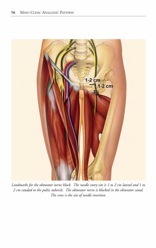
56 MAYO CLINIC ANALGESIC PATHWAY
Landmarks for the obturator nerve block. The needle entry site is 1 to 2 cm lateral and 1 to2 cm caudad to the pubic tubercle. The obturator nerve is blocked in the obturator canal.
The cross is the site of needle insertion.
Section_II.qxd 8/11/05 2:19 PM Page 56

Section II LUMBAR PLEXUS BLOCK 57
Evoked Motor ResponsesThe obturator nerve is identified by contraction of the adductor muscles.
Needle Redirection CuesAn alternative interadductor approach has been described. In this technique, theneedle is inserted behind the adductor longus tendon, near its pubic insertion, andis directed laterally with a slight posterior and superior inclination toward a markon the skin 1 to 2 cm medial to the femoral artery and immediately below theinguinal ligament representing the obturator canal. The needle is carefullyadvanced until contractions of the adductor muscles occur.
Side Effects and ComplicationsThe obturator canal contains vascular and neural structures, and there is a theoretical risk of intravascular injection, hematoma, and nerve damage.
Recommended ReadingBouaziz H, Vial F, Jochum D, Macalou D, Heck M, Meuret P, et al. An evaluation
of the cutaneous distribution after obturator nerve block. Anesth Analg.2002;94:445-9.
Jochum D, Iohom G, Choquet O, Macalou D, Ouologuem S, Meuret P, et al.Adding a selective obturator nerve block to the parasacral sciatic nerve block: an evaluation. Anesth Analg. 2004;99:1544-9.
Macalou D, Trueck S, Meuret P, Heck M, Vial F, Ouologuem S, et al.Postoperative analgesia after total knee replacement: the effect of an obturatornerve block added to the femoral 3-in-1 nerve block. Anesth Analg.2004;99:251-4.
Wassef MR. Interadductor approach to obturator nerve blockade for spastic conditions of adductor thigh muscles. Reg Anesth. 1993;18:13-7.
Section_II.qxd 8/11/05 2:19 PM Page 57

This page intentionally left blank

SAPHENOUS NERVE BLOCKS
59
Chapter 12
Clinical ApplicationsThe saphenous nerve is a cutaneous branch of the femoral nerve, arising in thefemoral triangle. It descends laterally to the femoral artery and enters the adductorcanal. The nerve exits from the lower part of the canal, emerging between the sartorius and gracilis muscles. The nerve becomes subcutaneous below the sartoriusat the medial side of the knee. It descends down the medial border of the tibiaimmediately behind the long saphenous vein. The nerve crosses with the vein infront of the medial malleolus and extends as far as the base of the great toe. Thesaphenous nerve supplies an extensive cutaneous area over the medial side of theknee, leg, ankle, and foot.
Saphenous nerve blocks are commonly performed as a component of knee andankle blocks. Supplemental block of the saphenous nerve is required for surgicalprocedures to the medial aspect of the leg, ankle, and foot or when a tourniquet or Esmarch’s bandage is applied. Patients with thigh tourniquet placement requirea more proximal block near the saphenous nerve origin in the femoral triangle.Various nerve block approaches are described, from its origin in the femoral triangle to the medial malleolus.
Saphenous Nerve Block at the Level of the Femoral Triangle
Patient PositionThe patient is positioned in the supine position. The leg is extended at the knee,and the long axis of the foot is at a 90° angle to the table. The anesthesiologiststands at the side to be blocked.
Section_II.qxd 8/11/05 2:19 PM Page 59

60 MAYO CLINIC ANALGESIC PATHWAY
Frontal view of the lower extremity, showing the saphenous nerve. Sites to block the saphenous nerve are indicated by crosses.
Section_II.qxd 8/11/05 2:19 PM Page 60

Section II LUMBAR PLEXUS BLOCK 61
LandmarksThe landmarks are the inguinal ligament, the femoral artery, and the proximalmedial border of the sartorius muscle. The femoral artery pulse is palpated andmarked. A mark is placed 1 to 2 cm lateral to the artery and medial to the upperborder of the sartorius muscle.
Vastus Medialus TechniqueA 22-gauge 5-cm (2-inch) insulated needle is introduced perpendicular to the skin at the needle insertion site and advanced until an evoked vastus medialis muscleresponse is elicited at 0.5 mA or less. A total of 5 to 10 mL of solution is injectedincrementally after negative aspiration. Alternatively, a more proximal block of thefemoral nerve may be performed if total volume of anesthetic solution is not an issue.
Evoked Motor ResponsesThe saphenous nerve is identified by elicitation of the vastus medialis motorresponse.
Saphenous Nerve Block at the Level of the Knee
Patient PositionThe patient is in the supine position, the leg is extended at the knee, and the longaxis of the foot is at a 90° angle to the table. The anesthesiologist stands at the sideto be blocked.
LandmarksThese saphenous nerve blocks are based on the relationship of the nerves to thesuperior pole of the patella, tibial tuberosity, medial head of gastrocnemius muscle,sartorius muscle, and saphenous vein.
Field Block TechniqueA 25-gauge 5-cm (2-inch) needle is inserted above the medial surface of the tibiaand 5 to 7 mL of solution is infiltrated subcutaneously in a fanlike pattern.Classically, this saphenous nerve block was performed as a blind subcutaneous infiltration over the medial surface of the tibia (between the tibial tuberosity andthe medial head of the gastrocnemius muscle). However, success rates with thistechnique ranged between 33% and 65%.
Section_II.qxd 8/11/05 2:19 PM Page 61

62 MAYO CLINIC ANALGESIC PATHWAY
Transsartorial TechniqueThe sartorius muscle is palpated on the medial side, just above the knee. At thelevel of the upper pole of the patella, a 22-gauge 5-cm (2-inch) needle is advanced45° from the coronal plane, through the muscle belly of the sartorius, until a fascial “pop” or click is noted (1.5-3 cm). After negative aspiration, 10 mL of localanesthetic is injected. Success rates for the transsartorial approach are 70% to 80%.
Paravenous TechniqueBelow the knee the saphenous nerve is immediately adjacent to the vein. Thesaphenous vein is identified by placing a thigh tourniquet and allowing the leg to hang down for at least 60 seconds. Ultrasound guidance also may be helpful. A subcutaneous paravascular injection is performed using a 25-gauge 5-cm (2-inch)needle with 5 mL of local anesthetic on each side of the vein, and care is taken toavoid intravenous injection. The success rate is approximately 100%. However,painless hematomas often occur with this approach.
Evoked Motor ResponsesNo motor response is sought. Although the saphenous nerve is pure sensory distalto the femoral triangle, the nerve may be localized using a nerve stimulator andeliciting sensory “electrical” pulsations.
Saphenous Nerve Block at the AnkleThe saphenous nerve follows the long saphenous vein to the medial malleolus and supplies the skin of the foot medially, to the midfoot. The saphenous nerve is usually one of the five peripheral nerves blocked at the ankle.
Patient PositionThe patient is in the supine position, and a padded table crutch support or pillowis placed under the calf to flex the leg 45°. The anesthesiologist stands at the footof the patient.
LandmarksThe medial malleolus, saphenous vein, and the needle insertion site for the deepperoneal nerve block are identified. The nerve is located between the saphenousvein and the medial malleolus.
Section_II.qxd 8/11/05 2:19 PM Page 62

Section II LUMBAR PLEXUS BLOCK 63
TechniqueA 25-gauge 5-cm (2-inch) needle is inserted at the site for the deep peroneal nerveblock and directed medially to subcutaneously infiltrate 5 mL of solution aroundthe great saphenous vein.
Evoked Motor ResponsesNo motor response is sought.
Needle Redirection CuesA 25-gauge 8.75-cm (3.5-inch) spinal needle may facilitate advancing the needle subcutaneously from the midline insertion site to the medial malleolus. An alternative is to insert the needle above and medial to the medial malleolus and 5 to 7 mL of solution is infiltrated subcutaneously about the saphenous vein in a fanlike pattern.
Side Effects and ComplicationsThere is a theoretical risk of intravascular injection, hematoma, and nerve damageat all approaches to the saphenous nerve.
Recommended ReadingComfort VK, Lang SA, Yip RW. Saphenous nerve anaesthesia: a nerve stimulator
technique. Can J Anaesth. 1996;43:852-7.De Mey JC, Deruyck LJ, Cammu G, De Baerdemaeker LE, Mortier EP. A
paravenous approach for the saphenous nerve block. Reg Anesth Pain Med.2001;26:504-6.
Mansour NY. Sub-sartorial saphenous nerve block with the aid of nerve stimulator.Reg Anesth. 1993;18:266-8.
van der Wal M, Lang SA, Yip RW. Transsartorial approach for saphenous nerveblock. Can J Anaesth. 1993;40:542-6.
Section_II.qxd 8/11/05 2:19 PM Page 63

This page intentionally left blank

Section_III.qxd 8/11/05 2:44 PM Page 65
SCIATIC NERVE BLOCK
SECTION III
Section_III.qxd 8/11/05 2:44 PM Page 65

This page intentionally left blank

CLASSIC POSTERIOR APPROACH OF LABAT
67
Chapter 13
Clinical ApplicationsBecause of its wide sensory distribution, the sciatic nerve block can be used, together with a saphenous or femoral nerve block, for any surgical procedure below the knee which does not require a thigh tourniquet. It can be used in combination with other peripheral nerve blocks to provide anesthesia for surgicalprocedures involving the thigh and knee.
Patient PositionThe patient is positioned laterally in a modified Sims’ position; the leg to beblocked is rolled forward onto the flexed knee as the heel rests on the knee of thedependent (nonoperative) leg.
LandmarksThis approach is based on the bony relationship of the posterior superior iliac spineand the greater trochanter of the femur. The dorsal tip of the greater trochanterand the posterior superior iliac spine are identified, and a line is drawn joiningthese two points. A perpendicular line is drawn bisecting this line and extending 3 to 5 cm caudally, the site of the needle insertion.
A confirmatory line can be drawn connecting the greater trochanter to a point 1 cm below the sacral hiatus. The needle insertion site is the intersectionbetween this line and that between the greater trochanter and the posterior superior iliac spine.
Section_III.qxd 8/11/05 2:44 PM Page 67

68 MAYO CLINIC ANALGESIC PATHWAY
TechniqueSome experts recommend that sciatic nerve block be initiated well before the scheduled time of operation to allow for long latencies.
A 21-gauge 10-cm (4-inch) insulated needle is advanced perpendicular to theskin until an evoked motor response is elicited.
The initial stimulating current should be kept at 0.75 mA or less because directstimulation of the gluteus maximus muscle with higher currents can mimic sciaticnerve stimulation. Also, high-current stimulation may result in vigorous rhythmiccontractions of the gluteus maximus, which are distracting to the proceduralist anduncomfortable for the patient.
Labat’s classic posterior approach.
Section_III.qxd 8/11/05 2:44 PM Page 68
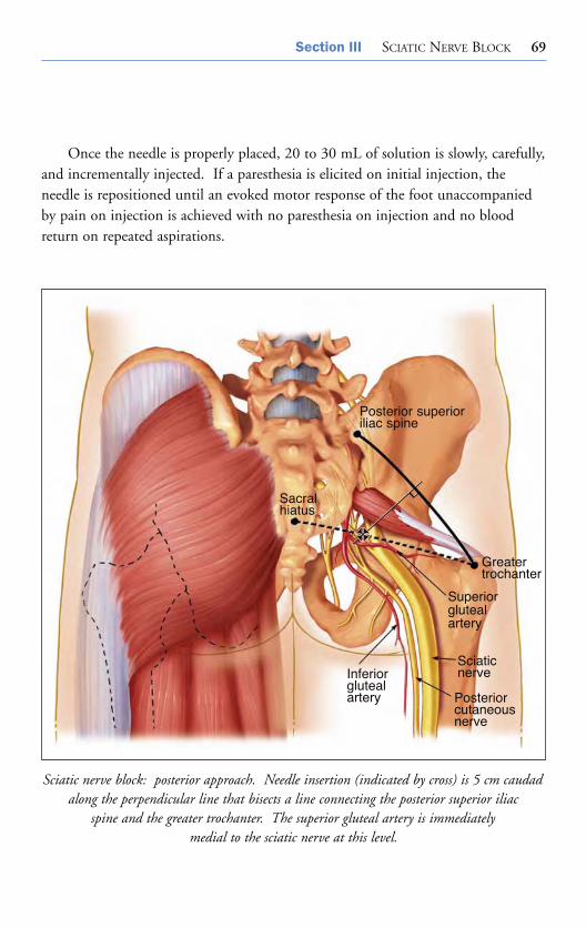
Section III SCIATIC NERVE BLOCK 69
Once the needle is properly placed, 20 to 30 mL of solution is slowly, carefully,and incrementally injected. If a paresthesia is elicited on initial injection, theneedle is repositioned until an evoked motor response of the foot unaccompaniedby pain on injection is achieved with no paresthesia on injection and no bloodreturn on repeated aspirations.
Sciatic nerve block: posterior approach. Needle insertion (indicated by cross) is 5 cm caudad along the perpendicular line that bisects a line connecting the posterior superior iliac
spine and the greater trochanter. The superior gluteal artery is immediately medial to the sciatic nerve at this level.
Posterior superioriliac spine
Sacralhiatus
Greatertrochanter
Superiorglutealartery
Sciaticnerve
Posteriorcutaneousnerve
Inferiorglutealartery
Section_III.qxd 8/11/05 2:44 PM Page 69
70 MAYO CLINIC ANALGESIC PATHWAY
Evoked Motor ResponsesStimulation of the tibial nerve component produces plantar flexion and inversionof the foot. Common peroneal nerve stimulation produces dorsiflexion and eversion of the foot.
Many methods have been tried to improve success with sciatic nerve blockade.Attempts to place the needle in the middle of the sciatic nerve by identifying a specific motor end point (foot inversion) may increase success rates. Anothermethod is the concept of multiple injections. The two major components (dorsiflexion and plantar flexion) of the sciatic nerve are separately identified and blocked.
Needle Redirection CuesKnowledge of neural anatomy allows the clinician to redirect the needle on thebasis of the motor response sought and that obtained. If bone is encountered, theneedle is directed medially. If blood is aspirated, the needle is directed laterally.
If knee flexion is elicited as a result of contraction of the tensor fascia lata, the superior gluteal nerve is stimulated. This response indicates the needle is posterior, lateral, and cephalad to the sciatic nerve and should be redirected.Hamstring contraction should not be accepted as a suitable response because it can be due to direct gluteal muscle stimulation.
Side Effects and ComplicationsThe block is technically not difficult to perform. Hematoma formation is possible;nerve damage has been reported, although persistent paresthesias usually are self-limited. A minimal degree of vasodilation may occur with sciatic nerve block.
Recommended ReadingDalans B, Tanguy A, Vanneuville G. Sciatic nerve blocks in children: comparison
of the posterior, anterior, and lateral approaches in 180 pediatric patients.Anesth Analg. 1990;70:131-7.
Labat G. Regional anesthesia: its technic and clinical application. Philadelphia:WB Saunders Company; 1922.
Winnie AP. Regional anesthesia. Surg Clin North Am. 1975;55(4):861-92.
Section_III.qxd 8/11/05 2:44 PM Page 70
PARASACRAL APPROACH
71
Chapter 14
Clinical ApplicationsThe parasacral approach is the most proximal of the posterior approaches to thesciatic nerve. As a result, the posterior cutaneous nerve usually is blocked, and thegluteal, pudendal, and obturator nerves frequently are blocked. This approach hasbeen used to provide analgesia after major knee, foot, and ankle reconstruction.For procedures above the knee, this approach may provide an advantage over moredistal approaches to the sciatic nerve. However, for procedures below the knee, the adductor weakness (from obturator and superior gluteal nerve block) may bedisadvantageous for mobilization of the patient.
Patient PositionThe patient is positioned laterally with the side to be blocked uppermost.
LandmarksThis approach is based on the bony relationship of the posterior superior iliac spineand the ischial tuberosity.
The most prominent aspects of the posterior superior iliac spine and the ischialtuberosity are identified, and a line is drawn joining these two points. Along theline, a mark is made at 6 cm inferior to the posterior superior iliac spine, definingthe needle insertion site.
Section_III.qxd 8/11/05 2:44 PM Page 71
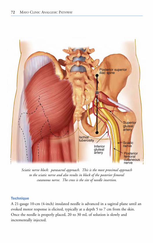
72 MAYO CLINIC ANALGESIC PATHWAY
TechniqueA 21-gauge 10-cm (4-inch) insulated needle is advanced in a sagittal plane until anevoked motor response is elicited, typically at a depth 5 to 7 cm from the skin.Once the needle is properly placed, 20 to 30 mL of solution is slowly and incrementally injected.
Sciatic nerve block: parasacral approach. This is the most proximal approach to the sciatic nerve and also results in block of the posterior femoral
cutaneous nerve. The cross is the site of needle insertion.
Posterior superioriliac spine
Superiorglutealartery
Sciaticnerve
Posteriorfemoralcutaneousnerve
Inferiorglutealartery
Ischialtuberosity
Section_III.qxd 8/11/05 2:44 PM Page 72

Section III SCIATIC NERVE BLOCK 73
Evoked Motor ResponsesPlantar flexion of the foot (tibial nerve component) or dorsiflexion (common peroneal nerve) is an acceptable motor response. Because of the proximal nature of the block, a hamstring motor response also is acceptable.
Needle Redirection CuesIf the needle is too cephalad, it may contact bone (ilium above the upper margin of the sciatic notch). The needle should be reinserted caudad 2 to 3 cm along the posterior superior iliac spine–ischial tuberosity marked line. If the needle isdirected too medially, the sacrum is encountered.
Side Effects and ComplicationsThis block is not technically difficult to perform. Hematoma formation and nerve damage are possible. Theoretical complications include penetration of pelvicstructures with the needle (rectum, internal iliac vessel perforation).
Recommended ReadingBruelle P, Cuvillon P, Ripart J, Eledjam MD. Sciatic nerve block: parasacral
approach [abstract]. Reg Anesth. 1998;23 Suppl:78.Mansour NY. Reevaluating the sciatic nerve block: another landmark for
consideration. Reg Anesth. 1993;18:322-3.Morris GF, Lang SA. Continuous parasacral sciatic nerve block: two case reports.
Reg Anesth. 1997;22:469-72.Morris GF, Lang SA, Dust WN, Van der Wal M. The parasacral sciatic nerve
block. Reg Anesth. 1997;22:223-8.
Section_III.qxd 8/11/05 2:44 PM Page 73

This page intentionally left blank

SUBGLUTEAL APPROACH
75
Chapter 15
Clinical ApplicationsThe indications for the subgluteal approach are those for sciatic nerve block: anesthesia or analgesia for knee, leg, and foot surgery. Because of the distal needleinsertion site, the posterior cutaneous femoral nerve may remain unblocked. Thishas implications for patients undergoing knee surgery or procedures requiring theuse of a thigh tourniquet.
Patient PositionThe patient is positioned laterally in a modified Sims’ position; the leg to beblocked is rolled forward onto the flexed knee as the heel rests on the knee of thedependent (nonoperative) leg.
LandmarksThis approach is based on the bony relationship of the greater trochanter and theischial tuberosity. The most prominent aspects of the greater trochanter and theischial tuberosity are identified by palpation, and a line is drawn joining these twopoints. A perpendicular line is drawn bisecting this line and extending 4 to 6 cmcaudad. The second line approximates the location of the sciatic nerve. The site of the needle insertion may be at the intersection of the two lines or as far as 6 cmdistally along the second line.
Section_III.qxd 8/11/05 2:44 PM Page 75
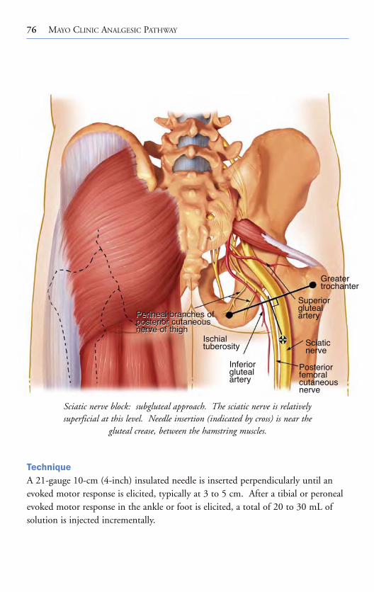
76 MAYO CLINIC ANALGESIC PATHWAY
TechniqueA 21-gauge 10-cm (4-inch) insulated needle is inserted perpendicularly until anevoked motor response is elicited, typically at 3 to 5 cm. After a tibial or peronealevoked motor response in the ankle or foot is elicited, a total of 20 to 30 mL ofsolution is injected incrementally.
Sciatic nerve block: subgluteal approach. The sciatic nerve is relatively superficial at this level. Needle insertion (indicated by cross) is near the
gluteal crease, between the hamstring muscles.
Greatertrochanter
Superiorglutealartery
Sciaticnerve
Posteriorfemoralcutaneousnerve
Inferiorglutealartery
Ischialtuberosity
Perineal brances ofposterior cutaneousnerve of thigh
Perineal branches ofposterior cutaneousnerve of thigh
Section_III.qxd 8/11/05 2:44 PM Page 76

Section III SCIATIC NERVE BLOCK 77
Evoked Motor ResponsesThe target response is an evoked response below the knee; stimulation of the tibialnerve component produces plantar flexion and inversion of the foot, and commonperoneal nerve stimulation produces dorsiflexion and eversion.
Needle Redirection CuesIf no motor response is elicited, the needle may be redirected 1 to 2 cm medially orlaterally to the original direction of the needle. It may be helpful to palpate orvisualize the groove that can be seen or palpated on the posterior aspect of thethigh. If bony contact is made, the needle is withdrawn and redirected medially.
Side Effects and ComplicationsThere is less risk of hematoma formation with this posterior approach. Nerveinjury is possible.
Recommended Readingdi Benedetto P, Bertini L, Casati A, Borghi B, Albertin A, Fanelli G. A new
posterior approach to the sciatic nerve block: a prospective, randomized comparison with the classic posterior approach. Anesth Analg. 2001;93:1040-4.
di Benedetto P, Casati A, Bertini L. Continuous subgluteus sciatic nerve blockafter orthopedic foot and ankle surgery: comparison of two infusion techniques. Reg Anesth Pain Med. 2002;27:168-72.
di Benedetto P, Casati A, Bertini L, Fanelli G, Chelly JE. Postoperative analgesia with continuous sciatic nerve block after foot surgery: a prospective,randomized comparison between the popliteal and subgluteal approaches.Anesth Analg. 2002;94:996-1000.
Section_III.qxd 8/11/05 2:44 PM Page 77

This page intentionally left blank

ANTERIOR APPROACH
79
Chapter 16
Clinical ApplicationsThe indications for the anterior approach are those for sciatic nerve block: anesthesia or analgesia for knee, leg, and foot surgery. The supine positioning ofthe patient with this approach is an advantage for patients in whom the posteriorapproaches would be difficult or painful. In addition, the supine position has the elegance of a single preparation of the patient for combined femoral and sciatic nerve block. Because of the distal needle insertion site, the posterior cutaneous femoral nerve may remain unblocked. This has implications for patients undergoing knee surgery or procedures requiring the use of a thigh tourniquet.
Patient PositionThe patient is in the supine position with the operative leg extended in the neutral position, avoiding external rotation of the leg.
LandmarksThe landmarks described by Chelly are based on the bony relationship between theanterior superior iliac spine and the pubic symphysis. The most caudal and mostmedial aspect of the anterior superior iliac spine and the most rostral surface of the pubic symphysis are identified by palpation, and a line is drawn joining thesetwo points. A perpendicular line is drawn bisecting this line and extending 8 cmdistally, the site of the needle insertion.
The original anterior approach described by Beck used the inguinal ligamentand the greater trochanter as landmarks.
Section_III.qxd 8/11/05 2:44 PM Page 79

80 MAYO CLINIC ANALGESIC PATHWAY
Sciatic nerve block: anterior approach.
TechniqueA 21-gauge 15-cm (6-inch) insulated needle is inserted perpendicularly with aslightly lateral angulation. The needle is advanced until it contacts bone, the lessertrochanter of the femur. The needle is redirected medially past the femur until anevoked motor response is elicited, typically 4 to 5 cm deep to the femur. After atibial or peroneal evoked motor response in the ankle or foot is elicited, a total of 20 to 30 mL of solution is injected incrementally.
The anterior sciatic approach is commonly used in combination with a femoral block for knee surgery. The advancing needle frequently stimulates thefemoral nerve at a distance of 3 to 5 cm from the skin. Blockade of the femoralnerve after sciatic block theoretically decreases the risk of femoral nerve injury due to needle trauma.
Section_III.qxd 8/11/05 2:45 PM Page 80
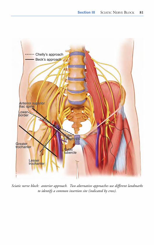
Section III SCIATIC NERVE BLOCK 81
Sciatic nerve block: anterior approach. Two alternative approaches use different landmarksto identify a common insertion site (indicated by cross).
Chelly’s approachBeck’s approach
Pubic symphysisPubic symphysis
PubictuberclePubictubercle
GreatertrochanterGreatertrochanter
LessertrochanterLessertrochanter
Anterior superioriliac spineAnterior superioriliac spine
Lower borderLower border
Section_III.qxd 8/11/05 2:45 PM Page 81

82 MAYO CLINIC ANALGESIC PATHWAY
Evoked Motor ResponsesAs the needle is slowly advanced, the first evoked response is commonly a quadriceps contraction as the needle glides past the femoral nerve. The targetresponse is an evoked response below the knee: stimulation of the tibial nervecomponent produces plantar flexion and inversion of the foot. Common peroneal nerve stimulation produces foot dorsiflexion and eversion. Contraction of the hamstring muscles is not accepted.
Needle Redirection CuesSeveral maneuvers have been reported to improve success with the anteriorapproach to the sciatic nerve. If bone (femur) is encountered, the needle is withdrawn to the skin, the leg is internally rotated, and the needle is readvanced. If the needle still will not pass by the femur, the process is repeated with the legexternally rotated. If these maneuvers are unsuccessful, the needle is reinserted 1 to 2 cm medially and redirected slightly laterally to advance past the femur. An insertion site 12 cm distal to the anterior superior iliac spine–pubic symphysisline also has been suggested.
Side Effects and ComplicationsThe needle passes near the femoral nerve and artery during advancement to the sciatic nerve. Hematoma formation is possible, and femoral nerve damage has been reported.
Recommended ReadingBeck GP. Anterior approach to sciatic nerve block. Anesthesiology.
1963;24:222-4.Chelly JE, Delaunay L. A new anterior approach to the sciatic nerve block.
Anesthesiology. 1999;91:1655-60.Ericksen ML, Swenson JD, Pace NL. The anatomic relationship of the sciatic
nerve to the lesser trochanter: implications for anterior sciatic nerve block.Anesth Analg. 2002;95:1071-4.
Vloka JD, Hadzic A, April E, Thys DM. Anterior approach to the sciatic nerveblock: the effects of leg rotation. Anesth Analg. 2001;92:460-2.
Section_III.qxd 8/11/05 2:45 PM Page 82

LATERAL POPLITEAL APPROACH
83
Chapter 17
Clinical ApplicationsThe sciatic nerve block at the popliteal fossa level may be used, together with asaphenous or femoral nerve block, for any surgical procedure below the knee which does not require a thigh tourniquet, such as tibia or fibula fractures, anklearthrodesis, or hindfoot procedures.
Patient PositionThe patient is in the supine position; the operative leg is extended at the knee, andthe long axis of the foot is at a 90° angle to the table.
LandmarksThe superior pole of the patella is palpated, and the groove between the lateral border of the biceps femoris and the vastus lateralis is identified and marked. A vertical line is drawn from the superior pole of the patella intersecting with the first line. The needle insertion site is the intersection of these two lines.
TechniqueA 21-gauge 10-cm (4-inch) insulated needle is slowly advanced in a horizontalplane between the biceps femoris and the vastus lateralis until the femur is encountered. The needle is then withdrawn to the skin and slowly advanced at a 30° angle posterior to the horizontal plane until an evoked motor response iselicited. After a tibial (preferred) or peroneal evoked motor response in the ankleor foot is elicited, a total of 30 to 40 mL of solution is injected incrementally.
Section_III.qxd 8/11/05 2:45 PM Page 83

84 MAYO CLINIC ANALGESIC PATHWAY
Knee block: lateral approach.
Section_III.qxd 8/11/05 2:45 PM Page 84

Section III SCIATIC NERVE BLOCK 85
Continuous popliteal block is performed by advancing a 20-gauge catheterthrough an 18-gauge insulated needle 5 to 6 cm into the popliteal fossa after theappropriate motor response is elicited.
Evoked Motor ResponsesAs the needle is slowly advanced, the first evoked response is direct stimulation ofthe vastus lateralis or biceps femoris. At a depth of approximately 3 to 4 cm, thecommon peroneal nerve (foot dorsiflexion and eversion) is commonly encountered.A deeper and more posterior angulation (45° from horizontal plane) results in tibialstimulation (plantar flexion and inversion).
Cross-sectional view of popliteal fossa. Needle position is shown for a lateral approach to thesciatic nerve in the supine patient at the level of the popliteal fossa.
ANTERIOR
LATERAL
POSTERIOR
MEDIAL
Vastus lateralismuscle
Biceps femorismuscle
Common peroneal nerve
Posterior femoralcutaneous nerveTibial nerveSemitendinous
muscle & tendon
Semimembranousmuscle
Sartorius muscle
Saphenousnerve
Distal femur
Vastus medialismuscle
Section_III.qxd 8/11/05 2:45 PM Page 85
86 MAYO CLINIC ANALGESIC PATHWAY
Needle Redirection CuesStimulating both tibial and common peroneal nerves has been described, with the local anesthetic dose divided equally between tibial and peroneal components. Aspiration of blood signifies that the needle is too deep and anterior. If neither component is localized, the needle is withdrawn to the skin, the femur is reidentified, and the needle angle is redirected in 5° increments until an appropriate motor response is elicited. Rarely, a more proximal skin insertion site is required.
Side Effects and ComplicationsHematoma formation and transient paresthesias are possible. Graft disruption orinfection may occur in patients with vascular grafts. Thus, this block is a relativecontraindication in patients with indwelling prostheses.
Recommended ReadingHadzic A, Vloka JD. A comparison of the posterior versus lateral approaches to
the block of the sciatic nerve in the popliteal fossa. Anesthesiology.1998;88:1480-6.
Paqueron X, Bouaziz H, Macalou D, Labaille T, Merle M, Laxenaire MC, et al.The lateral approach to the sciatic nerve at the popliteal fossa: one or two injections? Anesth Analg. 1999;89:1221-5.
Vloka JD, Hadzic A, April E, Thys DM. The division of the sciatic nerve in thepopliteal fossa: anatomical implications for popliteal nerve blockade. AnesthAnalg. 2001;92:215-7.
Vloka JD, Hadzic A, Kitain E, Lesser JB, Kuroda M, April EW, et al. Anatomicconsiderations for sciatic nerve block in the popliteal fossa through the lateralapproach. Reg Anesth. 1996;21:414-8.
Zetlaoui PJ, Bouaziz H. Lateral approach to the sciatic nerve in the popliteal fossa.Anesth Analg. 1998;87:79-82.
Section_III.qxd 8/11/05 2:45 PM Page 86
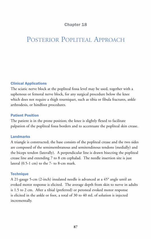
POSTERIOR POPLITEAL APPROACH
87
Chapter 18
Clinical ApplicationsThe sciatic nerve block at the popliteal fossa level may be used, together with asaphenous or femoral nerve block, for any surgical procedure below the knee which does not require a thigh tourniquet, such as tibia or fibula fractures, anklearthrodesis, or hindfoot procedures.
Patient PositionThe patient is in the prone position; the knee is slightly flexed to facilitate palpation of the popliteal fossa borders and to accentuate the popliteal skin crease.
LandmarksA triangle is constructed; the base consists of the popliteal crease and the two sidesare composed of the semimembranous and semitendinous tendons (medially) andthe biceps tendon (laterally). A perpendicular line is drawn bisecting the poplitealcrease line and extending 7 to 8 cm cephalad. The needle insertion site is just lateral (0.5-1 cm) to the 7- to 8-cm mark.
TechniqueA 21-gauge 5-cm (2-inch) insulated needle is advanced at a 45° angle until anevoked motor response is elicited. The average depth from skin to nerve in adultsis 1.5 to 2 cm. After a tibial (preferred) or peroneal evoked motor response is elicited in the ankle or foot, a total of 30 to 40 mL of solution is injected incrementally.
Section_III.qxd 8/11/05 2:45 PM Page 87

88 MAYO CLINIC ANALGESIC PATHWAY
Continuous popliteal block is performed by advancing a 20-gauge catheterthrough an 18-gauge insulated needle 5 to 6 cm into the popliteal fossa after theappropriate motor response is elicited.
Evoked Motor ResponsesPlantar flexion of the foot or toes and foot inversion indicate tibial nerve stimulation, the preferred response. Dorsiflexion of the foot, toe extension, andfoot eversion indicate common peroneal nerve stimulation. Attempts to placethe needle near the tibial nerve (foot inversion) may increase the success rate.
Knee block: posterior approach.
Section_III.qxd 8/11/05 2:45 PM Page 88

Section III SCIATIC NERVE BLOCK 89
Alternatively, with use of the multiple injection approach, the two majorcomponents (dorsiflexion and eversion, planter flexion and inversion) of the sciatic nerve are separately identified and blocked, and the local anesthetic dose is divided equally.
Posterior view of popliteal fossa. Landmarks for posterior approach. A perpendicular line isdrawn bisecting the popliteal crease line and extending 7 to 8 cm cephalad. The needle
entry site (indicated by cross) is just lateral (0.5-1 cm) to the perpendicular line.
Semimembranous muscle1 cm Biceps
femoris
Commonperonealnerve
Tibial nerve
Gastrocnemius muscle
Semitendinoustendon
7-8 cm
MEDIAL LATERAL
Section_III.qxd 8/11/05 2:45 PM Page 89

90 MAYO CLINIC ANALGESIC PATHWAY
Needle Redirection CuesIf the peroneal nerve is stimulated, medial redirection results in localization of thetibial component (and vice versa). Aspiration of blood signifies that the needle istoo deep and too medial and should be redirected laterally.
Side Effects and ComplicationsThe block is not technically difficult to perform. Hematoma formation and transient paresthesias are possible. A relative contraindication is presence of a vascular graft.
Recommended ReadingBenzon HT, Kim C, Benzon HP, Silverstein ME, Jericho B, Prillaman K, et al.
Correlation between evoked motor response of the sciatic nerve and sensoryblockade. Anesthesiology. 1997;87:547-52.
Hadzic A, Vloka JD. A comparison of the posterior versus lateral approaches tothe block of the sciatic nerve in the popliteal fossa. Anesthesiology.1998;88:1480-6.
Rorie DK, Byer DE, Nelson DO, Sittipong R, Johnson KA. Assessment of blockof the sciatic nerve in the popliteal fossa. Anesth Analg. 1980;59:371-6.
Vloka JD, Hadzic A, April E, Thys DM. The division of the sciatic nerve in thepopliteal fossa: anatomical implications for popliteal nerve blockade. AnesthAnalg. 2001;92:215-7.
Section_III.qxd 8/11/05 2:45 PM Page 90

ANKLE BLOCK
91
Chapter 19
Clinical ApplicationsThe peripheral nerves blocked at the ankle are terminal branches of both the sciatic(posterior tibial, superficial peroneal, deep peroneal, and sural) and the femoral(saphenous) nerves. The lumbosacral plexus blocked distally at the ankle providesanesthesia for surgery to the foot, including bunionectomy, amputation of the midfoot and toes, and debridement procedures.
Patient PositionThe patient is in the supine position, and a padded table crutch support or blankets are placed under the calf. The deep peroneal, superficial peroneal, andsaphenous nerves all may be blocked from the same needle insertion site, and anadditional insertion is required for posterior tibial and sural nerve blockade.
Posterior Tibial Nerve
LandmarksThe posterior tibial artery, posterior to the medial malleolus, is palpated andmarked at the cephalad tip of the medial malleolus. The medial border of theAchilles tendon is identified. The block also may be performed with the patient in the prone position.
Section_III.qxd 8/11/05 2:45 PM Page 91

92 MAYO CLINIC ANALGESIC PATHWAY
TechniqueA 25-gauge 3.8-cm (1.5-inch) needle is inserted perpendicularly to the skin at apoint 2 to 5 cm cephalad to the proximal tip of the medial malleolus and midwaybetween the posterior border of the upper malleolus and the medial border of theAchilles tendon. The nerve lies 1 to 2 cm deep to the skin. If a paresthesia isobtained, 5 mL of local anesthetic (epinephrine-free) solution is injected.Alternatively, 7 to 10 mL of solution is injected as the needle is slowly withdrawnfrom the posterior aspect of the tibia. At the supramalleolar level, blockade is
Cross-sectional view of ankle. The figure at the level of the malleolus shows the distributionof the terminal branches of the sciatic nerve that comprise the ankle block.
ANTERIOR
LATERAL
POSTERIOR
MEDIAL
Anterior tibial artery & vein
Extensor hallucis longus tendon
Anterior talofibular ligament
Superficial peronealnerve
Lateral anterior malleolar artery & vein
Lateral malleolus of fibula
Small saphenousvein
Sural nerve
Flexor hallucis longus muscleCalcaneal tendon
Posterior tibial nerve
Posterior tibial artery & vein
Flexor digitorumlongus tendon
Tibialis posterior tendon
Medial malleolus of tibia
Saphenous nerve
Medial deltoid ligament& synovial membrane
Great saphenous vein
Deep peroneal nerve
Tibialis anterior tendon
Section_III.qxd 8/11/05 2:45 PM Page 92

Section III SCIATIC NERVE BLOCK 93
Lumbosacral plexus block at the ankle.
theoretically more reliable because the calcaneal and plantar branches have not divided from the posterior tibial nerve.
Needle Redirection CuesStimulation with a 22-gauge 5-cm (2-inch) needle connected to a nerve stimulatormay be used to facilitate proper needle placement. The targeted evoked responseis plantar flexion. Nerve stimulation may not be effective in patients with aperipheral neuropathy. Ultrasound guidance may facilitate performance of thisblock.
Deep Peroneal Nerve Block
LandmarksAt the intramalleolus level, the tibialis anterior and the extensor hallucis tendonsare identified and marked.
Section_III.qxd 8/11/05 2:45 PM Page 93
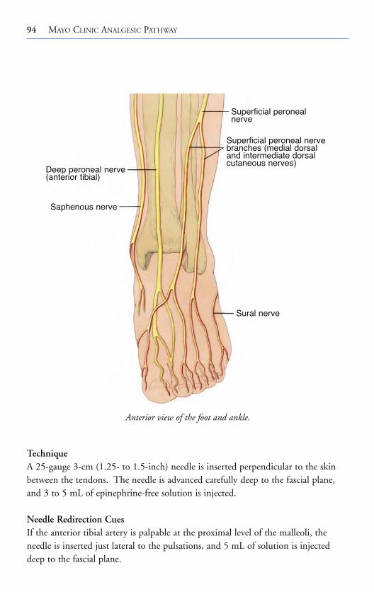
94 MAYO CLINIC ANALGESIC PATHWAY
Anterior view of the foot and ankle.
TechniqueA 25-gauge 3-cm (1.25- to 1.5-inch) needle is inserted perpendicular to the skinbetween the tendons. The needle is advanced carefully deep to the fascial plane,and 3 to 5 mL of epinephrine-free solution is injected.
Needle Redirection CuesIf the anterior tibial artery is palpable at the proximal level of the malleoli, the needle is inserted just lateral to the pulsations, and 5 mL of solution is injecteddeep to the fascial plane.
Superficial peronealnerve
Superficial peroneal nervebranches (medial dorsaland intermediate dorsalcutaneous nerves)
Sural nerve
Saphenous nerve
Deep peroneal nerve(anterior tibial)
Section_III.qxd 8/11/05 2:45 PM Page 94

Section III SCIATIC NERVE BLOCK 95
Saphenous Nerve Block
LandmarksThe medial malleolus, saphenous vein, and needle insertion site for the deep peroneal nerve block are identified. The saphenous nerve is located between thesaphenous vein and the medial malleolus.
TechniqueA 25-gauge 3- to 5-cm (1.5- to 2-inch) needle is inserted at the site for the deepperoneal nerve block and directed medially to subcutaneously infiltrate 5 mL ofsolution around the great saphenous vein.
Stabilizing the foot as the deep peroneal nerve is blocked.
Section_III.qxd 8/11/05 2:45 PM Page 95
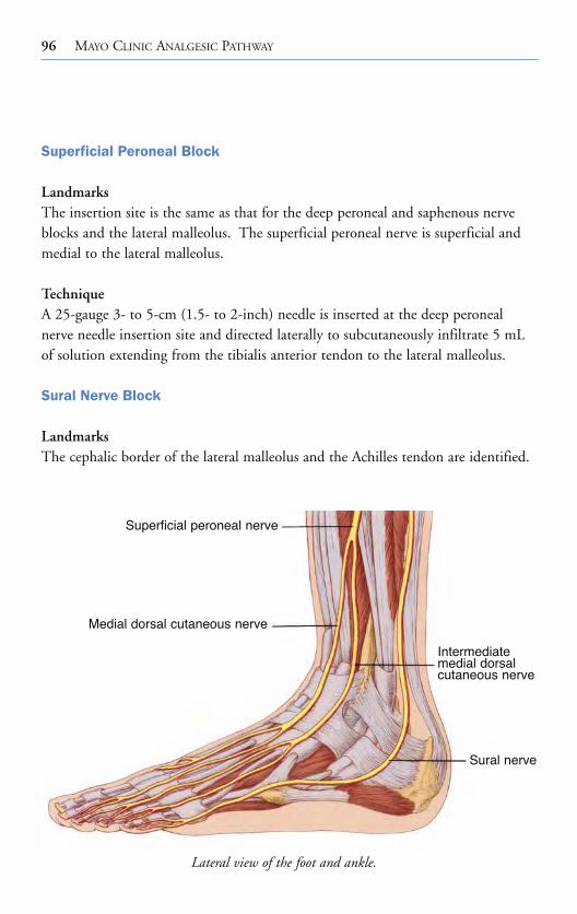
96 MAYO CLINIC ANALGESIC PATHWAY
Superficial Peroneal Block
LandmarksThe insertion site is the same as that for the deep peroneal and saphenous nerveblocks and the lateral malleolus. The superficial peroneal nerve is superficial andmedial to the lateral malleolus.
TechniqueA 25-gauge 3- to 5-cm (1.5- to 2-inch) needle is inserted at the deep peronealnerve needle insertion site and directed laterally to subcutaneously infiltrate 5 mLof solution extending from the tibialis anterior tendon to the lateral malleolus.
Sural Nerve Block
LandmarksThe cephalic border of the lateral malleolus and the Achilles tendon are identified.
Lateral view of the foot and ankle.
Superficial peroneal nerve
Medial dorsal cutaneous nerve
Intermediatemedial dorsalcutaneous nerve
Sural nerve
Section_III.qxd 8/11/05 2:46 PM Page 96

Section III SCIATIC NERVE BLOCK 97
View of the foot from above. Needle positioning is shown for blocking the terminal sciatic and saphenous nerves at the level of the malleolus.
Deep peroneal nerve
Superficial peroneal nerve Saphenous nerve
Posteriortibial nerve
Sural nerve
Section_III.qxd 8/11/05 2:46 PM Page 97
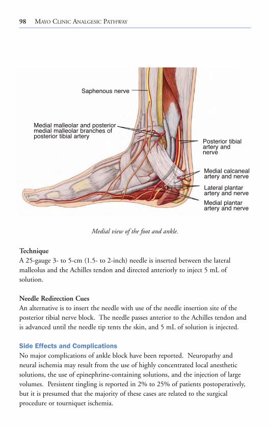
98 MAYO CLINIC ANALGESIC PATHWAY
TechniqueA 25-gauge 3- to 5-cm (1.5- to 2-inch) needle is inserted between the lateral malleolus and the Achilles tendon and directed anteriorly to inject 5 mL of solution.
Needle Redirection CuesAn alternative is to insert the needle with use of the needle insertion site of the posterior tibial nerve block. The needle passes anterior to the Achilles tendon andis advanced until the needle tip tents the skin, and 5 mL of solution is injected.
Side Effects and ComplicationsNo major complications of ankle block have been reported. Neuropathy and neural ischemia may result from the use of highly concentrated local anestheticsolutions, the use of epinephrine-containing solutions, and the injection of largevolumes. Persistent tingling is reported in 2% to 25% of patients postoperatively,but it is presumed that the majority of these cases are related to the surgical procedure or tourniquet ischemia.
Medial view of the foot and ankle.
Saphenous nerve
Medial malleolar and posteriormedial malleolar branches ofposterior tibial artery
Posterior tibialartery andnerve
Medial calcanealartery and nerve
Lateral plantarartery and nerve
Medial plantarartery and nerve
Section_III.qxd 8/11/05 2:46 PM Page 98

Section III
SC
IATIC
NE
RV
EB
LOC
K99
Sensory distribution of the foot and ankle.
Saphenous nerve(F)
Superficial peroneal nerve(S)
Deep peroneal nerve
(S)
Plantarnerve (PT)
Calcanealnerve (PT)
Sural nerve (S)
Saphenous nerve(F)
Superficial peroneal nerve
(S)
Sural nerve
(S)
Plantarnerve (PT)
Saphenousnerve
(F)
Deep peronealnerve
(S)
F = Femoral nerveS = Sciatic nervePT = Branch of posteriortibial nerve
LATERAL MEDIAL
Calcanealnerve (PT)
Section_III.qxd 8/11/05 2:46 PM Page 99

100 MAYO CLINIC ANALGESIC PATHWAY
Recommended ReadingColgrove RC. Posterior tibial nerve block. Foot Ankle Int. 2001;22:839-40.Gerbert J. The location of the terminal branching of the posterior tibial nerve and
its effect on administering a posterior tibial nerve block. J Am Podiatry Assoc.1971;61:8-11.
Needoff M, Radford P, Costigan P. Local anesthesia for postoperative pain reliefafter foot surgery: a prospective clinical trial. Foot Ankle Int. 1995;16:11-3.
Schurman DJ. Ankle-block anesthesia for foot surgery. Anesthesiology.1976;44:348-52.
Section_III.qxd 8/11/05 2:46 PM Page 100

Section_1V.qxd 8/11/05 2:50 PM Page 101
MAYO CLINIC
ANALGESIC PATHWAY
SECTION IV
Section_1V.qxd 8/11/05 2:50 PM Page 101

This page intentionally left blank

MAYO CLINIC TOTAL JOINT ANESTHESIAAND ANALGESIC PATHWAY
103
Chapter 20
Patients who undergo total joint arthroplasty experience significant postoperativepain. Failure to provide adequate analgesia impedes aggressive physical therapy and rehabilitation, which is critical to maintaining joint range of motion, andpotentially delays hospital dismissal. Traditionally, analgesia after total jointreplacement was provided by either intravenous patient-controlled analgesia or epidural analgesia. However, each technique has distinct advantages and disadvantages. For example, opioids do not consistently provide adequate painrelief and often cause sedation, constipation, nausea or vomiting, and pruritus.Epidural infusions containing local anesthetics (with or without an opioid) provide superior analgesia but are associated with hypotension, urinary retention,motor block that limits ambulation, and hematoma due to anticoagulation.
Recently, single-dose and continuous peripheral nerve techniques, which block the lumbar plexus (fascia iliaca, femoral, psoas compartment blocks) with or without sciatic nerve blockade, have been used among this patient population.Several European studies have reported that unilateral peripheral block provided a quality of analgesia and surgical outcomes similar to those of continuous epidural analgesia but with fewer side effects. This finding suggests that continuous peripheral techniques may be the optimal analgesic method after total joint arthroplasty.
Historically, total joint arthroplasty was associated with a hospitalizationextending nearly 3 weeks. During the past 2 decades, the typical duration of hospitalization progressively decreased (but plateaued) to 4 to 5 days. However,innovations in surgical approach, such as minimally invasive (incision) surgery, and the introduction of multimodal analgesia have increased the patient’s ability
Section_1V.qxd 8/11/05 2:50 PM Page 103
104 MAYO CLINIC ANALGESIC PATHWAY
to participate in physical therapy in the acute postoperative period and have shortened hospital stays by 1 or 2 days. Superior analgesia with minimal (opioid-related) side effects is crucial to perioperative management. Although alternativeprotocols have involved epidural infusions with supplemental intravenous opioidspostoperatively, the Mayo Clinic total joint analgesic pathway approach usesperipheral nerve blockade and oral analgesics.
This pathway is designed as a practical guide. An understanding of the pharmacology of opioid and non-opioid analgesics, anatomy, traditional and noveltechniques of neural localization, and methods of neural blockade is crucial to thesafe and efficient implementation of this clinical pathway. The clinician is encouraged to consult the standard references provided throughout for a morecomprehensive review of these elements.
Perioperative ManagementThe perioperative management of patients undergoing total joint replacement is divided into preoperative, intraoperative (anesthetic), and postoperative (analgesic) regimens. The rationale for the patient care components, as well as alternatives, is discussed. This patient care pathway is based on the experience of the authors. All patient care decisions remain the responsibility of the attending physicians and may require modification depending on local institutional practices, procedures, and treatment policies. This pathway may be revised as additional informationbecomes available.
IndicationsAll patients undergoing primary or revision joint replacement are candidates for the total joint analgesic pathway. Patients with preexisting neurologic deficits, suchas spinal stenosis, an active lumbar radiculopathy, sciatic or peroneal neuropathy, ordemyelinating disorder must be considered individually. Untreated infection andaltered hemostasis are considered relative contraindications to regional blockade.
Although the total joint analgesic pathway was developed for patients undergoingjoint replacement, it also can be applied to patients having other major lower extremity procedures such as hip disarticulation, repair of hip fracture, femoral rod placement,and tumor resection. The role of the technique for nonsurgical or medical indications,such as the treatment and prevention of complex pain syndromes, remains unstudied.
Section_1V.qxd 8/11/05 2:50 PM Page 104

Section IV MAYO CLINIC ANALGESIC PATHWAY 105
Preoperative PeriodAll patients are scheduled for a preoperative evaluation. Exclusion criteria for the total joint analgesic pathway include contraindications to regional anesthesia (progressive spinal or peripheral nerve disorder, infection at the needle insertionsite, anticoagulation with low-molecular-weight heparin, warfarin, or clopidogrel),and allergy to local anesthetics or other joint pathway medications. Although theAmerican Society of Anesthesiologists physical status I or II may facilitate physicaltherapy, increased age, significant cardiopulmonary disease, and obesity are notconsidered contraindications. Indeed, ongoing experience suggests that patientswith these factors may benefit the most from the total joint pathway and that their hospital course is uncomplicated and proportionally shortened.
The preoperative history and physical examination should include review of the following:
• Medication-related nausea or vomiting• Drugs affecting coagulation (discontinue in accordance with the American
Society of Regional Anesthesia and Pain Medicine Consensus Statements onNeuraxial Anesthesia and Anticoagulation, www.asra.com)
• Neurologic function, with documentation of any preexisting deficits• Hemoglobin and creatinine levels
The peripheral nerve techniques should be described to the patient, and thepatient should be informed that the presence of the blocks postoperatively willdecrease his or her ability to, for example, wiggle toes, and that this is a normaleffect of the blocks.
Patient Waiting AreaOn arrival to the patient waiting area, the following steps are accomplished:
1. Intravenous access is established 2. Oxycodone (sustained-release, 20 mg orally) is administered
These steps should not occur more than 1 hour preoperatively, or the likelihood ofnausea or gastric upset is increased.
Section_1V.qxd 8/11/05 2:50 PM Page 105

106 MAYO CLINIC ANALGESIC PATHWAY
Regional Anesthesia “Block Area”Ideally, peripheral blocks are performed in a designated “block area” to decreaseoperating room time and to allow adequate time for onset of regional blockade.However, this procedure may not be possible in all institutions. The time requiredto perform lower extremity blocks dramatically decreases with experience, typicallyrequiring less than 15 minutes (from patient positioning to completion of localanesthetic injection) after 25 blocks have been performed.
1. The patient is positioned with the operative side nondependent 2. Sedatives (midazolam 1-2 mg intravenously and fentanyl 50-100 μg
intravenously) are administered3. The total dose of local anesthetic must be monitored carefully. A maximum of
250 mg of bupivacaine or ropivacaine is recommended. Epinephrine should beadded to the initial bolus to allow identification of intravascular injection and todecrease systemic levels of local anesthetics
A complete block of the lumbar plexus (femoral, lateral femoral cutaneous,obturator nerves) is accomplished with the psoas block. In comparison, the fasciailiaca and femoral approaches reliably block the femoral but not the lateral femoralcutaneous and obturator nerves. Complete unilateral lower extremity blockade is achieved with a continuous psoas compartment (preferred), femoral, or fascia iliaca block, in combination with a proximal sciatic block. The sciatic block is performed first, because block onset often is protracted compared with lumbarplexus techniques. Selection of regional analgesic technique is dependent on thesurgical site:
• Total (or unicompartmental) knee replacement Psoas compartment or femoral catheterSciatic single injection block
• Total hip replacement • Psoas compartment or fascia iliaca catheter
Section_1V.qxd 8/11/05 2:50 PM Page 106

Section IV MAYO CLINIC ANALGESIC PATHWAY 107
Intraoperative CourseCombined lumbar plexus and sciatic blockade may be sufficient to provide the soleintraoperative anesthesia, if adequate onset time is allowed. However, the regionalblock typically is supplemented with an intravenous infusion of propofol.
• The airway may be secured with a laryngeal mask airway or endotracheal tube, particularly during the early stages of pathway implementation while theanesthesia and surgical teams are mastering techniques
• Opioid administration is limited to a maximum of 150 to 250 μg of fentanyl.No long-acting opioids (morphine, hydromorphone) are administered
• Normothermia and volume status are maintained to minimize the need for temperature or volume correction in the postoperative anesthesia recovery room
• Antiemetic prophylaxis is administered only to patients with a history of significant postoperative nausea and vomiting. The avoidance of neuraxial or intravenous opioids and general anesthesia places patients at a low risk forpostoperative nausea and vomiting; routine use of antiemetic therapy is not warranted
Postoperative Course
Anesthesia Care UnitManagement of the patient in the postoperative anesthesia care unit includes assessment of adequate regional blockade, administration of supplemental analgesics as needed, and correction of temperature and volume status.
• Immediately on arrival to the unit, the patient’s vital signs are measured. A warming device is placed or the hemoglobin level is assessed, as appropriate. A target hemoglobin of 10.0 g/dL is considered adequate in the absence of significant cardiopulmonary disease
Section_1V.qxd 8/11/05 2:50 PM Page 107
108 MAYO CLINIC ANALGESIC PATHWAY
• The presence of lumbar plexus (anterior thigh numbness and weakness) and sciatic (foot numbness and weakness) block is verified. Negative aspiration of the lumbar plexus catheter is confirmed, and a bolus is administered and infusion initiated:
Psoas compartment or femoral catheterBolus 10 mL bupivacaine 0.2% Infusion bupivacaine 0.2% at 10 mL/h
Fascia iliaca Bolus 20 mL bupivacaine 0.25% Infusion bupivacaine 0.1% at 20 mL/h
• A visual analog score more than 4 warrants supplemental analgesia. A combination of oxycodone and acetaminophen (2 tablets orally) is administered. Rarely, intravenous fentanyl (50-100 μg) is needed
Patient Care UnitPatient management on arrival to the surgical ward is a continuation of that implemented in the postoperative anesthesia care unit. Essentially all analgesicrequirements are met with the combination of peripheral nerve block and scheduled administration of oral acetaminophen and sustained-release oxycodone.Only a few patients require additional oral opioids for breakthrough pain.However, a nonfunctional or dislodged lumbar plexus catheter may necessitateintravenous opioids. A responsive anesthesia pain service is required to treat breakthrough pain immediately and effectively. An attentive nursing staff anticipates evolving analgesic requirements as physical therapy and activity increase.
Postoperatively, patients with an inadequate or failed block and patients withopioid dependence may require opioids intravenously (Table 1). Likewise, lowerdoses or alternative agents may be warranted in patients who are opioid-intolerant(Table 2).
• Oral intake should be encouraged (and rapidly advanced) to decrease nauseaassociated with oral analgesics
• Scheduled oral analgesics:Oxycodone 10 mg orally at 0800 and 2000 for patients 70 years or olderOxycodone 20 mg orally at 0800 and 2000 for patients younger than 70 years(use of oxycodone may be discontinued after 48 hours to avoid sedation andother opioid-related side effects)
Section_1V.qxd 8/11/05 2:50 PM Page 108

Section IV MAYO CLINIC ANALGESIC PATHWAY 109
Acetaminophen, 1,000 mg orally at 0800, 1200, and 1600 (maximal acetaminophen dose of 4,000 mg/d from all sources should not be exceeded)
• Supplemental analgesics:Oxycodone 5 to 10 mg orally every 4 hours as needed: 5 mg if patient complains of pain and rates pain score less than 4; 10 mg if pain score is 4 or more
Ketorolac 15 mg intravenously every 6 hours as needed for four doses.Ketorolac should not be administered to patients with renal insufficiency orpatients receiving thromboprophylaxis with low-molecular-weight heparin,warfarin, or aspirin
• Lumbar plexus infusions:Pain service rounds should be made in the morning to optimize analgesia for the upcoming day. Patients should be educated as to the oral analgesicregimen, including the option of additional medications for breakthroughpain. Reassurance regarding residual sciatic block may be necessary
Psoas compartment or femoral infusion
Table 1. Intravenous Opioids for Patient-Controlled Analgesia*
Lockoutinterval, 4-Hour
Agent Bolus min maximal dose Infusion rate
Fentanyl, 10 μg/mL 10-20 μg 5-10 300 μg 20-100 μg/h
Hydromorphone,
0.2 mg/mL 0.1-0.2 mg 5-10 3 mg 0.1-0.2 mg/h
Meperidine,
10 mg/mL 5-25 mg 5-10 200 mg† 5-15 mg/h†
Morphine sulfate,
1 mg/mL 0.5-2.5 mg 5-10 30 mg 1-10 mg/h
*Intravenous administration of opioids should be minimized. An intravenous patient-controlled analgesic regimen is typically required only if a peripheral block is inadequate or incomplete or thepatient is opioid-dependent.
†Meperidine limit in healthy patients should be 800 mg in the first 24 hours, then 600 mg every 24 hours thereafter.
Section_1V.qxd 8/11/05 2:50 PM Page 109

110M
AYO
CLIN
ICA
NA
LGE
SICP
ATH
WA
YTable 2. Oral Analgesics
Analgesic Dosing MaximalDrug* dose† interval daily dose Comments
Acetaminophen 500-1,000 mg q 4-6 h 4,000 mg As effective as aspirin; 1,000 mg more effectivethan 650 mg in some patients
Nonsteroidal anti-inflam-matory drugs
Celecoxib (Celebrex) 400 mg q 12 h Celecoxib is the only cyclooxygenase-2 inhibitor initially, available in North Americathen 200 mg Valdecoxib and rofecoxib recently were removed
from the market because of concerns about cardiovascular risk
Aspirin 325-1,000 mg q 4-6 h 4,000 mg Most potent antiplatelet effect
Ibuprofen (Advil, 200-400 mg q 4-6 h 3,200 mg 200 mg equal to 650 mg aspirin or acetaminophenMotrin, Nuprin, others)
Naproxen (Aleve, 500 mg q 12 h 1,000 mg 250 mg equal to 650 mg aspirin, but with longer Naprosyn, others) duration
Ketorolac (Toradol) 15-30 mg q 4-6 h 60 mg (≥65 Comparable to 10 mg morphine; reduce dose inIM/IV years); 120 mg patients <50 kg or with renal impairment; total
(<65 years) duration of administration is 5 daysOpioids
Oxycodone (Oxycontin) 10-20 mg q 12 h Limit to total of four doses to avoid accumulation and opioid-related side effects
Controlled-release 15-30 mg q 8-12 h Limit to total of four doses to avoid morphine (MS Contin) accumulation and opioid-related side effects
Section_1V.qxd 8/11/05 2:50 PM Page 110
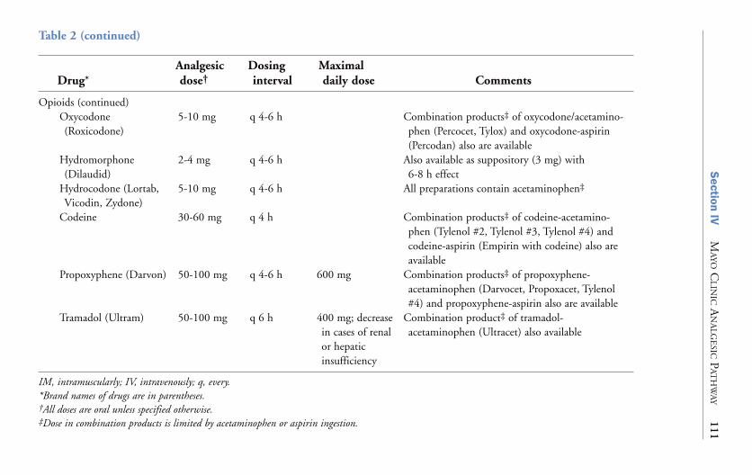
Section IV
MA
YOC
LINIC
AN
ALG
ESIC
PAT
HW
AY
111Table 2 (continued)
Analgesic Dosing MaximalDrug* dose† interval daily dose Comments
Opioids (continued)Oxycodone 5-10 mg q 4-6 h Combination products‡ of oxycodone/acetamino-(Roxicodone) phen (Percocet, Tylox) and oxycodone-aspirin
(Percodan) also are availableHydromorphone 2-4 mg q 4-6 h Also available as suppository (3 mg) with(Dilaudid) 6-8 h effect
Hydrocodone (Lortab, 5-10 mg q 4-6 h All preparations contain acetaminophen‡
Vicodin, Zydone)Codeine 30-60 mg q 4 h Combination products‡ of codeine-acetamino-
phen (Tylenol #2, Tylenol #3, Tylenol #4) andcodeine-aspirin (Empirin with codeine) also are available
Propoxyphene (Darvon) 50-100 mg q 4-6 h 600 mg Combination products‡ of propoxyphene-acetaminophen (Darvocet, Propoxacet, Tylenol #4) and propoxyphene-aspirin also are available
Tramadol (Ultram) 50-100 mg q 6 h 400 mg; decrease Combination product‡ of tramadol-in cases of renal acetaminophen (Ultracet) also availableor hepaticinsufficiency
IM, intramuscularly; IV, intravenously; q, every.*Brand names of drugs are in parentheses.†All doses are oral unless specified otherwise.‡Dose in combination products is limited by acetaminophen or aspirin ingestion.
Section_1V.qxd 8/11/05 2:50 PM Page 111

112 MAYO CLINIC ANALGESIC PATHWAY
Infusion of bupivacaine 0.2% at 10 mL/h is continued until 0600 the dayafter operation. At 0600 the day after operation, bupivacaine 0.1% at 12 mL/h is given for 24 hours. (Bupivacaine concentration is decreasedfrom 0.2% to 0.1% to ensure that complete weight bearing is possible.After conversion to bupivacaine 0.1%, a detectable block may not be reported)
Fascia iliaca infusionContinuous infusion bupivacaine 0.1% at 20 mL/h for 48 hours
Dismissal prescriptions consist of oxycodone and acetaminophen; patients are not dismissed with oxycodone (Oxycontin).
Section_1V.qxd 8/11/05 2:50 PM Page 112
REGIONAL ANESTHESIA ANDANALGESIA IN THE PATIENT
RECEIVING THROMBOPROPHYLAXIS
113
Chapter 21
As medical standards for the prevention of perioperative venous thromboembolismhave been established, new challenges have arisen in the management of patientshaving orthopedic surgery and neuraxial and peripheral blocks. Patients undergoing major orthopedic surgery, including hip and knee arthroplasty andrepair of hip fracture, are at particularly high risk for venous thromboembolism.Routine thromboprophylaxis has been the standard of care for nearly 2 decades.The most recent recommendations of the American College of Chest Physiciansregarding prevention of thromboembolism in patients undergoing total jointreplacement or repair of hip fracture include three anticoagulant regimens:
• Low-molecular-weight heparin (LMWH) therapy started 12 hours preoperative-ly, or 12 to 24 hours postoperatively, or 4 to 6 hours postoperatively at half the usual dose and the dose is increased to the usual high-risk dose the following day
• Fondaparinux (2.5 mg 6-8 hours postoperatively)• Adjusted-dose warfarin started preoperatively or the evening after operation
(target international normalized ratio [INR], 2.5; INR range, 2.0-3.0)
Intermittent pneumatic compression is an alternative to anticoagulant prophy-laxis in patients undergoing total knee (but not hip) replacement.
The recent introduction of more efficacious anticoagulants and antiplateletagents has further increased the complexity of patient management.Anesthesiologists must balance the risk of thromboembolic and hemorrhagic complications. Although spinal hematoma remains the most significant hemorrhagic complication of regional anesthesia because of the catastrophic
Section_1V.qxd 8/11/05 2:50 PM Page 113
114 MAYO CLINIC ANALGESIC PATHWAY
nature of bleeding into a fixed and noncompressible space, the associated risk afterplexus and peripheral techniques remains undefined. Clinicians must be aware ofthe coagulation status of the patient not only at the time of needle or catheterplacement but also during catheter maintenance and removal.
In response to these patient safety issues, the American Society of RegionalAnesthesia and Pain Medicine developed its Consensus Statements on NeuraxialAnesthesia and Anticoagulation (www.asra.com). The current information focuseson neuraxial blocks and anticoagulants. Conservatively, these neuraxial guidelinesmay be applied to plexus and peripheral techniques. However, they may be morerestrictive than necessary.
Preoperative Anticoagulant and Antiplatelet AgentsWith the exception of nonsteroidal anti-inflammatory drugs, the effects of preoperative warfarin, LMWH, standard heparin, or thienopyridine derivatives(clopidogrel, ticlopidine) should be allowed to resolve before regional block.
In patients who are therapeutically anticoagulated preoperatively because of a mechanical heart valve, hypercoagulability, or recent thromboembolic event,aggressive anticoagulation is done postoperatively, when the risk of subsequentthromboembolic complications further increases. The timing of the first anticoagulant dose and the degree of anticoagulation should be discussed with the surgeon preoperatively to determine anesthetic and analgesic management.
Postoperative Management and Removal of Plexus and Peripheral CathetersSpinal hematoma was considered a rare complication of neuraxial blockade untilthe introduction of LMWH as a thromboprophylactic agent in the 1990s. The calculated incidence (approximately 1 in 3,000 uses of epidural anesthetics)and the catastrophic nature of spinal bleeding (only 30% of patients had good neurologic recovery) warranted an alternative approach to analgesic managementafter total hip and knee replacement. Although psoas compartment and femoralcatheters are suitable (if not superior) alternatives to neuraxial infusions, no investigations have examined the frequency and severity of hemorrhagic complications after plexus or peripheral blockade in anticoagulated patients.Of reassurance, there are few reports of serious complications after neurovascularsheath cannulation for surgical, radiologic, or cardiac indications. For example,during interventional cardiac procedures, large-bore catheters are placed within
Section_1V.qxd 8/11/05 2:50 PM Page 114
Section IV MAYO CLINIC ANALGESIC PATHWAY 115
brachial or femoral vessels and heparin, LMWH, antiplatelet medications, orthrombolytics subsequently are administered. Despite the significant vessel traumaand coagulation deficiencies, neurologic complications are rare (although a bloodtransfusion occasionally is required). Thus, although bleeding into a neurovascularsheath may result in significant decreases in hematocrit values, the expandablenature of a peripheral site may decrease the chance of irreversible neural ischemia.
Several cases of vascular injury (with or without resultant nerve dysfunction)have been described in patients with normal and abnormal hemostasis. However,all cases of major bleeding (significant decreases in hemoglobin level or blood pressure) associated with nonneuraxial techniques occurred after psoas compartment or lumbar sympathetic blockade and involved heparin, LMWH, warfarin, and thienopyridine derivatives. These cases suggest that significant blood loss, rather than neural deficits, may be the most serious complication ofnonneuraxial regional techniques in the anticoagulated patient. Additional information is needed to make definitive recommendations. The followingAmerican Society of Regional Anesthesia and Pain Medicine Consensus Statements on Neuraxial Anesthesia and Anticoagulation may be applied to plexusand peripheral techniques. However, this approach may be more conservative thannecessary for superficial and compressible sites, such as the femoral sheath.
• LMWH: Needle placement should occur 10 to 12 hours after a dose.Indwelling neuraxial catheters are allowed with once-daily (but not twice-daily)dosing of LMWH. In general, the optimal technique is to place or removeindwelling catheters in the morning and administer LMWH in the evening toallow normalization of hemostasis before catheter manipulation
• Warfarin: Levels of all vitamin-K–dependent factors should be adequate duringcatheter placement and removal. Patients chronically receiving warfarin shouldhave normal INR before performance of regional technique. Prothrombin timeand INR should be monitored daily. The catheter is removed when the INR isless than 1.5
• Fondaparinux: Neuraxial techniques are not advised in patients who are anticipated to receive fondaparinux perioperatively
• Nonsteroidal anti-inflammatory drugs: No significant risk of regional anesthesia-related bleeding is associated with aspirin-type drugs. However, for patients receiving warfarin or LMWH, the combined anticoagulant andantiplatelet effects may increase the risk of perioperative bleeding.
Section_1V.qxd 8/11/05 2:50 PM Page 115
116 MAYO CLINIC ANALGESIC PATHWAY
Thromboembolism is a serious complication of total joint replacement. The development of new antithrombotic drugs, mechanical devices, and postoperative rehabilitation regimens is paramount for improving patient outcome. Peripheral techniques, which allow for early ambulation and earlier hospital dismissal, may prove to be an integral component of patient care.However, currently, they do not replace the need for pharmacologic thromboprophylaxis.
Recommended ReadingCapdevila X, Barthelet Y, Biboulet P, Ryckwaert Y, Rubenovitch J, d’Athis F.
Effects of perioperative analgesic technique on the surgical outcome and duration of rehabilitation after major knee surgery. Anesthesiology. 1999;91:8-15.
Geerts WH, Pineo GF, Heit JA, Bergqvist D, Lassen MR, Colwell CW, et al.Prevention of venous thromboembolism: the Seventh ACCP Conference on Antithrombotic and Thrombolytic Therapy. Chest. 2004;126 Suppl:338S-400S.
Horlocker TT. Thromboprophylaxis and neuraxial anesthesia. Orthopedics.2003;26 Suppl:s243-9.
Horlocker TT, Wedel DJ, Benzon H, Brown DL, Enneking FK, Heit JA, et al.Regional anesthesia in the anticoagulated patient: defining the risks (the secondASRA Concensus Conference on Neuraxial Anesthesia and Anticoagulation).Reg Anesth Pain Med. 2003;28:172-97.
Maier C, Gleim M, Weiss T, Stachetzki U, Nicolas V, Zenz M. Severe bleeding following lumbar sympathetic blockade in two patients under medication withirreversible platelet aggregation inhibitors. Anesthesiology. 2002;97:740-3.
Weller RS, Gerancher JC, Crews JC, Wade KL. Extensive retroperitonealhematoma without neurologic deficit in two patients who underwent lumbarplexus block and were later anticoagulated. Anesthesiology. 2003;98:581-5.
Section_1V.qxd 8/11/05 2:50 PM Page 116
117
Chapter 22
Effective postoperative pain management with peripheral nerve blocks and multimodal oral agents is dependent on successful nerve blocks and careful catheterplacement. Equally important is competent and committed management ofindwelling peripheral nerve catheters during hospitalization. Catheter failure due todislodgement or occlusion is dispiriting to the patient and discouraging to theattending caregivers. Care in securing the catheter hub and diligently placing thecatheter dressing at the time of placement decrease the number of unintendedcatheter evaluations at the bedside and may be the difference between success or failure. The catheter site must be inspected daily and the catheter dressing reinforced as needed.
Patients are evaluated daily for the presence and effectiveness of peripheral nerve blockade throughout their hospital stay. Assessment occurs after the block is performed in the block area or operating room, on the patient’s arrival in the postoperative anesthesia care unit, and on a daily basis.
Nerve Block AssessmentNeal described a simple and effective system for assessing the effectiveness of lowerextremity blocks. PUSH, PULL, PINCH, PUNT is a useful, simple, easy-to-learnmethod of assessing lower extremity blocks.
Sciatic NervePUSH: The patient is asked to push against the examiner’s hand or “to step on the gas” with his or her foot. This push method evaluates the adequacy of sciaticnerve block.
MANAGEMENT OF INPATIENT PERIPHERALNERVE CATHETERS
Section_1V.qxd 8/11/05 2:50 PM Page 117

118 MAYO CLINIC ANALGESIC PATHWAY
Obturator NervePULL: The examiner abducts the patient’s thigh and asks the patient to pull his orher thigh to the midline. This pull method evaluates the obturator nerve block bytesting the strength of the adductors of the thigh.
Lateral Femoral Cutaneous NervePINCH: The examiner pinches the skin on the lateral proximal thigh. This pinchmethod evaluates the lateral femoral nerve block.
Femoral NervePUNT: The examiner supports the patient’s knee, lifting it off the bed, and asksthe patient to punt an imaginary football. This punt method evaluates femoralnerve block by testing the strength of the quadriceps contraction to extend the leg.
Inadequate AnalgesiaThe first step in the management of inadequate analgesia is to assess the effectiveness of the nerve blocks and inspect the catheter insertion site. If thecatheter appears to be secure (and has not been obviously dislodged), a test bolusdose of local anesthetic is administered to reassess the effectiveness of the block. If appropriate sensory block occurs, correct catheter placement is confirmed.
If after the test bolus the patient reports adequate (improved) analgesia, insufficient volume or dose of local anesthetic likely was the cause of inadequatepain relief and the infusion rate is increased.
If assessment reveals no increase in sensory block, the catheter is no longer inthe appropriate location. The catheter is removed and a decision must be maderegarding replacement of a similar catheter or selection of an alternative technique.For example, although the fascia iliaca block is not as effective as the psoasapproach for blocking the lumbar plexus, a rescue fascia iliaca catheter is a suitable option when it is difficult to reposition the patient (e.g., after hip surgery).Also, it may be prudent, if the femoral nerve is partially blocked, to replace a nonfunctional femoral nerve catheter with a fascia iliaca catheter (theoretically, less risk of nerve injury). As a last resort, another mode of analgesic therapy can be chosen.
Section_1V.qxd 8/11/05 2:50 PM Page 118

Section IV MAYO CLINIC ANALGESIC PATHWAY 119
Blood AspirationAspiration of frank blood through the peripheral catheter indicates vessel cannulation. However, aspiration of slightly blood-tinged local anesthetic does notnecessarily mean venous cannulation. (Likewise, a negative aspiration does notguarantee that the catheter is extravascular.)
The first step in the management of blood aspiration is to inject a smallamount of normal saline and reaspirate. If frank blood again is aspirated, thecatheter is withdrawn until blood flow stops. The catheter is then reinjected withsaline and reaspirated to verify the absence of heme.
For more definitive confirmation of an extravascular location, a 3-mL test dose of saline containing epinephrine 1:200,000 may be injected. An increase in the systolic blood pressure or pulse 20% from baseline indicates that the catheteris intravascular and must be removed. Patients receiving β-adrenergic antagonistsmay not respond with an increase in heart rate. However, the systolic blood pressure will increase.
Catheter EntrapmentMost catheters are removed intact with deliberate, firm, steady traction. Rarelydoes removal of the catheter prove difficult. Catheter entrapment usually is due tocatheter kinking or knotting. Should this occur, the situation is frankly discussedwith the patient, including the possiblity of nerve injury and a retained catheterfragment, as well as the potential need for surgical or radiologic intervention.
The technique to help facilitate the removal of an entrapped catheter is dependent on the type of catheter placement. For removal of an entrapped psoascatheter, the patient is placed in the position used for catheter placement and a firm,steady pull technique is used, similar to that for removal of an epidural catheter.
In contrast, this approach may deter the removal of an entrapped femoral or fascia lata catheter; placing the patient in the supine position conceptually“tightens” the fascial planes, making removal of a kinked or knotted catheter much more difficult. Flexion of the ipsilateral hip “loosens” the fascial planes and facilitates removal of an entrapped catheter. Thus, for removal of an entrappedfascia iliaca or femoral catheter, the patient is seated with the ipsilateral hip flexed.The catheter is pulled with firm, steady traction.
Section_1V.qxd 8/11/05 2:50 PM Page 119

120 MAYO CLINIC ANALGESIC PATHWAY
Traction should cease if the catheter begins to attenuate, otherwise breakagewill occur. Subsequent consultation with an interventional radiologist or surgeon is recommended to determine the optimal method for catheter removal.
No definitive recommendation can be made regarding the removal of aretained fragment. Because neural catheters are inert, they are typically left in situand the patient is advised to return if pain or infection is noted.
Recommended ReadingBergman BD, Hebl JR, Kent J, Horlocker TT. Neurologic complications of 405
consecutive continuous axillary catheters. Anesth Analg. 2003;96:247-52.Gozal D, Gozal Y, Beilin B. Removal of knotted epidural catheters. Reg Anesth.
1996;21:71-3.Hubner T, Gerber H. Knotting of a catheter in the plexus brachialis: a rare
complication [German]. Anaesthesist. 2003 Jul;52:606-7. Epub 2003 Jul 10.Moore DC, Batra MS. The components of an effective test dose prior to epidural
block. Anesthesiology. 1981;55:693-6.Motamed C, Bouaziz H, Mercier FJ, Benhamou D. Knotting of a femoral
catheter. Reg Anesth. 1997;22:486-7.Neal JM. Assessment of lower extremity nerve block: reprise of the Four P’s
acronym. Reg Anesth Pain Med. 2002;27:618-20.
Section_1V.qxd 8/11/05 2:50 PM Page 120

MANAGEMENT OF AMBULATORYPERIPHERAL NERVE CATHETERS
121
Chapter 23
Outpatient postoperative analgesia after moderately invasive surgery about theknee, such as repair of the anterior cruciate ligament and unicompartmental knee replacement, is provided by continuous femoral nerve block infusion and supplemental oral analgesic medications.
The femoral catheter is placed preoperatively and a bolus of 30 mL of 0.5%bupivacaine is given. Patients undergoing unicompartmental knee replacement also undergo a single-injection sciatic block. Intraoperative anesthetic managementmay consist of a low-dose spinal (6-7 mg 0.5% bupivacaine) or a fast-track generalanesthetic. On arrival in the postoperative anesthesia care unit, the analgesic pathway is implemented: an infusion of 0.1% bupivacaine at 10 mL/h is initiated, breakthrough pain is treated with ketorolac, oxycodone, or combinationof oxycodone-acetaminophen, and the patient is educated regarding managementof an indwelling femoral catheter and infusion pump.
Patient EducationThe patient is informed that the infusion will continue for approximately 48 hoursand that use of the catheter and infusion pump will be discontinued and thecatheter and pump will be removed and disposed of at that time. Adverse eventsare reviewed and physician contact information and a set of instructions (see below)are given to the patient. The patient and family are encouraged to discuss theirunderstanding of how the pain will be managed, and any questions and concernsare addressed directly in a competent and compassionate manner. Patients areinformed that postoperative pain management is monitored by telephone at 24 and 48 hours. The patient’s home telephone number is verified.
Section_1V.qxd 8/11/05 2:50 PM Page 121

122 MAYO CLINIC ANALGESIC PATHWAY
Instructions for Use of At-Home Continuous Infusion System
Your postoperative pain is being managed, in part, with a continuous nerve blockinfusion system. The pump is attached to a catheter (small tube) in your groin whichhas been placed under the skin and next to a nerve that supplies the area where youroperation was performed. The pump automatically delivers the medicine at a slowflow rate. Listed below is some important information regarding the pain pump.
Drug solution: The solution contained within your pump is bupivacaine0.1%, and it is being infused at a rate of 10 mL/h. It will take approximately 48 hours for your pump to empty completely. This information is important andshould be provided to any health care provider who inquires about its contents.
Catheter information: The local anesthetic initially will make your legnumb. Over time, this degree of numbness will decrease, but usually your leg is not normal until the catheter is removed. Because your leg will not functionnormally, YOU SHOULD NOT WALK WITHOUT ASSISTANCE, ORDRIVE UNTIL AFTER THE CATHETER HAS BEEN REMOVED.
Complications that could occur include the following:
• The catheter may fall out. If this occurs, make sure to take your pain medicine and turn off the pump.
• Fluid may leak around the catheter. You can change or reinforce the dressingif necessary.
• The catheter may migrate into a blood vessel and cause high levels of localanesthetic. If you experience any of the side effects noted below, close theclamp on the infusion pump system and notify your physician immediately:
Drowsiness Blurred vision Poor balanceDizziness Slurred speech Tingling around the mouth or lips
Catheter removal: After the prescribed infusion time has passed (approximately 48 hours), the pain pump should be nearly empty. At that time:1. Close the clamp on the infusion pump system2. Remove from your groin all the tape that surrounds the catheter3. Gently pull the nerve catheter out from its location under the skin.
The catheter should slide out easily without pain or resistance. If pain orresistance occurs, notify your physician
4. Confirm that a small black dot is at the tip of the catheter. If this is not present, notify your physician
5. Discard the catheter, the entire pain pump, and all its tubingPhysician contact: If you have any questions or concerns regarding your
continuous nerve block infusion system, or if you experience any of the sideeffects described above, contact your physician for medical assistance.
Section_1V.qxd 8/11/05 2:50 PM Page 122

Section IV MAYO CLINIC ANALGESIC PATHWAY 123
Postoperative Assessment of Pain ManagementAt 24 and 48 hours, a nurse from the acute pain service calls the patient to assesspump function and postoperative pain management. The level of sedation, sideeffects, presence of nausea or emesis, the character and intensity of pain (at rest and movement), and the need for supplemental analgesics are documented in the patient’s medical record. The patient also is questioned regarding extremitynumbness, catheter site discomfort, and symptoms of local anesthetic toxicity. The assigned physician is informed of the patient’s condition.
Section_1V.qxd 8/11/05 2:50 PM Page 123

This page intentionally left blank

NURSING MANAGEMENT OFPERIPHERAL NERVE CATHETERS
125
Chapter 24
Local anesthetic is infused around nerves to control pain. This technique is alsoknown as continuous regional analgesia. Although peripheral nerve catheters are placed by anesthesiologists, nursing contributions are critical to the safe andefficacious management of peripheral nerve infusions.
Principles of medication administration include:
• Medications administered by peripheral nerve catheters are ordered by anesthesiologists or a member of an inpatient pain service and are supplied by the pharmacy
• Medications used for peripheral nerve administration are preservative-free anddiluted in normal saline
• Peripheral nerve medications should be clearly labeled with the specific catheterroute and infused with a designated regional anesthesia pump
Nursing AssessmentNursing assessments of the peripheral nerve catheter need to be conducted every 4 hours until use of the nerve catheter is discontinued. Nursing assessment includes:
• Visual inspection of the integrity of the infusion tubing, peripheral nerve catheter,connector between nerve catheter and infusion tubing, and sterile dressing over the catheter insertion site
• Subjective and objective evaluation of the patient’s pain experience, including the location, quality, and intensity
Section_1V.qxd 8/11/05 2:50 PM Page 125

126 MAYO CLINIC ANALGESIC PATHWAY
• Neurologic evaluation of the upper and lower extremities, including strength,movement, and sensation of the affected extremity
• Visual inspection and palpation of the catheter insertion site, noting the presenceof erythema, drainage, tenderness, swelling, or induration at the site
• Observation and questioning of the patient for symptoms of local anesthetic toxicity. Symptoms of local anesthetic toxicity, in increasing order of severity, are circumoral or tongue numbness or tingling sensation, metallic taste in themouth, tinnitus, restlessness, irritability, excitation, confusion, and seizures
Resources The inpatient pain service follows patients who have peripheral nerve catheters for 24 hours a day for the duration of the need for the catheter for pain control. In addition, nurses on the inpatient pain service are available by pager from 0700 to 1700 Monday through Friday for answering questions regarding patient care, troubleshooting catheter management issues, removing catheters, and providing education to staff nurses. The inpatient pain service should be called under the following conditions:
• Any significant change in assessment findings • Break in the integrity of the catheter to the connector or sterile dressing over
the insertion site • Uncontrolled pain • Increased weakness, changes, or loss of sensation in the extremities • Signs of infection at insertion site• Presence of symptoms of local anesthetic toxicity
Section_1V.qxd 8/11/05 2:50 PM Page 126
FUTURE DIRECTIONS
127
Chapter 25
The art and science of regional anesthesia progressed significantly during the lastcentury, resulting in improved safety and increased success rates. The frequency of serious complications related to neural blockade continues to decrease and issimilar, if not superior, to that of general anesthesia. Improved methods of neurallocalization and imaging such as fluoroscopy, high-resolution ultrasonography, and stimulating catheters have facilitated accurate needle and catheter placement.Most importantly, prospective randomized clinical investigations have foundimproved outcomes for patients undergoing major surgical procedures whenregional anesthesia and analgesia are utilized.
However, an understanding of anatomical relationships, neural innervation,and physiology remains paramount in the application of regional anesthetic andanalgesic techniques. Many clinicians do not have ready access to an anatomy laboratory. Classic anatomical atlases were constructed by anatomists, not regionalanesthesiologists, resulting in illustrations that depict neural anatomy with the“wrong” limb orientation or cross-sectional view. Finally, the majority of residenttraining programs do not provide formal training in peripheral blockade.Experienced clinicians and trainees must have access to anatomical sections andsimulators, allowing the proceduralist to explore the anatomical relationshipsbetween nerves and related structures before patient contact.
Section_1V.qxd 8/11/05 2:50 PM Page 127

128 MAYO CLINIC ANALGESIC PATHWAY
Continuous peripheral nerve blocks are an integral part of a multimodalapproach to postoperative analgesia after major orthopedic surgery. Additionalresearch is needed to complete our understanding of the ideal delivery devices and infusates. Further efforts to prolong analgesia may include improved drugdesign, such as controlled-release local anesthetics or innovative additives. Futureresearch should be directed toward improving the ease of performance to increasesuccess (and acceptance). These techniques clearly have a wider application forpostoperative pain relief. We envision a future in which peripheral nerve blocks for the lower extremity are widely taught and applied by all anesthesiologists, notjust enthusiasts of regional anesthesia.
Section_1V.qxd 8/11/05 2:50 PM Page 128

INDEX

This page intentionally left blank

131
Index
Note: “i” indicates an illustration; “t”indicates a table
AAccessory obturator nerve, 6Acetaminophen, total joint arthroplasty,
108, 109, 110tAcute pain service, 123Ambulatory peripheral nerve catheters,
121pain management assessment, 123patient education, 121-122
Analgesiaambulatory peripheral nerve catheters,
121indwelling peripheral nerve catheters,
117and nerve stimulation, 22and physical therapy, 103total joint arthroplasty, 103-112
Analgesic pathway, total joint arthroplas-ty, 103, 104
Anesthesia, and multiple nerve stimula-tion, 22
Ankleanterior view, 94iarthrodesis, 83cross-sectional view, 92ilateral view, 96ilumbrosacral plexus block, 93imedial view, 98ireconstruction, 71saphenous nerve block, 59, 62sensory distribution, 99i
Anterior sciatic nerve blockevoked motor response, 82landmarks, 79, 81ineedle redirection, 82side effects and complications, 82technique, 80
Anticoagulation, patient safety, 114Assessment
nerve block effectiveness, 117-118peripheral nerve catheters, 125-126postoperative pain management, 123preoperative, 15recovery room, 16stimulating catheters, 22
At-Home Continuous Infusion System,instructions, 122
LennonIndex.qxd 8/30/05 2:29 PM Page 131

132 MAYO CLINIC TOTAL JOINT PATHWAY
B“Block area” procedures, 206Bunionectomy, 91Bupivacaine, 25, 26t, 29
analgesia, 121total joint arthroplasty infusion, 112
Buttock, sensory and motor innervation, 3
CCatheters
entrapment, 119-120nerve localization, 21retained fragment, 120
Cerebral palsy, 552-Chloroprocaine, 26tClassic posterior approach of Labat. See
Labat’s classic posterior sciaticnerve block
Clonidine, 28Common peroneal nerve, 9Continuous regional analgesia,103, 125,
128
DDeep peroneal nerve block
landmarks, 93needle redirection, 94side effects and complications, 98technique, 94
Dermatomes, 11leg and foot, 13ilumbosacral plexus, 12i
EEducational session, preoperative, 16Electrical nerve stimulation, 19
needles, 21Electromyography, 32
Epinephrine, 26t, 27-28Esmarch’s bandage, 59Evoked motor responses
anterior sciatic nerve block, 82fascia iliaca block, 44femoral nerve block, 49Labat’s posterior sciatic nerve block,
70lateral femoral cutaneous nerve block,
52lateral popliteal sciatic nerve block, 85obturator nerve block, 57parasacral sciatic nerve block, 73psoas compartment block, 37saphenous nerve block, 61, 62, 63subgluteal sciatic nerve block, 77
Evoked potentials, 32Extravascular location assessment, 119
FFascia iliaca, total joint arthroplasty infu-
sion, 108, 112Fascia iliaca block
evoked motor response, 44, 46landmarks, 43, 44ineedle redirection, 46side effects and complications, 46technique, 43-44, 45i
Femoral nerve, 4i, 5Femoral nerve block
assessment, 118evoked motor response, 49landmarks, 48i, 49needle redirection, 49patient position, 47i, 48side effects and complications, 49technique, 49
Femoral shaft fractures, 43, 48Fentanyl, total joint arthroplasty, 107,
108, 109tFibula fracture, 83
LennonIndex.qxd 8/30/05 2:29 PM Page 132

Index 133
Field infiltration technique, 19Focused physical examination, 15-16Fondaparinux, 113, 115Foot
amputation, 91anterior sciatic nerve block, 79anterior view, 94idebridement, 91dermatomes and osteotomes, 12ifrom-above view, 97ilateral view, 96imedial view, 98ireconstruction, 71sensory distribution, 99isubgluteal sciatic nerve block, 75
GGenitofemoral nerve, 4i
HHindfoot procedures, 83Hip
arthroplasty, 43neurotomes, 11repair, 35thromboembolism, 113
IIlioinguinal nerve, 4iImaging
needle guidance, 23neurologic complications, 32
Indwelling peripheral nerve cathetersanalgesia assessment, 118effectiveness assessment, 117-118entrapment, 119-120extravascular location, 119nursing management, 125-126
Inpatient pain service, 126
Insulated nerve stimulation needles, 21Intermittent pneumatic compression, 113Intravascular injection, 16
KKetorolac, total joint arthroplasty, 109Knee
anterior sciatic nerve block, 79Labat’s sciatic nerve block, 67obturator nerve block, 55psoas compartment block, 35reconstruction, 71saphenous nerve block, 59subgluteal sciatic nerve block, 75thromboembolism, 113total arthroplasty, 43, 48
LL1-L5
dermatomes and osteotomes, 11, 12ilumbar plexus, 3, 4i
L2 and L3, lateral femoral cutaneousnerve, 6
L2-L4femoral nerve, 5obturator nerve, 6saphenous nerve, 5
L3, accessory obturator nerve, 6L4
accessory obturator nerve, 6obturator nerve, 6
L4 and L5common peroneal nerve, 9sciatic nerve, 7tibial nerve, 8
L5, sural nerve, 9Labat’s classic posterior sciatic nerve block,
67-70, 68ievoked motor response, 70landmarks, 67, 69i
LennonIndex.qxd 8/30/05 2:29 PM Page 133
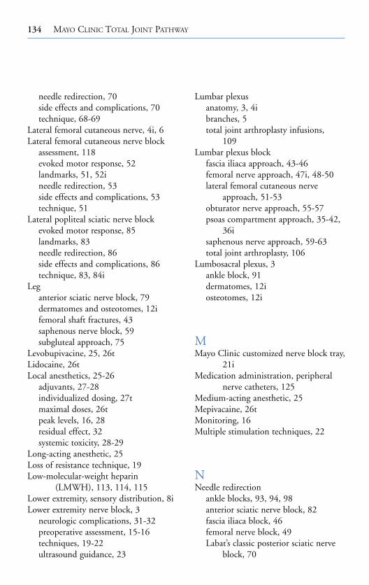
134 MAYO CLINIC TOTAL JOINT PATHWAY
needle redirection, 70side effects and complications, 70technique, 68-69
Lateral femoral cutaneous nerve, 4i, 6Lateral femoral cutaneous nerve block
assessment, 118evoked motor response, 52landmarks, 51, 52ineedle redirection, 53side effects and complications, 53technique, 51
Lateral popliteal sciatic nerve blockevoked motor response, 85landmarks, 83needle redirection, 86side effects and complications, 86technique, 83, 84i
Leganterior sciatic nerve block, 79dermatomes and osteotomes, 12ifemoral shaft fractures, 43saphenous nerve block, 59subgluteal approach, 75
Levobupivacine, 25, 26tLidocaine, 26tLocal anesthetics, 25-26
adjuvants, 27-28individualized dosing, 27tmaximal doses, 26tpeak levels, 16, 28residual effect, 32systemic toxicity, 28-29
Long-acting anesthetic, 25Loss of resistance technique, 19Low-molecular-weight heparin
(LMWH), 113, 114, 115Lower extremity, sensory distribution, 8iLower extremity nerve block, 3
neurologic complications, 31-32preoperative assessment, 15-16techniques, 19-22ultrasound guidance, 23
Lumbar plexusanatomy, 3, 4ibranches, 5total joint arthroplasty infusions,
109Lumbar plexus block
fascia iliaca approach, 43-46femoral nerve approach, 47i, 48-50lateral femoral cutaneous nerve
approach, 51-53obturator nerve approach, 55-57psoas compartment approach, 35-42,
36isaphenous nerve approach, 59-63total joint arthroplasty, 106
Lumbosacral plexus, 3ankle block, 91dermatomes, 12iosteotomes, 12i
MMayo Clinic customized nerve block tray,
21iMedication administration, peripheral
nerve catheters, 125Medium-acting anesthetic, 25Mepivacaine, 26tMonitoring, 16Multiple stimulation techniques, 22
NNeedle redirection
ankle blocks, 93, 94, 98anterior sciatic nerve block, 82fascia iliaca block, 46femoral nerve block, 49Labat’s classic posterior sciatic nerve
block, 70
LennonIndex.qxd 8/30/05 2:29 PM Page 134
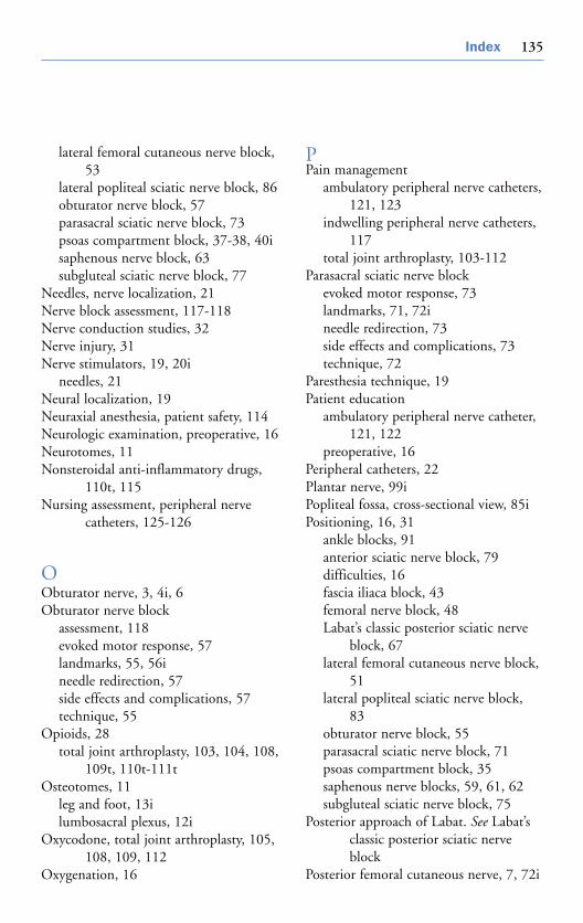
Index 135
lateral femoral cutaneous nerve block,53
lateral popliteal sciatic nerve block, 86obturator nerve block, 57parasacral sciatic nerve block, 73psoas compartment block, 37-38, 40isaphenous nerve block, 63subgluteal sciatic nerve block, 77
Needles, nerve localization, 21Nerve block assessment, 117-118Nerve conduction studies, 32Nerve injury, 31Nerve stimulators, 19, 20i
needles, 21Neural localization, 19Neuraxial anesthesia, patient safety, 114Neurologic examination, preoperative, 16Neurotomes, 11Nonsteroidal anti-inflammatory drugs,
110t, 115Nursing assessment, peripheral nerve
catheters, 125-126
OObturator nerve, 3, 4i, 6Obturator nerve block
assessment, 118evoked motor response, 57landmarks, 55, 56ineedle redirection, 57side effects and complications, 57technique, 55
Opioids, 28total joint arthroplasty, 103, 104, 108,
109t, 110t-111tOsteotomes, 11
leg and foot, 13ilumbosacral plexus, 12i
Oxycodone, total joint arthroplasty, 105,108, 109, 112
Oxygenation, 16
PPain management
ambulatory peripheral nerve catheters,121, 123
indwelling peripheral nerve catheters,117
total joint arthroplasty, 103-112Parasacral sciatic nerve block
evoked motor response, 73landmarks, 71, 72ineedle redirection, 73side effects and complications, 73technique, 72
Paresthesia technique, 19Patient education
ambulatory peripheral nerve catheter,121, 122
preoperative, 16Peripheral catheters, 22Plantar nerve, 99iPopliteal fossa, cross-sectional view, 85iPositioning, 16, 31
ankle blocks, 91anterior sciatic nerve block, 79difficulties, 16fascia iliaca block, 43femoral nerve block, 48Labat’s classic posterior sciatic nerve
block, 67lateral femoral cutaneous nerve block,
51lateral popliteal sciatic nerve block,
83obturator nerve block, 55parasacral sciatic nerve block, 71psoas compartment block, 35saphenous nerve blocks, 59, 61, 62subgluteal sciatic nerve block, 75
Posterior approach of Labat. See Labat’sclassic posterior sciatic nerveblock
Posterior femoral cutaneous nerve, 7, 72i
LennonIndex.qxd 8/30/05 2:29 PM Page 135

136 MAYO CLINIC TOTAL JOINT PATHWAY
Posterior popliteal sciatic nerve blockevoked motor response, 88-89landmarks, 87needle redirection, 90side effects and complications, 90technique, 87, 88i
Posterior tibial nerve blocklandmarks, 91needle redirection, 93side effects and complications, 98technique, 92
Preoperative examination, 15, 32Propofol, total joint arthroplasty, 107Psoas compartment block
landmarks, 35, 37i, 39ineedle redirection, 37-38, 40ineuroskeletal relationships, 38iside effects and complications, 40technique, 36-40, 36i, 41itotal joint arthroplasty infusion, 108,
109Psoas major, 3
QQuadratus lumborum, 3Quadriceps motor response, psoas com-
partment block, 37
RRegional anesthesia
“block area,” 106future directions, 127-128thromboprophylaxis, 113
Resuscitation preparation, 16, 28Ropivacaine, 25, 26t
SS1 and S2, sural nerve, 9
common peroneal nerve, 9sural nerve, 9
S1-S3, posterior femoral cutaneous nerve, 7
S1-S3, tibial nerve, 8S1-S4
dermatomes and osteotomes, 11, 12isacral plexus, 4i, 7
Sacral plexusanatomy, 4i, 7branches, 7
Saphenous nerve, 5, 59, 60i, 94iSaphenous nerve block
ankle technique, 95at ankle, 62-63, 95deep peroneal technique, 63evoked motor responses, 61, 62, 63at femoral triangle, 59, 61field block technique, 61at knee, 61-62landmarks, 61, 95needle redirection, 63paravenous technique, 62side effects and complications, 63transsartorial technique, 62vastus medialus technique, 61
Sciatic nerve, 4i, 7Sciatic nerve block
ankle approaches, 91, 93i, 93, 95, 96anterior approach, 79, 81iassessment, 117Labat’s classic posterior approach of,
67, 69ilateral popliteal approach, 83, 84iparasacral approach, 71, 72iposterior popliteal approach, 87, 88isubgluteal approach, 75, 76i
Sedation, 16Single stimulation techniques, 22Spinal hematoma, 113-114Stimulating catheters, 21Stimulating needles, 21Subgluteal sciatic nerve block
evoked motor response, 77landmarks, 75, 76ineedle redirection, 77
LennonIndex.qxd 8/30/05 2:29 PM Page 136
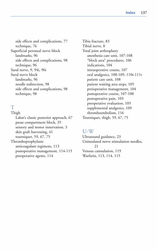
Index 137
side effects and complications, 77technique, 76
Superficial peroneal nerve blocklandmarks, 96side effects and complications, 98technique, 96
Sural nerve, 9, 94i, 96iSural nerve block
landmarks, 96needle redirection, 98side effects and complications, 98technique, 98
TThigh
Labat’s classic posterior approach, 67psoas compartment block, 35sensory and motor innervation, 3skin graft harvesting, 41tourniquet, 59, 67, 75
Thromboprophylaxisanticoagulant regimens, 113postoperative management, 114-115preoperative agents, 114
Tibia fracture, 83Tibial nerve, 8Total joint arthroplasty
anesthesia care unit, 107-108“block area” procedures, 106indications, 104intraoperative course, 107oral analgesics, 108-109, 110t-111tpatient care unit, 108patient waiting area steps, 105perioperative management, 104postoperative course, 107-108postoperative pain, 103preoperative evaluation, 105supplemental analgesics, 109thromboembolism, 116
Tourniquet, thigh, 59, 67, 75
U-WUltrasound guidance, 23Uninsulated nerve stimulation needles,
21Venous cannulation, 119Warfarin, 113, 114, 115
LennonIndex.qxd 8/30/05 2:29 PM Page 137